Cartilage and Bone Tissue 软骨组织和骨组织 Department of Histology and Embryology Medical...
84
Cartilage and Bone Tissue 软软软软软软软软 Department of Histology and Embryology Medical college in Three Gorges University
-
Upload
baldwin-mathews -
Category
Documents
-
view
426 -
download
0
Transcript of Cartilage and Bone Tissue 软骨组织和骨组织 Department of Histology and Embryology Medical...
Cartilage and Bone Tissue软骨组织和骨组织
Department of Histology and Embryology Medical college in Three Gorges University
Cartilage is composed of cartilage tissue( 软骨组织) and the perichondrium (软骨膜) .
Cartilage 软骨
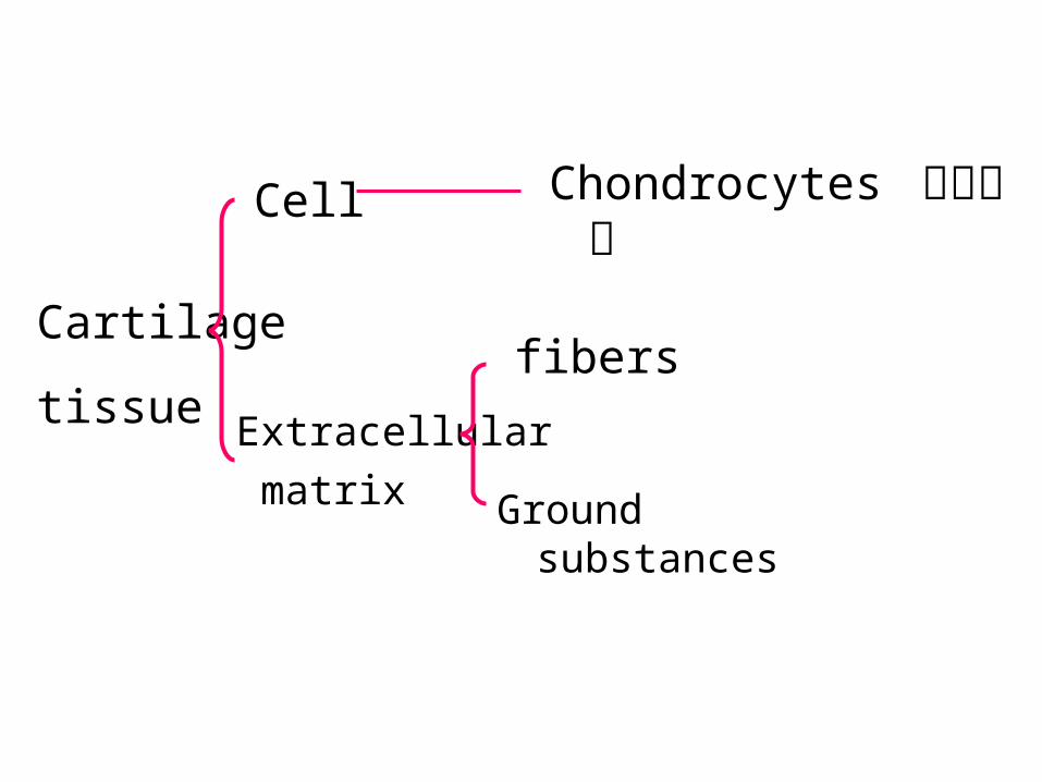
Cell
Cartilage
tissueExtracellular
matrix
fibers
Ground substances
Chondrocytes 软骨细胞
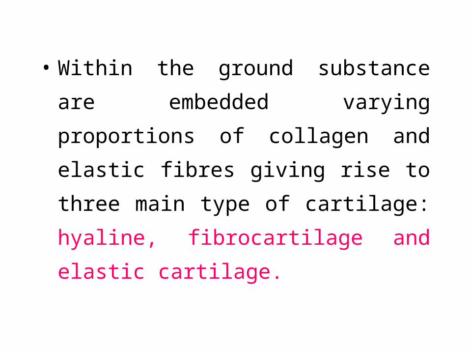
• Within the ground substance are embedded
varying proportions of collagen and elastic
fibres giving rise to three main type of
cartilage: hyaline, fibrocartilage and elastic
cartilage.
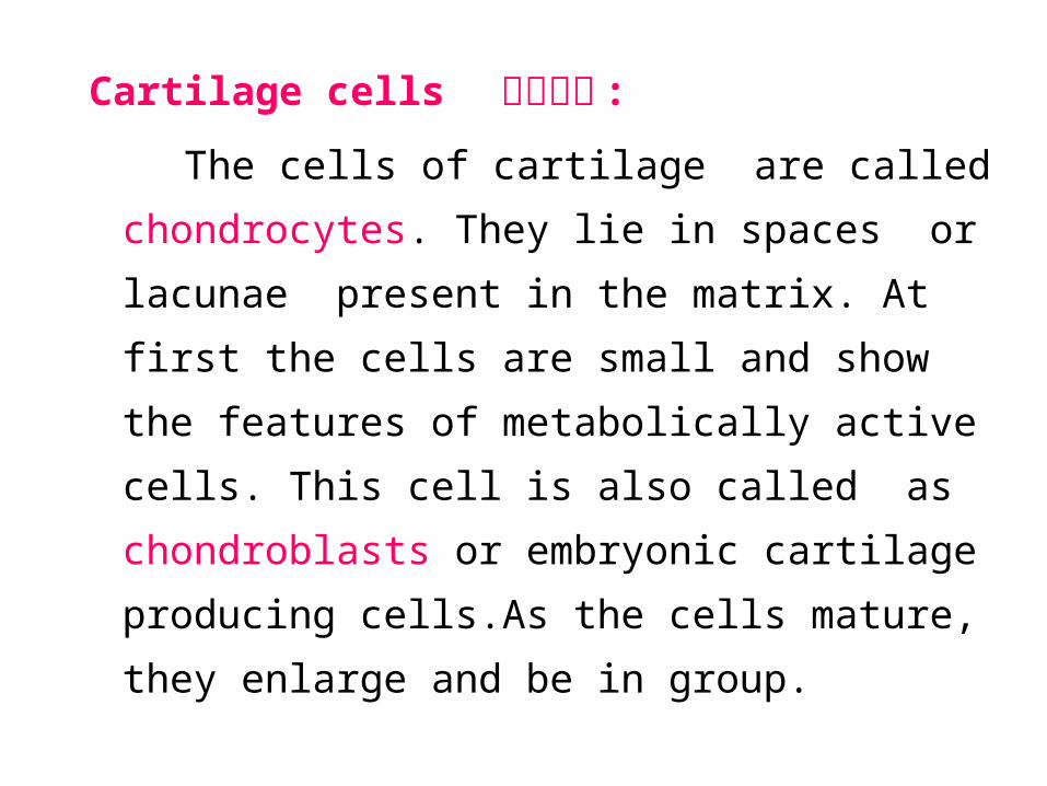
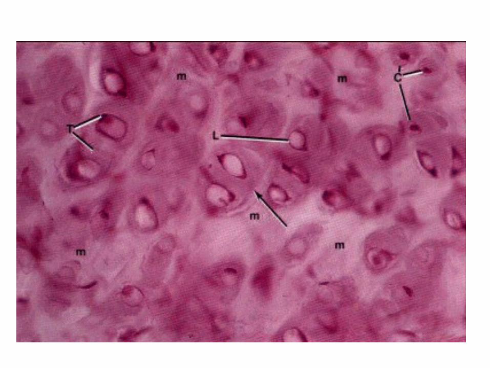
Cartilage cells 软骨细胞 :
The cells of cartilage are called
chondrocytes. They lie in spaces or lacunae
present in the matrix. At first the cells are
small and show the features of metabolically
active cells. This cell is also called as
chondroblasts or embryonic cartilage
producing cells.As the cells mature, they
enlarge and be in group.
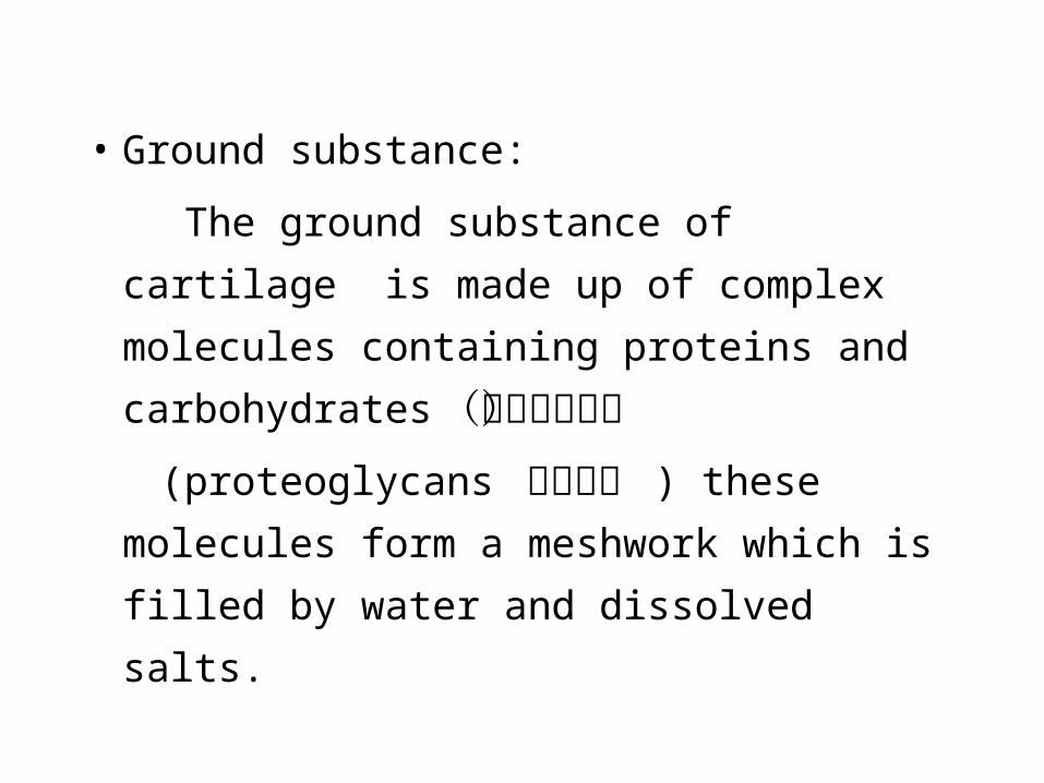
• Ground substance:
The ground substance of cartilage is made
up of complex molecules containing
proteins and carbohydrates (碳水化合物)
(proteoglycans 蛋白多糖 ) these molecules
form a meshwork which is filled by water
and dissolved salts.
Fibres of cartilage:
1.Collagen fibers:
type II collagen: hyaline cartilage
type I collagen: the normal collagen fiber
fibrocartilage, and the perichondrium,
2.Elastic fibers are in the Elastic cartilage
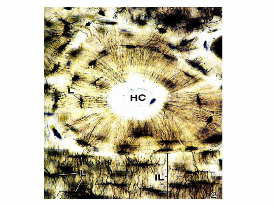
• Hyaline cartilage 透明软骨
• HC is the most common type of cartilage
found in the nasal septum, larynx ,tracheal
rings, most articular surfaces and the sternal
ends of the ribs etc.
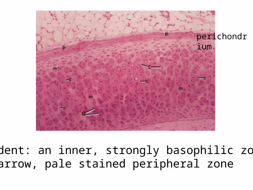
perichondrium
Evident: an inner, strongly basophilic zonea narrow, pale stained peripheral zone
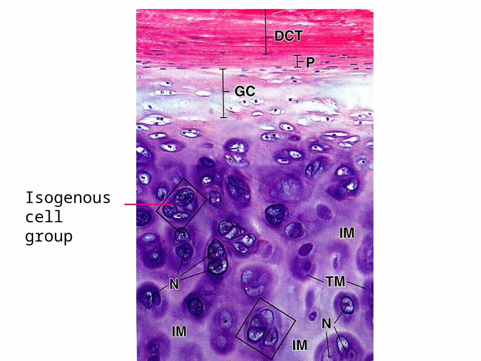
Isogenous cell group
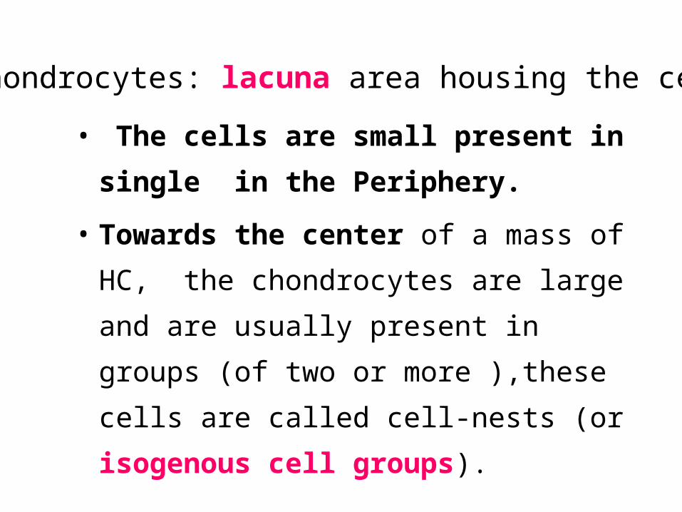
• The cells are small present in single in
the Periphery.
• Towards the center of a mass of HC, the
chondrocytes are large and are usually
present in groups (of two or more ),these
cells are called cell-nests (or isogenous cell
groups).
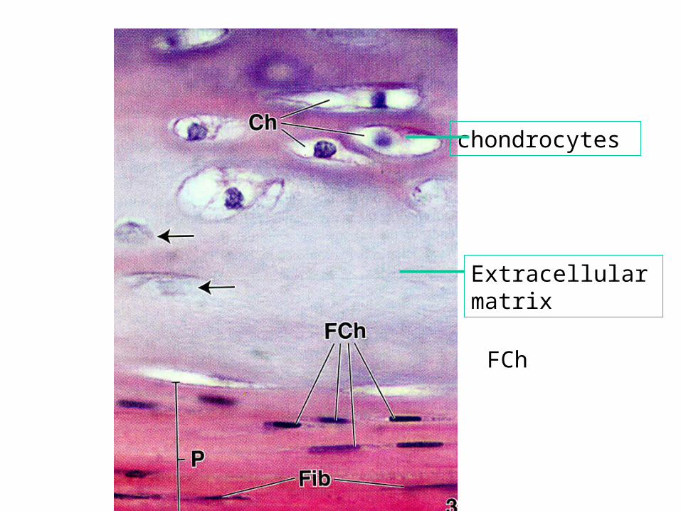
•Chondrocytes: lacuna area housing the cell
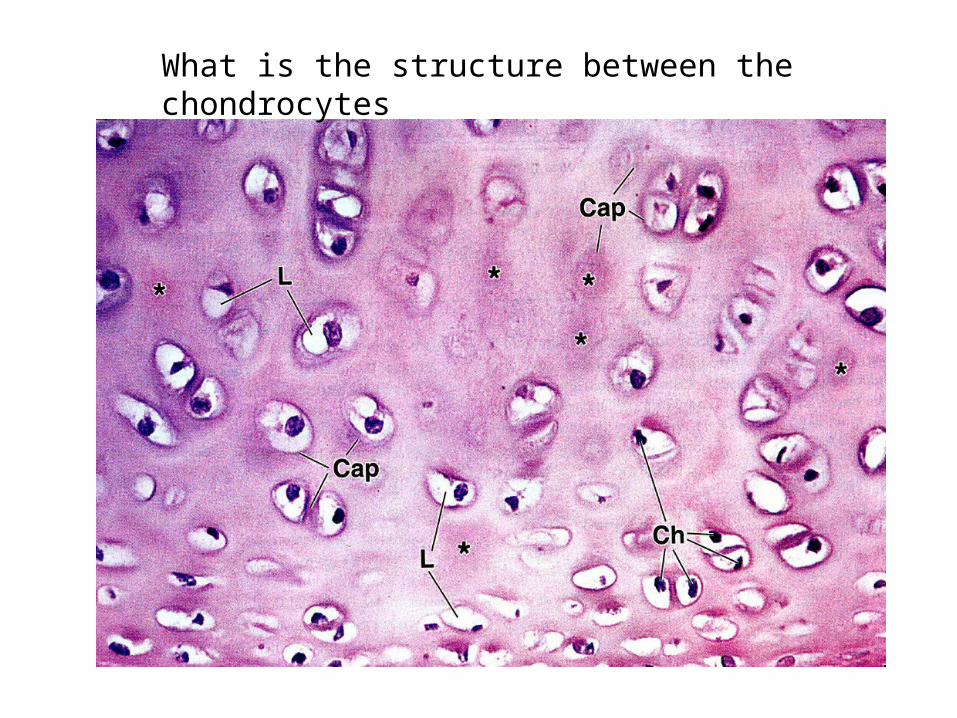
What is the structure between the chondrocytes
• The deep staining matrix around cell or
cell nests is newly formed and is called
the territorial matrix or lacunar capsule.
an amorphous matrix of ground substance reinforced by collagen fibres
Their tiny matrix enclosed compartments are
termed lacunae. (occupy space)
FCh
chondrocytes
Extracellular matrix
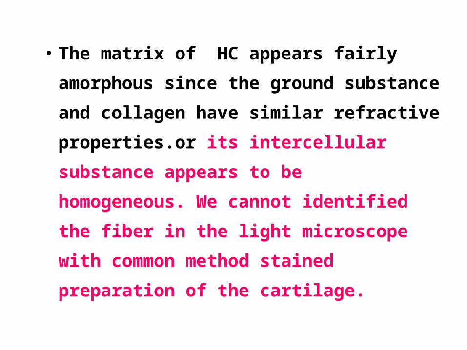
• The matrix of HC appears fairly
amorphous since the ground substance
and collagen have similar refractive
properties.or its intercellular substance
appears to be homogeneous. We cannot
identified the fiber in the light microscope
with common method stained preparation
of the cartilage.
• Fibrocartilage
Fibrocartilage, unlike hyaline and elastic
cartilage, does not possess a perichondrium
and its matrix possesses type I collagen.
•Distribution:the intervertebral discs( 椎间盘) ,
some Articular cartilage 关节软骨 ,
the pubic symphysis 耻骨联合 ,etc.

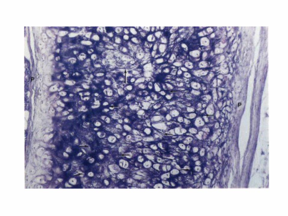
•Elastic cartilage
• The histological structure of EC is similar to
that of hyaline cartilage, its elasticity,
however, being derived from the presence of
numerous bundles of branching elastic fibres.
•Distribution: (1)in the external ear and external
auditory canal 外耳道 (2)the epiglottis 会厌 ,
parts of the laryngeal cartilage 喉软骨
Bone Tissue
• It is composed of cells and a predominantly
collagenous extracellular matrix (type I collagen )
called osteoid 类 骨 质 which becomes
mineralized by the deposition of calcium
hydroxyapatite 羟 磷 灰 石 , which produce an
extremely hard tissue capable of support and
protecting (rigidity and strength).
• Bone are an organ,and bone tissue is the structural
component of bones.
• Bone consist of bone tissue and other connective
tissue ,including hemopoietic 造血的 tissue, fat
tissue, blood vessels, and nerves.
• Bone is classified as either compact (dense ) or
spongy (cancellous).
•Bone and bone tissue 骨和骨组织 :
To 49
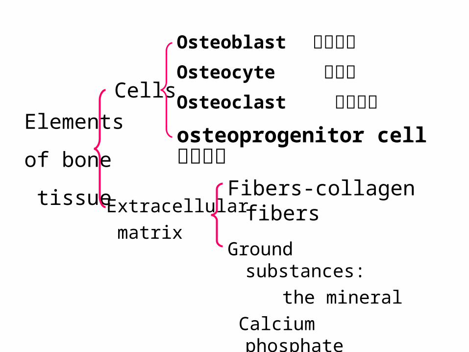
Cells
Elements
of bone
tissue Extracellular
matrix
Fibers-collagen fibers
Ground substances:
the mineral
Calcium phosphate
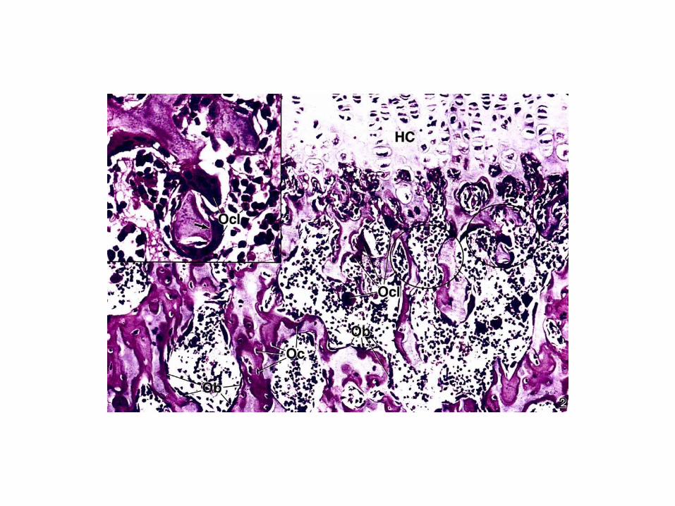
Osteoblast 成骨细胞Osteocyte 骨细胞Osteoclast 破骨细胞
osteoprogenitor cell 骨原细胞
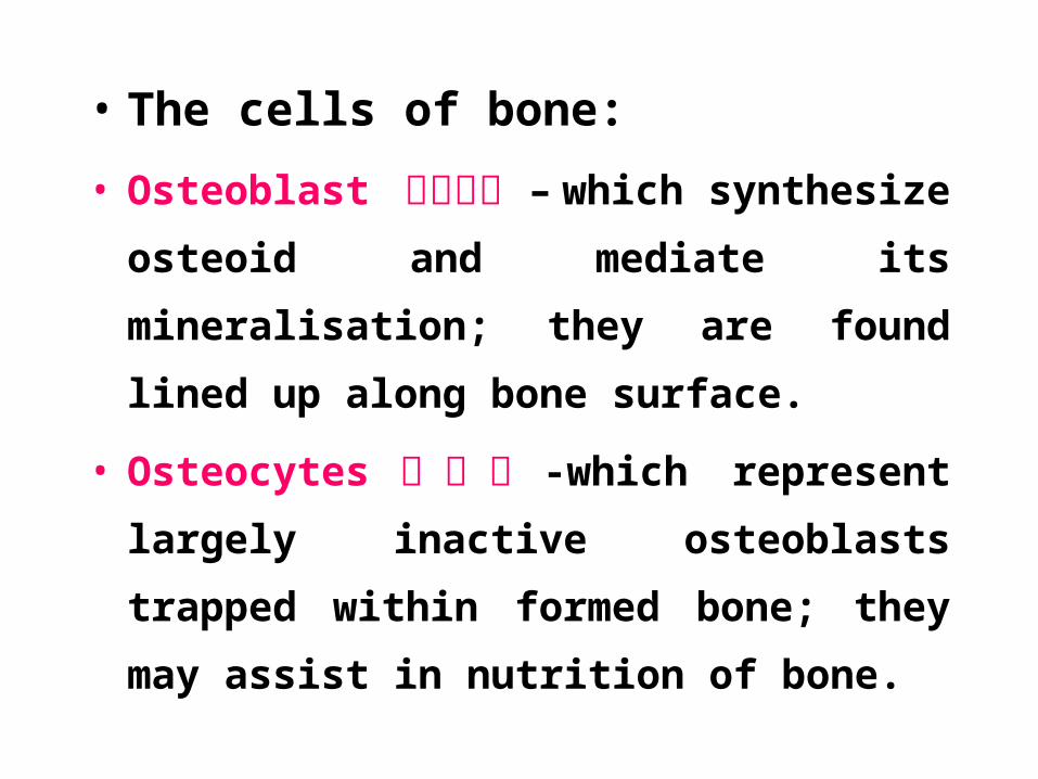
• The cells of bone:
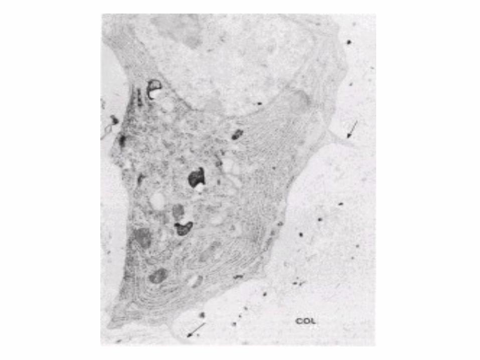
• Osteoblast 成骨细胞 – which synthesize osteoid
and mediate its mineralisation; they are found
lined up along bone surface.
• Osteocytes 骨 细 胞 -which represent largely
inactive osteoblasts trapped within formed
bone; they may assist in nutrition of bone.
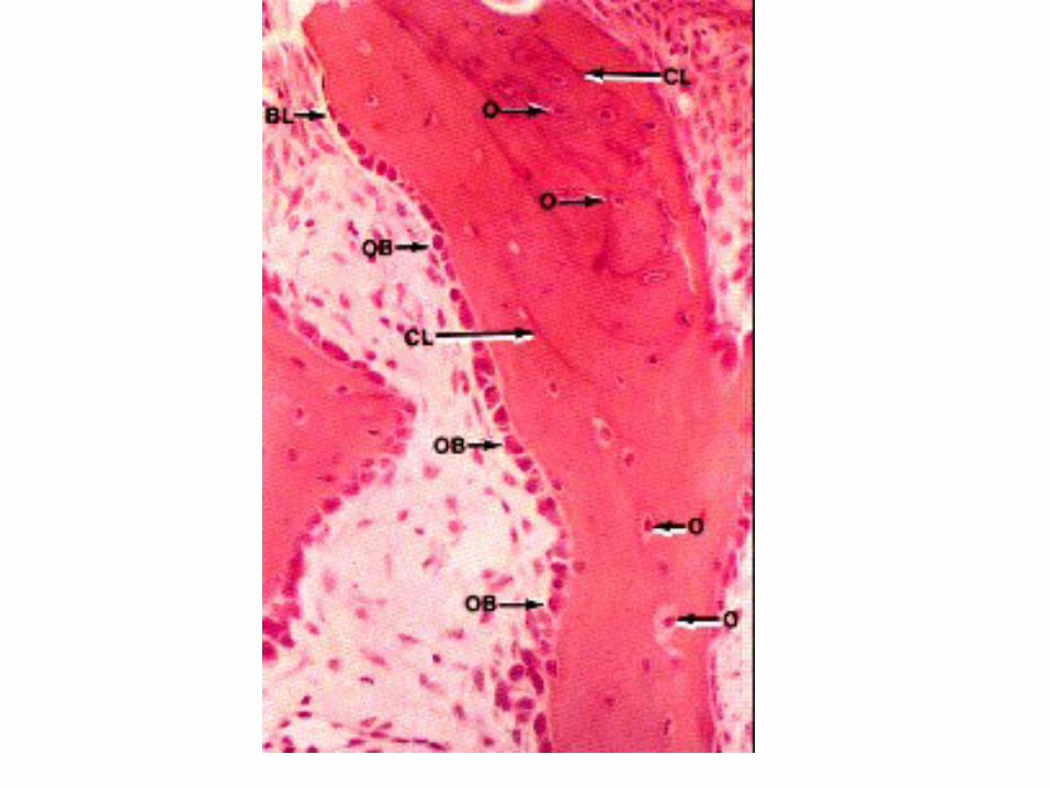
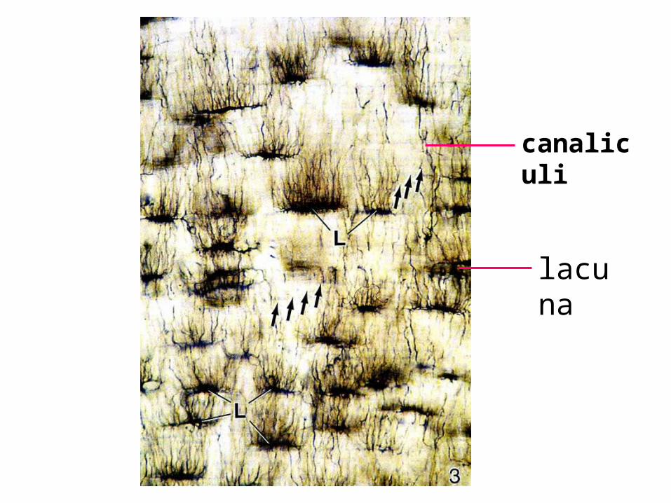
lacuna
canaliculi
• Bone lacuna 骨陷窝 : cell body of the osteocyte.
• The canaliculi 骨小管 are occupied by delicate
cytoplasmic processes of osteocytes, Spreading out
from the lacuna.
• (filled in tissue fluid).
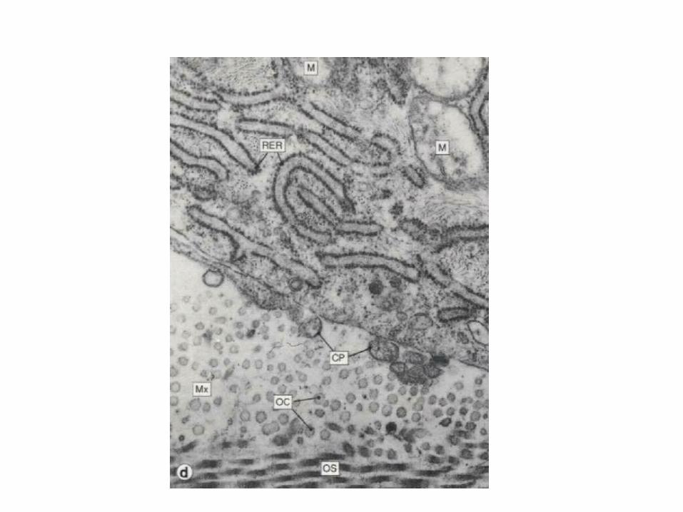
• Osteoclasts 破 骨 细 胞 -phagocytic cells
which are capable of eroding bone and
which are important, along with osteoblasts,
in the constant turnover and refashioning of
bone. Osteoclasts are multinucleate
phagocytic cells derived from the
macrophage-monocyte cell line
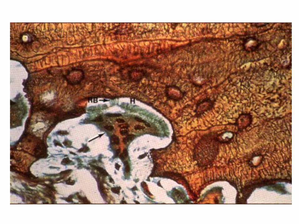
• Osteoprogenitor cell 骨祖细胞:
The osteoprogenitor cell is a resting cell that
can transform into an osteoblast and secrete
bone matrix. It is found on the external and
internal surfaces of bones. They resemble
fibroblast in appearance.
• Extracellular matrix :
The feature that distinguishes bone from
other connective tissue is the mineralisation
of its matrix,which produce an extremely
hard tissue capable of support and
protecting.The mineral is calcium
phosphate in the form of hydroxyapatite
crystals 羟磷灰石 .
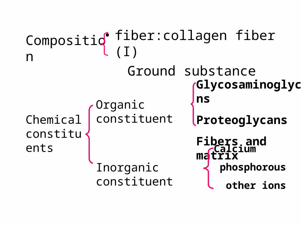
• fiber:collagen fiber (I)
Ground substance Composition
Chemical constituents
Organic constituent
Glycosaminoglycans
Proteoglycans
Fibers and matrix
Inorganic constituent
Calcium
phosphorous
other ions
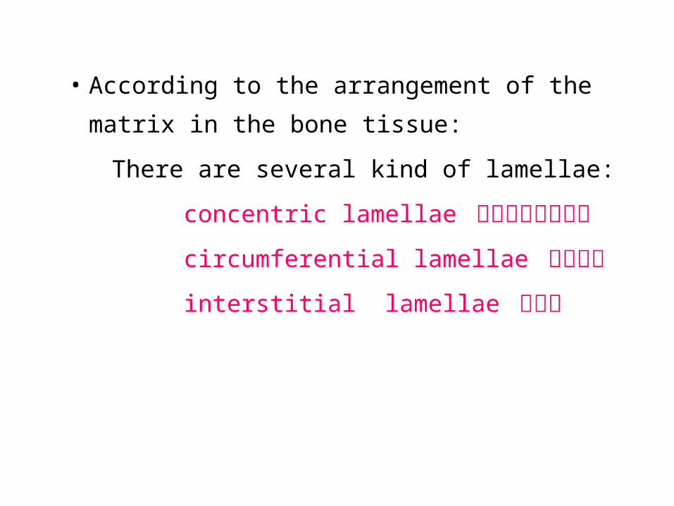
• According to the arrangement of the matrix
in the bone tissue:
There are several kind of lamellae:
concentric lamellae 同心圆排列的骨板
circumferential lamellae 环状骨板
interstitial lamellae 间骨板
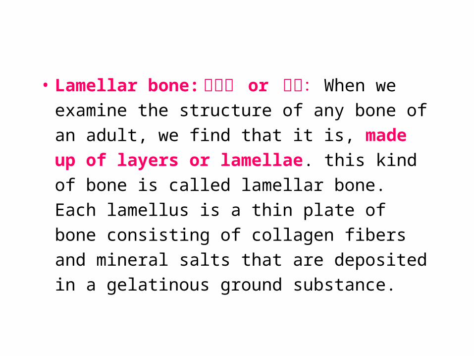
• Lamellar bone: 板层骨 or 骨板: When
we examine the structure of any bone of an
adult, we find that it is, made up of layers
or lamellae. this kind of bone is called
lamellar bone. Each lamellus is a thin plate
of bone consisting of collagen fibers and
mineral salts that are deposited in a
gelatinous ground substance.
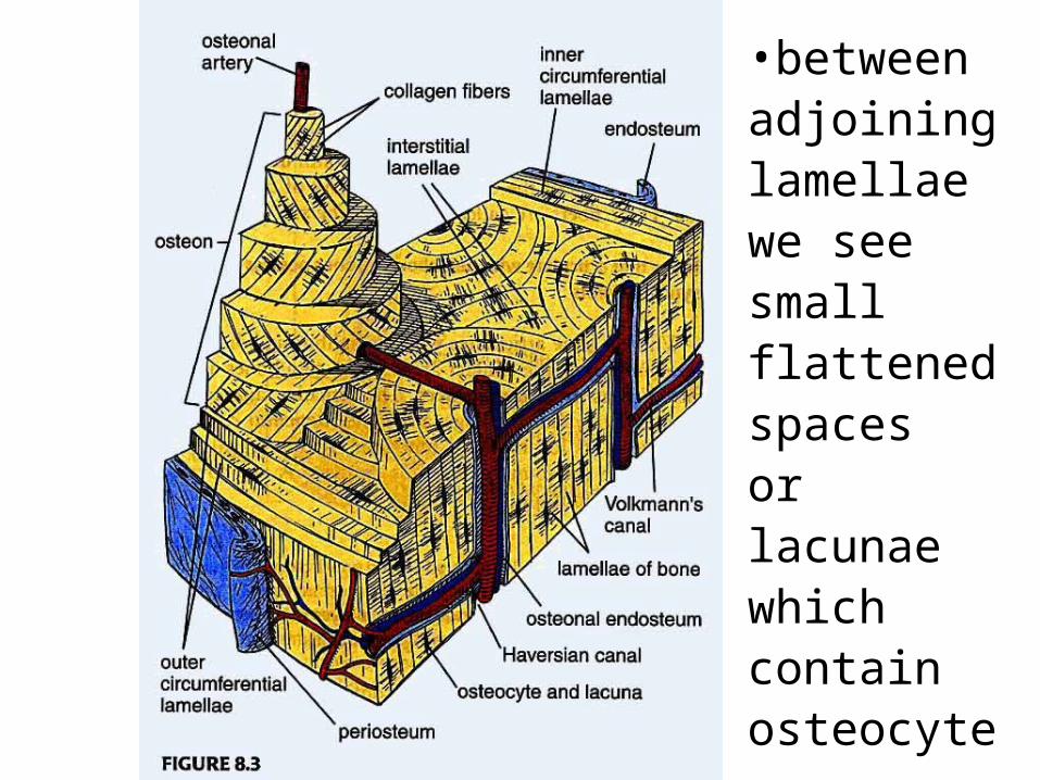
•between adjoining lamellae we see small flattened spaces or lacunae which contain osteocytes.
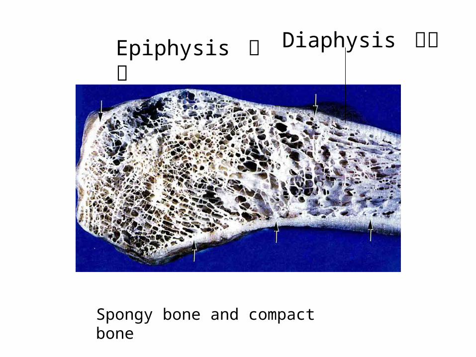
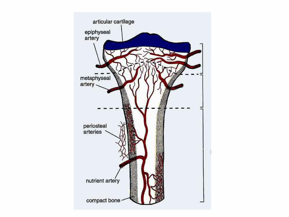
Spongy bone and compact bone
Epiphysis 骨骺 Diaphysis 骨干
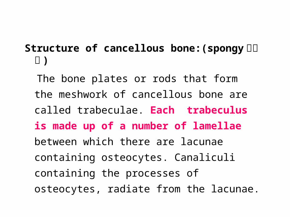
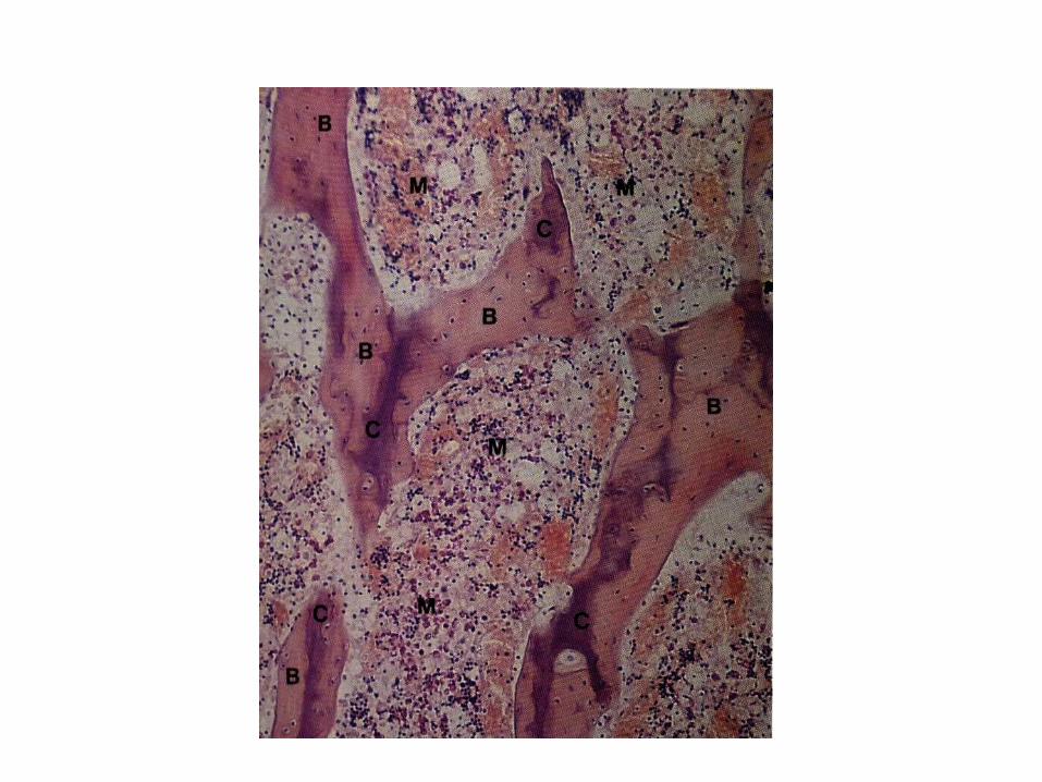
Structure of cancellous bone:(spongy 松质骨 )
The bone plates or rods that form the meshwork of
cancellous bone are called trabeculae. Each
trabeculus is made up of a number of lamellae
between which there are lacunae containing
osteocytes. Canaliculi containing the processes of
osteocytes, radiate from the lacunae.
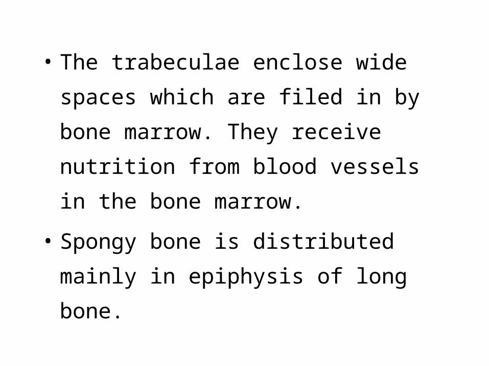
• The trabeculae enclose wide spaces which
are filed in by bone marrow. They receive
nutrition from blood vessels in the bone
marrow.
• Spongy bone is distributed mainly in
epiphysis of long bone.
Structure of compact bone 密质骨 :(dense)
When we examine a section of compact
bone we find that this type of bone is also
made up of lamellae.It is constitute the
main parts of diaphysis.
According to the arrangment of lamellae,
there are several kind of lamellae:
circumferential lamellae 环骨板
concentric lamellae 同心圆骨板
interstitial lamellae 间骨板
• ( 1 ) circumferential lamellae 环骨板 :
The outer circumferential lamellae are
just deep to the periosteum, forming the
outermost region of the diaphysis and
contain Sharpey’s fibres anchoring the
periosteum to the bone.
• The inner circumferential lamellae
completely encircle the marrow cavity.
Trabeculae of spongy bone extend from the
inner circumferential lamellae into the
marrow cavity, interrupting the endosteal
lining of the inner circumferential lamellae.

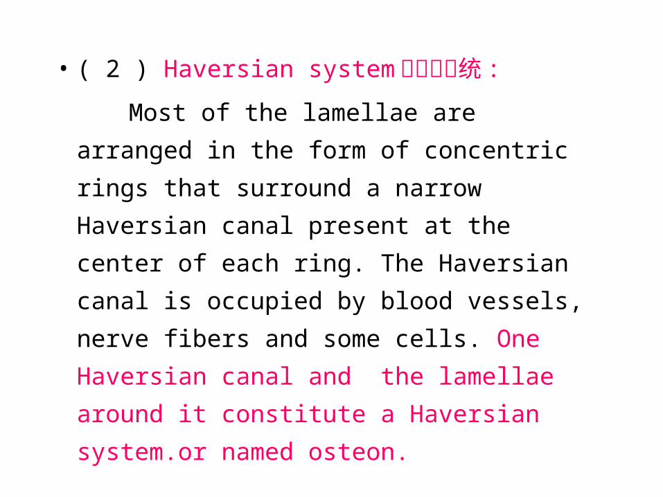
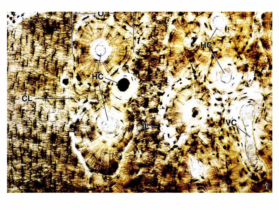
• ( 2 ) Haversian system 哈弗斯系统 :
Most of the lamellae are arranged in the
form of concentric rings that surround a
narrow Haversian canal present at the
center of each ring. The Haversian canal is
occupied by blood vessels, nerve fibers
and some cells. One Haversian canal and
the lamellae around it constitute a
Haversian system.or named osteon.
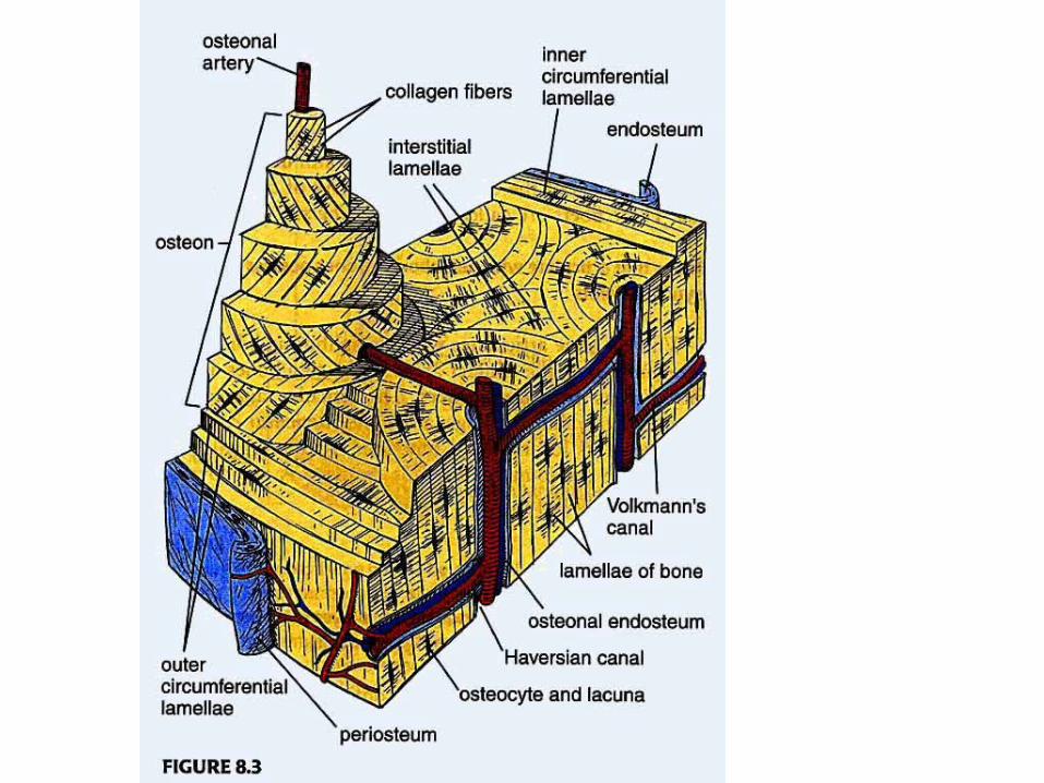
• ( 3 )Interstitial lamellae 间骨板 : It is
located between adjoining osteons.These
lamellae are remnants of osteons, the
greater parts of which have been destroyed.
• Formation of bone
• Histogenesis of Bone 骨发生• Two types:Intramembranous
Endochondral
• Intramembranous ossification 膜 内成骨
• Intramembranous bone formation occurs
within mesenchymal tissue.
• Most flat bones are formed by
intramembranous bone formation.
• (1) occurs in a richly vascularized
mesenchymal tissue, whose cells make
contact with each other via long processes.
• (2)Mesenchymal cells differentiate into
osteoblasts that secrete bone matrix,in which
fibers is embedded. This mass of swollen
fibers and matrix, no calcium, is called
osteoid.
• (3) Calcium salts is deposited in osteoid, this
process is named calcification and osteoblasts
trapped in their matrices become osteocytes.
As soon as this happens the layer of osteoid
can be said to have become one lamellus of
bone. This region of initial osteogenesis is
known as the primary ossification center.
• (4)In this way a number of lamellae are laid
down one over another,and these lamellae
together form a trabeculaus of bone
•Moreover, the spongy bone deep to the
periosteum and the periosteal layer of the
dura mater of flat bones are transformed
into compact bone, forming the inner and
outer tables with the intervening diploë.
• Endochondral Bone Formation 软骨内成骨
• Most of the long and short bones of the
body develop by endochondral bone
formation. This type of bone formation
occurs in two steps: (1)a miniature hyaline
cartilage model is formed, and (2) the
cartilage model continues to grow and
serves as a structural scaffold for bone
development, is resorbed, and is replaced
by bone.
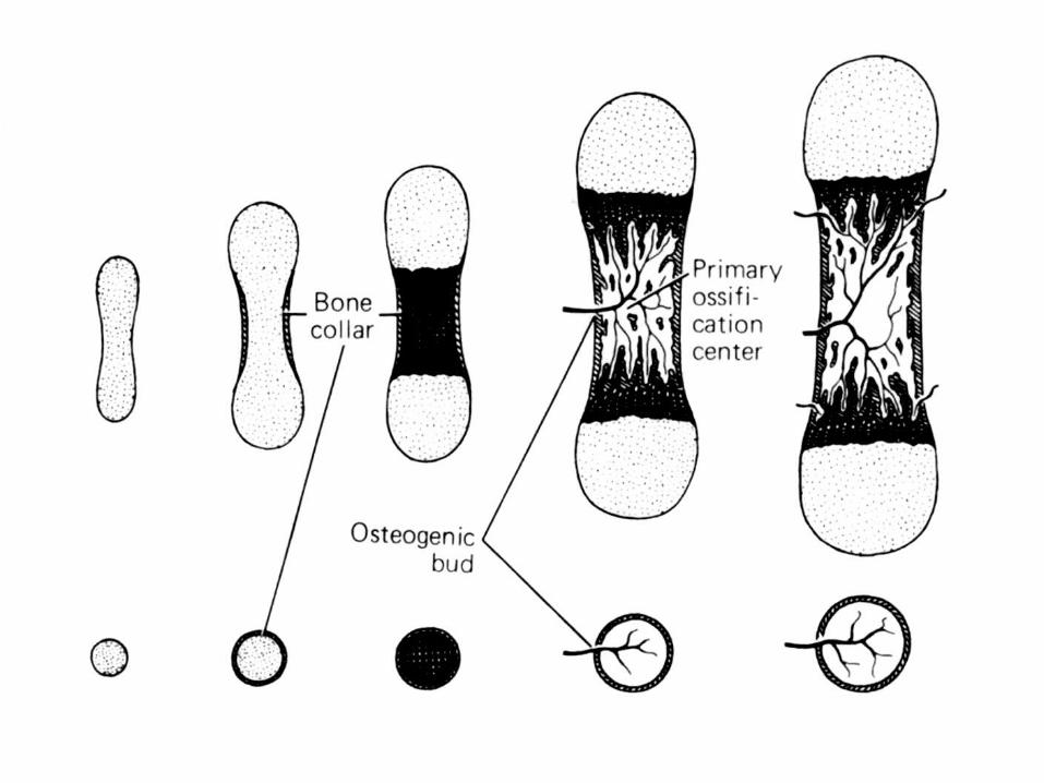
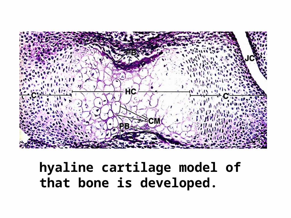
• 1. In the region where bone is to grow
within the embryo, a hyaline cartilage
model of that bone is developed.
• 2.Concurrently, the perichondrium at
the midriff of the diaphysis of
cartilage becomes vascularized .
When this happens, chondrogenic cells
become osteoprogenitor cells forming
osteoblasts, and the overlying
perichondrium becomes a periosteum.
• 3.The newly formed osteoblasts
secrete bone matrix, forming the
subperiosteal bone collar on the
surface of the cartilage template by
intramembranous bone formation.
• 4.The bone collar prevents the diffusion of
nutrients to the hypertrophied chondrocytes
within the core of the cartilage model, causing
them to die. This process is responsible for the
presence of empty, confluent lacunae forming
large concavities—the future marrow cavity in
the center of the cartilage model.
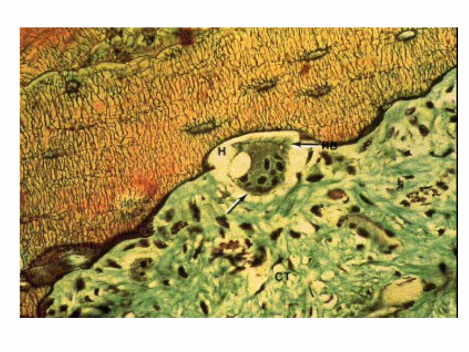
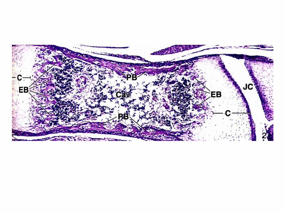
• 5. Holes etched in the bone collar by
osteoclasts permit a periosteal bud
(osteogenic bud), composed of
osteoprogenitor cells, hemopoietic cells,
and blood vessels, to enter the
concavities within the cartilage model .
• 6..Osteoprogenitor cells divide to form
osteoblasts. These newly formed cells
elaborate bone matrix on the surface of
the calcified cartilage. The bone matrix
becomes calcified to form a calcified
cartilage/calcified bone complex.
• This complex can be appreciated in
routinely stained histological sections
because calcified cartilage stains
basophilic, whereas calcified bone
stains acidophilic.
This is a first place for calsification,
primary center of ossification.
• 7. As the subperiosteal bone becomes
thicker and grows in each direction
from the midriff of the diaphysis
toward the epiphyses, osteoclasts
begin resorbing the calcified
cartilage/calcified bone complex,
enlarging the marrow cavity.
• .
• As this process continues, the cartilage of
the diaphysis is replaced by bone except for
the epiphyseal plates, which are responsible
for the continued growth of the bone for 18
to 20 years.
•
hyaline cartilage model of that bone is developed.
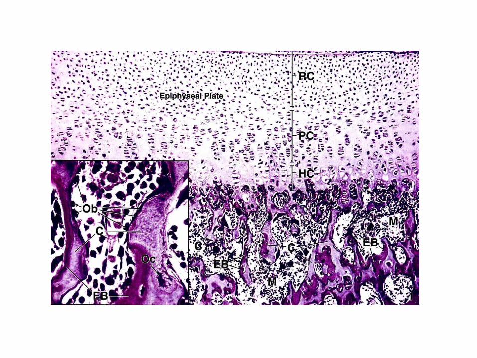
• Bone growth in length
• The continued lengthening of bone depends
on the epiphyseal plate.
• Histologically, the epiphyseal plate is
divided into five recognizable zones.
• ■Zone of reserve cartilage 软骨储备区 : Chondrocytes randomly distributed throughout the matrix are mitotically active.
• ■Zone of proliferation 软 骨 增 生 区 : Chondrocytes, rapidly proliferating, form rows of isogenous cells that parallel the direction of bone growth.
• ■Zone of maturation and hypertrophy:
软骨成熟区或软骨肥大区 Chondrocytes mature, hypertrophy, and
accumulate glycogen in their cytoplasm. The
matrix between their lacunae narrows with a
corresponding growth of lacunae.
• ■Zone of calcification 软 骨 钙 化 区 : Lacunae
become confluent, hypertrophied chondrocytes
die, and cartilage matrix becomes calcified.
• ■Zone of ossification 成骨区 :
Osteoprogenitor cells invade the area and
differentiate into osteoblasts, which
elaborate matrix that becomes calcified
on the surface of calcified cartilage. This
is followed by resorption of the calcified
cartilage/calcified bone complex.
• As long as the rate of mitotic activity in the
zone of proliferation equals the rate of
resorption in the zone of ossification, the
epiphyseal plate remains the same width
and the bone continues to grow longer.