The 37th the Japan Society for Biomedical …-31- 第37回日本基礎老化学会大会...
45
- 31 - 第 37 回日本基礎老化学会大会 発表抄録 シンポジウム ランチョンセミナー 一般発表(口頭発表、ポスター発表) The 37 th Annual Meeting of the Japan Society for Biomedical Gerontology ABSTRACTS Symposiums Luncheon Seminars General Presentations (Oral and Poster Presentations)
Transcript of The 37th the Japan Society for Biomedical …-31- 第37回日本基礎老化学会大会...
- 31 -
第 37 回日本基礎老化学会大会発表抄録
シンポジウムランチョンセミナー
一般発表(口頭発表、ポスター発表)
The 37th Annual Meeting ofthe Japan Society for Biomedical Gerontology
ABSTRACTS
SymposiumsLuncheon Seminars
General Presentations (Oral and Poster Presentations)
- 33 -
Symposium Session I:
Cellular Senescence and Aging
Chairpersons: Dr.Yoji Mitsui (Tokushima Bunri University)Dr. Sataro Goto (Juntendo University)
At 9:00 〜 12:00 on Thursday, 26th June, 2014in Health Science Theater, Aichi Health Plaza
Symposium
I・II
- 34 -
Chromatin dynamics during DNA damage– Consequences for repair and nuclear integrity
Dr. Philipp Oberdörffer
Head, Epigenetics of DNA Repair and Aging Section, National Cancer Institute, NIH
DNA double-strand breaks (DSBs) occur in the context of a highly organized chromatin environment and are, thus, a significant threat to the epigenomic integrity of eukaryotic cells. Changes in break-proximal chromatin structure are thought to be a prerequisite for efficient DNA repair and may help protect the structural integrity of the nucleus. Unlike most bona fide DNA repair factors, chromatin influences the repair process at several levels: the existing chromatin context at the site of damage directly affects the access and kinetics of the repair machinery; DSB induced chromatin modifications influence the choice of repair factors, thereby modulating repair outcome; lastly, DNA damage can have a significant impact on chromatin beyond the site of damage. Our lab has recently identified dynamic, DSB-induced chromatin condensation as an unexpected yet critical modulator of DSB repair outcome with direct implications for BRCA1-dependent genome maintenance. These findings will be discussed in the context of DNA damage-associated nuclear dysfunction to highlight the importance of dynamic and tightly orchestrated chromatin reorganization for DNA repair and tissue maintenance.
■ ContactCenter for Cancer Research,
National Cancer Institute
Building 41, Room B907
41 Library Drive, MSC 5055, Bethesda, MD 20892
USA
Phone: 301-594-0689
Fax: 301-496-4951
Email: [email protected]
■ Education1999-2004 Doctor of Philosophy, Genetics, University of Cologne, Cologne, Germany
1993-1999 Master of Science, Biology, Albert Ludwigs University, Freiburg, Germany
Symposium
I・II
- 35 -
■ Carrer2009-present Tenure-track Investigator, National Cancer Institute, NIH
2004-2009 Postdoctoral Fellow, Harvard Medical School, Paul F.
Glenn labs for Aging Research, Boston, MA
■ Major Research InterestEpigenetics of DNA Repair and Aging
■ Selected Resent Publications1. Metabolic modulation of chromatin: implications for DNA repair and genomic integrity.
Liu J, Kim J, Oberdoerffer P. 4:182. eCollection 2013. Review. Front Genet. 2013
2. Sirt1 ablation promotes stress-induced loss of epigenetic and genomic hematopoietic stem and progenitor cell maintenance.
Singh SK, Williams CA, Klarmann K, Burkett SS, Keller JR, Oberdoerffer P. 210(5): 987-1001. J Exp Med. 2013
3. DNA damage, chromatin, and transcription: the trinity of aging.
Burgess RC, Misteli T, Oberdoerffer P. 24(6):724-730. Review. Curr Opin Cell Biol. 2012
4. Chromatin dynamics in DNA double-strand break repair.
Shi L, Oberdoerffer P. 1819(7): 811-819. Review. Biochim Biophys Acta. 2012
5. CTCF-promoted RNA polymerase II pausing links DNA methylation to splicing.
Shukla S, Kavak E, Gregory M, Imashimizu M, Shutinoski B, Kashlev M, Oberdoerffer P, Sandberg R, Oberdoerffer S.
479 (7371):74-79. Nature 2011
6. An age of fewer histones. Oberdoerffer P. 12(11): 1029-1031, Nat Cell Biol. 2010
7. SIRT1 redistribution on chromatin promotes genomic stability but alters gene expression during aging. Oberdoerffer P,
Michan S, McVay M, Mostoslavsky R, Vann J, Park SK, Hartlerode A, Stegmuller J, Hafner A, Loerch P, Wright SM,
Mills KD, Bonni A, Yankner BA, Scully R, Prolla TA, Alt FW, Sinclair DA. 135(5): 907-918. Cell. 2008
- 36 -
The roles and mechanisms of cellular senescence
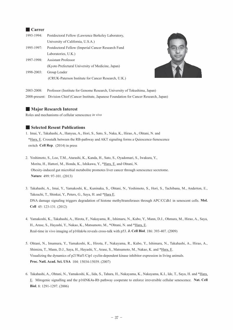
Dr. Eiji Hara
Chief, Division of Cancer Biology,The Cancer Institute of Japanese Foundation for Cancer Research (JFCR)
Over the last few decades, it has become apparent that oncogenic proliferative signals are coupled to a variety of growth inhibitory responses, such as the induction of apoptotic cell death or irreversible cell cycle arrest known as “cellular senescence.” Thus, both apoptosis and cellular senescence are thought to act as important tumor suppression mechanisms. Unlike apoptotic cells, however, senescent cells remain viable for long periods of time and accumulate with increasing age in various organs and tissues in vivo. Moreover, recent studies have revealed that although cellular senescence initially functions as a tumor suppressive process, it may eventually promote deleterious side effects, such as chronic inflammation and cancer promotion. It is therefore quite possible that accumulation of senescent cells during the aging process in vivo may contribute to aging-associated increases in homeostatic disorders. Here, I present our recent work regarding the roles and mechanisms of cellular senescence in vivo. We believe that a better understanding of the molecular mechanisms involved will lead to new strategies for the prevention of aging-associated diseases including cancer.
■ ContactDivision of Cancer Biology
The Cancer Institute,
Japanese Foundation for Cancer Research
3-8-31, Ariake, Koto-ku
Tokyo 135-8550
Japan
TEL +81-3-3570-0605 FAX +81-3-3570-0457
E-mail: [email protected] [email protected]
■ EducationMarch 1987: BSc. (Tokyo University of Science, Japan)
March 1993: Ph.D. (Tokyo University of Science, Japan)
- 37 -
■ Carrer1993-1994: Postdoctoral Fellow (Lawrence Berkeley Laboratory,
University of California, U.S.A.)
1995-1997: Postdoctoral Fellow (Imperial Cancer Research Fund
Laboratories, U.K.)
1997-1998: Assistant Professor
(Kyoto Prefectural University of Medicine, Japan)
1998-2003: Group Leader
(CRUK-Paterson Institute for Cancer Research, U.K.)
2003-2008: Professor (Institute for Genome Research, University of Tokushima, Japan)
2008-present: Division Chief (Cancer Institute, Japanese Foundation for Cancer Research, Japan)
■ Major Research InterestRoles and mechanisms of cellular senescence in vivo
■ Selected Resent Publications1. Imai, Y., Takahashi, A., Hanyuu, A., Hori, S., Sato, S., Naka, K., Hirao, A., Ohtani, N. and
*Hara, E. Crosstalk between the RB-pathway and AKT signaling forms a Quiescence-Senescence
switch Cell Rep. (2014) in press
2. Yoshimoto, S., Loo, T.M., Atarashi, K., Kanda, H., Sato, S., Oyadomari, S., Iwakura, Y.,
Morita, H., Hattori, M., Honda, K., Ishikawa, Y., *Hara, E. and Ohtani, N.
Obesity-induced gut microbial metabolite promotes liver cancer through senescence secretome.
Nature 499: 97-101. (2013)
3. Takahashi, A., Imai, Y., Yamakoshi, K., Kuninaka, S., Ohtani, N., Yoshimoto, S., Hori, S., Tachibana, M., Anderton, E.,
Takeuchi, T., Shinkai, Y., Peters, G., Saya, H. and *Hara E.
DNA damage signaling triggers degradation of histone methyltransferases through APC/CCdh1 in senescent cells. Mol.
Cell 45: 123-131. (2012)
4. Yamakoshi, K., Takahashi, A., Hirota, F., Nakayama, R., Ishimaru, N., Kubo, Y., Mann, D.J., Ohmura, M., Hirao, A., Saya,
H., Arase, S., Hayashi, Y., Nakao, K., Matsumoto, M., *Ohtani, N. and *Hara, E.
Real-time in vivo imaging of p16Ink4a reveals cross-talk with p53. J. Cell Biol. 186: 393-407. (2009)
5. Ohtani, N., Imamura, Y., Yamakoshi, K., Hirota, F., Nakayama, R., Kubo, Y., Ishimaru, N., Takahashi, A., Hirao, A.,
Shimizu, T., Mann, D.J., Saya, H., Hayashi, Y., Arase, S., Matsumoto, M., Nakao, K. and *Hara, E.
Visualizing the dynamics of p21Waf1/Cip1 cyclin-dependent kinase inhibitor expression in living animals.
Proc. Natl. Acad. Sci. USA 104: 15034-15039. (2007)
6. Takahashi, A., Ohtani, N., Yamakoshi, K., Iida, S., Tahara, H., Nakayama, K., Nakayama, K.I., Ide, T., Saya, H. and *Hara,
E. Mitogenic signalling and the p16INK4a-Rb pathway cooperate to enforce irreversible cellular senescence. Nat. Cell
Biol. 8: 1291-1297. (2006)
- 38 -
Molecular mechanisms of cellular senescence
Dr. Fabrizio d'Adda di Fagagna
PhD Principal InvestigatorIFOM Foundation - The FIRC Institute of Molecular Oncology Foundation, Italy
Cellular senescence acts both as a tumor suppressive mechanism and it is implicated in organismal ageing. I will discuss the molecular mechanisms controlling cellular senescence upon different stimuli and in stem cells. I will also discuss a novel class of non coding RNAs involved in DNA damage response control.
■ ContactIFOM Foundation - The FIRC Institute of Molecular Oncology Foundation
Via Adamello, 16
20139 Milan, Italy
tel. +39 02 574303.227
fax +39 02 574303.231
email: [email protected]
Skype: d'Adda di Fagagna Fabrizio
http://www.ifom-ieo-campus.it/research/dadda.php
http://www.semm.it
■ Education2014-2018 National Scientific Qualification (Abilitazione) as “Full and Associate University Professor in Medical
Genetics, General Pathology, Clinical Pathology, Molecular Biology, Applied Biology, Histology, Genetics
and Microbiology”.
1995 Doctor of Philosophy (PhD) awarded with full marks and Honours at the International School for Advanced
Studies (ISAS-SISSA), Trieste, Italy, with a thesis entitled "Molecular and functional regulation of HIV-1
expression by the Long Terminal Repeat". Advisors: Prof. A. Falaschi and Prof. M. Giacca. External reviewer:
Prof. N.J. Proudfoot, Oxford, UK. Experimental work carried out at the International Centre for Genetic
Engineering and Biotechnology (ICGEB), Trieste, Italy.
1993 Magister of Philosophy (Master of Science degree) awarded with full marks and Honours at ISAS-SISSA with
a thesis entitled "Interactions between the transcription factor USF/MLTF and the Long Terminal Repeat of
Human Immunodeficiency Virus type 1".
- 39 -
■ CarrerResearch experience2012 – present “Group leader” at Istituto di Genetica Molecolare (IGM) of the
Consiglio Nazionale delle Ricerche (CNR), Pavia, Italy http://www.igm.cnr.it/1/pagine-personali/dadda-di-fagagna-fabrizio/
2009 – present Tenured Principal Investigator, FIRC Institute of Molecular Oncology (IFOM), Milan, Italy (http://www.ifom-ieo-campus.it/research/dadda.php).
2003 – 2009 Principal Investigator, FIRC Institute of Molecular Oncology (IFOM), Milan, Italy (http://www.ieo-ifom-campus.it).
1996 – 2003 Research Associate, laboratory of Prof. S.P. Jackson (http://www.gurdon.cam.ac.uk/~jacksonlab/) at Wellcome Trust/Cancer Research UK Gurdon Institute of Cancer and Developmental Biology, University of Cambridge, UK. Discovered a DNA damage response in senescence triggered by telomere shortening in human fibroblasts. Studied the control of chromosomal stability and telomere length regulation by the DNA repair and DNA damage checkpoint apparatus in mammals. Analysed the fate of DNA repair and checkpoint factors in apoptosis. Identified a novel family of prokaryotic orthologues of the human Ku DNA repair factor.
1991 – 1995 PhD student, laboratory of Prof. M. Giacca, Department of Molecular Medicine, ICGEB (http://www.icgeb.trieste.it/), Trieste, Italy. Studied the transcriptional control of Human Immunodeficiency Virus type 1 (HIV-1).
1990 – 1991 ICGEB postgraduate fellowship.1988 – 1990 Internship at ICGEB leading to graduate thesis.
Teaching experience2007 – present Coordinator of the on Molecular and Cellular Biology course of European School of Medicine (SEMM).2004 – present Lecturer for European School of Medicine (SEMM). 1999 – 2003 Lecturer for University of Cambridge, Department of Zoology, third year undergraduate course on “Control
of Cell Growth and Genome Stability”. Responsible for supervisions to undergraduates and for marking final year essays.
■ Major Research InterestMolecular mechanisms of cellular senescence in somatic and stem cells & a novel role for RNA in DNA damage response.
■ Selected Resent Publications1. DNA damage in mammalian neural stem cells leads to senescence-associated secretory phenotype and BMP2/JAK-STAT
mediated astrocytic differentiation. Leonid Schneider, Rebecca Favaro, Paola Roncaglia, Serena Pellegata, Federica Pisati, Giuseppe Testa, Gaetano Finocchiaro, Silvia K Nicolis and Fabrizio d'Adda di Fagagna. Volume 1, Issue 2, 123-138, Stem cells reports, 25 July 2013.
2. Site-specific DICER and DROSHA RNA products control the DNA damage response. Francia S., Michelini F., Saxena A., Anelli V., Tang D., Dobreva M., Mione M., Carninci P., and d'Adda di Fagagna F., 488(7410):231-235. Nature 2012
3. Oncogene-induced telomere dysfunction enforces cellular senescence in human cancer precursorlesions. Suram A., Ruan H., Fishlock J., Mirani N., Fumagalli M., Di Micco R., Lal Gurung R., Prakash Hande M., d'Adda di Fagagna F., and Herbig U. EMBO J. 2012 May 8.
Commentary by KL Rudolph in the same issue and by JJ Jacobs in NRMCB
4. Telomeric DNA damage is irreparable and causes persistent DNA-damage-response activation. Fumagalli M., Rossiello F., Clerici M., Barozzi S., Spath L., Dobreva M., Herbig U., Beausejour M. C., Longhese M. P., and d'Adda di Fagagna F. Nature Cell Biology, 2012 Mar 18.
Commented by P. Adams in the same issue and by JJ Jacobs in NRMCB
5. Living on a break: cellular senescence as a DNA-damage response. d'Adda di Fagagna F. 8(7):512-522. Nature Reviews Cancer, 2008
6. Oncogene-induced senescence is a DNA damage response triggered by DNA hyper-replication. Di Micco R., Fumagalli M., Cicalese A., Piccinin S. Gasparini P., Luise C., Schurra C., Garre’ M., Nuciforo P., Bensimon A., Maestro R., Pelicci P.G., and d'Adda di Fagagna F. 444: 638-642. Nature, 2006
7. A DNA damage checkpoint response in telomere-initiated senescence. d'Adda di Fagagna F., Reaper P.M., Clay-Farrace L., Fiegler H., Carr P., von Zglinicki T., Saretzki G., Carter N.P. and Jackson S.P.; 426: 194-198. Nature, 2003
- 40 -
Molecular mechanisms underlying induction of cellular
senescence
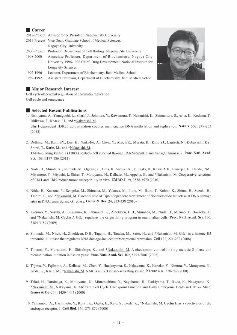
Dr. Makoto Nakanishi
Professor, Department of Cell Biology, Graduate School of Medical Sciences,Nagoya City University
Senescence is a state of permanent growth arrest and is a pivotal part of the anti-tumorigenic barrier in vivo. Although the tumor suppressor activities of p53 and pRb family proteins are essential for the induction of senescence, molecular mechanisms by which these proteins induce senescence are still not clear. Using time-lapse live-cell imaging, we demonstrate that normal human diploid fibroblasts (HDFs) exposed to various senescence-inducing stimuli undergo a mitosis skip before entry into permanent cell cycle arrest. This mitosis skip is mediated by both p53-dependent premature activation of APC/CCdh1 and pRb family protein-dependent transcriptional suppression of mitotic regulators. Importantly, mitotic skipping is necessary and sufficient for senescence induction. p16 is only required for maintenance of senescence. Analysis of human naevi also suggested the role of mitosis skip in in vivo senescence. Our findings provide decisive evidence for the molecular basis underlying the induction and maintenance of cellular senescence.
■ ContactDepartment of Cell Biology
Graduate School of Medical Sciences,
Nagoya City University
1 Kawasumi, Mizuho-cho, Mizuho-ku
Nagoya, 467-8601, Japan
TEL: +81-52-853-8146
E-mail: [email protected]
■ Education1979-1985 M.D., Nagoya City University Medical School
1985-1989 Ph.D., Nagoya City University Medical School
- 41 -
■ Carrer2012-Present Advisor to the President, Nagoya City University2011-Present Vice Dean, Graduate School of Medical Sciences, Nagoya City University2000-Present Professor, Department of Cell Biology, Nagoya City University1998-2000 Associate Professor, Department of Biochemistry, Nagoya City
University 1996-1998 Chief, Drug Development, National Institute for Longevity Sciences
1992-1996 Lecturer, Department of Biochemistry, Jichi Medical School1989-1992 Assistant Professor, Department of Biochemistry, Jichi Medical School
■ Major Research InterestCell cycle-dependent regulation of chromatin replicationCell cycle and senescence
■ Selected Resent Publications1. Nishiyama, A., Yamaguchi, L., Sharif, J., Johmura, Y., Kawamura, T., Nakanishi, K., Shimamura, S., Arita, K., Kodama, T.,
Ishikawa, F., Koseki, H., and *Nakanishi, M. Uhrf1-dependent H3K23 ubiquitylation couples maintenance DNA methylation and replication. Nature 502, 249-253
(2013) 2. Delhase, M., Kim, SY., Lee, H., Naiki-Ito, A., Chen, Y., Ahn, ER., Murata, K., Kim, SJ., Lautsch, N., Kobayashi, KS.,
Shirai, T., Karin, M., and *Nakanishi, M. TANK-binding kinase 1 (TBK1) controls cell survival through PAI-2/serpinB2 and transglutaminase 2. Proc. Natl. Acad.
Sci. 109, E177-186 (2012)
3. Niida, H., Murata, K., Shimada, M., Ogawa, K., Ohta, K., Suzuki, K., Fujigaki, H., Khaw, A.K., Banerjee, B., Hande, P.M., Miyamoto, T., Miyoshi, I., Shirai, T., Motoyama, N., Delhase, M., Appella, E., and *Nakanishi, M. Cooperative functions of Chk1 and Chk2 reduce tumor susceptibility in vivo. EMBO J. 29, 3558-3570 (2010)
4. Niida, H., Katsuno, Y., Sengoku, M., Shimada, M., Yukawa, M., Ikura, M., Ikura, T., Kohno, K., Shima, H., Suzuki, H., Tashiro, S., and *Nakanishi, M. Essential role of Tip60-dependent recruitment of ribonucleotide reductase at DNA damage sites in DNA repair during G1 phase. Genes & Dev. 24, 333-338 (2010)
5. Katsuno, Y., Suzuki, A., Sugimura, K., Okumura, K., Zineldeen, D.H., Shimada, M., Niida, H., Mizuno, T., Hanaoka, F., and *Nakanishi, M. Cyclin A-Cdk1 regulates the origin firing program in mammalian cells. Proc. Natl. Acad. Sci. 106, 3184-3189 (2009)
6. Shimada, M., Niida, H., Zineldeen, D.H., Tagami, H., Tanaka, M., Saito, H., and *Nakanishi, M. Chk1 is a histone H3 threonine 11 kinase that regulates DNA damage-induced transcriptional repression. Cell 132, 221-232 (2008)
7. Tonami, Y., Murakami, H., Shirahige, K., and *Nakanishi, M. A checkpoint control linking meiotic S phase and recombination initiation in fission yeast. Proc. Natl. Acad. Sci. 102, 5797-5801 (2005)
8. Tojima, Y., Fujimoto, A., Delhase, M., Chen, Y., Hatakeyama, S., Nakayama, K., Kaneko, Y., Nimura, Y., Motoyama, N., Ikeda, K., Karin, M., *Nakanishi, M. NAK is an IkB kinase-activating kinase. Nature 404, 778-782 (2000)
9. Takai, H., Tominaga, K., Motoyama, N., Minamishima, Y., Nagahama, H., Tsukiyama, T., Ikeda, K., Nakayama, K., *Nakanishi, M., Nakayama, K. Aberrant Cell Cycle Checkpoint Function and Early Embryonic Death in Chk1-/- Mice. Genes & Dev. 14, 1439-1447 (2000)
10. Yamamoto, A., Hashimoto, Y., Kohri, K., Ogata, E., Kato, S., Ikeda, K., *Nakanishi, M. Cyclin E as a coactivator of the androgen receptor. J. Cell Biol. 150, 873-879 (2000)
- 42 -
Symposium Session Ⅱ :
Cellular Metabolism and Age-Related Diseases
Chairpersons: Dr.Yoshikazu Higami(Tokyo Science University)Dr. Isao Shimokawa(Nagasaki University)
At 14:00 〜 17:00 on Friday, 27th June, 2014in Health Science Theater, Aichi Health Plaza
- 43 -
Ageing: What is it? New insights from C. elegans
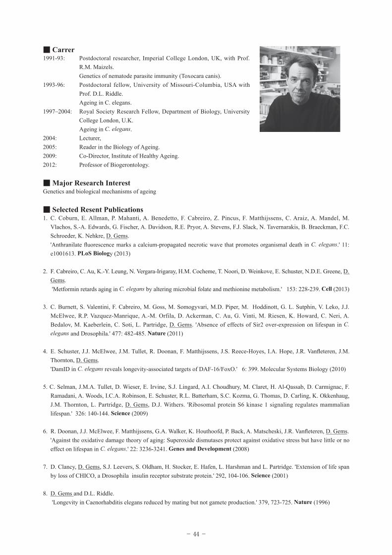
Dr. David Gems
Co-Director, Institute of Healthy Ageing, University College London
The biological mechanisms at the heart of the ageing process are a long-standing mystery. An influential theory has it that ageing is the result of an accumulation of molecular damage, caused in particular by reactive oxygen species (ROS) produced by mitochondria. This theory also predicts that processes that protect against oxidative damage (involving detoxification, repair and turnover) protect against ageing and increase lifespan. However, recent tests of the oxidative damage theory, some using the short-lived nematode worm C. elegans, have often failed to support the theory1-3. This motivates consideration of alternative models. One new theory, conceived by M.V. Blagosklonny, proposes that ageing is caused by the non-adaptive running on in later life of developmental and reproductive programmes. Such quasi-programmes give rise to hyperfunction, i.e. functional excess, leading via dysplasia (including hypertrophy and hyperplasia, and atrophy) to the age-related pathologies that cause the late-life increase in mortality4,5. Here we assess whether the hyperfunction theory is at all consistent with what is know about C. elegans ageing, and conclude that it is6. In particular, during ageing C. elegans show a number of changes that may reflect pathology and/or hyperfunction, including oocyte hypertrophy to form tumour-like masses, proximal gonad atrophy and disintegration, massive yolk accumulation, cuticular hypertrophy and neurite outgrowth. Such changes are retarded in long-lived mutants, and can contribute to mortality in at least one case (yolk accumulation). Our assessment implies that the hyperfunction theory is a plausible alternative to the molecular damage theory to explain ageing in C. elegans. To what extent the hyperfunction paradigm should replace the damage/maintenance paradigm remains an interesting open question.
1Genes Develop 22, 3236 2008. 2Cell Metab 6, 280 2007. 3Proc Natl Acad Sci USA 109, 5785 2012. 4Cell Cycle 5, 2087
2006. 5Cell Cycle 7, 3344 2008. 6Antioxid Redox Signal 2012 19, 321.
■ ContactInstitute of Healthy Ageing,
University College London,
Darwin Building, Gower Street,
London WC1E 6BT, U.K.
Tel: 020 7679 4381
E-mail: [email protected]
Lab URL: http://www.ucl.ac.uk/~ucbtdag/
■ Education1983 B.Sc. Biochemistry, University of Sussex, UK
1990 Ph.D. Genetics, Institute of Genetics, Glasgow, UK
- 44 -
■ Carrer1991-93: Postdoctoral researcher, Imperial College London, UK, with Prof.
R.M. Maizels. Genetics of nematode parasite immunity (Toxocara canis).1993-96: Postdoctoral fellow, University of Missouri-Columbia, USA with
Prof. D.L. Riddle. Ageing in C. elegans.1997–2004: Royal Society Research Fellow, Department of Biology, University
College London, U.K. Ageing in C. elegans.2004: Lecturer, 2005: Reader in the Biology of Ageing. 2009: Co-Director, Institute of Healthy Ageing.2012: Professor of Biogerontology.
■ Major Research InterestGenetics and biological mechanisms of ageing
■ Selected Resent Publications1. C. Coburn, E. Allman, P. Mahanti, A. Benedetto, F. Cabreiro, Z. Pincus, F. Matthijssens, C. Araiz, A. Mandel, M.
Vlachos, S.-A. Edwards, G. Fischer, A. Davidson, R.E. Pryor, A. Stevens, F.J. Slack, N. Tavernarakis, B. Braeckman, F.C. Schroeder, K. Nehkre, D. Gems.
'Anthranilate fluorescence marks a calcium-propagated necrotic wave that promotes organismal death in C. elegans.' 11: e1001613. PLoS Biology (2013)
2. F. Cabreiro, C. Au, K.-Y. Leung, N. Vergara-Irigaray, H.M. Cocheme, T. Noori, D. Weinkove, E. Schuster, N.D.E. Greene, D. Gems.
'Metformin retards aging in C. elegans by altering microbial folate and methionine metabolism.' 153: 228-239. Cell (2013)
3. C. Burnett, S. Valentini, F. Cabreiro, M. Goss, M. Somogyvari, M.D. Piper, M. Hoddinott, G. L. Sutphin, V. Leko, J.J. McElwee, R.P. Vazquez-Manrique, A.-M. Orfila, D. Ackerman, C. Au, G. Vinti, M. Riesen, K. Howard, C. Neri, A. Bedalov, M. Kaeberlein, C. Soti, L. Partridge, D. Gems. 'Absence of effects of Sir2 over-expression on lifespan in C. elegans and Drosophila.' 477: 482-485. Nature (2011)
4. E. Schuster, J.J. McElwee, J.M. Tullet, R. Doonan, F. Matthijssens, J.S. Reece-Hoyes, I.A. Hope, J.R. Vanfleteren, J.M. Thornton, D. Gems.
'DamID in C. elegans reveals longevity-associated targets of DAF-16/FoxO.' 6: 399. Molecular Systems Biology (2010)
5. C. Selman, J.M.A. Tullet, D. Wieser, E. Irvine, S.J. Lingard, A.I. Choudhury, M. Claret, H. Al-Qassab, D. Carmignac, F. Ramadani, A. Woods, I.C.A. Robinson, E. Schuster, R.L. Batterham, S.C. Kozma, G. Thomas, D. Carling, K. Okkenhaug, J.M. Thornton, L. Partridge, D. Gems, D.J. Withers. 'Ribosomal protein S6 kinase 1 signaling regulates mammalian lifespan.' 326: 140-144. Science (2009)
6. R. Doonan, J.J. McElwee, F. Matthijssens, G.A. Walker, K. Houthoofd, P. Back, A. Matscheski, J.R. Vanfleteren, D. Gems. 'Against the oxidative damage theory of aging: Superoxide dismutases protect against oxidative stress but have little or no
effect on lifespan in C. elegans.' 22: 3236-3241. Genes and Development (2008)
7. D. Clancy, D. Gems, S.J. Leevers, S. Oldham, H. Stocker, E. Hafen, L. Harshman and L. Partridge. 'Extension of life span by loss of CHICO, a Drosophila insulin receptor substrate protein.' 292, 104-106. Science (2001)
8. D. Gems and D.L. Riddle. 'Longevity in Caenorhabditis elegans reduced by mating but not gamete production.' 379, 723-725. Nature (1996)
- 45 -
Role of nutrient sensing pathways in stem cell fate determination
Dr. Atsushi Hirao
Professor, Division of Molecular Genetics, Cancer Research Institute,Kanazawa University
Hematopoietic stem cells (HSCs) are maintained in an undifferentiated quiescent state within a bone marrow niche. Although appropriate intrinsic and extrinsic controls are required for HSC homeostasis, the underlying molecular mechanisms are still unknown. Since we hypothesized that HSC fate may be controlled by molecules that are involved in longevity, we focused on mTOR complex 1 (mTORC1) and forkhead transcription factor FOXO, which function in nutrient sensing signaling pathways. In the quiescent HSCs, the phosphorylation of AKT is down-regulated, associated with activation of FOXO3a. We found that FoxO3a is critical for HSC self-renewal. FoxO3a-deficient HSCs showed increased phosphorylation of p38MAPK, an elevation of ROS, defective maintenance of quiescence, and heightened sensitivity to cell-cycle-specific myelotoxic injury. In addition, it was reported that dysregulation of mTORC1 causes abnormality in HSC behavior. Deficiency of Tsc1, a negative regulator of mTORC1, led to defective maintenance of the quiescence, associated with reduced HSC function. Thus, appropriate controls of these signaling pathways play a pivotal role in maintaining the HSC pool. In this symposium, I present recent data of nutrient sensing signals in stem cell fate determination.
■ ContactDivision of Molecular Genetics
Cancer Research Institute, Kanazawa University
Kakuma-machi, Kanazawa, Ishikawa, Japan 920-1192
TEL +81-76-264-6755
E-mail:[email protected]
■ Education1988 Jichi Medical University, Japan (MD)
1994 University of Tokushima, Japan (PhD)
- 46 -
■ Carrer2005- Professor, Cancer Research Institute,
Kanazawa University, Japan.
2004-2005 Associate professor,
Keio University School of Medicine, Japan
2002-2004 Assistant professor,
Keio University School of Medicine, Japan
2001-2002 Assistant professor, Institute of Molecular Embryology and Genetics,
Kumamoto University, Japan
1997-2001 Postdoctoral fellow, Ontario Cancer Institute, University of
Toronto,Canada
1995-1997 Postdoctoral fellow, Japan Society for the Promotion of Science (Kumamoto University)
■ Major Research InterestStem cell aging in hematopoiesis
■ Selected Resent Publications1. Hoshii T, Kasada A, Hatakeyama T, Ohtani M, Tadokoro Y, Naka K, Ikenoue T, Ikawa T, Kawamoto H, Fehling HJ, Araki
K, Yamamura K, Matsuda S, Hirao A. Loss of mTORC1 induces developmental blockage in early T-lymphopoiesis and
eradicates T-cell acute lymphoblastic leukemia cells. Proc Natl Acad Sci U S A. 2014, in press.
2. Hoshii T. TadokoroY. Naka K. Ooshio T. Muraguchi T. Sugiyama N. Soga T. Araki K. Yamamura K. Hirao A. mTORC1 is
essential for leukemia-propagation but not stem cell self-renewal. J Clin Invest. 122:2114-29, 2012.
3. Naka K, Hoshii T, Muraguchi T, Tadokoro Y, Ooshio T, Kondo Y, Nakao S, Motoyama N, Hirao A. TGFb-FOXO
signalling maintains leukaemia-initiating cells in chronic myeoid leukaemia. Nature, 463:676-80, 2010
4. Tamase A, Muraguchi T, Naka K, Tanaka S, Kinoshita M, Hoshii T, Ohmura M, Shugo H, Ooshio T, Nakada M, Sawamoto
K, Onodera M, Matsumoto K, Oshima M, Asano M, Saya H, Okano H, Suda T, Hamada JI, Hirao A. Identification of
tumor-initiating cells in a highly aggressive brain tumor using promoter activity of nucleostemin. Proc Natl Acad Sci U S A.
106:17163-8, 2009
5. Miyamoto K, Araki YK, Naka K, Arai F, Takubo K, Yamazaki S, Matsuoka S, Miyamoto T, Ito K, Ohmura M, Chen C,
Hosokawa K, Nakauchi H, Nakayama K, Nakayama KI, Harada M, Motoyama N, Suda T and Hirao A. Foxo3a is essential
for maintenance of the hematopoietic stem cell pool. Cell Stem Cell. 1:101-112: 2007
- 47 -
Methylation related to Transcription and Metabolism
Dr. Akiyoshi Fukamizu
Professor and Vice Director, Life Science Center, TARA, University of Tsukuba
S-adenosyl-L-methionine (SAM) synthesized by the methinone cycle is a principal biological methyl donor to a variety of acceptors, including DNA, RNA, proteins, and phospholipids. In addition, SAM is involved in polyamine biosynthesis and the conversion from glutathione to cysteine. Thus, SAM is an critical molecule participating in many biological methylations, suggesting the importance of SAM synthesis and its metabolism. The methionine cycle plays an integral part for producing SAM that serves as the methyl donor for many biological methylation reactions. The synthesis of SAM, an intermediate metabolite of this cycle, is catalyzed by SAM synthetase (SAMS), which transfers the adenosyl moiety of ATP to methionine. Although it is thought that about 1% gene encodes methyltransferases in the genome, it is unclear whether or not what is a methyltransferase that largely uses SAM. I would like to discuss about the methylation related to transcription and metabolism.
■ ContactLife Science Center, Tsukuba Advanced Research Alliance (TARA),
University of Tsukuba
Ten-nou dai, 1-1-1,
Tsukuba Ibaraki 305-8577 Japan
TEL: +81-029-853-6070
E-mail: [email protected]
■ Education1982 Faculty of Agricultural and Forestry,
University of Tsukuba: Awarded the B.S. degree.
1984 Master's Degree Program in Environmental Sciences,
University of Tsukuba: Awarded the M.S.
1986 Doctoral Degree Program in Agricultural Science,
University of Tsukuba.
1989 Awarded the Ph.D. (University of Tsukuba).
- 48 -
■ Carrer1999-2005 Professor, Center for Tsukuba Advanced Research Alliance (TARA),
University of Tsukuba
2002-2008 Program Leader in The 21st Century COE Program (Life Science)
2006-2010 Director and Professor, Center for TARA, University of Tsukuba
2011-present Professor, Life Science Center for TARA, University of Tsukuba
■ Major Research InterestTranscription and Metabolism
■ Selected Resent Publications1. Tamiya H, Hirota K, Takahashi Y, Daitoku H, Kaneko Y, Sakuta G, Iizaka,K, Watanabe S, Ishii N, Fukamizu A. Conserved
SAMS function in regulating egg-laying in C. elegans. J. Recept. Signal Transduct. 33, 56-62, 2013
2. Ozcan L, Wong CC, Li G, Xu T, Pajvani U, Park SK, Wronska A, Chen BX, Marks AR, Fukamizu A, Backs J, Singer HA,
Yates JR 3rd, Accili D, Tabas I. Calcium signaling through CaMKII regulates hepatic glucose production in fasting and
obesity. Cell Metab. 15, 739-751, 2012
3. Takahashi Y, Daitoku H, Hirota K, Tamiya H, Yokoyama A, Kako K, Nagashima Y, Nakamura A, Watanabe S, Yamagata
K, Yasuda K, Ishii N, Fukamizu A. Asymmetric arginine dimethylation determines lifespan in C. elegans by regulating
forkhead transcription factor DAF-16. Cell Metab. 13, 505-516, 2011
4. Sakamaki J-I, Daitoku H, Ueno K, Hagiwara A, Yamagata K, Fukamizu A. Arginine methylation of BAD counteracts its
phosphorylation and inactivation by Akt. Proc. Natl. Acad. Sci. USA 108, 6085-6090, 2011
5. Yamagata K, Daitoku H, Takahashi Y, Namiki K, Hisatake K, Kako K, Mukai H, Kasuya Y, Fukamizu A. Arginine
methylation of FOXO transcription factors inhibits their phosphorylation by Akt . Mol. Cell 32, 221-231, 2008

- 49 -
How much does it take for Purkinje cells to age prematurely?
Dr. I-hsin Su
Assistant Professor, Principle Investigator, Division of Molecular Genetics and Cell Biology, School of Biological Sciences, Nanyang Technological University, Singapore
Polycomb group protein complex 2 (PRC2) is involved in the regulation of various biological and pathogenic processes, including cell fate determination, proliferation, cancer progression and neurogenesis. However, functional importance of PRC2 in adult central nervous system is poorly understood. The PRC2 complex contains core proteins, Eed, suz12 and Ezh1 or Ezh2. While Ezh2 expression is largely restricted to neuronal precursor cells and immature brain, the Ezh1 is predominately expressed in mature brain cells. To determine functional implication of PRC2 in controlling cerebellar Purkinje cell (PC) dendrite formation, maturation and degeneration, we generated Ezh1-knockout mice with Ezh2 conditionally inactivated in Purkinje cells. Our preliminary data suggests that PRC2 is dispensable for PC formation, maturation and function under our current experimental setup in young mice, but may critically regulate PC function in aged mice. The underlying molecular mechanism will be determined.
■ Contact Division of Molecular Genetics and Cell Biology
School of Biological Sciences
Nanyang Technological University
SBS-02n-46, 60 Nanyang Drive
Singapore, 637551
Tel: +65-6513-8687
Fax: +65-6791-3856
e-mail: [email protected]
■ Education1986-1990 Agronomy major at National Taiwan University
1990 Bachelor of Science, National Taiwan University, Taiwan
1992-1994 Biology major at University of Cologne, Germany
1994 Received “Vordiplom”,
University of Cologne, Germany
1994-1998 Genetics Major, Biochemistry and Developmental Biology Minor at University of Cologne, Germany
1998 Received “Diplom”, University of Cologne, Germany
1998-2001 Ph.D. Program, Laboratory of Lymphocyte Signaling,
Institute for Genetics, University of Cologne, Germany
2001 Ph.D., Institute for Genetics, University of Cologne, Germany
- 50 -
■ Carrer2001-2003 Postdoctoral Associate/Fellow, Laboratory Lymphocyte Signaling,
The Rockefeller University, USA
2003-2006 Research Associate, Laboratory Lymphocyte Signaling,
The Rockefeller University, USA
2006-2007 Research Assistant Professor, Laboratory Lymphocyte Signaling,
The Rockefeller University, USA
2007-Present Assistant Professor, Division of Molecular Genetics and Cell
Biology, School of Biological Sciences,
Nanyang Technological University, Singapore
■ Major Research InterestOur lab is interested in the functional implication of lysine methylation under various physiological conditions including
immunological functions and age-related neurodegenerative disorders.
■ Selected Resent Publications1. Caganova M, Carrisi C, Varano G, Mainoldi F, Zanardi F, Germain PL, George L, Alberghini F, Ferrarini L, Talukder AK,
Ponzoni M, Testa G, Nojima T, Doglioni C, Kitamura D, Toellner KM, Su I-H, Casola S. “Germinal center dysregulation
by histone methyltransferase EZH2 promotes lymphomagenesis”. 123(12): 5009-5022. J Clin Invest 2013
2. Chan Y-S, Göke J, Lu X, Venkatesan N, Feng B, Su I-H*, Ng H-H* “A PRC2 dependent repressive role of PRDM14 in
human embryonic stem cells and iPSC reprogramming” 31(4):682-692 Stem cell 2013
3. Wyngaarden LA, Delgado-Olquin P, Su I-H, Bruneau BG, Hopyan S. Ezh2 regulates ateroposterior axis specification and
proximodistal axis elongation in the develop limb. 138, 3759-3767, Development 2011
4. Xu S, Huo J, Gunawan M, Su I-H, Lam KP. Activated dectin-1 localizes to lipid raft microdomains for signaling and
activation of phagocytosis and cytokine production in dendritic cells. 284(33):22005-22011, J Biol Chem, 2009
5. Chen N., Gu, X., Su, I-H., Bottino, R., Contreras, J.L. Tarakhovsky A., Kim S-K., Polycomb protein Ezh2 regulates
pancreatic β-cell Ink4a/Arf expression and regneration in diabetes mellitus. 23(8):975-985. Genes & Development, 2009
6. Ezhkova E., H. Pasolli A., Parker J., Stokes N., Su I-H., Hannon G., Tarakhovsky A., Fuchs E. Ezh2 Orchestrates Gene
Expression for the Stepwise Differentiation of Tissue Specific Stem Cells. 136, 1122-1135. Cell 2009
7. Su I-H.* and Tarakhovsky A*. Lysine methylation and “signaling memory“18(2):152-157, Curr. Op. Immunol. 2006,
8. Su I-H.*, Dobeneker MW., Dickinson E., Oser M., Basavaraj A., Margueron R., Viale A., Reinberg D., Wülfing C.,
Tarakhovsky A.* Polycomb group protein Ezh2 controls actin polymerization and cell signaling. 121:425-436. Cell 2005,
9. Saijo K., Schmedt C., Su I-H., Karasuyama H., Reth M., Patke A., Santana A., Tarakhovsky A. Essential role of Src-family
protein tyrosine kinases in NF-kB activation during B cell development. 4:274-279. Nature Immunology 2003
10. Su I-H., Basavaraj A., Krutchinsky A., Hobert O., Ullrich A., Chait B., Tarakhovsky A. Ezh2 controls B cell development
through histone H3 methylation and IgH gene rearrangement. 4: 124-131. Nature Immunology 2003
- 51 -
Lancheon Seminar I:
At 12:00 〜 13:00 on Thursday, 26th June, 2014
I-A 医学・生物学研究における糖鎖解析の重要性会場:ヘルスサイエンスシアター
講 師:岡島 徹也 先生
名古屋大学 大学院医学系研究科 准教授
座 長:古川 鋼一 先生
名古屋大学 大学院医学系研究科 教授
共催:住友ベークライト株式会社
I-B Lactobacillus gasseri STB2055 による線虫の 寿命延長効果とその作用機序の解明
会場:2 階 健康学習室 2・3
講 師:宮崎 忠昭 先生
北海道大学 遺伝子病制御研究所 特任教授
座 長:丸山 光生 先生
国立長寿医療研究センター研究所 部長
共催:雪印メグミルク株式会社
Lancheon Seminar I・
II

- 52 -
I-A……
医学・生物学研究における糖鎖解析の重要性
講師:岡島 徹也 先生
名古屋大学大学院医学系研究科 神経疾患腫瘍分子医学研究センター
タンパク質の主要な翻訳後修飾である糖鎖は、細胞周辺環境に豊富に存在し、細胞間のコミュニケーションを司る主要な構成要素である。高等生物における糖鎖に必要性については、糖鎖の生合成に関わる遺伝子の改変生物を用いた研究より、糖鎖の存在が細胞機能や生体機能に対して決定的な役割をすることが明らかになった。 一方で、老化や疾患などの多くの病態において糖鎖発現や構造の変化が報告されているが、先天性糖鎖合成異常症や先天性筋ジストロフィーなどの限られた例を除いては、老化や疾患の発症機序における糖鎖機能の役割は充分に明らかにされていない。また、糖鎖の機能発現には、糖鎖構造の理解が鍵となるが、タンパク質や脂質を修飾する形で存在するものや、主に糖鎖として存在するものがあるなど、糖鎖の構造は多様性に富んでいる。疾患や老化、そして、その背後にある生理現象において想定される糖鎖の多彩な生物学的役割は、多様な糖鎖構造に起因すると考えられ、タンパク質輸送、シグナル伝達、細胞間相互作用の制御における糖鎖構造の変化に起因するタンパク質機能の調節機序が解明されつつある。一方で、糖鎖解析技術の進歩により、プロテオミクス解析と平行した包括的な糖鎖構造解析が可能になり、老化や様々な疾患における糖鎖構造の変化が、俯瞰できるようになった。しかしながら、糖鎖修飾の変化は複数のタンパク質機能の変化と連動するため、糖鎖構造の解析結果を機能解析に結びつけるには、修飾を受けるタンパク質の情報も必要であり、糖鎖修飾を受けるタンパク質の情報も含めた分子情報を統合的に解析するグライコプロテオミクス技術の確立が望まれる。従って、糖鎖変化に伴うタンパク質機能変化がどのように細胞機能の変化へと統合されるか個体レベルで理解するためは、糖鎖解析技術の更なる革新や相補的な機能解析アプローチを融合した体系的な研究システムが、今後、必要となるだろう。 本セミナーでは、こうした背景を踏まえて、最近明らかになってきた新たな糖鎖機能と分子メカニズム、疾患との関連性に関した最近の知見を紹介し、糖鎖機能の統合的理解と疾患の解明への向けた糖鎖解析の重要性に関した話題を提供したい。
Lancheon Seminar I・
II
- 53 -
■連絡先
岡島 徹也名古屋大学大学院医学系研究科名古屋市昭和区鶴舞町 65
Tel: 052-744-2068 Fax: 052-744-2069
■略歷
平成 8 年 名古屋大学医学部卒業平成 12 年 名古屋大学大学院医学研究科卒業平成 13 年 米国ラトガース大学ワクスマン研究所 研究員平成 17 年 名古屋大学大学院生命農学研究科 助教平成 20 年 名古屋大学大学院医学系研究科 講師平成 21 年 名古屋大学大学院医学系研究科 准教授
■専門の研究分野
Biochemistry, Glycobiology
■本講演に関する参考文献
1. Okajima T, Irvine KD. Regulation of Notch signaling by O-linked fucose. Cell 111:893-904. (2002)
2. Okajima T, Xu A, Lei L, Irvine KD. Chaperone activity of Protein O-fucosyltransferase I within the endoplasmic reticulum
promotes folding of the Notch receptor. Science 307:1599-1603 (2005)
3. Matsuura A, Ito M, Sakaidani Y, Kondo T, Murakami K, Furukawa K, Nadano D, Matsuda T, Okajima T. O-linked
GlcNAc is present on the extracellular domain of Notch receptors. J. Biol. Chem. 283(51): 35486-35495. (2008)
4. Sakaidani Y, Nomura T, Matsuura A, Ito M, Suzuki E, Murakami K, Nadano D, Matsuda T, Furukawa K, Okajima
T. O-linked-N-acetylglucosamine on extracellular protein domains mediates epithelial cell-matrix interactions. Nat.
Commun. 583 2 (2011)
- 54 -
I-B……
Lactobacillus gasseri SBT2055 による線虫の寿命延長効果および作用機序の解明
講師:宮崎 忠昭 先生
北海道大学遺伝子病制御研究所 プロバイオティクス・イムノロジー研究部門
線虫 C.elegans は土壌に生息しており体長は約 1mm で、約 1000 個の細胞からなる生物である。寿命は約 3 週間と短いため寿命をコントロールしている遺伝子の探索に有用であるばかりでなく、全ゲノム情報が明らかになっており、分子遺伝学的な解析が可能である。これまでに長命・短命の変異体が報告され、様々な寿命関連遺伝子が明らかにされている。 近年、C.elegans を用いた乳酸菌の抗加齢・抗老化効果に関する研究が注目されているが、詳細なメカニズムは明らかにされていない。本研究では、C.elegans を用いて、Lactobacillus gasseri SBT2055 (LG2055)の寿命延長効果を評価し、その作用機序を解析した。 C.elegans N2 野生株の成虫に、標準食大腸菌(OP50)、または LG2055 を給餌し、生存率および運動能変化を解析した。その結果、LG2055 摂取群では、OP50 摂取群に比べ生存率の有意な上昇、および加齢に伴う運動能低下の抑制効果を示した。また、温度ストレスおよび酸化ストレスを負荷した生存解析においても、LG2055 摂取群で生存率の有意な上昇効果が認められた。この LG2055 による寿命延長の作用機序を検討するため、寿命関連遺伝子とアポトーシス制御遺伝子の発現を解析した結果、LG2055 給餌によりインスリン様シグナル関連遺伝子の発現変化は認められなかったが、抗酸化ストレスに関与する遺伝子群が有意に高い発現を示した。さらに、これらはミトコンドリアの機能に関与する事から ATP level およびミトコンドリア膜電位について比較解析した。その結果、LG2055 が ATP level およびミトコンドリア膜電位の制御に重要な働きを有することが示された。以上の事から、LG2055 の寿命延長効果は酸化ストレス抵抗性の亢進、およびミトコンドリアの機能制御によるものであることが示唆された。
- 55 -
■連絡先
宮崎 忠昭遺伝子病制御研究所プロバイオティクス・イムノロジー研究部門
〒 060-0815
札幌市北区北 15 条西 7 丁目TEL&FAX: 011-706-8095
http://www.igm.hokudai.ac.jp/pbi/index.html
■略歷
昭和 57 年 京都大学薬学部卒業昭和 57 年 ロート製薬株式会社研究所 研究員昭和 63 年 Nippon Boeringer Ingerheim Research Center,
Molecular Cellular Biology Department, Researcher 平成 1 年 大阪大学 医学研究科 細胞工学センター平成 7 年 同大学 医学博士取得(医学博士(大阪大学)第 11956 号)平成 7 年 東京大学医学部(現大学院医学系研究科)免疫学講座 助手 (平成 10 年から 13 年まで留学)平成 10 年 Sanford Burnham Institute
(La Jolla Cancer Research Center) Research Associate
平成 13 年 北海道大学 遺伝子病制御研究所 分子免疫分野 助教授平成 17 年 北海道大学 人獣共通感染症リサーチセンターバイオリソース部門 教授平成 23 年 北海道大学 遺伝子病制御研究所 分子免疫分野 特任教授平成 23 年〜現在 北海道大学 遺伝子病制御研究所
プロバイオティクス・イムノロジー研究分野 特任教授
■専門の研究分野
Molecular Biology, Immunology
■本講演に関する参考文献
1. Fujikura D, Ito M, Chiba S, Harada T, Perez F, Reed JC, Uede T, Miyazaki T. CLIPR-59 regulates TNF- α - induced
apoptosis by controlling ubiquitination of RIP1. Cell Death Dis., 3:e264, 2012
2. Harada T, Iwai A, Miyazaki T. Identification of DELE, a novel DAP3-binding protein which is crucial for death receptor-
mediated apoptosis induction. Apoptosis., Oct;15(10):1247-55, 2010
3. Kim,H.R., Chae, H.J., Thomas, M., Miyazaki, T., Monosov, A., Monosov, E., Krajewska, M.,
Krajewski, S., and Reed, J.C., Mammalian dap3 is an essential gene required for mitochondrial homeostasis in vivo and
contributing to the extrinsic pathway for apoptosis., FASEB J., Jan; 21(1), 188-96, 2007
4. Murata, Y., Wakoh, T., Uekawa, N., Sugimoto, M., Asai, A., Miyazaki, T., and Maruyama, M., Death-associated protein
3 regulates cellular senescence through oxidative stress response., FEBS Letters, Nov; 580(26), 6093-6099, 2006
- 56 -
Lancheon Seminar II:
At 11:50 〜 12:50 on Friday, 27th June, 2014
II-A サルコペニアにおける栄養の重要性会場:ヘルスサイエンスシアター
講 師:葛谷 雅文 先生
名古屋大学大学院医学系研究科 教授
座 長:丸山 光生 先生
国立長寿医療研究センター研究所 部長
共催:アボットジャパン株式会社
II-B 慢性痛が天気の影響を受けるメカニズム会場:2 階 健康学習室 2・3
講 師:佐藤 純 先生
名古屋大学動物実験支援センター 東山動物実験施設長
座 長:小木曽 昇 先生
国立長寿医療研究センター 実験動物管理室 室長
共催:中部科学資材株式会社
- 57 -
II-A……
サルコペニアにおける栄養の重要性
講師:葛谷 雅文 先生
名古屋大学大学院医学研究科老年科学教室 教授
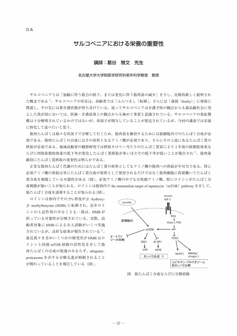
サルコペニアとは「加齢に伴う筋力の低下、または老化に伴う筋肉量の減少」をさし、比較的新しく提唱された概念である 1)。サルコペニアの存在は、高齢者では「ふらつき」、「転倒」、さらには「虚弱(frailty)」に密接に関連し、その先には要介護状態が待ち受けている。従ってサルコペニアは介護予防の観点からも超高齢社会に突入した我が国においては、医療・介護政策上の観点からも極めて重要と認識されている。サルコペニアの発症機構は十分解明されているわけではないが、多因子が関与していることが想定されているが、今回の講演では栄養に特化して述べたいと思う。 筋肉たんぱくは様々な状況下で分解して行くため、筋肉量を維持するためには筋細胞内でのたんぱく合成が必須である。筋肉たんぱくの合成にはその原料となるアミノ酸が必須であり、さらにその上流にあるたんぱく質の摂取が必須である。地域高齢者の観察研究では摂取カロリー当たりのたんぱく質量により 3 年後の除脂肪体重ならびに四肢除脂肪体重の低下率が変化したんぱく質摂取が多いほどその低下率が低いことが報告され 2)、筋肉量維持にたんぱく質摂取の重要性は明らかである。 正常な筋肉たんぱく代謝のためにはたんぱく質の材料としてもアミノ酸の筋肉への供給が不可欠である。特に必須アミノ酸の供給は単にたんぱく質合成の原料として使用されるだけではなく筋肉細胞に直接働いてたんぱく質合成を刺激している可能性がある(図)。必須アミノ酸の中でも分枝鎖アミノ酸、特にロイシンがたんぱく合成刺激が強いことが知られる。ロイシンは筋肉内で the mammalian target of rapamycin(mTOR)pathway を介して、筋たんぱく合成を誘導することが知られる(図)。 ロイシンは体内でその 5% 程度がβ -hydroxy-
β -methylbutyrate (HMB) に転換され、近年ロイシンの上記作用の少なくとも一部は、HMB が担っている可能性が示唆されている。実際、高齢者対象に HMB による介入試験がいくつ実施されているが、良好な結果が報告されている 3)。最近我々を含めいくつかの研究室が HMB はロイシンと同様 mTOR 経路の活性化を介して筋肉たんぱくの合成の促進のみならず、ubiquitin–
proteasome を介する分解亢進が抑制されることが関わっていることを報告している(図)。
図 筋たんぱく合成ならびに分解経路
- 58 -
■連絡先
葛谷 雅文名古屋大学大学院医学研究科老年科学教室教授[email protected]
■略歷
昭和 58 年 大阪医科大学卒業平成 元年 名古屋大学大学院医学研究科(内科系老年医学)卒業平成 3 年 米国国立老化研究所 研究員平成 8 年 名古屋大学医学部附属病院(老年科) 助手平成 11 年 同上 講師平成 14 年 4 月 名古屋大学大学院医学系研究科健康社会医学専攻発育・加齢医学講座 (老年科学分野)助教授平成 19 年4月 名古屋大学大学院医学系研究科健康社会医学専攻発育・加齢医学講座 (老年科学分野)准教授平成 21 年 9 月 名古屋大学医学部附属病院 NST 委員長(兼務)平成 23 年 4 月 名古屋大学大学院医学系研究科健康社会医学専攻発育・加齢医学講座 (地域在宅医療学・老年科学分野)教授平成 25 年 4 月 名古屋大学医学部附属病院地域医療センター センター長(兼務)平成 25 年 11 月 名古屋大学予防早期医療創成センター教授(兼務)平成 26 年 4月 名古屋大学未来社会創成機構 教授
■専門の研究分野
老年医学、栄養・代謝、サルコペニア、動脈硬化、認知症、地域在宅医療
■本講演に関する参考文献
1. 葛谷雅文.老年医学における Sarcopenia &Frailty の重要性.日老医誌 46: 279-285, 2009
2. Houston DK, Nicklas BJ, Ding J, et al. Health ABC Study. Dietary protein intake is associated with lean mass change
in older, community-dwelling adults: the Health, Aging, and Body Composition (Health ABC) Study. Am J Clin Nutr.
87:150-155, 2008.
3. Baier S, Johannsen D, Abumrad N, et al. Year-long changes in protein metabolism in elderly men and women
supplemented with a nutrition cocktail of beta-hydroxy-beta-methylbutyrate (HMB), L-arginine, and L-lysine. JPEN J
Parenter Enteral Nutr. 33:71-82, 2009.
- 59 -
II-B……
慢性痛が天気の影響を受けるメカニズム
講師:佐藤 純 先生
名古屋大学 動物実験支援センター 施設長
慢性痛やうつ病などの症状が天気の崩れによって悪化することが経験的によく知られている。このように気象変化の影響を受けて症状が悪化したり発症したりする疾患群を総称して「気象病」と呼ぶが、臨床研究では疾病と天気の関係について一定の結論が得られていない。そこで我々はラット、マウス、モルモットを用いた行動実験によって、気象要素(気圧、気温)と慢性痛、抑うつ症状の因果関係について科学的実証を試みてきた。本講演ではこれまでの研究成果(下記)を中心にお話ししたい。また、慢性痛患者を対象とした臨床研究も紹介する。1)慢性痛モデルの痛み行動は低気圧,低温環境で増強する
人工的な低気圧環境や低温環境への暴露は、慢性痛モデル動物の痛み行動を増強する。すなわち、「天気が悪くなったり寒くなったりすると痛みがひどくなる」現象を動物実験で再現することに成功した。
2)うつモデルの抑うつ行動は低気圧で増強する
うつモデル動物にみられる無動時間(無力・あきらめの指標)が、天候変化程度の人工的な低気圧環境への暴露によって増加した。すなわち,「天気が悪くなると抑うつ症状が悪化する」現象を動物実験で再現することに成功した。
3)低温環境,低気圧環境は交感神経を興奮させる
低温,低気圧環境では血圧、心拍数が上昇し、血液中のノルアドレナリンが増加する。すなわち、低温、低気圧環境は自律神経を刺激する。
4)病態メカニズムには交感神経が重要である
低気圧環境が痛みを増強する現象は、交感神経の活動を抑えると消失した。よって、メカニズムには交感神経の活動が重要である。
5)気圧の変化を感じるメカニズムが内耳に存在する
慢性痛モデル動物の内耳を破壊したところ、低気圧でも痛み行動は増強しない。また、内耳機能を支配する前庭神経の活動を調べると、気圧変化に反応する細胞がみつかった.よって、気圧検出機構は内耳に存在すると考えられる。
- 60 -
■連絡先
佐藤 純464-8601
名古屋市千種区不老町1名古屋大学動物実験支援センターhttp://www.care.nagoya-u.ac.jp
■略歷
昭和 58 年 東海大学医学部医学科 卒業昭和 62 年 名古屋大学大学院 医学研究科生理学専攻 満了平成 元 年 博士(医学)学位取得昭和 62 年 米国ノースカロライナ大学医学部 生理学講座 Research Associate
平成 3 年 名古屋大学環境医学研究所 神経性調節分野 助手平成 11 年 名古屋大学環境医学研究所 神経性調節分野 助教授平成 18 年 名古屋大学環境医学研究所 近未来環境シミュレーションセンター 准教授平成 19 年 愛知医科大学 学際的痛みセンター 客員研究員,非常勤医師(兼任)平成 22 年 名古屋大学発ベンチャー 株式会社 SKM 取締役(兼任)平成 25 年 名古屋大学 動物実験支援センター 教授平成 25 年 名古屋大学 東山動物実験施設 施設長(兼任)
学会役員 日本生理学会 評議員 日本疼痛学会 理事 日本ペインクリニック学会 評議員(倫理委員,利益相反委員) 日本生気象学会 幹事(学会誌 編集委員) 日本宇宙航空環境医学会 評議員 日本運動器疼痛学会 評議員
■専門の研究分野
sympathetically maintained pain, weather sensitive pain, biometeorology
■本講演に関する参考文献
1. J. Sato, M. Aoyama, M. Yamazaki, S. Okumura, K. Takahashi, M. Funakubo, K. Mizumura. Artificially produced
meteorological changes aggravate pain in adjuvant-induced arthritic rats. Neuroscience Letters, 354: 46-49, 2004.
2. J. Sato, K. Takanari, S. Omura, K. Mizumura. Effects of lowering barometric pressure on guarding behavior, heart rate and
blood pressure in a rat model of neuropathic pain. Neuroscience Letters, 299(1-2): 17-20, 2001
3. M. Funakubo, J. Sato, T. Honda, K. Mizumura. The inner ear is involved in the aggravation of nociceptive behavior
induced by lowering barometric pressure of nerve injured rats. European Journal of Pain, 14(1): 32-39, 2010.
- 61 -
A-01-O/P/YGADD34 works to suppress obesity-induced metabolic diseases including type 2 diabetes and NASH.
○ NISHIO Naomi, ITO Sachiko, YANAKA Yuriko, ISOBE Ken-ichiDepartment of Immunology, Nagoya University School of Medicine
In both western and Asian country, obesity resulted in an explosion of obesity-related health problems, including insulin resistance, type 2 diabetes, coronary artery disease, fatty liver disease and some cancers and degenerative diseases. Last year we presented the data that GADD34 KO mice got fat by high fat diet. This year we analyzed obesity by aging and myeloid cell infiltration into fad pad. Further we analyzed time course of GADD34 KO liver, which proceed to fatty liver to nonalchoholic fatty liver diseases by aging.
【Results】During the course of GADD34 knockout (KO) mice analysis in different age, we observed the higher increase of body weight caused by fat accumulation in tissue by male aged GADD34 KO mice. By flow cytometry analysis, we found that higher number of neutrophils were accumulated to GADD34 KO epididymal fat pad.These inflammatory cells induced type II diabetes. The analysis of GADD34 KO liver revealed infiltration myeloid cells to aged liver, which developed to nonalcoholic fatty liver disease.
【Discussion】We are currently trying to elucidate the molecular mechanism of GADD34 engagement in type II diabetes and fatty liver diseases. Our preliminary works suggest that GADD34 works to several points of signaling related to lipid development, insulin signaling.Keywords: GADD34, Obesity, Liver
A-03-PGADD34 inhibits activation-induced apoptosis of macrophages through enhancement of autophagy
○ ITO Sachiko, TANAKA Yuriko, THANASEGARAN Suganaya, OSHINO Reina, NISHIO Naomi, ISOBE Ken-ichiDepartment of Immunology, Nagoya University Graduate School of Medicine
【目的】 生体の細胞は、飢餓状態になるとオートファジー機能が働き、自己の細胞内小器官を分解しアミノ酸として再利用することで生存をはかっている。オートファジーがうまく働かない場合には、飢餓ストレスに耐えられず寿命の短縮につながる。また、オートファジーは細菌感染時にも働き、細胞内に侵入した細菌の分解を行う。GADD34 (growth arrest and DNA damage-inducible protein 34) は、ミエロイド系細胞の IL-6 応答遺伝子としてクローニングされ、DNA 傷害性ストレスで発現が上昇し、ストレスと老化、寿命に関連する遺伝子と考えられてきた。本研究では、マクロファージにおいて、飢餓状態で細菌感染刺激を与えた時に、細胞がどのように応答するかを検索し、オートファジー機構における GADD34 の関与について解析を行った。
【方法と結果】 マクロファージ系細胞株 RAW264.7 を Tyrosine/Cysteine 欠損培地で培養し、LPS 刺激を与えた。その結果、Tyrosine/Cysteine 欠損により GADD34 の発現 が誘導され、LPS によりさらに発現の増加が見られた。GADD34-shRNA により GADD34 を発現抑制した RAW264.7 細胞を作製し、同様の刺激を加えると、コントロールに比べ GADD34 発現抑制により、増殖シグナル伝達系が活性化された後、強い細胞死が観察された。さらにシグナル伝達系について検索したところ、GADD34 発現抑制により、mTOR シグナル経路の TSC2 の脱リン酸化が抑えられ、mTOR シグナルが促進していることが明らかとなった。その結果、GADD34 発現抑制により、オートファジーが抑制され、アポトーシスが促進し細胞死が誘導されていることが明らかとなった。これらの結果から、GADD34 は飢餓と感染ストレスに応答し、オートファジーを促進させ、生体を防御し、寿命を延長させていると考えられた。
Keywords: Macrophage, Autophagy, Apoptosis
A-02-O/PEvodiamine stimulates AMPK signaling and improves insulin resistance in adipocytes
○ YAMASHITA Hitoshi1, YAMASHITA Yukari 1, TAKEUCHI Tamaki1, TSUTSUMIUCHI Kaname2, HIRANO Shogo2, KUSUDO Tatsuya1
Department of Biomedical Sciences, College of Life and Health Sciences1 and Department of Biological Chemistry, College of Bioscience and Biotechnology2, Chubu University
生薬ゴシュユの主要成分であるエボジアミンは肥満を予防し、インスリン抵抗性を改善する作用をもつ機能性化合物である(Endcrinol,2008)。また最 近、そのインスリン抵抗性改善機序として、エボジアミンが主に脂肪細胞に おいて mTOR-S6K シグナル伝達の阻害を介して IRS-1 セリンリン酸化を抑 制することが明らかとなった(PLoS ONE, 2013)。今回、mTOR 経路の上流に位置するシグナル分子に対するエボジアミンの作用について検討を行った。成熟脂肪細胞をインスリン刺激すると、時間依存的に Akt、GSK3 及び mTOR のリン酸化が亢進した。また、mTOR 活性化を阻害する TSC2 の S939 と T1462 のリン酸化がインスリン刺激により上昇し、TSC2 活性の低下が示唆された。さらに、インスリン刺激は ERK のリン酸化を一過性に促進したが、AMPK のリン酸化には影響しなかった。一方、エボジアミン刺激は Akt、 GSK3 及び ERK のリン酸化には影響しなかったが、mTOR リン酸化を阻害し、AMPK のリン酸化は逆に促進することが明らかとなった。また、AMPK のターゲット部位である TSC2 の S1387 リン酸化が上昇し、TSC2 活性の上昇が示唆された。 これらの結果は、エボジアミンが AMPK のリン酸化を介して mTOR シグナル経路の活性化を制御し、インスリン抵抗性の改善に寄与することを示すと考えられる。現在、さらに詳細な分子メカニズムの検討を進めている。
Keywords: Evodiamine, Adipocyte, AMPK
A-04-P/YInhibition of autophagy by fatty acids in hepatocytes.
○ NEGISHI Arisa, MIZUNOE Yuhei, SUTO Yuka, HIGAMI Yoshikazu Molecular pathology and Metabolic Disease, Faculty of Pharmaceutical Sciences, Tokyo University of Science
【目的】 オートファジーは不要なタンパク質や細胞内小器官を分解することで細胞の恒常性を維持する機構である。隔離膜で囲まれた構造体はオートファゴソームと呼ばれ、リソソームと融合することでオートリソソームとなった後、隔離膜内の内容物を分解する。この隔離膜形成から分解までの一連の流れはオートファジーフラックスと呼ばれており、近年その低下が脂肪肝などの肝疾患の発症に関与していることが報告された。本研究では肥満症関連疾患におけるオートファジーの意義を解明することを目的とし、飽和脂肪酸および不飽和脂肪酸がオートファジーに及ぼす影響について解析した。
【方法・結果・考察】 脂肪酸を肝細胞に処理し、オートファジーの挙動やトリグリセリド(TG) の蓄積を解析した。その結果、飽和脂肪酸を処理した細胞においてのみオートファジーの機能低下がみられ、TG の蓄積は不飽和脂肪酸の処理によって顕著に増加した。オートファゴソームの分解にはリソソームによる分解が重要であることから、リソソームの pH を測定したところ、飽和脂肪酸の処理によりリソソームの pH が中性化していた。この結果から、リソソーム活性の低下によりオートファジーの機能低下が引き起こされることが示唆された。さらに、酸化ストレスに着目し ROS(reactive oxygen species)産生量を計測したところ、飽和脂肪酸を処理した細胞において ROS の増加がみられた。しかし、抗酸化剤の処理により、飽和脂肪酸による ROS 産生の増加が抑制され、オートファジー機能の低下が改善された。以上から、飽和脂肪酸を処理した細胞では、ROS レベルが過剰に増加するためリソソーム機能が障害を受けること、そのためにオートリソソームは内容物を分解できず、結果としてオートファジー機能の低下に繋がったと考えられる。本研究結果は、脂肪肝や老化動物の肝臓では、TG の蓄積よりも飽和脂肪酸によるリソソーム機能の低下が肝細胞機能への影響が大きいことを示唆する。
Keywords: Autophagy, Fatty acids, Hepatocytes
General Presentation
- 62 -
A-05-O/P/YLipid metabolism in cancer cachexia and caloric restriction in adipose tissue, effects of Rikkunshito
○ M I YA K AWA R y o t a 1 , S U D O Yu k a 1 , O T U K A H i r o k i 1 , G O T O Akihumi1,KASHIWASE Yohei1,2, TERAWAKI Kiyoshi2, UEZONO Yasuhito2, HIGAMI Yoshikazu1 Molecular pathology & Metabolic Disease, Faculty of Pharmaceutical Sciences, Tokyo University of Science1, Cancer Pathophysiology Division, National Cancer Center Research Institute2 Yoshikazu1
Molecular pathology & Metabolic Disease, Faculty of Pharmaceutical Sciences, Tokyo University of Science1, Cancer Pathophysiology Division, National Cancer Center Research Institute2
【目的】 がん悪液質(CC)は、体重減少や脂肪組織の委縮を特徴とする進行性消耗状態である。カロリー制限(CR)は加齢に伴う疾患の発症を抑制し、寿命を延伸する。CC と CR は共に摂食量の減少、白色脂肪組織(WAT)量が減少するなど類似した応答を示すが、その表現型は対照的である。本研究では、WAT における CC の病態解明を目的とし、CC による WAT での代謝への影響を検討し、CR による変化と比較した。また、CC の食欲不振に対してグレリン抵抗性の改善を介して食欲増進作用を示す漢方薬である六君子湯が WAT に及ぼす影響を明らかにした。
【方法】 8 週齢雄ヌードラットにヒト胃癌細胞株を皮下移植し、蒸留水を投与 (CC群 ) もしくは六君子湯を投与し(CC/TJ-43 群)、11 週齢で屠殺した。また、12 週齢 Wister 系雄ラットに自由摂食(AL)群の 70%(30%CR 群)もしくは 30%(70%CR 群)の食餌を与え、14 週齢で屠殺した。屠殺時、摘出したWAT を用いて脂質代謝に関連した因子の発現を解析した。
【結果・考察】 CC 群では、脂肪酸合成関連タンパク質発現は減少し、脂質分解関連タンパク質発現は亢進していた。30%CR 群では、脂肪酸合成関連タンパク質発現は顕著に増加したが、70%CR 群では増加が見られなかった。一方、脂質分解関連タンパク質の発現は CR の強度依存的に亢進した。以上より、CC 群と70%CR 群では脂質代謝関連タンパク質発現の変動が類似していることが確認された。CC/TJ-43 群では、CC によって減少した脂肪酸合成系因子の発現減少を抑制した。また、CC によって減少した p-Akt の発現が六君子湯によって改善した。以上より、30%CR では de novo 脂肪酸合成が亢進し、インスリン感受性が保たれること、一方、CC では摂餌量が 30%CR とほぼ同量であるにもかかわらず de novo 脂肪酸合成およびインスリン感受性が抑制されること、さらに六君子湯は CC 病態において中枢作用のみならず WAT においても、de novo 脂肪酸合成を活性化し、インスリン感受性を正に制御する可能性が示唆された。
Keywords: Cancer cachexia, Caloric restriction, Lipid metabolism
A-07-PExogenous administration of coenzyme Q10 restores mitochondrial function in the aged mouse brain
○ TAKAHASHI Mayumi, OHSAWA Ikuroh, TAKAHASHI KazuhideBiological Process of Aging, Tokyo Metropolitan Institute of Gerontology
ミトコンドリア呼吸鎖において電子伝達体として働いているコエンザイムQ (CoQ) は加齢に伴い減少することがヒトおよびラットで報告されている。 しかし CoQ 量と CoQ に依存するミトコンドリア機能との関連については明 らかではない。そこで 3 ヶ月および 12 ヶ月齢の雌雄マウスの各種臓器(大脳、心臓、肝臓、腎臓)から単離したミトコンドリアについて、CoQ が電子 伝達に関与している呼吸鎖複合体 I および II に依存した酸素消費量を測定 し、加齢に伴う変化を調べた。その結果、老齢雄マウスの大脳において、ミトコンドリア複合体 I および II 依存性の酸素消費速度が若齢に比べ有意に低 下していた。雌あるいは雄の大脳以外の臓器については月齢による有意な差 は認められなかった。また予想に反し、雄大脳の細胞質分画及びミトコンドリア分画のいずれにおいても、マウスの主要な CoQ である CoQ9 あるいは CoQ10 量に月齢による相違は認められなかった。従って、老齢雄マウス大脳 ミトコンドリアの酸素消費速度の低下は、電子伝達体である CoQ 量の加齢に伴う減少によるものではないことが示唆された。 一方、老齢雄マウス脳から単離したミトコンドリアに 10µM の水溶性 CoQ10 を添加したところ、ミトコンドリア内 CoQ10 量は有意に増加し、複合体 I および II 依存性の酸素消費速度が有意に上昇した。さらに老齢雄マウスに 150µM CoQ10 を1週間飲水投与した場合にも、脳細胞質の CoQ10 およびミ トコンドリア分画の CoQ9、CoQ10 量は有意に増加し、脳ミトコンドリアの複合体 I および II 依存性の酸素消費速度も有意に上昇し若齢マウスと同レベ ルにまで回復した。 これらの結果から、経口摂取された CoQ は体内で血流に乗った後に脳関門を通過し、大脳細胞さらにはミトコンドリア内に取り込まれ、老齢雄マウス 大脳ミトコンドリアの機能を若齢レベルまで回復させることが明らかとな った。
Keywords: Brain mitochondria, Oxygen consumption, Coenzyme Q
A-06-P/YElevated oxidative stress in OLETF (type 2 diabetes model) rat liver
○ TSUZUKI Takamasa1, NAKAMOTO Hideko2, KOBAYASHI Hiroyuki3, GOTO Sataro2, NAITO Hisashi1,2
Graduate School of Health and Sports Science, Juntendo University1, Institute of Health and Sports Science & Medicine, Juntendo University2, Mito Medical Center, Tsukuba University Hospital3
【背景】 2 型糖尿病では、インスリン抵抗性ならびに酸化ストレスが強く関与していることが知られている。本研究では運動等の糖尿病に対する介入研究のための基礎的データを得るために、2 型糖尿病モデルラットの Otsuka Long-Evans Tokushima Fatty(OLETF)において、糖尿病を発症するとされる 25 週齢時における酸化ストレスの指標を検討した。
【方法】 被験動物として OLETF ラットおよび糖尿病を発症しない対照である Long-Evans Tokushima Otsuka(LETO)ラットを用いた。25 週齢時に一晩の絶食後、全身麻酔下で屠殺し肝臓を摘出した。肝臓抽出液においてチオバルビツール反応物(TBARS)およびグルタチオン(GSH、GSSG)を測定した。さらに、RT-PCR を用いてグルタチオンレダクターゼ(GR)およびグルタチオンペルオキシダーゼ(GPx)の mRNA レベルを測定した。また、肝臓の細胞質分画およびミトコンドリア分画において、ウェスタンブロット法を用いてカルボニル化タンパク質を検出した。
【結果】 OLETF 群の TBARS、GSSG および GPx mRNA レベルは、LETO 群と比較して有意に高値を示した(P<0.05)。一方で、GSH は両群間で有意な差は認めなかった。また、OLETF 群の GR mRNA レベルは LETO 群と比較して有意に低値を示した(P<0.05)。細胞質分画におけるカルボニル化タンパク質は、両群間に有意差は見られなかったが、ミトコンドリア分画におけるにカルボニル化タンパク質は、OLETF 群は LETO 群に比較して有意に高値を示した
(P<0.05)。【結論】 2 型糖尿病モデルラットである OLETF では、25 週齢時の糖尿病発症初期において、肝臓での酸化ストレスが亢進していることが示された。今後、同モデル動物に対して運動介入等を行い、酸化ストレス指標への影響を検討していく。
Keywords: Type 2 diabetes, Glutathione, Calbonyl protein
A-08-O/PSearch for biomarkers that reflect mitochondrial dysfunction
○ FUJITA Yasunori1, ITO Masafumi1, KOJIMA Toshio2, YATSUGA Shuich3, KOGA Yasutoshi3, TANAKA Masashi4
Research Team for Mechanism of Aging, Tokyo Metropolitan Institute of Gerontology(TMIG)1, Research Center for Physical Fitness, Sports and Health, Toyohashi University of Technology2, Department of Pediatrics and Child Health, Kurume University School of Medicine3, Department of Genomics for Longevity and Health,TMIG4
ミトコンドリア機能異常は、アルツハイマー病、パーキンソン病、糖尿病などの高齢者疾患の病態に深く関係すると考えられている。一方、先天性疾患であるミトコンドリア病は、主にミトコンドリア DNA 変異によるミトコンドリア機能異常を起因とする疾患である。ミトコンドリア病の1つであるMELAS の患者の細胞から樹立した m.3243A>G 変異を有するサイブリッド細胞(2SD)は、ミトコンドリア病の細胞モデルとしてだけではなく、ミトコンドリア機能異常に起因する細胞応答を同定することができる有用な細胞モデルであると考えられた。本研究では、ミトコンドリア病の細胞モデルを用いて、ミトコンドリア機能異常により細胞外に分泌される分子の探索を行った。これまでに、m.3243A>G 変異を持たないサイブリッド細胞(2SA)と2SD 細胞のメタボローム解析から、ミトコンドリア機能異常を有する 2SD 細胞を高濃度の乳酸に暴露するとエネルギー代謝障害が増悪することを明らかにしている。そこで、網羅的遺伝子発現解析により、2SD 細胞において高濃度の乳酸暴露により特異的に発現変動する遺伝子を同定した。それらの中から分泌タンパクをコードする遺伝子を選抜し、発現が増加する遺伝子を 23 種類同定した。さらに、その中から GDF15、INHBE、IL1A に着目し、細胞培養上清中のタンパクレベルを調べたところ、GDF15 が 2SA 細胞と比較して 2SD 細胞で増加しており、高濃度の乳酸への暴露によりさらに増加することが分かった。また、ミトコンドリア病患者の血清中の GDF15 の濃度を調べた結果、コントロール群である他の小児疾患患者よりも有意に高値を示し、GDF15 がミトコンドリア病の診断マーカーになることが分かった。今後は、ミトコンドリア機能異常を反映する可能性のあるマーカーとして同定されたGDF15 が、高齢者疾患などの診断マーカーとなりうるかを検証する予定である。
Keywords: Mitochondria, Biomarker
General Presentation
- 63 -
A-09-PUbiquinol-10 Activates Mitochondria Functions and DecelerateSenescence in SAMP1 mice.
○ HIGUCHI Keiichi1, TIAN Geng1, XU Zhe1, KUBO Hiroshi2, NISHIO Shin-ya3, LI Lin1, SUZUKI Nobuyoshi3, HOSOE Kazunori4, USAMI Shin-ichi3, SAWASHITA Jinko1
Department of Aging Biology, Institute of Pathogenesis and Disease Prevention, Shinshu University Graduate School of Medicine, Matsumoto, Japan1, Frontier Biochemical & Medical Research Laboratories, Kaneka Corporation, Hyogo, Japan2, Department of Otorhinolaryngology, Shinshu University School of Medicine, Matsumoto, Japan3, QOL Division, Kaneka Corporation, Osaka, Japan4
【目的】 これまでに、還元型コエンザイム Q10(QH2)の老化促進モデルマウスSAMP1 への継続摂取が、加齢性難聴や促進老化の進行を遅延することを明らかにした。本研究では、QH2 が有する直接的な抗酸化作用以外の細胞内老化抑制機序を探索した。
【方法】 QH2 含有飼料を摂餌した SAMP1 の肝臓を用い、酸化ストレスマーカー、GSH/GSSG や SOD2 量を測定した。また、ミトコンドリア(Mt)機能関連分子の遺伝子発現量やタンパク質量変化と活性を測定した。さらに、SAMP1 の内耳も解析に用いた。次に、HepG2 細胞を用い、マウスの解析から推定した老化抑制機序の妥当性を検証した。
【結果】 SAMP1 マウスの解析:QH2 は酸化ストレスマーカーの加齢に伴う増加を抑制し、GSH/GSSG や SOD2 量の加齢に伴う減少を改善した。また、QH2は Mt 量を増加させ、電子伝達系機能も改善した。QH2 は Mt 機能関連分子
(SIRT1、SIRT3、PGC-1 α 等)の遺伝子やタンパク質の加齢に伴う減少も抑制し、SIRT1 による PGC-1 α の脱アセチル化も増加させた。さらに、QH2 は SIRT1 活性に関連する分子群(cAMP や AMPK)も増加させた。HepG2 細胞の解析:QH2 添加により ROS 産生量の減少と酸素消費量の増加がみられ、Mt 量や Complex I 活性マーカーも増加した。また、QH2 は SIRT1と PGC-1 α の複合体量と脱アセチル化 PGC-1 α を増加させ、これらの現象は SIRT1 活性阻害剤の添加で抑制された。さらに、QH2 により増加した SIRT1 活性調節分子 AMPK の活性は、cAMP 合成阻害剤の添加により抑制された。
【結論】 QH2 の新たな作用機序として、QH2 は SIRT1 および SIRT3 関連分子の活性化を促して Mt 機能を改善し、老化抑制作用をもたらす機構を解明した(Tian et al. Antioxid Redox Signal 2013)。
Keywords: Ubiquinol-10, Mitochondria, Sirtuin genes
A-11-P/YProteomic analysis of human erythrocyte proteins from individualswith diabetes
○ TSUMOTO Hiroki1, IWAMOTO Machiko1, CHIBA Yuko2, AKIMOTO Yoshihiro3, MORISAWA Hiraku1, ENDO Tamao1, MIURA Yuri1
Tokyo Metropolitan Institute of Gerontology1, Tokyo Metropolitan GeriatricHospital2, Kyorin University School of Medicine3
【【目的】 現在、糖尿病の診断には空腹時血糖値やヘモグロビン A1c 値などが使われている。しかしながら、早期診断あるいは合併症等の病態評価には新規バイオマーカーの開発が必要である。そこで本研究では、新規糖尿病バイオマーカーを開発するため、糖尿病患者赤血球の安定同位体標識法によるプロテオーム解析を行い、糖尿病で変動するタンパク質を同定した。
【方法】 糖尿病患者および健常者の血液より赤血球を分離し、超音波処理と遠心分離による赤血球タンパク質の可溶化と抽出、脱ヘモグロビン処理を行い、サンプルとして用いた。プロテオーム解析は糖尿病患者 17 例および健常者11 例のサンプルを混合して行った。サンプルの酵素消化により得られたペプチド混合物に対して iTRAQ® 試薬によるラベル化反応後、混合して強陽イオン交換スピンカラムと C18 スピンカラムによる分画および脱塩を行い、nanoLC-MALDI-TOF/TOF システムによるタンパク質の同定および変動解析を行った。
【結果および考察】 プロテオーム解析の結果、約 230 種類のタンパク質が同定され、糖尿病患者において 1.5 倍以上変動するタンパク質として 7 種類が減少し、7 種類が増加することが明らかになった。増加したタンパク質群のなかで酸化ストレスに関連するタンパク質が特に強く増加していた。このことは高血糖により引き起こされる酸化ストレスの亢進に対する防御機構が働いていることを示唆するものである。現在、ウェスタンブロッティングによる検証を行い、その有用性について検討中である。本発表では、その結果も合わせて報告する。
Keywords: Diabetes, Erythrocyte, Biomarker
A-10-P/YFiber specific studies in muscle atrophy with aging mice
○ FUKUNAGA Daichi1, MORI Shuuichi1, HIGAMI Kaichi2, SHIGEMOTO Kazuhiro1
Research Team for Geriatric Medicine, Tokyo Metropolitan Institute of Gerontology1, Molecular Pathology & Metabolic Diseases, Faculty of Pharmaceutical Sciences, Tokyo University of Science2
【背景】 加齢による筋萎縮は多様な要因で生じ、病因解明には様々なアプローチが必要である。そのためには、老化筋の病理学的変化を解析することが必要である。筋線維は収縮特性と代謝特性が異なる 4 種のタイプで構成される。そこで老齢マウスを用いて各筋線維タイプの骨格筋線維の形態変化、およびミトコンドリアの病理学的変化を解析した。
【方法】 1. C57BL/6 の 8, 20, 32 ヶ月齢のひらめ筋 (SOL, 遅筋優位筋 ) と長指伸筋(EDL, 速筋優位筋 ) の凍結切片を作製した。2. 筋線維数と断面積変化を解析するため、筋線維タイプ免疫染色を行った。ミトコンドリア呼吸酵素活性と蛋白質発現を COX 染色と COX 免疫染色で解析した。ミトコンドリア異常蓄積の評価は、Gomori 染色と電子顕微鏡観解析で行った。
【結果と考察】 32 ヶ月齢 SOL は 8 ヶ月齢と比べ、Type2X, Type2B 線維数減少が認められた。また、20・32 ヶ月齢において筋線維の平均面積には差はないが、面積分布の変化が観察された。ミトコンドリア病理解析の結果、20 ヶ月齢の時点でType1 特異的に COX 活性が低下していた。一方で、COX 蛋白の発現量低下とミトコンドリアの異常蓄積は全ての筋線維タイプで観察された。 20 ヶ月齢の EDL では、8 ヶ月齢に比べ、Type2B 特異的に筋線維の断面積が低下し、32 ヶ月齢では Type2B 特異的に筋線維数も減少していた。ミトコンドリアの病理解析では、8 ヶ月齢と 32 ヶ月齢との間に顕著な差がなかった。本研究により、老齢マウスにおいて各筋線維の断面積と数の減少は筋線維タイプで異なり、またミトコンドリアの病理学的変化は筋全体に均一に起きないことや、筋線維の断面積や数の減少と必ずしも一致しないことを明らかにした。加齢性筋萎縮とミトコンドリア機能低下との因果関係について更なる検討が必要である。
Keywords: Sarcopenia, Muscle fiber type, Mitochondria
A-12-O/P/YNeuropeptide Y deficiency enhances lipolysis and attenuates age-related changes of fat metabolism.
○ PARK Seongjoon, FUJISHITA Chika, KOMATSU Toshimitsu, KIM Sang Eun, KAWATA Takuya, MORI Ryoichi, SHIMOKAWA IsaoDepartment of Investigative Pathology, Nagasaki University
An orexigenic hormone, neuropeptide Y (NPY), not only plays a role in the hypothalamic regulation of appetite and energy expenditure, but also in the peripheral regulation of lipid metabolism. However, the intracellular mechanisms triggered by NPY to regulate lipid metabolism poorly understood. Here, we show NPY deficiency reduced white adipose tissue (WAT) mass in mice and attenuated age-related changes in adipose tissue metabolism. In particular, mRNA expression level of adipogenic/lipogenic genes were decreased and lipolytic proteins were increased in gonadal WAT (gWAT) of NPY-/- mice. In aged mice, both mRNA expression levels of lipogenic genes in gWAT and thermogenic genes in inguinal WAT were decreased, whereas NPY deficiency inhibited these age-related changes. A preadipocyte cell line, 3T3-L1, was used to analyze molecular mechanisms of NPY in adipocytes metabolism. NPY (100 nM) increased adipogenesis, and inhibited serum starvation-induced lipolysis in adipocytes. This anti-lipolytic action of NPY was blocked by Y1R antagonist, BIBO3304. Western blot analysis showed NPY inhibited SIRT1 and enhanced phosphorylation of FOXO1 in adipocytes. These results showed NPY enhances adipocytes differentiation through augmentation of classic adipogenic genes expression and inhibits lipolysis through suppression of SIRT1-FOXO1 signaling pathway. Taken together, these data reveal a novel intracellular mechanism of NPY in the regulation of lipid metabolism and highlight antagonism of NPY as a promising target for drug development to prevent age-related metabolic dysfunction.
Keywords: Adipose tissue, Age, NPY
- 64 -
A-13-O/PMetabolomic approach for human blood
○ KONDOH HiroshiDep. of Geriatric Medicine, Kyoto Univ. hospital
高齢社会の到来とともに、寝たきりや脆弱な人々に対し、健康長寿な方々も存在するという高齢者の多様性が拡大しつつあり、高齢者医療をより複雑にしている。我々は、血液メタボローム解析による高齢者の代謝特性解明を目指した。 沖縄科学技術大学院大学の柳田充弘教授との共同研究によりマススペクトロメトリーによる低分子物質の半定量的計測を行い、数千の低分子代謝物質検出が可能であることが判明し、この新規アプローチ(メタボローム)による新規代謝マーカーの検索が可能性となった。その基盤データをもとに、ヒト若年群(平均20歳代)と高齢群(平均80歳代)の血液メタボローム比較検討を行った(合計60名以上)。 詳細な解析の結果、ヒト血液メタボライトは、大きく3つに分類できることが判明した。すなわち、₁個人差の少ないもの、₂高齢者と若年群で明確な差があるもの、₃個人差の大きなもの、の3つである。₂に関しては、6つの候補メタボライトを見出した。₃に関しては、興味深いことに、若年群と高齢群で異なる挙動を示すメタボライトの存在が確認された。 我々の今回の知見より、高齢者と若年で個人差の違いの分布が大きく異なるメタボライトがいくつか存在することが判明した。このような高齢者特有のメタボライトは、年齢以外に、食生活、生活様式、健康状態、環境要因などを反映すると推測されるが、これまでに類似の報告は少ない。
Keywords: Metabolome, Blood, Ageing
A-15-P/YThe effect of Amino Acids on the Progression of Atherosclerosis and Aging. 2nd report using siRNA
○ INA Koicihiro, HAYASHI Toshio, YAMAGUCHI Tomoe, MAEDA Morihiko, KUZUYA MasafumiDepartment of Geriatrics, Nagoya University Graduate School of Medicine
【序論】 NO 合成酵素 (NOS) の基質 L-arginine は動脈硬化進行病変には有用ではない。NOS の反応物質 L-citrulline(L-Cit) から L-arginine(L-Arg) を生成する経路が内皮にある。L-Cit の老化と NO への影響を検討した。
【方法】 細胞を高グルコース等で刺激し、L-Cit、L-Arg、双方 (LA+LC) を投与し細胞老化マーカー等でみた。各種蛋白、細胞内 NO (DAF-2)、細胞内 ROS (H2-DCFDA) を調べた。アミノ酸トランスポーター siRNA を用いた。
【結果】1)生活習慣病刺激と細胞老化。高グルコース (HG) で細胞老化が進行し、アンギオテンシン II や酸化 LDL では軽度だった。2)内皮老化とアミノ酸。HG 下で L-Arg、L-Cit、LA+LC を投与した。細胞老化 SA- β -galactosidase は、HG で増加し、L-Arg 群で低下傾向、L-Cit 群、LA+LC 群で低下した。HG で peNOS は低下し、L-Arg、L-Cit、LA+LC で回復し Arginase2 を低下させ、Arginosuccinosynthase 活性を上昇させた。HG で低下する NO は L-Cit 及び L-Arg+L-Cit で増加。HG で増えた ROS はL-Arg、L-Cit にて減少傾向を認めた。3) L-Arg、L-Cit 各々のトランスポーター、Cationic Arginine Transporter (CAT-1) 及び SN-1 を使用し、eNOSsiRNA(siNOS)、siCAT1、SN1siRNA(siSN1) を作った。siCAT1、siSN1 にて細胞老化が亢進した。peNOS は siSN1 下で L-Cit で低下した。arginase2 は抑制され、ASS は増加した。細胞内 L-Arg、 L-Cit 濃度は siSN1 処置では変化しなかった。Cit 投与時は細胞内 Cit 濃度が上昇した。siRNA 処置は細胞内から細胞外へのアミノ酸移動が低下している可能性が示唆された。
Keywords: L-citrulline, Nitric Oxide, Senescence
A-14-O/P/YThe effect of Amino Acids on the Progression of Atherosclerosis and Aging. 3rd report. Animal Study
○ INA Koicihiro, HAYASHI Toshio, YAMAGUCHI Tomoe, MAEDA Morihiko, KUZUYA MasafumiDepartment of Geriatrics, Nagoya University Graduate School of Medicine
【序論】 L-arginine(L-Arg) は NO 合成酵素の基質で初期動脈硬化症の進展予防には有効だが,進行病変では有用でない。NO 合成酵素の反応物質 L-citrulline(L-Cit) から L-Arg を生成する経路が血管内皮にも存在し、報告者らは L-Cit 及び L-Arg+L-Cit の抗動脈硬化作用を in vivo で初めて証明し、L-Cit 等の NObioavailability への重要性を報告した。 血管内皮細胞老化は動脈硬化発症に先駆するので、L-Cit 及び L-Arg の動脈硬化進展、血管老化及び NO 産生への影響を検討した。糖尿病自然発症動脈硬化形成ラットにて、糖尿病での L-Cit の作用を検討した。
【方法】 糖尿病自然発症肥満ラット ZFDM-Lepr fa ラット雄性 6 週齢を、1週馴化後実験に供与した。I)Control 群、II)L-Cit. 群(2%自由飲水), III)L-Arg 群(同),IV)L-Arg+L-Cit 群(1%ずつ自由飲水)とし 4 週間投与し、体重、血糖値、血清脂質、肝機能、血中アミノ酸濃度、内皮機能として NO の動態、細胞老化等を検討した。
【結果】 ZFDM-Lepr fa ラ ッ ト 各 群、I)Control 群、II)L-Cit 群 , III)L-Arg 群, IV)L-Arg+L-Cit 群の体重は4週までに変化なく、血糖値は L-Cit 群がやや低下した。血中脂質は HDL-C 値が L-Cit 群で高値の他は、総コレステロール、中性脂肪値に4群間に有意差はなかった。肝機能は 4 群間に有意差はなかった。血中アミノ酸濃度は L-Cit 群、L-Arg+L-Cit 群にて血漿 Arg 濃度、血漿 Cit 濃度が有意に上昇した。 大動脈内皮細胞老化を SA- β -gal で検討した。胸腹部とも L-Cit 群が有意に低かった。血漿 NOx は L-Cit、L-Arg、L - Cit+L-Arg 群各々が高かった。
【結語】 L- シトルリンは細胞老化抑制とともに糖尿病性動脈硬化発症を抑制する可能性がある。
Keywords: Senescence, Diabetes mellitus, Nitric oxide
A-16-O/P/YBasic research of sex-specific aging in skeletal muscle
○ KITAJIMA Yuriko1,2, MASUDA Shinya2,ONO Yusuke2
Department of Reproductive Pathophysiology Nagasaki University Graduate School of Biomedical Sciences1, Department of Stem Cell Biology, Atomic Bomb Disease Institute, Nagasaki University2
今日までの骨格筋の基礎的研究は、主に男性(雄)を対象に行われてきた。しかし、女性は閉経後に、卵巣から分泌されるエストロゲンが低下・欠乏することで、骨量減少、脂質異常、動脈硬化など様々な全身的異常を引き起こすという点で,男性の老化とは異なる経過を示す。加齢性筋肉減弱症(サルコペニア)は、加齢に伴う急激な筋力低下を特徴とし、その原因としては筋萎縮および筋再生能の低下であると考えられている。近年の研究から、骨格筋においてエストロゲン受容体の発現が確認されているが、その機能は不明な点が多い。本研究は,女性特有の老化メカニズムとしてエストロゲンに着目し、エストロゲン欠乏が骨格筋に及ぼす影響を検討した。6 週齢の C57BL/6 マウスの両側卵巣を摘出し、エストロゲン欠乏群(OVX 群)を作成した。Control 群に比べて OVX 群では、前脛骨筋の筋横断面積が有意に萎縮した。とくに、OVX 群では、遅筋線維が減り速筋線維が増える速筋化が観察された。この変化は、いわゆる加齢でみられる遅筋化とは逆であり、不活動による廃用性筋萎縮に近い表現型であった。続いて、筋再生に必須の役割を担う骨格筋組織幹細胞であるサテライト細胞について検討した。加齢により筋線維あたりのサテライト細胞数は減少することが知られているが、OVX 群のサテライト細胞数には変化がないものの筋分化能は顕著に低下していた。さらに、薬剤により筋損傷を誘導し筋再生能を評価したところ、OVX 群では筋再生能の著しい低下が観察された。以上の結果から、エストロゲンは筋機能維持に重要な役割を担っており、その欠乏は女性特有の骨格筋老化に連なる可能性が示唆された。骨格筋を含む様々な組織・臓器における老化メカニズムの解明には、性差に着目した基礎研究が重要であると考えられた。
Keywords: Sex-specific aging, Skeletal muscle, Estrogen
- 65 -
A-17-P/YMetabolism of skin-absorbed resveratrol in mouse
○ MURAKAMI Itsuo, ITO Ken, KONDOH HiroshiDepartment of Geriatric Medicine, Graduate School of Medicine, Kyoto University
超高齢化社会に向かいつつある日本において、加齢性生活習慣病や寝たきりの解消は社会的に大きな課題である。健康長寿の達成や寝たきりの予防策として、健康効果を期待できる低分子化合物の摂取があげられる。レスベラトロールは、ポリフェノールの一種で、長寿遺伝子 Sirt1 や AMP キナーゼを活性化する。活性化された Sirt1 と AMP キナーゼはミトコンドリア活性化転写因子 PGC-1 を活性化する。レスベラトロールの経口摂取による血糖値改善効果や、筋肉増強効果が報告されている (Vernon W. 他 J Physiol 2012)。本研究では、レスベラトロールを経皮によって体内に吸収させることで、筋力回復・寝たきり予防効果や健康推進効果を経口摂取が困難な高齢者に対しても得る手法を開発する事を目指した。 レスベラトロールの経皮吸収の確認と経皮吸収による代謝経路の変化を検討するため、質量分析器を用いたメタボローム解析を行った。レスベラトロールを 3 種類の親水性基剤と混合してヘアレスマウスの皮膚に塗布し、4 時間後に各臓器(皮膚、肝臓、筋肉、血液)のサンプルを調製し解析した。それぞれの基剤を用いたマウスの臓器からレスベラトロールの代謝産物が検出された。レスベラトロール代謝産物は、臓器によって蓄積される量が異なっていた。経皮吸収のマウスからは、経口投与したマウスと同程度のピークが検出された。 皮膚からレスベラトロールが体内に吸収されることが確認され、寝たきりなど経口摂取が困難な場合でも経皮吸収によるレスベラトロールの摂取によって健康効果が得られる可能性が示唆された。
Keywords: Resveratrol, Skin absorption, Metabolomics
B-02-PMetabolic profiling of Alzheimer’s disease brain
○AKATSU Hiroyasu1, TSUTSUI Haruhito3, YAMAMOTO Takayuki2, HASHIZUME Yoshio2, OHARA Hirotaka1, TOYO’OKA Toshimasa3, INOUE Koichi3
Department of Community-Based Medical Education, Nagoya City,University Graduate School of Medical Sciences1, Choju Medical Institute, Fukushimura Hospital2, Laboratory of Analytical and Bio-Analytical Chemistry, School of Pharmaceutical Sciences, University of Shizuoka3
【目的】 アルツハイマー病(AD)は老人斑と神経原線維変化の病理所見で確定診断となる。しかしその原因の本質は不明で、脳内での代謝異常に関する報告は皆無である。我々は今回福祉村ブレインバンク患者脳を用い、低分子産物を解析することで脳内での代謝変化を解析した。
【方法】 ApoE 3/3 で遺伝子型が統一された AD, non AD 各 10 例の側頭葉と後頭葉の凍結サンプルを ultra performance liquid chromatography electrospray time-of-flight mass spectrometer analysis (UPLC-ESI/TOF/MS) で解析した。 S-plot 法を用いて定量的に差のある分子を抽出した。
【結果】 代謝データベースから spermine と spermidine が同定され、オルニチン代謝系がアルツハイマー病理に何らかの関与があることを示唆する結果を得た。
【考察】 オルニチン代謝系と AD との関連はこれまであまり注目されてきていないが、spermine、spermidine は NMDA 受容体に対する興奮毒性も報告されている。また NMDA 受容体阻害薬は臨床応用されており AD 患者病態のコントロールに用いられている。今回の結果は初期病変に対する2次的変化である可能性もあるが、発症後の臨床像に何らかの影響を与えている可能性も考えられる。今後、形態的な解析も含めてさらに検討を加えていきたい。
【倫理的配慮】 本研究は死亡時に質量分析、遺伝子解析等にもご遺体の一部を用いる事の許可を書面にて得ており、すべて匿名化により解析は行われた。福祉村病院および静岡県立大学の倫理委員会の承認も得ている。
Keywords: Alzheimer's disease, Metabolic profiling, Ornithine metabolism
B-01-O/P/YPhosphodiesterase 3 inhibitor cilostazol improves conditioned fear in SAM-P8 mice
○ YANAI Shuichi, KOJIMA Kai, ARASAKI Tomoko, ENDO ShogoAging Neuroscience Research Team, Tokyo Metropolitan Institute of Gerontology
cAMP 系は、様々な動物の記憶・学習機構に重要な役割を果たす。cAMPはホスホジエステラーゼ (Phosphodiesterase, PDE) により分解され、その生 理作用は終結する。それゆえ、PDE 阻害剤は神経活動により生成された細胞 内 cAMP 濃度を上昇・維持させ、cAMP 系の生理機能を増強すると考えられ る。我々はこれまでに PDE3 阻害剤である cilostazol が若齢マウスの海馬依 存性記憶を向上することを報告してきたが、PDE3 阻害剤について老化モデ ルマウスを対象とした報告は少ない。そこで本研究では、老化促進マウス SAM (Senescence-Accelerated Mouse) の中で比較的若齢から記憶障害が観察 される P8 を用い、cilostazol が条件性恐怖記憶に及ぼす効果を検討した。 8 ヶ月齢の老化促進マウス SAM P8 とその対照群である SAM R1 に対し、 音(10kHz, 70dB)と電気ショック(0.5mA)を対提示して恐怖条件づけを行 った。Cilostazol は各日の実験 30 分前にゾンデを用いて経口投与(0、30 ま たは 100mg/kgBW、急性投与)をするか、もしくは、cilostazol を含む餌を 3 ヶ月間(5 〜 8 ヶ月齢)自由摂食で与えた(0、0.3、1.5%、長期投与)。条件 づけから 24 時間後に手掛かり依存性記憶テストを、48 時間後に文脈依存性 記憶テストを行った。Cilostazol の効果は長期投与においてのみ観察され、 いずれのテストにおいても cilostazol 1.5% 投与 SAM P8 の条件性恐怖記憶が SAM R1 のそれと同程度まで改善することが示された。 これらの結果は、cilostazol の長期投与が扁桃体及び海馬依存性の長期記憶 を改善することを示している。
Keywords: Memory, Cilostazol, SAM
B-03-PEffect of acupuncture-like stimulation on cortical cerebral blood flow in aged rats○ UCHIDA Sae1, WATANABE Saori2, MISAWA Hidemi2, KAGITANI Fusako1
Department of Autonomic Neuroscience, Tokyo Metropolitan Institute of Gerontology1, Department of Pharmacology, Keio University, Faculty of Pharmacy2
【目的】 認知機能の維持に重要な大脳皮質血流の加齢に伴う低下を防ぐ手法の開発が望まれている。我々はこれまでに、鍼による皮膚・筋の機械的刺激がマイネルト核(NBM)コリン作動性神経を賦活させることにより、投射先の大脳皮質血流を増加させることを成熟ラットで報告した。老齢ラット(約 3 年齢)では成熟ラットと比較して、NBM の直接電気刺激で起こる大脳皮質血流増加反応が、低強度(50µA)刺激による反応は維持されるものの、高強度
(100-200µA)刺激による反応が有意に減弱する。これらの事実から、鍼刺激による大脳皮質血流増加反応も老齢ラットで減弱する可能性がある。そこで本研究では約 3 年齢の老齢ラットを用いて、鍼刺激が大脳皮質血流に及ぼす影響を調べ、成熟ラットの反応と比較した。
【方法】 老齢(35-36 ヶ月齢)の Wistar 系ラットを用いた。ウレタン麻酔下、人工呼吸下で頭頂葉の局所血流をレーザードップラー血流計を用いて連続測定した。一側の前肢足蹠に鍼灸針を深さ約 5mm 刺入し、1Hz で左右に回転させる刺激を 1 分間加えた。血圧変動を除去するために、第 1-2 胸髄レベルで脊髄を切断した。
【結果】 中枢無傷時の老齢ラットにおいて、前肢足蹠の鍼刺激は大脳皮質血流と平均血圧を同程度に上昇させた。第 1-2 胸髄レベルでの脊髄切断後、前肢足蹠刺激は血圧に影響を及ぼすことなく、頭頂葉血流を増加させた。この血流増加反応の大きさは平均約 112%であり、成熟ラットでの反応(約 111%)との間に差は認められなかった。
【結論と考察】 鍼刺激で誘発される大脳皮質血流増加反応は、約 3 年齢の老齢ラットにおいても成熟ラットと同程度に維持されることが明らかとなった。鍼などの自然な体性求心性刺激で活性化される NBM コリン作動性血管拡張系は老齢ラットでも十分機能することが示唆される。
Keywords: Acupuncture, Cerebral blood flow, Aging
- 66 -
B-04-O/YIdentification of the “toxic conformer” of amyloid β in Alzheimer’s disease
○ IZUO Naotaka1, MURAKAMI Kazuma2, KUME Toshiaki3, AKAIKE Akinori3, SHIRASAWA Takuji4, YOKOTE Koutaro1, IRIE Kazuhiro2, SHIMIZU Takahiko1
Graduate School of Medicine, Chiba University1, Graduate School of Agriculture2, Graduate School of Pharmacy3, Kyoto University, Graduate School of Medicine, Juntendo University4
【Introduction】 アルツハイマー病 (AD) の病態形成に関係する amyloidβ (Aβ) は、様々な立体構造を有することが知られているが、病態形成と強く関連する立体構造は明らかではない。これまで我々は、特に凝集能および神経毒性が強い 42 残基からなる Aβ42 に着目し、その凝集体の立体構造解析から、22-23 位においてターン構造を有するコンホマーの存在を明らかにした。そこで本研究では、Aβ42 のターン構造を有するコンホマーと AD 病態の関連について検討した。
【Results】 22-23 位においてターン構造を形成しやすい変異体 E22P-Aβ42 および形成しにくい変異体 E22V-Aβ42 を合成した。E22P-Aβ42 は、高い凝集能を示し、初代培養神経細胞に対し強力な毒性と酸化ストレスを惹起したのに対し、E22V-Aβ42 はいずれの活性も示さなかった。そこで、22-23 位においてターン構造を有するコンホマーを Aβ42 の「毒性コンホマー」と名付けた。毒性コンホマーの性質を解析するため、E22P-Aβ10-35 をハプテンとして特異抗体を作製した。得られた 11A1 抗体は、E22P-Aβ42 を認識し、E22V-Aβ42 を認識しないことから、毒性コンホマーに対する立体構造特異抗体であることが明らかとなった。AD 患者の剖検脳に対する免疫染色により、海馬および大脳皮質の老人斑において強い染色性が見られた。11A1 抗体は、培養神経細胞における Wt-Aβ42 および E22P-Aβ42 誘発神経毒性を完全に抑制した。さらに、11A1 抗体は強毒性を示す E22K-Aβ42 (Italian 型) およびE22G-Aβ42 (Arctic 型) と強く反応したことから、毒性コンホマー形成と神経毒性に強い相関が認められた。
【Discussion】 A β 42 の毒性コンホマーは、AD の病態形成に重要であり、有効な治療標的になりうることが示唆される。
Keywords: Amyloid β , "Toxic conformer", Alzheimer's disease
B-06-O/YChronic intermittent methamphetamine treatment effects on direct pathway neurons of the striatum.
○ INOUE Ritsuko, MIURA MasamiNeurophysiology, Tokyo Metropolitan Institute of Gerontology
高齢者で発症・有病率が高いパーキンソン病の症状は運動症状(震せん、固縮、無動、姿勢反射障害)を主とするが、常同行動などの衝動制御障害が伴うケースも知られてきている。大脳基底核の主要な神経核である線条体はマトリックスとストリオソームという性質が異なるふたつの領域から成り、それらの局所神経回路と常同行動との関連が指摘されている。しかし、詳細は未だ明らかではない。覚醒剤メタンフェタミンはドーパミンによる神経伝達を増幅することで自発活動を亢進させ、慢性投与すると常同行動を誘発する。メタンフェタミンを反復投与することで薬物投与時に誘発される常同行動が増悪したマウスを得ることができる。 本研究では、マトリックスとストリオソームの区別に加え、線条体の直接路と間接路を分けて、メタンフェタミン反復投与が線条体の抑制性入力(投射ニューロンの miniature IPSCs)に及ぼす影響を調べようと試みた。実験には線条体ストリオソームが同定でき、かつ D1 受容体を発現した直接路ニューロンが標識されたダブルトランスジェニック (double Tg) マウスを用いた。マトリックスの直接路が黒質網様部および淡蒼球内節の GABA ニューロンに投射するのに対し、ストリオソームの直接路は中脳黒質ドーパミンニューロンに直接投射することがわかっている。この double Tg マウスから皮質 - 線条体スライスを作成し、線条体投射ニューロンの電気生理記録を行った。コントロール(生理食塩水反復投与)群とメタンフェタミン反復投与群においてマトリックスからの記録とストリオソームからの記録を区別し、直接路投射ニューロンの miniature IPSCs の振幅と頻度をそれぞれ比較した。これまでの実験で明らかとなったメタンフェタミン反復投与の効果と合わせて報告する。
Keywords: Dopamine, Striatum, Motor stereotypy
B-05-O/YCharacterization of Amyloid-β Degrading Activity in Human Serum○ MIKAWA Ryuta1,3, OKUNO Alato1, TAKAYANAGI Akiko1, OKADA Ken2, YOSHIMI Tatsuya2, TAKIKAWA Osamu1,2
Laboratory of Radiation Safety, Research Institute, National Center for Geriatrics and Gerontology1 : Laboratory of Drug Screening, Department of Drug Discovery, Center for Development of Advanced Medicine for Dementia2 : Laboratory of Aging Research, Program in Integrated Medicine, Graduate School of Medicine, Nagoya University3
【目的】 アルツハイマー病 (AD) 脳ではアミロイドβペプチド (Aβ) の凝集体である老人斑の蓄積に加え、80%以上の高い頻度で脳微小血管周囲に Aβ 凝集体の沈着が生じる。この脳アミロイド血管症 (cerebral amyloid angiopathy: CAA) の発症機序は不明であり、特に Aβ の由来も解明されていない。脳微小血管は、血管内皮細胞、ペリサイト、アストロサイトで構成されており、これら3細胞に Aβ産生能が明らかにされつつある。従って、これら血管構成細胞由来の Aβ代謝障害が、CAA 発症原因となっている可能性が考えられる。我々は血管内皮細胞の Aβ 産生調節機構を解析中、ヒト血清中に Aβ分解活性を見出した。本研究では、その性状の解析を行った。【方法】 血清 Aβ分解活性を DEAE クロマトグラフィー、G-100 ゲル濾過およびハイドロキシアパタイトクロマトグラフィーで精製した。各カラムクロマトの分画の Aβ分解活性は Aβ40 を使用し、ELISA 法で測定した。Aβ分解活性を示す蛋白質は LC/MS により同定した。【結果】 血清中の Aβ分解活性は Sephadex G-100 のゲル濾過解析から分子量約200kDa を示した。その活性はハイドロキシアパタイトクロマトグラフィーにおいて分子量約 175 kDa と約 25 kDa の蛋白質に一致して溶出された。 LC/MS により約 175 kDa の蛋白質は α2-macroglobulin (α2M) であり、約25kDa の蛋白質は apolipoprotein A-I (apoA-I) と同定された。【考察】 apoA-I は Aβ分解活性を示さない。α2M も Aβ分解活性を示さないが、そのホモ 4 量体 (725 kDa) はカゴ状構造体を形成し、そのカゴ内にプロテアーゼ等を内包することが知られている。従って、apoA-I と複合体を形成している α2M に Aβ分解活性を示すある種のプロテアーゼが結合している可能性が考えられた。【今後の研究計画】 α2M と apoA-I 複合体の Aβ分解活性の本体 (ある種のプロテアーゼを想定) を同定し、さらに本 Aβ分解活性と CAA の関連を明らかにする。
Keywords:Amyloid angiopathy , A β degradation , Alzheimer's disease
B-07-PPrevention of cognitive decline due to psychosocial stress
○ UNNO Keiko1, SUMIYOSHI Akira2, NONAKA Hiroi2, KONISHI Tomokazu3, HARA Ayane1, NAKAGAWA Aimi1, IGUCHI Kazuaki1, TAKEDA Atsushi1, KAWASHIMA Rhuta2, HAYASHI Michiko4, NAKAMURA Noriyuki4
School of Pharmaceutical Sciences, University of Shizuoka1, Institute of Development, Aging and Cancer, Tohoku University2, Basic Life Science Research Group, Akita Prefectural University3, Tea Science Center, University of Shizuoka4
【目的】 ストレスの負荷が長期間持続すると、心身の疾患を引き起こすとともに、老化を促進させると考えられている。本研究では社会心理的ストレスによる脳の老化促進の機構を明らかにすることを目的とし、脳の形態的変化ならびに遺伝子発現の変化について検討した。また抗ストレス作用物質として、緑茶に含まれるテアニンの有効性を検討した。
【方法】 雄マウス間の縄張り意識を利用した対面飼育により、マウスに社会心理的ストレスを負荷した。マウスは、加齢に伴う脳の萎縮が認められている老化促進モデルマウス (SAMP10) を用いた。脳の萎縮がどのような部位でどのような時間経過で進行しているのか、動物用高磁場 MRI 装置を用いて観察した。また脳内の遺伝子発現の変化について DNA マイクロアレイにより解析した。
【結果及び考察】 SAMP10 マウスの場合、脳の萎縮および脳機能低下が有意に認められるのは 11 〜 12 月齢であるが、対面飼育条件下で社会心理的ストレスが負荷された場合は、8 〜 9 月齢から大脳の萎縮および学習能低下が認められることをこれまでに見いだしている。脳の形態変化を ex vivo による MRI 測定により調べた結果、対面飼育開始前のマウス(2 月齢)に比べ、対面飼育開始2ヶ月のマウス(4 月齢)で脳の萎縮傾向が認められた。一方、テアニンを摂取していたマウスでは脳萎縮は認められなかった。ストレスの負荷により、ストレス応答器官の一つである副腎の肥大が生ずるが、テアニン摂取により肥大が有意に抑制された。4 月齢のマウスにおいて、副腎が肥大しているマウスほど脳容積が減少する傾向にあることが見出された。また脳内遺伝子発現の変化は、ストレス開始初期の場合と長期間ストレス負荷した場合では大きく異なることが見出された。これらのことから、社会心理的ストレスの負荷により脳の萎縮は早期から始まっており、その変化は単純ではないことが示唆された。
Keywords: Brain, Atrophy, Psychosocial stress
- 67 -
B-08-O/PAge-dependent brain functional changes with mitochondrial oxidative stress
○ ISHII Takamasa1, TAKANASHI Yumi1, YANAGIHARA Rintaro1, YASUDA Kayo2, ISHII Naoaki1
Department of Molecular Life Science, Tokai University School of Medicine1,Education and Research Support Center, Tokai University2
筆者らは、ミトコンドリア電子伝達系複合体 II の SDHC サブユニットV69E アミノ酸点変異が電子伝達を部分的に阻害し、ミトコンドリアを過剰発生することを明らかにしてきた。近年、当該 SDHCV69E アミノ酸点変異を導入した Tet-mev-1 コンディショナルトランスジェニックマウスの表現型解析に尽力してきた。 その内、モリス水迷路による海馬依存的な学習記憶能力試験において、壮年期の Tet-mev-1 マウスは長期記憶形成に障害を生じていることが明らかにされた。しかし、脳内でのアミロイドβ 1-42 蓄積や乳酸アシドーシス、細胞死等の神経変性疾患様の異常は確認されなかった。そこで、酸化ストレス発生とその障害を解析した結果、Tet-mev-1 マウスは、若齢期では抗酸化能力を亢進することで活性酸素種(ROS)の産生を抑えているが、壮年期になるとその抗酸化能力の亢進は抑えられ、過剰な ROS を産生することが確認された。このような壮年期のマウスは JNK/SAPK ストレス応答シグナルを活性化し、アストロサイト細胞骨格を構成する中間径フィラメント GFAP 蛋白質量を低下させていた。 以上の結果から、ミトコンドリア電子伝達系複合体 II 異常によりミトコンドリアスーパーオキサイドを過剰産生する Tet-mev-1 マウスは、壮年期において酸化ストレス障害を生じ、脳内グリア環境を破綻していることが示唆された。これまで、加齢依存的な脳内病変部では神経膠症(グリオーシス)や神経膠腫(グリオーマ)などの発症が確認されていた。しかし今回の研究成果では、加齢依存的なミトコンドリア酸化ストレスの発生はアストロサイトの膠原線維形成を脆弱化し、長期記憶形成の障害を引き起こすことが新たに示された。 今後、当該モデルを生理的加齢記憶障害モデルと位置付け、その破綻が早期化する分子機序の解明を目指す。
Keywords: Mitochondria, Oxidative stress, Brain
B-10-P/YEffects of Aging and ROS on Nitric-Oxide Induced Calcium Release in Neuronal Cells
○ KAKIZAWA Sho1,2, YAMAMOTO Shinichiro1, ONGA Kazuko2, MORI Nozomu2, TAKESHIMA Hiroshi1
Department of Biological Chemistry, Graduate School of Pharmaceutical Sciences, Kyoto University1, Department of Anatomy, Graduate School of Biomedical Sciences, Nagasaki University2
加齢に伴う脳機能低下の主要因の一つとして可塑性の低下が考えられる。以前、我々は、マウス小脳皮質の平行線維 - プルキンエ細胞シナプスで見られる一酸化窒素 (NO) 依存的な長期増強現象 (NO-LTP) が、加齢及び酸化シグナルにより阻害されることを報告した ( 第33回大会、Kakizawa et al., Neurobiology of Aging (2012))。しかし酸化シグナルが、どのような標的分子の、どのような機能を阻害するのかについては不明であった。 その後、我々は、この NO-LTP の誘導に必要な細胞内メカニズムとして、「一酸化窒素依存的カルシウム放出」(Nitric-oxide induced calcium release; NICR)という新規細胞内カルシウム放出機構を、小脳皮質のプルキンエ細胞で発見した (Kakizawa et al., EMBO J (2012))。これまでに、NICR は NO-LTP を誘発する神経活動により誘導され、NO の作用でカルシウム放出チャネルの一種、1 型リアノジン受容体が S- ニトロシル化され活性化されることで起こることが示されている。 そこで我々は、NO-LTP が酸化シグナルにより阻害されること、及び S- ニトロシル化の標的であるシステインのチオール基は、酸化シグナルによるジスルフィド化の標的でもあることに着目し、「酸化シグナルはジスルフィド化修飾形成による S- ニトロシル化の阻害を介して、NICR ひいては NO-LTP を阻害する」との仮説をたて、これらの仮説を検証するための実験を行った。 本発表では、酸化シグナル及び老化による NICR の阻害と、そのメカニズムとしての S- ニトロシル化の阻害について報告する。
Keywords: Oxidation, S-nitrosylation, Ryanodine receptor
B-09-O/PThe role of small GTPases in the neuronal death induced by Cas/HEF1 associated signal transducer
○ GOMI Fujiya, UCHIDA YokoMolecular Neurobiology, Tokyo Metropolitan Institute of Gerontology
β - アミロイド (A β ) はアルツハイマー病の原因物質であり、培養系における神経毒性から、認知症の発症に重要な役割をはたしていると考えられている。しかし、A β毒性の分子機構はまだ明らかではない。A βがどのような遺伝子群の発現を変化させるのかを網羅的に解析し、A βで発現誘導される遺伝子候補のなかに Cas/HEF1 associated signal transducer (Chat) を見いだした。Chat は SH2 領域、a guanine nucleotide exchange factor for Ras like small GTPases (GEF) 様領域を持ちいろいろな分子と結合し、細胞の移動や形態などに関与する。しかし、細胞死に関与するという報告はない。 Northern hybridization により Chat の発現変化の確認をしたところ培養ニューロンで A β添加により 2.8 倍増加していた。 この遺伝子に Myc-tag をつけ培養神経細胞に強制発現させたところ、このタンパクを発現している細胞では神経細胞死が誘導されていた。C 末側のCas 結合領域を欠いたタンパクを強制発現させると細胞死の誘導はせず、かえって神経細胞死を抑制していた。 Chat C 末側に結合するp 130Cas や NEDD9 を Chat と共発現させると神経細胞死は抑制された。 Chat の持つ GEF 様領域に注目し、Chat による細胞死における R-Ras の効果を検討する。
Keywords: Chat, β amyloid, Neuronal death
B-11-O/PBrain cytokine change associated with enhanced bone marrow cell recruitment to the brain in SAM mice
○ ISHII Sanae1,2, IKEHARA Susumu3, INABA Muneo4, LI Ming3, SHI Ming3, SHIMADA Atsuyoshi1,5
Department of Pathology, Institute for Developmental Research, Aichi Human Service Center1, Research Fellow of the Japan Society for the Promotion of Science2, Department of Stem Cell Disorders, Kansai Medical University3, First Department of Internal Medicine, Kansai Medical University4, Department of Pathology and Laboratory Medicine, Central Hospital, Aichi Human Service Center5
Immune-brain interaction may be important to understand the mechanism of age-related neurodegenerative disorders. The senescence-accelerated mouse prone 10 (SAMP10) is a model of brain aging that undergoes an early onset cerebral neurodegeneration following immune senescence. We hypothesized that the brain-immune interaction is perturbed in SAMP10 mice. Four groups of radiation chimeras were prepared by intra-bone marrow bone marrow transplantation using young and aged SAMP10 and B6 mice as recipients with GFP transgenic B6 mice as donors and were analyzed immunohistochemically 4 months later. Donor’s marrow-derived cells of myeloid lineage entered discrete brain regions through the attachments of choroid plexus. In chimeric mice with aged SAMP10 mice, larger numbers of marrow cells entered more brain regions than the other groups, especially in the diencephalon. Multiplex cytokine assays were performed to determine tissue concentration of 10 cytokines in the diencephalon prepared from young and aged SAMP10 and B6 mice. Aged SAMP10 mice exhibited higher tissue concentrations of IL-6, G-CSF, CCL11, CXCL1 and CXCL10 than the other groups of chimera. Based on immunohistochemistry, these cytokines were expressed in astrocytic processes of the attachments of choroid plexus, periventricular astrocytes, tanycytes, and hypothalamic neurons. Therefore, the enhanced recruitment of bone marrow-derived cells into the brain may be associated with region-specific changes in profiles of tissue cytokine microenvironment, which represents a manifestation of perturbed brain-immune interaction in SAMP10 mice.
Keywords: Brain, Cytokine, Bone marrow
- 68 -
B-12-PProinflammatory cytokine milieu of the brain precedes age-related neurodegeneration in SAMP10 mice
○ SHIMADA Atsuyoshi1,2, HASEGAWA-ISHII Sanae2,3, INABA Muneo4
Department of Pathology and Laboratory Medicine, Central Hospital, Aichi Human Service Center, Japan1, Department of Pathology, Institute for Developmental Research, Aichi Human Service Center2, Research Fellow of the Japan Society for the Promotion of Science3, First Department of Internal Medicine, Kansai Medical University4
The senescence-accelerated mouse prone 10 (SAMP10) is a model of brain aging that undergoes an early onset neurodegeneration. Hippocampal microglia of young SAMP10 mice exhibit degenerative cytoplasmic processes and lack cytokine-mediated neuroprotective responses to excitotoxic injury. We hypothesized that brain tissue cytokine profiles are altered in SAMP10 mice. SAMP10 mice were investigated at the ages of 7 days, 4 weeks and 2, 7 and 13 months. Age-matched SAMR1 and B6 mice were used as control. Proteins extracted from the lymph node, spleen and hippocampus of individual mice were subjected to the multiple cytokine assays to determine tissue concentrations of 10 cytokines. In the lymph node, SAMP10 mice exhibited low IL-10 at ages 2 and 7 months, and high TNF- α and G-CSF levels at age 13 months. In the spleen, SAMP10 mice exhibited low IL-10 at ages 7 and 13 months, and high G-CSF at age 13 months. In the hippocampus, SAMP10 mice exhibited high TNF- α at ages 2 and 7 months, and high IL-1 β at age 7 months. Neither TNF- α nor IL-1 β was elevated in SAMP10 mice at ages 7 days or 4 weeks. Therefore, the hippocampus produced cytokines independent of systemic proinflammatory condition. Preliminary data of our immunohistochemical study raised a possibility that astrocytes produce proinflammatory cytokines. Receptor for TNF- α was expressed in astrocytes, whereas IL-1 β receptor accessory protein was expressed in neurons. Therefore, hippocampal astrocytes were activated to produce proinflammatory cytokines between ages 4 weeks and 2 months, which may be causally related to subsequent neuronal degeneration.
Keywords: Brain, Cytokine, Inflammation
B-14-O/P/YDynein dysfunction disrupts bidirectional vesicle transport and synaptic vesicle docking via endocytic disturbance
○ KIMURA Nobuyuki1, OKABAYASHI Sachi2, ONO Fumiko3
Section of Cell Biology and Pathology, Department of Alzheimer’s Disease Research, Center for Development of Advanced Medicine for Dementia, National Center for Geriatrics and Gerontology1 , Laboratory of Disease Control, Tsukuba Primate Research Center, National Institute of Biomedical Innovation2 , The Corporation for Production and Research of Laboratory Primates3
It remains unclear how aging contributes to Alzheimer’s disease (AD) onset. Growing evidence suggests that Aβ-independent mechanisms, such as altered intracellular signaling cascades and impaired neurotransmitter release, also are likely involved in AD pathogenesis. Cytoplasmic dynein, a microtubule-based motor protein, mediates minus end-directed vesicle transport via interactions with dynactin. We previously showed that aging attenuates the interaction between dynein-dynactin complexes in cynomolgus monkey brain, and that siRNA-induced dynein dysfunction reproduces age-dependent endocytic disturbance, resulting in intracellular Aβ accumulation. Here, we report that dynein dysfunction disrupts not only retrograde transport but also anterograde transport of synaptic vesicles via endocytic disturbance such as accumulated enlarged endosomes. Additionally, synaptic vesicle docking was disturbed via abnormally enhanced endocytosis at the neuritic terminals, and dynein dysfunction also induced neuritic swelling accompanied by a significant accumulation of neurofilaments. Furthermore, we also confirmed that the dynein dysfunction-related disturbance is associated with aging in monkey brains, and age-dependent endocytic disturbance precedes Aβ pathology. These findings suggest that dynein dysfunction can alter neuronal activity via endocytic disturbance and may underlie age-dependent impairment of cognitive function.
Keywords: Alzheimer's disease, Dynein, Endocytosis
B-13-O/PThe effect of aging in Alzheimer disease formation
○ TSUDA Leo, OMATA Masahiro, YAMASAKI Yasutoyo, LIM Young-mi Animal Models of Aging Project Team, CAMD, National Center for Geriatrics and Gerontology
現在、世界中で 3000 万人以上が罹患していると云われている認知症は、多くの研究機関で治療法の開発が模索されているにもかかわらず根本治療薬の開発には成功していない。主な要因としては、認知症の発症に時間がかかることが挙げられ、発症と加齢の関係を明らかにすることが治療法の開発につながるのではないかと期待されている。本研究ではアルツハイマー病 (AD)の重篤化と加齢との関係を明らかにすることにより、AD 治療薬開発における新たな作用点の同定を目指した。AD の発症から重篤化にいたるまでには、APP と呼ばれる前駆体からのアミロイド β (A β ) 蛋白質の産生が引き金になっていると考えられ、A β 42 蛋白質の蓄積が発症に先立って観察されている。その後、A β 42 からは N 末端切断後にピログルタミル化 (pE 化 ) されることにより A β 3pE-42 と呼ばれる修飾された A β が加齢にともなって産生されることが報告されている。しかし、AD 重篤化における A β 3pE-42 の役割に関しては不明な点が多く残されている。そこで、 A β 3pE-42 の機能を調べる目的で、ショウジョウバエを用いて A β 42 および A β 3pE-42 の過剰発現系を構築して、両者の作用を観察した。その結果、 A β 42 に比較して A β3pE-42 は強い神経機能低下および神経変性作用を示したことから、AD の重篤化には pE 化された A βの産生が重要であることが示唆された。加齢にともなった A β の pE 化メカニズムを探る目的で、pE 化に関わる酵素 ( グルタミルサイクラーゼ;QC) の発現を詳細に解析した結果、AD 重篤化にともなった正のフィードバック制御の存在が示唆された。本研究集会では QC の発現に関わるシグナル伝達経路について議論したい。
Keywords: Alzheimer disease, Drosophila, Neuron
B-15-O/PAnalyses on the use of oxidative eustress for potential Alzheimer's therapy
○ YOSHIIKE Yuji1, TSUDA Leo2, YAMASAKI Yasutoyo3, OMATA Yasuhiro6, SUGIMOTO Masataka4, HASHIMOTO Michihiro4, TAKASHIMA Akihiko5,SHEIK MOHIDEEN Sahabudeen1
Alzheimer's Disease Project Team1, Animal Model of Aging Project Team2, Department of Drug Discovery3, Cell Biology Project Team4,Department ofAging Neurobiology5,National Center for Geriatrics and Gerontology; Nagoya University Graduate School of Medicine, Department of Occupational and Environmental Health6
Oxidative stress has been suspected for its role in the acceleration of aging as well as age-related disorders like Alzheimer’s disease (AD). More than a few attempt for using anti-oxidants against aging and AD were made without great success. We asked whether oxidative stress induced by oxidants would do any good besides expected harmful effects. Alloxan, that generates ROS, is known to create type I diabetes model in mice. When we injected alloxan into human tau transgenic mice at a dose that didn’t cause diabetes, the accumulation of tau was decreased in contrast to the vehicle control. The formation of neurofibrillary tangle, which is built as tau aggregates in an insoluble fibrillar structure, is well correlated with the degree of cognitive decline in AD. Insoluble tau extracted from human tau overexpressing drosophila model was also reduced in flies treated with methylene blue (MB), which generates ROS as well. Both MB and alloxan inhibit tau protein aggregation in vitro. However, reduction of insoluble tau upon treatment with these oxidants is associated with reduction of soluble tau as well. Thus we speculate that these compounds’ mode of action in vivo is not simply attributed to inhibition of tau aggregation. In parallel with the reduction of insoluble tau, climbing deficit in the fly model was ameliorated by MB treatment although the survival rate was significantly lowered. How to achieve the beneficial effect without tremendous side effects is likely a key for the development of Alzheimer’s therapy based on the use of oxidative eustress.
Keywords: Alzheimer's disease, Oxidative stress, Hormesis
- 69 -
C-01-O/P/YMurine model of age-related COPD and lung defense system.
○ YANG Sun, ITO Sachiko, CHEN Nana, TANAKA Yuriko, LIU Lintao, NISHIO Naomi, ISOBE Ken-ichiDepartment of Immunology, Nagoya University School of Medicine
Chronic obstructive pulmonary disease (COPD) is one of age-related diseases. There exist many aged patient in Japan. Tobacco smoking has been shown to induce pulmonary damage and continued inflammation, which causes COPD. Tobacco smoke contains more than 60 mutagens that bind and chemically modify DNA and induces p53 mutations. Among them acrolein is a major mutagen to induce lung cancer. We investigated the effects of acrolein to lung epithelial cells and macrophages by nasal inhalation.
【Results and discussion】 By nasal exposure of acrolein, we found the destruction of alveolar type II epithelial cells. We found the increase of cleaved casparse 3. Further we found the increase of F4/80 macrophages in lung and the increase of NFkB p65. In order to investigate the macrophage responses to long-term exposure of acrolein, we used normal macrophage cells, which continued alive in GM-CSF. One-month treatment of acrolein induces monocyte-like macrophages to flat-form. Acrolein-treated macrophages expressed higher level of MHC class II, CD80 and CD86, which indicates capacity of antigen presentation of acrolein-treated macrophages are higher than non-treated macrophages. Acrolein-treated macrophages produce inflammatory cytokines higher than non-treated macrophages. Acrolein-treated macrophages engulf bacteria more strongly than non-treated macrophages.
Keywords: COPD, Macrophages, Acrolein
C-03-P/YEffects of GADD34 to TLR signaling
○ TANAKA Yuriko, ITO Sachiko, OSHINO Reina, NISHIO Naomi, ISOBE Ken-ichiDepartment of Immunology, Nagoya University Graduate School of Medicine
GADD34 は DNA 傷害や酸化ストレス、ER ストレスによって発現が増加するタンパク質ファミリーのメンバーのひとつであり、また、protein phosphatase 1(PP1)と複合体を形成しており、それが eIF2 α を脱リン酸化することでタンパク合成停止からの回復を引き起こすことを我々のグループが証明した(Kojima et al.,FASEB J, 2003)。ところが、GADD34KO マウスの表現系がなかなか現れないため、GADD34 の機能解析が滞っている。最近、我々のグループは老化に伴って、GADD34KO マウスに様々な変化があらわれることを見出してきた。例えば、GADD34 KO マウスは老化に伴って好中球系の増殖、分化が増加する(Nishio et al., Immunology & Cell Biology, 2013)。 GADD34 とほぼ同時に取られた Myd116 遺伝子は、IL-6 刺激により誘導される遺伝子であり、その後 GADD34 と同じ遺伝子であることが判明した。このように GADD34 は自然免疫系細胞で何らかの機能を果たしていることが考えられる。私たちは TLR のシグナル伝達系における GADD34 の役割を詳細に知るために、マクロファージ細胞株 RAW264-7 細胞の GADD34 をRNAi で欠損させ、TLR のリガンドで刺激することで、TLR シグナル伝達系のどの場所に GADD34 が関与するかを調べた。
【結果】 RAW 細胞の LPS 刺激で 1 時間後から8時間後 GADD34 が上昇した。様々な TLR リガンドでサイトカイン mRNA の発現を調べた。その結果、TNF α の産生は Poly(I:C) 刺激以外のすべてのリガンドで GADD34KO が control よりも高かった。一方、IL-10 の産生は調べたすべてのリガンドで GADD34KO がcontrol よりも低かった。GADD34 の TLR シグナル伝達系に及ぼす詳細な解析が進行中である。
Keywords: GADD34, TLR, Macrophages
C-02-PMurine model experiments of stem cell therapy to age-related infectious diseases
○ CHEN Nana, ITO Sachiko, CHENG Zhao, THANASEGRAN Suganya, YANG Sun, NISHIO Naomi, ISHOBE Ken-ichiDepartment of Immunology, Nagoya University School of Medicine
For future application of stem cells to age-related diseases, tissue stem cells and iPSCs will be useful. We have published murine model experiments to use stem cells especially to age-related diseases such as Atherosclerosis and Diabetes (Cheng Z et al., J Mol Cell Biol. 2011; Okawa T et al., Cell transplantation, 2013). Infections are a serious problem of aged, because of the decline of immune system. We try to establish macrophages in vitro from aged mice, which can be used at any emergency and try to establish iPSCs from these macrophages.
【Results】1) Establishment of long-term cultured macrophages from aged mice. We established macrophages from 15 Months old mice as described previously (Ito et al., PLOS ONE, 2013). They have macrophage characters cell surface markers, ability to kill bacteria and produce cytokines and chemokines by the stimulation of bacterial products.2) Establishment of iPSs from long-term cultured macrophages. We try to establish iPSCs from long-term cultured macrophages. We succeeded to establish iPSCs having pluripotent markers, which made teratoma by the transplantation to syngeneic mice.
【Discussion】 Now we can get macrophages from aged mice, which proliferate long-term and have the function to kill bacteria. Further we can establish iPSCs from these macrophages with relative easy way, which will be differentiate to any cell types.
Keywords: iPSCs, Macrophages, Stem cells
C-04-P/YRe-emergence of iPS like cells from differentiated human cells
○ KAMADA Mizuna, KUMAZAKI Tsutomu, MATSUO Taira, TAKAHASHI Tomoko, MITSUI YoujiFaculty of Pharmaceutical Sciences at Kagawa, Tokushima Bunri University
マウス細胞で、新型の万能細胞を創出したとの発表や、teratoma 中でepigenetic な細胞のがん化が起こるとの論文は老化研究に重要な示唆を与えるが、ヒト細胞での成功は無い。我々はヒト細胞の、培養条件に依る若返りや、細胞の形質転換と老化の可能性を示す例を得ているので、ここに報告する。 ヒト繊維芽細胞 TIG-1から、老化と分化との比較解析用に hiPS 株を開発した(#1)。それを移植して多様な分化組織を含む teratoma を作製し、細胞を分離してクローン培養したところ、クローンに応じて、同一系ヒト細胞から下記の様に老化、分化、形質転換、初期化の現象を見いだした。1) テロメア長が大延長した初期状態から、それが徐々に短縮化し、老化細胞に達した。 2) 老化細胞マーカーSa-β-Gal を呈さずに増殖停止し、最終分化に至った。3) 初期に soft agar 中でコロニー形成をする形質転換細胞も、長期寿命であるものの老化に至った。4) 分化した細胞のコロニーを、ES用培地に戻すと、初期化遺伝子を発現する細胞が出現し、増殖し続けた(#2)。DMEM 培地では増殖が続き、初期化細胞の出現もなかった。 この iPS 様細胞の出現は、分化細胞に適さない培地を用い、細胞死を伴う事がストレス条件と思われた。その後はES用培地で、初期化マーカーのNANOG、OCT4、SSEA4 の陽性細胞数の増加が確認された。我々がTIG細胞から樹立したヒト不死化細胞株(#3)を活用する戦略も含めて議論する。
#1 Human Cell 24, 96-103 (2011) Establishment of human iPS#2 doi: 10.1002/cbi3.10012 (2013) Re-emergence of undifferentiated cells#3 CBI 36, 519-527 (2012) Establishment of ultra long-lived cell line
Keywords: Reprogramming, Differentiation, Senescence
- 70 -
C-05-PMolecular mechanism of cellular senescence and transformation- regulation by CARF
○ WADHWA Renu, KALRA Rajkumar, KAUL SunilNational Institute of Advanced Industrial Science and Technology (AIST)
CARF (Collaborator of ARF), an interacting partner of ARF (Alternate Reading Frame), p53-tumor suppressor and HMD2 proteins, has been shown to regulate cell senescence or apoptosis.1,2 Whereas its overexpression caused growth arrest, itsknockdown triggered apoptosis.3 Consistent with these findings, we found that CARF is regulated in a cell cycle dependent manner and highly expressed during G1 and G2 phases of cell cycle with its half-life of less than 60 minutes.4
Furthermore, induction of growth arrest/senescence by (i) a variety of drugs (ii) oncogenic RAS and (iii) shRNA-mediated knockdown of TRF2 was associated with increase in CARF expression at the transcriptional and translational level suggesting its primary role in execution of cell senescence. Most recently, we found that the cells compromised for p53 function have normal induction of CARF in response to a variety of senescence-inducing stresses, but they fail to execute growth arrest. In contrary, these cells showed increase in cell proliferation both in vitro and in vivo assays, implying that (i) p53 is essential for CARF-induced growth arrest and (ii) CARF may promote cell transformation in absence of functional p53.
1. Hasan, M.K. et al., J. Biol. Chem. 277(2002) 37765-377702. Hasan, M.K. et al., J. Biol. Chem. 284 (2009) 1664-16723. Cheung, C.T. et al., Cell Death Differ. 18 (2010) 589-6014. Singh, R. et al., Exp Cell Res (2014) In Press
Keywords: CARF, p53, Cell senescence
C-07-P/YAnalysis of the glycan profile change leading to senescence indicator
○ ITAKURA Yoko, TOYODA MasashiResearch Team for Geriatric Medicine, Tokyo Metropolitan Institute of Gerontology
加齢とともに増加する様々な疾患は、細胞老化の蓄積が生体内において機能的悪影響を及ぼすことに起因すると予想される。しかし、細胞老化および個体老化を結び付ける科学的根拠や老化そのものの指標は未だ不明瞭である。 一方、細胞糖鎖はこれまでの研究からがん化や分化など様々な状態や状況に応じて変化することが明らかとなっており、生体機能における変化と密接な関係にあると予想される。それゆえ、細胞糖鎖の変化は老化においても例外ではないと考えられる。そこで、演者らは、細胞状態を敏感に反映する糖鎖を用いて、老化に伴う細胞の質的変化および細胞老化と個体老化の関係性を明らかにすることを目指した。 本研究では、由来する個体年齢の異なる(胎児および高齢者)ヒト正常繊維芽細胞(TIG 細胞)を用いた。各細胞を継代培養し、経時的に回収した細胞の表層糖鎖を網羅的に解析した。解析には、エバネッセント波励起型レクチンマイクロアレイ法を用い、統計的な比較を行った。 その結果、胎児及び高齢者ではそれぞれ異なる糖鎖プロファイルを示し、複数のレクチンにより個体に応じた細胞表層糖鎖が存在することが示唆された。また、各培養段階における糖鎖プロファイルは胎児および高齢者のどちらの細胞でも徐々に変化しており、細胞の老化に応じた糖鎖プルファイル変化を示した。さらに興味深いことに、培養が進むにつれて両細胞の糖鎖プロファイルは収束し、in vitro で継代培養の行われた細胞が老化した際に、特異的な糖鎖が存在することが示唆された。
Keywords: Aging, Glycan profile
C-06-PTherapeutic potential of Ashwagandha leaf extracts on age-associated brain pathologies
○ KAUL Sunil, WADHWA RenuNational Institute of Advanced Industrial Science and Technology (AIST)
Age is a major risk factor for common neurodegenerative diseases, including Alzheimer’s disease, Parkinson’s disease, Amnesia and cognitive impairment. Experimental evidence suggests that these age-related brain pathologies are most often mediated by reactive oxygen species and hence may be prevented or treated with antioxidants or other stress/damage preventing reagents. Indeed, advancements in diagnostic and therapeutic technologies have improved treatment outcomes and patient survival. The latter has in turn necessitated the use of natural therapies as integrated medicine for enhancing the quality of old age. Ashwagandha, a tropical herb used in Indian Ayurvedic medicine, has a long history of its health-promoting and therapeutic effects. We have investigated neuro-protective and neuro-differentiation potentials of Ashwagandha leaf extracts using mice and cultured brain derived cells in conjunction with neuro-toxicity induced by hydrogen peroxide, glutamate and scopolamine. We demonstrate that mice fed with Ashwagandha extract showed protection against scopolamine- induced amnesia. In in vitro models, RA-differentiated C6 and IMR-32 cells, when exposed to these stresses, showed loss of neural network and cell death. Ashwagandha leaf extract offered significant protection both in a preventive and therapeutic model assays as supported by analysis of several differentiation markers. The study proposes the use of Ashwagandha leaves to maintain brain functions in old age.
Keywords: Ashwagandha leaves, Aging, Oxidative stress
C-08-O/PCharacterization of the 5’-flanking region of the human TP53 gene and its response to Resveratrol
○ UCHIUMI Fumiaki1, SHOJI Koichiro1, TANUMA Sei-ichi2
Department of Gene Regulation1,Department of Biochemistry & Molecular Biology2, Faculty of Pharmaceutical Sciences,Tokyo University of Science
レスベラトロール(Rsv)は、種々の生物で寿命延長効果が認められている天然の化合物である。種々の培養細胞を用いた実験から、Rsv が腫瘍抑制因子 p53 タンパク質の活性化と量的増大をもたらす効果が示されている。本研究では、TP53 遺伝子発現制御メカニズムを明らかにするため、 Rsv 処理後の HeLa S3 細胞における TP53 遺伝子プロモーター活性と転写産物の量的変動について詳細に解析した。PCR 法によってクローニングされた 551-bp のヒト TP53 プロモーター領域に欠失あるいは点突然変異を導入し、種々のルシフェラーゼ(Luc)発現プラスミドを構築した。これらのプラスミドを HeLa S3 細胞にマルチプル DEAE- デキストラン法によりトランスフェクションし、Rsv 処理 24 時間後に細胞を回収して Luc アッセイを行った。また、RT-PCR 解析により TP53 遺伝子転写産物の定量も行った。その結果、TP53 遺伝子プロモーター活性と転写産物量ともに Rsv(10 µM)処理によって増大することが明らかとなった。さらに、TP53 遺伝子プロモーター領域への欠失・点突然変異導入実験の結果から、E2F1 結合エレメントに Rsv に対する応答性のあることが示唆された。本研究成果は、さらに活性の高い Rsv 誘導体を探索することにより、人為的に TP53 遺伝子の発現を増大させる新たな抗癌 / 抗老化薬開発の基盤が得られる可能性を示唆した。Rsv による TP53 遺伝子転写促進効果の分子メカニズムを解明すれば、細胞癌化ばかりでなく老化を予防する医療技術の確立に貢献できるだろう。
Keywords: E2F , Resveratrol , TP53
- 71 -
C-09-O/P/YImplication of a cellular senescence-related gene, TARSH in cellproliferation and cancer metastasis
○ IWASHITA Yuji1, HARADA Tanenobu1, MATSUDA Takenori1, SUGIMOTO Masataka2, MARUYAMA Mitsuo1
Department of Mechanism of Aging1, and Cell Biology Project Team2, ResearchInstitute, National Center for Geriatrics and Gerontology(NCGG)1
p53 などの多くのがん抑制遺伝子が細胞増殖に関与すること、細胞老化と不可逆的な細胞周期の停止が同義であることが数多く報告され、細胞のがん化と細胞老化には共通した細胞の増殖調節機構が関与している可能性が示唆されている。しかし、細胞老化とがん化、それぞれの制御機構や両者の関係、またそれらの破綻がどのような仕組みで個体老化につながるのか不明な点が多い。我々は、個体老化の仕組みの基礎となる、細胞老化の分子メカニズムを知るために、マウス胚性線維芽細胞(MEF)を継代培養し、増殖能が低下する過程で一過的に発現が上昇する遺伝子として、Target of NESH-SH3 (TARSH/ABI3BP) を同定した。TARSH はがんの異所性転移に対して抑制的に働く分子である NESH の SH3 ドメインに会合する分子として単離され、ヒト TARSH 遺伝子は肺がんや悪性甲状腺がんの患者で顕著な発現低下を示すことが報告されている。一方で、これまでの我々の解析により、TARSH 遺伝子の発現を抑制した MEF は細胞増殖能が低下し、p53 依存的なアポトーシス経路の活性化を示すことから、TARSH が細胞老化のみならずがん化にも関与する分子であることが示唆されている。以上のことからも、TARSH が細胞増殖能を正にも負にも制御しうる分子であり、近年報告された TARSH 遺伝子欠損マウスにおける、間葉系幹細胞の増殖能の上昇と骨や脂肪への分化能の低下という現象(Hodgkinson et.al Stem Cells. 2013)にも示されるとおり、TARSH は細胞増殖やそれに関連したがん化や細胞分化などに幅広く関与していると考えられる。本大会で我々は TARSH がどのような仕組みで、細胞増殖を正と負に制御しているのか知るために、いくつかの細胞株に TARSH を発現させて増殖能の変化を検証している。また、マウスを使った肺がん転移モデルにおいて、TARSH の発現やノックアウトが個体におけるガン転移能にどのような影響を与えるのか等についても得られた知見を今後の展望とあわせて紹介する。
Keywords: Cellular senescence , Cancer metastasis , TARSH/ABI3BP
C-11-O/P/YSIRT1 epigenetically regulates Senescence-associated secretory phenotype during cellular senescence.
○ HAYAKAWA Tomohisa, IWAI Mika, AOKI Satoshi, MARUYAMA Wakako, MOTOYAMA NoboruDepartment of Cognitive Brain Science, Research Institute, National Center forGeriatrics and Gerontology
Cellular senescence is a state of irreversible cell cycle arrest caused by various cellular stresses. Senescent cells secrete a number of pro-inflammatory factors, a phenotype termed the senescence-associated secretory phenotype (SASP). Because the SASP affects their surrounding cells and alters their microenvironments, SASP may be key phenomena to link with cellular senescence and individual aging and age-related diseases. SIRT1 is NAD-dependent protein deacetylase, which involves in various age-related diseases. SIRT1 regulates gene expression through deaceylation of histones or transcription factors. In this study we examined the role of SIRT1 on SASP during cellular senescence. The kinetics and amount of mRNA and protein expressions of SASP-factors were drastically accelerated and increased in SIRT1-depleted cells, indicating that SIRT1 negatively regulates the expression of SASP-factors at the transcriptional level. SIRT1 bound the promoter regions of SASP-factors. But SIRT1 dissociated from these regions during cellular senescence. The histone acetylation level of these regions was gradually increased during cellular senescence. In SIRT1-depleted cells, histone acetylations were promoted compared to those in control cells. These observations suggest that SIRT1 may repress the expression of SASP-factors through histone deacetylation of their promoter regions, and that SIRT1 dissociates from the promoter regions of SASP-factors during cellular senescnece, thereby inducing the expression of SASP-factors.
Keywords: SASP, SIRT1, Cellular Senescence
C-10-PThe identification of glycoconjugates related in aging and replicable senescence in human ECs
○ SASAKI Norihiko, TOYODA MasahiResearch Team for Geriatric Medicine (Vascular Medicine), Tokyo MetropolitanInstitute of Gerontology
Vascular endothelial cells (ECs) form the inner lining of blood vessels. They are critically involved in many physiological functions, including control of vasomotor tone, blood cell trafficking, hemostatic balance, permeability, proliferation, survival, and immunity. It is considered that impairment of EC functions leads to the development of vascular diseases with ageing and senescence. The carbohydrate antigens carried by glycoconjugates (e.g., glycoproteins, glycosphingolipids, and proteoglycans) mainly present on the cell surface serve not only as marker molecules, but also as functional molecules. Recent studies have revealed that the carbohydrate composition of the EC surface and extracellular proteoglycans are critical for these cells to perform their physiological functions. However, there is little evidence that glycoconjugates play important roles in human ECs during aging and senescence. Therefore, we have been investigating the expression and functional roles of endogenous glycoconjugates in human ECs during aging and senescence.
Keywords: Glycoconjugate, Senescence, Endothelial cell
C-12-P/YPretreatment with hydrogen molecule suppressed intracellular calcein accumulation
○ MURAKAMI Yayoi, NAKANISHI Shigeko , OHSAWA IkurohBiological Process of Aging, Tokyo Metropolitan Institute of Gerontology
加齢に伴う疾患の新規治療法として、分子状水素(H2)投与の可能性が動物モデルと臨床研究で数多く提示されている。しかし、反応性の高い活性酸素種のみを無毒化するという選択的還元作用のみでは、多様な疾患抑制効果を説明できない。分子レベルでの作用機序解明が H2 の臨床応用を広める鍵となる。我々は、酸化ストレスによる細胞死が H2 による前処置で抑制されることを一昨年度の当会で報告している。H2 による細胞変化を詳細に解析する過程で、生細胞内において calcein-AM からエステラーゼ活性により生成される蛍光物質 calcein の細胞内蓄積を観察したところ、H2 前処理により蛍光強度が低下する結果を得た。本研究では、H2 が直接作用する分子は何か、新たな H2 作用機序の解明を目的に、細胞内 calcein 蓄積低下の分子機構を解析した。ヒト神経芽細胞 SH-SY5Y を H2 で3時間前処理すると calcein の蓄積速度が H2 濃度依存的に低下していた。また、HeLa および C2C12 細胞においても H2 前処理による calcein の細胞内蓄積低下を確認した。しかし、H2 は生体高分子の粘性やリポソームの透過性には影響を与えなかった。そこでcalcein-AM の組み出しポンプとして報告されている multidrug transporter (MDR)の関与を検討した。MDR 阻害薬 verapamil ならびに MK-571 は、H2 前処理による calcein の蓄積低下を抑制した。また、MDR は ATP-binding cassette(ABC トランスポーター)として報告されている。細胞内 ATP 量を測定したところ H2 前処理により増加が認められた。 MDR は ATP 依存的であるため、H2 処理による細胞内 ATP の増加で MDR 活性が亢進され、結果として calcein の細胞内蓄積が抑制されたものと考えられる。H2 処理で、どの経路を介して ATP が増加するのか、現在、解析中である。
Keywords: Hydrogen molecule, Calcein, ATP-binding cassette
- 72 -
C-13-P/YHistological analysis of SMP30 in mouse liver during aging
○ MASUTOMI Hirofumi1,2, SHIMOKAGO Kentaro2, MARUYAMA Naoki1, ISHIGAMI Akihito1
Molecular Regulation of Aging, Tokyo Metropolitan Institute of Gerontology1, Geriatrics and Vascular Medicine, Tokyo Medical and Dental University 2
【目的】 加齢指標タンパク質 30(SMP30)は加齢に伴い肝臓や腎臓で減少するタンパク質として 1991 年にプロテオーム解析により同定した。また、我々はSMP30 がビタミン C 生合成経路に働く酵素、グルコノラクトナーゼ (GNL) であることを明らかにした。本研究では、肝臓における SMP30 の加齢に伴う減少、及び老化に関係する酸性のβ-ガラクトシダーゼ(SA-β-Gal)を組織化学的に解析した。【方法】 東京都健康長寿医療センター研究所、動物施設で飼育した 1、3、6、12、24、30 月齢の C57BL6 マウスを各 5 匹ずつ実験に用いた。各マウスから肝臓のパラフィン切片を作製し、SMP30 の免疫組織染色を行った。また、肝臓の凍結切片を作成し、SA-β-Gal の活性染色を行った。【結果および考察】 免疫組織染色により SMP30 は肝臓の中心静脈を中心とする同心円状に染色された。また、門脈や胆管周りの実質細胞はほとんど染色されなかった。SMP30 陽性細胞は 12 月齢まで増加し、それ以降は減少した。一方、SA-β-Gal陽性細胞は 12 月齢以降から増加した。このように、SMP30 と SA-β-Galは老化マーカーとして有用であると考えられる。
Keywords: SMP30, SA-β-Gal , Aging
C-15-O/PRASSF6 tumor suppressor regulates apoptosis and cell cycle via MDM2 and p53
○ IWASA Hiroaki1, KUDO Takumi2, MAIMAITI Sainawaer3, IKEDA Mitsunobu1, NAKAGAWA Kentaro1, MARUYAMA Junichi1, HATA Yutaka1
Department of Medical Biochemistry, Graduate School of Medical and Dental Sciences, Tokyo Medical and Dental University1, Department of Functional Neurosurgery, Graduate School of Medical and Dental Sciences, Tokyo Medical and Dental University 2, Department of Psychotherapy, The Fourth People’s Hospital of Urumqi3
Ras 結合ドメインをもつ蛋白質である Ras-association domain family (RASSF) 蛋白質は下等動物から哺乳動物まで保存されており、ヒトでは 10 個のRASSF 蛋白質が RASSF1-10 に命名されている。ヒトがん症例において、多くの RASSF プロモーター領域のメチル化による発現の低下が観察されていることから、RASSF 蛋白質の腫瘍抑制の役割が指摘されている。私たちは、RASSF の中でも RASSF3 と RASSF6 を研究対象として、細胞に DNA 損傷やoncogenic stress がかかる時に起こる細胞周期、細胞死の制御において果たす役割を解析している。本大会では、私たちが最近明らかにした RASSF6 による細胞死と細胞周期の制御について報告する。 私たちが行った RASSF6 の研究から以下のことが判明した。1)ヒト骨肉腫細胞株やヒト結腸線癌細胞株に RASSF6 を過剰発現すると G1 期停止とアポトーシスが誘起される。それらの制御はがん抑制遺伝子産物である p53 を介して起こる。2)上記の細胞株の RASSF6 を発現抑制した場合、DNA 損傷で起こる細胞死および G1 期停止が軽減される。DNA 損傷では p53 が誘導されるが、これは発現抑制によって減少する。加えて、発現抑制によってDNA 損傷後の修復応答が阻害され、その結果、倍数染色体が増幅する。3)MDM2 はユビキチンリガーゼとして p53 の量的な制御をつかさどる重要な蛋白質であるが、RASSF6 は MDM2 に結合してユビキチン化を促進する。以上のことから、RASSF6 は MDM2 と p53 を介した細胞死と細胞周期の制御を通して腫瘍抑制の役割を果たすことが示唆された。本大会では、さらに、ヒト正常細胞で起こる細胞老化と RASSF6 との関係について報告したい。Reference: Iwasa et al., 2013, JBC, 288, 30320-30329.
Keywords: p53, RASSF, Tumor suppressor
C-14-P/YThe role of p16 in the age-related functional decline of the submandibular gland
○ YAMAKOSHI KimiDepartment of Mechanism of Aging, Research Institute, National Center for Geriatrics and Gerontology
Bmi-1, a polycomb group transcriptional regulator, is known to play key roles in adult stem cell maintenance and tissue regeneration. Thus, the alteration of Bmi-1 function is likely to cause qualitative and/or quantitative decline of adult stem cells, leading to a certain age-associated degenerative diseases. However, to date, very little is known about exactly where and how Bmi-1 regulates adult stem cell maintenance and tissue regeneration in vivo. The expression of p16, a direct transcriptional target of Bmi-1, increases dramatically in many different tissues with age, and is thought to be a key event in regulation of aging and age-associated degenerative disease. To understand the role and mechanisms regulating Bmi-1/p16 pathway in aging, we performed bioluminescence in vivo imaging of p16 expression in the presence or absence of Bmi-1 gene during aging process. Interestingly, in mice lacking Bmi-1 gene, the induction of p16 expression in the submandibular gland (SMG) was occurred prematurely, accompanied by a significant reduction of saliva production. Notably, the induction of p16 expression in SMG was also observed during normal aging process in wild type mice. Functional analysis revealed that stem cell activities were reduced in SMG stem/progenitor cells prepared from aged mice as compared to those from young mice. Similar phenotypes were also observed when p16 was overexpressed in SMG cells prepared from young mice. Together, our findings suggested that age-associated decline of SMG function is caused at least partly by dis-regulation of Bmi-1/p16 pathway.
Keywords: Aging, Submandibular gland, p16
C-16-O/P/YSenescence-inducing stress promotes proteolysis of phosphoglycerate mutase via ubiquitin ligase Mdm2
○ MIKAWA Takumi, KONDOH HiroshiDepartment of Geriatric Medicine, Graduate School of Medicine, Kyoto University
ガン細胞の特性の一つに、解糖系代謝の亢進があり、Warburg 効果と呼ばれている。従来、Warburg 効果はガン細胞の低酸素環境に対する適応戦略と考えられてきたが、通常酸素環境下でもガン培養細胞では解糖系亢進維持され、低酸素適応だけでは説明できない謎が残った。 最近注目されるもう一つのガン細胞の特性に、細胞老化抑制がある。正常細胞の分裂寿命は有限で、最終的に老化する一方、ガン細胞は老化しない。また、様々なストレスが細胞老化誘導することが明らかとなり、細胞老化はガン化に対する防御機構として働くという結論に行き着きつつある。しかし、ガンにおいて、Warburg 効果と細胞老化の両者がどのような関係にあるのかは必ずしも明らかでない。 当研究室では、ストレス細胞老化抑制因子のスクリーニングにより、解糖系酵素 PGAM を同定した。PGAM 過剰発現は、MEF 細胞において解糖系を亢進させ、細胞老化をバイパスした。一方で、PGAM ノックダウンでは細胞老化が促進された (Kondoh et al, Cancer Res. 2005)。これらの結果は、PGAMを介した老化制御機構の存在と、それの破綻によるガン化の可能性を示唆した。さらに詳細な検討を行い、① PGAM が DNA 損傷や発ガンストレス依存的にユビキチン化分解を受け、②これらのストレスによる PGAM ユビキチン化には、Pak1 (p21 (Cdc42/Rac1)-activated kinase1) によるリン酸化修飾が必要であること、③さらにユビキチン化修飾酵素 Mdm2 を同定した。PGAM をユビキチン化できない Mdm2 変異はある条件では oncogenic であることも見出した。以上の結果より、PGAM 分解による細胞老化誘導とその分子機構がWarburg 効果とストレス老化シグナル抵抗性を結ぶ分子接点として明らかになった(Mikawa et al, J. Cell Biol, 2014)。
Keywords: Warburg effect, Ubiquitination
- 73 -
D-01-PBiological meanings of healthspan from survival analyses on a human- aging cohort
○ SUDA HitoshiLife Sciences, Course of Biosciences, Tokai University
遺伝的要因や環境的要因が寿命に寄与することは言うまでもないが、実は、第3の要素として「確率論的要因」も大きな寄与を果たしている。驚くことに、この第3のファクターを忘れている人が多い。筆者は、その第3の要因を含む「寿命方程式」を導き、その妥当性を究極のモデル生物(線虫 C. elegans)を用いてよく成立することを実証してきた。 そして最近、寿命方程式に含まれる1つのバラメータ biodemographic agingの開始時期(t0)が、個体1匹あたりの ATP 量あるいはミオシン II の遺伝子発現量の変動量(ノイズ)が突然増加する時期と極めて高い相関があることを見出した(文献1)。この成果は、逆に、ヒト系への応用の可能性を秘めている。つまり、モデル生物系で得られたこのような t0 の生物学的意味に基づいて、もし老衰でのみ死亡した日本人コホート集団系に対する生存曲線が作成でき、その生存曲線に対して寿命方程式を用いて解析し曲線回帰が高い精度で行うことができたならば、分析された統計的な結果に対して生物学的な解釈を行なうことができるはずである。 実際に解析に用いたヒト老衰コホート集団の基となる生データは、厚生労働省が公開している人口動態統計サイト「e-Stat」から取得した。そして2010 表 5-15 性・年齢別にみた年次推移死亡数 (1950-2010)「老衰のみ」から、縦断的研究方法として1つの後ろ向きコホート集団を確立し生存曲線を作成した。この曲線を寿命方程式で精密に分析したところ、3成分から構成されていることがわかった。各成分の興味深い生物学的意味については、本講演において詳細を述べる予定である。
【文献】(1)Hitoshi Suda, Noise-driven onset time of biodemographic aging. Exp. Gerontol. 48 (2013) 845-851.
Keywords: Lifespan equation, Noise, Human-aging cohort
D-03-P/YEffect of Lactobacillus gasseri SBT2055 on longevity and the mechanisms in Caenorhabditis elegans
○ NAKAGAWA Hisako1, HOSOYA Tomohiro2, MORIYA Tomohiro2, SAKAI Fumihiko2, NAKAYAMA Yosuke1, TARU Hidenori3, MIYAZAKI Tadaaki1
Department of Probiotics Immunology, Institute for Genetic Medicine, HokkaidoUniversity1, Milk Science Research Institute, Megmilk Snow Brand Co., Ltd 2, Laboratory of Neuronal Cell Biology, Graduate School of Pharmaceutical Sciences,
Lactobacillus gasseri SBT2055 (LG2055) は、整腸作用、免疫機能活性化や内臓脂肪蓄積抑制作用などを示すことが報告されており、感染症や免疫疾患などの疾病予防への有用性が期待されている。我々は、これまでに LG2055 摂取は Caenorhabditis elegans (C.elegans) の寿命を延長させることを示した。本検討ではその作用機序を明らかにするため、酸化ストレス関連遺伝子およびミトコンドリア機能に注目し解析した。ミトコンドリアは加齢に伴い形態異常を示し、ATP 産生低下や呼吸活性の低下が起こることが報告されている。LG2055 の寿命延長作用は、このミトコンドリアの機能低下の抑制または緩和によるものであることが想定されたため、ミトコンドリアの機能維持に重要な遺伝子の発現を解析し、ATP 量を測定した。その結果、LG2055 摂取群において酸化ストレス関連遺伝子 clk-1,hsp-16,trx-1,gst-4 などの遺伝子発現が有意に亢進していた。ATP 量を測定した結果、LG2055 摂取群では対照群と比較し、ATP 量の低下が有意に抑制されていた。またミトコンドリア膜電位依存的に取り込まれる Mitotracker を使用し、蛍光強度による膜電位の比較を行った結果、ATP 量と比例して LG2055 摂取群で有意に高い蛍光強度(膜電位)が認められた。ミトコンドリアはアポトーシスを制御することから、LG2055摂取によるアポトーシス関連遺伝子の発現への影響について検討を行った。その結果 LG2055 摂取群において cep-1 (p53 ortholog) などのアポトーシス関連遺伝子の発現が減少する傾向が認められた。これらの結果から、LG2055の寿命延長作用は抗酸化ストレス機構の制御、および加齢に伴うミトコンドリアの機能低下の抑制とアポトーシス誘導阻害によるものであることが示唆された。
Keywords: Lactic acid bacteria, Longevity, C.elegans
D-02-PAging-related acceleration of cardiac fibrosis in taurine transporter knockout mouse
○ ITO Takashi, AZUMA JunichiCollege of Pharmacy, Hyogo University of Health Sciences
【目的】 タウリンは生体内に広範囲且つ高濃度に存在する含硫アミノ酸で、細胞内カルシウム動態の維持や浸透圧調節などの多様な生理作用を持つ。これまでに、タウリンが心筋保護効果を持つことが知られているが、詳細は明らかではない。今回、我々はタウリン欠乏を呈するタウリントランスポーター欠損マウス(以下、TauTKO マウス)における加齢依存的な心臓組織の変化について検討を行った。
【方法・結果】 若齢(4-6 月齢)および高齢(18-20 月齢)の TauTKO マウス及び対照マウスを用いた。単離した心臓はヘマトキシリン&エオジン染色、ピクロシリウスレッド染色により組織学的に評価した。高齢 TauTKO マウスの心臓では高齢野生型マウスに比べて線維化の増加が認められた。次に Hydroxyprolineを定量し、組織コラーゲンの蓄積を評価した結果、Hydroxyproline は高齢TauTKO 心臓においてのみ顕著な増加がみられた。最後に、遺伝子発現の変動を Real time-RT-PCR 法により評価した。コラーゲン合成に関わるプロリン 4- 水酸化酵素(prolyl 4-hydroxylase)の蛋白発現は高齢 TauTKO マウス心臓において亢進が認められた。さらに、平滑筋型 α -actin、connective tissue growth factor(CTGF)などの線維化に関連した遺伝子発現の亢進も認められた。
【考察】 TauTKO マウスにおいて加齢依存的に心臓の線維化の亢進がみられたことから、タウリンが加齢に伴う心線維化に対して抑制的に働くことが示唆された。
Keywords: Taurine, TauTKO mouse, Cardiac fibrosis
D-04-P/YIncreased expression of the non-sulfated HNK-1 in α-Klotho mouse kidney
○ AKASAKA-MANYA Keiko1, MANYA Hiroshi1, KIZUKA Yasuhiko2, OKA Shogo3, ENDO Tamao1
Molecular Glycobiology, Research Team for Mechanism of Aging, Tokyo Metropolitan Institute of Gerontology, Tokyo Metropolitan Geriatric Hospital and Institute of Gerontology1, Department of Biological Chemistry, Graduate School of Pharmaceutical Sciences, Kyoto University2, Department of Biological Chemistry,Human Health Sciences, Graduate School of Medicine, Kyoto University3
The α-Klotho mouse is an animal model that prematurely exhibits phenotypes resembling human aging, such as arteriosclerosis, osteoporosis, skin atrophy and pulmonary emphysema, owing to mutation of the α-Klotho gene. Althoughα-Klotho mice appear normal at birth, they begin showing multiple age-associated disorders after 3-4 weeks. Meanwhile, overexpression of α-Klotho extends lifespan. Therefore, α-Klotho may be involved in the aging process. The α-Klotho protein has homology to β-glucosidase and is proposed to have glycosidase activity. However, it is unclear whether glycan alterations are present in α-Klotho mice. To elucidate the function of the α-Klotho protein in the kidney, we focused on the alteration of glycan modification, especially glycan containing glucuronic acid because α-Klotho was assumed to be a β-glucuronidase. We found higher expression of the non-sulfated human natural killer-1 (HNK-1) glyco-epitope in the kidneys of α-Klotho mice than in wild-type mice. Such a glycan change might be involved in the aging process, as a similar phenomenon has been observed in naturally aging mice and is correlated with decreased α-Klotho expression. To clarify the cause, the expression of glucuronyltransferase S (GlcAT-S) and the activity of β-glucuronidase were also examined. It was found from the result that α-Klotho expression was associated with expression of the non-sulfated HNK-1 epitope.
Keywords: α-Klotho, Non-sulfated HNK-1, Kidney
- 74 -
D-05-O/PCytoskeletal rearrangement by SIRT1 in podocytes
○ INAGI Reiko1, MOTONISHI Shuta2, NANGAKU Masaomi2
Division of Chronic Kidney Disease Pathophysiology1 and Division of Nephrology and Endocrinology2, The University of Tokyo Graduate School of Medicine
【背景】 長寿遺伝子 SIRT1 は多様な生命現象に関与する NAD+依存性脱アセチル化酵素で、近年は腎保護効果も報告されているが、糸球体におけるSIRT1 の役割は詳細には明らかでない。【方法と結果】 我々は、糸球体濾過機能を制御する足細胞(podocytes)特異的な Sirt1 欠損(SIRT1pod-/-)マウスを作製し、足細胞の形態や機能について野生型マウスと比較検討を行った。正常下では両群に有意な差は認めないが、糸球体腎炎誘発後は SIRT1pod-/-マウスは腎機能障害やアルブミン尿の増悪、糸球体障害(足突起消失の消失)の重症化、アクチン細胞骨格の異常(アクチン繊維の再構成)を認めた。さらに SIRT1pod-/-マウスの単離糸球体では足細胞関連分子(Nephrin, synaptopodin, WT-1)の発現低下を認め、SIRT1欠損足細胞の障害脆弱性を見いだした。培養マウス足細胞では糸球体腎炎を模した酸化ストレス(H2O2 刺激)負荷によるアクチン細胞骨格傷害が SIRT1 活性阻害(EX527, cambinol, nicotinamide 前処理) によって著明に増悪し、逆にSIRT1 活性化 (resveratrol 前処理) で改善した。その分子機序として、アクチン結合因子 cortatin が SIRT1 によって活性化制御を受けることを明らかにした。つまり Cortatin は足細胞の細胞質でアクチンと結合してアクチン細胞骨格形成を担っているが、その一部はアクチンと解離して核内でアセチル化されていることを見いだした。さらに重要なことに、それらは SIRT1 によって脱アセチル化されること、SIRT1 活性低下はアセチル化 cortatin の核内蓄積、ひいては細胞質内の脱アセチル化 cortactin 量の低下を招き、その結果アクチン細胞骨格異常(障害脆弱性)を引き起こすことを明らかにした。【結語】 SIRT1 は cortactin の脱アセチル化を介してアクチン細胞骨格を維持し、足細胞の構造・機能恒常性(スリット膜形成)に寄与することを明らかにした。
Keywords: Cortaction, Slit diaphragm, Deacetylation
D-07-PTelomere length of iPS cells measured by Q-FISH method
○ NAKAMURA Kenichi1, HIROSE Nobuyuki2, SHIMOMURA Naotaka1, AIDA Junko1, ISHIKAWA Naoshi1, TAKUBO Kaiyo1
Research Team for Geriatric Pathology, Tokyo Metropolitan Institute of Gerontology1, Geriatric Medicine, Dep. of Internal medicine, Keio University2
【背景】 iPS 細胞は移植治療への応用に期待が大きい。我々は iPS 化後における継代によるテロメア長の比較を行い、その変化の有無について検討した。今回、超百寿者と壮年例の細胞から作製した iPS 細胞のテロメア長を比較した。
【材料】 100 歳超 3 例(111 歳女性、2 例、113 歳男性 1 例)と 33 歳男性 1 例、32 歳女性 1 例のリンパ球にセンダイウィルスをベクターとして山中 4 因子を導入し作製された iPS 細胞の異なる培養期間(1 回目:7 〜 8P、2 回目:25 〜27P)について分裂中期展開標本を用いて、Q-FISH 法によりテロメア蛍光光度を測定し比較解析を行った。
【結果・結論】 樹立された iPS 細胞株のいずれもテロメア光度には大きな差異があった。iPS 細胞のテロメア光度と親細胞提供者の年齢とは相関はなく、超百寿者と壮年例から得た iPS 細胞のテロメア光度にも差を認めなかった。超百寿者から作製した iPS 細胞には、継代後に染色体数に異常を認めたものがあり、染色体数に異常を認めないものと比較してそれらのテロメア光度は小さかった。継代による iPS 細胞の再生能に変化が生じることが明らかになった。
Keywords: Telomere, FISH, iPS
D-06-O/P/YZizimin2/Dock11 promotes marginal zone B cell development
○ MATSUDA Takenori1, YANASE Shougo1, IWASHITA Yuji1, HAYAKAWA Tomoko1, MATSUI Naomi1, CASOLA Stefano2, MARUYAMA Mitsuo1
Department of Mechanism of Aging, Research Institute, National Center for Geriatrics and Gerontology 1, IFOM Foundation, Milan, Italy2
【背景】 Zizimin2(Ziz2/Dock11)は、T 細胞依存的抗原で刺激した後に形成されるマウス胚中心 B 細胞で発現上昇する遺伝子として我々の研究部で単離・同定されたグアニンヌクレオチド交換因子(GEF)であり、アクチン重合を介した細胞の形態変化や運動機能に関与する。Ziz2 は免疫組織ならびに免疫担当細胞において発現し、加齢に伴ってその発現量が低下することから、免疫応答のみならず免疫老化にも関わると考えられた。本研究では、Ziz2 あるいはそのファミリー遺伝子である Zizimin3(Ziz3)遺伝子欠失マウス(Ziz2KO、Ziz3KO)を用いて、B 細胞の形成過程や免疫応答における Ziz2 の関与を明らかにする事を目的とした。【研究方法】 Ziz2KO または Ziz3KO の骨髄、脾臓、胸腺内の B 細胞ならびに T 細胞の割合をフローサイトメトリーで解析した。次に、自然免疫系の解析では、高齢者において感染後の重篤度で問題となるインフルエンザウイルス(H1N1PR8)を野生型マウスならびに ZizKO マウスに経鼻感染し(103 pfu/mouse)、生存率ならびに体重変動を解析した。獲得免疫系の解析では、辺縁帯B細胞を標的とした抗原(TNP-LPS)を野生型ならびに ZizKO マウスの腹腔内に投与し(20 µg/mouse)、投与後7日目と14日目の血清中に含まれる抗体価をELISA 法にて解析した。【結果と考察】 Ziz2KO ならびに Ziz3KO の脾臓内において辺縁帯 B 細胞の割合が低下していた。しかしながら、自然免疫系の解析では、インフルエンザウイルス感染後の生存率ならびに体重変動では野生型と KO マウスとの間に有意な差は検出されなかった。一方で、獲得免疫系の解析では、投与後14日目の血清中に含まれる抗原特異的な抗体価において、Ziz2KO の方が野生型マウスよりも有意に高かった。これらの結果から、Ziz2 が細胞の形態変化や運動機能を制御することで辺縁帯 B 細胞の形成を促進し、辺縁帯 B 細胞を標的とした抗原に対する免疫応答を抑えるという事が示唆された。
Keywords: Marginal zone, B cell, Immunology
D-08-PQ-FISH measurement of hepatocyte telomeres in donor & graft after living-donor liver transplantation
○ ISHIKAWA Naoshi, IZUMIYAMA-SHIMOMURA Naotaka, AIDA Junko, NAKAMURA Ken-ichi, TAKUBO KaiyoResearch Team for Geriatric Pathology, Tokyo Metropolitan Institute of Gerontology
【背景】 肝臓移植は、胆道閉鎖症や劇症肝炎等による肝機能不全末期における究極的治療である。手術法の改良や免疫抑制剤の開発等により短期予後は飛躍的に向上したが、臓器提供者の不足により高齢提供者が増加している。最新治療でも、免疫抑制剤からの完全離脱例は少数である。また、先天性疾患の治療に関しては、60 〜 80 年に渡る長期予後が確保されることが求められている。私達は生体肝移植時の組織再生によるテロメア動態を解析することにより移植肝の加齢変化の有無と長期予後に関する基礎データを蓄積する目的で本実験を立案した。
【材料】 自治医科大学で実施された小児胆道閉鎖症根治手術(生体肝移植)症例中、5年以上経過例で、Volunteer として研究に同意したドナー・レシピエント両者の肝臓生検材料 12 対例。
【方法】 1)テロメア長計測:セントロメアシグナルを内部標準、TIG-1(34PDL) 細胞を外部標準とする組織 Q-FISH 法により、 Normalized Telomere Centromere Ratio を算出。2)組織診断。3)統計解析:ドナー・レシピエントの術前状態・術後状態・組織診断等とテロメア長の相関解析。
【結果】 ドナー・レシピエントのテロメア長比較から、レシピエントでテロメアが有意に短い群(4 例)と両者に有意差の無い群(8 例)に区分され、前群のドナー年齢が有意に高かった(中央値:40y8m vs. 35y2m, p = 0.017)。免疫抑制剤完全離脱の2例はいずれも後群に含まれ、有意差はないものの移植片でテロメアは延長していた。術前状態・術後状態・組織診断等とテロメア長との相関関係は認めなかった。[結論]肝臓における加齢/老化変化が若齢〜中年成人(30 歳代)で起こり、生体肝移植の予後に影響を与えることが示された。
謝辞:本研究は、日本医科大学・外科・川野陽一博士と自治医科大学・移植外科・眞田幸弘博士との共同研究である。
Keywords: Telomere, Liver transplantation, Aging at middle-age
- 75 -
D-09-O/PGlycoproteomics of plasma proteins in Japanese semisuper centenarians
○ MIURA Yuri1, OHTA Yuki2, TAKAKURA Daisuke2, HASHII Noritaka2, ARAI Yasumichi3, TSUMOTO Hiroki1, KAWASAKI Nana2, HIROSE Nobuyoshi3, ENDO Tamao1
Research Team for Mechanism of Aging, Tokyo Metropolitan Institute of Gerontology1, Division of Biological Chemistry and Biologicals, National Institute of Health Sciences2, School of Medicine, Keio University3
タンパク質はリン酸化や糖鎖修飾など、様々な翻訳後修飾を受けている。その中でも糖鎖修飾は、早老症や長寿との関連が示唆されるなど、老化に深く関わっている。また、糖鎖は DNA、RNA と違って鋳型なしに合成されるので、細胞の生理的変化を反映しやすい。このため、糖鎖に起こる加齢変化は、高齢者の様々な病理変化を解明するための重要なターゲットとなると考えられる。そのような観点から我々はこれまでに、健康長寿に特徴的な糖鎖修飾について明らかにするため、ヒトの長寿モデルである超百寿者(105 歳以上)を対象としてレクチンマイクロアレイ解析を行い、ECA (Erythrina cristagalli agglutinin) に対する血漿タンパク質の結合が超百寿者で特異的に増加することを明らかにした。そこで本研究では、さらに詳細に糖鎖構造を明らかにするため、LC-MS/MS を用いてグライコプロテオミクス解析を行った。 超百寿者群、老齢対照群、若齢対照群の日本人女性の血漿を解析対象とした。糖鎖解析サンプルは、それぞれ 5 人ずつの血漿について、還元・アルキル化後、N-Glycanase F で N- 型糖鎖を切り出し、タンパク質をエタノール沈殿により取り除いて調製した。糖タンパク質解析サンプルは、 DynaBeads を用いて血漿中より ECA に結合するタンパク質を濃縮し、還元・アルキル化後、トリプシン処理を行った。糖ペプチドはアセトン沈殿により濃縮し、解析はLC-MS/MS を用いて行った。 超百寿者群、老齢対照群、若齢対照群血漿の糖鎖解析サンプルについて主成分分析を行ったところ、高分岐の糖鎖及びシアル酸含有糖鎖が超百寿者で増加していることが明らかになった。
Keywords: Centenarians, Glycoproteomics, Plasma proteins
D-11-PA possible involvement of regeneration and 5hmC in age-related hypomethylation of the zebrafish genome
○ SHIMODA Nobuyoshi1, HIROSE Kentaro2, KIKUCHI Yutaka2, HASHIMOTO Naohiro1
Department of Regenerative Medicine, National Institute for Longevity Sciences, National Center for Geriatrics and Gerontology 1, Department of Biology, Hiroshima University2
ゼブラフィッシュのヒレでは日々の摩滅により失われていく細胞が恒常的な再生により補われ、それによりヒレの形態は維持される。したがって少なくともヒレを構成する細胞では加齢とともに再生を経る頻度が高くなると考えられる。我々はこれまでにゼブラフィッシュの体細胞において加齢依存的な DNA メチル化の低下が、CpG アイランドショアという領域に生じることを見出したが、それを誘引する事象は不明であった。再生とメチル化の低下の関連を調べるため、尾ビレを人為的に切断し再生させるという過程を連続して二度行い、再生したヒレにおけるメチル化を、加齢により低メチル化する EF1 α 遺伝子領域の CpG アイランドショアにおいて解析した結果、再生ごとにメチル化レベルがおよそ半減することが明らかになった。一方、加齢によりメチル化レベルが変化しないリピート領域では再生後もメチル化レベルに変化が見られなかった。次にいくつかの CpG アイランドショア領域をoxidative bisulfite 法により解析したところ、同領域には 5 ヒドロキシメチルシトシン (5hmC) が存在することを見出した。これらの結果はゼブラフィッシュに見られる加齢依存的なメチル化低下に組織再生および 5 ヒドロキシメチルシトシンが関連することを示唆する。
Keywords: DNA methylation, Regeneration, 5hmC
D-10-O/PAblation of senescent cells ameliorates age-associated pulmonary hypofunction
○ SUGIMOTO Masataka, HASHIMOTO Michihiro, ASAI AzusaCell Biology Project Team, Research Institute, National Center for Geriatrics and Gerontology
Senescent cells accumulate in many tissues as animals age, but little is known about their functions in age-associated pathologies. To investigate the role of cellular senescence in aging, we generated mouse model in which senescent cells can be ablated using toxin receptor cell-knockout system. These mice express luciferase and toxin receptor under the control of CDKN2A promoter/enhancer, thereby enabling the detection of senescent cells by in vivo imaging as well as the ablation of senescent cells by administrating diphtheria toxin (DT) to the animal. Luciferase activity was detected in lung and adipose tissues within 12 month after birth, which well-correlates with the expression of endogenous CDKN2A. Senile lung is characterized by an increased lung compliance, which is attributed to progressive loss of the elastic fibers during aging. DT treatment successfully ablated senescent cells from lung tissue of old animals, and the lung compliance was partially recovered with concomitant increase in the elastin fibers. Together, these results suggest that age-associated decline in pulmonary function is, at least in part, attributed to the senescent cell accumulation, which can be reversibly recovered by inhibiting the function of senescent cells.
Keywords: Senescence , Lung
D-12-P/YAge-related changes of α-tocopherol in various tissues from mice
○TAKAHASHI Keita1,2, SHIMOKADO Kentaro2, MARUYAMA Naoki1, ISHIGAMI Akihito1,Molecular Regulation of Aging, Tokyo Metropolitan Institute of Gerontology1, Geriatrics and Vascular Medicine, Tokyo Medical and Dental University, Tokyo, Japan2
【背景】 α-トコフェロール(α-Toc)は生体内の主要なビタミンEのひとつである。α-Tocの摂取は心血管疾患やアルツハイマー病の発症リスクを低下させることが報告されている。また、α-Tocの大量投与によりマウスの最長寿命が延長したとの報告もある。本研究では、加齢に伴うα-Toc 量の変化を様々な組織で測定、検討した。【方法】 3, 6, 12, 18, 24ヵ月齢のC57BL/6NCr雄マウスを一晩絶食後、肝臓、大脳皮質、海馬、小脳、心臓、膵臓、精巣、精巣上体周囲脂肪及び足底筋を採取した。これらの組織からα-Toc をn-ヘキサン抽出し、HPLC及び蛍光検出器(Ex 298 / Em 325)を用いて定量した。また、肝臓でのα-Tocに特異的な輸送タンパク質であるα-Toc輸送タンパク質(αTTP)量をウェスタン法により定量した。【結果】 肝臓及び心臓でのα-Toc量は6ヵ月齢で最大となり、それ以降24ヵ月齢まで減少した。大脳皮質及び海馬では3ヵ月齢から12ヵ月齢まで増加し、それ以降は変わらなかった。小脳、精巣、膵臓、足底筋では加齢変化は認められなかった。また、精巣上体周囲脂肪のα-Toc量は12ヶ月齢以降24ヵ月齢まで急激に増加した。一方、肝臓でのαTTPタンパク質量は12ヶ月齢まで増加したが、24ヶ月齢では3ヶ月齢とほとんど変わらないレベルにまで減少した。【考察】 いくつかの組織で α-Toc 量が増加または減少する加齢変化が認められた。一方で全く加齢変化を示さない組織もあった。これは、α-Toc の必要性が各組織で異なることを示している。また、肝臓での αTTP タンパク質量の加齢変化が、各組織での α-Toc 量の加齢変化に影響を及ぼしている可能性も考えられる。
Keywords: α-tocopherol , αTTP, Vitamin E
- 76 -
D-13-P/YAnalysis of aging factor in Xenopus early development
○ OHATA Yoshihisa1, KURODA Hiroki2, CHIBA Takuya3
Graduate School of Science and Technology, Shizuoka UniversityBiomedical Gerontology Laboratory School of Human Sciences Waseda University1, Faculty of Enviroment and Information Studies, Keio University2, Biomedical Gerontology Laboratory Faculty of Human Sciences Waseda University3
老化関連タンパク質は成体において働いていると考えられている分子群であるが、初期発生の段階では何の機能も有していないのだろうか。時間軸を制御するような働きを有している可能性もある。我々は老化関連タンパク質としてTORとSirtuinに焦点を絞り、阻害剤を用いた際の表現型の観察を行うことにした。 TORについては、その阻害剤であるRapamycinを用いた。Rapamycin 処理したアフリツメガエル胚では発生の遅れが観察することができた。また、色素沈着の阻害や、内臓形成において奇形が生じる事も判明した。Sirtuin については、Sirtuin1の阻害剤であるEx-527を用いた。Ex-527処理したツメガエルは、特に発生の遅れは生じなかったが、腹側領域に特徴的な膨らみを誘導し、且つ内臓形成において奇形が生じた。 以上の結果より、老化関連タンパク質が初期発生の段階から働いている事が判明したと言える。
Keywords: TOR, Sirtuin1, Xenopus
D-15-PProduction of SPF aging mouse model
○ OGISO Noboru1, TAKANO Satomi1, MUGURUMA Kaori1, YAMAGUCHI Kazumichi 2, HAYAKAWA Tomoko3, MARUYAMA Mitsuo3
1Laboratory of Research Animal, National Center for Geriatrics and Gerontology, Aichi, Japan 2KAC Corporation, Kyoto, Japan 3Department of Mechanism of Aging, National Center for Geriatrics and Gerontology, Aichi, Japan
近年、配偶子(精子と卵子)の老化による受精能および発生能への影響が示唆されているが、老化・老年病モデル動物を使用した生殖工学技術(体外受精、凍結配偶子)による個体復元に関する報告は少ない。そこで本研究では、当施設において飼育されているモデル動物の SPF 化 ( 体外受精による胚移植、卵子凍結 ) を行うにあたり、得られた受精率、出産率および離乳率について検討を行った。また、老齢雄マウス (18, 29 ヶ月齢 ) および老化促進モデルマウス (Senescence Accelerated Mouse; SAM) の複数の系統 (SAMR1, P1, P8, P10) についても体外受精を行ったので、その結果についても合わせて報告する。 60 系統の老化・老年病モデルマウスの SPF 化を実施した。その結果、受精率は 65.6 〜 97.7%、出産率は 0.0 〜 74.1%、離乳率は 0.0 〜 100.0% であった。老化・老年病モデルマウス、老齢マウスともに雄の加齢による受精率、出産率および離乳率の低下は認められなかった。SAM 系マウスでは系統間にばらつきが見られ、特に P10 マウスは低い出産率 (11.1%) を示し離乳に至らなかった。今後、SAM 系マウスを中心に培養液の組成や培養環境による影響および細胞周期や増殖に関わる遺伝子発現の影響について検討する予定である。
Keywords: Aging model animals, Reproduction technology, SAM
D-14-PHistopathology and cytokine expression of chronological aging and photoaging of mice model skin
○ SAKURA Masaaki1,2,3, KAWAMURA Noriko2, TAKEUCHI Minoru3, FURUKAWA Ayako2,4, ENOKIDO Yasushi2, HOSOKAWA Masanori2, CHIBA Yoichi2,5
Fundamental Research Laboratory, General Research & Development Institute, Hoyu Co., Ltd.1, Department of Pathology, Institute for Developmental Research, Aichi Human Service Center2, Faculty of Life Science, Kyoto Sangyo University3, Faculty of Pharmaceutical Sciences, Suzuka University of Medical Science4, Department of Pathology and Host Defense, Faculty of Medicine, Kagawa University5
【目的】皮膚の光老化は、紫外線(UV)の慢性暴露により皮膚構造と機能の変化が引き起こされる病態である。皮膚光老化でみられる組織変化は生理的老化(自然老化)で生じるものとは異なることが知られている。本研究では、この2つの老化過程が異なる組織変化を生じる要因について、特にサイトカイン発現の変化に着目して検討を行った。【材料と方法】6 週齢ヘアレスマウスを 2 週間馴化後、週 3 回、10 週間 UV照射を行った群(UV 照射群)と非照射群に分けた。また、30 週齢マウスを70 週齢まで飼育し、生理的老化群とした。マウスを屠殺後、背部皮膚を採取し、ホルマリンにて浸漬固定後パラフィン切片を作製し、組織学的に検索した。また、背部皮膚より抽出した RNA を用いて定量的 PCR 法を行い、遺伝子発現につき評価した。【結果】組織学的検討では、UV 照射群のみに皮膚小稜の消失、表皮肥厚が認められた。また、真皮層においては、表皮直下の弾性線維の減少、glycosaminoglycans(GAGs)の沈着が認められた。生理的老化群では皮膚小稜の消失や表皮肥厚は認められず、真皮層における弾性線維、GAGs 変化はわずかであった。定量的 PCR による検討では、UV 照射群で、炎症性サイトカインである IFN-γ の発現が上昇する一方、抗炎症性サイトカイン(IL-4、IL-10)の発現上昇は認められず、結果として IFN-γ/IL-4 比が有意に上昇した。一方、生理的老化群では炎症性サイトカイン(TNF-α、IFN-γ、iNOS)の上昇とともに抗炎症性サイトカイン(IL-4、IL-10)の上昇も認められ、IFN-γ/IL-4 比は UV 非照射群と同等であった。【考察】光老化における皮膚組織変化を引き起こす1つの要因として、炎症性サイトカインと抗炎症性サイトカインのバランス(IFN-γ/IL-4 比)の変化が寄与している可能性が考えられた。
Keywords: Photoaging, Chronological aging, Cytokines