Research Article Apoptosis Induction in Human Leukemia ...
11
Research Article Apoptosis Induction in Human Leukemia Cell Lines by Gold Nanoparticles Synthesized Using the Green Biosynthetic Approach Farideh Namvar, 1,2 Heshu Sulaiman Rahman, 3,4,5 Rosfarizan Mohamad, 1,6 Abdullah Rasedee, 3 Swee Keong Yeap, 4 Max Stanley Chartrand, 7 Susan Azizi, 8 and Paridah Mohd Tahir 1 1 Institute of Tropical Forestry and Forest Products, Universiti Putra Malaysia (UPM), 43400 Serdang, Selangor, Malaysia 2 Islamic Azad University, Mashhad Branch, Mashhad, Iran 3 Department of Veterinary Laboratory Diagnosis, Faculty of Veterinary Medicine, Universiti Putra Malaysia (UPM), 43400 Serdang, Selangor, Malaysia 4 Institute of Bioscience, Universiti Putra Malaysia (UPM), 43400 Serdang, Selangor, Malaysia 5 Department of Clinic and Internal Medicine, College of Veterinary Medicine, University of Sulaimani, Sulaymani Nwe, Street 27, Zone 11, Sulaimani, Kurdistan Region, Iraq 6 Faculty of Biotechnology and Biomolecular Sciences, Universiti Putra Malaysia (UPM), 43400 Serdang, Selangor, Malaysia 7 DigiCare Behavioral Research, Casa Grande, AZ, USA 8 Department of Chemistry, Faculty of Science, Universiti Putra Malaysia (UPM), 43400 Serdang, Selangor, Malaysia Correspondence should be addressed to Farideh Namvar; [email protected] and Heshu Sulaiman Rahman; [email protected] Received 15 June 2015; Accepted 23 August 2015 Academic Editor: Wolfgang Fritzsche Copyright © 2015 Farideh Namvar et al. is is an open access article distributed under the Creative Commons Attribution License, which permits unrestricted use, distribution, and reproduction in any medium, provided the original work is properly cited. Gold nanoparticles were grown on Sargassum muticum water extract (S-GNPs) using the green biosynthetic approach. e nanoparticles were characterized using UV-visible spectroscopy, zeta potential, and transmission electron microscopy (TEM). e resulting S-GNPs were spherical and crystalline with a size of <10 nm. e in vitro anticancer activity was demonstrated in human leukemia cell lines. e cancer cells were treated with different concentrations of S-GNPs, and calorimetric (MTT) assay used for the cytotoxicity test, which resulted in an IC 50 value of 4.22 ± 1.12, 5.71 ± 1.4, 6.55 ± 0.9, and 7.29 ± 1.7 g/mL for each of the K562, HL-60, Jurkat, and CEM-ss cells, respectively. us, the K562 was selected for the next experiments. Furthermore, apoptosis induction was confirmed by Hoechst 33342, annexin V staining, and caspase-3/-9 activity tests. e cell cycle analysis exhibited a significant increase in the accumulation of S-GNPs treated cells at the sub-G1 phase, demonstrating the induction of apoptosis by S-GNPs. e nature of the inhibition of cancer cell growth by S-GNPs could open the way for further research in the design of green synthesis therapeutic agents, particularly in nanomedicine, for the treatment of cancer. 1. Introduction Cancer, aſter heart disease and stroke, is the third leading cause of death in developing countries. According to WHO, cancers now account for approximately 13.3% of deaths and are expected to rise by at least 50% worldwide over the next 20 years [1]. Gold nanoparticles (GNPs) have great potential in the detection, diagnosis, and treatment of cancer [2]. For the preferred synthesis shape and size of GNPs there is a challenging and important mission with improvements of simple and flexible eco-friendly preparation methods. Typically, using organic solvents and chemical reducing agents for the synthesis of GNPs have raised environmen- tal concerns due to the consequences of processing with toxic compounds [3]. e use of nontoxic and eco-friendly materials via green synthesis of metallic nanoparticles is Hindawi Publishing Corporation Journal of Nanomaterials Volume 2015, Article ID 642621, 10 pages http://dx.doi.org/10.1155/2015/642621
Transcript of Research Article Apoptosis Induction in Human Leukemia ...
Research ArticleApoptosis Induction in Human Leukemia CellLines by Gold Nanoparticles Synthesized Using the GreenBiosynthetic Approach
Farideh Namvar,1,2 Heshu Sulaiman Rahman,3,4,5
Rosfarizan Mohamad,1,6 Abdullah Rasedee,3 Swee Keong Yeap,4
Max Stanley Chartrand,7 Susan Azizi,8 and Paridah Mohd Tahir1
1 Institute of Tropical Forestry and Forest Products, Universiti Putra Malaysia (UPM), 43400 Serdang, Selangor, Malaysia2Islamic Azad University, Mashhad Branch, Mashhad, Iran3Department of Veterinary Laboratory Diagnosis, Faculty of Veterinary Medicine, Universiti Putra Malaysia (UPM),43400 Serdang, Selangor, Malaysia4Institute of Bioscience, Universiti Putra Malaysia (UPM), 43400 Serdang, Selangor, Malaysia5Department of Clinic and Internal Medicine, College of Veterinary Medicine, University of Sulaimani, Sulaymani Nwe,Street 27, Zone 11, Sulaimani, Kurdistan Region, Iraq6Faculty of Biotechnology and Biomolecular Sciences, Universiti Putra Malaysia (UPM), 43400 Serdang, Selangor, Malaysia7DigiCare Behavioral Research, Casa Grande, AZ, USA8Department of Chemistry, Faculty of Science, Universiti Putra Malaysia (UPM), 43400 Serdang, Selangor, Malaysia
Correspondence should be addressed to Farideh Namvar; [email protected] Heshu Sulaiman Rahman; [email protected]
Received 15 June 2015; Accepted 23 August 2015
Academic Editor: Wolfgang Fritzsche
Copyright © 2015 Farideh Namvar et al.This is an open access article distributed under theCreativeCommonsAttribution License,which permits unrestricted use, distribution, and reproduction in any medium, provided the original work is properly cited.
Gold nanoparticles were grown on Sargassum muticum water extract (S-GNPs) using the green biosynthetic approach. Thenanoparticles were characterized using UV-visible spectroscopy, zeta potential, and transmission electron microscopy (TEM).Theresulting S-GNPs were spherical and crystalline with a size of <10 nm.The in vitro anticancer activity was demonstrated in humanleukemia cell lines. The cancer cells were treated with different concentrations of S-GNPs, and calorimetric (MTT) assay usedfor the cytotoxicity test, which resulted in an IC
50value of 4.22 ± 1.12, 5.71 ± 1.4, 6.55 ± 0.9, and 7.29 ± 1.7 𝜇g/mL for each of the
K562, HL-60, Jurkat, and CEM-ss cells, respectively. Thus, the K562 was selected for the next experiments. Furthermore, apoptosisinduction was confirmed by Hoechst 33342, annexin V staining, and caspase-3/-9 activity tests. The cell cycle analysis exhibiteda significant increase in the accumulation of S-GNPs treated cells at the sub-G1 phase, demonstrating the induction of apoptosisby S-GNPs. The nature of the inhibition of cancer cell growth by S-GNPs could open the way for further research in the design ofgreen synthesis therapeutic agents, particularly in nanomedicine, for the treatment of cancer.
1. Introduction
Cancer, after heart disease and stroke, is the third leadingcause of death in developing countries. According to WHO,cancers now account for approximately 13.3% of deaths andare expected to rise by at least 50% worldwide over the next20 years [1]. Gold nanoparticles (GNPs) have great potentialin the detection, diagnosis, and treatment of cancer [2].
For the preferred synthesis shape and size of GNPs thereis a challenging and important mission with improvementsof simple and flexible eco-friendly preparation methods.Typically, using organic solvents and chemical reducingagents for the synthesis of GNPs have raised environmen-tal concerns due to the consequences of processing withtoxic compounds [3]. The use of nontoxic and eco-friendlymaterials via green synthesis of metallic nanoparticles is
Hindawi Publishing CorporationJournal of NanomaterialsVolume 2015, Article ID 642621, 10 pageshttp://dx.doi.org/10.1155/2015/642621

2 Journal of Nanomaterials
being investigated to eliminate all possible biological risksin biomedical and pharmaceutical applications [3]. Manyresearchers are concentrating on bioactive natural productsfrom plants or other sources, such as bacteria, fungi, andyeast, for the synthesis of metal nanoparticles [4–7]. Seaweedis a functional food andmedicinal herb, the use ofwhich datesback at least 5,000 years in ancient China [8].
Marine algae, in a wide variety of different species withdifferentmedicinal behaviors, can be divided into two groups,namely, microalgae and macroalgae. Marine macroalgae orseaweeds are classified according to their pigmentation intogreen (chlorophytes), red (rhodophytes), and brown (phaeo-phytes) [9]. Seaweeds are rich sources of lipids, minerals, andcertain vitamins, and also several bioactive substances likepolysaccharides, proteins, and polyphenols, with potentialmedicinal uses against cancer [10], oxidative stress [11, 12],inflammation [13], allergy [14], diabetes [15], thrombosis [16],obesity [17], lipidaemia [18], hypertension [19], and otherdegenerative diseases.
The history, chemistry, biomedical effects, and medicaluses of seaweed species have been well documented andreviewed elsewhere [20–24]. Gold salts content within sea-weed antioxidant could be reduced to obtain nanoparticleswith specific properties offering potential applications in thebiomedical field. In a previous study, we synthesized andcharacterized gold NPs using brown seaweed (Sargassummuticum) extract via the green method [25]. The genusSargassum, a kind of brown algae comprising 150 species, is atropical and subtropical brown seaweed in subtidal and inter-tidal areas [26]. The chemical and nutritional compositionof seaweeds varies with individual species [27], habitats [28],and maturity and depends on the geographical origin or areaof cultivation [29], seasonal [30], environmental [29], andphysiological variations and water temperature [30, 31]. Theaim of this study was to investigate the cytotoxic effects ofgold NPs prepared by green biosynthesis on various humancancer cell lines using a number of experimental methods.
2. Materials and Methods
2.1. Raw Material. Specimens of the Sargassum muticumseaweed from the coastal areas of Persian Gulf waters werecollected, washed, and stored at −20∘C.
2.2. Chemicals. Hydrogen tetrachloroaurate (III) (HAuCl4⋅
3H2O, 99.98%), used as a good precursor, was obtained from
Sigma-Aldrich (St. Louis, MO, USA). All reagents in thisstudy were of analytical grade.
2.3. Synthesis of S-GNPs. Stabilized and biocompatible S-GNPs were prepared by suspending 1.0 g of seaweed in100mL of Milli-Q water. A volume of 50mL from 0.1MHAuCl
4solution was added to 50mL of the aqueous extract
of S. muticum under continuous stirring at 45∘C.The solutionchanged color (light yellow to pink to cherry red) withinone hour indicating the formation of S-GNPs. For completereaction the obtained solution was left under stirring fora further one hour. The S-GNPs formed were separated
from the residual seaweed by collecting the pellets aftercentrifugation at 6000 rpm/10minutes.Thepellets were againsuspended in double distilled water and the pH adjusted byadding 0.1mL of phosphate buffer to the whole volume tophysiological pH.
2.4. UV-Vis Absorption Spectroscopy. UV-Visible spectrawererecorded using a Lambda 25-Perkin Elmer (Waltham, MA,USA). The absorbance spectra were scanned in the range of200–800 nm with a 1 nm interval at room temperature.
2.5. Zeta Potential (ZP) Measurement. The laser Dopplerelectrophoresis technique was applied tomeasure the particleelectrostatic charge, in which 100𝜇L of the particle solutionwas diluted with 1.5mL of water and placed into a cuvetteof the Zetasizer-nanoinstrument (Malvern, UK); the resultsare expressed as zeta potential (ZP). The measurements wereperformed in triplicate at a pH of 7.26 ± 0.13 to mimicphysiological pH.
2.6. Transmission Electron Microscopy (TEM). The TEMmicrographs were obtained using an H-7100 electron micro-scope (Hitachi, Tokyo, Japan) instrument operated at anaccelerating voltage of 120KV. TEM samples were preparedby dispersing small quantities of the dried sample in distillatewater and depositing a few drops of the resulting suspensionon a copper grid.
2.7. Cell Culture Condition. The human leukemia cell linesK562 (chronic myelocytic leukaemia), Jurkat (acute lympho-blastic leukaemia), CEM-ss (acute lymphocytic leukaemia),and HL-60 (acute promyelocytic leukemia) were purchasedfrom the American Type Culture Collection (ATCC) (Mary-land, USA). All cell lines except HL-60 were maintained inRPMI-1640 (ATCC, USA) medium, supplemented with L-glutamine (2Mm), 10% fetal bovine serum (FBS) (ATCC,USA) and 100 units/mL penicillin, and 100 𝜇g/mL strepto-mycin (SigmaAldrich, USA). However, the basal medium forthe HL-60 cell line was ATCC-formulated Iscove’s ModifiedDulbecco’sMedium (IMDM) supplemented with fetal bovineserum to a final concentration of 20% and 100 units/mL peni-cillin and 100 𝜇g/mL streptomycin (Sigma Aldrich, USA).According to the ATCC protocol, all the cells were culturedand grown in 75 cm2 culture flasks (TPP, Switzerland) at 37∘Cin an incubator with a humidified atmosphere of 95% airand 5% CO
2. The cultures were frequently examined under
an inverted microscope (Micros, Austria) for confluency andviability.
2.8. Cytotoxicity Assay of S-GNPs toward Leukemia Cell Lines.The cytotoxicity effects of S-GNPs at concentrations of 1 to100 𝜇g/mL of cancer cells were quantified using the MTTkit (Sigma Aldrich, USA) according to the instructions ofthe manufacturing company. Briefly, the cells were allowedto grow in a 75 cm2 cell culture flask until 90% confluent.Then, the cells were plated at an initial cell count of 1 ×10³ cells per well, treated with various concentrations of S-GNPs, and incubated for 72 hours at 37∘C in a 5% CO
2.

Journal of Nanomaterials 3
The MTT solution (Microculture Tetrazolium) (SigmaAldrich, USA) (25 𝜇L) was added to each well, coveredwith aluminum foil, and incubated for an additional fourhours in the dark to allow the metabolically active viablecells to convert the water-soluble yellow MTT solution intowater insoluble purple formazan crystals. The optical density(OD) was measured at 570 nm using an ELISA plate reader(Universal Microplate reader) (Biotech Inc., USA). The IC
50
value (concentration at which 50% of the cells are killed) wasdetermined from the dose-response curve.TheDMSO (0.1%)was used as negative control. The assay was performed intriplicate.
2.9. Cytotoxicity of S-GNPs toward Human Blood Mononu-clear Cells. BD vacutainer (CPT ) was used for the separationof primary lymphocytic cells from whole human blood.About 8.0mL fresh blood was collected and mixed. Thetube was then centrifuged in a horizontal rotor (swing-outhead) (Hettich Zentrifugen, 32 R, Germany) for 15 minutes.Immediately after centrifugation, the buffy coat containinglymphocytes was gently aspirated with a Pasteur pipette andtransferred into 25 cm2 cell culture flasks containing RPMI-1640 medium (ATCC, USA) supplemented with L-glutamine(2Mm), 15% heat inactivated fetal calf serum (FCS) (ATCC,USA), 100 units/mL penicillin, and 100𝜇g/mL streptomycin(SigmaAldrich,USA).Theflaskwas incubated at 37∘C in a 5%CO2environment. After reaching 90% confluency, the cells
were counted and plated in triplicate in 96-well microtitreplate (TPP, Switzerland). The antiproliferative effect of S-GPNs on the lymphocytes was investigated for 24, 48, and 72hours by trypan blue exclusion assay.
2.10. Morphological Assessment of Apoptosis. K562 cells (1 ×10⁵ cells/mL) were seeded on a 25 cm2 culture flask andtreated with IC
50values of S-GNPs for 24, 48, and 72 hours.
Later, the cells were collected andwashed twice with cold PBSand stained in dark with 10𝜇L Hoechst 33342 (1mM) and5 𝜇L PI (100 𝜇g/mL) on glass slides. The cells were visualizedusing an Olympus BHZ, RFCAmicroscope (Japan) equippedwith a fluorescent light source with an excitation wavelengthof 330 nm and a barrier filter of 420 nm.
2.11. Annexin V-FITC Assay. Apoptosis was detected with anannexinV-FITCkit (SigmaAldrich,USA) in accordancewiththe instructions of the manufacturer without any modifica-tion. Briefly, K562 cells at a concentration of 1 × 10⁶ cells/mLwere exposed to S-GNPs for 6, 12, and 24 hours.The cells werethen collected and centrifuged at 1500 rpm for five minutesto remove the media.Then, the cell pellets were washed twicewith 1mLPBS and centrifuged. Subsequently, the pellets wereresuspended in 500 𝜇L ice-cold 1x binding buffer, to which5 𝜇L of annexin V-FITC conjugate and 10 𝜇L of propidiumiodide (PI) were added. The cells were gently vortexed andincubated for 15minutes in the dark. Flow cytometric analysiswas immediately conducted using laser emitting excitationlight at 488 nm and a BD flow cytometer equipped with anArgon laser (Cyan ADP, DAKO, Glostrup, Denmark). Lastly,the analysis was carried out using Summit V4.3 software.
2.12. Cell Cycle Assay. The flow cytometer was used to sup-port the cytotoxicity of S-GNPs towards K562 cells. Briefly,cells at a density of 2.5 × 10⁶ cells/mL were cultured withthe S-GNPs and incubated for 12, 24, and 48 hours. Thecells were harvested by centrifugation at 1500 rpm/5 minutesand washed with 1mL PBS. Subsequently, 600 𝜇L of 80% icecold ethanol was added to the cell pellets drop by drop withcontinuous vortexing to prevent clumping and aggregationof cells and then kept at 20∘C for five days. Then 1mL PBSwas added and spun down at 1500 rpm/5 minutes. Afterthat, the cell pellets were harvested again and washed twicewith 1mL PBS. Finally, collected cells were stained with aPBS staining buffer that contained 0.1% triton X-100, 10mMEDTA, 50 𝜇g/mL RNAase A, and 3 𝜇g/mL PI and incubatedin the dark on ice for 20 minutes. Flow cytometric analysiswas conducted using laser emitting excitation light at 488 nmusing a BD FACSCalibur flow cytometer equipped with anArgon laser (BD, USA). Data analysis was performed usingCellQuest Pro software.
2.13. Caspase Assay. The protease activity of caspases 3 and9 in K562 cells was performed using a colorimetric assaykit (Gene script kit, code: L00289, Piscataway, NJ 08854,USA). About 2 × 10⁶ cells were treated with S-GNPs at IC
50
concentration and incubated for 24 and 48 hours, while theuntreated cells incubated for 24 and 48 hours acted as control.Then, the cells were centrifuged for five minutes at 1500 rpmto remove the media, following which the cells were washedtwice with PBS, and centrifuged again at 1500 rpm/5minutes.The cell pellets were lysed by the addition of 50𝜇L coldprepared lysis buffer,mixedwell, and incubated on ice for onehour.The resulting cell lysate was centrifuged for one minuteat 10,000 rpm at 4∘C, and the supernatant was collected. Theprotein concentrations in each tube were quantified using theBradford method. Then 50𝜇L 2x reaction buffer was addedto 50 𝜇L supernatant containing 200𝜇g protein in each tube.Subsequently, 5 𝜇L caspase substrate was added, transferredto a 96-well plate, wrapped, and incubated at 37∘C for fourhours in dark. Finally, the samples were read at 405 nm ina microplate reader (Universal Microplate reader) (Biotech,Inc., USA). Data were presented as optical density (OD)(405 nm; mean SD) and a histogram was plotted.
2.14. Statistical Analysis. The experiments were done intriplicate and results were expressed as mean ± SD. Statisticalanalyses were done using SPSS version 20.0 (SPSS Inc.,Chicago,USA).Datawere initially evaluated for homogeneityof variance and normality. Probability values of less thanalpha (𝑃 < 0.05) were considered statistically significant.
3. Results and Discussion
Several natural products, including plants and herbs, havebeen successfully used in the efficient and rapid synthesis ofdifferent metal nanoparticles [3]. Such biomedically activecomponents found in plants and herbal preparations havethe advantage of being supportive in stabilizing nanoparticleswithout toxicity. Gold nanoparticles (GNPs) show potential
4 Journal of Nanomaterials
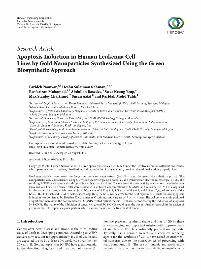
(a) (c)(b)
Figure 1: The color change of S. muticum aqueous extract at thebeginning (a); 30min (b); and 1 h (c) after synthesis of S-GNPs.
inmany areas of biomedical sciences, including the treatmentof cancer. Recently, natural products have been reportedelsewhere for the green synthesis of metallic nanoparticlesas reducing and stabilizing agents. Seaweed, one of the mostcommonly used medicinal extracts, has been acknowledgedto exhibit anticancer activity in vitro and in vivo and in humanclinical trials.
3.1. Synthesis of S-GNPs. In the current study, we synthesizedSargassum muticum stabilized gold nanoparticles via theone-step process, without the requirement of a stabilizationor reducing agent. This method allows the production ofgold nanoparticles within one hour (Figure 1). The goldnanoparticles generated through the seaweed-mediated pro-cess did not aggregate, suggesting that the cocktail of phyto-chemicals, including fucoxanthin, serve as excellent coatingson nanoparticles and thus provide strong protection fromaggregation. The AuCl
4reduction was visually obvious from
the changes in color from light purple to a ruby red colorindicating the completed formation of S-GNPs, within onehour.
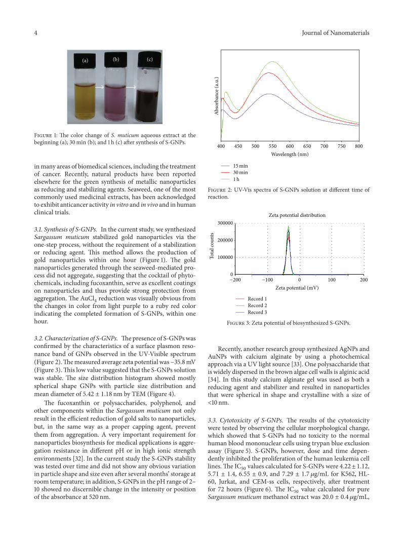
3.2. Characterization of S-GNPs. Thepresence of S-GNPswasconfirmed by the characteristics of a surface plasmon reso-nance band of GNPs observed in the UV-Visible spectrum(Figure 2).Themeasured average zeta potential was−35.8mV(Figure 3).This low value suggested that the S-GNPs solutionwas stable. The size distribution histogram showed mostlyspherical shape GNPs with particle size distribution andmean diameter of 5.42 ± 1.18 nm by TEM (Figure 4).
The fucoxanthin or polysaccharides, polyphenol, andother components within the Sargassum muticum not onlyresult in the efficient reduction of gold salts to nanoparticles,but, in the same way as a proper capping agent, preventthem from aggregation. A very important requirement fornanoparticles biosynthesis for medical applications is aggre-gation resistance in different pH or in high ionic strengthenvironments [32]. In the current study the S-GNPs stabilitywas tested over time and did not show any obvious variationin particle shape and size even after several months’ storage atroom temperature; in addition, S-GNPs in the pH range of 2–10 showed no discernible change in the intensity or positionof the absorbance at 520 nm.
400 450 500 550 600 650 700 750 800
Abso
rban
ce (a
.u.)
Wavelength (nm)
15 min30 min1h
Figure 2: UV-Vis spectra of S-GNPs solution at different time ofreaction.
300000
200000
100000
0
Tota
l cou
nts
Zeta potential distribution
−200 −100 0 100 200
Zeta potential (mV)
Record 1
Record 2
Record 3
Figure 3: Zeta potential of biosynthesized S-GNPs.
Recently, another research group synthesized AgNPs andAuNPs with calcium alginate by using a photochemicalapproach via a UV light source [33]. One polysaccharide thatis widely dispersed in the brown algae cell walls is alginic acid[34]. In this study calcium alginate gel was used as both areducing agent and stabilizer and resulted in nanoparticlesthat were spherical in shape and crystalline with a size of<10 nm.
3.3. Cytotoxicity of S-GNPs. The results of the cytotoxicitywere tested by observing the cellular morphological change,which showed that S-GNPs had no toxicity to the normalhuman blood mononuclear cells using trypan blue exclusionassay (Figure 5). S-GNPs, however, dose and time depen-dently inhibited the proliferation of the human leukemia celllines.The IC
50values calculated for S-GNPs were 4.22± 1.12,
5.71 ± 1.4, 6.55 ± 0.9, and 7.29 ± 1.7 𝜇g/mL for K562, HL-60, Jurkat, and CEM-ss cells, respectively, after treatmentfor 72 hours (Figure 6). The IC
50value calculated for pure
Sargassum muticum methanol extract was 20.0 ± 0.4 𝜇g/mL,

Journal of Nanomaterials 5
Au-NPs
Seaweed extract
10
8
6
4
2
0
Freq
uenc
y
1 2 3 4 5 6 7 8 9 10
Mean = 5.42nmSD = 1.18nm
Particle diameter (nm)
(a) (b)
(c)
100nm 25nm
Figure 4: TEM images ((a)-(b)) and corresponding size distribution graph (c) of biosynthesized S-GNPs.
0
20
40
60
80
100
120
0 20 40 60 80 100 120
Cel
l via
bilit
y (%
)
CEM-ssJurkat
HL-60K562
S-GNPs concentrations (𝜇g/mL)
Figure 5: Cytotoxic effect of S-GNPs onCEM-ss, Jurkat, HL-60, andK562 cells assessed by MTT assay. Each point is the mean value ofthree replicates.
which is three times higher than the S-GNPs. It is alsoimportant to point out that the vast majority of Gold (I) and
10094.7
89.285.4
0
20
40
60
80
100
120
Control
PBM
C vi
abili
ty (%
)
Time (h)72h48h24h
Figure 6: Cytotoxic effect of S-GNPs on human bloodmononuclearcells. Values are expressed as mean ± SD of three different experi-ments. The data has been analyzed using post hoc comparison test-one way ANOVA, means compared with Tukey’s-𝑏 test.
Gold (III) compounds show varying degrees of cytotoxicityto a variety of cells [35].
These results clearly demonstrated that the biomedicalactive components such as fucoxanthin within seaweed pro-vide a nontoxic coating on GNPs. The lack of any noticeable
6 Journal of Nanomaterials
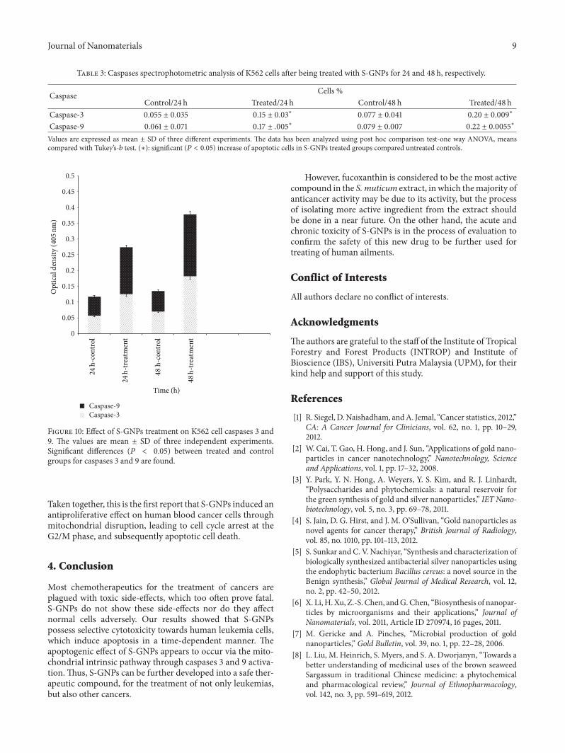
Table 1: Flow cytometric analysis of K562 cells after being treated with S-GNPs.The cells were stained with FITC-conjugated annexin V andPI and incubated at 37∘C for 6, 12, and 24 h, respectively.
Cell condition Control Treated/6 h Treated/12 h Treated/24 hViable cells 96.5 ± 0.55 89.19 ± 0.65 80.21 ± 0.11 71.5 ± 0.23Early apoptosis 1.81 ± 0.15 6.55 ± 0.75∗ 9.00 ± 0.33∗ 12.25 ± 0.25∗
Late apoptosis/necrosis 1.69 ± 0.35 4.26 ± 0.05∗∗ 10.79 ± 0.40∗∗ 16.25 ± 0.19∗∗
Values are expressed as mean ± SD of three different experiments. The data has been analyzed using post hoc comparison test-one way ANOVA, meanscompared byTukey’s-𝑏 test. (∗): significant (𝑃 < 0.05) increased early apoptotic cells in S-GNPs treated groups compared to untreated controls. (∗∗): significant(𝑃 < 0.05) increased late apoptotic/necrotic cells in S-GNPs treated groups compared to untreated controls.
50𝜇m
(a)
CCBL
BL
50𝜇m
(b)
BL
BL
CS
CS
50𝜇m
(c)
BL
AB
BL
CS
CS50𝜇m
(d)
Figure 7: Fluorescentmicrograph ofHoechst 33342 stainedK562 cells that were treated with S-GNPs. (a) Untreated cells showing normal sizeand cell structure. (b) Early apoptotic cells after 24 h treatment showing chromatin condensation and membrane blebbing. (c) Blebbing andcell shrinkage after 48 h treatment. (d)Membrane blebbing, cell shrinkage, and apoptotic body formation after 72 h treatment. CC: chromatincondensation, BL: blebbing, CS: cell shrinkage, and AB: apoptotic body.
toxicity of S-GNPs provides new opportunities for the safedelivery and applications in anticancer therapy.
3.4. Morphological Assessment of S-GNPs. Apoptosis wasidentified with Hoechst 33342 staining time dependentlyin the treated cells. Staining the 24-hour S-GNPs treatedK562 cell exhibited the typical features of apoptosis, such aschromatin condensation and morphology changes, as wellas cell shrinkage and membrane blebbing. The 48-hour S-GNPs treated cells showed even smaller nuclei; some hadperipherally condensed or clumped chromatin, whereas oth-ers showed membrane blebbing. Apoptotic bodies formationwas more prominent at 72 hours after treatment of S-GNPs.In contrast, the cells in the control group, without treat-ment, demonstrated normal nuclear and cellularmorphology(Figure 7). Lan et al. also showed that AuNPs with an averagediameter of 20.5 nm reduced cell viability of nasopharyngeal
carcinoma cell in a concentration-dependant manner bytrypan blue assay, especially at high concentration [36].
3.5. Apoptosis Evaluation. In order to quantify the apoptosis,an annexin V-FITC/PI staining experiment was performedto examine the occurrence of phosphatidylserine externaliza-tion onto the cell surface. The percentage of annexin V-FITCstained cells, both the early and late apoptotic cells, increasedgradually and significantly (𝑃 < 0.05) in all groups with thetime applied, while the percentage of viable cells subsequentlydecreased gradually (Figure 8). At six hours of treatment, anabundance of cells developed primarily in the early phase ofapoptosis (6.55 ± 0.75%), while increasing incubation timeof 12 and 24 hours, respectively, induced more cell apoptosisduring the late stage (9.00 ± 0.33% and 12.25 ± 0.25%, resp.)(Table 1).
Similar results in apoptosis enhanced cell death by(-)-epigallocatechin-3-flat gold nanoparticles (EGCG-PNG)

Journal of Nanomaterials 7
104
104
103
103
102
102
101
101100
100
Prop
idiu
m io
dide
Annexin V-FITC(a)
104
103
102
101
100
Prop
idiu
m io
dide
104103102101100
Annexin V-FITC(b)
104103102101100
104
103
102
101
100
Prop
idiu
m io
dide
Annexin V-FITC(c)
104103102101100
104
103
102
101
100
Prop
idiu
m io
dide
Annexin V-FITC(d)
Figure 8: Flow cytometric analysis of apoptosis induction by S-GNPs in K562 cells after staining with FITC-conjugated annexin V and PI.(a) Untreated (control) K562 cells. (b), (c), and (d) Effects of 6, 12, and 24 h S-GNPs treatment, respectively.
were found by other groups in B16F10murinemelanoma cellswith increasing fraction of annexin V positive cells. This, inturn, improved the anticancer efficacy of (-)-epigallocat-echin-3-flat gold nanoparticles in murine B16F10 melanomacells.
3.6. Cell Cycle Analysis. It demonstrated that the untreatedcells showed normal DNA content and cell cycle distribution.On the other hand, S-GNPs induced a concomitant andsignificant (𝑃 < 0.05) accumulation of K562 cell populationswith an apoptotic peak in the SubG0/G1 phase especially at 48hours of treatment (6.69±0.56%).Moreover, S-GNPs induced
cell cycle arrest in the G2/M phase with values of 27.14 ±0.41%, 30.47 ± 0.35%, and 33.83 ± 0.93% after 12, 24, and48 hours of treatment, respectively (Figure 9 and Table 2).
Huang et al. demonstrated that AuNPs with surfacemod-ification, using polyethylene glycol (PEG-AuNPs), inhibitedthe viability of human chronic myeloid leukemia K562 cells.These particles caused morphological changes typical of celldeath, and a marked increase in the sub-G1 population inDNA histogram, indicating apoptosis. In addition, PEG-AuNPs reduced the mitochondrial transmembrane potential,a hallmark of the involvement of the intrinsic apoptoticpathway in K562 cells [37].

8 Journal of Nanomaterials
Table 2: Flowcytometric analysis of K562 cells after being treated with S-GNPs. The cells were stained with PI and incubated at 37∘C for 12,24, and 48 h.
Cell cycle phases Cells %Control Treated/12 h Treated/24 h Treated/48 h
G0/G1 54.33 ± 0.06 51.36 ± 0.45 50.56 ± 0.52 47.10 ± 0.68G2/M 22.52 ± 0.76∗ 27.14 ± 0.41∗ 30.47 ± 0.35∗ 33.83 ± 0.93∗
Synthesis 15.26 ± 0.06 18.22 ± 0.33 14.97 ± 0.12 13.11 ± 0.18Sub-G0/G1 1.49 ± 0.23 3.39 ± 0.28∗∗ 4.73 ± 0.20∗∗ 6.69 ± 0.56∗∗
Values are expressed as mean ± SD of three different experiments. The data has been analyzed using post hoc comparison test-one way ANOVA, meanscompared with Tukey’s-𝑏 test. (∗): significant (𝑃 < 0.05) increased cells in G2/M phase in S-GNPs treated groups compared to untreated controls. (∗∗):significant (𝑃 < 0.05) increased of apoptotic cells in sub-G0/G1 phase in S-GNPs treated groups compared to untreated controls.
100
80
60
40
20
0
Cou
nts
0 200 400 600
DNA content
G0/G1
Sub-G0/G1
S
G2 + M
(a)
100
80
60
40
20
0
Cou
nts
0 200 400 600
DNA content
G0/G1
S
G2 + M
Sub-G0/G1
(b)
100
80
60
40
20
0
Cou
nts
0 200 400
DNA content
G0/G1
S
G2 + M
Sub-G0/G1
(c)
100
80
60
40
20
0
Cou
nts
0 200 400 600
DNA content
G0/G1
S
G2 + M
Sub-G0/G1
(d)
Figure 9: Cell cycle analysis of K562 cells treated with S-GNPs after staining with PI. (a) Untreated (control) K562 cells. (b), (c), and (d)Effects of 12, 24, and 48 h, respectively, exposure of K562 cells to S-GNPs. G0/G1, G2/M, and S indicate the cell phase, and sub-G0-G1 refersto the portion of apoptotic cells.
3.7. Caspase Activity. To investigate the involvement of cas-pases signaling cascade in S-GNPs induced apoptosis, K562cells were treated for various times and protease enzymaticactivities were determined. S-GNPs significantly (𝑃 < 0.05)stimulated both caspases 3 and 9 enzyme activities withmore than onefold activity in a time-dependent manner
in the treated K562 cells, compared to untreated controlgroups (Figure 10 and Table 3). Similar results of caspaseprotease activity were found in other types of nanoparticles,such as lipid nanoparticles, by Rahman et al. in humanlymphoblastoid leukemia Jurkat cells after treatment withzerumbone-loaded nanostructured lipid carrier (ZER-NLC).
Journal of Nanomaterials 9
Table 3: Caspases spectrophotometric analysis of K562 cells after being treated with S-GNPs for 24 and 48 h, respectively.
Caspase Cells %Control/24 h Treated/24 h Control/48 h Treated/48 h
Caspase-3 0.055 ± 0.035 0.15 ± 0.03∗ 0.077 ± 0.041 0.20 ± 0.009∗
Caspase-9 0.061 ± 0.071 0.17 ± .005∗ 0.079 ± 0.007 0.22 ± 0.0055∗
Values are expressed as mean ± SD of three different experiments. The data has been analyzed using post hoc comparison test-one way ANOVA, meanscompared with Tukey’s-𝑏 test. (∗): significant (𝑃 < 0.05) increase of apoptotic cells in S-GNPs treated groups compared untreated controls.
0
0.05
0.1
0.15
0.2
0.25
0.3
0.35
0.4
0.45
0.5
Caspase-9Caspase-3
Opt
ical
den
sity
(405
nm)
Time (h)
48
h-tre
atm
ent
48
h-co
ntro
l
24
h-tre
atm
ent
24
h-co
ntro
l
Figure 10: Effect of S-GNPs treatment on K562 cell caspases 3 and9. The values are mean ± SD of three independent experiments.Significant differences (𝑃 < 0.05) between treated and controlgroups for caspases 3 and 9 are found.
Taken together, this is the first report that S-GNPs induced anantiproliferative effect on human blood cancer cells throughmitochondrial disruption, leading to cell cycle arrest at theG2/M phase, and subsequently apoptotic cell death.
4. Conclusion
Most chemotherapeutics for the treatment of cancers areplagued with toxic side-effects, which too often prove fatal.S-GNPs do not show these side-effects nor do they affectnormal cells adversely. Our results showed that S-GNPspossess selective cytotoxicity towards human leukemia cells,which induce apoptosis in a time-dependent manner. Theapoptogenic effect of S-GNPs appears to occur via the mito-chondrial intrinsic pathway through caspases 3 and 9 activa-tion.Thus, S-GNPs can be further developed into a safe ther-apeutic compound, for the treatment of not only leukemias,but also other cancers.
However, fucoxanthin is considered to be the most activecompound in the S. muticum extract, in which themajority ofanticancer activity may be due to its activity, but the processof isolating more active ingredient from the extract shouldbe done in a near future. On the other hand, the acute andchronic toxicity of S-GNPs is in the process of evaluation toconfirm the safety of this new drug to be further used fortreating of human ailments.
Conflict of Interests
All authors declare no conflict of interests.
Acknowledgments
The authors are grateful to the staff of the Institute of TropicalForestry and Forest Products (INTROP) and Institute ofBioscience (IBS), Universiti Putra Malaysia (UPM), for theirkind help and support of this study.
References
[1] R. Siegel, D.Naishadham, andA. Jemal, “Cancer statistics, 2012,”CA: A Cancer Journal for Clinicians, vol. 62, no. 1, pp. 10–29,2012.
[2] W. Cai, T. Gao, H. Hong, and J. Sun, “Applications of gold nano-particles in cancer nanotechnology,” Nanotechnology, Scienceand Applications, vol. 1, pp. 17–32, 2008.
[3] Y. Park, Y. N. Hong, A. Weyers, Y. S. Kim, and R. J. Linhardt,“Polysaccharides and phytochemicals: a natural reservoir forthe green synthesis of gold and silver nanoparticles,” IET Nano-biotechnology, vol. 5, no. 3, pp. 69–78, 2011.
[4] S. Jain, D. G. Hirst, and J. M. O’Sullivan, “Gold nanoparticles asnovel agents for cancer therapy,” British Journal of Radiology,vol. 85, no. 1010, pp. 101–113, 2012.
[5] S. Sunkar and C. V. Nachiyar, “Synthesis and characterization ofbiologically synthesized antibacterial silver nanoparticles usingthe endophytic bacterium Bacillus cereus: a novel source in theBenign synthesis,” Global Journal of Medical Research, vol. 12,no. 2, pp. 42–50, 2012.
[6] X. Li, H. Xu, Z.-S. Chen, andG.Chen, “Biosynthesis of nanopar-ticles by microorganisms and their applications,” Journal ofNanomaterials, vol. 2011, Article ID 270974, 16 pages, 2011.
[7] M. Gericke and A. Pinches, “Microbial production of goldnanoparticles,” Gold Bulletin, vol. 39, no. 1, pp. 22–28, 2006.
[8] L. Liu, M. Heinrich, S. Myers, and S. A. Dworjanyn, “Towards abetter understanding of medicinal uses of the brown seaweedSargassum in traditional Chinese medicine: a phytochemicaland pharmacological review,” Journal of Ethnopharmacology,vol. 142, no. 3, pp. 591–619, 2012.

10 Journal of Nanomaterials
[9] F. Namvar, P. M. Tahir, R. Mohamad et al., “Biomedical proper-ties of edible seaweed in cancer therapy and chemopreventiontrials: a review,”Natural Product Communications, vol. 8, no. 12,pp. 1811–1820, 2013.
[10] F. Namvar, S. Mohamed, S. G. Fard et al., “Polyphenol-richseaweed (Eucheuma cottonii) extract suppresses breast tumourvia hormonemodulation and apoptosis induction,” Food Chem-istry, vol. 130, no. 2, pp. 376–382, 2012.
[11] A. A. El Gamal, “Biological importance of marine algae,” SaudiPharmaceutical Journal, vol. 18, no. 1, pp. 1–25, 2010.
[12] F. Namvar, J. Baharara, and A. A. Mahdi, “Antioxidant and anti-cancer activities of selected persian gulf algae,” Indian Journal ofClinical Biochemistry, vol. 29, no. 1, pp. 13–20, 2013.
[13] M. N. A. Khan, J. S. Choi, M. C. Lee et al., “Anti-inflammatoryactivities of methanol extracts from various seaweed species,”Journal of Environmental Biology, vol. 29, no. 4, pp. 465–469,2008.
[14] A. W. Zuercher, R. Fritsche, B. Corthesy, and A. Mercenier,“Food products and allergy development, prevention and treat-ment,” Current Opinion in Biotechnology, vol. 17, no. 2, pp. 198–203, 2006.
[15] G. R. M. Perez, S. M. A. Zavala, G. S. Perez, and G. C. Perez,“Antidiabetic effect of compounds isolated from plants,” Phy-tomedicine, vol. 5, no. 1, pp. 55–75, 1998.
[16] T. Nishino, A. Fukuda, T. Nagumo, M. Fujihara, and E. Kaji,“Inhibition of the generation of thrombin and factor Xa by afucoidan from the brown seaweed Ecklonia kurome,”Thrombo-sis Research, vol. 96, no. 1, pp. 37–49, 1999.
[17] K. Miyashita, “The carotenoid fucoxanthin from brown sea-weed affects obesity,” Lipid Technology, vol. 21, no. 8-9, pp. 186–190, 2009.
[18] S. Mohamed, S. N. Hashim, and H. A. Rahman, “Seaweeds: asustainable functional food for complementary and alternativetherapy,” Trends in Food Science and Technology, vol. 23, no. 2,pp. 83–96, 2012.
[19] K. Wada, K. Nakamura, Y. Tamai et al., “Seaweed intake andblood pressure levels in healthy pre-school Japanese children,”Nutrition Journal, vol. 10, no. 1, article 83, 2011.
[20] A. Bocanegra, S. Bastida, J. Benedı, S. Rodenas, and F. J.Sanchez-Muniz, “Characteristics and nutritional andcardiovascular-health properties of seaweeds,” Journal ofMedicinal Food, vol. 12, no. 2, pp. 236–258, 2009.
[21] I. A. Brownlee, A. Allen, J. P. Pearson et al., “Alginate as a sourceof dietary fiber,” Critical Reviews in Food Science and Nutrition,vol. 45, no. 6, pp. 497–510, 2005.
[22] I. Brownlee, A. Fairclough, A. Hall, and J. Paxman, DietarySeaweed and Human Health, Centre for Food Innovation,Sheffield Hallam University, Sheffield, UK, 2011.
[23] J. H. Fitton, “Therapies from fucoidan; multifunctional marinepolymers,”Marine Drugs, vol. 9, no. 10, pp. 1731–1760, 2011.
[24] C. Gerhauser, “Cancer chemoprevention and nutri-epigenetics:state of the art and future challenges,” Topics in CurrentChemistry, vol. 329, pp. 73–132, 2013.
[25] F. Namvar, S. Azizi, M. B. Ahmad et al., “Green synthesis andcharacterization of gold nanoparticles using the marine macro-algae Sargassummuticum,” Research on Chemical Intermediates,vol. 41, no. 8, pp. 5723–5730, 2015.
[26] R. Zahra, M. Mehrnaz, V. Farzaneh, and S. Kohzad, “Antiox-idant activity of extract from a brown alga, Sargassum bove-anum,”African Journal of Biotechnology, vol. 6, no. 24, pp. 2740–2745, 2007.
[27] P. Matanjun, S. Mohamed, N. M. Mustapha, and K. Muham-mad, “Nutrient content of tropical edible seaweeds, Eucheumacottonii, Caulerpa lentillifera and Sargassum polycystum,” Jour-nal of Applied Phycology, vol. 21, no. 1, pp. 75–80, 2009.
[28] K. H. Wong and P. C. K. Cheung, “Nutritional evaluation ofsome subtropical red and green seaweeds: part I—proximatecomposition, amino acid profiles and some physico-chemicalproperties,” Food Chemistry, vol. 71, no. 4, pp. 475–482, 2000.
[29] S. L. Holdt and S. Kraan, “Bioactive compounds in seaweed:functional food applications and legislation,” Journal of AppliedPhycology, vol. 23, no. 3, pp. 543–597, 2011.
[30] J. Ortiz, N. Romero, P. Robert et al., “Dietary fiber, amino acid,fatty acid and tocopherol contents of the edible seaweeds Ulvalactuca and Durvillaea antarctica,” Food Chemistry, vol. 99, no.1, pp. 98–104, 2006.
[31] P. Kumari, M. Kumar, V. Gupta, C. R. K. Reddy, and B. Jha,“Tropical marine macroalgae as potential sources of nutrition-ally important PUFAs,” Food Chemistry, vol. 120, no. 3, pp. 749–757, 2010.
[32] S. Pandey and G. Oza, “Green synthesis of highly stable goldnanoparticles using Momordica charantia as nano fabricator,”Archives of Applied Science Research, vol. 4, no. 2, pp. 1135–1141,2012.
[33] S. Saha, A. Pal, S. Kundu, S. Basu, and T. Pal, “Photochemicalgreen synthesis of calcium-alginate-stabilized Ag and Au nano-particles and their catalytic application to 4-nitrophenol reduc-tion,” Langmuir, vol. 26, no. 4, pp. 2885–2893, 2010.
[34] I.Wijesekara, R. Pangestuti, and S.-K.Kim, “Biological activitiesand potential health benefits of sulfated polysaccharides derivedfrom marine algae,” Carbohydrate Polymers, vol. 84, no. 1, pp.14–21, 2011.
[35] V. Balzani, “Nanoscience and nanotechnology: the bottom-upconstruction of molecular devices and machines,” Pure andApplied Chemistry, vol. 80, no. 8, pp. 1631–1650, 2008.
[36] M.-Y. Lan, Y.-B. Hsu, C.-H. Hsu, C.-Y. Ho, J.-C. Lin, and S.-W.Lee, “Induction of apoptosis by high-dose gold nanoparticles innasopharyngeal carcinoma cells,” Auris Nasus Larynx, vol. 40,no. 6, pp. 563–568, 2013.
[37] Y.-C. Huang, Y.-C. Yang, K.-C. Yang et al., “Pegylated goldnanoparticles induce apoptosis in human chronic myeloidleukemia cells,”BioMedResearch International, vol. 2014, ArticleID 182353, 9 pages, 2014.

Submit your manuscripts athttp://www.hindawi.com
ScientificaHindawi Publishing Corporationhttp://www.hindawi.com Volume 2014
CorrosionInternational Journal of
Hindawi Publishing Corporationhttp://www.hindawi.com Volume 2014
Polymer ScienceInternational Journal of
Hindawi Publishing Corporationhttp://www.hindawi.com Volume 2014
Hindawi Publishing Corporationhttp://www.hindawi.com Volume 2014
CeramicsJournal of
Hindawi Publishing Corporationhttp://www.hindawi.com Volume 2014
CompositesJournal of
NanoparticlesJournal of
Hindawi Publishing Corporationhttp://www.hindawi.com Volume 2014
Hindawi Publishing Corporationhttp://www.hindawi.com Volume 2014
International Journal of
Biomaterials
Hindawi Publishing Corporationhttp://www.hindawi.com Volume 2014
NanoscienceJournal of
TextilesHindawi Publishing Corporation http://www.hindawi.com Volume 2014
Journal of
NanotechnologyHindawi Publishing Corporationhttp://www.hindawi.com Volume 2014
Journal of
CrystallographyJournal of
Hindawi Publishing Corporationhttp://www.hindawi.com Volume 2014
The Scientific World JournalHindawi Publishing Corporation http://www.hindawi.com Volume 2014
Hindawi Publishing Corporationhttp://www.hindawi.com Volume 2014
CoatingsJournal of
Advances in
Materials Science and EngineeringHindawi Publishing Corporationhttp://www.hindawi.com Volume 2014
Smart Materials Research
Hindawi Publishing Corporationhttp://www.hindawi.com Volume 2014
Hindawi Publishing Corporationhttp://www.hindawi.com Volume 2014
MetallurgyJournal of
Hindawi Publishing Corporationhttp://www.hindawi.com Volume 2014
BioMed Research International
MaterialsJournal of
Hindawi Publishing Corporationhttp://www.hindawi.com Volume 2014
Nano
materials
Hindawi Publishing Corporationhttp://www.hindawi.com Volume 2014
Journal ofNanomaterials