The ASK family kinases differentially mediate induction of ...IMMUNOLOGY The ASK family kinases...
13
IMMUNOLOGY The ASK family kinases differentially mediate induction of type I interferon and apoptosis during the antiviral response Tomohiko Okazaki, 1 * Maiko Higuchi, 1 Kohsuke Takeda, 2 Kiyoko Iwatsuki-Horimoto, 3 Maki Kiso, 3 Makoto Miyagishi, 4 Hideyuki Yanai, 5,6 Atsushi Kato, 7 Mitsutoshi Yoneyama, 8 Takashi Fujita, 9 Tadatsugu Taniguchi, 5,6 Yoshihiro Kawaoka, 3,10,11,12 Hidenori Ichijo, 13 Yukiko Gotoh 1 Viral infection activates host defense mechanisms, including the production of type I interferon (IFN) and the apoptosis of infected cells. We investigated whether these two antiviral responses were differentially regulated in infected cells. We showed that the mitogen-activated protein kinase (MAPK) kinase kinase (MAPKKK) apoptosis signal–regulating kinase 1 (ASK1) was activated in cells by the synthetic double- stranded RNA analog polyinosinic:polycytidylic acid [poly(I:C)] and by RNA viruses, and that ASK1 played an essential role in both the induction of the gene encoding IFN-b (IFNB) and apoptotic cell death. In con- trast, we found that the MAPKKK ASK2, a modulator of ASK1 signaling, was essential for ASK1-dependent apoptosis, but not for inducing IFNB expression. Furthermore, genetic deletion of either ASK1 or ASK2 in mice promoted the replication of influenza A virus in the lung. These results indicated that ASK1 and ASK2 are components of the antiviral defense mechanism and suggested that ASK2 acts as a key modulator that promotes apoptosis rather than the type I IFN response. Because ASK2 is selectively present in epithelium-rich tissues, such as the lung, ASK2-dependent apoptosis may contribute to an antiviral de- fense in tissues with a rapid repair rate in which cells could be readily replaced. INTRODUCTION Mammalian cells deploy the innate immune system as the first line of de- fense against viruses. The type I interferons (IFNs) IFN-a and IFN-b play a central role in this innate immune response by activating the expression of hundreds of IFN-stimulated genes (ISGs) whose products establish an “antiviral state” to restrict viral replication within infected cells. Type I IFNs also promote the proliferation of effector lymphocytes to provide long-term and specific protection against the infecting virus through the production of various cytokines and chemokines, such as interleukin-15 (IL-15) ( 1). Thus, regulation of the expression of genes encoding type I IFNs is the subject of intense investigation. Cytoplasmic double-stranded RNA (dsRNA), a common byproduct of the replication of DNA and RNA viruses, is recog- nized by the retinoic acid–inducible gene I (RIG-I)–like helicase receptors (RLRs) RIG-I and melanoma differentiation–associated gene 5 (MDA5) (2, 3), which mediate the expression of genes encoding type I IFNs through their interaction with their common adaptor protein IFN-b promoter stim- ulator 1 (IPS-1) (also known as MAVS, VISA, or CARDIF) (4–7) and the subsequent tumor necrosis factor (TNF) receptor –associated factor (TRAF) – mediated activation of its downstream effectors, the transcription factors IFN regulatory factor–3 (IRF3) and nuclear factor kB (NF-kB) (8, 9). In addition to having IRF3- and NF-kB–binding sites, the promoter of the gene encoding IFNB contains an activator protein 1 (AP-1)–binding site that is essential for gene expression. Indeed, the AP-1 complex, which con- sists of c-Jun and activating transcription factor 2 (ATF-2), as well as the upstream activating mitogen-activated protein kinases (MAPKs) p38 and c-Jun N-terminal kinase (JNK) are necessary for the effective induction of IFNB in response to cytoplasmic dsRNA (10–13). However, although var- ious candidates have been proposed, the molecules that mediate activation of p38 and JNK in response to cytoplasmic dsRNA are unclear. In addition to the induction of type I IFN production, another main viral defense strategy of the innate immune response is the induction of apoptosis (14, 15). Given that viruses need host cells within which to replicate, the elimination of infected cells prevents further spread of infec- tion. Many viruses have evolved mechanisms to interfere with host cell apoptosis despite their being subjected to rigorous evolutionary selection to maintain small genomes, which reflects the importance of this strategy in antiviral immunity. Indeed, prevention of apoptosis, for example, by deletion of the gene encoding Bax, a proapoptotic B cell lymphoma 2 (Bcl-2) family member that triggers the mitochondrial apoptosis pathway, results in enhanced viral replication and pathogenesis in mice (16). Char- acterization of the mechanism underlying virus-induced apoptosis is thus critical to understanding antiviral host defense. As well as enabling type I 1 Laboratory of Molecular Biology, Graduate School of Pharmaceutical Sciences, The University of Tokyo, Tokyo 113-0033, Japan. 2 Division of Cell Regulation, Graduate School of Biomedical Sciences, Nagasaki University, Nagasaki 852-8521, Japan. 3 Division of Virology, Department of Microbiology and Immu- nology, Institute of Medical Science, The University of Tokyo, Tokyo 108-8639, Japan. 4 Molecular Composite Medicine Research Group, Biomedical Research Institute, National Institute of Advanced Industrial Science and Technology, Tsu- kuba 305-8566, Japan. 5 Department of Molecular Immunology and Center for International Research on Integrative Biomedical Systems, Institute of Industrial Science, The University of Tokyo, Tokyo 153-8505, Japan. 6 Max Planck–The Univer- sity of Tokyo Center for Integrative Inflammology, Tokyo 153-8505, Japan. 7 Depart- ment of Quality Assurance and Radiological Protection, National Institute of Infectious Diseases, Tokyo 162-8640, Japan. 8 Medical Mycology Research Center, Chiba University, Chiba 260-8673, Japan. 9 Laboratory of Molecular Ge- netics, Institute for Virus Research, Kyoto University, Kyoto 606-8507, Ja- pan. 10 Department of Special Pathogens, International Research Center for Infectious Diseases, Institute of Medical Science, The University of Tokyo, Tokyo 108-8639, Japan. 11 ERATO Infection-Induced Host Responses Project, Japan Science and Technology Agency, Saitama 332-0012, Japan. 12 Department of Pathobiological Sciences, School of Veterinary Medicine, University of Wisconsin– Madison, Madison, WI 53711, USA. 13 Laboratory of Cell Signaling, Graduate School of Pharmaceutical Sciences, The University of Tokyo, Tokyo 113-0033, Japan. *Corresponding author. E-mail: [email protected] RESEARCHARTICLE www.SCIENCESIGNALING.org 4 August 2015 Vol 8 Issue 388 ra78 1 CORRECTED 30 MAY 2017; SEE ERRATUM on March 12, 2020 http://stke.sciencemag.org/ Downloaded from
Transcript of The ASK family kinases differentially mediate induction of ...IMMUNOLOGY The ASK family kinases...
R E S E A R C H A R T I C L E
CORRECTED 30 MAY 2017; SEE ERRATUM
I M M U N O L O G Y
The ASK family kinases differentially mediateinduction of type I interferon and apoptosis duringthe antiviral responseTomohiko Okazaki,1* Maiko Higuchi,1 Kohsuke Takeda,2 Kiyoko Iwatsuki-Horimoto,3
Maki Kiso,3 Makoto Miyagishi,4 Hideyuki Yanai,5,6 Atsushi Kato,7 Mitsutoshi Yoneyama,8
Takashi Fujita,9 Tadatsugu Taniguchi,5,6 Yoshihiro Kawaoka,3,10,11,12
Hidenori Ichijo,13 Yukiko Gotoh1
http://D
ownloaded from
Viral infection activates host defense mechanisms, including the production of type I interferon (IFN) andthe apoptosis of infected cells. We investigated whether these two antiviral responses were differentiallyregulated in infected cells. We showed that the mitogen-activated protein kinase (MAPK) kinase kinase(MAPKKK) apoptosis signal–regulating kinase 1 (ASK1) was activated in cells by the synthetic double-stranded RNA analog polyinosinic:polycytidylic acid [poly(I:C)] and by RNA viruses, and that ASK1 playedan essential role in both the induction of the gene encoding IFN-b (IFNB) and apoptotic cell death. In con-trast, we found that the MAPKKKASK2, a modulator of ASK1 signaling, was essential for ASK1-dependentapoptosis, but not for inducing IFNB expression. Furthermore, genetic deletion of either ASK1 or ASK2 inmice promoted the replication of influenza A virus in the lung. These results indicated that ASK1 and ASK2are components of the antiviral defense mechanism and suggested that ASK2 acts as a key modulatorthat promotes apoptosis rather than the type I IFN response. Because ASK2 is selectively present inepithelium-rich tissues, such as the lung, ASK2-dependent apoptosis may contribute to an antiviral de-fense in tissues with a rapid repair rate in which cells could be readily replaced.
stke.
on March 12, 202sciencem
ag.org/
INTRODUCTION
Mammalian cells deploy the innate immune system as the first line of de-fense against viruses. The type I interferons (IFNs) IFN-a and IFN-b playa central role in this innate immune response by activating the expressionof hundreds of IFN-stimulated genes (ISGs) whose products establish an“antiviral state” to restrict viral replication within infected cells. Type I IFNsalso promote the proliferation of effector lymphocytes to provide long-termand specific protection against the infecting virus through the productionof various cytokines and chemokines, such as interleukin-15 (IL-15) (1). Thus,regulation of the expression of genes encoding type I IFNs is the subjectof intense investigation. Cytoplasmic double-stranded RNA (dsRNA), a
1Laboratory of Molecular Biology, Graduate School of Pharmaceutical Sciences,The University of Tokyo, Tokyo 113-0033, Japan. 2Division of Cell Regulation,Graduate School of Biomedical Sciences, Nagasaki University, Nagasaki852-8521, Japan. 3Division of Virology, Department of Microbiology and Immu-nology, Institute of Medical Science, The University of Tokyo, Tokyo 108-8639,Japan. 4Molecular Composite Medicine Research Group, Biomedical ResearchInstitute, National Institute of Advanced Industrial Science and Technology, Tsu-kuba 305-8566, Japan. 5Department of Molecular Immunology and Center forInternational Research on Integrative Biomedical Systems, Institute of IndustrialScience, The University of Tokyo, Tokyo 153-8505, Japan. 6Max Planck–The Univer-sity of Tokyo Center for Integrative Inflammology, Tokyo 153-8505, Japan. 7Depart-ment of Quality Assurance and Radiological Protection, National Institute ofInfectious Diseases, Tokyo 162-8640, Japan. 8Medical Mycology ResearchCenter, Chiba University, Chiba 260-8673, Japan. 9Laboratory of Molecular Ge-netics, Institute for Virus Research, Kyoto University, Kyoto 606-8507, Ja-pan. 10Department of Special Pathogens, International Research Center forInfectious Diseases, Institute of Medical Science, The University of Tokyo, Tokyo108-8639, Japan. 11ERATO Infection-Induced Host Responses Project, JapanScience and Technology Agency, Saitama 332-0012, Japan. 12Department ofPathobiological Sciences, School of Veterinary Medicine, University of Wisconsin–Madison, Madison, WI 53711, USA. 13Laboratory of Cell Signaling, Graduate Schoolof Pharmaceutical Sciences, The University of Tokyo, Tokyo 113-0033, Japan.*Corresponding author. E-mail: [email protected]
ww
0
common byproduct of the replication of DNA and RNAviruses, is recog-nized by the retinoic acid–inducible gene I (RIG-I)–like helicase receptors(RLRs) RIG-I and melanoma differentiation–associated gene 5 (MDA5)(2, 3), which mediate the expression of genes encoding type I IFNs throughtheir interaction with their common adaptor protein IFN-b promoter stim-ulator 1 (IPS-1) (also known as MAVS, VISA, or CARDIF) (4–7) and thesubsequent tumor necrosis factor (TNF) receptor–associated factor (TRAF)–mediated activation of its downstream effectors, the transcription factorsIFN regulatory factor–3 (IRF3) and nuclear factor kB (NF-kB) (8, 9). Inaddition to having IRF3- and NF-kB–binding sites, the promoter of thegene encoding IFNB contains an activator protein 1 (AP-1)–binding sitethat is essential for gene expression. Indeed, the AP-1 complex, which con-sists of c-Jun and activating transcription factor 2 (ATF-2), as well as theupstream activating mitogen-activated protein kinases (MAPKs) p38 andc-Jun N-terminal kinase (JNK) are necessary for the effective induction ofIFNB in response to cytoplasmic dsRNA (10–13). However, although var-ious candidates have been proposed, the molecules that mediate activationof p38 and JNK in response to cytoplasmic dsRNA are unclear.
In addition to the induction of type I IFN production, another mainviral defense strategy of the innate immune response is the induction ofapoptosis (14, 15). Given that viruses need host cells within which toreplicate, the elimination of infected cells prevents further spread of infec-tion. Many viruses have evolved mechanisms to interfere with host cellapoptosis despite their being subjected to rigorous evolutionary selectionto maintain small genomes, which reflects the importance of this strategyin antiviral immunity. Indeed, prevention of apoptosis, for example, bydeletion of the gene encoding Bax, a proapoptotic B cell lymphoma 2(Bcl-2) family member that triggers the mitochondrial apoptosis pathway,results in enhanced viral replication and pathogenesis in mice (16). Char-acterization of the mechanism underlying virus-induced apoptosis is thuscritical to understanding antiviral host defense. As well as enabling type I
w.SCIENCESIGNALING.org 4 August 2015 Vol 8 Issue 388 ra78 1
R E S E A R C H A R T I C L E
on March 1
http://stke.sciencemag.org/
Dow
nloaded from
IFN production, RLRs and IPS-1 mediate the induction of caspase activa-tion and apoptosis in response to cytoplasmic dsRNA (17–19). However,although IFN-b promotes apoptosis through p53-dependent signaling(20), both IFN-b and p53 are, at least in some cases, not required for ap-optosis induced by cytoplasmic dsRNA and IPS-1 (21, 22). IRF3 is im-plicated in the induction of apoptosis by IPS-1 in some instances (16, 23),but not in others (18, 19). The effectors of IPS-1–induced apoptosis thusremain to be elucidated. Although apoptosis appears to be an effectivemeans of suppressing viral replication, the elimination of cells can bedamaging to the organism. The type I IFN response can also be harmfulin some instances (24–28). Therefore, it is reasonable to assume that hostcells may differentially regulate the production of type I IFN and the induc-tion of apoptosis to optimize the benefit to the organism in a context-dependent manner. It remains unclear whether such differential regulationexists and, if it does, how it is controlled.
The activities of the MAPKs JNK and p38 are strictly regulated by theirupstream MAPK kinases (MAPKKs) and MAPKK kinases (MAPKKKs)(29). Among a number of MAPKKKs, apoptosis signal–regulating kinase1 (ASK1) is an evolutionarily conserved enzyme that activates both JNKand p38 through the MAPKKs MEK3, MEK4, MEK6, or MEK7 (30).ASK1 mediates the induction of apoptosis in response to various stimuli,including oxidative stress, endoplasmic reticulum (ER) stress, and TNF-a(31). ASK1 is part of a large complex (>1500 kD) known as the ASK1signalosome, which includes TRAF2 and TRAF6 (32). ASK2, a MAPKKKclosely related to ASK1, forms a hetero-oligomer with ASK1 and pro-motes its proapoptotic function (33, 34). Studies have revealed that mouseASK1 and its Caenorhabditis elegans ortholog neuronal symmetry-1 (NSY-1) both mediate the innate immune response to bacteria (35, 36).Here, we showed that ASK1 mediated the activation of p38 and JNK inresponse to the synthetic dsRNA analog polyinosinic:polycytidylicacid [poly(I:C)] and RNAviruses, and acted as a link between IPS-1 ac-tivation and the AP-1–dependent expression of the gene encoding IFN-b.We found that ASK1 stimulated both IFNB expression and apoptosis inresponse to poly(I:C) and RNAviruses, and thereby played an essentialrole in preventing viral spread. Furthermore, our results indicate thatASK2 differentially regulated type I IFN production and apoptotic re-sponses and suggest that ASK2 may thereby determine the cellular out-come of viral infection.
2, 2020
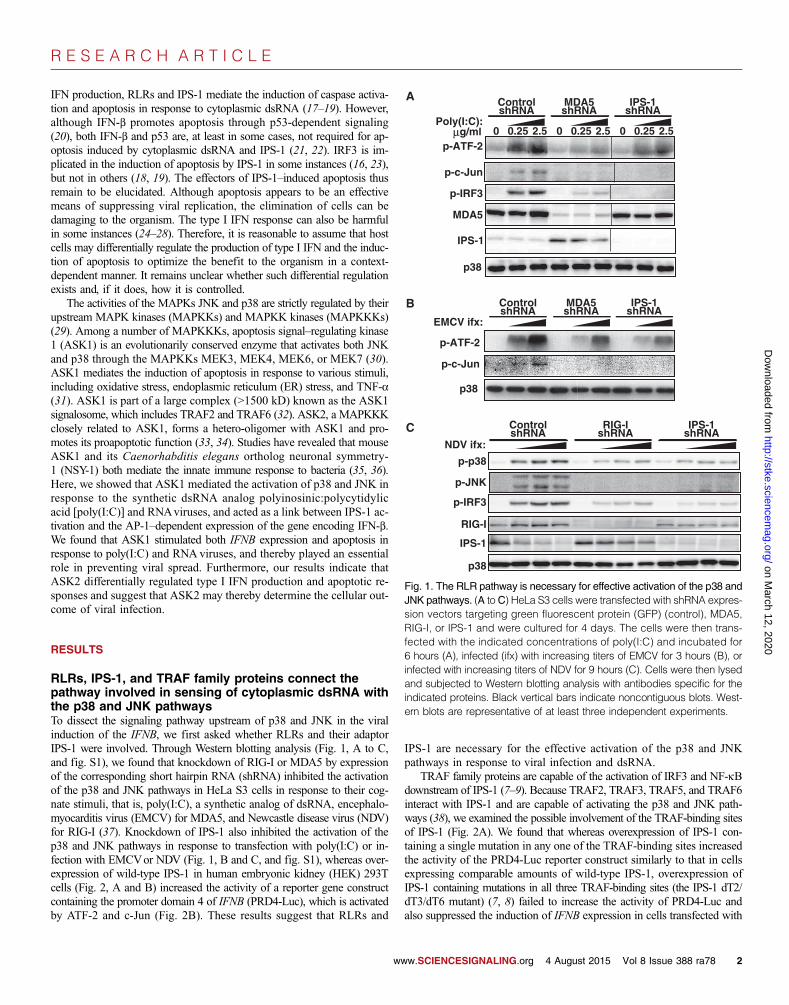
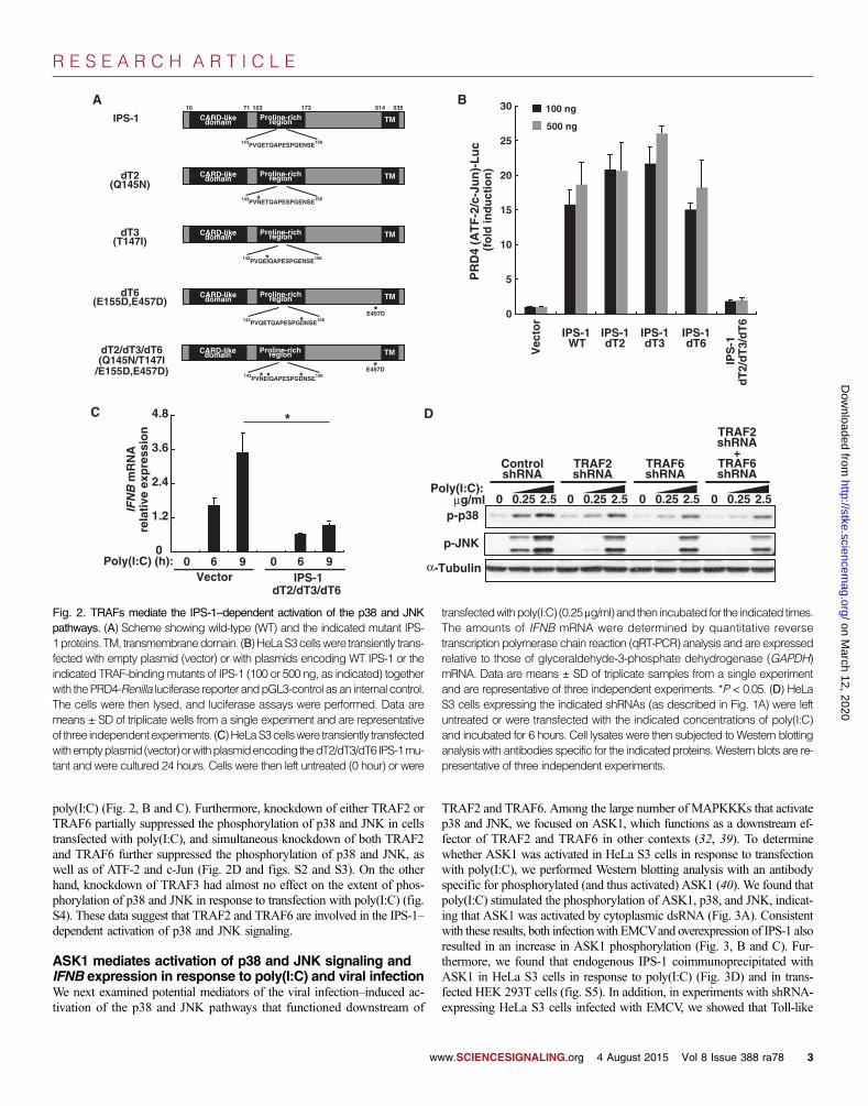
RESULTSRLRs, IPS-1, and TRAF family proteins connect thepathway involved in sensing of cytoplasmic dsRNA withthe p38 and JNK pathwaysTo dissect the signaling pathway upstream of p38 and JNK in the viralinduction of the IFNB, we first asked whether RLRs and their adaptorIPS-1 were involved. Through Western blotting analysis (Fig. 1, A to C,and fig. S1), we found that knockdown of RIG-I or MDA5 by expressionof the corresponding short hairpin RNA (shRNA) inhibited the activationof the p38 and JNK pathways in HeLa S3 cells in response to their cog-nate stimuli, that is, poly(I:C), a synthetic analog of dsRNA, encephalo-myocarditis virus (EMCV) for MDA5, and Newcastle disease virus (NDV)for RIG-I (37). Knockdown of IPS-1 also inhibited the activation of thep38 and JNK pathways in response to transfection with poly(I:C) or in-fection with EMCVor NDV (Fig. 1, B and C, and fig. S1), whereas over-expression of wild-type IPS-1 in human embryonic kidney (HEK) 293Tcells (Fig. 2, A and B) increased the activity of a reporter gene constructcontaining the promoter domain 4 of IFNB (PRD4-Luc), which is activatedby ATF-2 and c-Jun (Fig. 2B). These results suggest that RLRs and
w
IPS-1 are necessary for the effective activation of the p38 and JNKpathways in response to viral infection and dsRNA.
TRAF family proteins are capable of the activation of IRF3 and NF-kBdownstream of IPS-1 (7–9). Because TRAF2, TRAF3, TRAF5, and TRAF6interact with IPS-1 and are capable of activating the p38 and JNK path-ways (38), we examined the possible involvement of the TRAF-binding sitesof IPS-1 (Fig. 2A). We found that whereas overexpression of IPS-1 con-taining a single mutation in any one of the TRAF-binding sites increasedthe activity of the PRD4-Luc reporter construct similarly to that in cellsexpressing comparable amounts of wild-type IPS-1, overexpression ofIPS-1 containing mutations in all three TRAF-binding sites (the IPS-1 dT2/dT3/dT6 mutant) (7, 8) failed to increase the activity of PRD4-Luc andalso suppressed the induction of IFNB expression in cells transfected with
A
p-p38
p-JNK
p38
NDV ifx:
ControlshRNA
RIG-IshRNA
IPS-1shRNA
B
C
p-ATF-2
p-c-Jun
p38
p-ATF-2
p-c-Jun
p-IRF3
Poly(I:C):g/ml 0 0.25 2.5 0 0.25 2.5 0 0.25 2.5
ControlshRNA
MDA5shRNA
IPS-1shRNA
EMCV ifx:
ControlshRNA
MDA5shRNA
IPS-1shRNA
IPS-1
MDA5
p38
p-IRF3
RIG-I
IPS-1
μ
Fig. 1. The RLR pathway is necessary for effective activation of the p38 and
JNK pathways. (A toC) HeLa S3 cells were transfected with shRNA expres-sion vectors targeting green fluorescent protein (GFP) (control), MDA5,RIG-I, or IPS-1 and were cultured for 4 days. The cells were then trans-fected with the indicated concentrations of poly(I:C) and incubated for6 hours (A), infected (ifx) with increasing titers of EMCV for 3 hours (B), orinfected with increasing titers of NDV for 9 hours (C). Cells were then lysedand subjected to Western blotting analysis with antibodies specific for theindicated proteins. Black vertical bars indicate noncontiguous blots. West-ern blots are representative of at least three independent experiments.ww.SCIENCESIGNALING.org 4 August 2015 Vol 8 Issue 388 ra78 2
R E S E A R C H A R T I C L E
on March 12, 2020
http://stke.sciencemag.org/
Dow
nloaded from
poly(I:C) (Fig. 2, B and C). Furthermore, knockdown of either TRAF2 orTRAF6 partially suppressed the phosphorylation of p38 and JNK in cellstransfected with poly(I:C), and simultaneous knockdown of both TRAF2and TRAF6 further suppressed the phosphorylation of p38 and JNK, aswell as of ATF-2 and c-Jun (Fig. 2D and figs. S2 and S3). On the otherhand, knockdown of TRAF3 had almost no effect on the extent of phos-phorylation of p38 and JNK in response to transfection with poly(I:C) (fig.S4). These data suggest that TRAF2 and TRAF6 are involved in the IPS-1–dependent activation of p38 and JNK signaling.
ASK1 mediates activation of p38 and JNK signaling andIFNB expression in response to poly(I:C) and viral infectionWe next examined potential mediators of the viral infection–induced ac-tivation of the p38 and JNK pathways that functioned downstream of
ww
TRAF2 and TRAF6. Among the large number of MAPKKKs that activatep38 and JNK, we focused on ASK1, which functions as a downstream ef-fector of TRAF2 and TRAF6 in other contexts (32, 39). To determinewhether ASK1 was activated in HeLa S3 cells in response to transfectionwith poly(I:C), we performed Western blotting analysis with an antibodyspecific for phosphorylated (and thus activated) ASK1 (40). We found thatpoly(I:C) stimulated the phosphorylation of ASK1, p38, and JNK, indicat-ing that ASK1 was activated by cytoplasmic dsRNA (Fig. 3A). Consistentwith these results, both infectionwith EMCVand overexpression of IPS-1 alsoresulted in an increase in ASK1 phosphorylation (Fig. 3, B and C). Fur-thermore, we found that endogenous IPS-1 coimmunoprecipitated withASK1 in HeLa S3 cells in response to poly(I:C) (Fig. 3D) and in trans-fected HEK 293T cells (fig. S5). In addition, in experiments with shRNA-expressing HeLa S3 cells infected with EMCV, we showed that Toll-like
p-p38
p-JNK
α-Tubulin
Poly(I:C):μg/ml 0 0.25 2.5 0 0.25 2.5 0 0.25 2.5
ControlshRNA
TRAF2shRNA
TRAF6shRNA
0 0.25 2.5
TRAF2shRNA
+TRAF6shRNA
A B
C
CARD-likedomain TMProline-rich
regionIPS-1
dT2(Q145N)
dT3(T147I)
dT6(E155D,E457D)
dT2/dT3/dT6(Q145N/T147I
/E155D,E457D)
CARD-likedomain TMProline-rich
region
10 71 103 173 514 535
CARD-likedomain TMProline-rich
region
*
*
CARD-likedomain TMProline-rich
region
*E457D*
143PVQETQAPESPGENSE158
143PVNETQAPESPGENSE158
143PVQEIQAPESPGENSE158
143PVQETQAPESPGDNSE158
CARD-likedomain TMProline-rich
region
*E457D*
143PVNEIQAPESPGDNSE158* *
0
5
10
15
20
25
30
PR
D4
(AT
F-2
/c-J
un
)-L
uc
(fo
ld in
du
ctio
n)
Vec
tor
IPS-1 WT
IP
S-1
dT
2/d
T3/
dT
6
IPS-1dT2
IPS-1dT3
IPS-1dT6
IFN
B m
RN
Are
lati
ve e
xpre
ssio
n
0
1.2
2.4
3.6
4.8
Poly(I:C) (h): 0 96 0 96Vector IPS-1
dT2/dT3/dT6
D*
100 ng
500 ng
Fig. 2. TRAFs mediate the IPS-1–dependent activation of the p38 and JNK transfectedwithpoly(I:C) (0.25mg/ml)and then incubated for the indicated times.The amounts of IFNB mRNA were determined by quantitative reverse
pathways. (A) Scheme showing wild-type (WT) and the indicated mutant IPS-1proteins. TM, transmembranedomain. (B)HeLaS3cellswere transiently trans-fected with empty plasmid (vector) or with plasmids encoding WT IPS-1 or theindicated TRAF-bindingmutants of IPS-1 (100 or 500 ng, as indicated) togetherwith thePRD4-Renilla luciferase reporter andpGL3-control asan internal control.The cells were then lysed, and luciferase assays were performed. Data aremeans ± SD of triplicate wells from a single experiment and are representativeof three independentexperiments. (C)HeLaS3cellswere transiently transfectedwithemptyplasmid(vector)orwithplasmidencoding thedT2/dT3/dT6 IPS-1mu-tant and were cultured 24 hours. Cells were then left untreated (0 hour) or were
transcription polymerase chain reaction (qRT-PCR) analysis and are expressedrelative to those of glyceraldehyde-3-phosphate dehydrogenase (GAPDH)mRNA. Data are means ± SD of triplicate samples from a single experimentand are representative of three independent experiments. *P < 0.05. (D) HeLaS3 cells expressing the indicated shRNAs (as described in Fig. 1A) were leftuntreated or were transfected with the indicated concentrations of poly(I:C)and incubated for 6 hours. Cell lysates were then subjected to Western blottinganalysis with antibodies specific for the indicated proteins. Western blots are re-presentative of three independent experiments.
w.SCIENCESIGNALING.org 4 August 2015 Vol 8 Issue 388 ra78 3
R E S E A R C H A R T I C L E
on March 12, 2020
http://stke.sciencemag.org/
Dow
nloaded from
receptor 3 (TLR3) was required for efficient activation of p38 and JNK(fig. S6). Together with previous data (32, 39), these results suggest thatASK1 is commonly activated in response to viral infection and poly(I:C)through its interaction with IPS-1 and TRAFs.
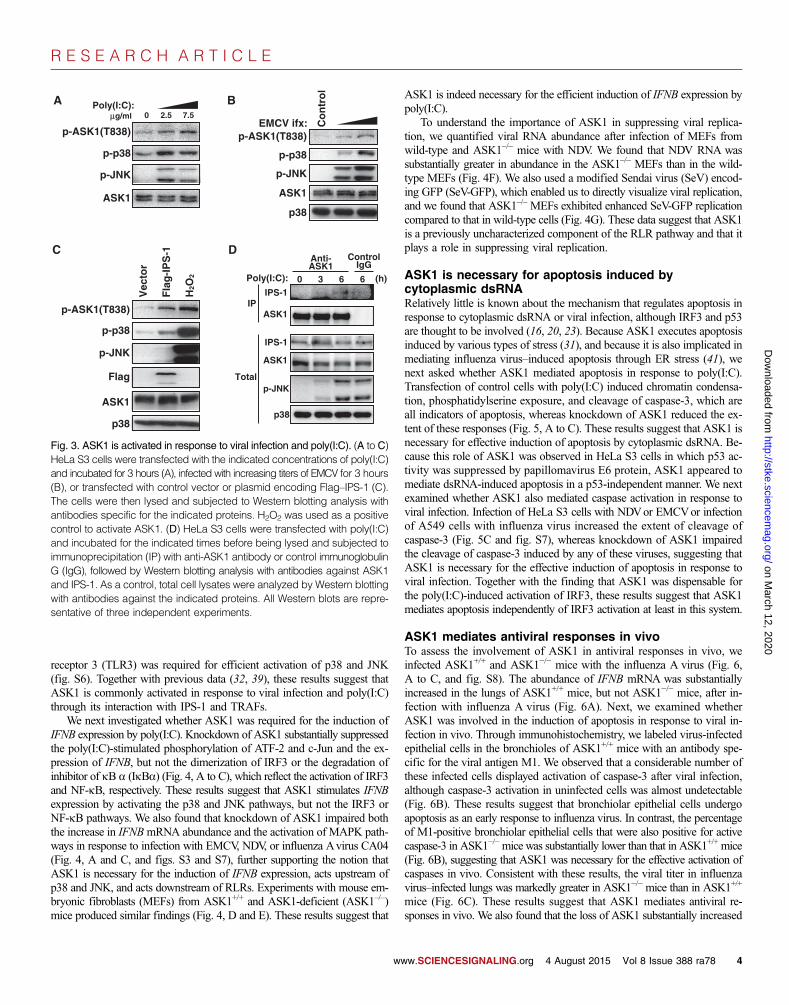
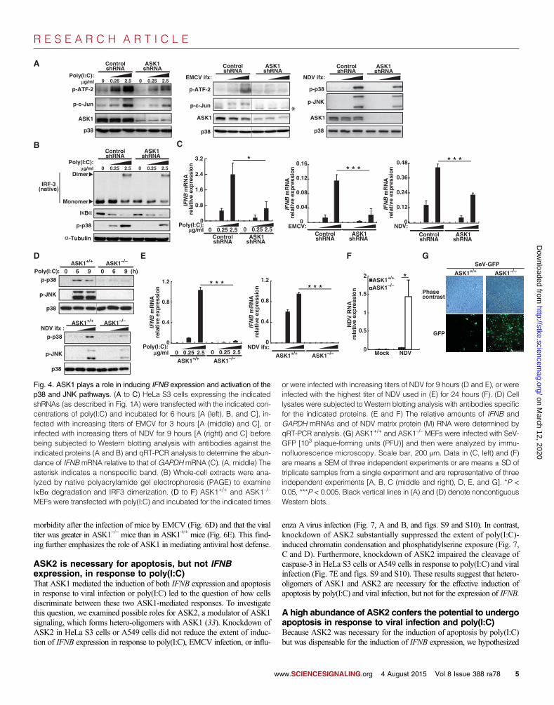
We next investigated whether ASK1 was required for the induction ofIFNB expression by poly(I:C). Knockdown of ASK1 substantially suppressedthe poly(I:C)-stimulated phosphorylation of ATF-2 and c-Jun and the ex-pression of IFNB, but not the dimerization of IRF3 or the degradation ofinhibitor of kB a (IkBa) (Fig. 4, A to C), which reflect the activation of IRF3and NF-kB, respectively. These results suggest that ASK1 stimulates IFNBexpression by activating the p38 and JNK pathways, but not the IRF3 orNF-kB pathways. We also found that knockdown of ASK1 impaired boththe increase in IFNBmRNA abundance and the activation of MAPK path-ways in response to infection with EMCV, NDV, or influenza Avirus CA04(Fig. 4, A and C, and figs. S3 and S7), further supporting the notion thatASK1 is necessary for the induction of IFNB expression, acts upstream ofp38 and JNK, and acts downstream of RLRs. Experiments with mouse em-bryonic fibroblasts (MEFs) from ASK1+/+ and ASK1-deficient (ASK1–/–)mice produced similar findings (Fig. 4, D and E). These results suggest that
w
ASK1 is indeed necessary for the efficient induction of IFNB expression bypoly(I:C).
To understand the importance of ASK1 in suppressing viral replica-tion, we quantified viral RNA abundance after infection of MEFs fromwild-type and ASK1–/– mice with NDV. We found that NDV RNA wassubstantially greater in abundance in the ASK1–/– MEFs than in the wild-type MEFs (Fig. 4F). We also used a modified Sendai virus (SeV) encod-ing GFP (SeV-GFP), which enabled us to directly visualize viral replication,and we found that ASK1–/– MEFs exhibited enhanced SeV-GFP replicationcompared to that in wild-type cells (Fig. 4G). These data suggest that ASK1is a previously uncharacterized component of the RLR pathway and that itplays a role in suppressing viral replication.
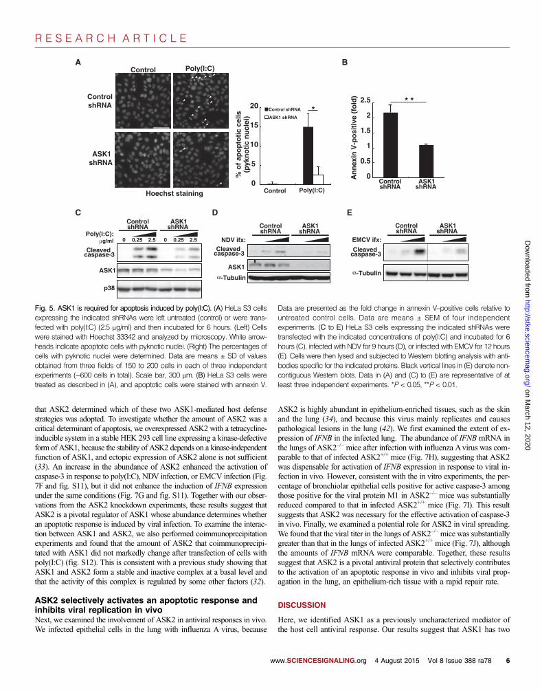
ASK1 is necessary for apoptosis induced bycytoplasmic dsRNARelatively little is known about the mechanism that regulates apoptosis inresponse to cytoplasmic dsRNA or viral infection, although IRF3 and p53are thought to be involved (16, 20, 23). Because ASK1 executes apoptosisinduced by various types of stress (31), and because it is also implicated inmediating influenza virus–induced apoptosis through ER stress (41), wenext asked whether ASK1 mediated apoptosis in response to poly(I:C).Transfection of control cells with poly(I:C) induced chromatin condensa-tion, phosphatidylserine exposure, and cleavage of caspase-3, which areall indicators of apoptosis, whereas knockdown of ASK1 reduced the ex-tent of these responses (Fig. 5, A to C). These results suggest that ASK1 isnecessary for effective induction of apoptosis by cytoplasmic dsRNA. Be-cause this role of ASK1 was observed in HeLa S3 cells in which p53 ac-tivity was suppressed by papillomavirus E6 protein, ASK1 appeared tomediate dsRNA-induced apoptosis in a p53-independent manner. We nextexamined whether ASK1 also mediated caspase activation in response toviral infection. Infection of HeLa S3 cells with NDVor EMCVor infectionof A549 cells with influenza virus increased the extent of cleavage ofcaspase-3 (Fig. 5C and fig. S7), whereas knockdown of ASK1 impairedthe cleavage of caspase-3 induced by any of these viruses, suggesting thatASK1 is necessary for the effective induction of apoptosis in response toviral infection. Together with the finding that ASK1 was dispensable forthe poly(I:C)-induced activation of IRF3, these results suggest that ASK1mediates apoptosis independently of IRF3 activation at least in this system.
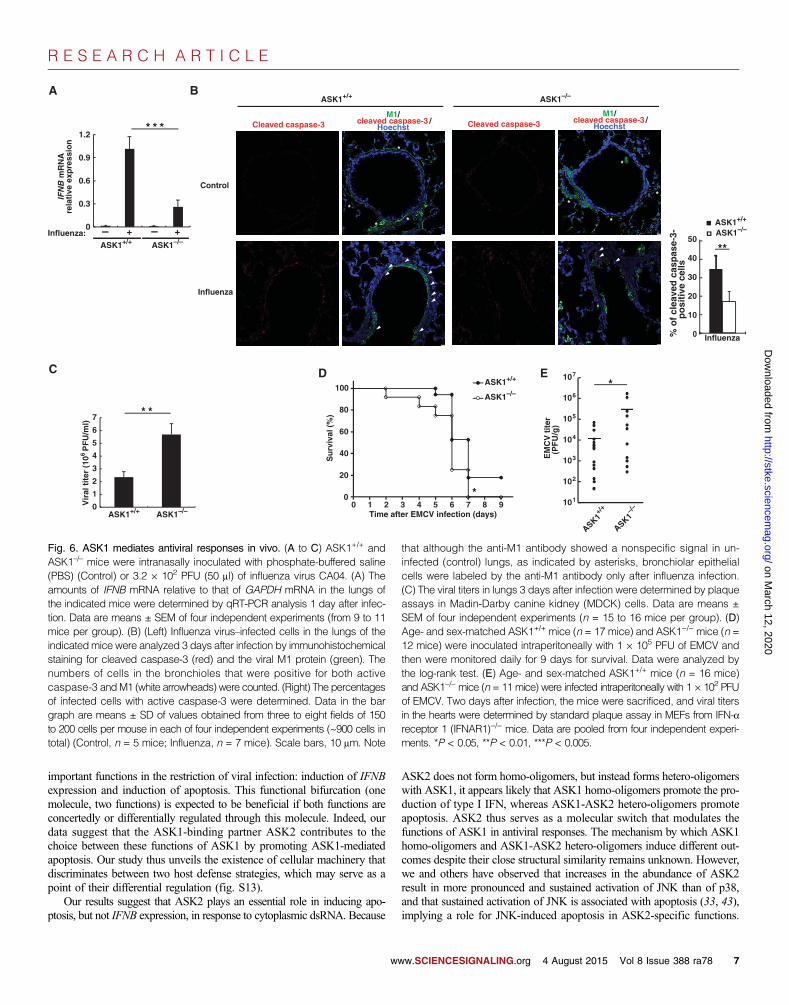
ASK1 mediates antiviral responses in vivoTo assess the involvement of ASK1 in antiviral responses in vivo, weinfected ASK1+/+ and ASK1−/− mice with the influenza A virus (Fig. 6,A to C, and fig. S8). The abundance of IFNB mRNA was substantiallyincreased in the lungs of ASK1+/+ mice, but not ASK1−/− mice, after in-fection with influenza A virus (Fig. 6A). Next, we examined whetherASK1 was involved in the induction of apoptosis in response to viral in-fection in vivo. Through immunohistochemistry, we labeled virus-infectedepithelial cells in the bronchioles of ASK1+/+ mice with an antibody spe-cific for the viral antigen M1. We observed that a considerable number ofthese infected cells displayed activation of caspase-3 after viral infection,although caspase-3 activation in uninfected cells was almost undetectable(Fig. 6B). These results suggest that bronchiolar epithelial cells undergoapoptosis as an early response to influenza virus. In contrast, the percentageof M1-positive bronchiolar epithelial cells that were also positive for activecaspase-3 in ASK1−/−mice was substantially lower than that in ASK1+/+ mice(Fig. 6B), suggesting that ASK1 was necessary for the effective activation ofcaspases in vivo. Consistent with these results, the viral titer in influenzavirus–infected lungs was markedly greater in ASK1−/− mice than in ASK1+/+
mice (Fig. 6C). These results suggest that ASK1 mediates antiviral re-sponses in vivo. We also found that the loss of ASK1 substantially increased
A B
C
Vec
tor
Fla
g-I
PS
-1
H2O
2
p-ASK1(T838)
p-p38
p-JNK
Flag
ASK1
p38
p-ASK1(T838)
p-p38
p-JNK
ASK1
p38
EMCV ifx: Co
ntr
ol
p-ASK1(T838)
p-p38
p-JNK
ASK1
Poly(I:C):g/ml 0 2.5 7.5
D
ASK1
ASK1
IPS-1
IPS-1
IP
Total
ControlIgG
Anti-ASK1
p-JNK
p38
Poly(I:C): 0 3 6 6 (h)
Fig. 3. ASK1 is activated in response to viral infection and poly(I:C). (A to C)
HeLa S3 cells were transfected with the indicated concentrations of poly(I:C)and incubated for 3 hours (A), infected with increasing titers of EMCV for 3 hours(B), or transfected with control vector or plasmid encoding Flag–IPS-1 (C).The cells were then lysed and subjected to Western blotting analysis withantibodies specific for the indicated proteins. H2O2 was used as a positivecontrol to activate ASK1. (D) HeLa S3 cells were transfected with poly(I:C)and incubated for the indicated times before being lysed and subjected toimmunoprecipitation (IP) with anti-ASK1 antibody or control immunoglobulinG (IgG), followed by Western blotting analysis with antibodies against ASK1and IPS-1. As a control, total cell lysates were analyzed by Western blottingwith antibodies against the indicated proteins. All Western blots are repre-sentative of three independent experiments.ww.SCIENCESIGNALING.org 4 August 2015 Vol 8 Issue 388 ra78 4
R E S E A R C H A R T I C L E
on March 12, 2020
http://stke.sciencemag.org/
Dow
nloaded from
morbidity after the infection of mice by EMCV (Fig. 6D) and that the viraltiter was greater in ASK1−/− mice than in ASK1+/+ mice (Fig. 6E). This find-ing further emphasizes the role of ASK1 in mediating antiviral host defense.
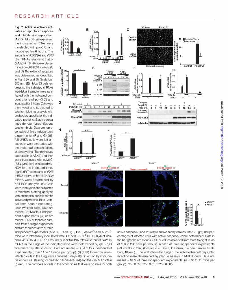
ASK2 is necessary for apoptosis, but not IFNBexpression, in response to poly(I:C)That ASK1 mediated the induction of both IFNB expression and apoptosisin response to viral infection or poly(I:C) led to the question of how cellsdiscriminate between these two ASK1-mediated responses. To investigatethis question, we examined possible roles for ASK2, a modulator of ASK1signaling, which forms hetero-oligomers with ASK1 (33). Knockdown ofASK2 in HeLa S3 cells or A549 cells did not reduce the extent of induc-tion of IFNB expression in response to poly(I:C), EMCV infection, or influ-
ww
enza A virus infection (Fig. 7, A and B, and figs. S9 and S10). In contrast,knockdown of ASK2 substantially suppressed the extent of poly(I:C)-induced chromatin condensation and phosphatidylserine exposure (Fig. 7,C and D). Furthermore, knockdown of ASK2 impaired the cleavage ofcaspase-3 in HeLa S3 cells or A549 cells in response to poly(I:C) and viralinfection (Fig. 7E and figs. S9 and S10). These results suggest that hetero-oligomers of ASK1 and ASK2 are necessary for the effective induction ofapoptosis by poly(I:C) and viral infection, but not for the expression of IFNB.
A high abundance of ASK2 confers the potential to undergoapoptosis in response to viral infection and poly(I:C)Because ASK2 was necessary for the induction of apoptosis by poly(I:C)but was dispensable for the induction of IFNB expression, we hypothesized
A
C
F
p-ATF-2
p-c-Jun
p38
Poly(I:C):g/ml
ASK1
0 0.25 2.5 0 0.25 2.5
ControlshRNA
ASK1 shRNA
B
Poly(I:C):g/ml 0 0.25 2.5 0 0.25 2.5
ControlshRNA
ASK1shRNA
-Tubulin
p-p38
I B
Dimer
Monomer
IRF-3(native)
p-ATF-2
p-c-Jun
p38
EMCV ifx:
ControlshRNA
ASK1 shRNA
p-p38
p-JNK
p38
NDV ifx:
ControlshRNA
ASK1 shRNA
p-p38
p-JNK
p38
Poly(I:C):ASK1+/+ ASK1–/–
6 )h(906 90
p-p38
p-JNK
p38
NDV ifx :ASK1+/+ ASK1–/–
Poly(I:C):g/ml 0 0.25 2.5 0 0.25 2.5
ASK1+/+ ASK1–/–
IFN
BA
NR
mn
oi sserpxe
evit al er
ASK1+/+ ASK1–/–SeV-GFP
Phasecontrast
GFP
D E
BN
FIA
NR
mn
oi sserpxe
evit aler
BN
FIA
NR
mn
oi sserpxe
evit al er
EMCV:ControlshRNA
ASK1shRNA
0
0.8
1.6
2.4
3.2
Poly(I:C):g/ml 0 0.25 2.5
ControlshRNA
ASK1shRNA
0 0.25 2.50
0.04
0.08
0.12
0.16
IFN
BA
NR
mn
oi sserpx e
evi tal er
NDV ifx:
ASK1+/+ ASK1–/–
0
0.4
0.8
1.2
NDV:
0.12
0.24
0.36
0.48
0
BN
FIA
NR
mn
oi s serpxe
evit al er
ControlshRNA
ASK1shRNA
0
0.4
0.8
1.2
ASK1 ASK1
** * *
* * *
* * * * * *
0
0.5
1
1.5
2
Mock NDV
ASK1+/+
ASK1–/–
AN
RV
DN
noi sser
pxeevit al er
*
G
Fig. 4. ASK1 plays a role in inducing IFNB expression and activation of thep38 and JNK pathways. (A to C) HeLa S3 cells expressing the indicated
or were infected with increasing titers of NDV for 9 hours (D and E), or wereinfected with the highest titer of NDV used in (E) for 24 hours (F). (D) Cell
shRNAs (as described in Fig. 1A) were transfected with the indicated con-centrations of poly(I:C) and incubated for 6 hours [A (left), B, and C], in-fected with increasing titers of EMCV for 3 hours [A (middle) and C], orinfected with increasing titers of NDV for 9 hours [A (right) and C] beforebeing subjected to Western blotting analysis with antibodies against theindicated proteins (A and B) and qRT-PCR analysis to determine the abun-dance of IFNB mRNA relative to that of GAPDHmRNA (C). (A, middle) Theasterisk indicates a nonspecific band. (B) Whole-cell extracts were ana-lyzed by native polyacrylamide gel electrophoresis (PAGE) to examineIkBa degradation and IRF3 dimerization. (D to F) ASK1+/+ and ASK1–/–
MEFs were transfected with poly(I:C) and incubated for the indicated times
lysates were subjected to Western blotting analysis with antibodies specificfor the indicated proteins. (E and F) The relative amounts of IFNB andGAPDH mRNAs and of NDV matrix protein (M) RNA were determined byqRT-PCR analysis. (G) ASK1+/+ and ASK1–/– MEFs were infected with SeV-GFP [103 plaque-forming units (PFU)] and then were analyzed by immu-nofluorescence microscopy. Scale bar, 200 mm. Data in (C, left) and (F)are means ± SEM of three independent experiments or are means ± SD oftriplicate samples from a single experiment and are representative of threeindependent experiments [A, B, C (middle and right), D, E, and G]. *P <0.05, ***P < 0.005. Black vertical lines in (A) and (D) denote noncontiguousWestern blots.
w.SCIENCESIGNALING.org 4 August 2015 Vol 8 Issue 388 ra78 5
R E S E A R C H A R T I C L E
on March 12, 2020
http://stke.sciencemag.org/
Dow
nloaded from
that ASK2 determined which of these two ASK1-mediated host defensestrategies was adopted. To investigate whether the amount of ASK2 was acritical determinant of apoptosis, we overexpressed ASK2 with a tetracycline-inducible system in a stable HEK 293 cell line expressing a kinase-defectiveform of ASK1, because the stability of ASK2 depends on a kinase-independentfunction of ASK1, and ectopic expression of ASK2 alone is not sufficient(33). An increase in the abundance of ASK2 enhanced the activation ofcaspase-3 in response to poly(I:C), NDV infection, or EMCV infection (Fig.7F and fig. S11), but it did not enhance the induction of IFNB expressionunder the same conditions (Fig. 7G and fig. S11). Together with our obser-vations from the ASK2 knockdown experiments, these results suggest thatASK2 is a pivotal regulator of ASK1 whose abundance determines whetheran apoptotic response is induced by viral infection. To examine the interac-tion between ASK1 and ASK2, we also performed coimmunoprecipitationexperiments and found that the amount of ASK2 that coimmunoprecipi-tated with ASK1 did not markedly change after transfection of cells withpoly(I:C) (fig. S12). This is consistent with a previous study showing thatASK1 and ASK2 form a stable and inactive complex at a basal level andthat the activity of this complex is regulated by some other factors (32).
ASK2 selectively activates an apoptotic response andinhibits viral replication in vivoNext, we examined the involvement of ASK2 in antiviral responses in vivo.We infected epithelial cells in the lung with influenza A virus, because
w
ASK2 is highly abundant in epithelium-enriched tissues, such as the skinand the lung (34), and because this virus mainly replicates and causespathological lesions in the lung (42). We first examined the extent of ex-pression of IFNB in the infected lung. The abundance of IFNB mRNA inthe lungs of ASK2–/– mice after infection with influenza Avirus was com-parable to that of infected ASK2+/+ mice (Fig. 7H), suggesting that ASK2was dispensable for activation of IFNB expression in response to viral in-fection in vivo. However, consistent with the in vitro experiments, the per-centage of bronchiolar epithelial cells positive for active caspase-3 amongthose positive for the viral protein M1 in ASK2–/– mice was substantiallyreduced compared to that in infected ASK2+/+ mice (Fig. 7I). This resultsuggests that ASK2 was necessary for the effective activation of caspase-3in vivo. Finally, we examined a potential role for ASK2 in viral spreading.We found that the viral titer in the lungs of ASK2–/–mice was substantiallygreater than that in the lungs of infected ASK2+/+ mice (Fig. 7J), althoughthe amounts of IFNB mRNA were comparable. Together, these resultssuggest that ASK2 is a pivotal antiviral protein that selectively contributesto the activation of an apoptotic response in vivo and inhibits viral prop-agation in the lung, an epithelium-rich tissue with a rapid repair rate.
DISCUSSION
Here, we identified ASK1 as a previously uncharacterized mediator ofthe host cell antiviral response. Our results suggest that ASK1 has two
A
0
5
10
15
20
% o
f ap
op
toti
c ce
lls(p
ykn
oti
c n
ucl
ei)
Poly(I:C) Control
*
Poly(I:C):μg/ml 0 0.25 2.5 0 0.25 2.5
ControlshRNA
ASK1shRNA
ASK1
Cleaved caspase-3
α-Tubulin
ASK1
Cleaved caspase-3
NDV ifx:
ControlshRNA
ASK1shRNA
B
C
p38
ControlshRNA
ASK1shRNA
0
0.5
1
1.5
2.5
2
An
nex
in V
-po
siti
ve (
fold
)
**
α-Tubulin
Cleaved caspase-3
EMCV ifx:
ControlshRNA
ASK1shRNA
ControlshRNA
ASK1shRNA
Control Poly(I:C)
Hoechst staining
D E
Control shRNA
ASK1 shRNA
Fig. 5. ASK1 is required for apoptosis induced by poly(I:C). (A) HeLa S3 cells Data are presented as the fold change in annexin V–positive cells relative to
expressing the indicated shRNAs were left untreated (control) or were trans-fected with poly(I:C) (2.5 mg/ml) and then incubated for 6 hours. (Left) Cellswere stained with Hoechst 33342 and analyzed by microscopy. White arrow-heads indicate apoptotic cells with pyknotic nuclei. (Right) The percentages ofcells with pyknotic nuclei were determined. Data are means ± SD of valuesobtained from three fields of 150 to 200 cells in each of three independentexperiments (~600 cells in total). Scale bar, 300 mm. (B) HeLa S3 cells weretreated as described in (A), and apoptotic cells were stained with annexin V.untreated control cells. Data are means ± SEM of four independentexperiments. (C to E) HeLa S3 cells expressing the indicated shRNAs weretransfected with the indicated concentrations of poly(I:C) and incubated for 6hours (C), infected with NDV for 9 hours (D), or infected with EMCV for 12 hours(E). Cells were then lysed and subjected to Western blotting analysis with anti-bodies specific for the indicated proteins. Black vertical lines in (E) denote non-contiguous Western blots. Data in (A) and (C) to (E) are representative of atleast three independent experiments. *P < 0.05, **P < 0.01.
ww.SCIENCESIGNALING.org 4 August 2015 Vol 8 Issue 388 ra78 6
R E S E A R C H A R T I C L E
on March 12, 2020
http://stke.sciencemag.org/
Dow
nloaded from
important functions in the restriction of viral infection: induction of IFNBexpression and induction of apoptosis. This functional bifurcation (onemolecule, two functions) is expected to be beneficial if both functions areconcertedly or differentially regulated through this molecule. Indeed, ourdata suggest that the ASK1-binding partner ASK2 contributes to thechoice between these functions of ASK1 by promoting ASK1-mediatedapoptosis. Our study thus unveils the existence of cellular machinery thatdiscriminates between two host defense strategies, which may serve as apoint of their differential regulation (fig. S13).
Our results suggest that ASK2 plays an essential role in inducing apo-ptosis, but not IFNB expression, in response to cytoplasmic dsRNA. Because
ww
ASK2 does not form homo-oligomers, but instead forms hetero-oligomerswith ASK1, it appears likely that ASK1 homo-oligomers promote the pro-duction of type I IFN, whereas ASK1-ASK2 hetero-oligomers promoteapoptosis. ASK2 thus serves as a molecular switch that modulates thefunctions of ASK1 in antiviral responses. The mechanism by which ASK1homo-oligomers and ASK1-ASK2 hetero-oligomers induce different out-comes despite their close structural similarity remains unknown. However,we and others have observed that increases in the abundance of ASK2result in more pronounced and sustained activation of JNK than of p38,and that sustained activation of JNK is associated with apoptosis (33, 43),implying a role for JNK-induced apoptosis in ASK2-specific functions.
BN
FIA
NR
m n
oi sserpxe evi t al er
Influenza:ASK1+/+ ASK1–/–– + – +
Vir
al t
iter
(10
6 PF
U/m
l)
ASK1+/+ ASK1–/–
A B
C
0
10
20
30
40
50
ASK1+/+
ASK1–/–
Influenza % o
f cl
eave
d c
asp
ase-
3-
p
osi
tive
cel
ls
*
*
ASK1+/+ ASK1–/–
Control
Influenza
M1/cleaved caspase-3 /
Hoechst
*
**
*
*
*
*
*
M1/cleaved caspase-3 /
HoechstCleaved caspase-3 Cleaved caspase-3
*
** *
*
0
0.3
0.6
0.9
1.2
0
1
2
3
4
5
6
7
101
102
103
104
105
106
107
ASK1+/
+
ASK1–/–
retit V
CM
E)
g/U
FP(
ASK1+/+
ASK1–/–
0
100
80
60
40
20
0 1 2 3 4 5 6 7 8 9
)%( lavivr
uS
Time after EMCV infection (days)
*
*
D E
Fig. 6. ASK1 mediates antiviral responses in vivo. (A to C) ASK1+/+ andASK1–/– mice were intranasally inoculated with phosphate-buffered saline
that although the anti-M1 antibody showed a nonspecific signal in un-infected (control) lungs, as indicated by asterisks, bronchiolar epithelial
(PBS) (Control) or 3.2 × 10² PFU (50 ml) of influenza virus CA04. (A) Theamounts of IFNB mRNA relative to that of GAPDH mRNA in the lungs ofthe indicated mice were determined by qRT-PCR analysis 1 day after infec-tion. Data are means ± SEM of four independent experiments (from 9 to 11mice per group). (B) (Left) Influenza virus–infected cells in the lungs of theindicatedmice were analyzed 3 days after infection by immunohistochemicalstaining for cleaved caspase-3 (red) and the viral M1 protein (green). Thenumbers of cells in the bronchioles that were positive for both activecaspase-3 andM1 (white arrowheads) were counted. (Right) The percentagesof infected cells with active caspase-3 were determined. Data in the bargraph are means ± SD of values obtained from three to eight fields of 150to 200 cells per mouse in each of four independent experiments (~900 cells intotal) (Control, n = 5 mice; Influenza, n = 7 mice). Scale bars, 10 mm. Note
cells were labeled by the anti-M1 antibody only after influenza infection.(C) The viral titers in lungs 3 days after infection were determined by plaqueassays in Madin-Darby canine kidney (MDCK) cells. Data are means ±SEM of four independent experiments (n = 15 to 16 mice per group). (D)Age- and sex-matched ASK1+/+ mice (n = 17 mice) and ASK1−/− mice (n =12 mice) were inoculated intraperitoneally with 1 × 10⁵ PFU of EMCV andthen were monitored daily for 9 days for survival. Data were analyzed bythe log-rank test. (E) Age- and sex-matched ASK1+/+ mice (n = 16 mice)and ASK1−/−mice (n = 11 mice) were infected intraperitoneally with 1 × 10² PFUof EMCV. Two days after infection, the mice were sacrificed, and viral titersin the hearts were determined by standard plaque assay in MEFs from IFN-areceptor 1 (IFNAR1)−/− mice. Data are pooled from four independent experi-ments. *P < 0.05, **P < 0.01, ***P < 0.005.
w.SCIENCESIGNALING.org 4 August 2015 Vol 8 Issue 388 ra78 7
R E S E A R C H A R T I C L E
on March 12, 2020
http://stke.sciencemag.org/
Dow
nloaded from
A B C
0
5
10
15
sllec citot
po
pa fo
%)ielc
un cit
onky
p(
Poly(I:C) Control
*Control shRNAASK2 shRNA
-Tubulin
Poly(I:C):g/ml
p-p38
Cleaved caspase-3
p-JNK
0 0.25 2.5 0 0.25 2.5
ControlshRNA
ASK2 shRNA
0
0.08
0.16
0.24
0.32
Poly(I:C):g/ml 0 0.25 2.5
ControlshRNA
ASK2shRNA
0 0.25 2.5
BN
FIA
NR
m n
oi sserpxe evi t al er0
0.3
0.6
0.9
1.2
ControlshRNA
ASK2shRNA
2K
SA
AN
Rm
noi sser
pxe evit al er
p38
Flag-ASK2
Cleaved caspase-3
Poly(I:C) (h): 0 12 6 9 0 6 912 12
Tet ng/ml: 00
1 3
Flag-ASK2
Cleaved caspase-3
-Tubulin
9 240 12 9 240 12Poly(I:C):Tet ng/ml:
NDV (h):+ +
0 3
BN
FIA
NR
m BN
FIA
NR
m n
oi sserpxe evit al er
E F
Poly(I:C):0
0.4
0.8
1.2
ASK2: – +
noisser
pxe evit al er 0
0.1
0.2
0.3
0.4
NDV ifx:
D
0
0.5
1
1.5
2.5
2
)dl
of( evitiso
p-V
n
ixen
nA Control
shRNAASK2
shRNA ASK2: – +
G
BN
FIA
NR
m n
oisserpxe evit al er
Influenza: ASK2+/+ ASK2–/–– + – + V
iral
tit
er (
106 P
FU
/ml)
ASK2+/+ ASK2–/–
H J**
N.S.
*
0
0.8
1.2
1.8
2.4
0123456789
10
I ASK2+/+ ASK2–/–
Control
Influenza
ASK2+/+
ASK2–/–
% o
f C
leav
ed c
asp
ase-
3-
p
osi
tive
cel
ls
*
M1/cleaved caspase-3 /
Hoechst
M1/cleaved caspase-3 /
HoechstCleaved caspase-3 Cleaved caspase-3
010203040506070
Influenza
*
**
*
ControlshRNA
ASK2shRNA
Control Poly(I:C)
Hoechst staining
– + – +
0 0− − − − − –−−
**
Fig. 7. ASK2 selectively acti-
vates an apoptotic responseand inhibits viral replication.(A toD)HeLaS3cellsexpressingthe indicated shRNAs weretransfected with poly(I:C) andincubated for 6 hours. Theamounts ofASK2 (A) and IFNB(B) mRNAs relative to that ofGAPDH mRNA were deter-mined by qRT-PCR analysis. (Cand D) The extent of apoptosiswas determined as describedin Fig. 5 (A and B). Scale bar,300 mm. (E) HeLa S3 cells ex-pressing the indicated shRNAswere left untreatedorwere trans-fected with the indicated con-centrations of poly(I:C) andincubated for6hours.Cellswerethen lysed and subjected toWestern blotting analysis withantibodies specific for the indi-cated proteins. Black verticallines denote noncontiguousWestern blots. Data are repre-sentative of three independentexperiments. (F and G) 293-ASK2/1KN cells were left un-treatedorwerepretreatedwiththe indicated concentrationsof tetracycline (Tet) (to induceexpression of ASK2) and thenwere transfected with poly(I:C)(7.5mg/ml) (left) or infectedwithNDV for the indicated times(right). (F) Theamounts of IFNBmRNArelative to thatofGAPDHmRNA were determined byqRT-PCR analysis. (G) Cellswere then lysedandsubjectedto Western blotting analysiswith antibodies specific for theindicatedproteins.Black verti-cal lines denote noncontig-uous Western blots. Data aremeans±SEMof four indepen-dent experiments (D) or aremeans ± SD of triplicate sam-ples from a single experimentandare representative of three independent experiments (A to C, F, and G). (H to J) ASK2+/+ and ASK2–/–mice were intranasally inoculated with PBS or 3.2 × 10² PFU (50 ml) of influ-enza virus CA04. (H) The amounts of IFNBmRNA relative to that of GAPDHmRNA in the lungs of the indicated mice were determined by qRT-PCRanalysis 1 day after infection. Data are means ± SEM of four independentexperiments (from 11 to 14 mice per group). (I) (Left) Influenza virus–infected cells in the lung were analyzed 3 days after infection by immuno-histochemical staining for cleaved caspase-3 (red) and the viralM1protein(green). The numbers of cells in the bronchioles that were positive for both
ww
active caspase-3 andM1 (white arrowheads)werecounted. (Right) Theper-centages of infected cells with active caspase-3 were determined. Data inthe bar graphs are means ± SD of values obtained from three to eight fieldsof 150 to 200 cells per mouse in each of three independent experiments(~900 cells in total) (Control, n = 3 mice; Influenza, n = 5 to 6 mice). Scalebars, 10 mm. (J) The viral titers in the lungs of the indicatedmice 3 days afterinfection were determined by plaque assays in MDCK cells. Data aremeans ± SEM of three independent experiments. (n = 10 to 11 mice pergroup). *P < 0.05, **P < 0.01, ***P < 0.005.
w.SCIENCESIGNALING.org 4 August 2015 Vol 8 Issue 388 ra78 8
R E S E A R C H A R T I C L E
on March 12, 2020
http://stke.sciencemag.org/
Dow
nloaded from
The mechanism by which ASK1-ASK2 hetero-oligomers are activatedupon viral infection is still an open question.
In which contexts would these antiviral strategies be differentiallyused? Although apoptosis eliminates an infected cell, such eliminationmay be costly for cells that are in limited supply. For example, lost neuronsare not normally replaced by new neurons in most parts of the adult brain,so neurons should not easily undergo apoptosis. The benefits of apoptosisoutweigh the risks, however, for cells in plentiful supply with a high turn-over rate, such as those in epithelial tissues. Indeed, ASK2 is highly abun-dant in epithelial tissues, but not nonepithelial tissues, whereas ASK1appears to be ubiquitously expressed (34). Consistent with this scenario,deletion of ASK2, which reduced the activation of caspase-3, but not theexpression of IFNB, enhanced the replication of influenza virus in the lungs,a representative, epithelium-rich tissue with a rapid repair rate (Fig. 7). Wetherefore speculate that renewable epithelial cells eliminate viruses in partby ASK2-dependent apoptosis, whereas other cell types with less ASK2abundance eliminate viruses by other means (such as type I IFN produc-tion). It is also possible that ASK2-dependent apoptosis takes place in acontext in which the production of type I IFN is harmful. Because type IIFN production reduces hematopoietic stem cells and compromises hostdefense against some bacterial infections (22, 24–28), it would be interest-ing to investigate whether ASK2 plays a role in this context in future studies.
Although the regulation of IFNB expression by viral dsRNA has beenstudied extensively, the signaling pathway that activates p38 and JNK andthe AP-1 element of IFNB was unclear. Among members of the MAPKKKfamily, transforming growth factor–b (TGF-b)–activated kinase 1 (TAK1)has been implicated in the induction of type I IFN production (44); how-ever, deletion of the gene encoding TAK1 does not affect type I IFN pro-duction in response to cytoplasmic dsRNA or infection with vesicularstomatitis virus (13, 45). Knockdown of MEKK1 partially inhibits theproduction of type I IFN in response to dsRNA, although the activationof MEKK1 by dsRNA has not been described (13). Here, we found thatASK1 was phosphorylated at its critical site for activation in response topoly(I:C) or viral infection and that ASK1 played an essential role in theactivation of p38 and JNK signaling and of AP-1, as well as in the expres-sion of IFNB. Furthermore, endogenous ASK1 coimmunoprecipitated withIPS-1, a key adaptor protein for RLRs, in response to poly(I:C). A propor-tion of total cellular ASK1 is thought to be localized at the mitochondria,where IPS-1 resides, consistent with the notion that ASK1 mediatessignaling from IPS-1 by forming a protein complex (46, 47). Together, theseresults suggest that ASK1 connects IPS-1 and the p38 and JNK signalingpathways in the viral induction of IFNB expression.
A point that still remains to be resolved is how IPS-1 activates ASK1.Our results suggest that TRAF2 and TRAF6, both of which are recruitedby IPS-1 to the insoluble fraction that includes the mitochondrial mem-brane (7), play a role in activating ASK1. Given that a proportion of ASK1and IPS-1 are located at the mitochondrial outer membrane, the associationof ligand-bound RLRs with IPS-1 may stimulate the activation of an IPS-1–TRAF–ASK1 complex by inducing conformational changes or through ad-ditional modulators. Reactive oxygen species (ROS) increase the activity ofASK1 under oxidative stress by removing the inhibitor thioredoxin fromASK1 (48), and IPS-1 induces IFNB expression in a ROS-dependent manner(49). ROS might therefore contribute to activation of an IPS-1–TRAF–ASK1complex that is induced by cytoplasmic dsRNA. As a common mediatorof cellular stress, ASK1 might sense cellular conditions (such as the extentof oxidative stress) and modulate the outcome of viral infection.
The history of host-pathogen interactions has given rise to the evolu-tion of viral molecules that antagonize host defense mechanisms. For ex-ample, the HIV gene product Nef binds to and inhibits the activity ofASK1 (50), which is consistent with the importance of ASK1 in antiviral
ww
strategies. Our findings may help to explain how ASK1 restricts the spreadof such viruses. Our identification of ASK1 and ASK2 as regulators ofthe innate immune response and our elucidation of the underlying mecha-nisms by which the ASK family kinases differentially mediate type I IFNinduction and apoptosis may provide a basis for the development of newtherapies for the effective elimination of viruses.
MATERIALS AND METHODS
Plasmids and reagentsThe constructs encoding wild-type human IPS-1 and human ASK1 weredescribed previously (48, 51). Site-directed mutagenesis was performedwith a QuikChange site-directed mutagenesis kit (Agilent Technologies)to generate mutant human IPS-1 that could not interact with TRAFs, asdescribed previously (7, 8). The reporter gene construct containingpromoter domain 4 of IFNB (PRD4-RLuc) was generated as described pre-viously (11). Poly(I:C) was purchased from GEHealthcare. To stimulate cells,poly(I:C) was mixed with Lipofectamine 2000 (Invitrogen) and then added tocells at final concentrations of 0.25, 2.5, or 7.5 mg/ml.
RNA interferenceSmall interfering RNA (siRNA) vectors were constructed by inserting oli-gonucleotides into the Bsp MI sites of the pcPUR-U6i expression vector,as described previously (52). HeLa S3 cells were transfected with 2.0 mgof siRNA vector in 5.0 ml of Lipofectamine 2000 transfection reagent(Invitrogen). The cells were then used for subsequent assays after incuba-tion for 48 hours in the presence of puromycin (2.0 mg/ml; Sigma). Thesequences of the siRNA targeting sequences used in this study are fol-lows: GFP (control), 5′-GGGTGCTCAGGTAGTGGTT-3′; human ASK1-1,5′-GGAACAGCCTTCAAATCAA-3′; human ASK1-2, 5′-GAAAGA-GAAAGAATTACAA-3′; human ASK2-1, 5′-GCCCCGACATCATCAT-GAA-3′; human ASK2-2, 5′-GACAAAGCGTATTAAACAA-3′; humanMDA5-1, 5′-GATTGAGAATTTATCACAA-3′; human MDA5-2, 5′-AG-TTAAACATTTAATATGA-3′; human RIG-I-1, 5′-GCGACAAATTT-AAATACAT-3′; human RIG-I-2, 5′-GCAGAAATGTCCAAATGAT-3′;human IPS-1-1, 5′-GACAAGACCTATAAGTATA-3′; human IPS-1-2,5′-GCTGAAGACAAGACCTATA-3′; human TRAF2-1, 5′-AGGG-GACCCTGAAAGAATA-3′; human TRAF2-2, 5′-GGCTGGACCAA-GACAAGAT-3′; human TRAF3-1, 5′-GTTGCAGAATGAAAGTGTA-3′;human TRAF3-2, 5′-GTGCCAGGGTCTACCTGAA-3′; human TRAF6-1,5′-GGTGAAATGTCCAAATGAA-3′; human TRAF6-2, 5′-GTTTA-AACCCTAAATATAA-3′; human TLR3-1, 5′-GGAGAAACTTTCT-CAATTT-3′; and human TLR3-2, 5′-GTGAAGAACTGGATATCTT-3′.Stealth RNA interference (RNAi) (Invitrogen) was used for ASK1 andASK2 knockdown experiments in A549 cells. A549 cells were transfectedwith siRNA oligonucleotides with the Lipofectamine RNAiMAX Reagent(Invitrogen). The cells were then used for subsequent assays after incubationfor 72 hours. The siRNA sequences were 5′-UGAAGCUAAGUAGU-CUUCUUGGUAA-3′ and 5′-UUACCAAGAAGACUACUUAGCUU-CA-3′ for ASK1, and 5′-GAGGUCAGAGGAGCUGAGUAAUGAA-3′and 5′-UUCAUUACUCAGCUCCUCUGACCUC-3′ for ASK2, respectively.The negative control Med GC #1 siRNA was used as a control.
Real-time qPCR analysisTotal RNA was obtained from cells with RNAiso (TaKaRa) according tothe manufacturer’s instructions. Reverse transcription was performed with1 mg of total RNA, oligo(dT)12–18 (Invitrogen) primers, and ReverTra Ace(Toyobo). The resulting complementary DNA was subjected to real-timeqPCR analysis in a Roche LightCycler with SYBR Premix Ex Taq
w.SCIENCESIGNALING.org 4 August 2015 Vol 8 Issue 388 ra78 9
R E S E A R C H A R T I C L E
on March 12, 2020
http://stke.sciencemag.org/
Dow
nloaded from
(TaKaRa). The abundance of target mRNAwas normalized relative to thatof GAPDH mRNA. The sense and antisense primers, respectively, wereas follows: human GAPDH, 5′-ACTCCTCCACCTTTGACGCT-3′ and5′-TCCTCTTGTGCTCTTGCTGG-3′; human IFNB, 5′-GCTCTCC-TGTTGTGCTTCTC-3′ and 5′-AGTCTCATTCCAGCCAGTGC-3′;human ASK2, 5′-TGCTGGTCCTGGAGATGAA-3′ and 5′-TCAGGGT-CACTGTGCTTAC-3′; human TLR3, 5′-GAAACTAGAAATTCTC-GATTTGCAG-3′ and 5′-CAAGTTAAGGATGTGGAGGTGA′; mouseGAPDH, 5′-ATGAATACGGCTACAGCAACAGG-3′ and 5′-CTCTT-GCTCAGTGTCCTTGCTG-3′; mouse IFNB, 5′-TCCACCAGCAGA-CAGTGTT-3′ and 5′-CTTTGCACCCTCCAGTAATAGC-3′; and NDVAPMV1, 5′-AGTGATGTGCTCGGACCTTC-3′ and 5′-CCTGAGGA-GAGGCATTTGCTA-3′.
Viral infectionSeV-GFP virus was used for infections and was titered as previously described(53). Cells were infected with EMCV and NDV as described previously(3). Mice were infected with EMCVor influenza Avirus (A/California/04/09;Ca04), and lungs from infected mice were analyzed by standard plaqueassays, as described previously (42, 54). The 50% mouse lethal dose(MLD 50) for Ca04 was 102.8 PFU.
MiceThe generation of ASK1-deficient (Map3k5−/−) mice and ASK2-deficient(Map3k6−/−) mice was described previously (34, 55). All mice were main-tained according to protocols approved by the Animal Care and Use Com-mittee of the University of Tokyo.
AntibodiesThe antibodies used in this study, and their sources, are as follows: anti-ASK1 (F9) and anti-ASK1 (H300), anti–hemagglutinin (HA) (Y11), anti-IkBa, anti-Myc (9E10), anti-p38, anti–phospho–c-Jun, anti-TRAF2 (C-20),and anti-TRAF3 (G-6) (Santa Cruz Technology); anti–phospho-p38, anti–phospho-JNK, anti–cleaved caspase-3, and anti–phospho-IRF3 (CellSignaling Technology); anti-Flag (M2) and anti–a-tubulin (Sigma); anti–IPS-1 (Abcam); anti-IRF3 and anti-TRAF6 (MBL); anti–phospho-ATF-2(New England Biolabs); and anti-M1 (Serotec). Antibodies specific forphospho-ASK1, MDA5, and RIG-I were described previously (40, 56).
Cell lines and transfectionsHeLa S3 cells, HEK 293T cells, A549 cells, MDCK cells, and MEFs fromwild-type mice, ASK1–/– mice, and IFNAR1−/− mice were maintained inDulbecco’s modified Eagle’s medium containing 10% fetal bovine serum.293-ASK2/1KN cells were maintained as described previously (34). Trans-fection of HEK 293T cells was performed with the FuGENE6 Transfec-tion Reagent (Roche).
Western blotting analysisWestern blotting analysis was performed as described previously (57). NativePAGE to detect IRF3 dimers was performed as described previously (3).
ImmunohistochemistryMouse lungs were inflated with up to 1 ml of 4% paraformaldehyde (PFA)and fixed for 4 hours at 4°C in PFA, cryoprotected in a 10% sucrose so-lution in PBS at 4°C overnight, followed by overnight incubations at 4°Cin 20 and 30% sucrose solutions, and finally embedded and frozen in op-timal cutting temperature compound (Tissue-Tek). Lungs were thensectioned into 12-mm sections with a cryostat. Sections were exposed totris-buffered saline containing 0.1% Triton X-100 and 3% bovine serum(blocking buffer), and were first incubated for 24 hours at 4°C with primary
ww
antibodies in blocking buffer and then were incubated for 1 hour at roomtemperature with Alexa Fluor–conjugated secondary antibodies inblocking buffer. Images were acquired with TCS SP5 (Leica) confocalmicroscopes and were processed with Photoshop CS software (Adobe).
CoimmunoprecipitationsWe performed coimmunoprecipitations with anti-Flag antibody (M2,Sigma) and anti-HA antibody (Y11, Santa Cruz Technology) for cells ec-topically expressing Flag–IPS-1 and HA-ASK1, and with anti-ASK1 anti-body (F9, Santa Cruz Technology) and anti–IPS-1 antibody (Abcam) forcells with endogenous ASK1 and IPS-1. The cells were washed with PBSand lysed with the cell lysis buffer described earlier.
Luciferase reporter analysisHeLa S3 cells seeded on 48-well plates were transiently transfected with2 ng of the Renilla luciferase reporter plasmid together with a total of100 ng of various expression plasmids or empty control plasmids. As aninternal control, 20 ng of pGL3-control (Promega) was used. Twenty-fourhours later, the luciferase activity was measured with a dual-luciferase re-porter assay system (Promega).
Annexin V binding assaysCells were stained with annexin V coupled to Cy5 (TaKaRa) in accord-ance with the manufacturer’s instructions. Flow cytometric analysis wasperformed with a BD FACSAria flow cytometer (BD Biosciences).
Statistical analysisAll quantitative data are shown as means ± SD or SEM, unless otherwisenoted. Values were compared with unpaired Student’s t test and log-ranktests. P < 0.05 was considered to be statistically significant.
SUPPLEMENTARY MATERIALSwww.sciencesignaling.org/cgi/content/full/8/388/ra78/DC1Fig. S1. The RLR pathway is necessary for the activation of IRF3 after infection with EMCV.Fig. S2. TRAF2 and TRAF6 mediate the activation of ATF-2 and c-Jun in response to poly(I:C).Fig. S3. Quantification of Western blots.Fig. S4. Analysis of the effect of TRAF3 knockdown on the activation of the p38 and JNKpathways.Fig. S5. ASK1 interacts with IPS-1.Fig. S6. TLR3 is necessary for the effective activation of the p38 and JNK pathways afterinfection with EMCV.Fig. S7. ASK1 is necessary for effective activation of caspase-3 and induction of IFNBexpression in response to influenza virus.Fig. S8. Survival andweight loss of ASK1+/+ andASK1–/–mice after infectionwith influenza virus.Fig. S9. ASK2 selectively activates an apoptotic response to infection with EMCV.Fig. S10. ASK2 selectively activates an apoptotic response to infection with influenza virus.Fig. S11. Overexpression of ASK2 selectively promotes the activation of caspase-3 inresponse to infection by EMCV.Fig. S12. Stable association between ASK1 and ASK2 before and after transfection ofcells with poly(I:C).Fig. S13. Schematic overview of the antiviral strategies mediated by ASK family members.
REFERENCES AND NOTES1. D. B. Stetson, R. Medzhitov, Type I interferons in host defense. Immunity 25, 373–381
(2006).2. L. Gitlin, W. Barchet, S. Gilfillan, M. Cella, B. Beutler, R. A. Flavell, M. S. Diamond,
M. Colonna, Essential role of mda-5 in type I IFN responses to polyriboinosinic:polyribocytidylic acid and encephalomyocarditis picornavirus. Proc. Natl. Acad. Sci. U.S.A.103, 8459–8464 (2006).
3. M. Yoneyama, M. Kikuchi, T. Natsukawa, N. Shinobu, T. Imaizumi, M. Miyagishi, K. Taira,S. Akira, T. Fujita, The RNA helicase RIG-I has an essential function in double-strandedRNA-induced innate antiviral responses. Nat. Immunol. 5, 730–737 (2004).
4. T. Kawai, K. Takahashi, S. Sato, C. Coban, H. Kumar, H. Kato, K. J. Ishii, O. Takeuchi,S. Akira, IPS-1, an adaptor triggering RIG-I- and Mda5-mediated type I interferon induc-tion. Nat. Immunol. 6, 981–988 (2005).
w.SCIENCESIGNALING.org 4 August 2015 Vol 8 Issue 388 ra78 10
R E S E A R C H A R T I C L E
on March 12, 2020
http://stke.sciencemag.org/
Dow
nloaded from
5. R. B. Seth, L. Sun, C. K. Ea, Z. J. Chen, Identification and characterization of MAVS,a mitochondrial antiviral signaling protein that activates NF-kB and IRF 3. Cell 122,669–682 (2005).
6. E. Meylan, J. Curran, K. Hofmann, D. Moradpour, M. Binder, R. Bartenschlager, J. Tschopp,Cardif is an adaptor protein in the RIG-I antiviral pathway and is targeted by hepatitis Cvirus. Nature 437, 1167–1172 (2005).
7. L. G. Xu, Y. Y. Wang, K. J. Han, L. Y. Li, Z. Zhai, H. B. Shu, VISA is an adapter proteinrequired for virus-triggered IFN-b signaling. Mol. Cell 19, 727–740 (2005).
8. S. K. Saha, E. M. Pietras, J. Q. He, J. R. Kang, S. Y. Liu, G. Oganesyan, A. Shahangian,B. Zarnegar, T. L. Shiba, Y. Wang, G. Cheng, Regulation of antiviral responses by adirect and specific interaction between TRAF3 and Cardif. EMBO J. 25, 3257–3263(2006).
9. E. D. Tang, C. Y. Wang, TRAF5 is a downstream target of MAVS in antiviral innateimmune signaling. PLOS One 5, e9172 (2010).
10. W. M. Chu, D. Ostertag, Z. W. Li, L. Chang, Y. Chen, Y. Hu, B. Williams, J. Perrault,M. Karin, JNK2 and IKKb are required for activating the innate response to viral infection.Immunity 11, 721–731 (1999).
11. W. Du, T. Maniatis, An ATF/CREB binding site is required for virus induction of thehuman interferon b gene. Proc. Natl. Acad. Sci. U.S.A. 89, 2150–2154 (1992).
12. T. Fujita, S. Ohno, H. Yasumitsu, T. Taniguchi, Delimitation and properties of DNAsequence: Required for the regulated expression of human interferon-b gene. Cell 41,489–496 (1985).
13. R. Yoshida, G. Takaesu, H. Yoshida, F. Okamoto, T. Yoshioka, Y. Choi, S. Akira, T. Kawai,A. Yoshimura, T. Kobayashi, TRAF6 and MEKK1 play a pivotal role in the RIG-I-like heli-case antiviral pathway. J. Biol. Chem. 283, 36211–36220 (2008).
14. R. J. Clem, L. K. Miller, Apoptosis reduces both the in vitro replication and the in vivoinfectivity of a baculovirus. J. Virol. 67, 3730–3738 (1993).
15. J. Tschopp, M. Thome, K. Hofmann, E. Meinl, The fight of viruses against apoptosis.Curr. Opin. Genet. Dev. 8, 82–87 (1998).
16. S. Chattopadhyay, M. Yamashita, Y. Zhang, G. C. Sen, The IRF-3/Bax-mediated apo-ptotic pathway, activated by viral cytoplasmic RNA and DNA, inhibits virus replication.J. Virol. 85, 3708–3716 (2011).
17. J. Rintahaka, D. Wiik, P. E. Kovanen, H. Alenius, S. Matikainen, Cytosolic antiviralRNA recognition pathway activates caspases 1 and 3. J. Immunol. 180, 1749–1757(2008).
18. R. Besch, H. Poeck, T. Hohenauer, D. Senft, G. Häcker, C. Berking, V. Hornung, S. Endres,T. Ruzicka, S. Rothenfusser, G. Hartmann, Proapoptotic signaling induced by RIG-Iand MDA-5 results in type I interferon–independent apoptosis in human melanomacells. J. Clin. Invest. 119, 2399–2411 (2009).
19. Y. Lei, C. B. Moore, R. M. Liesman, B. P. O’Connor, D. T. Bergstralh, Z. J. Chen, R. J. Pickles,J. P. Ting, MAVS-mediated apoptosis and its inhibition by viral proteins. PLOS One 4,e5466 (2009).
20. A. Takaoka, S. Hayakawa, H. Yanai, D. Stoiber, H. Negishi, H. Kikuchi, S. Sasaki,K. Imai, T. Shibue, K. Honda, T. Taniguchi, Integration of interferon-a/b signalling top53 responses in tumour suppression and antiviral defence. Nature 424, 516–523(2003).
21. J. T. Marques, D. Rebouillat, C. V. Ramana, J. Murakami, J. E. Hill, A. Gudkov, R. H. Silverman,G. R. Stark, B. R. Williams, Down-regulation of p53 by double-stranded RNA modulatesthe antiviral response. J. Virol. 79, 11105–11114 (2005).
22. B. K. Weaver, O. Ando, K. P. Kumar, N. C. Reich, Apoptosis is promoted by thedsRNA-activated factor (DRAF1) during viral infection independent of the action ofinterferon or p53. FASEB J. 15, 501–515 (2001).
23. S. Chattopadhyay, J. T. Marques, M. Yamashita, K. L. Peters, K. Smith, A. Desai,B. R. Williams, G. C. Sen, Viral apoptosis is induced by IRF-3-mediated activationof Bax. EMBO J. 29, 1762–1773 (2010).
24. M. J. White, K. McArthur, D. Metcalf, R. M. Lane, J. C. Cambier, M. J. Herold, M. F. van Delft,S. Bedoui, G. Lessene, M. E. Ritchie, D. C. Huang, B. T. Kile, Apoptotic caspasessuppress mtDNA-induced STING-mediated type I IFN production. Cell 159, 1549–1562(2014).
25. R. M. O’Connell, S. K. Saha, S. A. Vaidya, K. W. Bruhn, G. A. Miranda, B. Zarnegar,A. K. Perry, B. O. Nguyen, T. F. Lane, T. Taniguchi, J. F. Miller, G. Cheng, Type I interferonproduction enhances susceptibility to Listeria monocytogenes infection. J. Exp. Med. 200,437–445 (2004).
26. G. Guarda, M. Braun, F. Staehli, A. Tardivel, C. Mattmann, I. Förster, M. Farlik, T. Decker,R. A. Du Pasquier, P. Romero, J. Tschopp, Type I interferon inhibits interleukin-1 produc-tion and inflammasome activation. Immunity 34, 213–223 (2011).
27. Y. G. Kim, J. H. Park, T. Reimer, D. P. Baker, T. Kawai, H. Kumar, S. Akira, C. Wobus,G. Núñez, Viral infection augments Nod1/2 signaling to potentiate lethality associatedwith secondary bacterial infections. Cell Host Microbe 9, 496–507 (2011).
28. R. M. Teles, T. G. Graeber, S. R. Krutzik, D. Montoya, M. Schenk, D. J. Lee, E. Komisopoulou,K. Kelly-Scumpia, R. Chun, S. S. Iyer, E. N. Sarno, T. H. Rea, M. Hewison, J. S. Adams,S. J. Popper, D. A. Relman, S. Stenger, B. R. Bloom, G. Cheng, R. L. Modlin, Type Iinterferon suppresses type II interferon–triggered human anti-mycobacterial responses.Science 339, 1448–1453 (2013).
ww
29. J. M. Kyriakis, J. Avruch, Mammalian mitogen-activated protein kinase signal transduc-tion pathways activated by stress and inflammation. Physiol. Rev. 81, 807–869 (2001).
30. H. Ichijo, E. Nishida, K. Irie, P. ten Dijke, M. Saitoh, T. Moriguchi, M. Takagi, K. Matsumoto,K. Miyazono, Y. Gotoh, Induction of apoptosis by ASK1, a mammalian MAPKKK thatactivates SAPK/JNK and p38 signaling pathways. Science 275, 90–94 (1997).
31. K. Takeda, T. Noguchi, I. Naguro, H. Ichijo, Apoptosis signal-regulating kinase 1 instress and immune response. Annu. Rev. Pharmacol. Toxicol. 48, 199–225 (2008).
32. T. Noguchi, K. Takeda, A. Matsuzawa, K. Saegusa, H. Nakano, J. Gohda, J. Inoue, H. Ichijo,Recruitment of tumor necrosis factor receptor-associated factor family proteins toapoptosis signal-regulating kinase 1 signalosome is essential for oxidative stress-inducedcell death. J. Biol. Chem. 280, 37033–37040 (2005).
33. K. Takeda, R. Shimozono, T. Noguchi, T. Umeda, Y. Morimoto, I. Naguro, K. Tobiume,M. Saitoh, A. Matsuzawa, H. Ichijo, Apoptosis signal-regulating kinase (ASK) 2 functionsas a mitogen-activated protein kinase kinase kinase in a heteromeric complex with ASK1.J. Biol. Chem. 282, 7522–7531 (2007).
34. T. Iriyama, K. Takeda, H. Nakamura, Y. Morimoto, T. Kuroiwa, J. Mizukami, T. Umeda,T. Noguchi, I. Naguro, H. Nishitoh, K. Saegusa, K. Tobiume, T. Homma, Y. Shimada,H. Tsuda, S. Aiko, I. Imoto, J. Inazawa, K. Chida, Y. Kamei, S. Kozuma, Y. Taketani,A. Matsuzawa, H. Ichijo, ASK1 and ASK2 differentially regulate the counteractingroles of apoptosis and inflammation in tumorigenesis. EMBO J. 28, 843–853 (2009).
35. D. H. Kim, R. Feinbaum, G. Alloing, F. E. Emerson, D. A. Garsin, H. Inoue, M. Tanaka-Hino,N. Hisamoto, K. Matsumoto, M. W. Tan, F. M. Ausubel, A conserved p38 MAP kinasepathway in Caenorhabditis elegans innate immunity. Science 297, 623–626 (2002).
36. A. Matsuzawa, K. Saegusa, T. Noguchi, C. Sadamitsu, H. Nishitoh, S. Nagai, S. Koyasu,K. Matsumoto, K. Takeda, H. Ichijo, ROS-dependent activation of the TRAF6-ASK1-p38pathway is selectively required for TLR4-mediated innate immunity. Nat. Immunol. 6,587–592 (2005).
37. H. Kato, O. Takeuchi, S. Sato, M. Yoneyama, M. Yamamoto, K. Matsui, S. Uematsu,A. Jung, T. Kawai, K. J. Ishii, O. Yamaguchi, K. Otsu, T. Tsujimura, C. S. Koh, C. Reis e Sousa,Y. Matsuura, T. Fujita, S. Akira, Differential roles of MDA5 and RIG-I helicases in therecognition of RNA viruses. Nature 441, 101–105 (2006).
38. V. Baud, M. Karin, Signal transduction by tumor necrosis factor and its relatives.Trends. Cell Biol. 11, 372–377 (2001).
39. H. Nishitoh, M. Saitoh, Y. Mochida, K. Takeda, H. Nakano, M. Rothe, K. Miyazono,H. Ichijo, ASK1 is essential for JNK/SAPK activation by TRAF2. Mol. Cell 2, 389–395(1998).
40. K. Tobiume, M. Saitoh, H. Ichijo, Activation of apoptosis signal-regulating kinase 1 bythe stress-induced activating phosphorylation of pre-formed oligomer. J. Cell Physiol.191, 95–104 (2002).
41. S. Maruoka, S. Hashimoto, Y. Gon, H. Nishitoh, I. Takeshita, Y. Asai, K. Mizumura, K. Shimizu,H. Ichijo, T. Horie, ASK1 regulates influenza virus infection-induced apoptotic cell death.Biochem. Biophys. Res. Commun. 307, 870–876 (2003).
42. Y. Itoh, K. Shinya, M. Kiso, T. Watanabe, Y. Sakoda, M. Hatta, Y. Muramoto, D. Tamura,Y. Sakai-Tagawa, T. Noda, S. Sakabe, M. Imai, Y. Hatta, S. Watanabe, C. Li, S. Yamada,K. Fujii, S. Murakami, H. Imai, S. Kakugawa, M. Ito, R. Takano, K. Iwatsuki-Horimoto,M. Shimojima, T. Horimoto, H. Goto, K. Takahashi, A. Makino, H. Ishigaki, M. Nakayama,M. Okamatsu, D. Warshauer, P. A. Shult, R. Saito, H. Suzuki, Y. Furuta, M. Yamashita,K. Mitamura, K. Nakano, M. Nakamura, R. Brockman-Schneider, H. Mitamura, M. Yamazaki,N. Sugaya, M. Suresh, M. Ozawa, G. Neumann, J. Gern, H. Kida, K. Ogasawara,Y. Kawaoka, In vitro and in vivo characterization of new swine-origin H1N1 influenzaviruses. Nature 460, 1021–1025 (2009).
43. J. J. Ventura, A. Hübner, C. Zhang, R. A. Flavell, K. M. Shokat, R. J. Davis, Chemical geneticanalysis of the time course of signal transduction by JNK. Mol. Cell 21, 701–710 (2006).
44. S. S. Mikkelsen, S. B. Jensen, S. Chiliveru, J. Melchjorsen, I. Julkunen, M. Gaestel,J. S. Arthur, R. A. Flavell, S. Ghosh, S. R. Paludan, RIG-I-mediated activation ofp38 MAPK is essential for viral induction of interferon and activation of dendriticcells: Dependence on TRAF2 and TAK1. J. Biol. Chem. 284, 10774–10782 (2009).
45. H. Konno, T. Yamamoto, K. Yamazaki, J. Gohda, T. Akiyama, K. Semba, H. Goto, A. Kato,T. Yujiri, T. Imai, Y. Kawaguchi, B. Su, O. Takeuchi, S. Akira, Y. Tsunetsugu-Yokota,J. Inoue, TRAF6 establishes innate immune responses by activating NF-kB and IRF7upon sensing cytosolic viral RNA and DNA. PLOS One 4, e5674 (2009).
46. K. Takeda, Y. Komuro, T. Hayakawa, H. Oguchi, Y. Ishida, S. Murakami, T. Noguchi,H. Kinoshita, Y. Sekine, S. Iemura, T. Natsume, H. Ichijo, Mitochondrial phosphoglyceratemutase 5 uses alternate catalytic activity as a protein serine/threonine phosphatase toactivate ASK1. Proc. Natl. Acad. Sci. U.S.A. 106, 12301–12305 (2009).
47. R. Zhang, R. Al-Lamki, L. Bai, J. W. Streb, J. M. Miano, J. Bradley, W. Min, Thioredoxin-2inhibits mitochondria-located ASK1-mediated apoptosis in a JNK-independent manner.Circ. Res. 94, 1483–1491 (2004).
48. M. Saitoh, H. Nishitoh, M. Fujii, K. Takeda, K. Tobiume, Y. Sawada, M. Kawabata, K. Miyazono,H. Ichijo, Mammalian thioredoxin is a direct inhibitor of apoptosis signal-regulating kinase(ASK) 1. EMBO J. 17, 2596–2606 (1998).
49. M. C. Tal, M. Sasai, H. K. Lee, B. Yordy, G. S. Shadel, A. Iwasaki, Absence of autophagyresults in reactive oxygen species-dependent amplification of RLR signaling. Proc. Natl.Acad. Sci. U.S.A. 106, 2770–2775 (2009).
w.SCIENCESIGNALING.org 4 August 2015 Vol 8 Issue 388 ra78 11
R E S E A R C H A R T I C L E
Do
50. R. Geleziunas, W. Xu, K. Takeda, H. Ichijo, W. C. Greene, HIV-1 Nef inhibits ASK1-dependent death signalling providing a potential mechanism for protecting the infectedhost cell. Nature 410, 834–838 (2001).
51. K. Onoguchi, M. Yoneyama, A. Takemura, S. Akira, T. Taniguchi, H. Namiki, T. Fujita,Viral infections activate types I and III interferon genes through a common mechanism.J. Biol. Chem. 282, 7576–7581 (2007).
52. M. Miyagishi, K. Taira, Strategies for generation of an siRNA expression librarydirected against the human genome. Oligonucleotides 13, 325–333 (2003).
53. T. Akaike, S. Fujii, A. Kato, J. Yoshitake, Y. Miyamoto, T. Sawa, S. Okamoto, M. Suga,M. Asakawa, Y. Nagai, H. Maeda, Viral mutation accelerated by nitric oxide productionduring infection in vivo. FASEB J. 14, 1447–1454 (2000).
54. K. Honda, H. Yanai, H. Negishi, M. Asagiri, M. Sato, T. Mizutani, N. Shimada, Y. Ohba,A. Takaoka, N. Yoshida, T. Taniguchi, IRF-7 is the master regulator of type-I interferon-dependent immune responses. Nature 434, 772–777 (2005).
55. K. Tobiume, A. Matsuzawa, T. Takahashi, H. Nishitoh, K. Morita, K. Takeda, O. Minowa,K. Miyazono, T. Noda, H. Ichijo, ASK1 is required for sustained activations of JNK/p38MAP kinases and apoptosis. EMBO Rep. 2, 222–228 (2001).
56. K. Onomoto, M. Jogi, J. S. Yoo, R. Narita, S. Morimoto, A. Takemura, S. Sambhara,A. Kawaguchi, S. Osari, K. Nagata, T. Matsumiya, H. Namiki, M. Yoneyama, T. Fujita,Critical role of an antiviral stress granule containing RIG-I and PKR in viral detectionand innate immunity. PLOS One 7, e43031 (2012).
57. T. Okazaki, M. Higuchi, Y. Gotoh, Mitochondrial localization of the antiviral signalingadaptor IPS-1 is important for its induction of caspase activation. Genes Cells 18,493–501 (2013).
ww
Acknowledgments: We thank E. Nigh and K. Tysowsky for critical reading of the manuscript; N.Igarashi, M. Okajima, R. Uraki, H. Akashi, T. Iriyama, and H. Katsura for their technical assistance;and members of Gotoh laboratory for helpful discussions. Funding: This work was supported byGrant-in-Aid from the Ministry of Education, Culture, Sports and Technology (MEXT) of Japan, andCore Research for Evolutionary Science and Technology from the Japan Science and TechnologyAgency. This work was also supported in part by research fellowships from the Japan Society for thePromotion of Science and theGlobal Centers of Excellence Program (Integrative Life Science Basedon the Study of BiosignalingMechanisms), MEXT, Japan.Author contributions: T.O. performed allof the experiments; M.H. supported the experiments; K.T. and H.I. supported all experiments aboutASK family members; M.Y. and T.F. provided precious materials; M.M. helped to designRNAi experimental protocols and provided invaluable advice; A.K. assisted with the SeV-GFP infection experiments; H.Y. and T.T. helped with all of the in vivo experiments withEMCV; M.K., K.I.-H., and Y.K. supported all of the experiments with influenza virus; andT.O. and Y.G. conceived all of the experiments, analyzed data, and wrote the manuscript.Competing interests: The authors declare that they have no competing interests.
Submitted 27 April 2015Accepted 17 July 2015Final Publication 4 August 201510.1126/scisignal.aab1883Citation: T. Okazaki, M. Higuchi, K. Takeda, K. Iwatsuki-Horimoto, M. Kiso, M. Miyagishi,H. Yanai, A. Kato, M. Yoneyama, T. Fujita, T. Taniguchi, Y. Kawaoka, H. Ichijo, Y. Gotoh,The ASK family kinases differentially mediate induction of type I interferon and apoptosisduring the antiviral response. Sci. Signal. 8, ra78 (2015).
wn
w.SCIENCESIGNALING.org 4 August 2015 Vol 8 Issue 388 ra78 12
on March 12, 2020
http://stke.sciencemag.org/
loaded from
during the antiviral responseThe ASK family kinases differentially mediate induction of type I interferon and apoptosis
GotohAtsushi Kato, Mitsutoshi Yoneyama, Takashi Fujita, Tadatsugu Taniguchi, Yoshihiro Kawaoka, Hidenori Ichijo and Yukiko Tomohiko Okazaki, Maiko Higuchi, Kohsuke Takeda, Kiyoko Iwatsuki-Horimoto, Maki Kiso, Makoto Miyagishi, Hideyuki Yanai,
DOI: 10.1126/scisignal.aab1883 (388), ra78.8Sci. Signal.
abundance of ASK1 and ASK2 determines the type of response that is elicited by viral infection.relativeASK2 was required for ASK1-dependent apoptosis, but not IFN production. Together, these data suggest that the
infected mice showed that the related kinase−IFN production and apoptosis. However, experiments with influenza virusRLR-dependent signaling in response to viral infection to activate the downstream kinases JNK and p38, resulting in both
. found that in epithelial cells, the kinase ASK1 mediatedet aldeath (apoptosis) to limit viral spread. Okazaki stimulate the production of type I interferons (IFNs) to activate the immune response and also stimulate programmed cell
like receptors (RLRs) sense double-stranded RNA and−When a host cell is infected by a virus, cytosolic RIG-IViruses ASK for it
ARTICLE TOOLS http://stke.sciencemag.org/content/8/388/ra78
MATERIALSSUPPLEMENTARY http://stke.sciencemag.org/content/suppl/2015/07/31/8.388.ra78.DC1
CONTENTRELATED
http://stke.sciencemag.org/content/sigtrans/10/488/eaah5054.fullhttp://stke.sciencemag.org/content/sigtrans/10/481/eaan8208.fullhttp://stke.sciencemag.org/content/sigtrans/9/436/ra70.fullhttp://stke.sciencemag.org/content/sigtrans/9/415/ec30.abstracthttp://stke.sciencemag.org/content/sigtrans/8/406/ra126.fullhttp://stm.sciencemag.org/content/scitransmed/6/219/219ec11.fullhttp://science.sciencemag.org/content/sci/345/6201/1256070.fullhttp://science.sciencemag.org/content/sci/347/6227/aaa2630.fullhttp://stke.sciencemag.org/content/sigtrans/8/377/ec130.abstracthttp://stke.sciencemag.org/content/sigtrans/8/366/ra25.fullhttp://stke.sciencemag.org/content/sigtrans/7/313/ec52.abstracthttp://stke.sciencemag.org/content/sigtrans/7/307/ra3.fullhttp://stke.sciencemag.org/content/sigtrans/7/309/ra8.full
REFERENCES
http://stke.sciencemag.org/content/8/388/ra78#BIBLThis article cites 57 articles, 23 of which you can access for free
PERMISSIONS http://www.sciencemag.org/help/reprints-and-permissions
Terms of ServiceUse of this article is subject to the
is a registered trademark of AAAS.Science SignalingYork Avenue NW, Washington, DC 20005. The title (ISSN 1937-9145) is published by the American Association for the Advancement of Science, 1200 NewScience Signaling
Copyright © 2015, American Association for the Advancement of Science
on March 12, 2020
http://stke.sciencemag.org/
Dow
nloaded from