Title Studies on shoot redifferentiation from cultured...
113
Title Studies on shoot redifferentiation from cultured tobacco cells( Dissertation_全文 ) Author(s) Sekiya, Jiro Citation Kyoto University (京都大学) Issue Date 1976-05-24 URL https://doi.org/10.14989/doctor.k1756 Right Type Thesis or Dissertation Textversion author Kyoto University
Transcript of Title Studies on shoot redifferentiation from cultured...
Title Studies on shoot redifferentiation from cultured tobacco cells(Dissertation_全文 )
Author(s) Sekiya, Jiro
Citation Kyoto University (京都大学)
Issue Date 1976-05-24
URL https://doi.org/10.14989/doctor.k1756
Right
Type Thesis or Dissertation
Textversion author
Kyoto University
i' ,tlS!, t, .7.1}Åq:. t "' l.',,srEt:l::iilll`J'Q'{}'
STUDIES ON SHOOT REDIFFERENTIATION
CULTURED TOBACCO CELLS
" -.. 'r t - -tr - ""' - JIRO SEKIYA - -" --J--1- "r-"-"-X
" r-- Prw T- -- -r== lt g-"- - - i nH-
t-s J. "F --
r rt.- rm" L" L.h

STUDIES ON SHOOT REDIFFERENTIATION FROM CULTURED TOBACCO CELLS
JIRO SEKIYA
1976

CONTENTS
INTRODUCTION
CHAPTER I, SHOOT REDIFFERENTIATION FROM CULTURED TOBACCO CELLS BY CYTOKININS IN VITRO.
CHAPTER II. SHOOT FORIViATION FROM CULTURED TOBACCO
CELLS BY OPTICALLY ACTIVE CYTOKININS.
14 C INCORPORATEDCHAPTER III. METABOLISM OF KINETIN-8- INTO CULTURED TOBACCO CELLS DURING THE
EARLY STAGES OF SHOOT REDIFFERENTIATION.
CHAPTER IV CHANGES IN PROTEIN SYNTHESIS DURING THE EARLY STAGES OF SHOOT REDIFFERENTIATION
FROM CULTURED TOBACCO CELLS.
CHAPTER V. DNA-DEPENDENT RNA POLYMERASES DURrNG SHOOT FORMATION FROM CULTVRED TOBACCO CELLS.
CHAPTER VI. CELL-CELL INTERACTiON IN CULTURED TOBACCO
CELLS. --THE INDUCTION OF PHENYLALANINE
AMMONIA LYASE IN CULTURED TOBACCO CELLS.--
CONCLUSION
ACKNOWLEDGEMENT
1
11
36
42
54
71
93
105
109
INTRODUCTION
Differentiation in organisms is one of the basic problems in
physiology and biochemistry. The most remarkable difference
between animals and plants is that the direction of differentiation
for each cell is determined: a) in the early stage of development
in animals, b) at a certain time in appropriate environment in
plants. This characteristic of plant cell differentiation reflects
its great capacity to adapt to the environment, Research on
differentiation in microorganisms and animals has made great
progress. However, research on higher plants has been insufficient.
Techniques for plant tissue and cell culture have, however,
advanced rapidly, which has given us an advantageous experimental
system for differentiation: differentiated cells -. dedifferentiated
cultured cells . redifferentiated cells. It has now been demon-
strated that many species of living cells from mature plant organs
retain their capacity to divide after being excised and cultured.
Since it is generally held that these cultured cells are totipotent,
it should be possible to direct them into a variety of develop-
mental pathways by placing them in appropriate environments, in
particular hormonal environments. This is the basic assumption
which underlies differentiation studies with cultured plant cells.
Plant tissue and cell culture is believed to have originated
with the work of Haberlandt in 19021). He stated clearly the
desirability of culturing isolated vegetative cells of higher plants,
' -1-

"To my knowledge, no systematically organized attempts to
culture isolated vegetative cells from higher plants in simple
nutrient solutions have been made, Yet the results of such cultureexperiments should give some interesting insight into the properties
and potentialities which the cell as an elementary organism possesses.
Moreover, it would provide information about the inter-relationships
and complementary influences to which cells within the multicellular
whole organism are exposed. - - -, if we could culture isolatedplant cells, then we could demonstrate experimentally the suspected
totipotency of all the living cells of higher plants and, in so doing,
the way might be opened to direct and reverse experimentally the
processes of cellular differentiation."
While he was the first to express the idea of plant tissue and
cell culture, he was not dble to succesfully carry out his hypothesis,
which was probably due to the fact that he attempted to culture
mature cells and that the nutrients used may not have been
adequate2-5). rn spite of his failure, his idea continued to live,
and many experiments were done during the following 30 years,
In 1934, White6) succeeded in the continuous culture of tomato
root tip on artificial media. Gautheret7) also reported that
pieces of cambial tissue continued to proliferate for some months.
In lg3g, white8), Nobe'court9) and GautheretlO) independently
reported their successes in the first unlimited culture of callus
tissue cornposed of unorganized and dedifferentiated plant cells.
The reasons for their successes may be numerous but probably use
of a favorable type of tissue coupled with a favorable medium(vitaminsii) and indole-3-acetic acid (iAA)i2'i3) etc.) were the
-2-
major contributing factors, Since 1939, many significant contri-
butions have been made: the use of coconut milk as a growthpromotori4), the successfui cuiture of singie ceusi5), the dis-
covery of kinetin as a promotor of ceii divisionl6) and mass
culture17) to name a few. In the past ten years, the techniques
for tissue and cell culture have greatly progressed and have been
used in many areas of the plant sciences5'18).
Nobe'court9) showed in his pioneer paper that cultured carrot
cells could differentiate roots and white19) described the differ•-
entiation of leafy buds when his cultured cells of Nicotiana were
transferred to a liquid medium, Thus, it was seen that as cultured
cells aged they showed an increasing degree of organization and
subsequent differentiation. However, no irmediate progress wasmade towards identifying the factor controlling this organogenesis20)
Late in the lgsos steward et a121) showed that the whole carrot
plant could be regenerated from cultured somatic cells through
induction and the development of the embryoid (embryogenesis), using
a medium containing coconut milk and growth regulators. Reinertalso reported the same results22'23). in contrast skoog et a124)
demonstrated that in cultured tobacco cells, roots or shoots could
be induced separately by varying the ratio of kinetin/IAA in a
completely synthetic rnedium (organogenesis). These two types of
differentiation (embryogenesis and organogenesis) showed that
cultured cells are totipotent and that the direction of differenti-
ation can be mainly controlled by growth regulators. In particular,
-3-
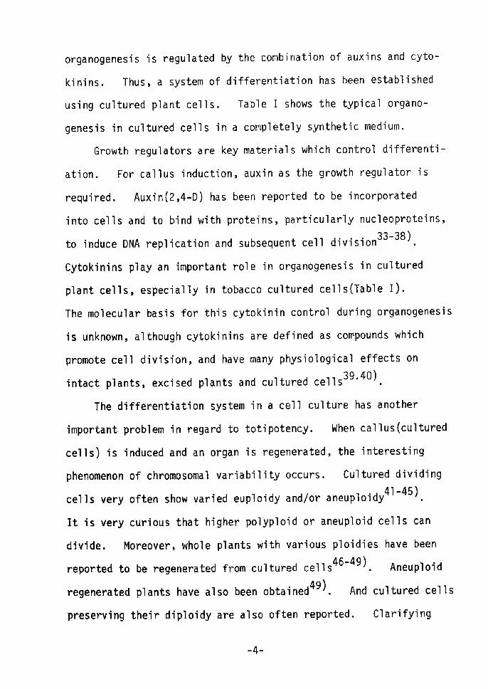
organogenesis fis regulated by the combination of auxins and cyto-
kinins. Thus, a system of differentiation has been established
using cultured plant cells. Table I shows the typical organo-
genesis in cultured cells in a completely synthetic medium.
Growth regulators are key materials which control differenti-
ation. For callus induction, auxin as the growth regulator is
required. AuxinÅq2,4-D) has been reported to be incorporated
into cells and to bind with proteins, particularly nucleopreteins,
to induce DNA replicatien and subsequent cen division33-38).
Cytokinins play an important role in organogenesis in cultured
plant cells, especially in tobacco cultured cells(Table I).
The molecular basis for this cytokinin control during organogenesis
is unknown, although cytokinins are defined as compounds which
promote cell division, and have many physiological effects onintact plants, excised plants and cultured cens39'40).
The differentiation system in a cell culture has another
important problem in regard to totipotency. When callus(cultured
cells) is induced and an organ is regenerated, the interesting
phenomenon of chromosornal variability occurs. Cultured dividingcells very often show varied euploidy andlor aneuploidy41-45).
It is very curious that higher polyploid or aneuploid cells can
divide. Moreover, whole plants with various ploidies have beenreported to be regenerated from cultured cells46-49). Aneuploid
regenerated plants have also been obtained49). And cultured cells
preserving their diploidy are also often reported. Clarifying
-4-
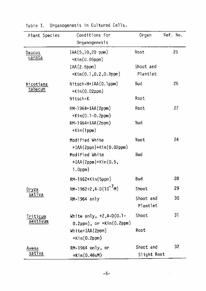
Table I. Organogenesis in Cultured Cells.
Plant Species Conditions for
Organogenesis
Daucus carota
Nicotiana tabacum
!orug sativa
Triticum aestivum
Avena sativa
IAA(5,1O,20 ppm)
+Kin(O.05ppm)
IAA(2,5ppm)
+Kin(e,1,O.2,O.3ppm)
Nitsch-H+IAA(e.lppm)
+Kin(O.02ppm)
Nitsch-K
RM-1 964+IAA(2ppm)
+Kin(O.1•-O.2ppm)
RM-1964+rAA(2ppm)
+Kin(1ppm)
Modified White +IAA(2ppm)+Kin(O.02ppmÅr
Modified White +IAA(2ppm)+Kin(O.5,
1.0ppm)
RM-1 962+Kfi n(5ppm )
RM-1 962+2 ,4-D (1 o-7MÅr
RM-1964 only
White only, +2,4-D(O.1- O.2ppm), or +Kin(O.2ppm)
White+1AA(2ppm)
+Kin(O.2ppm)
RM-1964 only, or +Kin(O,46uM)
-5-
Organ Ref. No.
Root
Shoot and
Plantlet
Bud
Root
Root
'Bud
Root
Bud
Bud
Shoot
Shoot and
Plantlet
Shoot
Root
Shoot and
Slight Root
25
26
27
24
28
29
30
31
32

eALSRg.!zgg!,!.E-aus
officina1isRM-1964 only Bud 33
these features of cultured cells is a most difficult problem,
but it represents one possible way for understanding the nature of
the regulation of differentiation.
Some changes in the metabolic patterns observed in dedifferen-
tiated cultured cells dre associated with the beginning of morpho-
genesis. These changes have been studied with the hope of deter-
mining the relationship between metabolic differentiation and
morphogenesis• changes in nucleic acid metabolism50-52), protein
metabolisrn5i'53-55), enzyme activity56'57), starch metaboiism58'59)
and aikaioid production60'61)have been reported in relation to
organogenesis. But there has been no inclusive biochemical and
molecular explanation of organogenesis. The following appears
to be the key to the most basic mechapism of organogenesis; cell
differentiation is based almost certainly on the regulation of
gene activity62). The regulation of gene activity invoives the
regulation of DNA replication and transcription etc. However, to
date very few studies from this point have been made in relation
63)to organogenesis in cell culture .
To understand organogenesis in cultured plant cell systems
the following four points must be clarified: 1) the properties of
cultured dedifferentiated cells; 2) the mode of action of growth
regulators; auxins and cytokinins; 3) the gene activation and in-
activation which directs differentiation; and 4) the biochemical
-6-

changes associated with morphological development.
The purpose of the present study was to obtain information
on the biochemical and molecular processes of organogenesis in a
cultured tobacco cell system and on the properties of cultured
cells, themselves. First the systern of shoot redifferentiation
by cytokinins was established for cultured tobacco cells. Then
the relationships of the structure of cytokinins to shoot formation
and the metabolism of 14c-labeled kinetin were described. The
following biochemical changes accompanying shoot redifferentiation
were studied: changes in protein synthesis and the properties of
RNA polymerase as one participant in transcription during shoot
redifferentiation. Based on the results of this investigation,
the mechanism of shoot redifferentiation by cytokinins in cultured
cells is discussed.
REFERENCES
1) Haberlandt, G,, Sitzben. Akad, Wiss. Wien Math-Naturwiss. Kl,,
111, 69 (1902) 2) Gautheret, R. J., Ann. Plant Physiol., fi., 433 (1955)
3) Carew, D, P. and Staba, E. J,, Lloydia, Zt,1 (l965)
4) Street, H. E. and Henshaw, G. G., in The Biology of Cells and
Tissues in Culture, vol. 3, ed. Willmer, E, N., p 459, Academic
Press (1966) 5År Street, H, E., in Plant Tissue and Cell Culture, ed. Street, H. E., p 1, Blackwell Scientific Publications (1973)
6) White, P. R,, Plant Physiol., 9, 585 (1934)
7) Gautheret, R. J., Compt. Rend., 198, 2195 (1934)
--7-

8)
9)
1O)
ll)
l2)
l3)
l4)
15)
16)
17)
18)
19)
20)
21)
22)
23)
24)
25)
26)
27)
28)
29)
30)
31)
32)
White, P. R., Am. J. Bot., 26, 59 (1939)
Nobe'court, P., Compt, Rend. Sci. Biol,, 130, 1270 (1939)
Gautheret, R. J., Compt. Rend., 208, 118 (1939)
White, P. R., Plant Physiol., 12, 803 (1937)
Gautheret, R. J., Compt. Rend., 205, 572 Åq1937)
Gautheret, R. J., Compt. Rend., 206, 125 (1938)
von Overbeek, J., Conklin, M. E. and Blakeslee, A. F.,
Science, 94, 350 (1941)
IVIuir, W. H,, Hildebrandt, A. C. and Riker, A. J,, Science, 119,
877 (1954)
Miller, C. O., Skoog, F., Okumura, F, S,, von Saltza, M. H.and Strong, F. M., J, Am. Chem. Soc., Lt, 1375 (1956)
Tulecke, W. and Nickell, L, G., Science, 130, 863 (1959)
See abstract of III Intl. Cong. Plant Tissue Cell Culture,
Leicester, 1974, etc,White, P. R., Bull. Torrey Bot. Club,, SStL, 507 (1939)
Gautheret, R. J., in Cell Differentiation and Morphogenesis.,ed. Beerman, W. M. et al, p 56, North Holland Publishing (1966)
Steward, F, C., Mapes, M. O. and Mears, K., Am. J. Bot., 45,
705 (1959)
Reinert, J., Ber. Dtsch. Bot. Ges., 71, 15 (1958)
Refinert, J., Planta, 53, 318 (1959)
Skoog, F. and Miller, C. O,, Symp. Soc. Exp. Biol., 11, 118
(1957)
Ibrahim, P, K., Can. J. Bot., 47, 825 (1969)
Nitsch, J, P. and Nitsch, C., Science, 163, 85 (1969År
Linsmaier, E. M. and Skoog, F., Physiol. Plant., Lt, 100 (1965)
Vasil, V. and Hildebrandt, A. C,, Planta, 75, 139 (1967)
Maeda, E., Proc. Crop. Sci. Soc. Japan, 37, 551 (1968)
Nishi, T., Yamada, Y. and Takahashi, E., Nature, 219, 508 (1968)
Shimada, T., Sasakuma, T. and Tsunewaki, K., Can. J. Genet.Cytol., 11, 294 Åq1969År
Carter, O. Y., Yamada, Y, and Takahashi, E., Nature, 214,
-8-

33)
34)
35)
36År
37)
38)
39)
40)
41)
42)
43)
44)
45)
46)
47)
48)
49)
50)
51)
1029 (1967)
Yamada, Y., Yasuda, T., Koge, M. and Sekiya, J., Colloques
internationaux C. N. R. S., 193, 137 (1971)
Yamada, Y., Yasuda, T., Koge, M. and Sekiya, J., Agr. Bio1.
Chem., 35, 99 (1971)
Yasuda, T. and Yamada, Y., Biochem. Biophys. Res, Commun., SÅítL,
649 (1970)
Yamada, Y. and Yasuda, T., Biochem, Biophys. Res. Commun., 43,
488 (1971)
Yasuda, T., Studies on the mechanism of callus induction
(dedifferentiation) by 2,4--dichlorophenoxyacetic acid., Ph. D.
Thesis, Kyoto Univ., 1Åqyoto (1971)
Yasuda, T., Yajima, Y. and Yarnada, Y., Plant Cell Physiol., Lt,
321 (1974)
Skoog, F. and Armstrong, D. J., Ann. Rev. Plant Physiol., 21,
359 (1970)Kende, H., Intl. Rev. Cytol., 31, 301 (1971)
Mitra, J., Mapes, M. O. and Steward, F. C., Am. J. Bot., 47,
357 (1960)
Fox, J. E., Physiol. Plant., 16, 793 (1963)
Torrey, J. G., Physiol. Plant., 20, 265 (1967)
Shimada, T. and Tabata, M., Japan J. Genet., St, 195 (1967)
Murashige, T. and Nakano, R., Am. J. Bot., 54, 963 (1967)
Nishiyama, I, and Taira, T., Japan J. Genet., 41, 357 (1966)
Tabata, M., Yamamoto, H. and Hiraoka, N., Japan J. Genet.,
43, 319 (l968)Nishi, T. and Mitsuoka, S., Japan J. Genet., 44, 341 (1969)
Sacristan, M. D. and Melchers, G., Mol. Gen. Genet., 105,
317 (1969)Yamada, Y., Matsumoto, H. and Takahashi, E., Soil Sci. Plant
Nut., 14, 35 (1968)Thorpe, T. A. and Murashige, T., Can. J. Bot., 48, 277 (1970)
-9--
52)
53)
54År
55)
56)
57)
58)
59)
60)
61)
62)
63)
Simard, A,, Can. J. Bot., fLEtL, 1541 (1971)
Butenko, R. G. and Volodarsky, A. D., Physiol. Veg., S., 299
(1968)Butenko, R. G., Colloques internationaux C. N. R, S., 193,
155 (1971)Matsushima, H., Wada, M. and Takeuchi, M., Bot. Mag. (Tokyo),
82, 417 (1969)DeJong, D, W., Jansen, E. F, and Olson, A. C., Exp. Cell Res.,
47, 138 (1967)Werner, D. and Gogolin, D., Planta, 9:1!.., 155 (1970)
Grant, M. E. and Fuller, K. W., J, Exp. Bot., Lt, 667 (1968)
Thorpe, T. A, and Murashige, T., Science, 160, 421 (1968)
Bhendary, R., Collin, H. A., Thomas, E. and Street, H. E., Ann.
Bot., 33, 647 (1969)Tabata, M., Yamamoto, H,, Hiraoka, N., Marumoto, Y. and
Konoshima, M., Phytochem., Lt, 723 (1971)Britten, R. J. and Davidson, E. H., Science, 165, 349 (1969)
Matsumoto, H., Gregor, D. and Reinert, J., Phytochem., L4,
41 (1975)
-1 O-
CHAPTER I
SHOOT REDIFFERENTIATrON FROM CULTURED TOBACCO
CELLS BY CYTOKININs IN vlTRol,2).
INTRODUCTION
Cultured plant cells(callus) are believed to have totipotency
and this is thought to be influenced by environmental conditions,
in particular by the presence of plant growth regulators, whfich
developes embryogenesis or organogenesis(organ formationÅr.
Experimental studies of organogenesis took a modest leap
forward during the 1950s as a result of the discovery of cytokinins.
skoog et a13) demonstrated that in cultured tobacco cells a high
,cytokinin!auxin ratio induced shoot forrnation and a low ratio
induced root formation. Others have also reported organ formation
from cultured cel1s(dedifferentiated cens)4,5).
Organ induction is divided into two types, and is mainly
dependent on the plant species. A low concentration of auxin or
its absence in a medium induces organ formation in such cultures
as rice cells6). cytokinins are required for organ formation with
3) or without auxins in such cultures as tobacco cells .
Some naturally occurring cytokinins and many synthetic ones
have been reported5År, since the synthetic kinetin was found to
possess cytokinin activity8'9). These cytokinins can promote
cell growth and induce organ formation from cultured cells as
described above.
-11-
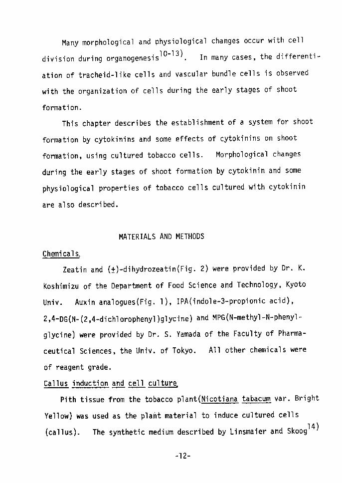
Many morphological and physiological changes occur with cell
division during organogenesislO'"i3). in many cases, the differenti-
ation of tracheid-like cells and vascular bundle cells is observed
with the organization of cells during the early stages of shoot
fonmation.
This chapter describes the establishment of a system for shoot
formation by cytokinins and some effects of cytokinins on shoot
forrnation, using cultured tobacco cells, Morphological changes
during the early stages of shoot formation by cytokinin and some
physiological properties of tobacco cells cultured with cytokinin
are also described.
MATERIALS AND METHODS
Chemicals.
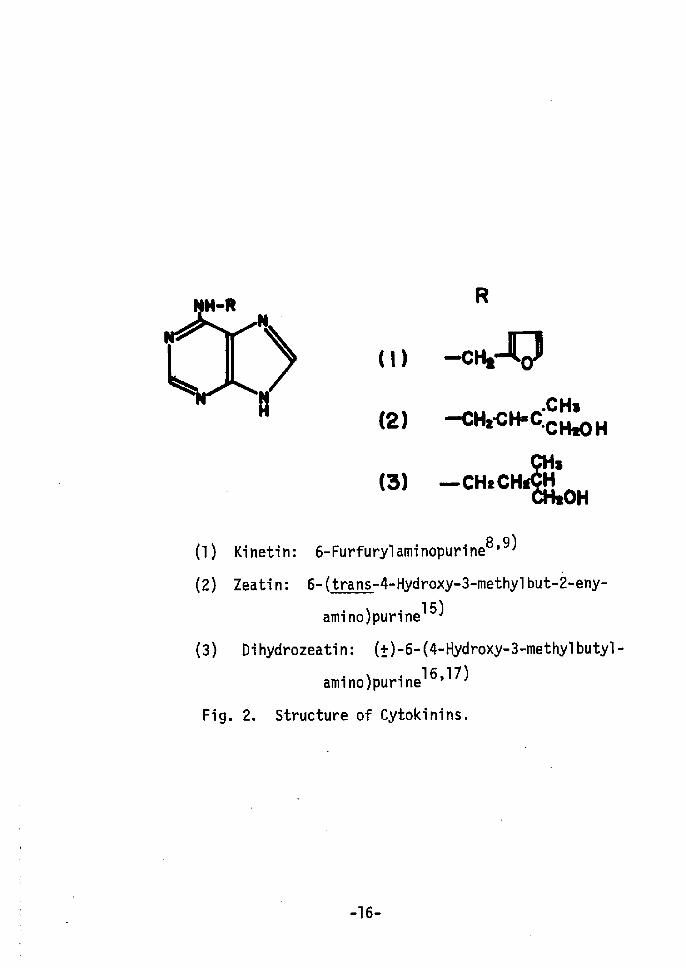
Zeatin and (Å})-dihydrozeatin(Fig. 2) were provided by Dr. K.
Koshimizu of the Department of Food Science and Technology, Kyoto
Univ. Auxin analogues(Fig. 1), IPA(indole-3-propionic acid),
2,4-DG(N-(2,4-dichlorophenyl)glycineÅr and MPG(N-methyl•-N-phenyl-
glycine) were provided by Dr. S. Yamada of the Faculty of Pharma-
ceutical Sciences, the Univ. of Tokyo. All other chemicals were
of reagent grade.
cal1us induction and cel1 guu !iy!zg,1ture
Pith tissue from the tobacco plant(Nicotiana tabacum var. Bright
Yellow) was used as the plaht material to induce cultured cells
14)(callus). The synthetic medium described by Linsmaier and Skoog
--12-
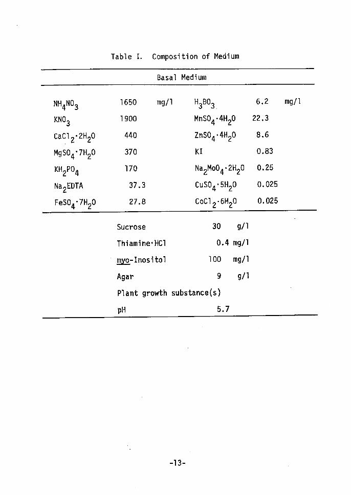
Table I. Composition of Medium
Basal Medium
NH4N03
KN03
caCl2•2H20
MgS04•7H20
KH2P04
Na2EDTA
FeS04•7H20
1650
1900
440
370
170
37.3
27.8
mgll H3B03.
MnS04•4H20
ZnS04•4H20
KI
Na2Mo04•2H20
cuS04•5H20
CoCl2•6H20
6
22
8
o
o
o
o
.
-
.
.
2
3
6
83
25
025
025
mg!1
Sucrose
Thiamine.HCI
!tllxg:- I nosi to1
Agar
Plant growth
pH
30
O.4
1OO
9
substance(s)
5.7
gll
mg/1
mg/1
g/1
-13-

was used as the basal medium in the following experiments(Table I).
For the suspension culture t the same mediun with agar omitted was
used.
Segments of sterilized tobacco pith were inoculated on the agar
medium with 10-5 M2t 4-D. The resulting callus (cultured cells)
was subcultured on the agar medium with 10-6 M2 t 4-D. Culture
conditions were 25-28°C and darkness. The cell line obtained in
1968 was designated as strain T5. Other cultured tobacco cells
were similarly induced and subcultured, using the various auxins
shown in Fig. 1.
To investigate shoot formation(redifferentiation) from cultured
cells(dedifferentiated cells), cells cultured with auxins were
transferred to an agar medium containing cytokinins (Fig. 2), then
cultured at 25-28°C in the dark. Shoot formation was measured
after 50-70 days of culture.
These cultured cells were designated; 2,4-0 cells, IBA cells,
zeatin cells, kinetin cells etc.
Microscopic observation.
2,4-0 cells and zeatin cells in static cultures were fixed with
acetic acid:alcohol (1 :3) for 3 hr. After fixation cells were
dipped in polyethyleneg1yco1 #1500 (50°C) for 1 hr, then in a
mixture of polyethyleneglycol #1500 and #4000 (1:1, v/v) for 1 hr.
These dehydrated cells were embedded in a mixture of polyethylene
glycol #1500 and #4000 (1:1) and sectioned into 20-25 u slices.
Each section was placed on a slideglass and the polyethyleneg1ycol
-14--

R
H
R (1)
(2)
(3)
-CH2COOH-- (C H2) 2 COOH
-(CH2)3COOH
Cl
OCH2CooH
Cl
cl
NHCH2CooH
Cl
N(CH3)CH2 COOH
(4) (5) (6)
Fig. 1.
(1År Indole-3-acetic acid (IM)
(2) Indole-3-propionic acid (!PA)
(3) Indole-3-butyric acid (IBA) (4) 2,4-Dichlorophenoxyacetic acid (2,4-D)
(5) N-(2,4-DichlorophenylÅrglycine (2,4-DG)
(6) N-Methyl-N-phenylglyci'ne (MPG)
Structures of the Auxins and Their Analogues.
Abbrebiations are in parentheses.
-15-
RH-R
) (t) -cHt-fi5J
N (2) iKH,•CHsCIcCII:'oH
CHs (3) .,.cH2CHdZlll,oH
(1) Kinetin: 6-Furfurylaminopurine8'9)
(2År Zeatin: 6-(trans-4-Hydroxy-3-methylbut•-2-eny-
amino)purine15)
(3) Dihydrozeatin: (Å}År-6-(4-Hydroxy-3-methylbutyl-
amino)purinel6,17)
Fig. 2. Structure of Cytokinins.
-16-

was removed with water. The section covered with glass served as
the preparation for microscopic observation. To detect lignified
cells, 5 % phloroglucin in 95 % ethanol was dropped on one of these
sections, followed by conc, HCI. Lignin was stained purple by this
method .
Ribonuc1ease RNase gs:t!!.!.Y.!!IM.t .
2,4-D cells and kinetin cells were cultured in suspensions,
Cells were homogenized in a chilled mortar with O.05 M phosphate
buffer, pH 6.5, containing Polyclar AT and 1 % Na-ascorbate. The
homogenate was filtered through gauze and the filtrate centrffuged
at 10,OOO g for 20 min. The supernatant obtained was dialyzed by
Sephadex G-25 gel filtration, The eluate was used as the enzyme
solution. The reaction mixture contained O.1 M acetate buffer,
4 mg of purified yeast RNA and 2 ml of the enzyme solution (200 ug
as protein) in a final volume of 4 ml(pH 5,5). After incubation
for 1 hr at 350C, the reaction was stopped by adding 1 ml of 25 %
trichloroacetic acid(TCA) containing uranium acetate, After cool-
ing it in an ice bath, the supernatant was separated by centri-
fugation and the decornposed products were determined from the
absorbance at 260 nm. A non-incubated mixture undergoing the
above procedures served as the reference. One enzyme unit was
defined as the enzyme activity causing changes in the absorbance
at 260 nm of 1 for 1 hr,
-17-
RESULTS AND DISCUSSION
(1) Conditions for shoot forrnation from cultured tobacco cells ib
Åíx!t!g!sL!!!nynE:.k
Table II shows the conditions for shoot formation from
-6cultured tobacco cells. Tobacco cells cultured with 10 M 2,4-D
(2,4-D cells) were transferred to an agar media containing a combi-
nation of 2,4-D and zeatin. Shoot formation was observed 6-8 weeks
after inoculation at a concentration of sxlo-5 M zeatin in the
presence of a low concentration of 2,4-D (o - lo-6 M), while no
shoot formation was found in the presence of lo-5 M 2,4-D(Fig. 3). As
the concentration of 2,4-D decreased, increased shoot formation
was observed. In contrast, cell proliferation occurred in the
presence of both 2,4-D and zeatin. A combination of a high concent-
ration of 2,4-D (lo-5 - lo"6 M) and a low concentration of zeatin
Åqo - lo-7 MÅr gave particularly good cell proliferation. At a
high concentration of zeatin, cell proliferation was repressed.
Consequently, it is presumed that, when shoot formation occurs, cell
proliferation is reduced. skoog et a13) reported that o.o2, O.2,
and O.5 mgll of kinetin with 2 mgll of IAA induced root formation,
cell proliferation and shoot formation, respectively. Othershave aiso reported similar resuitsi4'18). in the strain Ts
cell line which required only 2,4-D for cell growth, shoot formaton
could be induced and developed with zeatinÅq5xlO"-5 M). The presence
of 2,4-D had an inhibitory effect on shoot formation, thus no exo-
genous 2,4-D was required.
-18-

B
.
Fig. 3. Redifferentiated Shoots from Cultured Tobacco Cells.(A): Dedifferentiated 2,4-D cells (left),
(right).
(B): Subcultured redifferentiated-shoots.
redifferentiated shoots
-1 9-
i

Table II.
Formation
Effects of
in Cultured
2,4-D and Zeatin
Tobacco Cel1s.
on Cell Growth and Shoot
Concn.
Zeatin
of,(M)
Concentration of 2 4-D M -510 -610 10
d7 d810 o
o
-1O5xl O
-95xl O
-85xl O
-7 10
-75xl O
-6 10
-65xl O
-55xlO
•H-•t-
+++
--l-+
++
+-H-
+- - +
-++--+ ++
+- - - -s++
++
++
--
-+ - - -s-
----++
++
- +
s-
++
-+•H-i-
++
++
++
-
-
S+e
Number ef
indicates
+ indicates
the degree
the degree of cell
of shoot formation.
growth. Numberof s+
-20-
The zeatin requirements shown in Table III indicate that short-
term treatment with zeatin does not induce shoot formation.
Therefore, zeatin(cytokininÅr is required not only for triggering
redifferentiation, but also for the development of the shoot during
the culture period.
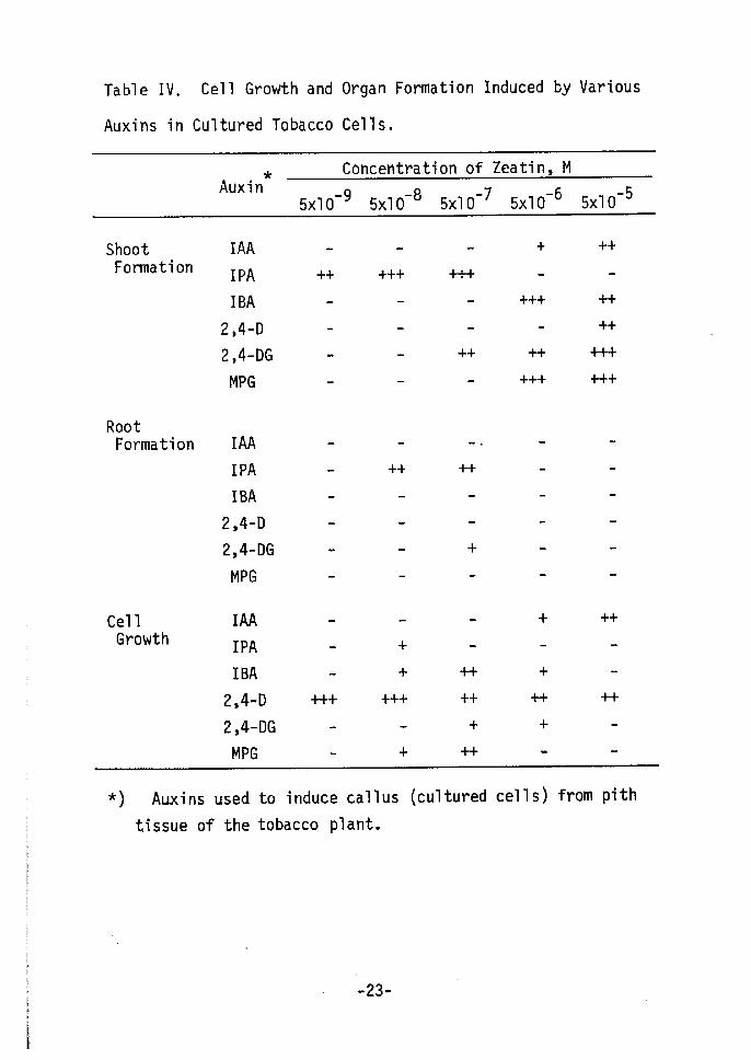
Table IV shows the effects of callus-inducing auxins on organ
formation. Calluses were induced from tobacco pith by various
auxins and their analogues(lo-5 M) as shown in Fig. 1. These were
transferred to an agar media, containing the indicated concent-
ration of zeatin (no auxins) to test organ forTnation. The shoot-
forming potency of cultured cells varied according to the auxin
used to induce callus. IPA cells could easily redifferentiate
shoots at a low concentration of zeatin (sxlo-9 - sxlo-7 M). IBA,
2,4-DG and MPG cells also easily redifferentiated shoots. While
IPA and the 2,4-DG cells showed only slight cell proliferation, IBA
cells proliferated and could be subcultured for years. Among
the tested cells, the 2,4-D ones showed the greatest cell prolifer-
ation over a wide range of 2,4-D concentrations and shoots were
induced at 5xlO-5 M of zeatin. IAA cells had low potencies both
for cell growth and shoot formation. Only root formation was found
in IPA cell and 2,4-DG cells in these systems. Results shown in
Table IV suggest that the degree of dedifferentiation varies
according to the auxin used and that this reflects the potency for
redifferentiating shoots a$ the reverse phenomenon of dedifferenti-
ation,
-21-
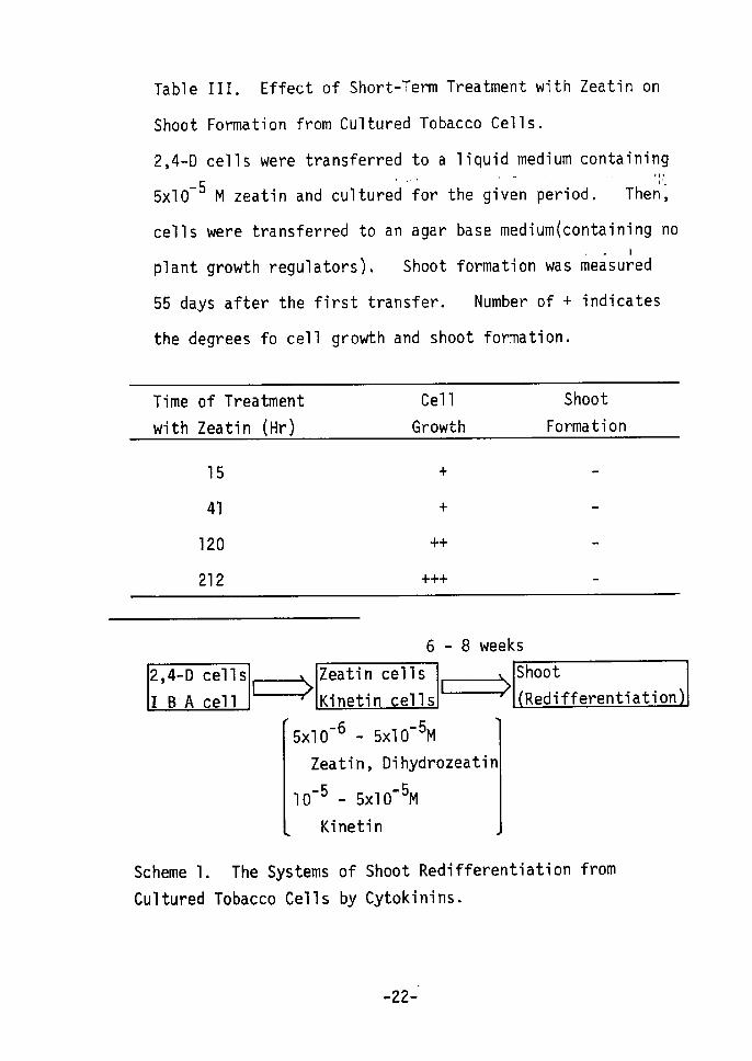
Table III. Effect of Short-Term Treatment with Zeatin on
Shoot ForTnation from Cultured Tobacco Cells,
2,4-•D cells were transf'erred to a liqui,d.med,ium containill,g
sxlo-5 M zeatin and cultured ior the given perfiod. ThenL';
cells were transferred to an agar base medium(containing no
.bplant growth regulators), Shoot formation was measured
55 days after the first transfer. Number of + indicates
the degrees fo cell growth and shoot formation.
Time
wi th
of TreatmentZeatin CHr)
Cel1
Growth
Shoot
Formation
15
41
120
212
+
+
++
+++
2,4-bD
IBAcel1s
cel1 EÅr
Scheme 1.
Cultured
The Systems
Tobacco
6
Zeatin cel1s Kinetin cel1s
--5 -6 - 5xlO M 5xlO
Zeatin, Dihydrozeatin
-5 -5 - 5xlO M 10
Kinetin
of Shoot
Cells by Cytokinins.
- 8 weeks
Shoot[=År Redifferentiation
Redifferentiation from
•-22-'
Il
ii
Ta bl e
Auxins
IV.
in
Cell Growth and
Cultured Tobacco
Organ Formation
Cel1s.
Induced by Various
*Concentration of Zeatin, M
Auxin -95Å~1O -7 --8 5xl O5xlO 5Å~1O -5-6 5xlO
Shoot Formation
Root Formation
Cel1 Growth
1AA
IPA
IBA
2,4-D
2,4-DG
MPG
IM IPA
IBA
2,4-D
2,4•-DG
MPG
IAA
IPA
IBA
2,4-D
2,4-DG
MPG
-
-+
+++
++
+
+
+++
+
+-
++
++
+
-++
+
++
+
+++
++
+++
+
+
- +
++
++
++
-++++
++
.--
++
*) Auxinstissue of
used
the
to induce callus
tobacco plant.
-23-
(cul tured cel1s) from pith
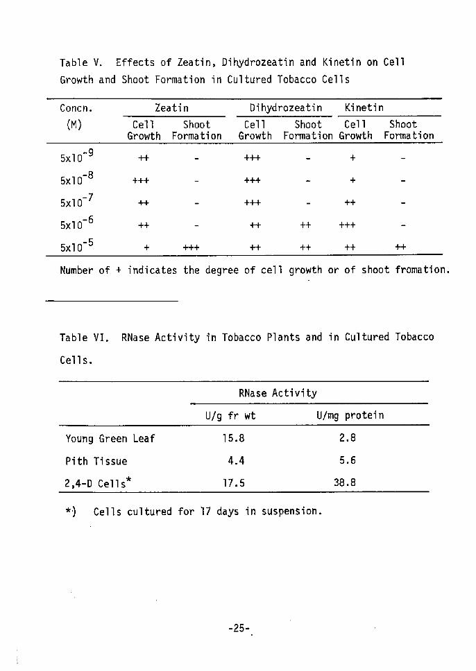
Table V shows that dihydrozeatin (Fig. 2) had activity for
producing shoot formation from 2,4-D cells, as did zeatin. The
naturally occurring cytokinins, dihydrozeatin and zeatin, were
more active than the synthetic kinetin for shoot formation. These
cytokinins also enhanced cell growth in 2,4-D cells at low concent--
rations. Concentrations needed for shoot formation and cell growth
-6 -8 M of zeatin and dihydrozeatin and 5xlO M kinetindiffered: 5xlO
for maximum cell growth, and 5xlO-5 M of zeatin, dihydrozeatin and
kinetin for shoot formaiton. Cell growth and shoot formation seem
to be opposite effects of cytokinins in cultured tobacco cells.
Due to the results in Tables II, IV and V the following culture
systems were used for shoot redifferentiation(Scheme 1). Sub-
cultured 2,4-D cellsÅqdedifferentiated cells) were transferred to a
medium with lo-5 - sxlo'5 M of zeatin or lo'5 M kinetin and were
cultured in static or suspension cultures. In some experiments
IBA cells were used. 2,4-D cells or IBA cells served as dediffer-
entiated control cells,
(2) Mor holo ical and h siolo ical ush st!u!ztag the sw1
E:t!i9El9IÅ} S2:f[. shoot formation.
Growth curves shown in Fig, 4 indicate that over a period of
20 days the 2,4-D cells increased more in fresh weight than did
zeatin cells ,in a static culture. In zeatin cells, the growth rate
was slower, reaching a plateau 15 days after inoculation, whereas
the 2,4-D cells grew logarithmically for more than 20 days, A
-24-
i
Table
Growth
v.
and
Effects
Shoot
of Zeatin
Formation'
.In
Dihydrozeatin and
Cultured Tobacco
Kinetin on
Cel1s
Cel1
Concn.
(M)
Zeatin
Cell ShootGrowth Formation
Dihydrozeatin
Cell ShootGrowth Formation
Kinetin
Cel1Growth
ShootFormation
5xlO
5xlO
5xlO
5xlO
5xlO
-9
-8
-7
-6
-5
++
+++
++
++
+ +++
+++
+-+++
++
++
++
++
+
+
++
+++
++ ++
Number of + indicates the
Table VI.
Cel1s.
RNaseActivity
degree of
in Tobacco
cell growth
Plants and
or
ln
of shoot
Cul tured
fromation.
Tobacco
RNase Activity
U/g fr wt Ulmg protein
Young
Pi th
2,4-D
Green
Tissue
t Cel1s
Leaf 15.8
4.4
17.5
2
5
38
.
.
.
8
6
8
*, ) Cel1s cu1tured for 17 days in suspension.
-25-

A thE -P=en'rw3=caÅë
sLvao"
4
3
2
Days
Fig. 4. Growth Curves for Tobacco Cells Cultured on a Mediumwith 1o'6 M 2,4-D (-e-) and sxlo-5 M zeatin(-o-).
About 50 mg of cells was .inoculated in a test tube containing
10 ml of agar medium. Fresh weight was measured at specified
intervals. The average fresh weight of 3 replicates is given
in the figure.
-26-

Åë
"rdct
=Psose
2.5
2.0
1.5
1.0 o
Days
4 5
Fig. 5. Growth Rates of Tobacco Cells Cultured with 2,4-D or
Kinetin in Suspension.Cell growth is represented as the growth rate of the initial
inoculum as seen from the turbidity at 610 nm. (-e-) shows
the growth rate of 2,4-D cells; (-O-), of kinetin cells.
;
I
ii
-27-
similar phenomenon was observed in the growth of 2,4-D cells and
kinetin cells cultured in suspension(Fig. 5). However, 3 days
after inoculation a reduced growth rate was found for kinetin cells.
In many cases, an additional low concentration of cytokinin with
auxin promotes cell growth19). In contrast, Fig. 4 shows that
a high concentration(sxlo-5 M) of zeatin inhibits callus growth.
Nudel e:t!L gJl.20) also reported inhibitory effects of a high concent-
ration of kinetin on growth, as well as on the synthetic activity
of other components in tobacco cells.
Ratios of dry to fresh weights also changed a few days after
inoculation(Fig. 6). 2,4-D cells showed a temporary increase in
dry material which later decreased to a constant level (about 2.0 -
2.5 % dry materials, Fig. 6-A). rn contrast, zeatin cells had 4 -
5 % dry materials. This indicates that cell components in zeatin
cells are enriched. The tirnes of the beginning of the changes
in dry materials were consistent with those of the changes in the
growth rates in Fig. 4.
As shown in Fig. 7, organization was found in cells cultured
for 15 days with zeatin, while 2,4-D cells were amorphous and
difficult to prepare in sections. Organized compact cells of
small size were found 10 days after inoculation in the zeatin
culture. Simultaneously, the lignified cells and tracheid-like
cells shown in Figs. 7 and 8, were observed in these cells. These
type$ of cells were seen 6 days after inoculation, and the number
of these cells, as well as lignification,increased during culture•
-28.-

ARv '"3LLx"shLa
6
4
2
oo
(AÅr
o coo Zeatin
o
2,4-D
Cel1s
Cel1s
o
Days
A? 2.0''"'
ny.s"t:' s'
NV
-1.5o"
'p'
E
1.0
o 10 20Days30
Fig.
the
Dry
was
6. Changes in Dry Weight
Ratio for Zeatin Cells as
weight was measured after
dried at 1000C for 3 hr.
to Fresh
Compared
about 400
Weight
to 2,4-D
mg (fr
Percents (A) and
Cells (B).
wt) of the cells
t
ik
-29-
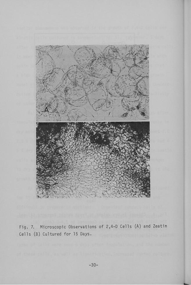
Fig.
Cel1s
7.
xei'
År
)Il'?1
yvedee
Y-IC-r.
(.B)
ic(fij
7t$
Microscopic Observations
Cultured for 15 Days,
-30d
,sx
,I.l.lge
.-"
""ibeg6;,t
ll$;.24"LSt:L x .
IiÅql-"e:?tt9,,",,x•lr.
i.. ;' i. K:-" S
of 2,4--D Cel1s CA) and Zeatin
l
[
i
1
il
lI!
}
l
l

'it
//
l
t
!
'
t
,
T
'
t
l-
4
7 K'-
c
.Y t? ":" FA:?-s)p'
h.f-.u 1
Åqt',
1
'..rT7"
-.h-
t'et" tz ti
"-- .T ,-e Le.' t' `"'
'i-t
J
.t-jLV '
.' r., g•N NN
t.J.l,'ijdt;::gS
-et"- ge
g. Yxl-
.(rh-dN
1
'ASk ' N -".'F"S'-
" .l:.. .
•lgJÅriEc':
N
L"-x
twk
.-.v-
--- 2---- .; "'Vf ect.
{-
g
.--
s.L•K.-,..
vS . I) Lx '" ,
s- vf
li
"
N{t
s
'
:
'
:
l
i
l
,
it
'
'
ttL
t'
'
i
l
"
:[iPi
Fig. 8.
Zeatin
Microsco
Cel1s
.PlC
Cultured
Observations of
for 15 Days.
Tracheid--Li ke Cel1s .In
'
ti
'
gIll
ll
-31 -
Cell organization in the early stage seemed to occur first around
the tracheid-like cells. However, these cells could not develop
into vascular bundleslO). In 2,4-D cells no tracheid-like cells
were found. rn some reports on tobacco 21) and the soybean22)
kinetin was also shown to promote tracheid-like cell production.
In surmary, tracheid-like cells first appeared 6 days after inocu-
lation and subsequently cell organization was observed in zeatin
cells during the early stages of shoot formation. The beginning
of morphological change is consistent with that of changes in cell
growth .
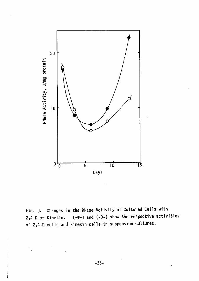
As one physiological change, changes in RNase activi ty were
studied. Cultured cells(2,4-D cells) showed high RNase activity
compared to the original plant tissue, in particular pith tissue
(Table VI). Changes in the RNase activities of 2,4-D and kinetin
cells are shown in Fig. 9. A few days after inoculation, RNase
activities in both cell types decreased to 50 % of the initial
activity. Then a rapid increase was found in 2,4•-D cells, while
only a gradual one occurred in kinetin cells. Dedifferentiated
2,4-D cells show much higher RNase activity than does the original
pith tissue. However, kinetin cells (redifferentiation cells)
show lower activity than 2,4-D cells. During the senescence of
excised leaves increased RNase activity is repressed by kinetin
treatment23), which repressed the progress of senescence24).
At the onset of shoot formation early physiological and
morphological changes have already occurred a few days after inocu-
-32o
=-r-opoLamE-x= Ah--l-År
'r-
-v(Åë
mnt
Zct
20
10
o
Days
Fig. 9.
2,4-LD, or
of 2,4-D
Changes in
Kinetin.
cells and
the RNase Activity of Cultured Cells
C-e-) and C-O-) show the respective
kinetin cells in suspension cultures.
with
activitie$
L
'r
E•- 33-

lation. These changes were not found in 2,4-D cells.
SUMMARY
Shoot redifferentiation in tobecco cells cultured with auxins
(dedifferentiated cells) was examined. Shoot formation could by
induced by the continuous presence of exogenous cytokfinin(sxlo-5 M
zeatin, dihydrozeatin and kinetin) as plant growth regulator.
The presence of 2,4-D had an inhibitory effect on shoot formation.
The concentrations for shoot formation and cell growth differed.
The cytokinin concentration causing shoot formation was higher than
that giving maximum cell growth. Zeatin and dihydrozeatfin showed
higher activities for shoot forTnation than did kinetin. Cultured
cells induced by various auxins showed different degrees of potency
for shoot formation by zeatin.
Morphological changes and changes in cell growth, the amount
of dry materials and RNase activity were found about 1 week after
transfer to the cytokinin medium. The onset of shoot redifferenti-
ation is believed to occur a few days after transfer to the cyto-
kinin medium and physiological and morphological changes to continue,
REFERENCES
1) Yamada, Y., Kiso, K,, Sekiya, J. and Yasuda, T., Agr. Biol.
Chem., gLt, lo55 (1971)
2) Sekiya, J,.and Yamada, Y., Bull. Inst. Chem. Res., iÅqyoto Univ.,
!2;t, 246 (1974)
-34•-
3) Skoog, F. and Miller, C. O., Symp. Soc. Exp. Biol., L/,118
(1957)
4) Halperin, W., Ann. Rev. Plant Physiol., Lt, 395 Åq1969)
5) Niizeki, H. and Ohno, K., Recent Advances in Plant Breeding,
(Japanese), 11, 68 (1970År
6) Nishi, T., Yamada, Y. and Takahashi, E., Nature, 219, 508 (1968)
7) Skoog, F. and Armstrong, D. J., Ann. Rev. Plant Physiol., gr, 359 (1970)
8) Miller, C, O., Skoog, F., von Saltze, M. H. and Strong, F, M,, J. Am. Chem. Soc., 77, 1392 (1955År
9) Miller, C. O., Skoog, F., Okumura, F. S., von Saltze, M, H. and
Strong, F. M., J. Am, Chem. Soc., 78, 1375 Åq1956)
10) Gautheret, R. J., in Cell Differentiation and Morphogenesis, ed.
Beerman, W. et al, p 55, North-Holland Publishing, Arnsterdam
(1 966)
11) Halperin, W., Ann. Rev. Plant Physiol., Lt, 395 (1969År
12År Reinert, J., in Plant Tissue and Cell Culture, ed. Street, H. E.,
p 338, Blackwell Scientific Publications, (1973)
13) Halperin, W., Can, J. Bot., 51, 1801 (1973)
14) Linsmaier, E. M. and Skoog, F,, Physiol. Plant., Lt, 100 (1965)
15) Letham, D, S., Proc. Chem. Soc., 230 (1964)
16) Koshimizu, K., Kusaki, T., Mitsui, T. and Matsubara, S., Tetrahedron Letters, 1317 (1967)
17) Koshimizu, K., Matsubara, S. and Mitsui, T., Agr. Biol. Chem.,
31, 795 (1967)18) Vasil, V. and Hildebrandt, A. C., Planta, Lt,139 (1967)
19) Helgeson, J. P., Kruger, S. M. and Upper, C. D., Plant Physiol.,
44, 193 (1969)20) Nudel, U. and Bamberger, E. S., Plant Physiol., !!Zt., 400 (1971)
21) Bergman, L., Planta, SL2, 221 (1964)
22) Fosket, D, E. and Torrey, J. G,, Plant Physiol., !!!4L, 871 (1969)
23) Dove, L. D,, Phytochem., Lt, 2561 (1973)24) Atkin, R. K. and Srivastava, B. I. S. Physiol. Plant.,g22., 742(1969)
-35-
CHAPTER II
SHOOT FORMATION FROM CULTURED TOBACCO CELLS BY OPTICALLY ACTIVE CYTOKININsl).
INTRODUCTION After the synthetic compound kinetin2'3) was found to possess
cytokinin activity, the naturally occurring cytokinins zeatin4) and
(-)-dihydrozeatin5'6) were isolated from Zea g!g,ys. and LtyRLn!tLs. 1uteus,
respectively. These compounds promote cell division in cultured
tobacco cells, Kinetin also induces organ formation in cultured
cells but it is not known whether the naturally occurring cyto-
kinins affect organogenesis in cultured cell system.
Dihydrozeatin is optically active, although kinetin is not;
of the two possible enantiomers, one mirror image may be more active
than the other. Unfortunately, both optically active dihydrozeatins
are not available for study. Therefore, optically active N6-sub-
stituted adenines5) with asymmetric carbons adjacent to exocyclic
nitrogen atoms(Table I) were tested for their ability to induce
organ development in cultured tobacco cells.
TVtATERIALS AND METHODS
Dihydrozeatin and optically active cytokinins were provided
by Dr. K. Koshimizu, the Department of Food Science and Technology,
kyoto Univ. Tobacco(Nicotiana tabacum var. Bright Yellow) cultured
-36-

cells were induced from pith tissue with a concentration of lo-5 M
of indole-3•-butyric acid(rBA) in Linsmaier and Skoog medium8).
About 40 days after callus induction, this IBA cells were used in
the test of zeatin and (Å})-dihydrozeatin and ln the test of optically
active cytokinins. Shoot formation was observed 28 days after
inoculation of cells on Linsmaier and Skoog media containing each
optically active cytokinins, and 53 day$ after inoculation of cells
with zeatin and (Å})-dihydrozeatin,
RESULTS AND DISCUSSION
Letham9) tested the growth promoting ability of zeatin and
kinetin in a number of axenic systems. In all systems zeatin
was considerably more effective than kinetin. Table II shows the
ability of zeatin and (Å})-dihydrozeatin to promote cell growth and
to ' induce shoot formation in vitro from cultured tobacco cells.
For both growth promotion and organ redifferentiation) zeatin
and (Å})•-dihydrozeatin was equally active. Hence zeatin and (Å})-
dihydrozeatin, like kinetin, have the ability to induce bud form-
'ation from cultured tobacco cells.
The abilities of optically active N6-substituted adenines
to induce shoot formation and to promote cell growth are compared
fin Table III. Both R-(-)-6-(nt-methyl-ct•-phenylmethylamino)purine
(R-PE) and S-(-)-6-(d-hydroxymethyl-tS-phenylethylamino)purine(S-PA)
induced shoot formation. R•-PE and S-PA were active in inducing
shoot formation at concentration of 4xlod5 M and 2xlo'4 M, respec-
--37-
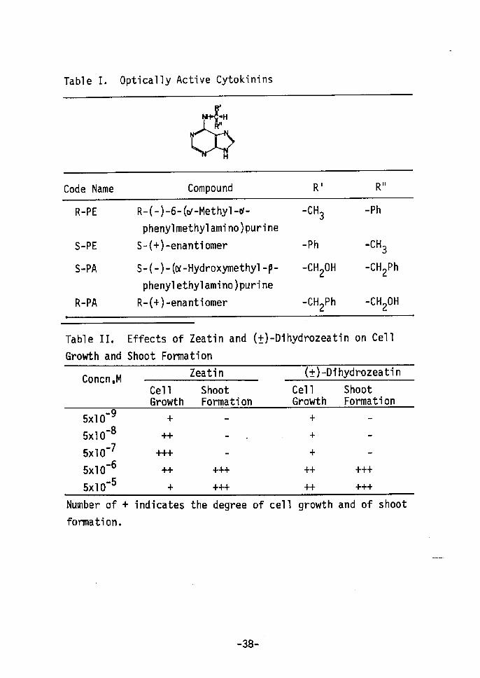
Table I. Optica11y Active Cytokinins
t)H•S.lH
,Åq)]iillHÅr
Code Narne Compound R' Rii
R-PE
S-PE
S-tPA
R-PA
R-(-)-6-(c,t-Methy1-or-
phenylmethylamino)purine
S-Åq+)-enantiomer
S-(-)- (or-Hydroxymethyl -P-
phenylethylamino)purine
R-C+)-enantiomer
-cH3
-Ph
-CH20H
-CH2Ph
-Ph
.cH3
-CH2Ph
-CH20H
Tabl e
Growth
II,
and
Effects of Zeatin
Shoot Formation
and (Å})-Dihydrozeatin on Cel1
Concn ,MZeatin (Å}År-Dihydrozeatin
Cel1Growth
ShootFormation
Cel1Growth
ShootFormation
•• 95xlO -85xlO -75xlO -65xlO -55xlO
+'i"t"
-+-
++-+++
+
+
+
--
+++
+-Number of +
formation.
indicates the degree of cel1 growth and of shoot
-38-
e
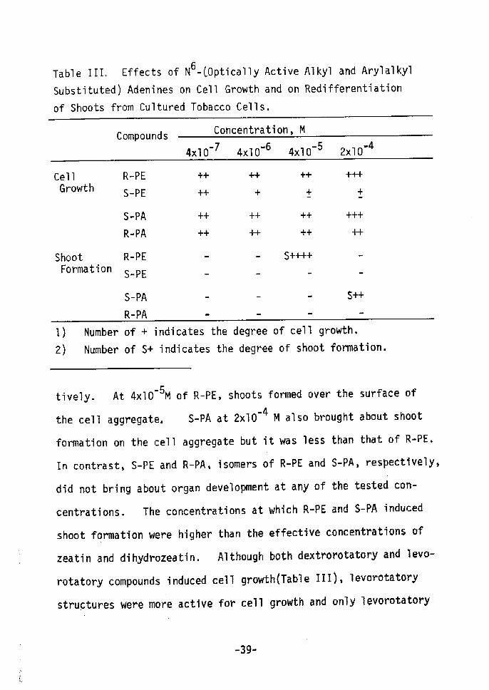
Table III, Effects of N6-COptically Active Alkyl and Arylalkyl
Substituted) Adenines on Cell Growth and on Redifferentiation
of Shoots from Cultured Tobacco Cells,
Compounds Concentration, M
-74xlO -64xlO -54xlO -42xlO
Cel1 Growth
Shoot Formation
R-PE
S-PE
S-PA
R-PA
R-PE
S-PE
S-PA
R-PA
++
++
++
++
-
++
+
++
++
++ Å}
++ ++
s++++
.
+++
+
+++
++
s-
I) Number of + indicates the degree of cell growth,2År Number of S+ indicates the degree of shoot forrnation.
tively. At 4xlO'5M of R-PE, shoots formed over the surface of
-4 M also brought about shoetthe cell aggregate, S-PA at 2xlO
f'ormation on the cell aggregate but it was less than that ot' R-PE,
In contrast, S-PE and R-PAs isomers of R-PE and S-PA, respectlvely,
did not bring about organ development at any of the tested cen-
centrations. The concentrations at which R-PE and S-PA induced
shoot formation were higher than the effective concentrations ef
zeatin and dihydrozeatin. Altheugh both dextroretatory and levo-
rotatery eompounds induced cell growthÅqTable ITIÅr, levoretatery
structures were more active fov cell growth and only levorotatery
-
-39-
ones brought about shoot formation. Essentially similar results
both for cell growth and shoot formation have been obtained by
Matsubara et allO). since configuration markedly influences the
ability to induce differentiation, binding of cytokinin N6group to
a receptor site is probably involved in organogenesis.
'
SUMMARY
(+)-Dihydrozeatin ttnd optically active cytokinins(asymmetric
carbon a to the exocyclic nitrogen) were tested for their ability
to induce development of shoots in cultured tobacco cells.
(Å})-Dihydrozeatin was active to induce shoot formation. The levo-
rota'tory compounds tested were active in inducing shoot formation
but the corresponding dextrorotatory compounds were inactive at
all concentrations tested. These findings suggest that the
group attached to the N6position of cytokinins binds to a stereo-
specific receptor site to brfing about shoot redifferentiation.
REFERENCES
1) Yamada, Y., Sekiya, J. dnd Koshimizu, K., Phytochem., L/,1019
(1972)
2) Miller, C. O., Skoog, F., von Saltze, M. H. and Strong, F. M.,
J. Am. Chem. Soc., 77, 1392 (1955)
3) Miller, C. O., Skoog, F., Okumura, F, S., von Saltze, M. He
and Strong, F. M., J. Am. Chem. Soc., 78, 1375 (1956)
4) Letham, D. S., Proc. Chem. Soc., 230 (1964)
5) Koshimizu, K., Kusaki, T., Mitsui, T. and Matsubara, S.,
Tetrahedron Letters, 1317 (1967År
-40-

6) Koshimizu, K., Matsubara, S and Mitsui, T,, Agr. Biol. Chem.,
31, 795 (l967)
7) Koshimizu, K., Kobayashi, A., Fujita, T. and Mitsui, T.,
Phytochem., Z., 1989 Åq1969)
8) Linsmaier, E. M. and Skoog, F., Physiol. Plant., L8, 100 (1965)
9) Letham, D. S., in Biochemistry and Physiology of Plant Growth
Substances; Proc. VI Intl. Conf. Plant Growth Substances, ed.
Wighrman, F. and Setterfield, G., p 19, Runge Press, Ottawa (1 968)
10) Matsubara, S., Koshimizu, K, and Fujii, T., in Plant Growth
Substances 1973; Proc. VII! rntl. Conf, Growth Subst'ances,
p 456, Hirokawa, Tokyo (1974)
-41 -
CHAPTER III
14 C rNCORPORATED INTO METABOLrSM OF KINETIN-8-
CULTURED TOBACCO CELLS DURING THE EARLY STAGES
OF SHOOT REDIFFERENTIATrON.
INTRODUCTION
In order to induce the shoot from cultured tobacco cells
(dedifferentiated cells), exogenous cytokinins are essentially
requisite,
During callus induction, 2,4-D incorporated into cells are
accumulated in free form and thereafter forms high molecular weight
complexes with proteins (but not with nucleic acids), particularly
with lysine-rich histone in pea nucleus presumably to bring about
cell divisionl,2),
Low molecular weight complexes are also formed for detoxinl).
After the hydrolysis of these complexes, the radioactivity is
recovered as 2,4-D.
Fox e:t; g21-3) presented the hypothesis that exogenous cytokinins
are incorporated into s-RNA and thereafter give the physiological
effects of plant cells3'4År, since the cytokinins are located in
s-RNA as one of minor base components5). But this hypothesis
was denied by Kende g:tL g.1!.6) and Hall et a17'8); Hall et al showed
that cytokinin, N6-to2-isopentenyl)adenine (2ipA), in t-RNA was
synthesized by the addition of A2-isopentenylpyrophosphate, derived
-42-

from mevalonate, to adenine residue in t-RNA, However, cytokinins
in t-RNA are not seem to be active forms since the occurrence in
t-RNA are found in wide range of microorganisms, animals and
plants. It seems to be one of the source of cytokinins.
On the other hand, cytokinins (zeatin and benzyladenine) are
metabolized to a number of metabolites, adenine, adenosine, AMP,
riboside and ribotide of cytokinin etc., after incorporated into
some kinds of plant cells9-16),
To date, it has not been clear what kinds of metabolites of
cytokinin are actually active forms to induce organogenesis in
cultured tobacco system. This chapter describes the incorporation
14 C during the early stages of shootand metabolism of kinetin-8-
redifferentiation in cultured tobacco cells.
TVtATERrALS AND METHODS
Cell culture and incor oration of kinetin-s-14c.
2,4-D cells in suspension culture were used. 2,4-D cells
were transferred into the medium containing lo-5 M kinetin and
o.2 uci of kinetin-s-14c Csp. act. Is mcilmmol, Radiochemical
Center, England) and cultured in suspension for given periods at
250C in the dark. 14Extraction of radioactive materials de"ived from kinetin-8- c
incor otated.
Cultured kinetin cells were harvested at 24 hr intervals and
washed with the basal medium. Five grams of each sample was
-43 --

homogenized with cold 80 % EtOH in a chilled mortar. The homoge-
nate was filtered through Whatman 3MM disc filter paper and the
residue on the filter paper was washed with cold 80 % EtOH. The
combined 80 % EtOH extract (80 % EtOH soluble materials) was filled
up to 100 ml and taken up 2 ml into a vial tube. After removing
the solvent (80 % EtOH) and pouring toluene scintillator, radio-
activity of 80 % EtOH soluble materials was determined with
Beckman LS-100 liquid scintillation spectrometer.
The residue on the t'ilter paper (80 % EtOH insoluble materialsÅr
was washed and dried with 100 % EtOH and ether. Radioactivity of
80 % EtOH insoluble materials was also determined with liquid
scintillation spectrometer.
!t!1!J([!9tigt sdiLggrs!s!gti!2nd t ptf:. 80 % EtOH insoluble materials.
The 80 % EtOH insoluble materials radiolabeled were prepared
from cells cultured with kinetin-s-14c for 24 or g6 hr. A 2o mg
of EtOH insoluble materials was incubated at 30eC for 60 min with
a following enzyme solution; 1) 1 mg of Pronase E (70,OOO PUKIg,
Kaken Kagaku Co.) in 2 ml of 50 mM Tris-HCI buffer (pH 7,8), 2År
1.5 mg of cellulase (type I from As er i1lus !t!:l.ger , Sigma Chemical
Co,) in 2 ml of 50 mM phosphate buffer, pH 6.5, 3) 1 mg of RNase
Ctype IA from bovine pancreas, Sigma Chemical CO.) in 2 ml of
50 mM phosphate buffer, pH 6.5. Control was run without enzyrne
in each buffer. Enzyme reaction was stopped by addition of O•5
ml of cold 20 % trichloroacetic acid(TCA). Reaction mixture was
filtered through Whatman 3MM disc filter paper and the paper was
-44-
washed with 5 O/o TCA, After the paper was washed and dried with
EtOH and ether, radioactivity remained on the disc paper as high
molecular weight components was determined with liquid scintillation
spectrometer.
ItlnglyEj-E.1 i of 80 % EtOH soluble materials.
Each 80 % EtOH extract (80 % EtOH soluble materials) was
concentrated to O.5 ml and 100 ul of concentrate was spotted on
Whatman 3MM cellulose paper. The paper was developed with
!L-butanol: acetic acid: water (4:1:1). After drying, the paper
was cut into 1 cm pieces and the radioactivity of each pieces was
determined with liquid scintillation spectrometer.
RESULTS AND DISCUSSION
14Incor oration of kinetin-8- C st!!,u .Å}ng. the !grJl.)L E]tEgggEe of shoot
redifferentiation.
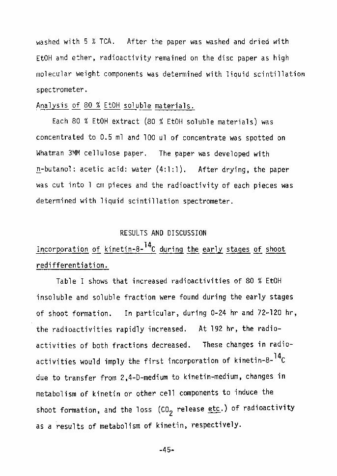
Table 1 shows that increased radioactivities of 80 % EtOH
insoluble and soluble fraction were found during the early stages
of shoot formation. In particular, during O-24 hr and 72-120 hr,
the radioactivities rapidly increased. At 192 hr, the radio-
activities of both fractions decreased. These changes in radio-
activities would imply the first incorporation of kinetin-s-14c
due to transfer from 2,4-D-medium to kinetin-medium, changes in
metabolism of kinetin or other cell components to induce the
shoot formation, and the loss (c02 release etc.) of radioactivity
as a ' results of metabolism of kinetin, respectively•
-45-

Table I.
Cel1s.
rncorporation of Kinetin-8-14C into Cultured Tobacco
Time(hr)
14Kinetin-8- c CPM/g.
Incorporatedfr. wt.
80 % EtOH lnsol. 80 % EtOH Sol. Total
24
48
72
96
120
192
1,443
1,995
2,974
5,024
5,086
4,038
9,464
12,166
12,646
17,304
20,866
14,976
1O,
14,
15,
22,
25,
19,
907
161
620
328
952
Ol4
Table rr. Effects oi Enzyrnes on 80 % EtOH Insoluble Materials.
Radioactivity Remained, CPMTreatment pH 24 Hrl) 96 Hr2)
Control
Pronase E
Control
RNase
Cel1ulase
7.
7.
6.
6.
6.
8
8
5
5
5
1093
1060
1250
709
1320
(1OO)
(97)
(1 OO)
(57)
(1 06År
1763
1257
2122
953
2569
(100)
(71)
(1OO)
(45)
(121)
1)
2)
Culture time with
For experimental
14 kinetin-8-
details, see
Ci
in nMethods".
-46-
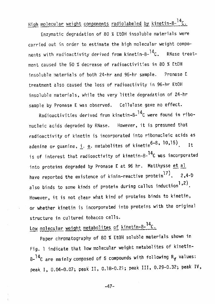
EtÅ}ELb.h mo1ecu1ar l!e.!gbt!t gg!gp!;!]gn!tE radio1abe1ed !b2)t kinetin-s-14c.
Enzymatic degradation of 80 % EtOH insoluble materials were
carried out in order to estimate the high molecular weight compo-
14 C. RNase treat-nents with radioactivity derived from kinetin-8-
ment caused the 50 % decrease of radioactivities in 80 % EtOH
insoluble materials of both 24-hr and 96-hr sample. Pronase E
treatment also caused the loss of radioactivity in 96-hr EtOH
insoluble materials, while the very little degradation of 24-hr
sarnple by Pronase E was observed. Cellulase gave no effect.
Radioactivities derived from kinetin-s-14c were found in ribo-
nucleic acids degraded by RNase. However, it is presumed that
radioactivity of kinetin is incorporated into ribonucleic acids asadenine or guanine, L, e. metabolites of kinetin6-8, 10,15). It
is of interest that radT'oactivity of kinetin-s-i4c was incorporated
into proteins degraded by Pronase E at 96 hr. Matthysse et g.1!.
17) . 2,4-D have reported the existence of kinin-reactive proteinalso binds to some kinds of protein during callus inductionl'2).
However, it is not clear what kind of proteins binds to kinetin,
or whether kinetin is incorporated into proteins with the original
structure in cultured tobacco cells.Lowmo1ecu1ar)"[g.!.g!t)lt!ngtSg!2glj:tgstabo1itess2:f[.k:/l.ng!Il.n:!L:!2,-netin-8-14c.
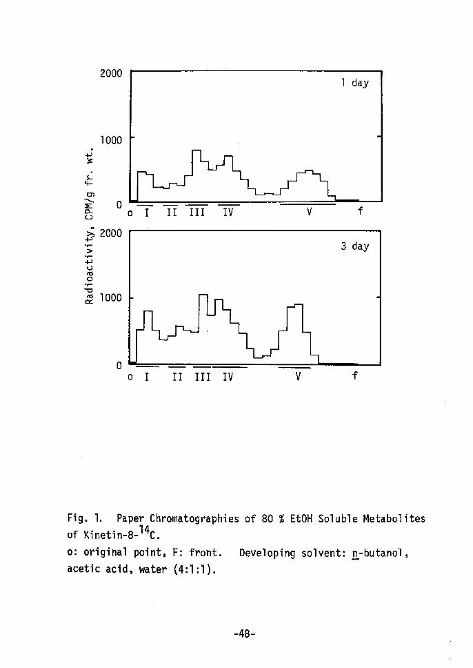
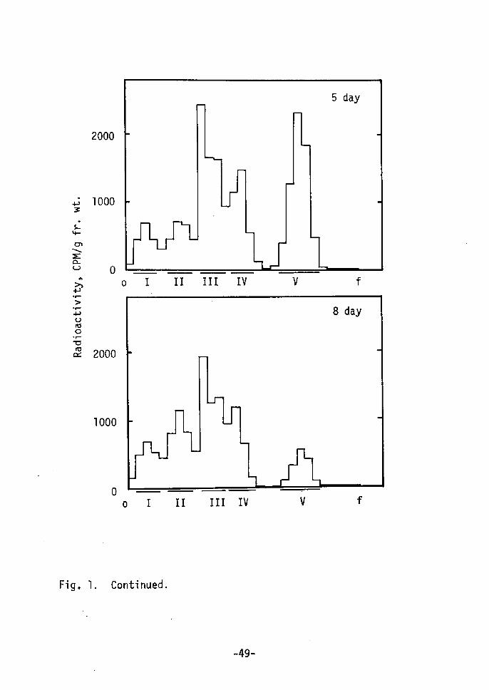
Paper chromatography of 80 % EtOH soluble materials shown in
Fig. 1 indicate that low molecular weight metabolites of kinetin-
s-14c are mainly composed of s compounds with fouowing Rf values:
peak I, O.04-O.07; peak rl, O.18-O.21; peak III, O.29-O,32; peak IV,
-47-
V3
clp
en
NEao "hv.T-År.Ppvnto
-rvntct
2000
1OOO
o
2000
1OOO
o
o : II Irl IV v f
ol II lrl TV v f
Fig, 1. Paper Chromatographiesof Kinetin-s-14c.
o: original point, F: front.
acetic acid, water (4:1:1).
of 80 % EtOH Soluble Metabolites
Developing solventt n-butanol,
-48-
PslvmxZavb"
.;•--
x.O-voct
2000
1OOO
o
2000
1OOO
o
ol rr I!r rv v f
ol II !r! lv v f
Fig. 1. Conti' nued.
-49•-

M:
o r xA p 3 s . a "s. E a ov A" ra o a = v o w = 'r
h p-e-
År -l- P U " o .r v rd ct
10
5
o
o 48 96 144Culture Time, hr
III
rv
II
192
20
10
o
cot
o r xA P 3 L " a x Z a ov - h p 'r År -t- - v nt o -r- v rd ct
r to - oH
Fig• 2• Changes in so% EtoH sol. Metabolites of Kinetin-s-14c.
Radioactivity of each peak (peak 1, II, III, IV and V) was
calculated from Fig. 1. (-O--År shows the radioactivity of
peak I; (.-e-), peak II; C-O-), peak III; Åq,-A-), peak IV;
C-A-), peak V; and (-•o•--), total radioactivity, respectively.
-50-

o.39-O.50; peak V, O,75-O.82. Peak r, IV and V were identified
as AMP, adenine and kinetin, respectively, since the Rf values of
AMP, adenine and kinetin were O.07, O.43-O.54 and O.81-O.86,
respectively, Peak II (Å~) and III ÅqYÅr were not identified.
Change in radioactivity of each peak is shown in Fig. 2.
Peak IV, V and III increased during 72-120 hr in this order, while
no significant changes were not found in peak I and II. Therefore,
it is considered that incorporated kinetin is imrnediately metabolized
to adenine, AMP, (Å~) and (Y) and a part of kinetin remains in free
form, Kineitn (peak V), after accumulation to a certain concent-
ration, is seemed to be converted to (Y). Free form of kinetin
or (.Y) is considered to be active form of kinetin. Details is 12-14)not clear. Recently, 6-benzylamino-7-glucofuranosylpurineand 7-glucosyl zeatin15) have been reported in addition to ribo-
nucleosides and 5i-ribonucleotides of cytokinins. These glucosides
are reported to be major and stable metabolites of benzyladenine
and zeatin, and to be active as cytokinin. Compound (Y) may be
glucoside or riboside of kinetin. It has also been reported that
cytokinins interact with their action site by loose, probably non-
covalent bonds18År. And cytokinin, itself, can not directly
affect enzyme activity etc. Lt vitro. Therefore,a possibility
is considered during the shoot redifferentiation: kinetin incorpo-
rated into cells is metabolized rapidly; free kinetin or metabolite
(Y) acts as trigger of shoot redifferentiation with kinetin-reactive
Protei'n rather than directly without mediator.
-51 -
SUMIVIARY
Kinetin-s-14c was incorporated rapidly into cultured tobacco
cells during the early stages of shoot redifferentiation, About
80 % of radioactivity was located in 80 % EtOH soluble materials.
About 50 % of radioactivity in 80 % EtOH insoluble materials was
lost by RNase treatment and 30 % was lost by Pronase E treatment.
Radioactivity in RNA is presumably due to adenine etc., rnetabolites
of kinetin. Radioactivity in 80 % EtOH soluble materials distri-
buted mainly 5 fractions, AMP, (X), (,Y), adenine and kinetin.
Metabolite (Y) and free kinetin increased during the culture. It
is considered that free kinetin or metabolite (Y) acts as trigger
of shoot redifferentiation with kinetin-reactive protein,
REFERENCES
1) Yamada, Y., Yasuda, T., Koge, M. and Sekiya, J,, Colloques
internationaux C. N. R. S., 193, 137 (1971År
2) Yasuda, T. and Yamada, Y., Biochem, Biophys. Res. Commun., A!t;,
649 C1971)
3) Fox, J. E, and Chen, C. M., J. Biol. Chem., 242, 4490 (1967)
4) Fox, J. E. and Chen, C. M., in Biochemistry and Physiology of
Plant Growth Substances; Proc. Vr lntl. Conf. Plant Growth
Substances C1967), ed. wightman, F. and Setterfield, G., p777,
Runge Press, Ottawa (1968)
5) Skoog, F, and Armstrong, J., Ann. Rev. Plant Physiol., !l:IL,
359 (1970År
6) Kende, H. and Tavares, J. E., Plant Physiol.i 43, 1244 C1969)
7) Hall, R, H., in Biochemistry and Physiology of Plant Growth
Substances; Proc. V: Intl. Conf. Plant Growth Substances (1967),
-52-

8)
9)
10)
11)
12)
13)
14)
15)
16)
17)
18)
ed. Wightrnan, F. and Setterfield, G,, p 47, Runge Press, Ottawa,(1 968)
Chen, C. M. and Hall, R. H., Phytochem., S, 1687 (1969)
McCalla, D. R., Morre, D. J. and Osborne, D. J,, Biochim,Biophys. Acta, S.Lt, 522 Cl962)
Fox, J. E,, Stood, C., Dyson, W, D. and McChesney, J. D,, in
Plant Growth Substances 1970; Proc. Vrl Intl. Conf. Plant
Growth Substances, ed. Carr, D, J,, p 449, Springer-VerlagCl 972)
Dyson, l-i. D., Fox, J. E. and McChesney, J. D., Plant Physiol.,
49, 506 (1972)
Deleuze, G,, McChesney, J. D. and Fox, J. E., Biochem.Biophys.
Res. Commun., 48 1426 C1972)
Fox, J, E., Cornette, J., Deleuze, G., Dyson, W., Giersak, C,,
Niu, P., Zapata, J. and McChesney, J. D., Plant Physiol.,
52, 627 (1973)
Fox, J. E, and McChesney, J. D., in Plant Growth Substances
1973; Proc. VIrl Intl. Conf. Plant Growth Substances, p 468,
Hirokawa, Tokyo (1974)
Parker, C. W. and Letham, D., Planta, 114, 199 (1973)
Doree, M. and Guern, J., Biochim, Biophys. Acta, 304, 611
(1 972)
Matthysse, A. G. and Abrams, M., Biochim. Biophys, Acta, 199,
511 (1970)Brandes, H. and Kende, H., Plant Physiol., 43, 827 (1969)
t53-

CHAPTER IV
CHANGES IN PROTEIN SYNTHESIS DURrNG THE EARLY
STAGES OF SHOOT REDIFFERENTIATION FROM CULTURED TOBAcco cELLsl).
- INTRODUCTION
Cytokinins appear to regu]ate many different actions in plant,
2)for example, cell division, enlargernent, and differentiation .
However, conclusive evidence for specific regulatory functions has
yet to be reported. In some studies of cytokinin actions, the
investigation has been made into their influence on protein synthesis, For example, Jouanneau et a13'4) reported that kinetin promotes
protein synthesis in tobacco cell suspensions and that at least
one specific difference in protein pattern could be observed
before the first cell division occurred. During senescence, it
has been reported that cytokinins affect the nucleic acid and
protein metabolism5-8). in relation to protein metabolism, the
cytokinin actions on the expansion of cotyledons9) and on dormant
duckweedlO) have been reported.
During cell culture cytokinins can induce the formation of
shoots or roots from cultured tobacco cellsll) but the mechanism
is not understood yet. This chapter reports the changes in
protein synthesis during shoot formation(.redifferentiation) in
cultured tobacco cells in vitro.
-54-
MATERIALS AND METHODS
Cell culture.
Tobacco callus, strain Ts12År, and Linsmaier and skoog basic
medium13) were used. For shoot formation cultured ce"s were
inoculated in an agar medium containing sxlo-5 M zeatin(zeatin
cells) or 10h5 M kinetin(kinetin cells). 2,4-D cells were served
as control cells. All cell cultures were incubated at 250C in the
dark.
DNA, RNA and png:tl!gin. determination.
DNA and RNA were extracted according to the method ofschneider14), except that the acid insoluble material was defatted
by heating it at 500C in ethanol-ether (1:1, v!v) for 15 min before
hot trichloroacetic acid(TCA) hydrolysis. DNA content was deter-
mined by the diphenylamine method as described by Burton15). RNA
was determined by the orcinol method as described by Mejbaum16).
Soluble protein was extracted by homogenizing cells in a glass
homogenizer containing O,1 M phosphate buffer, pH 7.5, centri-
fuging the homogenate at 10,OOO g for 15 min and collecting the
supernatant. Protein content was determined by the method
described by Lowry et al17).
14C-Leucine l.nsgn2g!t:g!l.gntion and DEAE cel1u1ose co1umn chromato ra h .
cultured cells, incubated with 14c-leucine (L-(u-14c)-ieucine,
SP• act. 270 mCilrnmol, the Radiochemical Center, England), were
homogenized with O.1 M phosphate buffer (pH 7.5) in a chilled
MOrtar] then the homogenate was centrifuged at 10,OOO g for 15 min.
-55-

The supernatant was dialysed against O,Ol M phosphate buffer (pH
7.5) overnight. The extract was applied tb a DEAE cellulose
column and eluted with a combination of linear and step gradients
of NaCl. Rddioactivity was determined as follows. An equal
volume of 10 % TCA was added to Part of the fractionated solution
and the precipitate was 6ollected on a glass fiber paper (whatman
GFIC) and washed with 5 % TCA, ethanol and ether. Using a toluene
base scintillator, the radioactivity was counted with a Beckman
LS-100 liquid scintillation spectrometer.
Discg!/gg!ngRhg!gsLEth •
Polyacrylamide gel electrophoresis of the fractionated protein
was performed according to the method of Davis18).
Pre aration of gn:t!itsgni and antiserum.
Cultured tobacco cells or tobacco leaves grown in a green
house for 3 months, were homogenized in a chilled mortar with
1!5 its weight of Polyclar AT and an equal volume of O.05 M
phosphate buffer (pH 7.5) containing 1 % Na-ascorbate. The pH
was adjusted to 7.5 with 1 N NaOH. The homogenate was squeezed
through gauze, then centrifuged at 10,OOO g for 30 min. Protein
was precipitated by sdturating the mixture with (NH4)2S04• The
precipitate was collected by low speed centrifugation, after which
it was dissolved in small amounts of and dialysed against O.05 M
phosphate buffer CpH 7.5). All procedu"es were carried out at
40C. The protein solution (antigen) was stored in a freezer after
the addition of 12.s % of glucose19), Two male rabbits were
-56-
immunized with antigen from 15 day old 2,4-D cells and two others
with antigen from tobacco leaf. Five doses of the same antigen
(the first three, 20 mg as protein and the last two, 10 mg) in
incomplete Freund adjuvant were administered intraperitoneally at
1 week intervals, blood was taken from the carotid artery 1 week
after the last injection. Sera were prepared by the usual method20).
It!n!:L!!gE!y:A!)!iggnntibodyatienreactions,
Antibody--antigen reactions were studied using the double
diffusion method in agar gel as described by ouchterlony21).
RESULTS AND DISCUSSION
AIthough 2,4-D cells, strain T5, grow well in the absence
of cytokinins, it will only form shoots when transferred to a
-5 M zeatin as the cytokinin.medium with 5xlO
Table I shows that in zeatin cells RNA and protein concent-
ration per gram fresh weight began to increase 5 to 6 days after
inoculation, then 15 days later DNA increased, whereas the contents
per fresh weight in 2,4-D cells rernained constant. Increased of
RNA, soluble protein and DNA in this order suggest that protein
synthesis changes during the shoot redifferentiation. Thorpe gt
A-1!:22) have reported that RNA and protein are increased in shoot-
forming regions of tobacco cultured cells, and werner st sL123)have
also described that sorne qualitative changes in the proteins of
Carrot eccur prior to visible morphological onset of root form-
ation.
•-57-

Table I. Contents of DNA, RNA and Protein.
Days
o 6 15
DNA
RNA
(mglg fresh
2,4-D (A)
Zeatin (B)
BIA
(mglg fresh
2,4-D (C)
Zeatin (D)
DIC
wt)
wt)
o
1
2
1
.
.
.
'
038
oo
80
oo
o
o
o
2
4
1
.
.
'
.
065
052
80
72
11
51
o
o
1
3
4
1
.
.
.
056
104
86
08
14
34
Days
o 5 10 16
Buffer Soluble Protein (mglg
2,4-D (E)
Zeatin (F)
FlE
fresh wt)
1.34
1.00
1.54
1,83
1.19
1.35
2.28
1.69
1.33
1.98
1.48
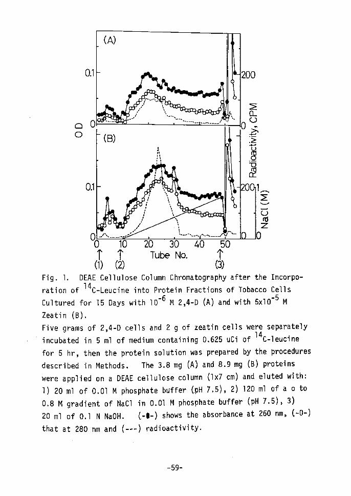
14 Incorporation of C-leucine into proteins and DEAE cellulose
column chromatography of these proteins were investigated and the
results are shown in Figs. 1 and 2. rn Fig. 1, protein peaks for
zeatin cells were eluted at a higher concentration of NaCl than were
those for 2,4-D cells. The radioactivity of 14c-leucine, incorpo-
rated into protein showed a relatively high level. A remarkable
and characteristic peak at the concentration of O.3 M NaCl for
zeatin cells(B) was obtained as cempared to that for 2,4-D cells (A)t
-58-
O.1
(A)
''
r.xr"4" ,ix
o
O.1
It fl,
200
Ea
OO 10 20 30 40 50 T T Tube No. 'r (1) (2) (3)Fig. 1. DEAE Cellulose Column Chromatography after the Incorpo- 14 C-Leucine into Protein Fractions of Tobacco Cellsration ofCultured for 15 Days with lo-6 M 2,4-D (A) and with sxlo-5 M
Zeatin (B).
Five grams of 2,4-D cells and 2 g of zeatin cells were separatelyfincubated in s ml of medium containing o.62s uci of 14c-leucine
for 5 hr, then the protein solution was prepared by the procedures
described in Methods. The 3.8 mg (A) and 8.9 mg (B) proteins
Were applied on a DEAE cellulose column (lx7 cm) and eluted with:
1) 20 ml of O.Ol M phosphate buffer CpH 7.5), 2) 120 ml of a o toO•8 M gradient of NaCl in O.Ol M phosphate buffer (pH 7•5), 3)
20 ml of O.1 N NaOH. C-e-) shows the absorbance at 260 nm, C-O-)
that at 280 nm and (-•--) radioactivity•
•-"'"""'--.-.-...--...•'"Ei•lr" oo 'År(B) •--.
.2.--d,
l`:l, gll 'v'
:' dicr
-- -' o 11 Ag--,l, , 2t11 vIti l -tS1-t Utl
xtllxlt..1"/X.t-..-.t''`-.'Sv.-".5".!V
ft v
rd
-59-
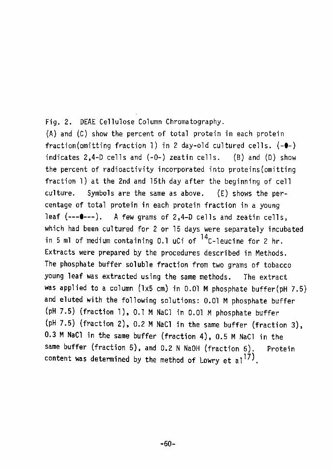
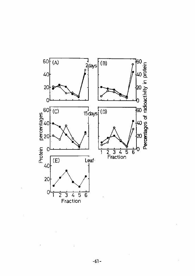
Fig. 2. DEAE Cellulose' Column Chromatography.
(A) and CC) show the percent of total protein in each protein
fraction(omitting fraction 1) in 2 day-old cultured cells. (-e-)
indicates 2,4-D cells and (-O-) zeatin cells. (B) and (D) show
the percent of radioactivity incorporated into proteinsÅqomitting
fraction 1) at the 2nd and 15th day after the beginning of cell
culture. Symbols are the same as above. (E) shows the per-
centage of total protein in each protein fraction in a young
leaf C---e--). A few grams of 2,4-D cells and zeatin cells,
which had been cultured for 2 or 15 days were separately incubatedin s mi of medium containing o.1 uci of i4c-leucine for 2 hr.
Extracts were prepared by the procedures described in Methods.
The phosphate buffer soluble fraction from two grams of tobacco
young leaf was extracted using the same methods. The extractwas applied to a column (1Å~5 cm) in O.Ol M phosphate buffer(pH 7.5)
and eluted with the following solutions: O.Ol M phosphate buffer(pH 7.5) (fraction 1), O.1 M NaCl in O.Ol M phosphate buffer
CpH 7.5) (fraction 2), O.2 M NaCl in the same buffer (fraction 3),
O.3 M NaCl in the same bui'fer (fraction 4), O.5 M NaCl in the
same buffer Cfraction 5), and O.2 N NaOH (fraction 6). Proteincontent was determined by the method of Lowry et al17).
-60-
60
40
20
o
608.9-4o
syg2o
2sdays(B) 60
t.=-
co g
.E.E!i
9a 40
20
Leaf -
o 1 234 56 Fraction
Fraction
.EOh •.-- -.År--
ta .9
868gs
,lli
-61-

The eluent with O.1 N NaOH shows a high absorption but much less
radioactivity. Phenolics seem to cause the high U. V. absorption
at 260 and 280 nm,
14 To compare protein patterns at different stages, C-leucine
was incorporated and DEAE cellulose column chromatography was
performed. As shown in Fig. 2, patterns for the total protein
and the radioactivity incorporated into 2 day-old zeatin cells are
not much different from those for the 2 day-old 2,4-D cells (A and
B in Fig 2). However, 15 day-old zeatin cells showed quite
different patterns from the 15 day-old 2,4-D cells for both total
protein and incorporated radioactivity in protein (C and D in Fig.
2), A 15 day culture period on a medium containing zeatin enhanced
the synthesis of certain characteristic proteins (fraction 3 on C
and D in Fig. 2) which are specific to young leaves of the tobacco
plant (E in Fig, 2). Even the protein pattern of incorporated
14c-leucine showed the specific peak at fraction 3 in Hg• 2 (D),
the same as in Fig. 2 (E). This means that protein synthesis in
zeatin cells differs from that in 2,4-D cells, and that zeatin
cells protein content becomes similar to that of leaves after
about 15 days of culture. Therefore, 1 investigated, in deteil,
the differences in protein patterns between 15 day-old 2,4-D cells
and 15 day-eld zeatin cells using electrophoresis.
Protein fractions from 15 day-old cells obtained from DEAE
cellulose column chromatography (Fig. 2) were electrophoresed wiih
polyacrylamide gel. As shown in Fig. 3 slight discrepancies were
-62-
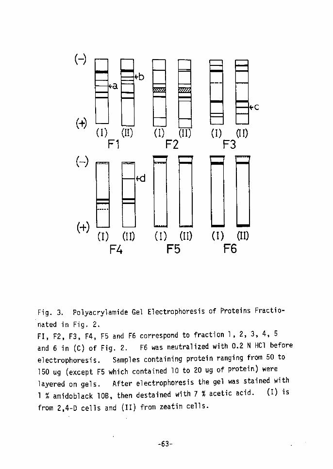
(-)
(+)
(-)
(+)
a
(I) (II) Fl
"b
rd
(I)
F2(II)
-
(I)
F3aI)
ec
(1) (II) (I) (II) (I) (II)
F4 F5 F6
Fig. 3. Polyacrylamide Gel Electrophoresis of Proteins Fractio-
nated in Fig. 2.FI, F2, F3, F4, F5 and F6 correspond to fraction 1, 2, 3, 4, 5
and 6 in (C) of Fig. 2. F6 was neutralized with O.2 N HCI before
electrophoresis. Samples containing protein ranging from 50 to150 ug (except F5 which contained 10 to 20 ug of protein) were
layered on gels. After electrophoresis the gel was stained with1 % amidoblack 10B, then destained with 7 % acetic acid. (I) is
frOm 2,4-D cells and (II) from zeatin cells•
. -63- .
detected between 2,4-D and zeatin cells. In zeatin cells 3 bands,
(b), (c) and Cd) appeared in Fl, F3, and F4, which were not found
in 2,4-D cells. Band (a) was found only in 2,4-D cells.
Fractions 5 and 6, which were eluted with O.5 M NaCl and O.2 N NaOH,
respectively, were not well mapped by electrophoresis. Jouanneau
et ai3'4År reported the enhancement of i4c-ieucine incorporation by
cytokinin in tobacco cultured cells and using disc electrophoresis,
that at least one specific band appeared after the first generation,
The present results showed that at least 4 different bands were
found in cultured tobacco cells treated with and without zeatin
for 15 days. The characteristic synthesized proteins in zeatin
cells (C and D in Fig. 2) which occurred in quantitative amounts,
did not give quantitative amounts as the very specific protein
bands at F3 in Fig. 3. However, band (c) is a characteristic
protein band in F3 from zeatin cells and band (c), as well as
band (b) and (d), is believed to contain proteins which are
significantly invelved in regenerating leaves from cultured cells•
To confirm and determine qualitatively the protein changes
due to cytokinins, which accompany shoot formation in cultured
cells, we used immunological techniques. rn these experiments,
kinetin cells were used instead of zeatin cells,
To obtain antigen from kinetin cells, cells were inoculated
on a medium containing lo-5 M kinetin and cultured for 5, 10, 15
and 32 days. Kinetin cells cultured for 32 days produced no
visible organogenesis; though the cells, themselves, became harder
-64-

(l)
(ll)
c
c
(lll)
(IVÅr
p
Ac
Fig. 4, (I) and ÅqIII), Photographs: (IIÅr and (IV), Diagramatic
Representation ef Irrununodiffusion Patterns Observed under the
Microscope.
Fifty ul of antiserum was put into the central he]e and 150 ug(as protein) of antigen into the outer holes. Agar plates were
incubated for 36 hr at 25eC. [Ac]; anti-2,4-D cell serum. [Al];
anti-leaf serum. [1]; antigen of 2,4-D cells cultured for l5days, [2]; antigen ef kinetin cells cultured for 5 days, [3];
kinetin ceMs cultured for 10 days, [4]; kinetin cells cultured
for l5 days, [5]; kinetin ceHs cultured for 32 days, EL]; antigen
of tobacco leaves grown in a green house.
-65-

and more compact. Fig. 4 shows results obtained with the double
diffusion method and indicates an antigenic difference between
2,4-D and kinetin cells. In kinetin cells, Fig. 4 (I) and (II),
a specific precipitin line (b) appeared between [Al],and [4] and
[5], which was not observed in 2,4•-D cells. The precipitin line
(aÅr between [Al] and antigens of cultured cells were observed, but
there was no line [All and [1] 12 hr after the beginning of diffu-
sion (not shown in the figure). Line '(a) between [Al] and [5]
was the strongest, while line (a) between [Al] and [2] was the
weakest at that time. As in Fig. 4 (rll) and (IV), antigens of
kinetin cells were similar to those of 2,4-D cells. However,
line (d) disappeared between [Ac] and [5]. These results show
that some types of proteins specific to the 2,4-D cells do not
exist in the kinetin cells, and that new proteins, similar to leaf
proteins, are synthesized. Furthermore 2,4-D and kinetin cells
retain some common protein constituents which seem differ consider-
ably from those of the tobacco leaf.
These results also indicate that 2,4-D and kinetin cells
retain cormon protein constituents, However, it appears that
kinetin cells are synthesizing some characteristic proteins which
cause the formation of or become part of the leaves.
In the cultured tobacco cell line, strain T5, these results
show that cytokinins cause the induction of certain characteristic
proteins, which begin to form leaves from cultured cells in vitro.
Simultaneously, other protein syntheses are repressed by cytokininS
-66-
;1

for shoot formation• However, it is not yet known how cytokinins
act and regulate the protein syntheses for organogenesis in cultured
cel1s.
"Cell differentiation is based almost certainly on the regu-
lation of gene activity."24) if some genes are activated, RNA and
protein syntheses may, subsequently, be induced or repressed. By
adding a high concentration of cytokinin to cultured dedifferenti-
ated cells, genes characteristic to a specific organ rnay be activated,
and a factor specific to constantly dividing cells may be inacti-
vated. The results of gene activation and inactivation would
then appear in the protein constituents. The present results,
showing that cytokinins induced the formation of some characteristic
proteins and repressed the formation of othersi, are consistent with
this hypothesis.
Evidence that cytokinin affects RNA synthesis has beenreported by Roychoudhury et a125) and Matthysse et a126),
Roychoudhury et al reported that kinetin stimulates the synthesis
and release of nuclear RNA in coconuts. Matthysse g:t!L al showed
that kinetin enhances the rate of RNA synthesis by isolated nuclei
of tobacco and soybean calluses and by pea buds.
It would thus seem that cytokinin reacts with certain cell
components to regulate gene activity, which is followed by the
Promotion of RNA synthesis and the effects on protein rnetabolism
shown in the present results. Thereafter, organ fotrrmation should
OCcur morphologically. However, from the results en callus
-67-

induction (dedifferentiationÅr from differentiated tfissue by
auxins27-30), the actions of cytokinins for redifferentiation
seem to have conjugated metabolic relationships with auxin action,
Dedifferentiation by auxins is the reverse reaction to rediffer-
entiation by cytokinins.
SUMMARY
During tobacco cell culture cytokinins can induce and re-
differentiate shoots from cells cultured in vitro.
During the early stages of shoot formation RNA, soluble
protein and DNA increased in this order.
Relationships between the synthesis of certain proteins and
the disappearance of other, proteins during the early stages of
shoot redifferentiation, under the specific actions of cytokinins,
were sought using DEAE cellulose column chromatography, poly-
acrylamide gel electrophoresis and imrnunological rnethods,
The present Fesults show that cytokinins cause the induction
of certain characteristic proteins' , when leaves begin to foum
fron cultured cells .Lt vitro, During shoot redifferentiation some
proteins synthesized for cell proliferation are repressed. These
changes in ptotein synthesis caused by cytokinins in cultured
tobacco cells are discussed in r,egard to redifferentiation.
-68-
REFERENCES
1) Sekiya, J, and Yamada, Y., Bull. rnst. Chem. Res., Kyoto Univ.,
!1;t, 246 (1974)
2) Skoog, F. and Avtnstrong, D, J., Ann. Rev. Plant Physiol., 21,
359 (l970)
3) Jouanneau, J. P, and Pesud-Leonol. C., Physiol. Plant,, 20,
834 (1967)
4) Jouanneau, J, P,, Physiol. Plant., 23, 232 (1970)
5) Osborne, D. J., Plant Physiol., 37, 595 (1962)
6) vonAbrams, G. J. and Pratt, H. K., Plant Physiol., 43, 1271
(1968)
7) Atkin, R. K. and Sriv' astava, B. I. S., Physiol. Plant., 23,
304 (1970)
8) Martin, C, and Thimann, K. V., Plant Physiol., 49, 64 (1972)
9) Rijven, A. H. G. C. and Parkash, V,, Plant Physiol., 47,
59 (1971)
10) McComb, P. J. A, and Ralph, R. K., Biochem. J., 129, 403 (1972)
11) Skoog, F. end Miller, C, O., Syrnp. Soc. Exp, Biol., 11, 118
(1957)
12) Yamada, Y., Kiso, K., Sekiya, J, and Yasuda, T., Agr, Biol.
Chem., 36, 1055 (1971)
13) Linsmaier, E. M. and Skoog, F., Physiol. Plant., 18, 100
(1 965)
14) Schneider, i-1. C., J. Biol, Chem., 161, 293 (1945)
15) Burton, K., Biochem. J., 62, 315 Åq1956)
16) Mejbaum, W., Z. Physiol. Chem., 258, 117 (1939)
17) Lowry, O. H., Rosebrough, N. J,, Farr, A. L. and Randall, R, J.,
J. Biol. Chem., 193, 265 (1951År
18) Davis, B. J., Ann. N. Y. Acad. Sci., 121, 404 (1964)
19) Heitefuss, R., Buchanan-Davfidson, D. J. and Stahmann, M. A.,
Arch. Biochem. Biophys., 85, 200 (1959)
-69-

20)
21)
22)
23)
24)
25År
26)
27)
28)
29)
30)
Campbell, D. H., Garvey, J. S,, Cremer, N. E. and Sussdorf, D.H., in Methods in Xmmunology, p 22, IAI. A. Benjamin Co. (1963)
Ouchterlony, O., Acta Pathol. Microbiol. Scand., :!Zt., 231 (1953)
Thorpe, T. A. and Murashige, T,, Can. J. Bot,, f!et., 277 C1970)
Werner, D, and Gogolin, D., Planta, g:IL, 155 (1970)
Britten, R, J. and Davidson,r E. H,, Science, 165, 349 (1969)
Roychoudhury, R., Datta, A. and Sen, S, P,, Biochim. Biophys,
Acta, 107, 346 C1965)
Matthysse, A. G. and Abrams, M,, Biochim. Biophys. Acta, 199,
511 (1970)
Yasuda, T. and Yamada, Y., Biochem. Biophys. Res. Cormnun.,
40, 649 C1970)
Yamada, Y., Yasuda, T., Koge, M. and Sekiya, J., Colloques
internationaux C. N. R, S,, 193, 137 (1971)
Yamada, Y. and Yasuda, T., Biochem. Biophys. Res. Commun,, EStL,4,88 C1971)
Yasuda, T., Yajima, Y.,and Yamada, Y., Plant Cell Physiol., 15,321 C1974)
-7e-
CHAPTER V
DNA-DEPENDENT RNA POLYMERASES DURING SHOOT FORIVIATION
FROM CULTURED TOBACCO CELLslÅr.
INTRODUCTION
Since the first DNA-dependent RNA polymerases (RPases) in the
nuclei of animal cells were solubilized, purified and identified
as multiple forms2), Rpases from many animal cells have been
reported3). Recent studies also show that rnultiple forms occur
in higher plants as well. The RPases which have been purified and
characterized from higher piants4-i7) show in most cases, properties
similar to the RPases from animal.cells.
Hormonal control of the activities of the RPases from plant
cens by inddle-3-acetic acid (IM)10'18'19) and gibbereuic acid
(GA)20) has been reported. However, cultured plant cells (callusÅr
usually continue cell division and growth in the presence of auxins.
Shoots and roots can be formed by cytokinins from cultured tobacco
cens20-22År. During organogenesis from these cultured -cells, many
metabolic changes involving, for example, changes in RNA and proteinsyntheses have been observed23). cytokinins and auxins are pre-
sumed to play an important role in organogenesis and transcriptive
regulation by cytokinins and!or auxins seems to exist.
This chapter describes the partial purification of DNA-depen-
clent RNA polymerases frorn cultured tobacco cells at dffferent
stages of shoot formation and presents a comparison of these RPases.
-71 -

MATERIALS AND METHODS
Chemica1s.
uridine-s-3H-s'-triphosphate (3H-UTP, sp, act. 26.g cilmM)
was obtained from New England Nuclear and ethanol as solvent was
removed by bubling with N2. Hi.ghly polymerized calf thymus DNA
was purchased from Warthington Biochemical Corp.; whole histones,
from Sigma Chemical Co.; ot-amanitin, from C. H. Boehringer Sohn;
rifamycin SV, from Calbiochem, and four kinds of nucleoside tri-
phosphates, from Sigma Chemical Co. All other chemicals were of
reagent grade.
Cell culture.
Three kinds of cultured cells (2,4-D cells, kinetin cells and
redifferentiated shoots) originating from the pith tissue of the
tobacco plant, Nicotiana tabacum var. Bright Yellow, were used,
2,4-D ceils24) were transferred to a liquid medium containing io-5
M kinetin and cultured at 270C in the dark. Three days after
this transfer the kinetin cells were harvested (3DK5 cells). Re-
differentiated green shoots (Rs cells) were induced with 10-5 M
kinetin using cells cultured with 10-5 M indoled-3-butyric acid(IBA),
then subcultured on an agar medium containing lo'-5 M kinetin at
250C in the light, RS cells were harvested 15-20 days after
transfer to the same medium. The basic composition was that of
the Linsmaier and skoog medium25), These three day-old 2,4-D
cells (3DD6 cells), 3DK5 and RS cells were stored at -20eC for
use as the enzyme source.
-72-

IEtnz,yg!ga.ssgM.•
Rpase activity was determined as described previouslyl'11)
with some modifications. The standard reaction mixture contained
in O.15 ml: 50 mM Tris-HCI, pH 7.8; 2 mM DTT; 1-2 mM MnC12; O.32
mM of ATP, GTP and CTP; O.05 mM of UTP containing 1-2 uCi of 3H-
UTP;' 12 ug of native calf thyrnus DNA (highly polymerized) or pea
DNA; and 50 ul of enzyme solution. Incubated lasted for 10-15
min at 370C RPase activity was represented by the 3H-UMP incorpo-
rated into the trichloroacetSc acid (TCA) insoluble materials.
Radioactivity was determined with a Beckman LS-100 liquid scinti-
11ation spedtrometer.
Purification of RNA ol erases.
RPase preparation was essentially according to the methods .
described by sasaki et aiii). sixty grams of frozen tobacco
cells and 20 g of Polyclar AT were ground in a chilled mortar for
10 min in a final volume of 120 ml containing O.15 M Tris-HCI
(pH 7.8); O.1 M EDTA; 5 mM DTT; O,Ol M ÅqNH4)2S04 and 25 %(V!VÅr
glycerol. The mixture was sonicated for 2 min. The homogenate
was filtered through gauze and centrifuged at 100,OOO g for 60
min. The supernatant was dialysed for 4 hr against 3 liters of
50 mM Tris-HCI (pH 7.8); O.5 mM MgC12; 1 mM DTT; O•Ol M (NH4År2S04
and 25 % glycerol (O.Ol M ammonium sulfate standard buffer; O.Ol M
ASS buffer). After the enzyme selution was loaded on a DEAE-
Sephadex column (1.5 x 11 cm) equilibrated with O.Ol M ASS buffer.
-73-
The column was washed with 150 ml of O.Ol M ASS buffer and eluted
with 150 rn1 of O.1 M ASS buffer, then with 100 ml of O.1-O.4 M
linear gradient ASS buffer. RPase rl fractions after DEAE-Sepha-
dex chromatography were collected and dialysed for several hours
against 50 mM Tris-HCI buffer CpH 7,8) containing 1 mM DTT and
50 X glycerol. Purifitid RPase IIs were stored in liquid nitrogen,
Å}P!wrg!L!.gnti gzf.ane DNA.
About 50 g of etiolated Alaska pea seedlings which had been
grown for 5 days, was homogenized in a Waring Blendor with 50 mM
Tris-HCI, pH 7.8, containing O.25 M sucrose, 10 mM mercaptoethanol
and 1 mM MgC12 (Buffer I). After passage through 6 layers of
gauze, the filtrates were centrifuged at 6,OOO g for 30 min, The
resulting pellet was resuspended in Buffer l and collected by
centrifugation at 10,OOO g for 15 min. This pellet was washed
with 1110 x SSC buffer, then suspended in a small amount of 1110 x
SSC buffer containing 2 M NaCl and stirred gently for a few hours,
The viscous solution was centrifuged at 10,OOO g for 15 min, Three
volumes of cold ethanol was added to the supernatant obtained.
The resulting white fibrous precipitate (crude chromatin) was
collected with tweezers, then dissolved in 50 rnM Tris-HCI, pH 7.8,
containing 1 % SDS after which the solution was stirred gently
for several hours. An equal volume of phenol saturated with
buffer was added to the solution and the whole was stSrred for 30
min at room temperature. After low speed centrifugation the
-74-
water phase was collected. The phenol treatment was repeated 2
more times, then three volumes of cold ethanol was added to the
collected water phase solution. A white fibrous precipitate
(crude DNA) was collected and dissolved in a small amount of SSC
buffer. After adding the RNase (100 uglml) the solution was
incubated at 370C for 10 min. DNA was purified three times using
the phenol treatment. This purified DNA was precipitated with
cold ethanol, then washed with acetone and dried.
RESULTS
Cell culture.
-6 When tobacco cells cultured with 10 M 2,4-D (2,4-D cells)
were transferred to a medium containing lo-5 M kinetin (kinetin
cells) shoot formation was observed. In the early stage of shoot
formation the growth rate of the kinetin cells decreased 3 days
after transfer compared to that for the 2,4-D cells, as shown in
Fig. 5 in CHAPTER r. The degree of organogenesis proceeded in
the or'der of 2,4-D cells (3DD6 cells) through kinetin cells (3DK5
cells) to RS cells (redifferentiated shoots).
Iso1ation and urification of RNA ]29.IL)!!!!9! 9.E9E.•
Fig. 1 shows that cultured tobacco cells (3DD6) had two peaks
of RPases. ot--Amanitin inhibited RPase activity of peak Ir
completely but did not inhibit peak I (Table I). From this result
cultured tobacco cells had two different types of RPases, RPase r
and RPase' Ir, which were.eluted with O.1 M and O.25-O,30 Fl
--75-
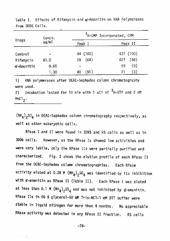
Table I, Effects of Rifamycin and o(-Amanitin on RNA Polyrnerases
from 3DD6 Cells, ,
DrugsConcn.uglml
3 H••UMP Incorporated, CPM
Peakr Peak rlContro1
Rifamycin
of••Amanitin
63.0
O,65
1,30
44 CIOO)
28 (64)
40 (91)
637 CIOO)
627 (98)
59 (9) 21 (3)
1) RNA polymerases after DEAE-Sephadex column chromatography
were used.2) Incubation lasted for 10 nin with 1 uCi of 3H-UTp and 2 mM
MnC12•
ÅqNH4)2S04 in DEAE-Sephadex column chromatography respectively, as
well as other eukaryotic cells.
RPase r and rl were found in 3DK5 and RS cells as well as in
3Db6 cells. However, as the RPase Is showed low activities and
were very labile, only the RPase IIs were partially purified and
characterized. Fig. 2 shows the elution profile of each RPase II
from the DEAE-Sephadex column chromatographies. Each RPase
activity eluted at O.28 M (NH4)2S04 was identified by its inhibition
with or-amanitin as RPase II CTable :I), Each RPase I was eluted
at less than O.1 M (NH4)2S04 and was not inhibited by of-amanitin.
RPase !Is in 50 % glycerol-50 mM Tris-HCI-1 mM DTT buffer were
stable in liquid nitrogen for more than 4 months. No appreciable
RNase activity was detecited in any RPase II fraction. RS cells
-76-

E a Q " v di p nt L o a s o U = H a E = 1 =M
300
200
1OO
o o
Fig. 1. DEAE-Sephadex
from 3DD6 Cells(••••) shows OD2so and
Fraction Number
Column Chromatography
(•-O-), radioactivity
of
of
o
o
o co N a o.o
O.4
o
E - ttoen NA er=zv
RNA PolyTnerases
3 H-UMP incorporated.
had other RPase activity (peak rll) eluted with O.22 M (NH4)2S04•
This seems to be an RPase originating from chloroplasts because
rifamycin inhibited this activity b.ut oC-amanitin did not.
Table III show changes in the total RPase II activities after
DEAE-Sephadex column chromatography. Total activity decreased
after to the kinetin-medium C3DK5 cells), then the activity of the
RS cells increased to the level of the 3DD6 cells.
p77'
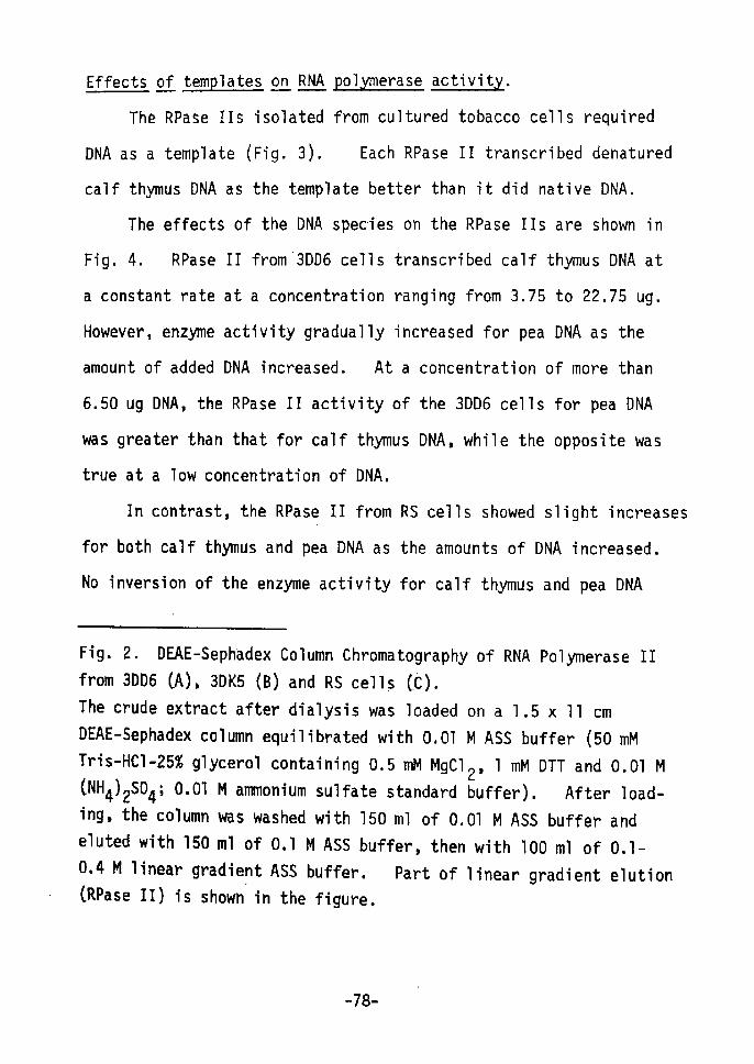
Effects gzt :t!gu!R.tgEesLl t g!,; RNA pg-ILE!gngEg ggStaytwt•
The RPase IIs isolated from cultured tobacco cells required
DNA as a template (Fig. 3), Each RPase II transcribed denatured
calf thyTnus DNA as the template better than it did native DNA.
The effects of the DNA species on the RPase IIs are shown fin
Fig. 4. RPase II t'rom'3DD6 cells transcribed calf thymus DNA at
a constant rate at a concentration ranging from 3.75 to 22.75 ug.
However, enzyme activity gradually increased for pea DNA as the
amount of added DNA increased. At a concentration of more than
6.50 ug DNA, the RPase II activity of the 3DD6 cells for pea DNA
was greater than that for calf thymus DNA, while the opposite was
true at a low concentration of DNA.
In contrast, the RPase II from RS cells showed slight increases
for both calf thymus and pea DNA as the amounts ot' DNA increased,
Ne inversion of the enzyme activity for calf thymus and pea DNA
'
Fig. 2. DEAE-Sephadex Column Chromatography of RNA Polyrnerase rr
from 3DD6 CA), 3DK5 (B) and RS cells CC).
The crude extract after dialysis was loaded on a 1.5 x 11 cm
DEAE-Sephadex column equilibrated with O.Ol M ASS buffer (50 mM
Tris-HCI-25% glycerol containing O.5 mM MgC12, 1 mM DTT and O.Ol MCNH4)2S04; O•Ol M anmionium sulfate standard buffer). After load-
ing, the column was washed with 150 ml of O.Ol M ASS buffer and
eluted with 150 ml of O,1 M ASS buffer, then with 100 ml of O.1-
O.4 M linear gradiept ASS buffer. Part of linear gradient elution(RPase II) is shown in the figure.
-78-
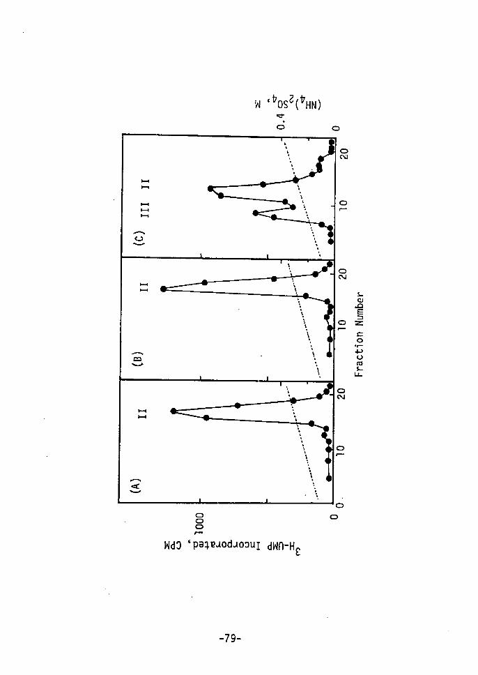
w `t7osl(VHN)
g•o
H---1
HHt--t
Aov
Acov
A't..v
oN
o
LoAE=Z=o
-t-
pvas
sL
ooor"-o
WdO`pe;paodaoouI dWA-H t
-79-

Table
from
li.
3DD6,
Effects
3DK5 and
ot' Rifarnycin and
RS Cel1s.
of-Amanitin en RNAPolymerase IIs
DrugConcn.uglml
3 H-UMP Incorporated, CPM
3DD6 3DK5 RS
Control
Rifamycin
of-Amanitin
63,O
1.30
,1826 (100)
1913 (105)
31 (2)
836
834
11
(1OO)
(1OO)
(1)
428
31O
51
(1OO)
(73)
(12)
1)
2År
and 1
Figures in
Incubation
mtvl MnC12•
parentheses show
lasted for 15 min
the
at
relative
37ec with
activities(%År 1•5 uci of 3
.
H-UTP
Table
Shoot
IIr. ChangesFormation.
in Total RNA Polymerase II Activities during
Cel1s TotalActivityl)
3DD6
3DK5
RS
396
193
351
x lo3
x lo3
x lo3
(1OO)
(49)
(89)
1) Total activity is represented as total 3H-UMp incorporated
(CPM) per 60 g of original material, after DEAE-Sephadex column
chromatography,2) Incubation lasted for 15 min with 2 uCi of 3H-UTP and 1 mM
MnC12,
-80-
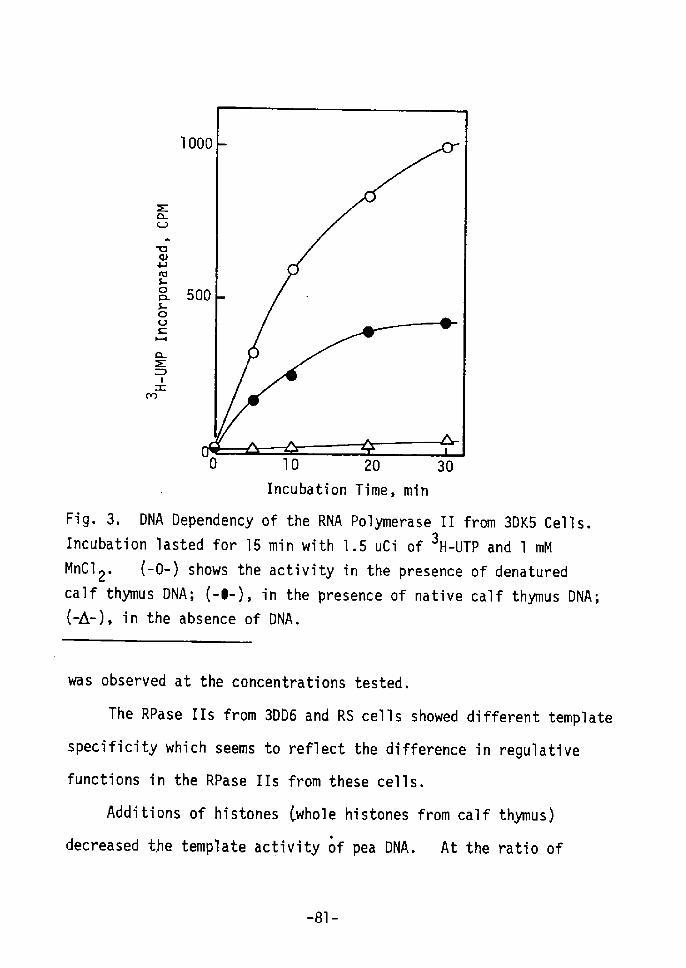
E a o - v o p as L o a L o v c N a E = l =rn
o
Fig. 3, DNA Dependency
Incubation lasted for 15
MnC12• (-O-) shows thecalf thymus DNA; (-e-),Åq-A•-), in the absence of
10 20 30Incubation Time, min
of the RNA Polymerase II from 3DK5 Cells. min with 1.5 uCi of 3H-uTp and 1 mM
activity in the presence of denatured
in the presence of native calf thymus DNA;
DNA.
was observed at the concentrations tested.
The RPase IIs from 3DD6 and RS cells showed different template
specificity which seems to reflect the difference in regulative
functions in the RPase Ils from these cells.
Additions of histones Cwhole histones from calf thymus)
.decreased t,he template activity of pea DNA. At the ratio of
-81-
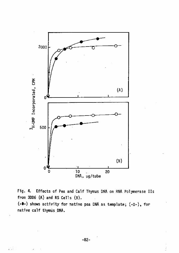
=Els
isggts
8 H i E = l =n
2000
o
500
o•
(A)
o '10 DNA, vg!tube
20
Fig. 4. Effects of Pea and Calffrom 3DD6 (AÅr and RS Cells CB).
C-e-) shows activity for native
native calf thymas.DNA.
Thymus
pea
DNA on
DNA as
RNAPolymerase
temp1ate; c-o-),
rrs
for
-82-
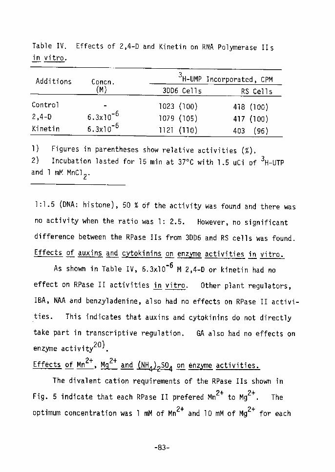
Table IV. Effects of 2,4-D and Kinetin on RNA Polymerase I!s
in vitro.
Additions Concn. (M)
3 H-UMP Incorporated, CPM
3DD6 Cells RS Cel1s
Control
2,4-D
Kinetin
•- 66.3xlO -66.3xlO
1023 Åq100)
1079 (105)
1121 (11e)
418 (100)
417 (100År
403 (96)
1) Figures in parentheses show relative activities (%).2) Incubation lasted for 15 min at 370C with 1.s uci of 3H-UTP
and 1 mM MnC12•
1:1.5 (DNA: histone), 50 % Of the activity was found and there was
no activity when the ratio was 1: 2.5. However, no significant
difference between the RPase IIs from 3DD6 and RS cells was found.
Effects of auxins and c tokinins on erLbc[[ig activities int vitro,
As shown in Table IV, 6.3xlO'6 M 2,4-D or kinetin had no
effect on RPase II activities Lt vitro. Other plant regulators,
IBA, NAA and benzyladenine, also had no effects on RPase II activi-
ties. This indicates that auxins and cytokinins do not directly
take part in transcriptive regulation. GA also had no effects onenzyme activity20).
Effects of Mn2+, !t!!sLll[t+ and S!!uzR.2:st,t4o on enzM!ng activities•
The divalent cation requirements of the RPase IIs shown in
Fig• 5 indicate that each Rpase II prefered Mn2+ to Mg2+. The
2+ 2+optimum concentration was 1 mM of Mn and 10 mM of Mg for each
-83-

Rpase r:. The ratio of maximum activity at le mM of Mg2+ to that
at 1 mM'of Mn2+ decreased from 74 to 43 % as organogenesis pro:
2+ceeded (from 3DD6 to RS cells). A high concentration of Mg
repressed the activity of the RPase II from the 3DD6 cells, while
the RPase II from the RS cells inaintained 74 % of its activity at
4o miv[ of Mg2+. Repres' sion at a high concentration of Mg2+ was
enhanced in the presence of (NH4)2S04 (about O.08 M) as shown in
Fig. 6. The addition of (NH4)2S04 .to the reaction mixture caused
changes in the enzyme activities (Fig, 7År. In the presence of
1 mM Mn2+, maximum activities occurred at O•05 M of (NH4)2S04 for
the RPase IIs from the 3DD6 and 3DK5 cells, and when there was no
addition of (NH4)2S04 there was maximum activity for the RPase II
from the RS cells. In contrast, in the presence of lo mM Mg2+,
(NH4)2S04 repressed the transcribing activities of each RPase II
and no activi.ty was found at O.1 M of (NH4)2S04. However, the
degree of ' repression differed for each RPase r: at the concent-
rations tested ÅqFig. 7).
Fig. 5. Divalent Cation Requirements of the RNA Polymerase rlsfrom 3DD6 CA), 3DK5 CB).and RS cells (C).Incubation lasted for 10 min at 370C with 1.5 uci of 3H-uTP and
the given concentration of divalent cation. (-O-), activity inthe presence of MgC12; (-e-), in the presence of MnC12.
-84-
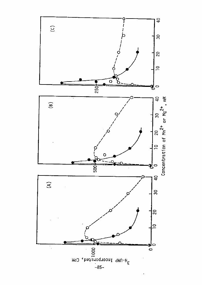
#
g
NO
po
oN
m
8 9wdo `pe;plodaoDur dwn-HE
-85-
Ri:iil
CON,i
-+outnEts
+NE=E
bo=- .9 x 'iio fi 8 88co.,
NO
.o
oo
+

g loo v v- s N g ts 8 H k so en;t
'
Oo 10 20 30 40 concentration of Mn2+ or Mg2+
Fig, 6. Effects of Mn2+ and Mg2+ on RNA polymerase II Activity
from 3DD6 Cells.Indicated concentration of Mn2+ or Mg2+ was added instead of 2 mM
of MnC12• About O.08 M ÅqNH4)2S04 and O,17 mM Mg2+, originating
from enzyme solution after DEAE-Sephadex chromatography, were
included in all reaction mixture. rncubation lasted for 10 minat 37oc with 1 uci of 3H-uTp. Åq-e-) and (-o-) indicate the Mn2+and Mg2+ effects, respectively.
-86-

ls"04
-hp'rÅr
'rpvÅq
oÅr-l-
Pnt
Pwct
1oo
oo
(A)
concentration of (NH4)2S04, M
O.2
Fig. 7. Effect ot' (NH4)2S04 on RNA Polymer'ase Ir Activity.Incubation lasted for 15 min with 2 uCi of 3H-UTp and given
concentration of (NH4)2S04 in the presence of 1 mM MnC12 (A)
and 10 mM MgC12 (B). (-e-) shows RPase II activity from 3DD6cells; (-O-), from 3DK5 cells; (-A-), from RS cells.
-87-
DrscussloN
Changes in protein and RNA syntheses have been found during
shoot formation from cultured tobacco cens by cytokinins23) and
during callus forTnation from Jerusalem artichoke tubers26). To
clarify what this transcriptive ' regulation is, RPases from the
different stages of shoot formation were purified and characterized.
The RPases of cultured tobacco cellsC3DD6 and 3DK5 cells) and re-
differentiated shoots CRS cells) were shown to have multiple forms
as in many other plants. The total RPase II activity of the 3DD6
cells was higher than that of the 3DK5 and RS cells. Changes in
the levels of RPase II activities have been observed during theembryogenesis of ,xlsgnsmyE27) and the germination of wheat14), one
possibility is that increased or decreased sLt novo synthesis
of RPase II occurs duning shoot formation.
The RPase IIs purified from 3DD6, 3DK5 and RS cells possessed
differing properties for template specificity (Fig. 4), their
divalent cation requirements CFig. 5). and their behaviors toward
ionic strength CFig. 7). The difference in transcribing activity
for pea and calf thymus DNAs CFig.'4) indicates that the RPase IIs
from 3DD6 and RS cells differ in their regulatory functions.
These RPase 11s from the 3DD6 and 3DK5 cells were repressed at a
high concentration o"F Mg2+, especially in the presence of CNH4)2S04,
wh"e less repression was found in the RPase Ir from RS cells.
RPase II activity decreased rapidly as the concentration of
-88-
(NH4)2S04 increased. In preliminary experiments the RNAs synthe-
sized by RPase lIs from 3006 and RS cells with pea DNA appeared
to be different in molecular size. Changes in divalent cations
and the ionic strength of the reaction mixture are known to produce
a qualitative difference in the RNA synthesized by various animal
nuclear preparations 28 ). These findings might imply the presence
of polymerases with different ionic requirements or they could be
the results of the effects of divalent cations and ionic strength
on the template, or on other regulatory components of the systems.
The detailed relationships between auxins and cytokinins and
RPase II activity are not clear. But auxins or cytokinins had
no direct effect on RPase II activity ~ vitro (Table IV) nor did
GA 20 ) . Non-histone proteins29 ) and some types of activating
factors 18 ,19,30,3l) involved with auxins or kinetin, have been
reported to be regulatory factors for plant RPases.
Two molecular species for RPase II have been reported. Thus,
an RPase II with a low molecular weight may be derived from one
with a high molecular weight by proteolysis8 ,13,30).
Because of the different properties of the RPase lIs from the
three kinds of cells, modifications in the structure of the enzyme
are presumed. However, it is not yet clear what kinds of modifi
cations exist in cUltured cells.
-89-
SUMMARY
During tobacco (Nitotiana tabacum var. Bright Yellow) cell
culture, cytokinins can induce shoot formation. As the syntheses
of specific proteins progressed during shoot formation from cultured
cells, the RNA polymerases (RPases) were extracted and partially
purified from 3 day-old cells cultured with 2,4-D (control) or
kinetin, and from redifferentiated shoots. RPases from cultured
tobacco cells were composed of at least 2 kinds, RPase I and II.
RPase I was low activity and very labile. Total RPase activities
changed during shoot formation. Furthermore, the isolated RPase
lIs from redifferentiating shoot cells showed properties which
differed in template specificity, divalent cation requirements and
behavior to ionic strength when compared with the control. In
contrast, no direct effects of auxins and cytokinins on RPase II
activities were found in vitro. It is presumed that changes in
de~ RPase II synthesis and some modifications of RPase II occur
during shoot formation.
REFERENCES
1) Sekiya, J. and Yamada, Y., Agr. Bio1. Chern., 38, 110l (1974)2) Roeder, R. G. and Rutter, W. J., Nature, 224, 234 (1969)3) Jacob, S. T., Prog. Nucleic Acid Res. Mol. Biol., "ll, 93
(1913)
4) stout, E. R. and Mans, R. J., Biochim. Biophys. Acta~ 134,327 (1967)
5) Polya, G. M. and Jagendorf, A. T., Arch. Biochem. Biophys.,
-90-

l46, 635 C1971)
6) Mondal, H., Mdndal, R. K. and Biswas, B. B,, Eur. J. Biochem.,
25, 463 (1972)
7) Strain, G. C., Mullinix, K. P. and Bogorad, L., Proc. Nat.
Acad. Sci., USA, 68, 2647 (1971) 8) Mullinix, K. P,, Strain, G. C, and Bogorad, L., Proc. Nat.
Acad. Sci., USA, 70, 2386 (1973) 9) Horgen, P. A. and Key, j. 1., Biochim. Biophys. Acta, 294,
227 (1973)10) Teissere, M., Penon, P. and Ricard, J., FEBS Let., 30, 65
(1973)
11) Sasaki, Y., Sasaki, R., Hashizume, T. and Yamada, Y., Biochem.
Biophys. Res Commun., 50, 785 Åq1973)12) Jendrisak, J. and Becker, W. M., Biochim. Biophys, Acta, 314,
48 (1973)
13) Jendrisak, J. and Becker, W. M., Biochern. J., 139, 771 (1974)
14) Mazus, B., Phytochem., 12, 2809 (1973)
15) Rizzo, P. J., Cherry, J. H., Pedersen, K. and Dunham, V. L.,
Plant Physiol., 54, 349 (1974)16) Glicklich, D., Jendrisak, J. and Becker, W. M., Plant Physiol.,
54, 356 (1974)
17) Fukasawa, H. and Mori, K., Plant Sci, Let., 2, 391 (1974)
18) Mondal, H., Mandal, R. K. and Biswas, B. B., Nature, New Biol.,
240, 111 (1972År
19) Hardin, J. W., Cherry, J. H., Morre, D. J. and Lembi, C. A.,
Proc. Nat. Acad. Sci., USA, 69, 3146 (1972)
20) Hou, G. C. and Pillay, D. I. N., Phytochem., 14, 403 (1975)
21) Skoog, F. and Miller, C. O., Symp, Soc. Exp. Biol., ll,
118 0957)22) Yamada, Y., •Sekiya, J. and Koshimizu, K., Phytochem., 11,
1Ol9 (.1972)
23) Sekiya,. J. and Yamada, Y., Bull. Inst. Chem. Res., Kyoto Univ.,
52, 246 (1974)
-91 -
24)
25)
26)
27)
28)
29)
30)
31)
Yamada, Y., Kiso, K., Sekiya, J. and Yasuda, T., Agr. Biol.
Chem.i'36, 1055 (1972)
Linsmaier, E. M. and Skoog, F., Physiol. Plant. Lt,100 C1965)
Yasuda, T., Yajima, Y. and Yamada, Y., Plant Cell Physiol,,
15, 321 (1974)
Roeder, R. G., J. Biol. Chem., 249, 249 (1974)
Widnell, C. C. and 'Tata, J. R., Biochim. Biophys. Acta, 123,
478 (1966)
Matsumoto, H., Gregor, D. and Reinert, J., Phytochem., Lt,
41 Åq1975)
Matthysse, A. G. and Phillips, C., Proc. Nat. Acad. Sci.,
USAi 63, 897 C1969)
Matthysse, A. G. and Abrams, M., Biochim. Biophys. Acta, 199,
511 (1970)
-92-
CHAPTER VI
CELL-CELL INTERACTION IN CULTURED TOBACCO CELLS.
-THE INDUCTION OF PHENYLALANINE AMMONIA LYASE IN
CULTURED TOBACCO CELLS1).-
INTRODUCTION
Phenylalanine ammonia lyase(PAL) plays an important role in
plants. Many phenolics~ including a large number of secondary
metabolites~ are developed from the t-cinnamic acid produced from
~-phenylalanine by a PAL catalyzed reaction. Since Koukol et ~2)
discovered this enzyme, a number of reports on the interrelationship
of PAL with the synthesis of phenolic ~ompounds have been made.
However~ PAL responds to a wide variety of stimuli in a short time;
especially to illumination3,4). In cultured cells~ Rubery et ~5) ~
Thorpe et ~6), Davies?), Gregor et ~8) and Innerarity et ~9)
reported evidence of PAL or a large increase in PAL activity due
to different regulations. This chapter describes PAL induction
after the transfer of cells to a fresh culture medium and cell-
cell interaction is discussed.
MATERIALS AND METHODS
Cell cul tures.
Cultured tobacco cells(Nicotiana tabacum var. Bright Yellow)~
Strain T5S~ derived from tobacco callus Strain T5 and cultured in
a liquid medium containing 10-6 M2,4-D were used. The basic
-93-

composition of the medium was the same as that of Linsmaier and
skoogll). All cells were cultured in suspension under reciprocal
shaking (80 rlmin) at 250C in the dark.
The inoculate of cells was about 10 g in 20 ml of liquid
medium in a 100 ml Erlenmeyer flask. Growth was measured by the
turbidity at 610 nm. Protein content was also measured during
growth. Protein was extracted with O.05 M Tris-HCI buffer, pH 7.8,
then precipitated by 5 % trichloroacetic acid. The precipitate
was collected by centrifugation and dissolved in O.2 N NaOH.
Protein content was determined by the method described by Lowry
et al12).
Extraction of PAL.
The enzyme was extracted from acetone powder. To do this,
about 10 g of cultured cells was ground in a mortar with acetone
previously chilled to -20eC. The homogenate was filtered on a
glass filter funnel and its residue was washed with cold acetone,
then dried Lt vacuo. The acetone powder was homogenized with
75 rnM borate buffer, pH 8.8, and Polyclar AT. The homogenate was
centrifuged at 10,OOO g for 20 min. The supernatant was used as
the enzyme solution.
!EE!!IX!!!9:aAEE9)L.
The spectrophotometric rnethod13) was used for the enzyme
assay. The reaction mlxture contained 112 u moles of borate at
pH 8.8; 30 umoles of L-phenylalanine; 1.5 ml of enzyme solution
(O.4-O.7 mg of protein) in a volume of 3 ml. The reaction at 370C
-94-
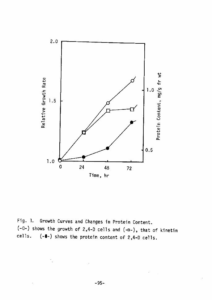
oPasct
=v3o"eoÅr
-t--
vorocr
1
2.0
.5
1.0o 24 48
Time, hr72
s e1,O NM Ea •,-",
di t vo .s .il e a
O.5
Fig. 1
(-o-)
cel1s.
. Growthshows the
C-e-)
Curves and Changes in Protein Content,
growth of 2,4-D cells and (-e-År, that of
shows the protein content of 2,4-D cells.kinetin
-95-
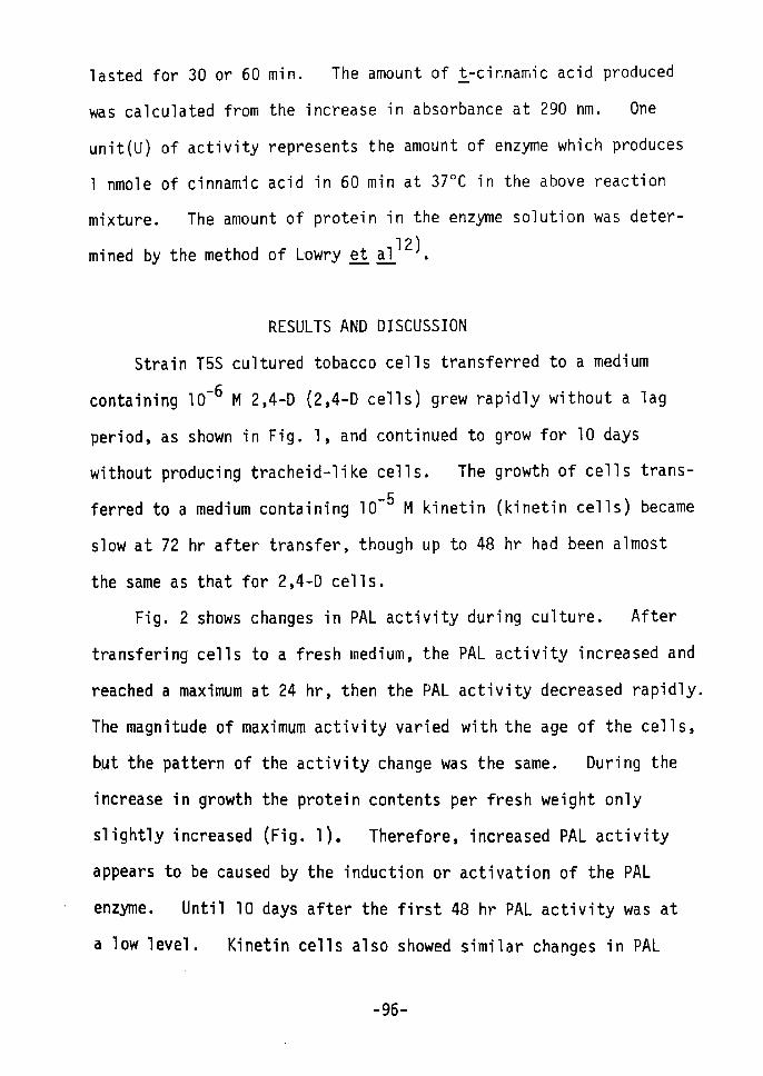
lasted for 30 or 60 min. The amount of !-cinnamic acid produced
was calculated from the increase in absorbance at 290 nm. One
unit(U) of activity represents the amount of enzyme which produces
1 nmole of cinnamic acid in 60 min at 370C in the above reaction
mixture. The amount of protein in the enzyme solution was deter- 12)mined by the method of Lowry gt sul. •
RESULTS AND DISCUSSION
Strain T5S cultured tobacco cells transferred to a medium
containing 10-6 M 2,4-D Åq2,4-D cells) grew rapidly without a lag
period, as shown in Fig. 1, and continued to grow for 10 days
without producing tracheid-like cells, The growth of cells trans-
ferred to a medium containing lo-5 M kinetin (kinetin cells) became
slow at 72 hr after transfer, though up to 48 hr had been almost
the same as that for 2,4-D cells.
Fig. 2 shows changes in PAL activity during culture. After
transfering cells to a fresh medium, the PAL activity increased and
reached a maximum at 24 hr, then the PAL activity decreased rapidly.
The magnitude of maximum activity varied with the age of the cells,
but the pattern of the activity change was the same. During the
increase in growth the protein contents per fresh weight only
slightly increased (Fig. 1). Therefore, increased PAL activity
appears to be caused by the induction or activation of the PAL
enzyme. Until 10 days after the first 48 hr PAL activity was at
a low level. Kinetin cells also showed similar changes in PAL
-96-

s::.....aJ+'o~
c-100t:TIE.......=:l..>,+'.....>.....+'u«-'«0.
ao 24 48 72
Time, hr
I
120 240
. fig. 2. Changes in PAL Activity in 2,4-0 Cells.
-97-

.5.i!l
9amEN=
-hv-t-År
-t"
voq"(a
1OO
o 12
e ---.-LuL-o
24 36
Time, hr
Fig. 3.
by Cel1
added to
Effect of Cycloheximide
Transfer. Arrows show the medium.
CCHr) on
the time
the PAL
when CHI
Activity(5xlo-5
Induced
M) was
-98d
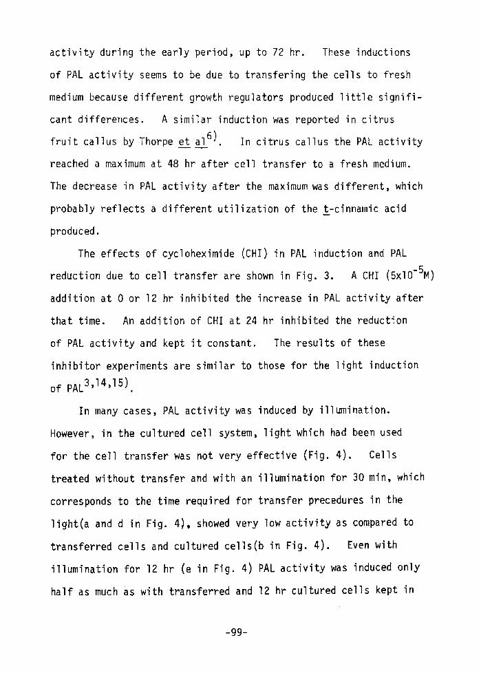
activity during the early period, up to 72 hr. These inductions
of PAL activity seems to be due to transfering the cells to fresh
medium because different growth regulators produced little signifi-
cant differences. A similar induction was reported in citrus
fruit callus by Thorpe et ~6). In citrus callus the PAL activity
reached a maximum at 48 hr after cell transfer to a fresh medium.
The decrease in PAL activity after the maximum was different, which
probably reflects a different utilization of the t-cinnamic acid
produced.
The effects of cycloheximide (CHI) in PAL induction and PAL
reduction due to cell transfer are shown in Fig. 3. A CHI (5xlO-5M)
addition at 0 or 12 hr inhibited the increase in PAL activity after
that time. An addition of CHI at 24 hr inhibited the reduction
of PAL activity and kept it constant. The results of these
inhibitor experiments are similar to those for the light induction
of PAL3,14,15).
In many cases, PAL activity was induced by illumination.
However, in the cultured cell system. light which had been used
for the cell transfer was not very effective (Fig. 4). Cells
treated without transfer and with an illumination for 30 min, which
corresponds to the time required for transfer precedures in the
light(a and d in Fig. 4), showed very low activity as compared to
transferred cells and cultured cells(b in Fig. 4). Even with
illumination for 12 hr (e in Fig. 4) PAL activity was induced only
half as much as with transferred and 12 hr cultured cells kept in
-99-

Treatment
Cell Transfer
Treatment Time, hr , PAL Activity, Ulmg protein
Fig. 4. Effect of Illumination on PAL Activity.(a-f) show the different treatments in various experiments with
cell culture: a) 30 min of illumination without cell transfer;
b) 30 min of illumination and a continuous 23.5 hr in the dark;
c) cell transfer and 24 hr in the dark; d) 30 min of illumination
without cell transfer; e) 12 hr of illuminaiton; f) cell transfer
and 12 hr in the dark. rn treatments, aand -indicatecell culture in light and dark conditions, respectively. Time O
is the point of cell transfer.
--1 OO-
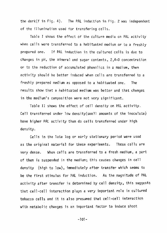
the dark(f in Fig. 4). The PAL induction in Fig. 2 was independent
of the illumination used for transfering cells.
Table I shows the effect of the culture media on PAL activity
when cells were transferred to a habituated medium or to a freshly
prepared one. If PAL induction in the cultured cells is due to
changes in pH, the mineral and sugar contents, 2,4-D concentration
or to the reduction of accumUlated phenolics in a medium, then
activity should be better induced when cells are transferred to a
freshly prepared medium as opposed to a habituated one. The
results show that a habituated medium was better and that changes
in the medium's composition were not very significant.
Table II shows the effect of cell density on PAL activity.
Cell transferred under low density(small amounts of the inoculate)
have higher PAL actiVity than do cells transferred under high
density.
Cells in the late log or early stationary period were used
as the original material for these experiments. These cells are
very dense. When cells are transferred to a fresh medium, a part
of them is suspended in the medium; this causes changes in cell
density (high to low), immediately after transfer which seems to
be the first stimulus for PAL induction. As the magnitude of PAL
activity after transfer is determined by cell density, this suggests
that cell-cell interaction plays a very important role in cultured
tobacco cells and it is also presumed that cell-cell interaction
with metabolic changes is an important factor to induce shoot
-101-
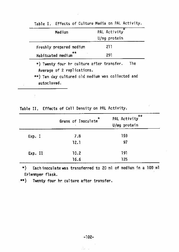
Table I. Effects of Culture Media on PAL Activity.
Medium PAL
UImg
Activity
protein
Freshly prepared
Habituated medium
medium
tde -211
291
*) Twenty four hr culture after
Average of 2 replications.
** ) Ten day cultured old medium
autoc1aved.
transfer.
was collected
The
and
Table II. Effects of Cell Density on PAL Activity.
**Grams of Inoculate
t PAL
U/mg
Activity
pretei n
Exp. r
Exp. Ir
7
12
10
16
.
.
.
.
8
1
2
6
159
97
191
125
t) Eachinoculate was transferred to 20 ml of
Erlenmyer flask.
-nk) Twenty four hr.culture after transfer,
medium in a 100 ml
-' 102-

formation.
SUMMARY
The induction of phenylalanine ammonia lyase (PAL) activity
in a suspension of cUltured tobacco cells(2,4-D cells) was observed
when the cells were transferred to fresh medium. It was concluded
that this induction of PAL activity was caused by changes in cell
density in the medium and was independent of illumination during
cell transfer and of changes in the composition of the medium.
Hence, it is considered that cell-cell interaction is important in
cultured cells.
REFERENCES
1) Yamada, Y. and Sekiya, J., Phytomorphology, in press (1975)2) Koukol, J. and Conn, E., J. Biol. Chern., 236,2692 (1961)3) Zucker, M., Ann. Rev. Plant Physiol., 23, 133 (1972)4) Carom, E. L. and Towers, G. H. N., Phytochem., }£, 961 (1973)5) Rubery, P. H. and Fosket, D. E. Planta, 87, 54 (1969)6) Thorpe, T. A., Maier, V. P. and Hasegawa, S., Phytochem.,
]Q, 711 (l971)
7) Davies, M. E., Planta, 104, 50 (1972)8) Gregor, H. D. and Reinert, J., Protoplasma, 74, 307 (1972)9) Innerarity, L. T., Smith, E. C. and Wender, S. H., Phytochem.,
11, 83 (1972)10) Yamada, Y., Kiso, K., Sekiya, J. ~nd Yasuda, T., Agr. Biol.
Chern., 36, 1055 (1972)11) Linsmaier,· E. M. and Skoog, F., Physiol. Plant., ~, 100
(1965)
-103-

12)
13)
14)
15)
Lowry, O. H., Rosebrough, N. J., Farr, A. L
J. Biol. Chem., 193, 265 (1951)
Zucker, M., Plant. Physiol.i A!tlL, 779 (1965)
Engelsma, G., Naturwissenschaften, :Sf4;, 319
Zucker, M., Plant Physiol., Agt., 365 (1968)
. and
(1 967)
Randal1, R. J.,
-1[04-
CONCLUSION
Recent developments in plant tissue and cell culture have
provided a basis for an experimental system for differentiation.
To date, results indicate that cultured plant cells produce de
differentiation of differentiated tissue and cells and, in addition,
that some organs or the whole plant can be regenerated from de
differentiated cultured cells under appropriate condition (re
differentiation). The purpose of this study was to obtain infor
mation on the biochemical mechanism of shoot redifferentiation in
cultured tobacco cells and on some properties of these cultured
cells.
When 2,4-D cells or IBA cells were cultured on a medium with
5xlO-5 Mof kinetin, zeatin or (±)-dihydrozeatin with little or
no auxin, shoot formation was observed after 6 to 8 weeks of
culture. The natural cytokinins, zeatin and (±)-dihydrozeatin
showed higher activity for shoot formation than did the synthetic
cytokinin, kinetin. Cytokinin was required continuously throughout
the culture for shoot formation.
Growth rates for the zeatin and kinetin cells were reduced
after several days of culture with cytokinin, while the dry material
percentage increased.
Morphological changes, cell organization and tracheid-like
cell production were simultaneously observed about 1-2 weeks after
inoculation to the zeatin medium. RNase activity also changed in
-105-
correspondence to a change in the growth. The initiation of shoot
redifferentiation may have already begun morphologically and physio-
logically one week after transfer to the cytokinin medium.
(Å})--Dihydrozeatin and optically active cytokinins were tested
for their ability to induce shoot redifferentiation in cultured
tobacco cells. (Å}År-Dihydrozeatin was active in inducing shoot
formation. Levorotatory cornpounds were active in inducing shoot
formation, while the corresponding dextrorotatory compounds were
inactive at all the concentrations tested. This suggests that the
N6 position of the cytokinins binds to a stereo-specific receptor
site to bring about shoot formation.
Kinetin-s-14c was incorporated into cultured cells and sub-
sequently metabolized. About 20 % of its radioactivity was located
in the 80 % EtOH insoluble materials and 80 %, in the 80 % EtOH
soluble materials. About 50 % of the EtOH insoluble radioactivity
was found in RNA and 3-30 % in protein. The EtOH soluble materials
were separated into 5 main fractions. It is presumed that cyto-
kinins are metabolized after their incorporation into cells and
that free cytokinin or some metabolite acts as a trigger for shoot
redifferentfiation with some types of proteins.
RNA, buffer-soluble proteins and DNA increased in this order
in zeatin cells during the early stages of shoot formation. A
different incorporation pattern of i4c-ieucine into the protein
fraction was observed in zeatin cells when compared to for 2,4-D
cells, using DEAE cellulose column chromatography.
-1 06-
The resu]ts of disc electrophoresis of the proteins in the
2,4-D and zeatin cells indicated that qualitative differences
occurred during shoot formation, These qualitative changes in
protein synthesis were also verified by immunological studies
which indicated that cytokinins caused the induction of certain
characteristic proteins vvhen leaves began to form from cultured
cells Ut!. vitro, During shoot formation some proteins which are
synthesized for cell proliferation were repressed. These changes
in protein synthesis suggests the existence of transcriptive
regulation during organogenesis.
One participant in transcription, the RNA polymerases(RPases),
was investigated. RPases were found in multiple forms in cultured
tobacco cells. RPase I showed very low activity and was very
Tabile while RPase II was relatively stable. RPase II was influ-
enced by the degree of redifferentiation. The levels of RPase II
activities varied during shoot formation. Template specificity,
the requirement for divalent cations and the behavior toward the
ionic environment changed according to the degree of redifferenti-
ation. The RPase II in dedifferentiated cultured cells is believed
to differ from other plant RPase Irs. Thus,the existence of
transcriptional regulation by RPase during shoot formation is
indicated, In addition, the fact that the RPase in cultured cells
had different properties involves a problem: the difficulty of
preserving a cell-specific phenotype when cell culture is used for
practical purposes.
-107-

When dedifferentiated cultured tobacco cells were cultured with
relatively high concentrations of cytokinins, shoot redifferenti-
ation occurred. Cytokinin is incorporated into the cell and free
cytokinin or some metabolite acts as a trigger for shoot rediffer-
entiation. At this time the cytokinin (or its metabolite) takes
part in transcriptional tegulation through the RPases probably
with a cytokinin-reactive protein. SubsequentTy, organ-specific
or tissue-specific RNA and protein are synthesized. Simultaneously,
such morphological changes as cell organization and the production
of tracheid-like cells take place. These changes have begun 1 to
2 weeks after the transfer to the cytokinin medium. These changes
also enhance the process of the development of organ formation and
produce an organ specific shape.
-1 08-
ACKNOELEoGEMENT
The author wishes to thank Or. Eiichi Takahashi. Professor of
Kyoto University. for his kind guidance and encouragement during
the cource of this work. The author also greatly appreciates to
Dr. Yasuyuki Yamada, Associate Professor of Kyoto University. for
his continuous guidance and advice in carrying out this investi
gation. Thanks are due to members of Laboratory of Plant Nutri
tion, Department of Agricultural Chemistry, Kyoto University, for
their valuable discussion and encouragement.
The author thanks Dr. Shunichi Yamada. Professor of Tokyo
University, for a gift of auxin analogues and also Dr. Kooichi
Koshimizu. Professor of Kyoto University. for a gift of cytokinins.
The author is indebted to Dr. Yukiko Sasaki. Department of Food
Science and Technology of Kyoto University, for a technical advice
for the work of RNA polymerase.
-109-