Pulmonary Sarcoidosis Presenting with Acute Respiratory ...
5
1 doi: 10.2169/internalmedicine.4624-20 Intern Med Advance Publication http://internmed.jp 【 CASE REPORT 】 Pulmonary Sarcoidosis Presenting with Acute Respiratory Failure: A Report of a Case Diagnosed by Endobronchial Ultrasound-guided Transbronchial Needle Aspiration on Ventilation after Intubation Jumpei Taniguchi, Kei Nakashima, Hiroyuki Ito, Yu Tanaka, Ayumu Otsuki, Akihiro Shiroshita, Michinori Yoshimi, Norihiko Kubota and Masahiro Aoshima Abstract: Sarcoidosis is a multisystem granulomatous disease of unknown etiology and is pathologically character- ized by non-caseating granulomas in the organs involved. We herein report a case of sarcoidosis in a Japa- nese woman with acute respiratory failure, diagnosed using endobronchial ultrasound-guided transbronchial needle aspiration (EBUS-TBNA) on the ventilator after intubation. Only a few cases of previously undiag- nosed sarcoidosis presenting acute respiratory failure have been reported. It is important to be aware that un- diagnosed sarcoidosis may present with acute respiratory failure. Therefore, EBUS-TBNA under mechanical ventilation may be useful for the immediate diagnosis of patients. Key words: Sarcoidosis, acute respiratory failure, endobronchial ultrasound-guided transbronchial needle aspiration (EBUS-TBNA) (Intern Med Advance Publication) (DOI: 10.2169/internalmedicine.4624-20) Introduction Sarcoidosis is a multisystem granulomatous disease of un- known etiology. It most commonly affects the mediastinal lymph nodes, lungs, skin, liver, and eyes (1). It may also af- fect the heart, central nervous system, spleen, and upper res- piratory tract. The progress of pulmonary diseases is typically slow, and some do not progress. The progress of pulmonary sarcoido- sis leads to chronic interstitial lung disease accompanied by dyspnea, cough, and fatigue (2). Acute respiratory disorders are rare and usually occur in advanced sarcoidosis based on previous respiratory infectious diseases, like pneumonia or, more specifically, bronchospasm, in cases where the airways are hyperresponsive due to granulomatous inflammation or are injured due to fibrocystic pulmonary sarcoidosis (3). An acute initial presentation with respiratory failure in a previ- ously undiagnosed patient is rare, and only a few cases have been reported (4-11). A sarcoidosis diagnosis is based on the compatible clini- cal and radiologic findings supported by histological find- ings in one or more organs of noncaseating epithelioid cell granulomas while excluding other similarly developing dis- eases (1). For patients with parenchymal lung disease or ap- parent mediastinal adenopathy, flexible bronchoscopy with bronchoalveolar lavage (BAL), an endobronchial biopsy, and a transbronchial biopsy (TBLB) are the traditional methods for a minimally invasive diagnosis of sarcoidosis. Recent re- ports have confirmed the diagnostic value of endobronchial ultrasound-guided transbronchial needle aspiration (EBUS- TBNA) for a mediastinal lymph node biopsy in patients with suspected sarcoidosis (12, 13). Compared to a trans- bronchial biopsy, TBNA of the mediastinal nodes by en- dobronchial or esophageal ultrasound results in a higher di- agnostic yield, is less invasive, and is safer (14, 15). How- ever, there have been no reported cases of sarcoidosis diag- nosis by EBUS-TBNA on the ventilator after intubation, and there are very few case reports of sarcoidosis presenting with acute respiratory failure. Department of Pulmonology, Kameda Medical Center, Japan Received: February 12, 2020; Accepted: April 20, 2020; Advance Publication by J-STAGE: June 15, 2020 Correspondence to Dr. Jumpei Taniguchi, [email protected]
Transcript of Pulmonary Sarcoidosis Presenting with Acute Respiratory ...
1
doi: 10.2169/internalmedicine.4624-20
Intern Med Advance Publication
http://internmed.jp
【 CASE REPORT 】
Pulmonary Sarcoidosis Presenting with Acute RespiratoryFailure: A Report of a Case Diagnosed by EndobronchialUltrasound-guided Transbronchial Needle Aspiration on
Ventilation after Intubation
Jumpei Taniguchi, Kei Nakashima, Hiroyuki Ito, Yu Tanaka, Ayumu Otsuki,
Akihiro Shiroshita, Michinori Yoshimi, Norihiko Kubota and Masahiro Aoshima
Abstract:Sarcoidosis is a multisystem granulomatous disease of unknown etiology and is pathologically character-
ized by non-caseating granulomas in the organs involved. We herein report a case of sarcoidosis in a Japa-
nese woman with acute respiratory failure, diagnosed using endobronchial ultrasound-guided transbronchial
needle aspiration (EBUS-TBNA) on the ventilator after intubation. Only a few cases of previously undiag-
nosed sarcoidosis presenting acute respiratory failure have been reported. It is important to be aware that un-
diagnosed sarcoidosis may present with acute respiratory failure. Therefore, EBUS-TBNA under mechanical
ventilation may be useful for the immediate diagnosis of patients.
Key words: Sarcoidosis, acute respiratory failure, endobronchial ultrasound-guided transbronchial needle
aspiration (EBUS-TBNA)
(Intern Med Advance Publication)(DOI: 10.2169/internalmedicine.4624-20)
Introduction
Sarcoidosis is a multisystem granulomatous disease of un-
known etiology. It most commonly affects the mediastinal
lymph nodes, lungs, skin, liver, and eyes (1). It may also af-
fect the heart, central nervous system, spleen, and upper res-
piratory tract.
The progress of pulmonary diseases is typically slow, and
some do not progress. The progress of pulmonary sarcoido-
sis leads to chronic interstitial lung disease accompanied by
dyspnea, cough, and fatigue (2). Acute respiratory disorders
are rare and usually occur in advanced sarcoidosis based on
previous respiratory infectious diseases, like pneumonia or,
more specifically, bronchospasm, in cases where the airways
are hyperresponsive due to granulomatous inflammation or
are injured due to fibrocystic pulmonary sarcoidosis (3). An
acute initial presentation with respiratory failure in a previ-
ously undiagnosed patient is rare, and only a few cases have
been reported (4-11).
A sarcoidosis diagnosis is based on the compatible clini-
cal and radiologic findings supported by histological find-
ings in one or more organs of noncaseating epithelioid cell
granulomas while excluding other similarly developing dis-
eases (1). For patients with parenchymal lung disease or ap-
parent mediastinal adenopathy, flexible bronchoscopy with
bronchoalveolar lavage (BAL), an endobronchial biopsy, and
a transbronchial biopsy (TBLB) are the traditional methods
for a minimally invasive diagnosis of sarcoidosis. Recent re-
ports have confirmed the diagnostic value of endobronchial
ultrasound-guided transbronchial needle aspiration (EBUS-
TBNA) for a mediastinal lymph node biopsy in patients
with suspected sarcoidosis (12, 13). Compared to a trans-
bronchial biopsy, TBNA of the mediastinal nodes by en-
dobronchial or esophageal ultrasound results in a higher di-
agnostic yield, is less invasive, and is safer (14, 15). How-
ever, there have been no reported cases of sarcoidosis diag-
nosis by EBUS-TBNA on the ventilator after intubation, and
there are very few case reports of sarcoidosis presenting
with acute respiratory failure.
Department of Pulmonology, Kameda Medical Center, Japan
Received: February 12, 2020; Accepted: April 20, 2020; Advance Publication by J-STAGE: June 15, 2020
Correspondence to Dr. Jumpei Taniguchi, [email protected]
Intern Med Advance Publication DOI: 10.2169/internalmedicine.4624-20
2
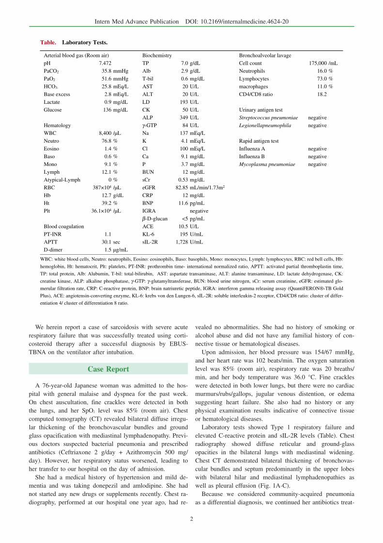
Table. Laboratory Tests.
Arterial blood gas (Room air) Biochemistry Bronchoalveolar lavage
pH 7.472 TP 7.0 g/dL Cell count 175,000 /mL
PaCO2 35.8 mmHg Alb 2.9 g/dL Neutrophils 16.0 %
PaO2 51.6 mmHg T-bil 0.6 mg/dL Lymphocytes 73.0 %
HCO3- 25.8 mEq/L AST 20 U/L macrophages 11.0 %
Base excess 2.8 mEq/L ALT 20 U/L CD4/CD8 ratio 18.2
Lactate 0.9 mg/dL LD 193 U/L
Glucose 136 mg/dL CK 50 U/L Urinary antigen test
ALP 349 U/L Streptococcus pneumoniae negative
Hematology γ-GTP 84 U/L Legionellapneumophila negative
WBC 8,400 /μL Na 137 mEq/L
Neutro 76.8 % K 4.1 mEq/L Rapid antigen test
Eosino 1.4 % Cl 100 mEq/L Influenza A negative
Baso 0.6 % Ca 9.1 mg/dL Influenza B negative
Mono 9.1 % P 3.7 mg/dL Mycoplasma pneumoniae negative
Lymph 12.1 % BUN 12 mg/dL
Atypical-Lymph 0 % sCr 0.53 mg/dL
RBC 387×104 /μL eGFR 82.85 mL/min/1.73m2
Hb 12.7 g/dL CRP 12 mg/dL
Ht 39.2 % BNP 11.6 pg/mL
Plt 36.1×10⁴ /μL IGRA negative
β-D-glucan <5 pg/mL
Blood coagulation ACE 10.5 U/L
PT-INR 1.1 KL-6 195 U/mL
APTT 30.1 sec sIL-2R 1,728 U/mL
D-dimer 1.5 μg/mL
WBC: white blood cells, Neutro: neutrophils, Eosino: eosinophils, Baso: basophils, Mono: monocytes, Lymph: lymphocytes, RBC: red bell cells, Hb:
hemoglobin, Ht: hematocrit, Plt: platelets, PT-INR: prothrombin time- international normalized ratio, APTT: activated partial thromboplastin time,
TP: total protein, Alb: Alubumin, T-bil: total-bilirubin, AST: aspartate transaminase, ALT: alanine transaminase, LD: lactate dehydrogenase, CK:
creatine kinase, ALP: alkaline phosphatase, γ-GTP: γ-glutamyltransferase, BUN: blood urine nitrogen, sCr: serum creatinine, eGFR: estimated glo-
merular filtration rate, CRP: C-reactive protein, BNP: brain natriuretic peptide, IGRA: interferon gamma releasing assay (QuantiFERON®-TB Gold
Plus), ACE: angiotensin-converting enzyme, KL-6: krebs von den Lungen-6, sIL-2R: soluble interleukin-2 receptor, CD4/CD8 ratio: cluster of differ-
entiation 4/ cluster of differentiation 8 ratio.
We herein report a case of sarcoidosis with severe acute
respiratory failure that was successfully treated using corti-
costeroid therapy after a successful diagnosis by EBUS-
TBNA on the ventilator after intubation.
Case Report
A 76-year-old Japanese woman was admitted to the hos-
pital with general malaise and dyspnea for the past week.
On chest auscultation, fine crackles were detected in both
the lungs, and her SpO2 level was 85% (room air). Chest
computed tomography (CT) revealed bilateral diffuse irregu-
lar thickening of the bronchovascular bundles and ground
glass opacification with mediastinal lymphadenopathy. Previ-
ous doctors suspected bacterial pneumonia and prescribed
antibiotics (Ceftriaxone 2 g/day + Azithromycin 500 mg/
day). However, her respiratory status worsened, leading to
her transfer to our hospital on the day of admission.
She had a medical history of hypertension and mild de-
mentia and was taking donepezil and amlodipine. She had
not started any new drugs or supplements recently. Chest ra-
diography, performed at our hospital one year ago, had re-
vealed no abnormalities. She had no history of smoking or
alcohol abuse and did not have any familial history of con-
nective tissue or hematological diseases.
Upon admission, her blood pressure was 154/67 mmHg,
and her heart rate was 102 beats/min. The oxygen saturation
level was 85% (room air), respiratory rate was 20 breaths/
min, and her body temperature was 36.0 °C. Fine crackles
were detected in both lower lungs, but there were no cardiac
murmurs/rubs/gallops, jugular venous distention, or edema
suggesting heart failure. She also had no history or any
physical examination results indicative of connective tissue
or hematological diseases.
Laboratory tests showed Type 1 respiratory failure and
elevated C-reactive protein and sIL-2R levels (Table). Chest
radiography showed diffuse reticular and ground-glass
opacities in the bilateral lungs with mediastinal widening.
Chest CT demonstrated bilateral thickening of bronchovas-
cular bundles and septum predominantly in the upper lobes
with bilateral hilar and mediastinal lymphadenopathies as
well as pleural effusion (Fig. 1A-C).
Because we considered community-acquired pneumonia
as a differential diagnosis, we continued her antibiotics treat-
Intern Med Advance Publication DOI: 10.2169/internalmedicine.4624-20
3
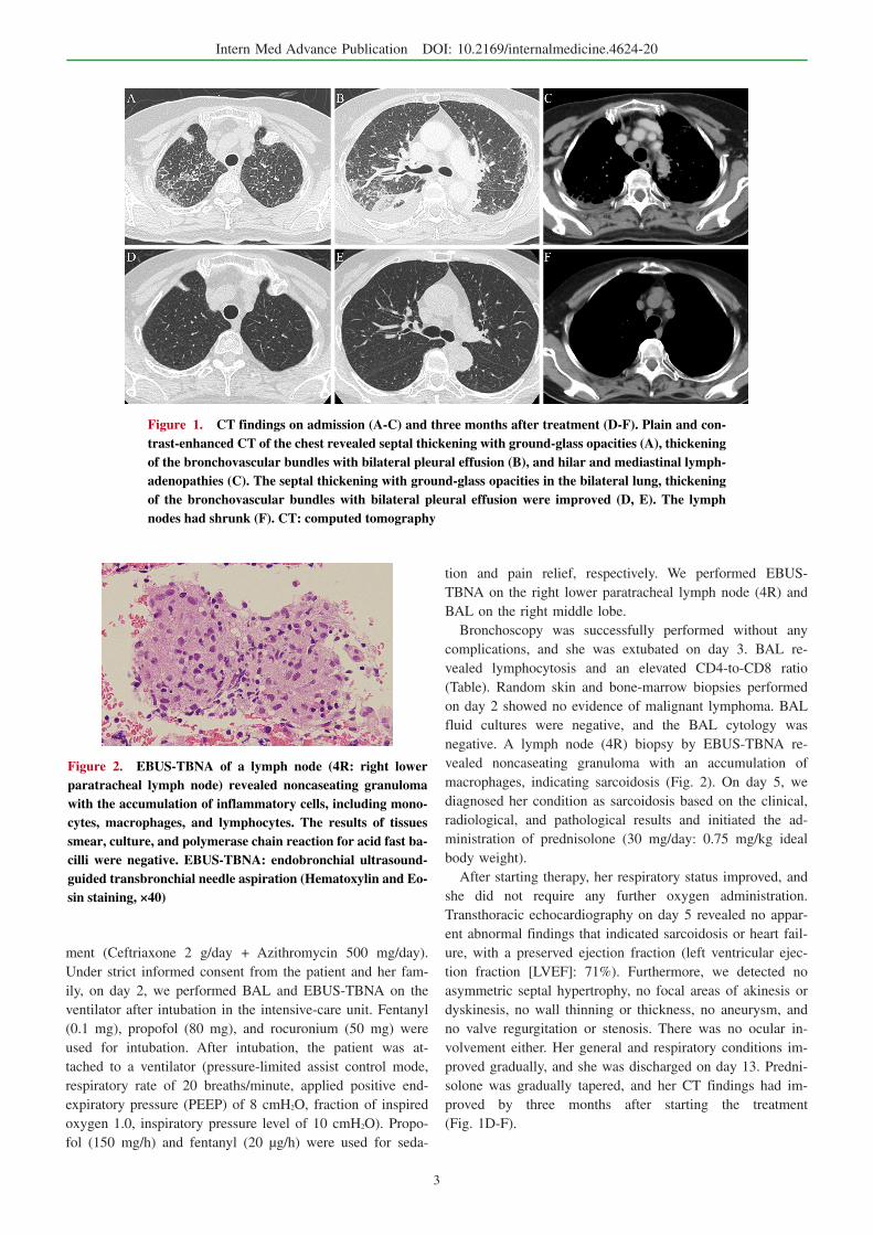
Figure 1. CT findings on admission (A-C) and three months after treatment (D-F). Plain and con-trast-enhanced CT of the chest revealed septal thickening with ground-glass opacities (A), thickening of the bronchovascular bundles with bilateral pleural effusion (B), and hilar and mediastinal lymph-adenopathies (C). The septal thickening with ground-glass opacities in the bilateral lung, thickening of the bronchovascular bundles with bilateral pleural effusion were improved (D, E). The lymph nodes had shrunk (F). CT: computed tomography
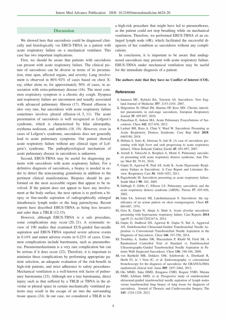
Figure 2. EBUS-TBNA of a lymph node (4R: right lower paratracheal lymph node) revealed noncaseating granuloma with the accumulation of inflammatory cells, including mono-cytes, macrophages, and lymphocytes. The results of tissues smear, culture, and polymerase chain reaction for acid fast ba-cilli were negative. EBUS-TBNA: endobronchial ultrasound-guided transbronchial needle aspiration (Hematoxylin and Eo-sin staining, ×40)
ment (Ceftriaxone 2 g/day + Azithromycin 500 mg/day).
Under strict informed consent from the patient and her fam-
ily, on day 2, we performed BAL and EBUS-TBNA on the
ventilator after intubation in the intensive-care unit. Fentanyl
(0.1 mg), propofol (80 mg), and rocuronium (50 mg) were
used for intubation. After intubation, the patient was at-
tached to a ventilator (pressure-limited assist control mode,
respiratory rate of 20 breaths/minute, applied positive end-
expiratory pressure (PEEP) of 8 cmH2O, fraction of inspired
oxygen 1.0, inspiratory pressure level of 10 cmH2O). Propo-
fol (150 mg/h) and fentanyl (20 μg/h) were used for seda-
tion and pain relief, respectively. We performed EBUS-
TBNA on the right lower paratracheal lymph node (4R) and
BAL on the right middle lobe.
Bronchoscopy was successfully performed without any
complications, and she was extubated on day 3. BAL re-
vealed lymphocytosis and an elevated CD4-to-CD8 ratio
(Table). Random skin and bone-marrow biopsies performed
on day 2 showed no evidence of malignant lymphoma. BAL
fluid cultures were negative, and the BAL cytology was
negative. A lymph node (4R) biopsy by EBUS-TBNA re-
vealed noncaseating granuloma with an accumulation of
macrophages, indicating sarcoidosis (Fig. 2). On day 5, we
diagnosed her condition as sarcoidosis based on the clinical,
radiological, and pathological results and initiated the ad-
ministration of prednisolone (30 mg/day: 0.75 mg/kg ideal
body weight).
After starting therapy, her respiratory status improved, and
she did not require any further oxygen administration.
Transthoracic echocardiography on day 5 revealed no appar-
ent abnormal findings that indicated sarcoidosis or heart fail-
ure, with a preserved ejection fraction (left ventricular ejec-
tion fraction [LVEF]: 71%). Furthermore, we detected no
asymmetric septal hypertrophy, no focal areas of akinesis or
dyskinesis, no wall thinning or thickness, no aneurysm, and
no valve regurgitation or stenosis. There was no ocular in-
volvement either. Her general and respiratory conditions im-
proved gradually, and she was discharged on day 13. Predni-
solone was gradually tapered, and her CT findings had im-
proved by three months after starting the treatment
(Fig. 1D-F).
Intern Med Advance Publication DOI: 10.2169/internalmedicine.4624-20
4
Discussion
We showed here that sarcoidosis could be diagnosed clini-
cally and histologically via EBUS-TBNA in a patient with
acute respiratory failure on a mechanical ventilator. This
case has two important implications.
First, we should be aware that patients with sarcoidosis
can present with acute respiratory failure. The clinical pic-
ture of sarcoidosis can be diverse in terms of its presenta-
tion, time span, affected organs, and severity. Lung involve-
ment is observed in 86%-92% of cases based on chest X-
ray, either alone or, for approximately 50% of cases, in as-
sociation with extra-pulmonary disease (16). The most com-
mon respiratory symptom is a chronic dry cough. Dyspnea
and respiratory failure are uncommon and usually associated
with advanced pulmonary fibrosis (17). Pleural effusion is
also very rare, but sarcoidosis with acute respiratory failure
sometimes involves pleural effusion (4, 5, 11). The acute
presentation of sarcoidosis is well recognized as Lofgren’s
syndrome, which is characterized by hilar adenopathy,
erythema nodosum, and arthritis (18, 19). However, even in
cases of Lofgren’s syndrome, sarcoidosis does not generally
lead to acute pulmonary symptoms. Our patient showed
acute respiratory failure without any clinical signs of Lof-
gren’s syndrome. The pathophysiological mechanism of
acute pulmonary disease in sarcoidosis is unknown.
Second, EBUS-TBNA may be useful for diagnosing pa-
tients with sarcoidosis with acute respiratory failure. For a
definitive diagnosis of sarcoidosis, a biopsy is needed in or-
der to detect the noncaseating granuloma in addition to the
pertinent clinical manifestations. Biopsies should be per-
formed on the most accessible organs that appear to be in-
volved. If the patient does not appear to have any involve-
ment at the body surface, the next option is to perform a bi-
opsy or fine-needle aspiration of radiographically enlarged
intrathoracic lymph nodes or the lung parenchyma. Recent
reports have described EBUS-TBNA as being less invasive
and safer than a TBLB (12-15).
However, although EBUS-TBNA is a safe procedure,
some complications may occur (20, 21). A systematic re-
view of 190 studies that examined EUS-guided fine-needle
aspiration and EBUS-TBNA reported severe adverse events
in 0.14% and minor adverse events in 0.22% of cases. Com-
mon complications include barotrauma, such as pneumotho-
rax. Pneumomediastinum is a very rare complication but can
be serious if it does occur (22). Therefore, it is important to
minimize these complications by performing appropriate pa-
tient selection, an adequate evaluation of the risk-benefit in
high-risk patients, and strict adherence to safety procedures.
Mechanical ventilation is a well-known risk factor of pulmo-
nary barotrauma (23). Although not a true barotrauma, direct
injury such as that suffered by a TBLB or TBNA in the al-
veolar or pleural space in certain mechanically ventilated pa-
tients may result in the escape of air into the surrounding
tissue spaces (24). In our case, we considered a TBLB to be
a high-risk procedure that might have led to pneumothorax,
as the patient could not stop breathing while on mechanical
ventilation. Therefore, we performed EBUS-TBNA of an en-
larged lymph node (4R), which facilitated the successful di-
agnosis of her condition as sarcoidosis without any compli-
cations.
In conclusion, it is important to be aware that undiag-
nosed sarcoidosis may present with acute respiratory failure.
EBUS-TBNA under mechanical ventilation may be useful
for the immediate diagnosis of a patient.
The authors state that they have no Conflict of Interest (COI).
References
1. Iannuzzi MC, Rybicki BA, Teirstein AS. Sarcoidosis. New Eng-
land Journal of Medicine 357: 2153-2165, 2007.
2. Shigemitsu H, Oblad JM, Sharma OP, Koss MN. Chronic intersti-
tial pneumonitis in end-stage sarcoidosis. European Respiratory
Journal 35: 695-697, 2010.
3. Panselinas E, Judson MA. Acute Pulmonary Exacerbations of Sar-
coidosis. Chest 142: 827-836, 2012.
4. Lashari BH, Raza A, Chan V, Ward W. Sarcoidosis Presenting as
Acute Respiratory Distress Syndrome. Case Rep Med 2018:
6465180, 2018.
5. Shibata S, Saito K, Ishiwata N, Ieki R. [A case of sarcoidosis pre-
senting with high fever and rash progressing to acute respiratory
failure]. Nihon Kokyuki Gakkai Zasshi 45: 691-697, 2007.
6. Arondi S, Valsecchi A, Borghesi A, Monti S. Pulmonary sarcoido-
sis presenting with acute respiratory distress syndrome. Ann Tho-
rac Med 11: 79-81, 2016.
7. Gupta D, Agarwal R, Paul AS, Joshi K. Acute Hypoxemic Respi-
ratory Failure in Sarcoidosis: A Case Report and Literature Re-
view. Respiratory Care 56: 1849-1852, 2011.
8. Pugazhenthi M. Sarcoidosis presenting as acute respiratory failure.
South Med J 98: 265, 2005.
9. Sabbagh F, Gibbs C, Efferen LS. Pulmonary sarcoidosis and the
acute respiratory distress syndrome (ARDS). Thorax 57: 655-656,
2002.
10. Sahn SA, Schwarz MI, Lakshminarayan S. Sarcoidosis: the sig-
nificance of an acinar pattern on chest roentgenogram. Chest 65:
684-687, 1974.
11. Gera K, Gupta N, Ahuja A, Shah A. Acute alveolar sarcoidosis
presenting with hypoxaemic respiratory failure. Case Reports 2014(apr30 2): bcr2013202247-b, 2014.
12. Gupta D, Dadhwal DS, Agarwal R, Gupta N, Bal A, Aggarwal
AN. Endobronchial Ultrasound-Guided Transbronchial Needle As-
piration vs Conventional Transbronchial Needle Aspiration in the
Diagnosis of Sarcoidosis. Chest 146: 547-556, 2014.
13. Tremblay A, Stather DR, Maceachern P, Khalil M, Field SK. A
Randomized Controlled Trial of Standard vs Endobronchial
Ultrasonography-Guided Transbronchial Needle Aspiration in Pa-
tients With Suspected Sarcoidosis. Chest 136: 340-346, 2009.
14. von Bartheld MB, Dekkers OM, Szlubowski A, Eberhardt R,
Herth FJ, in ’t Veen JC, et al. Endosonography vs conventional
bronchoscopy for the diagnosis of sarcoidosis: the GRANULOMA
randomized clinical trial. Jama 309: 2457-2464, 2013.
15. Oki MMD, Saka HMD, Kitagawa CMD, Kogure YMD, Murata
NMD, Ichihara SMD, et al. Prospective study of endobronchial
ultrasound-guided transbronchial needle aspiration of lymph nodes
versus transbronchial lung biopsy of lung tissue for diagnosis of
sarcoidosis. Journal of Thoracic and Cardiovascular Surgery, The
143: 1324-1329, 2012.
Intern Med Advance Publication DOI: 10.2169/internalmedicine.4624-20
5
16. Newman LS, Rose CS, Maier LA. Sarcoidosis. 336: 1224-1234,
1997.
17. DeRemee RA, Andersen HA. Sarcoidosis: a correlation of dysp-
nea with roentgenographic stage and pulmonary function changes.
Mayo Clin Proc 49: 742-745, 1974.
18. Grunewald J, Eklund A. Sex-Specific Manifestations of Löfgren’s
Syndrome. American Journal of Respiratory and Critical Care
Medicine 17: 40-44, 2007.
19. Oshima M, Maeda H, Furonaka O, Doi M, Nlshlzaka T, Kuwabara
M. Sarcoidosis with Multiple Organ Involvement Emerging as
Loefgren’s Syndrome. Internal Medicine. 42: 534-537, 2003.
20. Facciolongo N, Patelli M, Gasparini S, Lazzari L, Salio M,
Simonassi C, et al. Incidence of complications in bronchoscopy.
Multicentre prospective study of 20,986 bronchoscopies.. Monaldi
Arch Chest Dis 71: 8-14, 2009.
21. Eapen GA, Shah AM, Lei X, Jimenez CA, Morice RC, Yarmus L,
et al. Complications, Consequences, and Practice Patterns of En-
dobronchial Ultrasound-Guided Transbronchial Needle Aspiration.
Chest 143: 1044-1053, 2013.
22. Hayama M, Izumo T, Matsumoto Y, Chavez C, Tsuchida T,
Sasada S. Complications with Endobronchial Ultrasound with a
Guide Sheath for the Diagnosis of Peripheral Pulmonary Lesions.
Respiration 90: 129-135, 2015.
23. Anzueto A, Frutos-Vivar F, Esteban A, Alía I, Brochard L, Stewart
T, et al. Incidence, risk factors and outcome of barotrauma in me-
chanically ventilated patients. Intensive Care Medicine 30: 612-
619, 2004.
24. Pue CA, Pacht ER. Complications of Fiberoptic Bronchoscopy at
a University Hospital 107: 430-432, 1995.
The Internal Medicine is an Open Access journal distributed under the Creative
Commons Attribution-NonCommercial-NoDerivatives 4.0 International License. To
view the details of this license, please visit (https://creativecommons.org/licenses/
by-nc-nd/4.0/).
Ⓒ The Japanese Society of Internal Medicine
Intern Med Advance Publication