Production of a Chimeric Fibroin Light-chain Polypeptide in a Fibroin Secretion-deficient Naked Pupa...
12
J. Mol. Biol. (1995) 251, 217–228 Production of a Chimeric Fibroin Light-chain Polypeptide in a Fibroin Secretion-deficient Naked Pupa Mutant of the Silkworm Bombyx mori Kazuyuki Mori, Kazunori Tanaka, Yoshimi Kikuchi, Miho Waga Shou Waga and Shigeki Mizuno* Laboratory of Molecular The allelic Nd-s and Nd-s D mutations of the silkworm Bombyx mori are mapped to the same locus as that of the fibroin light (L)-chain gene (Fib-L). Biology, Department of The silkworm carrying the homozygous Nd-s or Nd-s D mutation secretes less Applied Biological Chemistry Faculty of Agriculture than 0.3% of the normal level of fibroin and produces a thin cocoon (naked Tohoku University, 1-1 pupa). In this study, cDNA sequences of the Nd-s and Nd-s D L-chains were Tsutsumidori-Amamiyamachi compared with the cDNA and genomic sequences of the L-chain of the B. mori J-139 strain, a normal-level producer of fibroin. The two mutant cDNA Aoba-ku, Sendai 981, Japan sequences are almost identical except for one base change in the coding region. The N-terminal half of the L-chain encoded by exons I to III is identical between the mutants and J-139, but the rest of the molecule is completely different. The C-terminal half of the Nd-s D mutant L-chain is encoded by two exons, IV' and V', which are brought into proximity with the exon III by recombination between sequences in the third intron and in the far downstream region with concomitant loss of a region containing exons IV to VII. Sequences corresponding to exons IV' and V' are present about 10 kb downstream of the L-chain gene in the J-139 genome. Their homologous sequences have not been found in the DNA and protein databases. The chimeric L-chain molecule of about 27 kDa is present in posterior silk glands of Nd-s and Nd-s D strains without disulfide-bonding to the fibroin heavy (H-) chain, as revealed by Western blotting with the antibody specific to the C-terminal half of the mutant L-chain. 7 1995 Academic Press Limited Keywords: Bombyx mori; secretion-deficient mutant; chimeric fibroin L-chain gene; intronic recombination; exon shuffling *Corresponding author Introduction Fibroin produced by the silkworm Bombyx mori consists of a heavy(H)-chain polypeptide of approxi- mately 350 kDa and a light(L)-chain polypeptide of 25 kDa, which are linked by a disulfide bond (Yamaguchi et al ., 1989). The importance of the H-L subunit combination for efficient secretion of fibroin from posterior silk gland (PSG) cells has been implicated from the analysis of a naked pupa mutation, Nd-s D (Takei et al ., 1987). The Nd-s mutation, which is allelic to Nd-s D (Gamo & Sato, 1985), was mapped to the same locus as that of the gene for the L-chain (Fib-L) on the 14th chromosome (Takei et al ., 1984a), suggesting that a mutation of Fib-L caused the naked pupa phenotype. A silkworm carrying Nd-s /Nd-s or Nd-s D /Nd-s D homozygous mutations produces a thin cocoon consisting mostly of sericin, which is produced in the middle silk gland (MSG), and a pupa (naked pupa) is visible from outside. In these mutant silkworms, the development of a pair of PSG is retarded severely but MSGs develop well as in a wild-type silkworm (Figure 1 of Takei et al ., 1987). Previous studies demonstrated that the amount of fibroin secreted from PSG cells into the lumen of the silk gland of the homozygous Nd-s D mutant was less than 0.3% of that in a normal-level fibroin-producing strain, B. mori J-139 (a Japanese breed), and that the H and L-chain polypeptides were not combined within the PSG cells of the homozygous Nd-s D mutant, although the L-chain mRNA and the L-chain polypeptide were produced in the PSG cells as demonstrated by Northern and Present address: M. Waga and S. Waga, Cold Spring Harbor Laboratory, Cold Spring Harbor, NY. Abbreviations used: PSG, posterior silk gland; MSG, middle silk gland; H-chain, heavy chain; L-chain, light chain; ER, endoplasmic reticulum; BiP, immunoglobulin heavy-chain binding protein. 0022–2836/95/320217–12 $12.00/0 7 1995 Academic Press Limited
-
Upload
kazuyuki-mori -
Category
Documents
-
view
213 -
download
1
Transcript of Production of a Chimeric Fibroin Light-chain Polypeptide in a Fibroin Secretion-deficient Naked Pupa...

JMB—MS 669 Cust. Ref. No. CAM 121/94 [SGML]
J. Mol. Biol. (1995) 251, 217–228
Production of a Chimeric Fibroin Light-chainPolypeptide in a Fibroin Secretion-deficient NakedPupa Mutant of the Silkworm Bombyx mori
Kazuyuki Mori, Kazunori Tanaka, Yoshimi Kikuchi, Miho WagaShou Waga and Shigeki Mizuno*
Laboratory of Molecular The allelic Nd-s and Nd-sD mutations of the silkworm Bombyx mori aremapped to the same locus as that of the fibroin light (L)-chain gene (Fib-L).Biology, Department ofThe silkworm carrying the homozygous Nd-s or Nd-sD mutation secretes lessApplied Biological Chemistry
Faculty of Agriculture than 0.3% of the normal level of fibroin and produces a thin cocoon (nakedTohoku University, 1-1 pupa). In this study, cDNA sequences of the Nd-s and Nd-sD L-chains wereTsutsumidori-Amamiyamachi compared with the cDNA and genomic sequences of the L-chain of the B.
mori J-139 strain, a normal-level producer of fibroin. The two mutant cDNAAoba-ku, Sendai 981, Japansequences are almost identical except for one base change in the codingregion. The N-terminal half of the L-chain encoded by exons I to III isidentical between the mutants and J-139, but the rest of the molecule iscompletely different. The C-terminal half of the Nd-sD mutant L-chain isencoded by two exons, IV' and V', which are brought into proximity withthe exon III by recombination between sequences in the third intron and inthe far downstream region with concomitant loss of a region containingexons IV to VII. Sequences corresponding to exons IV' and V' are presentabout 10 kb downstream of the L-chain gene in the J-139 genome. Theirhomologous sequences have not been found in the DNA and proteindatabases. The chimeric L-chain molecule of about 27 kDa is present inposterior silk glands of Nd-s and Nd-sD strains without disulfide-bondingto the fibroin heavy (H-) chain, as revealed by Western blotting with theantibody specific to the C-terminal half of the mutant L-chain.
7 1995 Academic Press Limited
Keywords: Bombyx mori; secretion-deficient mutant; chimeric fibroinL-chain gene; intronic recombination; exon shuffling*Corresponding author
Introduction
Fibroin produced by the silkworm Bombyx moriconsists of a heavy(H)-chain polypeptide of approxi-mately 350 kDa and a light(L)-chain polypeptide of25 kDa, which are linked by a disulfide bond(Yamaguchi et al., 1989). The importance of the H-Lsubunit combination for efficient secretion of fibroinfrom posterior silk gland (PSG) cells has beenimplicated from the analysis of a naked pupamutation, Nd-sD (Takei et al., 1987). The Nd-smutation, which is allelic to Nd-sD (Gamo & Sato,1985), was mapped to the same locus as that of the
gene for the L-chain (Fib-L) on the 14th chromosome(Takei et al., 1984a), suggesting that a mutation ofFib-L caused the naked pupa phenotype. A silkwormcarrying Nd-s/Nd-s or Nd-sD/Nd-sD homozygousmutations produces a thin cocoon consisting mostlyof sericin, which is produced in the middle silk gland(MSG), and a pupa (naked pupa) is visible fromoutside. In these mutant silkworms, the developmentof a pair of PSG is retarded severely but MSGsdevelop well as in a wild-type silkworm (Figure 1 ofTakei et al., 1987). Previous studies demonstrated thatthe amount of fibroin secreted from PSG cells into thelumen of the silk gland of the homozygous Nd-sD
mutant was less than 0.3% of that in a normal-levelfibroin-producing strain, B. mori J-139 (a Japanesebreed), and that the H and L-chain polypeptideswere not combined within the PSG cells of thehomozygous Nd-sD mutant, although the L-chainmRNA and the L-chain polypeptide were producedin the PSG cells as demonstrated by Northern and
Present address: M. Waga and S. Waga, Cold SpringHarbor Laboratory, Cold Spring Harbor, NY.
Abbreviations used: PSG, posterior silk gland; MSG,middle silk gland; H-chain, heavy chain; L-chain, lightchain; ER, endoplasmic reticulum; BiP, immunoglobulinheavy-chain binding protein.
0022–2836/95/320217–12 $12.00/0 7 1995 Academic Press Limited

JMB—MS 669
Chimeric Fibroin L-chain Gene218
Western blot analyses (Takei et al., 1987). In thosestudies, some unusual features of Fib-L carryingthe Nd-sD mutation were suggested because themolecular mass of the L-chain mRNA was smallerbut that of the L-chain polypeptide was larger thanthose in J-139.
In the present study, we aimed at elucidating themechanism of Nd-sD mutation by comparing cDNAand genomic sequences of Fib-L of this mutant strainwith those of the normal-level fibroin-producingstrain, J-139. Primary structure of the fibroin L-chainand the whole sequence of the Fib-L of J-139 weredetermined in our laboratory (Yamaguchi et al., 1989;Kikuchi et al., 1992). The results obtained in thisstudy revealed that the Nd-sD mutation created achimeric Fib-L by intronic recombination and exonshuffling mechanisms. The L-chain polypeptideencoded by the mutant gene has a completelydifferent C-terminal half region and cannot form adisulfide linkage with the H-chain.
Results
Comparison of cDNA and deduced amino acidsequences of L-chains from a normal-levelfibroin-producing strain, J-139, and two nakedpupa mutant strains, Nd-s and Nd-s D
cDNA libraries prepared from PSGs of Nd-s andNd-sD strains were screened with the insert of pFL18as a probe, which contained a full length cDNAsequence of the J-139 L-chain (Yamaguchi et al., 1989),and clones pNSL64 (from Nd-s) and pNDL55 (fromNd-sD), each containing an insert of about 1 kb, wereobtained. Nucleotide sequences and deduced aminoacid sequences of these cDNA clones are comparedwith those of the J-139 cDNA clone, pFL18 inFigure 1. Although the 5' ends of these three cDNAclones are slightly different, the two mutantsequences and the J-139 sequence are identical fromnucleotide position 5 to position 357 (numberedalong the J-139 sequence from the 5' end), except forone base change between J-139 and the two mutantsat nucleotide position 219. However, the two mutantsequences after nucleotide position 358 are entirelydifferent from the J-139 sequence. Nd-s and Nd-sD
sequences are the same except that the 5' end of theNd-s sequence is 9 bp longer and one base change ispresent in the coding region (position 613). Totallengths of cDNAs from the 5' end to thepolyadenylation site are 1175, 978 and 969 bp forJ-139, Nd-s and Nd-sD, respectively. Two canonicalpolyadenylation signals (AATAAA) are present inthe 3' non-coding region of the J-139 sequence(double underlined) but the L-chain mRNAs of twodifferent sizes have not been detected by Northernblotting (Yamaguchi et al., 1989). In the two mutantsequences, no canonical sequence but AATACA andAATATA, both of which have been suggested to beless efficient signals for polyadenylation (Birnstielet al., 1985), are present (underlined).
When deduced amino acid sequences of the two
mutant L-chains and the sequence of the J-139L-chain are compared as shown in Figure 2A, thethree sequences are found to be identical from Met1to Asn105. The above-mentioned base change at thenucleotide position 219 (ATC to ATT, Figure 1) doesnot cause an amino acid change (both encode Ile59,marked with a filled triangle). But, after Asn105through the C terminus, the two mutant sequencesare entirely different from the J-139 sequence.Between Nd-s and Nd-sD, one base change atnucleotide position 613 (CCC to GCC, Figure 1)caused an amino acid change from Pro191 to Ala191.Total amino acid residues constituting the mutantL-chains including the signal sequence (Yamaguchiet al., 1989) are both 276, which is 14 residues longerthan the J-139 L-chain. The molecular mass of themutant L-chain, from which the putative signalpeptide is omitted, is calculated to be 28,765 Da. Thedifference in molecular masses is consistent withthe previous estimation by Western blotting for theL-chains produced in the strains carrying homo-zygous (27 kDa) and heterozygous (25 + 27 kDa)Nd-sD mutations (Takei et al., 1987). Of the three Cysresidues in the J-139 L-chain, only Cys101 is includedwithin the region common to J-139 and the twomutants. Three Cys residues, 225, 235 and 275, arepresent in the region specific to the mutant L-chains,but these residues are located closer to the Cterminus than in the J-139 L-chain (Cys160 andCys190). The mutant-specific region contains morecharged residues than in the counterpart region inthe J-139 L-chain; acidic plus basic residues per totalresidues of this region are 40/171 for the mutantL-chains and 18/157 for the J-139 L-chain.
Hexapeptide hydrophobicity-hydrophilicity indi-ces of the regions that are different between the J-139and Nd-sD L-chains are compared in Figure 2B. Theregion incorporated into the mutant L-chain islargely hydrophilic except for a hydrophobic blockconsisting of about 25 residues near the C terminus,whereas the counterpart region in the J-139 L-chainis generally hydrophobic.
Chimeric structure of the Nd-s D Fib-L
It was noticed that nucleotide position 357 in theJ-139 L-chain cDNA sequence, which was theendpoint of the common sequences among J-139,Nd-s and Nd-sD cDNAs (Figure 1), corresponded tothe last nucleotide of exon III of the J-139 Fib-L(Kikuchi et al., 1992). Thus, it seemed likely that theNd-s and Nd-sD mutant genes had been created by anexon shuffling mechanism. As the first step toelucidate the mechanisms of these mutations,Southern blot hybridization to genomic DNAs ofNd-s and Nd-sD was carried out using probescontaining each of the exon IV, V, VI and VIIsequences of the J-139 Fib-L. It was found that noneof these probes hybridized to the mutant DNAs (datanot shown), which suggested that a large deletionincluding these four exons had occurred in the twomutant genomes.
Next, a cDNA probe for the mutant-specific

JMB—MS 669
Chimeric Fibroin L-chain Gene 219
C-terminal half region, i.e. the HpaII-HpaII fragmentcorresponding to nucleotide positions 392 to 720(SDHp2 probe, see Figure 1), was employed inSouthern blot hybridization to various restrictionfragments obtained from the Nd-sD genomic DNA oran Nd-sD cosmid clone, pSD02. Hybridizationpatterns obtained with the exon III probe and theSDHp2 probe are shown in Figure 3A and B,respectively. These results are interpreted to showthat the sequence hybridizable with the SDHp2probe is present in the HindIII-BglII fragment that isadjacent to the HindIII-HindIII region containing theexon III, as illustrated in Figure 3C.
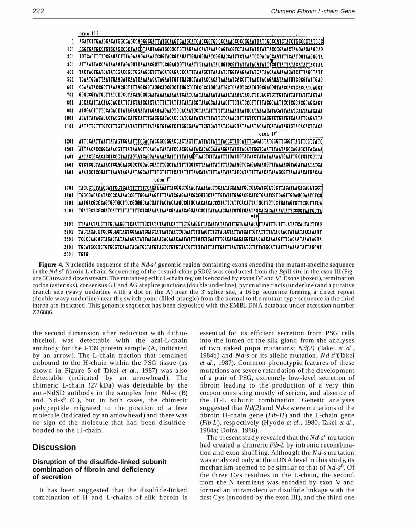
Nucleotide sequencing of the Nd-sD cosmid clonepSD02 from the BglII site within exon III toward theabove-described HindIII-BglII region revealed thatthe mutant-specific cDNA region was derived fromtwo exons, IV' and V', which were located about 1 to2 kb downstream from the end of the exon III(Figure 4). Splice junctions conformed to the GT/AGrule (Breathnach et al., 1978). Pyrimidine-rich tracts(Shapiro & Senapathy, 1987) and a putative branchsite (YNYTRAY, where Y, N and R representpyrimidine, any and purine bases, respectively;Krainer & Maniatis, 1988) are present near the 3'splice site in introns between exons III and IV' and
Figure 1. Sequences of fibroin L-chain cDNA clones from B. mori J-139 (pFL18), Nd-s (pNSL64) and Nd-sD (pNDL55).A common base to all the three cDNAs ( − ), a common base to Nd-s and Nd-sD cDNAs (asterisk), initiation and terminationcodons (boxed), the canonical (double underline) and minor (underline) polyadenylation signals, and positions of introns(downward and upward filled triangles; Kikuchi et al., 1992 and this study) are indicated. The sequence between twoarrows corresponds to that of the mutant-specific SDHp2 probe. The AluI site utilized to construct a subclone forexpression in E. coli is indicated (wavy underline). The cDNA sequences of Nd-s and Nd-sD L-chains have been depositedwith the EMBL DNA database under accession numbers Z26884 and Z26885, respectively.

JMB—MS 669
Chimeric Fibroin L-chain Gene220
Figure 2. A, Deduced amino acid sequences of the fibroin L-chain of B. mori J-139, Nd-s and Nd-sD. A residue commonto all three sequences ( − ), a residue common to Nd-s and Nd-sD (asterisk), the Ile residue encoded by different codonsbetween J-139 (ATC) and the two mutants (ATT; filled triangle) and Cys residues (over or underline) are indicated. B,Hexapeptide hydrophobicity-hydrophilicity indices of the C-terminal half region unique to the J-139 or Nd-sD (and Nd-s)L-chain.
between exons IV' and V'. It was noted that theswitch from the J-139-type sequence to the Nd-sD-type sequence occurred in the third intron (filledtriangle in Figure 4) and that a 16 bp sequence at theend of the J-139-type sequence formed a direct repeat(double-wavy underlines in Figure 4) with aninsertion of T in between.
Presence of the exons IV' and V' sequences inthe far downstream of the J-139 Fib-L
The genomic DNA of J-139 or a J-139 cosmid clone,pKYFL139-1 containing the exons II to VII of theFib-L and its downstream region (Figure 5C), wassubjected to digestion with restriction enzymes and

JMB—MS 669
Chimeric Fibroin L-chain Gene 221
Figure 3. Genomic Southern blot hybridization indicat-ing location of the sequence (exon IV' ~) encoding themutant-specific region of the fibroin L-chain in the genomeof Nd-sD. Genomic DNA of Nd-sD (lanes 1 to 4) or a cosmidclone, pSD02 (lanes 5 to 8), was digested with EcoRI(lanes 1 and 5), EcoRI + HindIII (lanes 2 and 6),EcoRI + BglII (lanes 3 and 7), or EcoRI + XbaI (lanes 4and 8) and subjected to Southern blot hybridization withthe 32P-labeled probe for the exon III (A) or the 32P-labeledSDHp2 probe for the mutant-specific region (B). C, Arelevant restriction map of pSD02 and interpretation of theresults in A and B. Restriction enzymes are abbreviated asfollows. N, NotI; E, EcoRI; B, BamHI; H, HindIII; X, XbaI;G, BglII; K, KpnI; and V, EcoRV.
present at about 16 to 18 kb downstream from theend of the exon III in the genome of J-139. Splicejunctions conform to the GT/AG rule and thepyrimidine-rich tracts and a putative branch site arepresent near the 3' splice site of each intron. Thus,these two exons in the J-139 genome seem to havebeen conserved as potentially functional forms.However, transcripts from these exons have not beendetected in the PSG of J-139 at the level of Northernblotting (data not shown). Sequence similaritiesbetween these far downstream exons in J-139 and theexons IV' and V' of Nd-sD are high; 94% each at thenucleotide level, and 96 and 90%, respectively, at theamino acid level. However, the deduced amino acidsequence for the J-139 exon V' is 11 residues shorterthan that for the Nd-sD exon V' because of differentlocations of the termination codon (Figures 4 and 6).
It is noted that the sequence ACTAATACTA– – –,with which the Nd-sD-specific region starts after thedirect repeat of the 16 bp sequence in the Nd-sD
genome (Figure 4), is conserved in the J-139 genomeat about 15 kb downstream from the end of theexon III (indicated with a filled upward triangle inFigure 6). However, a direct repeat of the 16 bpsequence is not present at this site, instead, a directrepeat of TACTACT with two A bases between ispresent (Figure 6).
Immunological detection of the chimericL-chain unbound to the H-chain in theposterior silk glands of Nd-s and Nd-s D
The cDNA region specific to Nd-s and Nd-sD
L-chains was subcloned and expressed into apolypeptide in E. coli. The polypeptide wasseparated by SDS-PAGE, reduced with dithiothreitol,carboxymethylated, and used as an antigen toimmunize mice. The polyclonal antibody thusobtained was designated as an anti-NdSD antibody.
PSG protein samples containing both lumen andtissue proteins were prepared from the fifth-instarlarvae of J-139, Nd-s, and Nd-sD, reduced with2-mercaptoethanol, separated by SDS-PAGE andsubjected to Western blotting with the anti-L-chainantibody or the anti-NdSD antibody (Figure 7).Translation products of the chimeric Fib-L are presentin the PSGs of Nd-s and Nd-sD as an about 27 kDapolypeptide (lanes 2, 3, 6 and 7). This molecularmass is close to that (28,765 Da) calculated fromthe deduced amino acid sequence. The anti-NdSDantibody did not react with any polypeptide in theprotein blot from the PSG of J-139 (lane 5), Themutant-specific antigen polypeptide (lane 9) wasreacted with the anti-NdSD antibody (lane 8) but notwith the anti-L-chain antibody (lane 4).
Figure 8 shows Western blotting profiles of thenormal and the chimeric L-chains in the PSG proteinsamples separated by two-dimensional SDS-PAGE,in which samples were treated in gel with di-thiothreitol after the first-dimensional electrophor-esis. The L-chain (25 kDa), which had been bound tothe H-chain by a disulfide bond but was separated in
Southern blot hybridization with the probe contain-ing the sequence of Nd-sD exon IV' or V'. The resultsshown in Figure 5A suggest that the exon IV'sequence is present in the 4.5 kb EcoRI-HindIIIregion (lanes 4 and 8) that is located about 10 kbdownstream from the end of the exon VII (Fig-ure 5C). Similarly, the results shown in Figure 5Bsuggest that the exon V' sequence is spread from the1 kb HindIII-EcoRI region (lanes 3 and 6) to the 2.5 kbEcoRI-BamHI region (deduced from the combinedresults of lanes 1 to 6) that are adjacent to the regioncontaining the exon IV' sequence (Figure 5C).
Nucleotide sequences were determined startingfrom the 1 kb HindIII-EcoRI region (Figure 5C) andthen towards upstream and downstream sides. Thedetermined sequence and sequences of the exon IIIand a part of the third intron are shown in Figure 6.Sequences corresponding to the exons IV' and V' are
JMB—MS 669
Chimeric Fibroin L-chain Gene222
Figure 4. Nucleotide sequence of the Nd-sD genomic region containing exons encoding the mutant-specific sequencein the Nd-sD fibroin L-chain. Sequencing of the cosmid clone pSD02 was conducted from the BglII site in the exon III (Fig-ure 3C) toward downstream. The mutant-specific L-chain region is encoded by exons IV' and V'. Exons (boxed), terminationcodon (asterisks), consensus GT and AG at splice junctions (double underline), pyrimidine tracts (underline) and a putativebranch site (wavy underline with a dot on the A) near the 3' splice site, a 16 bp sequence forming a direct repeat(double-wavy underline) near the switch point (filled triangle) from the normal to the mutant-type sequence in the thirdintron are indicated. This genomic sequence has been deposited with the EMBL DNA database under accession numberZ26886.
the second dimension after reduction with dithio-threitol, was detectable with the anti-L-chainantibody for the J-139 protein sample (A, indicatedby an arrow). The L-chain fraction that remainedunbound to the H-chain within the PSG tissue (asshown in Figure 5 of Takei et al., 1987) was alsodetectable (indicated by an arrowhead). Thechimeric L-chain (27 kDa) was detectable by theanti-NdSD antibody in the samples from Nd-s (B)and Nd-sD (C), but in both cases, the chimericpolypeptide migrated to the position of a freemolecule (indicated by an arrowhead) and there wasno sign of the molecule that had been disulfide-bonded to the H-chain.
Discussion
Disruption of the disulfide-linked subunitcombination of fibroin and deficiencyof secretion
It has been suggested that the disulfide-linkedcombination of H and L-chains of silk fibroin is
essential for its efficient secretion from PSG cellsinto the lumen of the silk gland from the analysesof two naked pupa mutations; Nd(2) (Takei et al.,1984b) and Nd-s or its allelic mutation, Nd-sD(Takeiet al., 1987). Common phenotypic features of thesemutations are severe retardation of the developmentof a pair of PSG, extremely low-level secretion offibroin leading to the production of a very thincocoon consisting mostly of sericin, and absence ofthe H-L subunit combination. Genetic analysessuggested that Nd(2) and Nd-s were mutations of thefibroin H-chain gene (Fib-H) and the L-chain gene(Fib-L), respectively (Hyodo et al., 1980; Takei et al.,1984a; Doira, 1986).
The present study revealed that the Nd-sD mutationhad created a chimeric Fib-L by intronic recombina-tion and exon shuffling. Although the Nd-s mutationwas analyzed only at the cDNA level in this study, itsmechanism seemed to be similar to that of Nd-sD. Ofthe three Cys residues in the L-chain, the secondfrom the N terminus was encoded by exon V andformed an intramolecular disulfide linkage with thefirst Cys (encoded by the exon III), and the third one

JMB—MS 669
Chimeric Fibroin L-chain Gene 223
was encoded by exon VI and is suggested to beinvolved in the linkage with the H-chain (Yamaguchiet al., 1989; Kikuchi et al., 1992). In the Nd-sD mutation,an event of recombination between the 16 bp repeatsequence in the third intron and presumably asimilar repeat sequence in the far downstreamregion of the gene seemed to have resulted in a largedeletion including exons IV to VII, and acquisition oftwo new exons, IV' and V', from the downstreamregion. Thus, the second and the third Cys residuesin the normal L-chain are eliminated in the chimericL-chain, as illustrated in Figure 9. The chimericL-chain is incapable of forming a disulfide linkagewith the H-chain in spite of the presence of three newCys residues encoded by exon V'.
The presence of a free sulfhydryl group in aprotein has been suggested to be inhibitory for itsintracellular transport and secretion from studiesincluding a mutant form of major histocompatibilitycomplex class I antigen (Miyazaki et al., 1986), mutantLDL receptors (Esser & Russel, 1988) and IgM heavychain (Sitia et al., 1990). The monomeric IgM heavychain is not secreted from B lymphocytes, but whenCys575 at the C terminus of the ms chain was replacedby Ala, it was secreted efficiently. Similarly, when asmall region containing Cys575 was removed, therest of the molecule was secreted efficiently as amonomer. It has been shown that monomericimmunoglobulin heavy chains are retained in ER bybinding to BiP (Bole et al., 1986; Munro & Pelham,1986). In the case of monomeric IgM heavy chain, thefree sulfhydryl group of Cys575 was suspected tointeract with either BiP or protein disulfideisomerase present in ER (Sitia et al., 1990). Retentionof the free fibroin H-chain within the PSG cells in
Nd-s and Nd-sD seems to be caused by a similarmechanism, because Gamo & Sato (1985) observedby electron microscopy that the ER was markedlyenlarged but the Golgi complex and fibroin secretorygranules were poorly developed in PSG cells of thehomozygous Nd-sD mutant silkworm.
It is of interest to note that homologs of Bombyxfibroin H and L-chains have been found in Galleriamellonella, which belongs to another family, thePyralidae, in the Order Lepidoptera (Zurovec et al.,1992). Overall identity of the deduced amino acidsequences of Bombyx and Galleria L-chains is 42% andthe three Cys residues are conserved. Thus, themechanism of efficient secretion of fibroin by forminga disulfide-linked H-L complex seems to have beenconserved among different fibroin-producing insects.
Mechanism of formation of the chimeric Fib-L
The Nd-s mutant was found in the Thanghpre-Spopulation of B. mori in the early 1960s and the Nd-sD
mutant was isolated from the fourth generation afterinjecting diethyl sulfate into a male pupa of a normalfibroin-producing B. mori of unknown breed (citedby Gamo & Sato, 1985), but parental breeds of thesetwo mutants have not been maintained. Thus, in thisstudy, we had to compare Fib-L structures of Nd-sD
and a non-parental breed, J-139. The presence of adirect repeat of a 16 bp sequence at the junctionbetween normal and mutant-type sequences inthe third intron of Nd-sD Fib-L suggested stronglythat recombination between the 16 bp repeat in thethird intron and the similar repeat in the far down-stream region had taken place in the parental breed(Figure 9), although the corresponding repeat
Figure 5. Genomic Southern blothybridization revealing existence ofexons IV' and V' sequences in the fardownstream region of Fib-L in thegenome of J-139. A, J-139 genomicDNA (lanes 1 to 4) or a cosmid clone,pKYFL139-1 (lanes 5 to 8), wasdigested with EcoRI (lanes 1 and 5),HindIII (lanes 2 and 6), BamHI (lanes3 and 7) or EcoRI + HindIII (lanes 4and 8), and subjected to Southernblot hybridization with the 32P-labeled exon IV' probe. B, J-139genomic DNA or pKYFL139-1 wasdigested with EcoRI (lanes 1 and 4),BamHI (lanes 2 and 5) or EcoRI +HindIII (lanes 3 and 6), and hybrid-ized with the 32P-labeled exon V'probe. C, Exons and relevant restric-tion sites (abbreviated as in Figure 3)in and around the J-139 Fib-L, andinterpretation of the results in A andB, suggesting locations of exons IV'and V' sequences.

JMB—MS 669 DEEP PAGE
Figure 6. Nucleotide sequences of the exon III and a part of the third intron of the Fib-L and the far downstream regioncontaining the exon IV' and V' sequences in the J-139 genomic DNA. Exons (boxed), termination codon (asterisks), splicesignals (as in Figure 4), the 16 bp sequence that forms a direct repeat in the Nd-sD Fib-L (Figure 4; double-wavy underline),TACTACT repeat (overline), the switching point where the J-139-type sequence ends (T) or the Nd-sD-type sequence starts(R) in the Nd-sD Fib-L are indicated. This genomic sequence has been deposited with the EMBL DNA database underaccession number Z26887.

JMB—MS 669
Chimeric Fibroin L-chain Gene 225
Figure 7. Immunological detection of the normal or thechimeric fibroin L-chain in the PSG protein samples.Samples containing both lumen and tissue proteins of PSGfrom J-139 (lanes 1 and 5; 4 mg/lane), Nd-s (lanes 2 and 6;40 mg/lane), and Nd-sD (lanes 3 and 7; 40 mg/lane), and theantigen polypeptide that was synthesized in E. coli andused to produce the anti-NdSD antibody (lanes 4, 8 and 9;00.5 mg/lane), were dissolved in 60% LiSCN, dialyzedagainst 20 mM Tris-HCl (pH 7.5) containing 5 M urea, 1%SDS, 1% 2-mercaptoethanol at 37°C and subjected toSDS/12.5% PAGE. Western blotting was carried out withthe anti-L-chain antibody (lanes 1 to 4) or the anti-NdSDantibody (lanes 5 to 8). Lane 9, stained with Coomassiebrilliant Blue R-250. Molecular mass markers (in kDa)were: myosin heavy chain (200), phosphorylase B (97),ovalbumin (45), carbonic anhydrase (31), trypsin inhibitor(21.5) and lysozyme (14.4).
Figure 8. Examination of the association with theH-chain of the normal or the chimeric fibroin L-chain bytwo-dimensional electrophoresis of the PSG proteinsamples. Samples containing both lumen and tissueproteins of PSG from J-139 (A), Nd-s (B), and Nd-sD (C)were dissolved in 60% LiSCN, dialyzed against 20 mMTris-HCl (pH 7.5) containing 5 M urea, 1% SDS at 37°C, andfirst subjected to SDS/4% PAGE. Lanes were then cut out,disulfide bonds were reduced in 63 mM Tris-HCl (pH 6.8)containing 10% glycerol, 2% SDS, 10 mM dithiothreitol at25°C for one hour, and subjected to the second-dimen-sional SDS/12.5% PAGE. Western blotting was carried outwith the anti-L-chain antibody (A) or the anti-NdSDantibody (B and C). Markers (M) were fibroin H andL-chains of J-139, detected with the anti-H29 antibody(polyclonal rabbit antibody against a C-terminal regionof the H-chain; to be reported elsewhere) and theanti-L-chain antibody. The L-chain that had been di-sulfide-bonded to the H-chain (arrow) and the L-chain orthe chimeric L-chain that had not been disulfide-bonded tothe H-chain (arrowhead) are marked.
sequence was not found in the far downstreamregion of the J-139 Fib-L. This mechanism resemblesthe case reported for the formation of primateglycophorin B and E genes (Rearden et al., 1993; Ondaet al., 1993); these two genes were suggested to havebeen formed by homologous recombination be-tween the Alu repetitive sequences in the fifth intronof the glycophorin A gene and at about 9 kb down-stream from the end of the gene. The B and E genesthus formed lacked exon 6 and the exon for the 3'untranslated region of the A gene but acquired a 3'untranslated exon from the downstream region. Aunique feature of the chimeric Fib-L is that theacquired exons IV' and V' are expressed as a part ofthe chimeric L-chain polypeptide.
We searched for the existence of the 16 bpsequence in genomic DNAs of nine different breedsof B. mori, all of which produced normal-level fibroin,and of a wild species, Bombyx mandarina, by South-ern blot hybridization using a 32-mer, i.e. a tandemrepeat of the 16 bp, synthetic DNA probe under twodifferent stringencies; one for detection of the 16 bpmonomer and the other for detection of its tandemrepeat. At least ten to 15 monomeric 16 bp units werepresent in all of the genomic DNAs tested, but itstandem form was detected only in Nd-s and Nd-sD
at the position in the third intron of the chimeric Fib-L(data not shown). From these results, we concludethat the 16 bp sequence is not a general hotspot ofrecombination in Bombyx and suppose that the 16 bprepeat was fortuitously present in the far down-stream region of Fib-L in the parental breeds.
Materials and Methods
Silkworm breeds
B. mori J-139 and two naked pupa mutant strains of B.mori, Nd-s and Nd-sD, carrying homozygous Nd-s/Nd-sand Nd-sD/Nd-sD mutations, respectively, were supplied

JMB—MS 669
Chimeric Fibroin L-chain Gene226
Figure 9. A model illustrating the creation of thechimeric Fib-L of Nd-sD. Intronic recombination within the16 bp repeats, deletion of a large region containing exonsIV to VII, and inclusion of the far downstream exons IV'and V' by exon shuffling are postulated. Location of a Cyscodon is shown with -SH.
kit (United States Biochem. Corp.). The determinednucleotide sequences and the deduced amino acidsequences were subjected to similarity searches against thecurrent versions of GenBank DNA database and the PIRprotein database through DDBJ (DNA Databank of Japanin the National Institute of Genetics, Mishima). Splicingsignals in genomic DNA sequences were searched foras described (Kikuchi et al., 1992). Hydrophobicity-hydrophilicity indices of hexapeptide windows for thededuced amino acid sequences were determined using theprogram based on Hopp & Woods (1981) in DNASISversion 7.00 (Hitachi Software Engineering Co., Ltd.,Yokohama, Japan).
Probes
Probes containing each one of exons III to VII sequencesof the J-139 Fib-L were prepared as described (Kikuchiet al., 1992). The SDHp2 probe was the HpaII fragmentobtained from the Nd-sD L-chain cDNA insert of pNDL55;nucleotide positions 392 to 720 as shown in Figure 1. Thisprobe contained a cDNA region that was derived from bothexons IV' and V', and was specific to the L-chains of Nd-sand Nd-sD. The exon IV' probe (nucleotide positions 1243(SpeI site) to 1465 in Figure 4) and the exon V' probe(nucleotide positions 1701 to 2175 (AseI site) in Figure 4)were selected from deletion constructs derived frompSD02. All probes were 32P-labeled by the random primingmethod (Feinberg & Vogelstein, 1983) using [a-32P]dCTP(0110 TBq/mmol, Amersham).
Southern blot hybridization
DNA fragments electrophoresed on a 1% (w/v) aga-rose gel were transferred to Hybond-N + membrane(Amersham) in 0.4 M NaOH, 0.6 M NaCl. Hybridizationwith one of the above probes was carried out in 50 mMTris-HCl (pH 7.5), 1 M NaCl, 1% (w/v) SDS, 10% (w/v)dextran sulfate, 100 mg/ml sheared, denatured salmonsperm DNA at 65°C for 14 to 16 hours. The membrane waswashed successively in 2 × SSC at room temperature, in2 × SSC containing 1% SDS at 65°C, and in 0.1 × SSC atroom temperature, and then subjected to autoradiography.
Preparation of polyclonal antibody against themutant-specific region of the Nd-s D L-chain andWestern blot analysis
The Nd-sD L-chain cDNA clone pNDL55 was digestedwith AluI, and the 0.55 kb fragment corresponding to thenucleotide position 476 (Figure 1) to the AluI site in thevector and encoding Tyr146 (the coding frame starts fromnucleotide position 478) to Glu276 (Figure 2) wasrecovered and subcloned into the blunt-ended EcoRI site ofpUC118. Escherichia coli MV1184, transformed with thissubclone, was cultured in 2 × YT medium (Sambrook et al.,1989) containing ampicillin for five hours, and then theexpression of cDNA was induced with 1 mM isopropyl-thio-b-D-galactoside (IPTG). Cells were harvested afteran overnight culture and resuspended in 20 mM Tris-HCl(pH 7.5), 30 mM EDTA containing lysozyme at a concen-tration of 2 mg/ml and placed on ice for one hour.Sodium deoxycholate was added to 1 mg/ml and the sus-pension was warmed to 37°C. Cells were then disruptedby sonication. The polypeptide corresponding to themutant-specific region, as judged by its transformation-de-pendent and IPTG-dependent production and expectedmolecular mass of about 15 kDa, was present in the
from the Department of Silkworm Breeding, NationalInstitute of Sericultural and Entomological Science, theMinistry of Agriculture, Forestry and Fisheries, Japan.These two mutant strains had been isolated independentlybut the two loci are allelic (Gamo & Sato, 1985).
Cloning of fibroin L-chain cDNA
A cDNA clone, pFL18, containing the 1.2 kb full-lengthL-chain cDNA sequence, was obtained from B. mori J-139(Yamaguchi et al., 1989). Two L-chain cDNA clones,pNSL64 and pNDL55, each containing about 1 kb insert,were obtained from the poly(A)+ RNAs of PSGs of Nd-sand Nd-sD, respectively, using the same procedures as forpFL18, except that AMV reverse transcriptase (Bio-Rad)was used instead of RAV-2 reverse transcriptase and thatthe cDNA libraries were screened with the 32P-labeledinsert of pFL18.
Preparation of genomic DNA and cloning ofgenomic sequences of the fibroin L-chain
Large molecular mass genomic DNAs were preparedfrom the nuclei isolated from PSGs of B. mori J-139, Nd-sand Nd-sD according to the method of Kondo et al. (1987).A cosmid clone, pKYFL139-1, containing most of J-139Fib-L was obtained previously (Kikuchi et al., 1992). ANd-sD genomic library was constructed from the partiallyMboI-digested genomic DNA using the BamHI-cut pWE15cosmid vector (Wahl et al., 1987). A portion of this librarycontaining about 2 × 10⁵ cosmids was screened with the32P-labeled insert of the Nd-sD L-chain cDNA clonepNDL55 as a probe and a clone, pSD02, was obtained.
Nucleotide sequencing and computer analysis
cDNA inserts in pNSL64 and pNDL55 were digestedwith HpaII, HpaII plus BglII or Sau3AI, or BglII plus Sau3AIand subcloned into M13mp10/11 vectors. Single-strandedplasmids were prepared and sequences of the inserts weredetermined using the 7-deaza sequencing kit version 2(Takara Shuzo Co., Ltd., Kyoto, Japan). Genomic sequencesin cosmid clones pKYFL139-1 and pSD02 were digestedwith appropriate restriction enzymes, subcloned intopUC118/119 vectors, converted to a series of deletionconstructs as described (Kikuchi et al., 1992), and theirinserts were sequenced using the Sequenase sequencing

JMB—MS 669
Chimeric Fibroin L-chain Gene 227
pellet fraction obtained after centrifugation at 13,000 g for15 minutes. The pellet was suspended in 20 mM Tris-HCl (pH 7.5), 5 M urea, incubated at 37°C overnight andrecovered by centrifugation. Proteins remaining in thepellet fraction were then solubilized by incubating in50 mM Tris-HCl (pH 7.5), 2% SDS, 5% 2-mercaptoethanolat 37°C overnight, followed by boiling in the same solutionfor five minutes. The polypeptide of about 15 kDa wasseparated by SDS/15% polyacrylamide slab gel electro-phoresis, recovered from the gel by electro-elution in25 mM Tris base, 192 mM glycine (Garfin, 1990), andconcentrated using Molcut L (Millipore, NMWL 5000). Theconcentrated polypeptide sample was reduced with50 mM dithiothreitol at 25°C for two hours, followedby carbamoylmethylation with 125 mM iodoacetamide(pH 8.0) at 25°C for 30 minutes in the dark and dialysisagainst 25 mM Tris base, 192 mM glycine. Four-week-oldfemale BALB/c mice were immunized with this re-duced polypeptide sample mixed with Freund’s completeadjuvant (Difco), followed by three more injections withthe same polypeptide sample mixed with Freund’sincomplete adjuvant (Difco). The polyclonal antibody,designated anti-NdSD antibody, was obtained as animmunoglobulin G fraction from the antiserum byadsorption to and elution from the Protein A-SepharoseCL-4B (Sigma) column according to Sambrook et al. (1989).
For Western blotting, proteins separated by SDS/12.5%polyacrylamide gel electrophoresis were transferred to anitrocellulose membrane (Schleicher & Schuell, pore size0.45 mm) according to the method of Towbin et al. (1979),and subjected to reaction with the rabbit polyclonalanti-L-chain antibody (Tanaka et al., 1993) or the mouseanti-NdSD antibody, followed by reaction with alkalinephosphatase (AP)-conjugated anti-rabbit IgG (Promega) orAP-conjugated anti-mouse IgG (Promega) and colordevelopment with 5-bromo-4-chloro-3-indoryl phosphate(BCIP) and nitro blue tetrazolium chloride (NBT).
AcknowledgementsThis work was supported by Grant-in-Aid for Scientific
Research 03453133 and for Developmental ScientificResearch 06556012 from the Ministry of Education, Scienceand Culture, Japan.
ReferencesBirnstiel, M. L., Busslinger, M. & Strub, K. (1985).
Transcription termination and 3' processing: the end isin site! Cell, 41, 349–359.
Bole, D. G., Hendershot, L. M. & Kearney, J. F. (1986).Posttranslational association of immunoglobulinheavy chain binding protein with nascent heavychains in nonsecreting and secreting hybridomas.J. Cell Biol. 102, 1558–1566.
Breathnach, R., Benoist, C., O’Hare, K., Gannon, F. &Chambon, P. (1978). Ovalbumin gene: Evidence for aleader sequence in mRNA and DNA sequences at theexon-intron boundaries. Proc. Natl Acad. Sci. USA, 75,4853–4857.
Doira, H. (1986). Linkage maps of Bombyx mori – Revisedin 1986. Sericologia, 26, 485–488.
Esser, V. & Russell, D. W. (1988). Transport-deficientmutations in the low density lipoprotein receptor.Alterations in the cysteine-rich and cysteine-poor
regions of the protein block intracellular transport.J. Biol. Chem. 263, 13276–13281.
Feinberg, A. P. & Vogelstein, B. (1983). A technique forradiolabeling DNA restriction endonuclease frag-ments to high specific activity. Anal. Biochem. 132, 6–13.
Gamo, T. & Sato, S. (1985). Ultrastructural study of theposterior silk gland in the Nd, Nd-s and Nd-sD
mutants with a defect of fibroin synthesis. J. Seric. Sci.Jpn, 54, 412–419.
Garfin, D. E. (1990). One-dimensional gel electrophoresis.Methods Enzymol. 182, 425–441.
Hopp, T. P. & Woods, K. R. (1981). Prediction of proteinantigenic determinants from amino acid sequences.Proc. Natl Acad. Sci. USA, 78, 3824–3828.
Hyodo, A., Gamo, T. & Shimura, K. (1980). Linkageanalysis of the fibroin gene in the silkworm, Bombyxmori. Jpn. J. Genet. 55, 297–300.
Kikuchi, Y., Mori, K., Suzuki, S., Yamaguchi, K. &Mizuno, S. (1992). Structure of the Bombyx morifibroin light-chain-encoding gene: upstream sequenceelements common to the light and heavy chain. Gene,110, 151–158.
Kondo, K., Aoshima, Y., Hagiwara, T., Ueda, H. & Mizuno,S. (1987). Tissue-specific and periodic changes in thenuclease sensitivity of the fibroin gene chromatin inthe silkworm Bombyx mori. J. Biol. Chem. 262,5271–5279.
Krainer, A. R. & Maniatis, T. (1988). RNA splicing. InTranscription and Splicing (Hames, B. D. & Glover,D. M., eds), pp. 131–206, IRL Press, Oxford.
Miyazaki, J., Appella, E. & Ozato, K. (1986). Intracellulartransport blockade caused by disruption of thedisulfide bridge in the third external domain of majorhistocompatibility complex class I antigen. Proc. NatlAcad. Sci. USA, 83, 757–761.
Munro, S. & Pelham, H. R. B. (1986). An Hsp70-like proteinin ER: identity with the 78 kd glucose-regulatedprotein and immunoglobulin heavy chain bindingprotein. Cell, 46, 291–300.
Onda, M., Kudo, S., Rearden, A., Mattei, M.-G. & Fukuda,M. (1993). Identification of a precursor genomicsegment that provided a sequence unique toglycophorin B and E genes. Proc. Natl Acad. Sci. USA,90, 7220–7224.
Rearden, A., Magnet, A., Kudo, S. & Fukuda, M. (1993).Glycophorin B and glycophorin E genes arose from theglycophorin A ancestral gene via two duplicationsduring primate evolution. J. Biol. Chem. 268,2260–2267.
Sambrook, J., Fritsch, E. F. & Maniatis, T. (1989). MolecularCloning: A Laboratory Manual, 2nd edit., Cold SpringHarbor Laboratory Press, Cold Spring Harbor, NY.
Shapiro, M. B. & Senapathy, P. (1987). RNA splice junctionsof different classes of eukaryotes: sequence statisticsand functional implications in gene expression. Nucl.Acids Res. 15, 7155–7174.
Sitia, R., Neuberger, M., Alberini, C., Bet, P., Fra, A. Valetti,C., Williams, G. & Milstein, C. (1990). Developmentalregulation of IgM secretion: The role of thecarboxy-terminal cysteine. Cell, 60, 781–790.
Takei, F., Kimura, K., Mizuno, S., Yamamoto, T. &Shimura K. (1984a). Genetic analysis of the Nd-smutation in the silkworm Bombyx mori. Jpn. J. Genet. 59,307–313.
Takei, F., Oyama, F., Kimura, K., Hyodo, A., Mizuno S. &Shimura, K. (1984b). Reduced level of secretion andabsence of subunit combination for the fibroinsynthesized by a mutant silkworm, Nd(2). J. Cell Biol.99, 2005–2010.

JMB—MS 669
Chimeric Fibroin L-chain Gene228
Takei, F., Kikuchi, Y., Kikuchi, A., Mizuno, S. & Shimura,K. (1987). Further evidence for importance of thesubunit combination of silk fibroin in its efficientsecretion from the posterior silk gland cells. J. Cell Biol.105, 175–180.
Tanaka, K., Mori, K. & Mizuno, S. (1993). Immunologicalidentification of the major disulfide-linked lightcomponent of silk fibroin. J. Biochem (Tokyo), 114, 1–4.
Towbin, H., Staehelin, T. & Gordon, J. (1979). Electrophor-etic transfer of proteins from polyacrylamide gels tonitrocellulose sheets: Procedure and some appli-cations. Proc. Natl Acad. Sci. USA, 76, 4350–4354.
Wahl, G. M., Lewis, K. A., Ruiz, J. C., Rothenberg, B., Zhao,J. & Evans, G. A. (1987). Cosmid vectors for rapidgenomic walking, restriction mapping, and genetransfer. Proc. Natl Acad. Sci. USA, 84, 2160–2164.
Yamaguchi, K., Kikuchi, Y., Takagi, T., Kikuchi, A., Oyama,F., Shimura, K. & Mizuno, S. (1989). Primary structureof the silk fibroin light chain determined by cDNAsequencing and peptide analysis. J. Mol. Biol. 210,127–139.
Zurovec, M., Sehnal, F., Scheller, K. & Kumaran, A. K.(1992). Silk gland specific cDNAs from Galleriamellonella L. Insect Biochem. Mol. Biol. 22, 55–67.
Edited by M. Yaniv
(Received 22 March 1994; accepted in revised form 22 May 1995)