Philips - radiography.0catch.comradiography.0catch.com/8 knobology hd11.pdf · Color Power Doppler...
66
Transcript of Philips - radiography.0catch.comradiography.0catch.com/8 knobology hd11.pdf · Color Power Doppler...


Philips HD11


1
7
6
3
5
2
12 11 10
9A
9
8
7A
16
15 14
1312A 12B
4B4A 4
18
17


2019

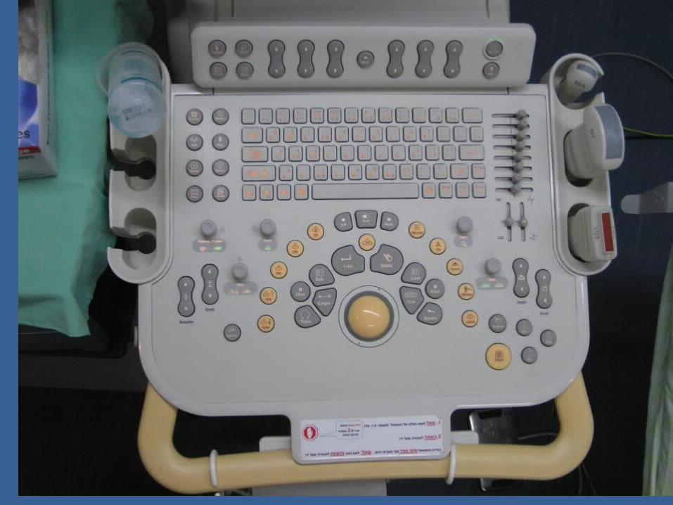
המכשיר – 1 הדלקת
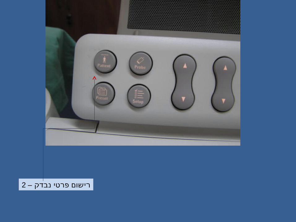
נבדק – 2 פרטי רישום
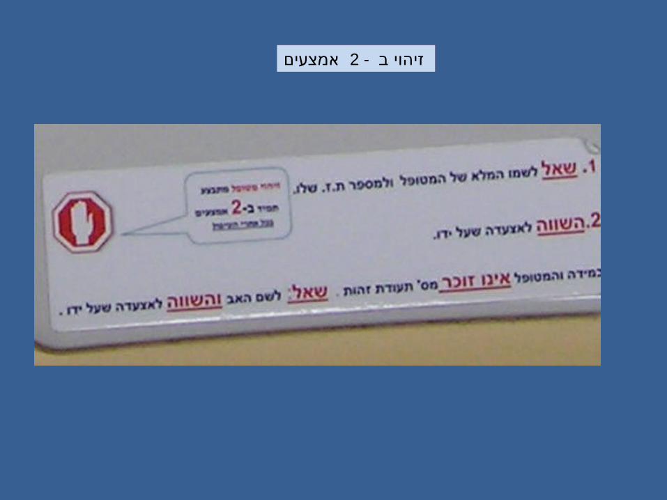
ב - אמצעים 2זיהוי
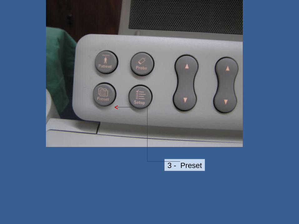
3 - Preset
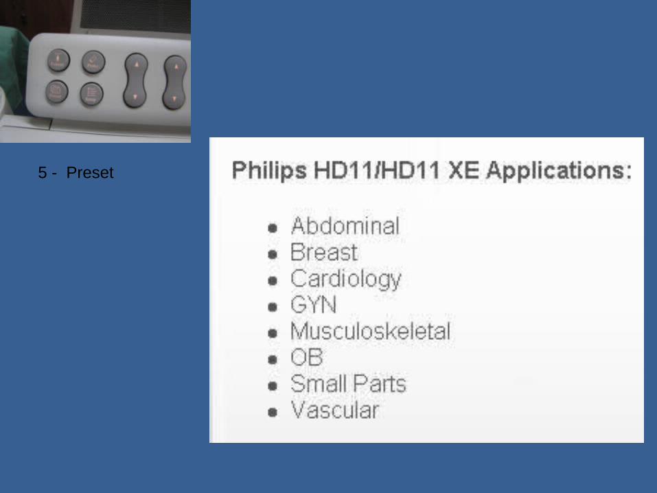
5 - Preset

3 - Preset
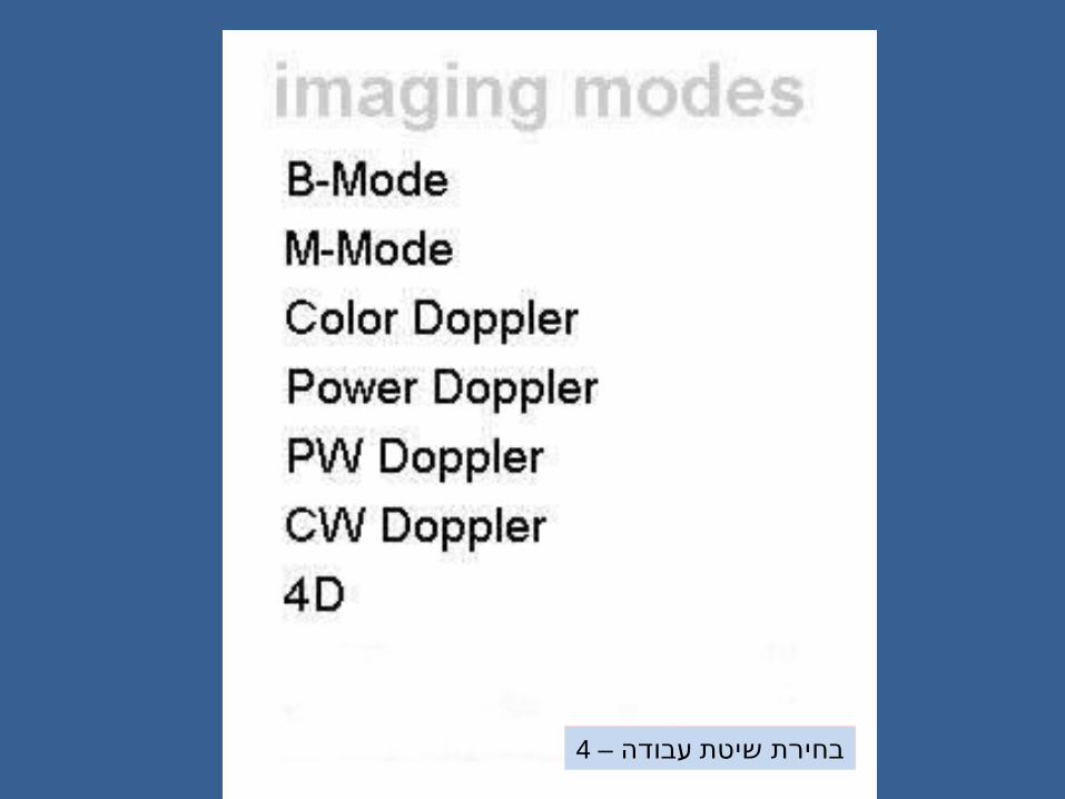
עבודה – 4 שיטת בחירת
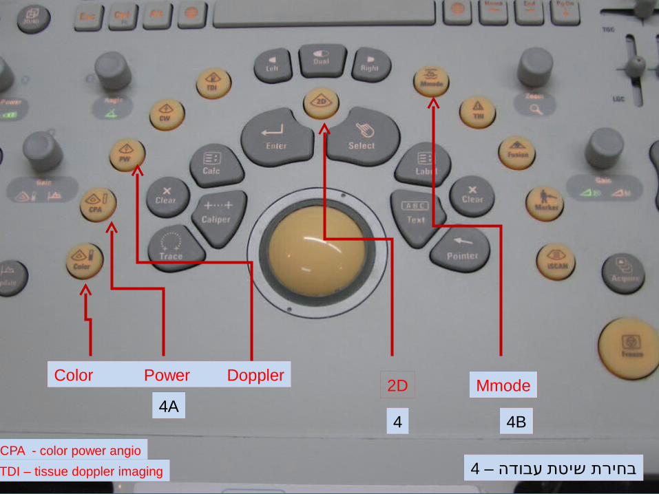
Color Power Doppler
4A2D Mmode
CPA - color power angio
TDI – tissue doppler imaging
4 4B
עבודה – 4 שיטת בחירת

Color Power Doppler
4A
2D Mmode
CPA - color power angio
TDI – tissue doppler imaging
4 4B
עבודה – 4 שיטת בחירת
דופלרכווןזווית
דופלרכוון
Gain
2D + MmodeGain כוון
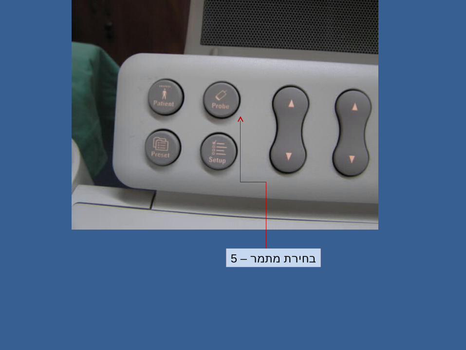
מתמר – 5 בחירת

מתמר – 5 בחירת
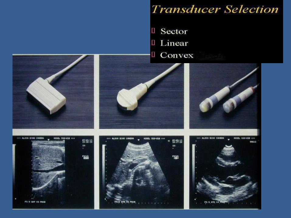

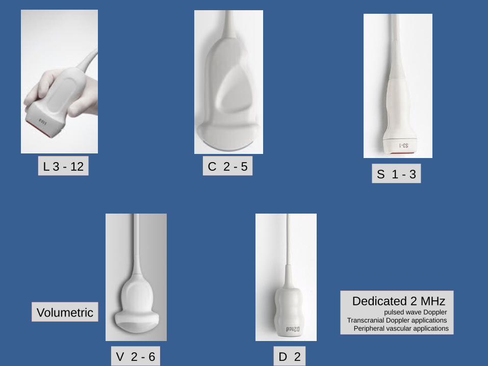
L 3 - 12 C 2 - 5S 1 - 3
V 2 - 6
Volumetric
D 2
Dedicated 2 MHz pulsed wave Doppler
Transcranial Doppler applications Peripheral vascular applications
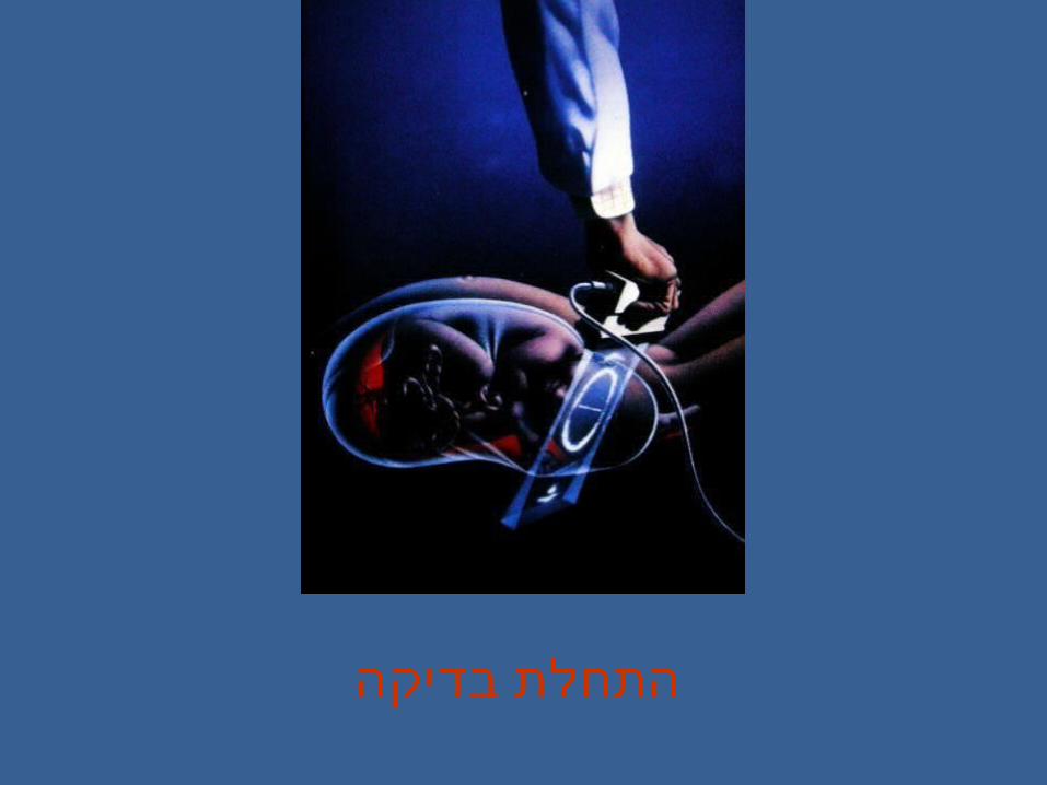
בדיקה התחלת
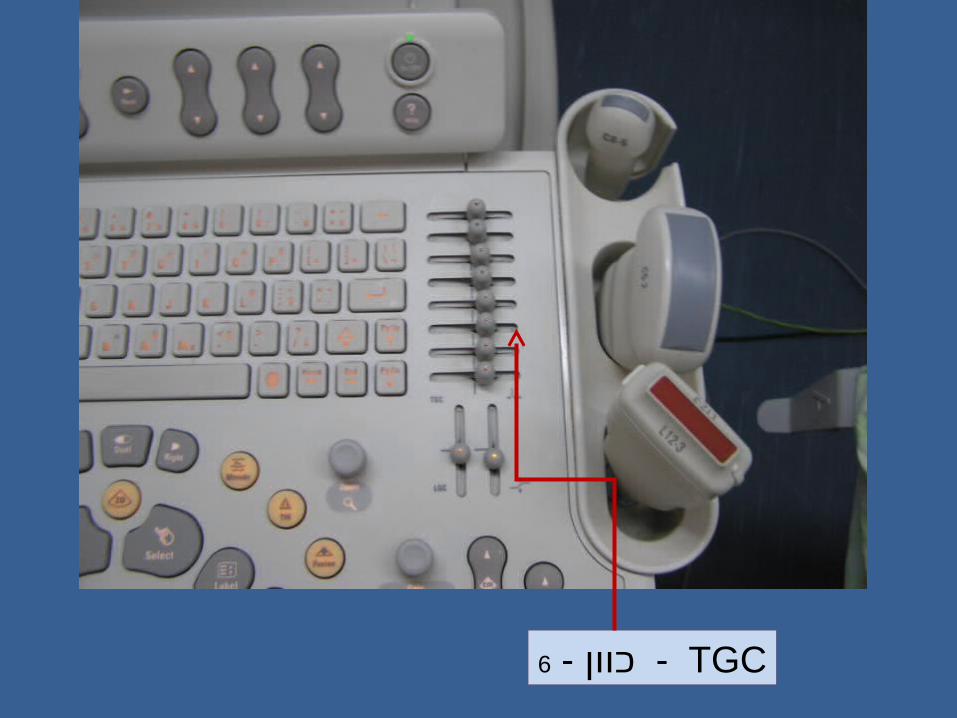
TGC - כוון - 6

TGClateral gain control - LGC - כוון - 6
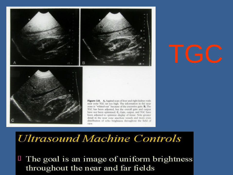
TGC
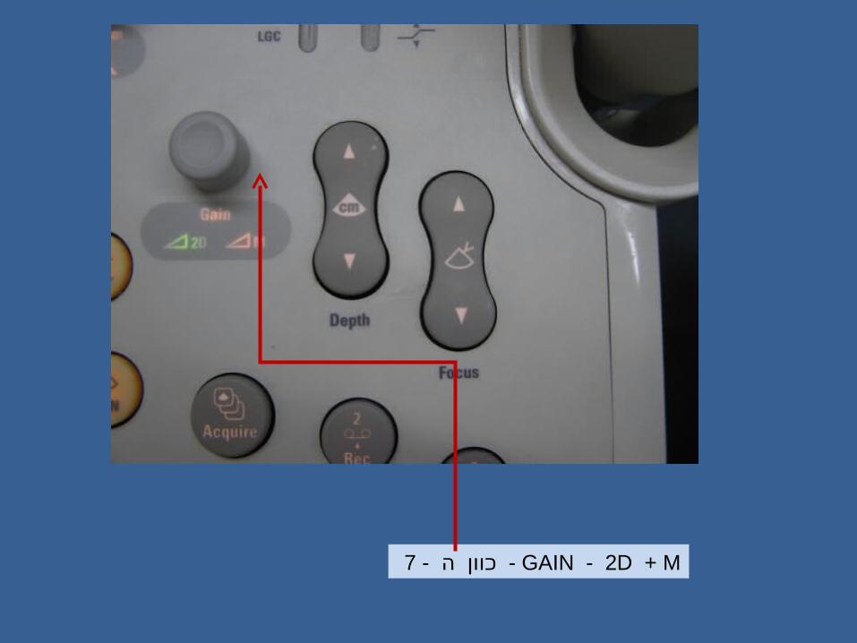
ה - 7 GAIN - 2D + M - כוון

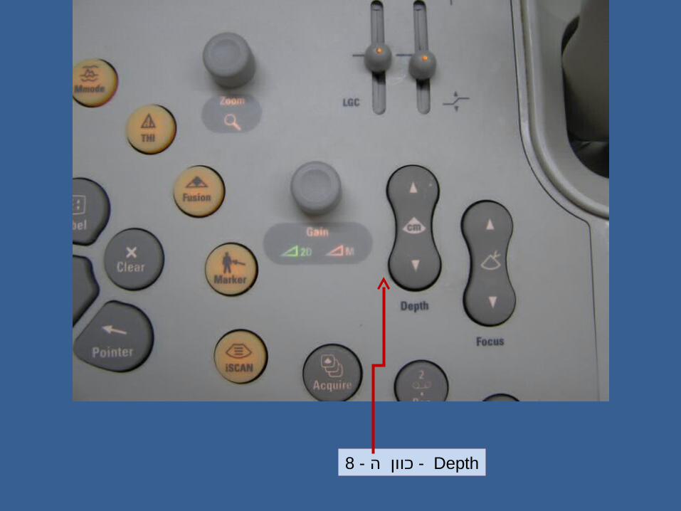
ה - 8 Depth - כוון


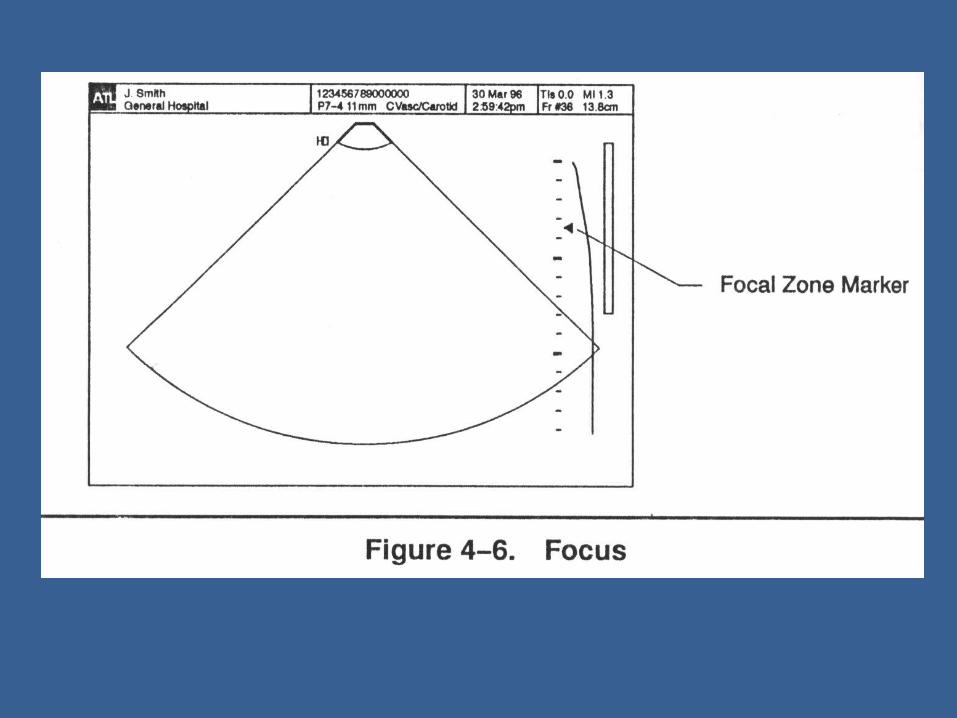
ה – 9 FocusContinuous Focus - כוון
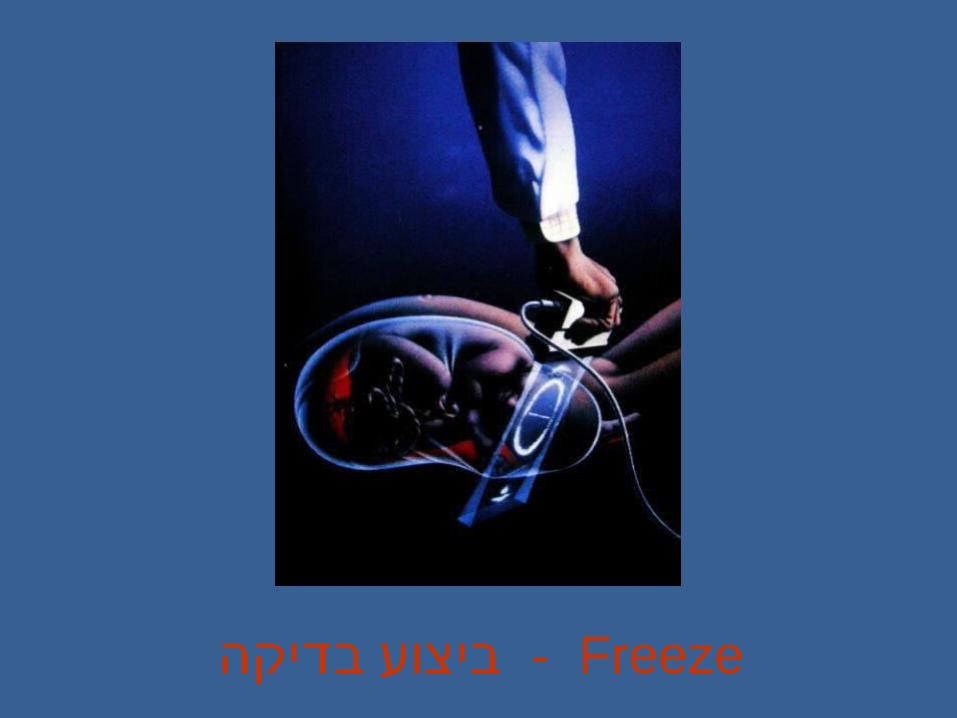
בדיקה Freeze - ביצוע

התמונה – 10 עצירת
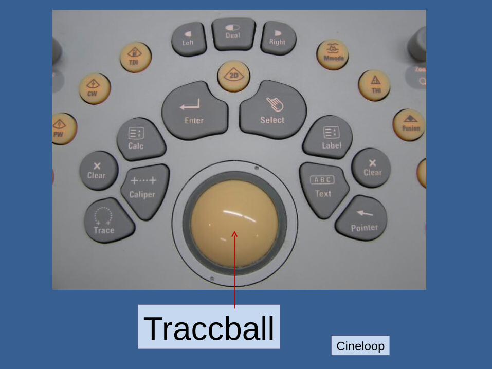
TraccballCineloop

מדידות , , . 12 רישום סימון

מדידות , , . 12 רישום סימון
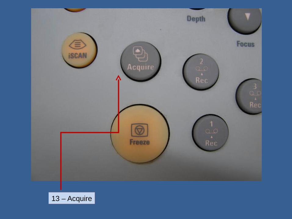
13 – Acquire

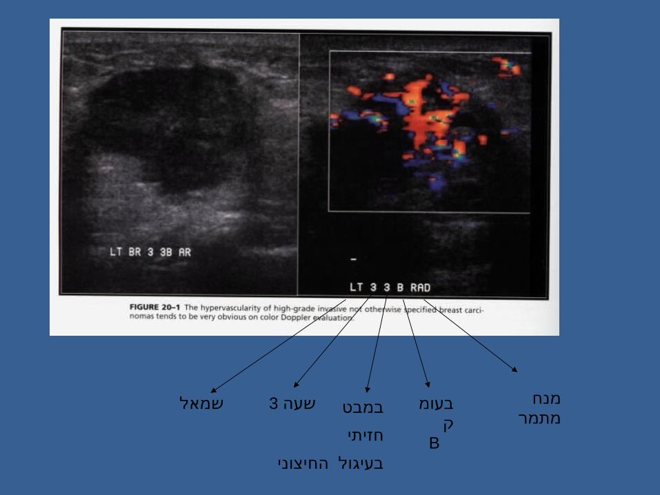
שמאל 3שעה במבט
חזיתי
החיצוני בעיגול
בעומק
B
מנחמתמר

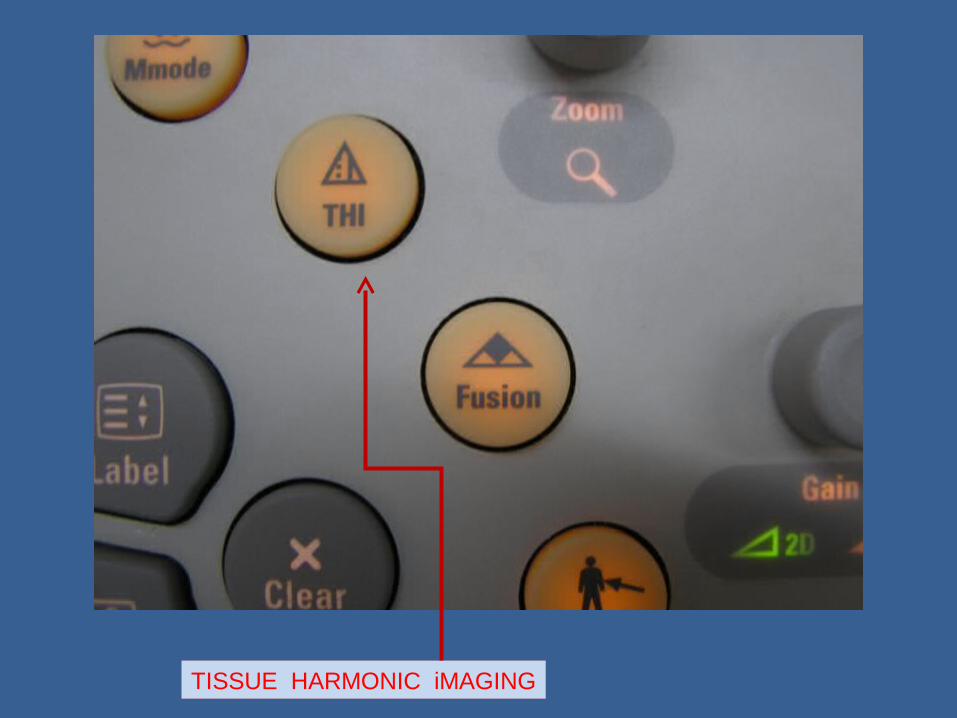
TISSUE HARMONIC iMAGING
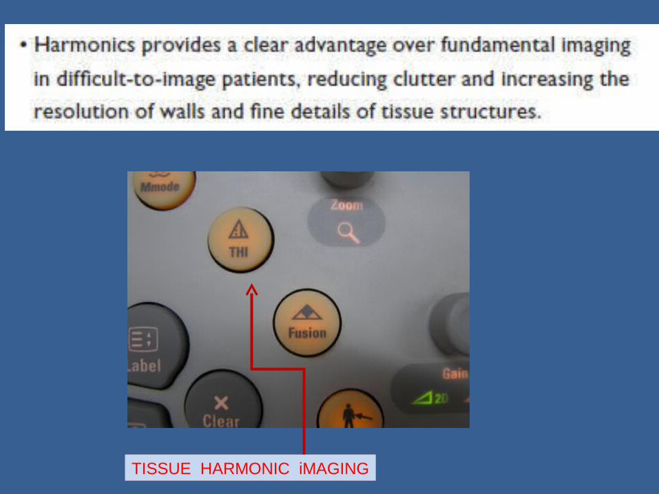
TISSUE HARMONIC iMAGING


17 - 3D/4D 16 - Panoramic
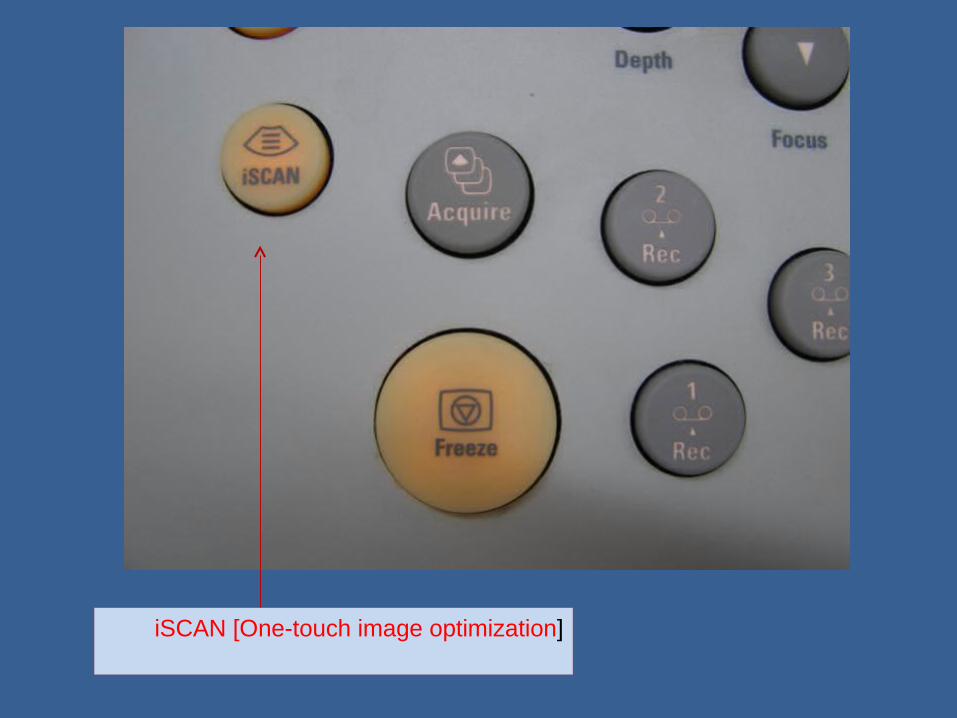
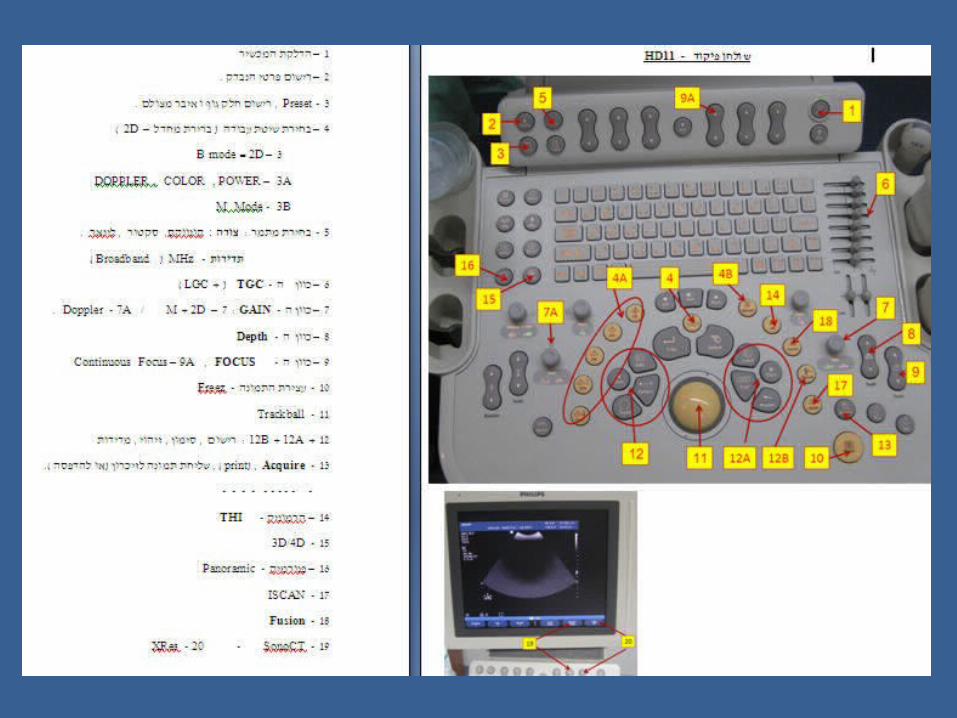
iSCAN [One-touch image optimization]

iSCAN [One-touch image optimization]
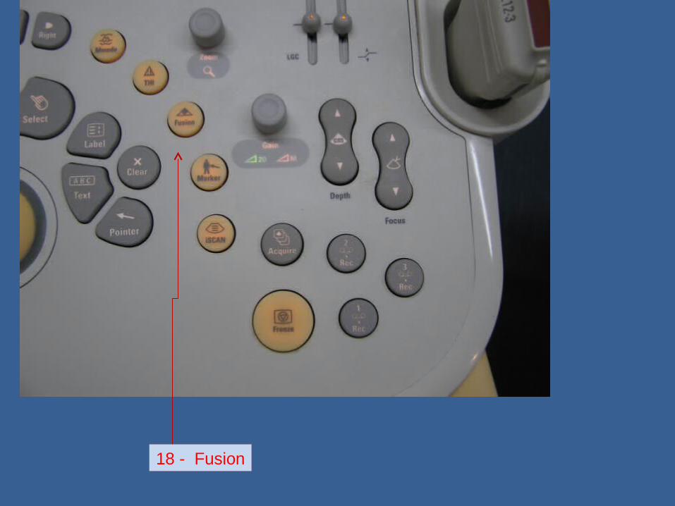
18 - Fusion
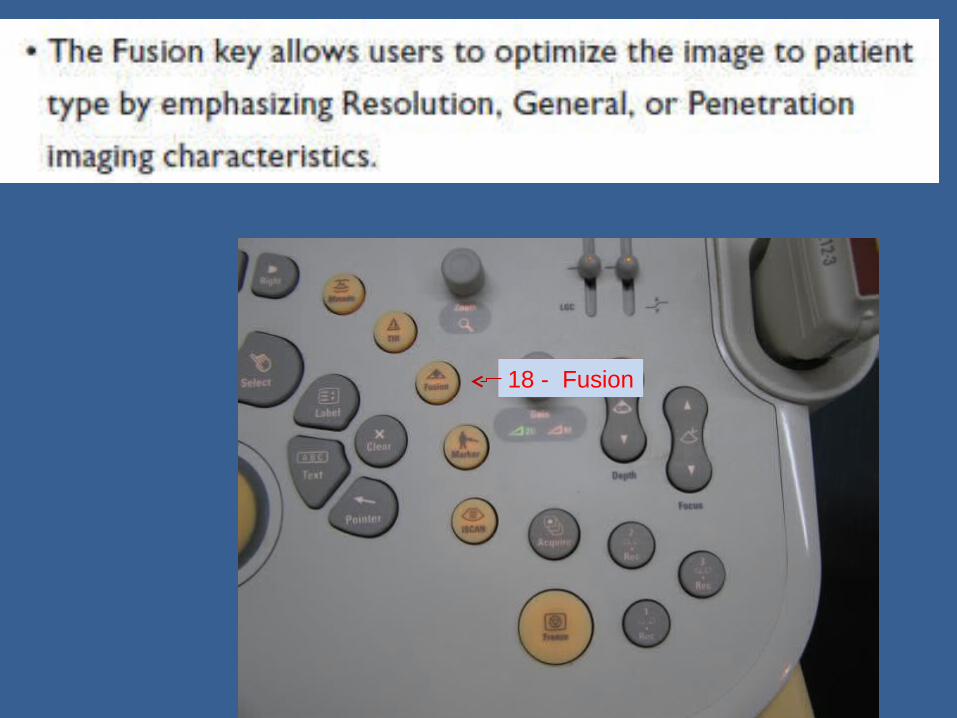
18 - Fusion
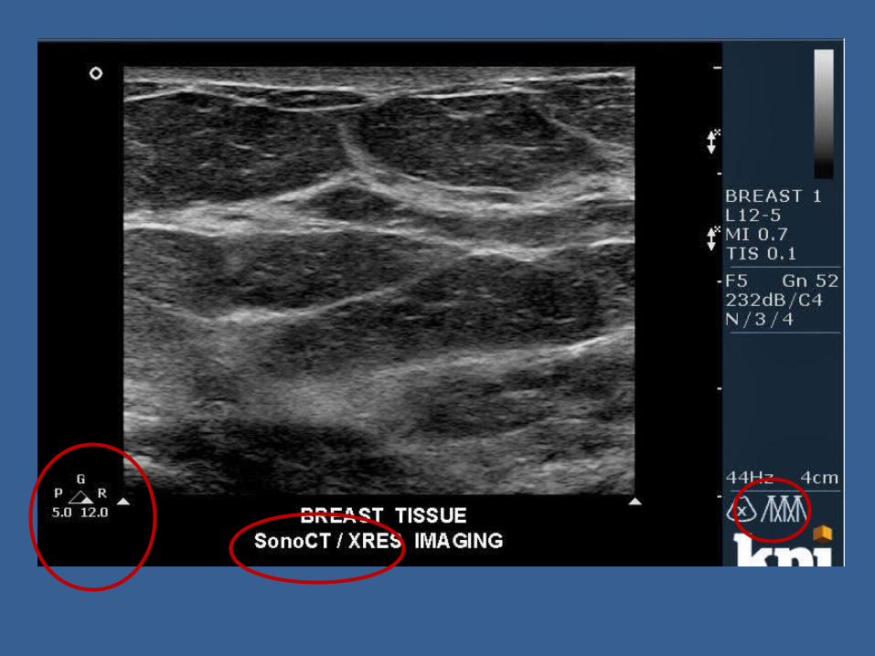

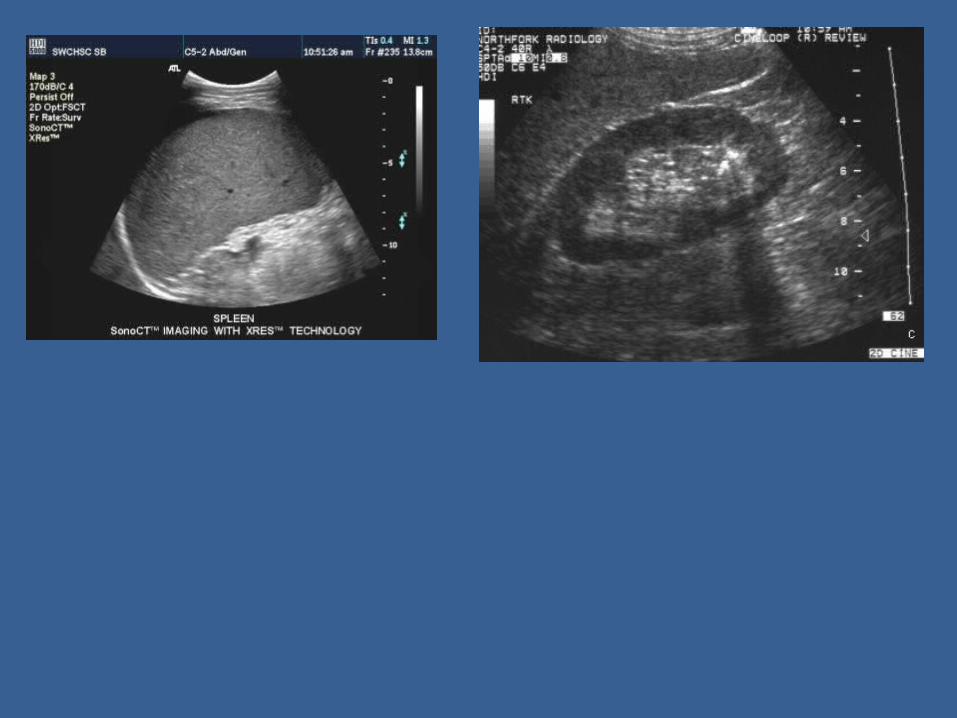
19 - SonoCT

SonoCT Real-time Compound Imaging technology is a
unique approach to overcome the inherent artifacts of conventional ultrasound that compromise image quality. SonoCT imaging technology uses transmit beam-steering techniques to obtain coplanar, tomographic images from different viewing angles, then combines these micro-angulated images into a single compounded image at real-time frame rates.
With SonoCT, angle-generated and speckle noise artifacts are reduced, and structures with curved and irregular borders are more readily visualized. Contrast resolution is improved and tissue margins are more discernable
SonoCT is supported in most 2D, Doppler, harmonic and 3D imaging modes, increasing image clarity for most exam needs and patient types.

SONO CT

20 - XRES

XRES Adaptive Image Processing provides real-
time image enhancement using proprietary contextural algorithms that reduce speckle, haze and clutter artifacts. At the same time, XRES enhances edges by
correcting discontinuities between textured regions allowing improved visualization of real tissue information.
The result is images virtually free from noise, with extraordinary clarity and border definition.


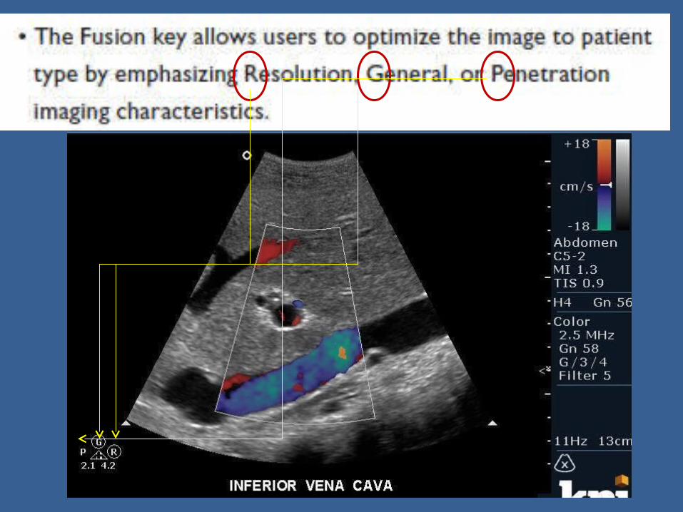
Fusion


The End