Non-epithelial tumors Uterine Tumors - IOP Pathology/… · · 2015-04-06muscle tumor S08-17284of...
10
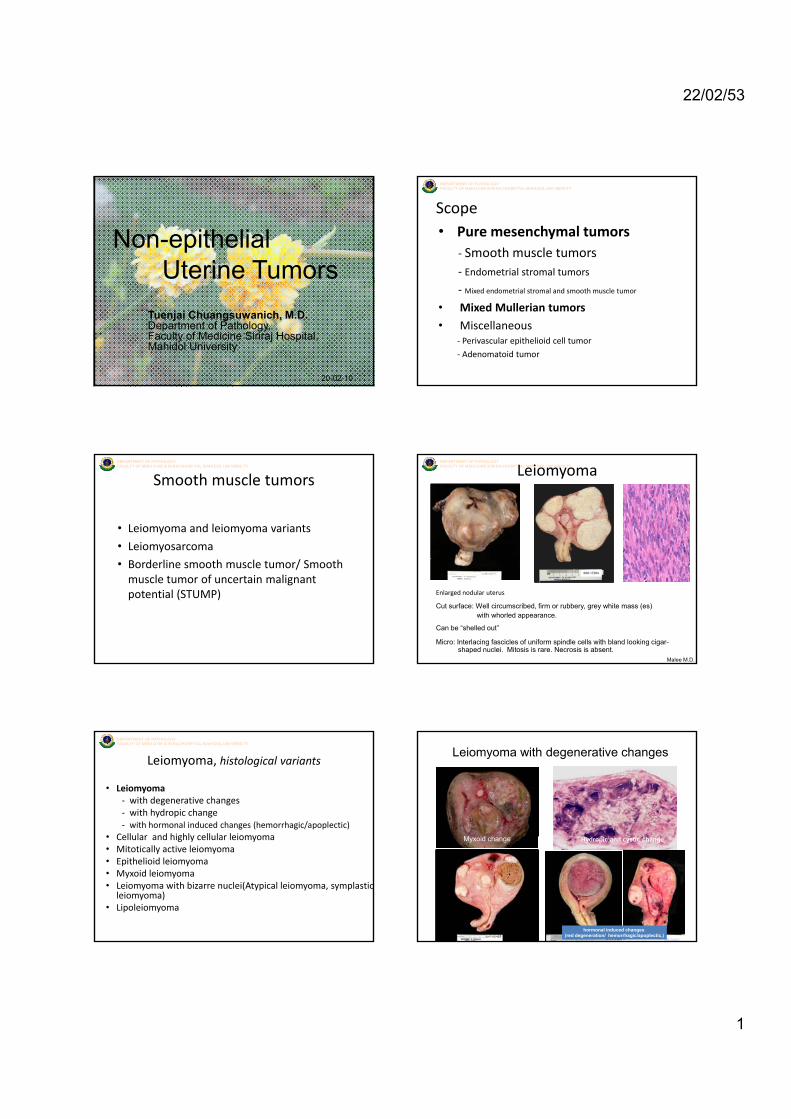
22/02/53 1 DEPARTMENT OF PATHOLOGY FACULTY OF MEDICINE SIRIRAJ HOSPITAL MAHIDOL UNIVERSITY Non-epithelial Uterine Tumors Tuenjai Chuangsuwanich, M.D. Department of Pathology, Faculty of Medicine Siriraj Hospital, Mahidol University. 20-02-10 DEPARTMENT OF PATHOLOGY FACULTY OF MEDICINE SIRIRAJ HOSPITAL MAHIDOL UNIVERSITY Scope • Pure mesenchymal tumors ‐ Smooth muscle tumors ‐ Endometrial stromal tumors ‐ Mixed endometrial stromal and smooth muscle tumor • Mixed Mullerian tumors • Miscellaneous ‐ Perivascular epithelioid cell tumor ‐ Adenomatoid tumor DEPARTMENT OF PATHOLOGY FACULTY OF MEDICINE SIRIRAJ HOSPITAL MAHIDOL UNIVERSITY Smooth muscle tumors • Leiomyoma and leiomyoma variants • Leiomyosarcoma • Borderline smooth muscle tumor/ Smooth muscle tumor of uncertain malignant potential (STUMP) DEPARTMENT OF PATHOLOGY FACULTY OF MEDICINE SIRIRAJ HOSPITAL MAHIDOL UNIVERSITY Leiomyoma Enlarged nodular uterus Cut surface: Well circumscribed, firm or rubbery, grey white mass (es) with whorled appearance. Can be “shelled out” Micro: Interlacing fascicles of uniform spindle cells with bland looking cigar- shaped nuclei. Mitosis is rare. Necrosis is absent. S08-17284 Malee M.D. DEPARTMENT OF PATHOLOGY FACULTY OF MEDICINE SIRIRAJ HOSPITAL MAHIDOL UNIVERSITY Leiomyoma, histological variants • Leiomyoma ‐ with degenerative changes ‐ with hydropic change ‐ with hormonal induced changes (hemorrhagic/apoplectic) • Cellular and highly cellular leiomyoma • Cellular and highly cellular leiomyoma • Mitotically active leiomyoma • Epithelioid leiomyoma • Myxoid leiomyoma • Leiomyoma with bizarre nuclei(Atypical leiomyoma, symplastic leiomyoma) • Lipoleiomyoma Leiomyoma with degenerative changes M id h hormonal induced changes (red degeneration/ hemorrhagic/apoplectic,) Hydropic and cystic change Myxoid change
Transcript of Non-epithelial tumors Uterine Tumors - IOP Pathology/… · · 2015-04-06muscle tumor S08-17284of...
22/02/53
1
DEPARTMENT OF PATHOLOGYFACULTY OF MEDICINE SIRIRAJ HOSPITAL MAHIDOL UNIVERSITY
Non-epithelial Uterine Tumors
Tuenjai Chuangsuwanich, M.D.Department of Pathology, Faculty of Medicine Siriraj Hospital, Mahidol University.
20-02-10
DEPARTMENT OF PATHOLOGYFACULTY OF MEDICINE SIRIRAJ HOSPITAL MAHIDOL UNIVERSITY
Scope• Pure mesenchymal tumors
‐ Smooth muscle tumors‐ Endometrial stromal tumors
‐ Mixed endometrial stromal and smooth muscle tumor
• Mixed Mullerian tumors
• Miscellaneous ‐ Perivascular epithelioid cell tumor
‐ Adenomatoid tumor
DEPARTMENT OF PATHOLOGYFACULTY OF MEDICINE SIRIRAJ HOSPITAL MAHIDOL UNIVERSITY
Smooth muscle tumors
• Leiomyoma and leiomyoma variants
• Leiomyosarcoma
• Borderline smooth muscle tumor/ Smooth muscle tumor of uncertain malignant potential (STUMP)
DEPARTMENT OF PATHOLOGYFACULTY OF MEDICINE SIRIRAJ HOSPITAL MAHIDOL UNIVERSITYLeiomyoma
Enlarged nodular uterus
Cut surface: Well circumscribed, firm or rubbery, grey white mass (es) with whorled appearance.
Can be “shelled out”
Micro: Interlacing fascicles of uniform spindle cells with bland looking cigar-shaped nuclei. Mitosis is rare. Necrosis is absent.
S08-17284
Malee M.D.
DEPARTMENT OF PATHOLOGYFACULTY OF MEDICINE SIRIRAJ HOSPITAL MAHIDOL UNIVERSITY
Leiomyoma, histological variants
• Leiomyoma‐ with degenerative changes‐ with hydropic change‐ with hormonal induced changes (hemorrhagic/apoplectic)
• Cellular and highly cellular leiomyoma• Cellular and highly cellular leiomyoma• Mitotically active leiomyoma• Epithelioid leiomyoma• Myxoid leiomyoma • Leiomyoma with bizarre nuclei(Atypical leiomyoma, symplastic
leiomyoma)• Lipoleiomyoma
Leiomyoma with degenerative changes
M id h
hormonal induced changes (red degeneration/ hemorrhagic/apoplectic,)
Hydropic and cystic changeMyxoid change
22/02/53
2
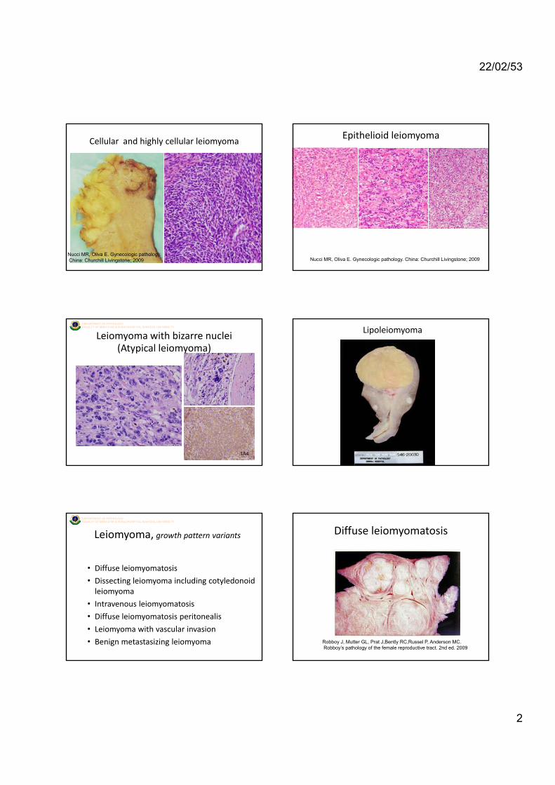
Cellular and highly cellular leiomyoma
Nucci MR, Oliva E. Gynecologic pathology.China: Churchill Livingstone; 2009
Epithelioid leiomyoma
Nucci MR, Oliva E. Gynecologic pathology. China: Churchill Livingstone; 2009
DEPARTMENT OF PATHOLOGYFACULTY OF MEDICINE SIRIRAJ HOSPITAL MAHIDOL UNIVERSITY
Leiomyoma with bizarre nuclei(Atypical leiomyoma)
1A4
Lipoleiomyoma
DEPARTMENT OF PATHOLOGYFACULTY OF MEDICINE SIRIRAJ HOSPITAL MAHIDOL UNIVERSITY
Leiomyoma, growth pattern variants
• Diffuse leiomyomatosis
• Dissecting leiomyoma including cotyledonoid leiomyoma
• Intravenous leiomyomatosis
• Diffuse leiomyomatosis peritonealis
• Leiomyoma with vascular invasion
• Benign metastasizing leiomyoma
Diffuse leiomyomatosis
Robboy J, Mutter GL, Prat J,Bently RC,Russel P, Anderson MC.Robboy’s pathology of the female reproductive tract. 2nd ed. 2009

22/02/53
3
Dissecting leiomyoma includingcotyledonoid leiomyoma
Nucci MR, Oliva E. Gynecologic pathology. China: Churchill Livingstone; 2009
Diffuse leiomyomatosis peritonealis
Nucci MR, Oliva E. Gynecologic pathology. China: Churchill Livingstone; 2009
DEPARTMENT OF PATHOLOGYFACULTY OF MEDICINE SIRIRAJ HOSPITAL MAHIDOL UNIVERSITY
Intravenous leiomyomatosis
DEPARTMENT OF PATHOLOGYFACULTY OF MEDICINE SIRIRAJ HOSPITAL MAHIDOL UNIVERSITY
Leiomyosarcoma
‘Malignant smooth muscle tumor’• 1% of all uterine malignancies
• 0.64 cases per 100,000 women
• present later in life around or aftermenopause • unsuspected or presumed to be leiomyoma before patho. exam.
DEPARTMENT OF PATHOLOGYFACULTY OF MEDICINE SIRIRAJ HOSPITAL MAHIDOL UNIVERSITY
Leiomyosarcoma
Macro:- intramural , usually not associated with leiomyoma- average 8 cm , fleshy, poorly defined margins
Hemorrhage and necrosis
Nucci MR, Oliva E. Gynecologic pathology. China: Churchill Livingstone; 2009
DEPARTMENT OF PATHOLOGYFACULTY OF MEDICINE SIRIRAJ HOSPITAL MAHIDOL UNIVERSITY
Micro:• Atypia• Mitotic figure……Atypical mitosis
Leiomyosarcoma
• Necrosis: geographic coagulation necrosis• Cellularity• Infiltrative pattern• Vascular invasion• Metastasis
22/02/53
4
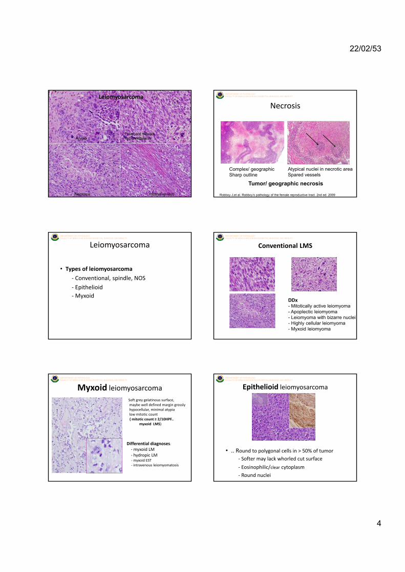
DEPARTMENT OF PATHOLOGYFACULTY OF MEDICINE SIRIRAJ HOSPITAL MAHIDOL UNIVERSITYLeiomyosarcoma
Frequent mitosishypercellularityAtypia
InfiltrativenessNecrosis
DEPARTMENT OF PATHOLOGYFACULTY OF MEDICINE SIRIRAJ HOSPITAL MAHIDOL UNIVERSITY
Necrosis
Complex/ geographicSharp outline
Atypical nuclei in necrotic areaSpared vessels
Tumor/ geographic necrosis
Robboy J,et al. Robboy’s pathology of the female reproductive tract. 2nd ed. 2009
DEPARTMENT OF PATHOLOGYFACULTY OF MEDICINE SIRIRAJ HOSPITAL MAHIDOL UNIVERSITY
• Types of leiomyosarcoma
‐ Conventional, spindle, NOS
E ith li id
Leiomyosarcoma
‐ Epithelioid‐Myxoid
DEPARTMENT OF PATHOLOGYFACULTY OF MEDICINE SIRIRAJ HOSPITAL MAHIDOL UNIVERSITY
Conventional LMS
DDx- Mitotically active leiomyoma - Apoplectic leiomyoma- Leiomyoma with bizarre nuclei- Highly cellular leiomyoma- Myxoid leiomyoma
DEPARTMENT OF PATHOLOGYFACULTY OF MEDICINE SIRIRAJ HOSPITAL MAHIDOL UNIVERSITY
Soft grey gelatinous surface, maybe well defined margin grosslyhypocellular, minimal atypialow mitotic count( mitotic count ≥ 2/10HPF..
myxoid LMS)
Myxoid leiomyosarcoma
Differential diagnoses:‐myxoid LM‐ hydropic LM‐myxoid EST‐ intravenous leiomyomatosis
DEPARTMENT OF PATHOLOGYFACULTY OF MEDICINE SIRIRAJ HOSPITAL MAHIDOL UNIVERSITY
Epithelioid leiomyosarcoma
• .. Round to polygonal cells in > 50% of tumor‐ Softer may lack whorled cut surface
‐ Eosinophilic/clear cytoplasm
‐ Round nuclei
22/02/53
5
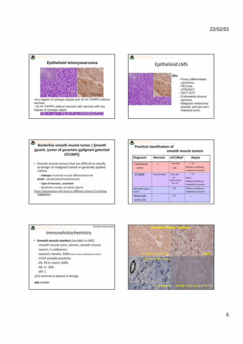
DEPARTMENT OF PATHOLOGYFACULTY OF MEDICINE SIRIRAJ HOSPITAL MAHIDOL UNIVERSITY
Epithelioid leiomyosarcoma
-Any degree of cytologic artypia and ≥5 mf /10HPFs without necrosis- ≥5 mf /10HPFs without necrosis with necrosis with any degree of cytologic atypia
AE1/AE3
DEPARTMENT OF PATHOLOGYFACULTY OF MEDICINE SIRIRAJ HOSPITAL MAHIDOL UNIVERSITY
DDx- Poorly differentiated carcinoma
- PEComa
Epithelioid LMS
- PEComa- UTROSCT- PSTT /ETT- Endometrial stromal sarcoma
- Malignant melanoma/ alveolar soft part sarc/ rhabdoid tumor
DEPARTMENT OF PATHOLOGYFACULTY OF MEDICINE SIRIRAJ HOSPITAL MAHIDOL UNIVERSITY
Borderline smooth muscle tumor / Smooth muscle tumor of uncertain malignant potential
(STUMP)]
• Smooth muscle tumors that are difficult to classify as benign or malignant based on generally applied criteriacriteria‐ Subtype of smooth muscle differentiation in
doubt…standard/epithelioid/myxoid
‐ Type of necrosis…uncertain‐ Borderline number of mitotic figures
These interpretation will result in different criteria of justifying malignancy.
DEPARTMENT OF PATHOLOGYFACULTY OF MEDICINE SIRIRAJ HOSPITAL MAHIDOL UNIVERSITY
Practical classification of smooth muscle tumors
Diagnosis Necrosis mf/10hpf Atypia
Leiomyosar ‐coma
+
‐
Any rate
>10
+ or ‐
Diffuse/ multifocal;moderate to severe
STUMP Questionable
‐‐
Any rate>15
Approaching but<10
+ or ‐
None
Diffuse/multifocal;moderate to severe
LM with bizarre nuclei
‐ ≤10 Diffuse/ multifocal;moderate to severe
Mitotically active LM
‐ ≤15 ‐
DEPARTMENT OF PATHOLOGYFACULTY OF MEDICINE SIRIRAJ HOSPITAL MAHIDOL UNIVERSITY
Immunohistochemistry
• Smooth muscle markers:(variable in LMS)‐ smooth muscle actin, desmin, smooth muscle myosin, h‐caldesmon‐ oxytocin, keratin, EMA (more often epithelioid variant)
bl
Smooth muscle tumor
‐ CD10 variable positivity‐ ER, PR in nearly 100%‐ AR in 30%‐WT‐1
p53 minimal to absent in benign
MiB-1( Ki-67)
DEPARTMENT OF PATHOLOGYFACULTY OF MEDICINE SIRIRAJ HOSPITAL MAHIDOL UNIVERSITYSmooth muscle markers
Smooth muscle actin,1A4 Desmin
h-caldesmon
Smooth muscle markers
Crum CP, Lee KR. Diagnostic gynecologic and obstetric pathology. 2006
Smooth muscle myosin heavy chain
22/02/53
6
DEPARTMENT OF PATHOLOGYFACULTY OF MEDICINE SIRIRAJ HOSPITAL MAHIDOL UNIVERSITY
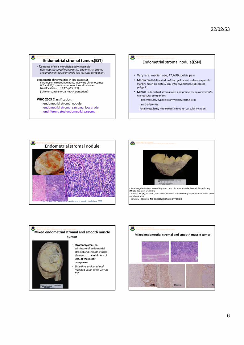
Endometrial stromal tumors(EST)‐ Compose of cells morphologically resemble
nonneoplastic proliferative phase endometrial stroma and prominent spiral arteriole‐like vascular component.
Cytogenetic abnormalities in low grade ESS chromosome rearrangements involving chromosomes 6,7 and 17/ most common reciprocal balanced ptranslocation:‐ t(7;17)(p15;q21) …( chimeric JAZF1‐JJAZ1 mRNA transcripts)
WHO 2003 Classification:‐ endometrial stromal nodule‐ endometrial stromal sarcoma, low grade‐ undifferentiated endometrial sarcoma
DEPARTMENT OF PATHOLOGYFACULTY OF MEDICINE SIRIRAJ HOSPITAL MAHIDOL UNIVERSITY
Endometrial stromal nodule(ESN)
• Very rare; median age, 47;AUB ;pelvic pain
• Macro: Well delineated, soft tan yellow cut surface, expansile margin; mean diameter,7 cm; intramyometrial, subserosal,
l idpolypoid
• Micro: Endometrial stromal cells and prominent spiral arteriole‐like vascular component;
‐ hypercellular/hypocellular/myxoid/epithelioid;
‐mf 1‐5/10HPFs.Focal irregularity not exceed 3 mm; no vascular invasion
DEPARTMENT OF PATHOLOGYFACULTY OF MEDICINE SIRIRAJ HOSPITAL MAHIDOL UNIVERSITY
Endometrial stromal nodule
Crum CP, Lee KR. Diagnostic gynecologic and obstetric pathology. 2006
DEPARTMENT OF PATHOLOGYFACULTY OF MEDICINE SIRIRAJ HOSPITAL MAHIDOL UNIVERSITY
- focal irregularities not exceeding 3 mm ; smooth muscle metaplasia at the periphery. (Mitotic figures=1-2/10 HPFs. - diffuse CD10(+), focal 1A4, and smooth muscle myosin heavy chain(+) in the tumor and thperipheral area; - diffusely (-)desmin. No angiolymphatic invasion
DEPARTMENT OF PATHOLOGYFACULTY OF MEDICINE SIRIRAJ HOSPITAL MAHIDOL UNIVERSITY
Mixed endometrial stromal and smooth muscle tumor
• Stromomyoma.. an admixture of endometrial stromal and smooth muscle elements…….a minimum of 30% f h i30% of the minor component
• Should be evaluated and reported in the same way as EST
DEPARTMENT OF PATHOLOGYFACULTY OF MEDICINE SIRIRAJ HOSPITAL MAHIDOL UNIVERSITY
Mixed endometrial stromal and smooth muscle tumor
CD10 Desmin 1A4
22/02/53
7
DEPARTMENT OF PATHOLOGYFACULTY OF MEDICINE SIRIRAJ HOSPITAL MAHIDOL UNIVERSITY
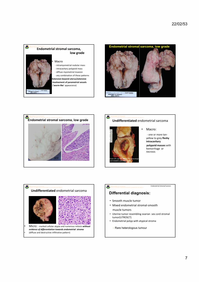
Endometrial stromal sarcoma, low grade
• Macro‐ intramyometrial nodular mass
‐ intracavitary polypoid mass
‐ diffuse myometrial invasion
‐ any combination of these patterns
Extension beyond uterus/extensive
involvement of parametrial vessels(‘worm‐like’ appearance)
DEPARTMENT OF PATHOLOGYFACULTY OF MEDICINE SIRIRAJ HOSPITAL MAHIDOL UNIVERSITYEndometrial stromal sarcoma, low grade
DEPARTMENT OF PATHOLOGYFACULTY OF MEDICINE SIRIRAJ HOSPITAL MAHIDOL UNIVERSITY
Endometrial stromal sarcoma, low grade
AFIP
DEPARTMENT OF PATHOLOGYFACULTY OF MEDICINE SIRIRAJ HOSPITAL MAHIDOL UNIVERSITY
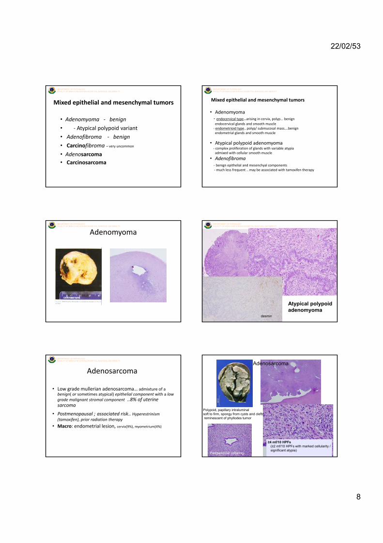
Undifferentiated endometrial sarcoma
• Macro:
‐ one or more tan‐yellow to grey fleshy i t itintracavitary
polypoid masses with hemorrhage or necrosis
Crum CP, Lee KR. Diagnostic gynecologicand obstetric pathology. 2006
DEPARTMENT OF PATHOLOGYFACULTY OF MEDICINE SIRIRAJ HOSPITAL MAHIDOL UNIVERSITY
Undifferentiated endometrial sarcoma
• Micro: ‐marked cellular atypia and numerous mitosis without evidence of differentiation towards endometrial stroma
• (diffuse and destructive infiltrative pattern)
DEPARTMENT OF PATHOLOGYFACULTY OF MEDICINE SIRIRAJ HOSPITAL MAHIDOL UNIVERSITY
Endometrial stromal tumors
• Smooth muscle tumor
• Mixed endometrial stromal‐smooth
muscle tumors
Differential diagnosis:
usc e tu o s• Uterine tumor resembling ovarian sex cord stromal tumor(UTROSCT)
• Endometrial polyp with atypical stroma
Rare heterologous tumour
22/02/53
8
DEPARTMENT OF PATHOLOGYFACULTY OF MEDICINE SIRIRAJ HOSPITAL MAHIDOL UNIVERSITY
Mixed epithelial and mesenchymal tumors
• Adenomyoma ‐ benign
• ‐ Atypical polypoid variant
• Adenofibroma benign• Adenofibroma ‐ benign
• Carcinofibroma – very uncommon
• Adenosarcoma• Carcinosarcoma
DEPARTMENT OF PATHOLOGYFACULTY OF MEDICINE SIRIRAJ HOSPITAL MAHIDOL UNIVERSITY
• Adenomyoma‐ endocervical type..arising in cervix, polyp… benign endocervical glands and smooth muscle‐ endometrioid type.. polyp/ submucosal mass….benign endometrial glands and smooth muscle
Mixed epithelial and mesenchymal tumors
• Atypical polypoid adenomyoma‐ complex proliferation of glands with variable atypia admixed with cellular smooth muscle
• Adenofibroma ‐ benign epithelial and mesenchyal components‐much less frequent .. may be associated with tamoxifen therapy
DEPARTMENT OF PATHOLOGYFACULTY OF MEDICINE SIRIRAJ HOSPITAL MAHIDOL UNIVERSITY
AdenomyomaDEPARTMENT OF PATHOLOGYFACULTY OF MEDICINE SIRIRAJ HOSPITAL MAHIDOL UNIVERSITY
desmin
Atypical polypoid adenomyoma
DEPARTMENT OF PATHOLOGYFACULTY OF MEDICINE SIRIRAJ HOSPITAL MAHIDOL UNIVERSITY
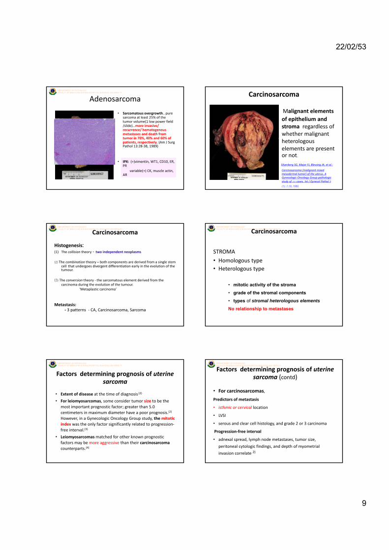
Adenosarcoma
• Low grade mullerian adenosarcoma… admixture of a benign( or sometimes atypical) epithelial component with a low grade malignant stromal component ..8% of uterine sarcomasarcoma
• Postmenopausal ; associated risk.. Hyperestrinism (tamoxifen), prior radiation therapy
• Macro: endometrial lesion, cervix(9%), myometrium(4%)
Adenosarcoma
Polypoid, papillary intraluminalsoft to firm, spongy from cysts and cleftsreminescent of phyllodes tumor
≥4 mf/10 HPFs(≥2 mf/10 HPFs with marked cellularity /significant atypia)
Periglandular collaring..
22/02/53
9
DEPARTMENT OF PATHOLOGYFACULTY OF MEDICINE SIRIRAJ HOSPITAL MAHIDOL UNIVERSITY
Adenosarcoma
• Sarcomatous overgrowth.. pure sarcoma at least 25% of the tumor volume(1 low power field /slide)…more invasive/ recurrence/ hematogenous metastases and death from tumor in 70% 40% and 60% oftumor in 70%, 40% and 60% of patients, respectively. (Am J Surg Pathol 13:28‐38, 1989)
• IPX: (+)vimentin, WT1, CD10, ER, PR
variable(+) CK, muscle actin, AR
Carcinosarcoma
• Malignant elements of epithelium and stroma regardless of whether malignant heterologous elements are presentor not.
Silverberg SG, Major FJ, Blessing JA, et al.:
Carcinosarcoma (malignant mixed mesodermal tumor) of the uterus. A Gynecologic Oncology Group pathologic study of 203 cases. Int J Gynecol Pathol 9 (1): 1-19, 1990.
DEPARTMENT OF PATHOLOGYFACULTY OF MEDICINE SIRIRAJ HOSPITAL MAHIDOL UNIVERSITY
Histogenesis:(1) The collision theory ‐ two independent neoplasms.
(2) The combination theory – both components are derived from a single stem cell that undergoes divergent differentiation early in the evolution of the tumour.
Carcinosarcoma
(3) The conversion theory ‐ the sarcomatous element derived from the carcinoma during the evolution of the tumour.
‘Metaplastic carcinoma’
Metastasis:‐ 3 patterns ‐ CA, Carcinosarcoma, Sarcoma
DEPARTMENT OF PATHOLOGYFACULTY OF MEDICINE SIRIRAJ HOSPITAL MAHIDOL UNIVERSITYCarcinosarcoma
STROMA
• Homologous type• Heterologous type
• mitotic activity of the stroma
• grade of the stromal components
• types of stromal heterologous elements
No relationship to metastases
DEPARTMENT OF PATHOLOGYFACULTY OF MEDICINE SIRIRAJ HOSPITAL MAHIDOL UNIVERSITY
Factors determining prognosis of uterine sarcoma
• Extent of disease at the time of diagnosis (2)
• For leiomyosarcomas, some consider tumor size to be the most important prognostic factor; greater than 5.0
i i i di h i (2)centimeters in maximum diameter have a poor prognosis.(2)
However, in a Gynecologic Oncology Group study, the mitotic index was the only factor significantly related to progression‐free interval.(3)
• Leiomyosarcomas matched for other known prognostic factors may be more aggressive than their carcinosarcomacounterparts.(4)
DEPARTMENT OF PATHOLOGYFACULTY OF MEDICINE SIRIRAJ HOSPITAL MAHIDOL UNIVERSITY
• For carcinosarcomas,
Predictors of metastasis
• isthmic or cervical location
Factors determining prognosis of uterine sarcoma (contd)
• LVSI
• serous and clear cell histology, and grade 2 or 3 carcinoma
Progression‐free interval
• adnexal spread, lymph node metastases, tumor size,
peritoneal cytologic findings, and depth of myometrial
invasion correlate 2)
22/02/53
10
DEPARTMENT OF PATHOLOGYFACULTY OF MEDICINE SIRIRAJ HOSPITAL MAHIDOL UNIVERSITY
Perivascular epithelioid cell tumor
(PEComa)• Composed predominantly or exclusively of HMB‐45 positive perivascular
epithelioid cells with eosinophilic granular cytoplasm
• Age 40‐75 yrs( mean 54)/….. AUB/ …uncertain malignant potential• Macro: mass in uterine corpus
• Micro:1) tongue‐like growth pattern of low grade ESS, abundant eosinophilic granular/clear cytoplasm( diffuse HMB‐45 and variable muscle marker expressions)
• 2) lesser tongue‐like growth pattern of epithelioid cells with less prominent clear cytoplasm and a smaller number HMB‐45 (+)cells but more extensive muscle marker differentiation
• Genetic susceptibility:
‐ pelvic nodes involved by lymphangioleiomyomatosis
‐ one fourth had tuberous sclerosis
DEPARTMENT OF PATHOLOGYFACULTY OF MEDICINE SIRIRAJ HOSPITAL MAHIDOL UNIVERSITY
Adenomatoid tumor
• Benign tumor of mesothelium forming gland like structures• Site: uterine serosa, myometrium• Macro: softer, less well defined than leiomyoma • Micro: multiple small, often slit‐like interconnecting spaces
within the myometrium lined by cuboidal or attenuatedwithin the myometrium lined by cuboidal or attenuated cells….infiltrative appearance..
‐may be confused with lymphangioma ‐ little nuclear atypia, signet ring like‐ no stromal desmoplastic response
• IPX... Positive keratin and mesothelial markers
DEPARTMENT OF PATHOLOGYFACULTY OF MEDICINE SIRIRAJ HOSPITAL MAHIDOL UNIVERSITY
References1. Crum CP, Lee KR. Diagnostic gynecologic and obstetric pathology.
20062. Major FJ, Blessing JA, Silverberg SG, et al. Prognostic factors in early
stage uterine sarcoma. A Gynecologic Oncology Group Study. Cancer 71( 4Suppl);1993:1702-9
3. Evans HL, Chawla SP, Simpson C, et al. Smooth muscle neoplasm of the uterus other than ordinary leiomyoma. A study of 46 cases, with emphasis on diagnostic criteria and prognostic factors Cancer 62(10);emphasis on diagnostic criteria and prognostic factors. Cancer 62(10); 1988:2239-47
4. Oláh KS, Dunn JA, Gee H. Leiomyosarcoma have a poorer prognosis than mixed mesodermal tumor when adjusting for known prognostic factors: the result of a retrospective study of 423 cases of uterine sarcoma. Br J Obstet Gynecol 99(7);1992:590-4
5. Nucci MR, Oliva E. Gynecologic pathology. China: Churchill Livingstone; 2009
6. Tavassoli FA, Devilee P. WHO classification of tumors. Tumors of the breast and female genital organs. 2003
7. Robboy J, Mutter GL, Prat J,Bently RC,Russel P, Anderson MC. Robboy’s pathology of the female reproductive tract. 2nd ed. 2009
DEPARTMENT OF PATHOLOGYFACULTY OF MEDICINE SIRIRAJ HOSPITAL MAHIDOL UNIVERSITY
Any questions?
Thank You for Your Attention