MR Imaging of Pituitary Dwarfism - ajnr.org · MR Imaging of Pituitary Dwarfism The purpose of this...
4
Tashiro Kuroiwa 1 · 2 Yasufumi Okabe 3 Kanehiro Hasuo 1 Kotaro Yasumori 1 Akira Mizushima 1 Kouji Masuda 1 Received October 30, 1989; revision requested January 8, 1990; final revision received August 16, 1990; accepted August 17, 1990. ' Department of Radiology, Faculty of Medicine, Kyushu University, 3-1-1 Maidashi, Higashi-ku, Fukuoka 812, Japan. ' Present address: Department of Radiology, Saga Medical School , 5-1-1 Nabeshima, Saga 849, Japan. Address reprint requests to T. Kuroiwa. 3 Department of Pediatrics, Faculty of Medicine, Kyushu University, Fukuoka 812, Japan. 0195-6108/91/1201-0161 © American Society of Neuroradiology 161 MR Imaging of Pituitary Dwarfism The purpose of this study was to determine whether the MR findings of idiopathic pituitary dwarfism correlated with a clinical history of perinatal abnormalities and abnormal levels of pituitary hormones _ MR examinations of 18 patients with pituitary dwarfism were performed; these patients were divided into two groups: those with ectopic posterior lobes (group 1) and those with normal posterior lobes (group 2). Among the seven patients in group 1, MR showed hyperintense signal at the median eminence of the hypothalamus, which was regarded as the ectopic posterior lobe of the pituitary gland. The mean anteroposterior length and height of the pituitary gland in group 1 patients were significantly smaller than those dimensions in the normal control group; the pituitary stalk was not detected in three of seven patients. Six of seven patients were products of breech presentation with perinatal asphyxia. The peak serum growth hormone level was less than 5 ngfml when assessed by insulin-induced hypoglycemia or the clonidine test. MR findings in the 11 patients with pituitary dwarfism and normal posterior lobes were normal except that the mean size of the pituitary gland was slightly smaller than that of normal controls. The clinical history of these patients was normal except for perinatal asphyxia in one case. Our findings in patients with pituitary dwarfism, with or without an ectopic posterior pituitary lobe suggest that the ectopic lobe, visualized as a bright spot at the median eminence of the hypothalamus, may be common when pituitary dwarfism follows perinatal anoxic/ischemic episodes. AJNR 12:161-164, January/February 1991 The cause of pituitary dwarfism is not clear, but some investigators propose that perinatal abnormalities such as asphy xi a or breech presentation may cause pituitary ischemia, resulting in pituitary dwarfism [1] . In the diagnosis of pituitary dwarfism, only a few reports have discussed MR findings [2-4]. It has been reported that in some cases of pituitary dwarfism, MR shows an ectopic posterior lobe at the median eminence of hypothalamus; authors have speculated that this may be due to perinatal ischemia of the pituitary stalk or pituitary gland. However, while investigating MR findings in clinically proven cases of pituitary dwarfism, we found that there were two distinct groups: one group had an ectopic posterior lobe located at the median eminence of the hypothalamus that was seen as a bright spot on MR and a small anterior lobe with or without a thin pituitary stalk; the second group had a normally positioned posterior lobe and very few abnormal MR findings . In the first group, a history of perinatal abnormalities was more common than in the latter. We present MR findings in 18 pituitary dwarfs who were classified into two groups on the basis of whether their posterior lobes were ectopic or normal in position and discuss the relationship between the position of the posterior pituitary lobe and perinatal history.
Transcript of MR Imaging of Pituitary Dwarfism - ajnr.org · MR Imaging of Pituitary Dwarfism The purpose of this...
Tashiro Kuroiwa1·2
Yasufumi Okabe3
Kanehiro Hasuo 1
Kotaro Yasumori 1
Akira Mizushima 1
Kouji Masuda 1
Received October 30, 1989; revision requested January 8, 1990; final revision received August 16, 1990; accepted August 17, 1990.
' Department of Radiology, Faculty of Medicine, Kyushu University, 3-1-1 Maidashi, Higashi-ku, Fukuoka 812, Japan.
' Present address: Department of Radiology, Saga Medical School , 5-1-1 Nabeshima, Saga 849, Japan. Address reprint requests to T. Kuroiwa.
3 Department of Pediatrics, Faculty of Medicine, Kyushu University, Fukuoka 812, Japan.
0195-6108/91/1201-0161 © American Society of Neuroradiology
161
MR Imaging of Pituitary Dwarfism
The purpose of this study was to determine whether the MR findings of idiopathic pituitary dwarfism correlated with a clinical history of perinatal abnormalities and abnormal levels of pituitary hormones_ MR examinations of 18 patients with pituitary dwarfism were performed; these patients were divided into two groups: those with ectopic posterior lobes (group 1) and those with normal posterior lobes (group 2). Among the seven patients in group 1, MR showed hyperintense signal at the median eminence of the hypothalamus, which was regarded as the ectopic posterior lobe of the pituitary gland. The mean anteroposterior length and height of the pituitary gland in group 1 patients were significantly smaller than those dimensions in the normal control group; the pituitary stalk was not detected in three of seven patients. Six of seven patients were products of breech presentation with perinatal asphyxia. The peak serum growth hormone level was less than 5 ngfml when assessed by insulin-induced hypoglycemia or the clonidine test. MR findings in the 11 patients with pituitary dwarfism and normal posterior lobes were normal except that the mean size of the pituitary gland was slightly smaller than that of normal controls. The clinical history of these patients was normal except for perinatal asphyxia in one case.
Our findings in patients with pituitary dwarfism, with or without an ectopic posterior pituitary lobe suggest that the ectopic lobe, visualized as a bright spot at the median eminence of the hypothalamus, may be common when pituitary dwarfism follows perinatal anoxic/ischemic episodes.
AJNR 12:161-164, January/February 1991
The cause of pituitary dwarfism is not clear, but some investigators propose that perinatal abnormalities such as asphyxia or breech presentation may cause pituitary ischemia, resulting in pituitary dwarfism [1] . In the diagnosis of pituitary dwarfism, only a few reports have discussed MR findings [2-4]. It has been reported that in some cases of pituitary dwarfism, MR shows an ectopic posterior lobe at the median eminence of hypothalamus; authors have speculated that this may be due to perinatal ischemia of the pituitary stalk or pituitary gland. However, while investigating MR findings in clinically proven cases of pituitary dwarfism, we found that there were two distinct groups: one group had an ectopic posterior lobe located at the median eminence of the hypothalamus that was seen as a bright spot on MR and a small anterior lobe with or without a thin pituitary stalk; the second group had a normally positioned posterior lobe and very few abnormal MR findings. In the first group, a history of perinatal abnormalities was more common than in the latter.
We present MR findings in 18 pituitary dwarfs who were classified into two groups on the basis of whether their posterior lobes were ectopic or normal in position and discuss the relationship between the position of the posterior pituitary lobe and perinatal history.
162 KUROIWA ET AL. AJNR:12, January{February 1991
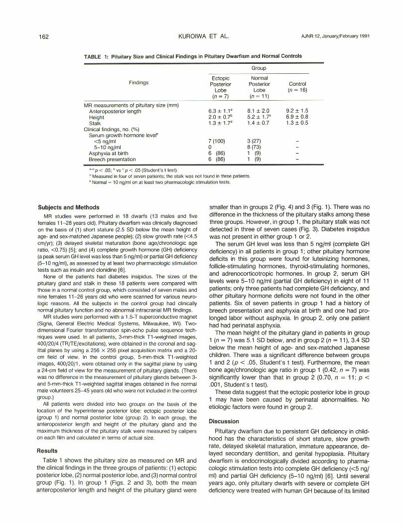
TABLE 1: Pituitary Size and Clinical Findings in Pituitary Dwarfism and Normal Controls
Group
Ectopic Normal Findings Posterior Posterior Control
Lobe Lobe (n = 16) (n = 7) (n = 11)
MR measurements of pituitary size (mm) Anteroposterior length 6.3±1.1a 8.1 ± 2.0 9.2 ± 1.5 Height 2.0 ± 0.7° 5.2 ± 1.7' 6.9 ± 0.8 Stalk 1.3± 1.7" 1.4 ± 0.7 1.3 ± 0.5
Clinical findings, no. (%) Serum growth hormone level•
< 5 ngjml 7 (100) 3 (27) 5-10 ngfml 0 8 (73)
Asphyxia at birth 6 (86) 1 (9) Breech presentation 6 (86) 1 (9)
•-c p < .05; b vs ' p < .05 (Student 's t test). " Measured in four of seven patients; the stalk was not found in three patients. • Normal = 1 0 ng{ml on at least two pharmacologic stimulation tests.
Subjects and Methods
MR studies were performed in 18 dwarfs (13 males and five females 11-28 years old). Pituitary dwarfism was clinically diagnosed on the basis of (1) short stature (2.5 SD below the mean height of age- and sex-matched Japanese people); (2) slow growth rate (<4.5 cmjyr); (3) delayed skeletal maturation (bone agejchronologic age ratio , < 0.75) (5]; and (4) complete growth hormone (GH) deficiency (a peak serum GH level was less than 5 ngjml) or partial GH deficiency (5-1 0 ngfml) , as assessed by at least two pharmacologic stimulation tests such as insulin and clonidine (6].
None of the patients had diabetes insipidus. The sizes of the pituitary gland and stalk in these 18 patients were compared with those in a normal control group, which consisted of seven males and nine females 11 -26 years old who were scanned for various neurologic reasons. All the subjects in the control group had clinically normal pituitary function and no abnormal intracranial MR findings.
MR studies were performed with a 1.5-T superconductive magnet (Signa, General Electric Medical Systems, Milwaukee, WI). Twodimensional Fourier transformation spin-echo pulse sequence techniques were used. In all patients, 3-mm-thick T1-weighted images, 400/20/4 (TR/TEfexcitations) , were obtained in the coronal and sagittal planes by using a 256 x 256 pixel acquisition matrix and a 20-cm field of view. In the control group, 5-mm-thick T1-weighted images, 400/20/1 , were obtained only in the sagittal plane by using a 24-cm field of view for the measurement of pituitary glands. (There was no difference in the measurement of pituitary glands between 3- . and 5-mm-thick T1-weighted sagittal images obtained in five normal male volunteers 25-45 years old who were not included in the control group.)
All patients were divided into two groups on the basis of the location of the hyperintense posterior lobe: ectopic posterior lobe (group 1) and normal posterior lobe (group 2). In each group, the anteroposterior length and height of the pituitary gland and the maximum thickness of the pituitary stalk were measured by calipers on each film and calculated in terms of actual size.
Results
Table 1 shows the pituitary size as measured on MR and the clinical findings in the three groups of patients: (1) ectopic posterior lobe, (2) normal posterior lobe, and (3) normal control group (Fig. 1 ). In group 1 (Figs. 2 and 3), both the mean anteroposterior length and height of the pituitary gland were
smaller than in groups 2 (Fig. 4) and 3 (Fig. 1 ). There was no difference in the thickness of the pituitary stalks among these three groups. However, in group 1, the pituitary stalk was not detected in three of seven cases (Fig. 3). Diabetes insipidus was not present in either group 1 or 2.
The serum GH level was less than 5 ngjml (complete GH deficiency) in all patients in group 1; other pituitary hormone deficits in this group were found for luteinizing hormones, follicle-stimulating hormones, thyroid-stimulating hormones, and adrenocorticotropic hormones. In group 2, serum GH levels were 5-1 0 ngjml (partial GH deficiency) in eight of 11 patients; only three patients had complete GH deficiency, and other pituitary hormone deficits were not found in the other patients. Six of seven patients in group 1 had a history of breech presentation and asphyxia at birth and one had prolonged labor without asphyxia. In group 2, only one patient had had perinatal asphyxia.
The mean height of the pituitary gland in patients in group 1 (n = 7) was 5.1 SD below, and in group 2 (n = 11 ), 3.4 SD below the mean height of age- and sex-matched Japanese children. There was a significant difference between groups 1 and 2 (p < .05, Student's t test) . Furthermore, the mean bone agejchronologic age ratio in group 1 (0.42, n = 7) was significantly lower than that in group 2 (0.70, n = 11; p < .001 , Student's t test).
These data suggest that the ectopic posterior lobe in group 1 may have been caused by perinatal abnormalities. No etiologic factors were found in group 2.
Discussion
Pituitary dwarfism due to persistent GH deficiency in childhood has the characteristics of short stature, slow growth rate, delayed skeletal maturation, immature appearance, delayed secondary dentition, and genital hypoplasia. Pituitary dwarfism is endocrinologically divided according to pharmacologic stimulation tests into complete GH deficiency (<5 ngj ml) and partial GH deficiency (5-1 0 ngjml) [6]. Until several years ago, only pituitary dwarfs with severe or complete GH deficiency were treated with human GH because of its limited
A B
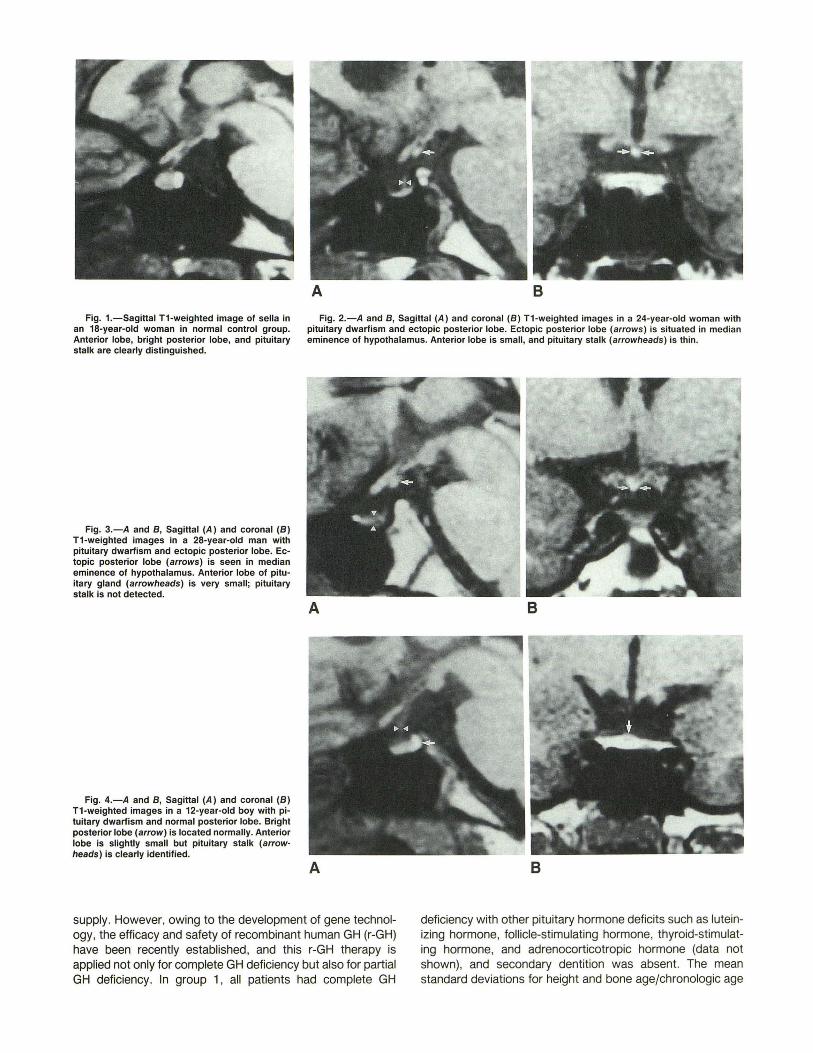
Fig. 1.-Sagittal T1-weighted image of sella in an 18-year-old woman in normal control group. Anterior lobe, bright posterior lobe, and pituitary stalk are clearly distinguished.
Fig. 2.-A and 8, Sagittal (A) and coronal (8) T1-weighted images in a 24-year-old woman with pituitary dwarfism and ectopic posterior lobe. Ectopic posterior lobe (arrows) is situated in median eminence of hypothalamus. Anterior lobe is small, and pituitary stalk (arrowheads) is thin.
Fig. 3.-A and 8, Sagittal (A) and coronal (8) T1-weighted images in a 28-year-old man with pituitary dwarfism and ectopic posterior lobe. Ectopic posterior lobe (arrows) is seen in median eminence of hypothalamus. Anterior lobe of pituitary gland (arrowheads) is very small; pituitary stalk is not detected.
Fig. 4.-A and 8, Sagittal (A) and coronal (8) T1-weighted images in a 12-year-old boy with pituitary dwarfism and normal posterior lobe. Bright posterior lobe (arrow) is located normally. Anterior lobe is slightly small but pituitary stalk (arrow· heads) is clearly identified.
A
A
supply. However, owing to the development of gene technology, the efficacy and safety of recombinant human GH (r-GH) have been recently established, and this r-GH therapy is applied not only for complete GH deficiency but also for partial GH deficiency. In group 1, all patients had complete GH
B
B
deficiency with other pituitary hormone deficits such as luteinizing hormone, follicle-stimulating hormone, thyroid-stimulating hormone, and adrenocorticotropic hormone (data not shown), and secondary dentition was absent. The mean standard deviations for height and bone age/chronologie age
164 KUROIWA ET AL. AJNR:12, January/February 1991
ratio in group 1 were significantly smaller than those in group 2. It is evident that patients in group 1 had more severe clinical findings than those in group 2.
The fact that most of our patients with MR findings of ectopic hyperintensity of the posterior lobe at the median eminence of the hypothalamus (group 1) experienced perinatal asphyxia suggests that pituitary dwarfism may be caused by perinatal abnormalities. The size of the pituitary gland in our control group (Fig. 1) was approximately equal to that in previous reports (7, 8) . Both anteroposterior length and height of the pituitary gland in the group with ectopic posterior lobes (Figs. 2 and 3) were smaller than those of control group, and pituitary stalks were not found in almost half of these patients (Fig. 3).
The posterior lobe of the pituitary gland is formed by downward budding from the diencephalon and secretes antidiuretic hormone and oxytocin. The anterior lobe of the pituitary gland is formed by upward development of Rathke's pouch toward the hypothalamus, and is mainly supplied by the hypophyseal-portal system (2].
The cause of pituitary dwarfism is not clearly known, but three hypotheses are discussed here. One is that the pituitary gland and stalk may be damaged by hypoxia or decreased perfusion during abnormal delivery [2, 9]. When a neonate experiences perinatal hypoxia or asphyxia because of breech presentation or other perinatal abnormalities, there may be disturbance of the microcirculation in the hypophyseal-portal system, causing pituitary ischemia or infarction of the pituitary stalk (2), resulting in a hypoplastic anterior lobe and a hypoplastic or absent pituitary stalk. However, since our pituitary dwarfs had no ischemic/anoxic changes in the brain or elsewhere in the body, other causes must be considered. The second hypothesis is that the stalk may be mechanically cut by the posterior clinoid plate or diaphragma sellae during breech delivery, the so-called "stalk transection" (4, 10). Kikuchi et al. (1 0] stated that the pituitary stalk is anatomically vulnerable to transection during breech or foot presentation. However, transection of the pituitary stalk was not always seen in patients who had ectopic posterior lobes on MR imaging, so we think that mechanical transection of the pituitary stalk was not the only cause of pituitary dwarfism. The third hypothesis is that congenital hypoplasia or dysplasia of the pituitary gland and pituitary stalk or hypothalamus may coexist in pituitary dwarfism (11 , 12]. We speculate that stalk infarction due to the disturbance of microcirculation in the hypophyseal-portal system may be accelerated by such congenital defects, and that the rest of the brain and other organs in the body may be spared such damage.
The presence of an ectopic hyperintense area suggests that neurosecretion continued in the median eminence of the hypothalamus, and consequently , diabetes insipidus is absent in these patients. In patients with diabetes insipidus, bright signal intensity may be absent in the posterior lobe (13]. The cause of a hyperintense nodule at the median eminence of the hypothalamus is not known , but tissue similar to that of the normal posterior lobe of the pituitary gland should show hyperintensity . Fujisawa et al. [7] stated that the signal intensity of the posterior lobe of the pituitary gland was higher than that of fatty tissue on proton-density and T2-weighted
images, and chemical-shift artifact was absent. Furthermore, an ectopic hyperintense area in the hypothalamus in patients with stalk transection seems to be due to the normal contents of the hypothalamohypophyseal tract, such as neurosecretory granules (4, 14). Others report, however, that the ectopic or normal pituitary hyperintensity of the posterior lobe shows intracellular lipid signal in the glial cell pituicytes [2, 15], so that the precise cause of the posterior pituitary hyperintensity has yet to be elucidated.
Our group 2 patients had fewer abnormal findings on MR (Fig. 4) than did group 1 patients. Most patients in this group were delivered normally and their serum GH levels were in the range of 5-1 0 ngjml (partial GH deficiency). Most patients with partial GH deficiency have no history of perinatal abnormality (6). The cause of dwarfism in group 2 is completely unknown.
Although an ectopic posterior lobe is rarely seen as a normal variation (16), the correlation between the ectopic posterior hyperintensity on MR and the history of perinatal abnormalities in our study of pituitary dwarfism is very interesting, and MR may provide a clue as to its cause in some patients with idiopathic pituitary dwarfism.
REFERENCES
1. Shizume K, Harada Y, lbayashi H. Survey studies on pituitary diseases in Japan. Endocrinol Jpn 1977;24:139-147
2. Kelly WM, Kucharczyk W, Kucharczyk J, et al. Posterior pituitary ectopia: an MR feature of pituitary dwariism. AJNR 1988;9 :453-460
3. Root AW, Martinez CR , Muroff LR. Subhypothalamic high-intensity signals identified by magnetic resonance imaging in children with idiopathic anterior hypopituitarism. Am J Dis Child 1989;143 :366-367
4. Fujisawa I, Kikuchi K, Nishimura K, et al. Transection of the pituitary stalk: development of an ectopic posterior lobe assessed with MR imaging. Radiology 1987;165:487-489
5. Greulich WW, Pyle Sl. Radiographic atlas of skeletal development of the hand and wrist. 2nd ed . Stanford, CA: Stanford University, 1983
6. Okabe Y, Ohkubo K, Mizuno Y, et al. Clinical appearance of partial GH deficiency; correlation of complete GH deficiency and primordial dwariism. Clin Endocrinol (Oxf) 1986;34 : 119-122
7. Fujisawa I, Asato R, Nishimura K, et al. Anterior and posterior lobes of the pituitary gland: assessment by 1.5 T MR imaging. J Comput Assist Tomogr 1987;11 :214-220
8. Wiener SN , Rzeszotarski MS, Droege RT, et al. Measurement of pituitary gland height with MR imaging. AJNR 1985;6 :717-722
9. Inoue Y, Nemoto Y, Fujita K, et al. Pituitary dwariism: CT evaluation of the pituitary gland. Radiology 1986;159:171-173
10. Kikuchi K, Fujisawa I, Momoi T, et al. Hypothalamic-pituitary function in growth hormone-deficient patients with pituitary stalk transection. J Clin Endocrinol Metab 1988;67:817-823
11. Natale BD, Scotti G, Pellini C, et aL Empty sella in children with pituitary dwariism: Does it exist? Pediatrician 1987;14:246-252
12. Craft WH , Underwood LE, VanWyk JJ . High incidence of perinatal insult in children with idiopathic hypopituitarism. J Pediatr 1980;96:397-402
13. Fujisawa I, Nishimura K, Asato R, et al. Posterior lobe of the pituitary in diabetes insipidus: MR findings . J Comput Assist Tomogr 1987;11 : 221-225
14. El Gammal T, Brooks BS, Hoffman WH . MR imaging of the ectopic bright signal of posterior pituitary regeneration . AJNR 1989;10:323-328
15. Kucharczyk J, Kucharczyk W, Berry I, et al. Histochemical characterization and functional significance of the hyperintense signal on MR images of the posterior pituitary. AJNR 1988;9 : 1 079- 1 083
16. Brooks BS, El Gammal T, Allison JD, et al. Frequency and variation of the posterior pituitary bright signal on MR images. AJNR 1989;10: 943- 948