Microbiota Landscape of Gut System of Guppy Fish ...
11
Research Article Microbiota Landscape of Gut System of Guppy Fish (Poecilia reticulata) Plays an Outstanding Role in Adaptation Mechanisms Christian Aim´ e Kayath , 1,2 Armel Ibala Zamba, 1,3 Joseph Goma-Tchimbakala, 1,3 Victor Mamon´ ek´ en´ e, 1,3 Gloire Moniceth Mombo Makanga, 1 Aim´ e Augustin Lebonguy, 1 and Etienne Nguimbi 1,2 1 Institut National de Recherche en Sciences Exactes et Naturelles (IRSEN), Avenue de l’Auberge Gascogne, B.P 2400 Brazzaville, Congo 2 Laboratoire de Biologie Cellulaire et Mol´ eculaire (BCM), Facult´ e des Sciences et Techniques, Universit´ e Marien Ngouabi, BP. 69 Brazzaville, Congo 3 Ecole Nationale Sup´ erieure d’Agronomie et de Foresterie, Universit´ e Marien Ngouabi, BP. 69 Brazzaville, Congo Correspondence should be addressed to Christian Aim´ e Kayath; [email protected] Received 29 October 2018; Revised 22 January 2019; Accepted 28 January 2019; Published 17 February 2019 Academic Editor: Simona Nardoni Copyright © 2019 Christian Aim´ e Kayath et al. is is an open access article distributed under the Creative Commons Attribution License, which permits unrestricted use, distribution, and reproduction in any medium, provided the original work is properly cited. Microbial consortium that is present in fish gut systems works together to achieve unknown specific roles. Here, we collected guppy fish from hydrocarbon- and trace metal-contaminated wastewater to assess the relationships between gut microbiota and host fish adaptation. Targeted genes and 16S rRNA amplicon sequencing have been used to identify gut bacteria of guppies. Mineral-conditioned medium contributes to identify bacteria with the ability to grow and/or to tolerate hydrocarbon and trace metals. Additionally, trace metals’ tolerance minimum inhibitory concentration (MIC) of microbiota was evaluated. We first isolated bacteria from the gut system, and we showed that Bacillus spp., Staphylococcus spp., Shigella spp., Salmonella spp, Pseudomonas spp., Citrobacter spp., Salmonella enterica ssp.arizonae sp., Enterobacter spp, and Acinetobacter spp. are part of guppy gut microbiota. Some representative species are able to degrade and/or tolerate gasoline and/or diesel fuel hydrocarbons. Tolerance to trace metals was observed in Gram-positive and Gram-negative bacteria. We showed that minimal inhibitory concentration (MIC) of some microbiota isolated from gut systems has been found including for mercury (Hg) between 2 and 4‰,cobalt(Co)Co(2and5‰),zinc(Zn)(9and18‰),andplomb(Pb)(22and27‰).ZnandPbwerethetracemetalsforwhich the rate of tolerance was significantly higher. Finally, we showed that cytochrome c oxidase is not interfering in presence of trace metals. e working consortium showed that bacteria should work together to achieve their best. 1. Introduction Guppy fish has been introduced in many countries around the world. Its great adaptability including rapid re- production and unquenchable appetite for mosquito larvae make it a valuable tool in combating a couple of diseases such as malaria that can be transmitted by mosquito bites [1]. In 1976, some collaboration between the Republic of Congo government and the World Health Organization (WHO) was made introducing Poecilia reticulata (Peters, 1959) in Brazzaville. One of the main approaches of this collaboration was to control mosquito epidemics for better fighting malaria. Guppies and mosquito larvae have been unfortunately coevoluated towards a balance relationship (Victor Mamonekene, data submitted). By the way, P. reticulata is a fascinating vertebrate fish model with a large landscape of facets linked to its ability to quickly grow by having a high population that is able to resist the envi- ronment pressures such as evolution of fish size [2], changes in predation environment and the ability to be anatomically modified [3], and behavioral trait in a population of in- dividuals under the effect of natural selection [4]. is is Hindawi International Journal of Microbiology Volume 2019, Article ID 3590584, 10 pages https://doi.org/10.1155/2019/3590584
Transcript of Microbiota Landscape of Gut System of Guppy Fish ...
Research ArticleMicrobiota Landscape of Gut System of Guppy Fish(Poecilia reticulata) Plays an Outstanding Role inAdaptation Mechanisms
Christian Aime Kayath ,1,2 Armel Ibala Zamba,1,3 Joseph Goma-Tchimbakala,1,3
Victor Mamonekene,1,3 Gloire Moniceth Mombo Makanga,1 Aime Augustin Lebonguy,1
and Etienne Nguimbi1,2
1Institut National de Recherche en Sciences Exactes et Naturelles (IRSEN), Avenue de l’Auberge Gascogne,B.P 2400 Brazzaville, Congo2Laboratoire de Biologie Cellulaire et Moleculaire (BCM), Faculte des Sciences et Techniques, Universite Marien Ngouabi,BP. 69 Brazzaville, Congo3Ecole Nationale Superieure d’Agronomie et de Foresterie, Universite Marien Ngouabi, BP. 69 Brazzaville, Congo
Correspondence should be addressed to Christian Aime Kayath; [email protected]
Received 29 October 2018; Revised 22 January 2019; Accepted 28 January 2019; Published 17 February 2019
Academic Editor: Simona Nardoni
Copyright © 2019 Christian Aime Kayath et al. *is is an open access article distributed under the Creative Commons AttributionLicense, which permits unrestricted use, distribution, and reproduction in any medium, provided the original work is properly cited.
Microbial consortium that is present in fish gut systems works together to achieve unknown specific roles. Here, we collectedguppy fish from hydrocarbon- and trace metal-contaminated wastewater to assess the relationships between gut microbiota andhost fish adaptation. Targeted genes and 16S rRNA amplicon sequencing have been used to identify gut bacteria of guppies.Mineral-conditioned medium contributes to identify bacteria with the ability to grow and/or to tolerate hydrocarbon and tracemetals. Additionally, trace metals’ tolerance minimum inhibitory concentration (MIC) of microbiota was evaluated. We firstisolated bacteria from the gut system, and we showed that Bacillus spp., Staphylococcus spp., Shigella spp., Salmonella spp,Pseudomonas spp., Citrobacter spp., Salmonella enterica ssp.arizonae sp., Enterobacter spp, and Acinetobacter spp. are part ofguppy gut microbiota. Some representative species are able to degrade and/or tolerate gasoline and/or diesel fuel hydrocarbons.Tolerance to trace metals was observed in Gram-positive and Gram-negative bacteria. We showed that minimal inhibitoryconcentration (MIC) of some microbiota isolated from gut systems has been found including for mercury (Hg) between 2 and4‰, cobalt (Co) Co (2 and 5‰), zinc (Zn) (9 and 18‰), and plomb (Pb) (22 and 27‰). Zn and Pb were the trace metals for whichthe rate of tolerance was significantly higher. Finally, we showed that cytochrome c oxidase is not interfering in presence of tracemetals. *e working consortium showed that bacteria should work together to achieve their best.
1. Introduction
Guppy fish has been introduced in many countries aroundthe world. Its great adaptability including rapid re-production and unquenchable appetite for mosquito larvaemake it a valuable tool in combating a couple of diseasessuch as malaria that can be transmitted by mosquito bites[1]. In 1976, some collaboration between the Republic ofCongo government and the World Health Organization(WHO) was made introducing Poecilia reticulata (Peters,1959) in Brazzaville. One of the main approaches of this
collaboration was to control mosquito epidemics for betterfighting malaria. Guppies and mosquito larvae have beenunfortunately coevoluated towards a balance relationship(Victor Mamonekene, data submitted). By the way, P.reticulata is a fascinating vertebrate fish model with a largelandscape of facets linked to its ability to quickly grow byhaving a high population that is able to resist the envi-ronment pressures such as evolution of fish size [2], changesin predation environment and the ability to be anatomicallymodified [3], and behavioral trait in a population of in-dividuals under the effect of natural selection [4]. *is is
HindawiInternational Journal of MicrobiologyVolume 2019, Article ID 3590584, 10 pageshttps://doi.org/10.1155/2019/3590584
creating a new phenotypic selection inducing a rapid escapeability [5]. Guppies’ physiological changes linked to dietshave involved gut length plasticity [6]. Fish guts containmicrobiota playing different roles. Bacterial composition ofthe digestive tract of some fish has been documented. *isdepends on age, size, diet, and environmental conditions [7].Different genera of lactic acid bacteria such as Streptococcus,Leuconostoc, Lactobacillus, Lactococcus, and Carnobacte-rium are also part of this microbiota [8]. Anaerobic bacteriasuch as Escherichia coli and Streptococcus sp. are the first toinitiate colonization creating a propitious environment tothe development of many other strict anaerobic microor-ganisms [9]. Numerous groups of microorganisms have alsobeen successfully colonized guppy gut. Gut bacterial com-munities are now known to influence a wild range of fitness-related aspects of organisms. *is includes Vibrionales,Bacillales, Actinomycetales, Clostridiales and Enter-obacteriales [10]. Bacteria communities are known to playimportant physiological inputs by influencing metabolicprocesses, such as the digestion of complex carbohydrates[11, 12], regulation of fat storage and fish nutrition bymicrobiota [13], enzymes production from microbiota [14],and antibiotic resistance profile [15].
Most of the time, Brazzaville ecosystem waters con-taminated with hydrocarbon are associated with tracemetals. Some trace metals have been shown to be essentialfor a couple of biological functions for living organismsincluding Cu, Zn, Co, Fe, Mn, Ni, Cr, Se, and As, but theincrease in their concentration can lead to phenomena oftoxicity to organisms. Associations between trace metals andP. reticulata have been documented [16]. Some heavy-metalconcentrations such as Cd, Cr, Cu, Hg, Pb, and Zn have beenidentified in the muscle tissue of some fish species includingguppies [17–19].
Wastewater fish are exposed to high hydrocarbon con-centrations and trace metals. Several investigations are stillunclear in terms of adaptive mechanisms linked to the gutmicrobial communities. Adaptation strategies of guppiesliving in wastewaters contaminated with hydrocarbon andheavy metal involving gut microbial populations is stillmissing. *is work aims to study the role of microbiota in theadaptive mechanisms in wastewaters. By isolating intestinebacteria of gut track and identifying microbiota using icsB,invG, and 16S rRNA gene, by determining the emulsion index(E24), by investigating the capacity to grow and/or to toleratein mineral media supplemented with hydrocarbons, by cal-culating minimal inhibitory concentration (MIC) of heavy-metal inhibition, and by studying interferences between cy-tochrome oxidases and trace metals, this study will allowcontributing to the understanding of the knowledge gap. *iswork will try to provide a deep assessment of adaptationmechanisms occurring in the guppy gut system once incontact with pollutants.
2. Materials and Methods
2.1. Collection of Guppies and Isolation of Gut Microbiota.Fresh wastewater guppies were collected from differentgutters close to the laboratory and from small streams of
Brazzaville and stocked in ziplocks and transported from thesite of sampling to the laboratory (lab GPS coordinates:elevation (127m), distance (5.7 km), S04.27643°, E015.29297°; station GPS coordinates: elevation (127m),distance (6.1 km), S04.27643°, E 015.29297°). Fish had beencollected by using deep net. Samples were systematicallybrought from the station to the laboratory (about 0.4 km) fordissection. Tricaine methane-sulfonate (MS-222) has beenused as an anesthetic and euthanasia agent, and the fish wassurface-sterilized with alcohol (70%). *e intestine of Poe-cilia reticulata gut track was removed by dissection withsterile instruments and then washed in 70% ethanol to avoidcontamination. *e intestine was immersed in sterile saline.*is was vigorously vortexed to separate microbiota fromtissue. Dilutions were done, and bacterial suspension wasstreaked on nutrient agar media. Enumeration of colonieswas done in triplicate on plate count agar (PCA). *e Petridishes were incubated at 37°C for 24 h to 48 h. After the firstisolation on Petri dishes, different colonies were obtained.Each colony of different appearance was separately isolated.Purification of the isolates was rigorously done by successiveand alternating subcultures. Purity was estimated by using amicroscope for morphological characterization. Gram statuswas determined by using 3% KOH. Sporulation, hydrogenperoxide (H2O2), and oxidases tests were used for bio-chemical characterization.
2.2. Identification of Isolate andGenomicDNAExtraction andSequencing. Conventional methods and Enterosystem 18R(Liofilchem kits) were first done for identification of all Gram-negative bacterial strains. *is was performed according tothe manufacturer’s instructions. SS medium has been used forShigella spp. and Salmonella spp. preidentification. To easilyconfirm Shigella or Salmonella, targeted primers were used.For Shigella spp., ACKicsBs (5′-ATGAGCCTCAAAAT-TAGCAA-3′) and ACKicsBas (5′-CTATATATTA-GAATGAGAGTTATTC-3′) primers have been used bydirect amplification from colonies. In terms of Salmonellaspp., ACKinvGs primer (5′-ATGAAGACACATATTCTT-TTGGCC-3′) and ACKinvGas primer (5′-TCATTTAATT-GCCTCCTGACCTCTA-3′) have been used by the samemethod as Shigella. For other bacteria, genomic DNA ex-traction and purification was performed using NucleoSpinMicrobial DNA kit (Macherey-NAGEL). Briefly, the targetedisolate is grown in 5mL of LB broth for 24 h at 37°C withstirring. DNA purity was assessed by electrophoresis on 1%agarose gel and by the ratio of optical densities 260/280 nm.*e housekeeping 16S rRNA gene has been amplified by PCR(*ermal Cycler, Bio-Rad) by using universal primers fD1(5′-AGACTTTGATCCTGGCTCAG-3′ and rP2 (5′-AC-GGCTACCTTGTTACGACTT-3′). 5 μL of each amplifica-tion product was mixed with 2 μL of loading buffer (BIOKE).Mixtures were subjected to electrophoresis on 1% agarose gel(w/v). *e 10 kb 2-Log (BIOKE) was used as a molecularweight marker. *e PCR products were purified usingthe solution of Gel Extraction kit (Omega Bio-tek), and thepurified products were subjected to sequencing by the Sangertechnique (3130× l Genetic Analyser (Applied Biosystems)).
2 International Journal of Microbiology
*e sequences obtained were aligned with the softwareBioNumerics 7.5 (Applied Maths, Belgium) and correctedmanually to resolve discrepancies between the sense andantisense strands. Sequences were compared with homolo-gous sequences contained in the sequence databanks throughNCBI (National Center for Biotechnology Information(http://www.ncbi.gov/Blast.cgi) using the BLASTn programbased on the identification criterion published by Drancourt[20]. All sequences have been stored in NCBI GenBank data.
2.2.1. Tolerance of Microbiota to Hydrocarbon and Evalua-tion of Emulsion Index (E24). *e capacity of bacterialisolates and consortium to degrade hydrocarbons wasrandomly evaluated according to the capacity to utilizegasoline and diesel fuel hydrocarbons. An additional testwas performed by studying the production of bio-surfactants that emulsify hydrocarbons. *e emulsionindex (E24) was calculated as an indicator for bio-surfactants production. McFarland standards were used asa reference to adjust the turbidity of bacterial consortium.Isolates and consortium were cultivated for 14 days at37°C by using an adapted Bushnell-Haas (BH) mineral saltmedia composed of 10 g/L NaCl, 0.29 g/L KCl, 0.42 g/LMgSO4.7H2O, 0.83 g/L KH2PO4, 0.42 g/L NH4SO4, and1.25 g/L K2HPO4 [21]. *e medium was adjusted to pH 7.2and supplemented with gasoline or diesel fuel (1 mL for300mL of medium). *is experiment was done in trip-licate. *e E24 was investigated by adding crude oil withLB medium in 1 : 1 ratio (v/v). *e solution was vortexedfor 5min and incubated for 24 h. *e emulsion rate wascalculated through the height of the emulsion layer. Inaddition, E24 was determined for gasoline and diesel fuelhydrocarbons. All the experiments were performed intriplicates, E24 � height of emulsion layer/total height ofsolution × 100.
2.3. Determination of Minimal Inhibitiory Concentration(MIC). *e isolates were subsequently submitted to theircapacity to tolerate trace metals. Four different heavymetals combined with salt were used including PbNO3,HgCl2, ZnCl2, and CoSO4. All the solutions were preparedin deionized water with specific concentrations.*e bufferstock solution was diluted to the working concentration asrequired. Controls for overnight inoculum viability anddensity were performed in tryptic soy broth at 37°C.100 µL of randomly chosen strain cultures in accordancewith McFarland standards was plated on LB Petri dishesfor overnight subcultures. 50 µL of metal working con-centration was deposited on Petri dishes. Cultures wereincubated at 37°C, and MICs were evaluated.
2.4. Effect of Metals Interference on Cytochrome c OxidaseActivity. *e metal interferences on the production andexpression of cytochrome c oxidase in metal-tolerantstrains were assessed indirectly by the microbiologicaloxidase test. For this experiment, randomly chosen strainswith positive results in the presence of the metals were
analyzed. LB medium was used for culture supplementedwith PbNO3 (15‰), HgCl2 (1.5‰), ZnCl2 (8‰), andCoSO4 (1.5‰). All tests were made in triplicates. *einoculation conditions, incubation, and reading of thetests were in accordance with the methods of McFarland.
2.5. Statistical Analysis. Principal component analysis(PCA) was used to investigate possible correlations betweengrowth after 2 to 8 and 11 days or heavy trace metals and (1)strains assemblages or (2) strains assemblages and Con-sortium of Gram-negative/Gram-positive bacteria groups.Prior to ordination, strains abundance data were trans-formed to better meet the assumptions of normality [22]using ln (x+ 1). In addition, a PCA of cytochrome oxidaseenzymatic activity was conducted in order to see the effect ofmetals on enzymatic activity. All analyses were performedusing CANOCO (Canonical Community Ordination, ver-sion 4.5) [23].
3. Results
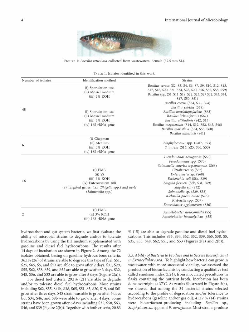
3.1. Microbiological and Biochemical Assessment. In thisstudy, the intestine of Poecilia reticulata (Figure 1) guttrack has been aseptically dissected as mentioned inMethods and Materials.
Seventy-two (72) isolates have been purified and ob-tained (Table 1). *e 3% KOH Gram test identified 25% ofGram-negative cultural bacteria and 75% of Gram-positive cultural bacteria. In order to estimate the totalnumber of microbial florae contained in the digestive tractof guppy fish on PCA media, male and of female guppyfish have been dissected. Using PCA medium, we found1.3 ± 0.51 (105 UFC/ml/intestine track) in the male gutsystem and 1.5 ± 0.68 (105 UFC/ml/intestine track) in thefemale gut system. Mossels, SS, Chapman media, andEnterobacter, and Enterosystem 18R allowed the pre-sumptive identification. 66.66% (48) of Bacillaceae are themost dominant genera in the digestive tract of P. retic-ulata isolated from Brazzaville wastewater, 8.33 % (6) ofStaphylococcaceae and 25 % (18) Enterobacteriaceae. *eidentification of Shigella spp. and Salmonella spp. hasbeen confirmed using direct PCR of targeted specific genessuch as icsB and invG, respectively. Using 16S rRNA gene,Acinetobacter haemolyticus (GenBank: MK099885.1),Bacillus spp. including Bacillus subtilis (GenBank:MK099888.1), Bacillus cereus (GenBank: MK099886.1,MK099887.1, and MK099891.1), Bacillus amyloliquefa-ciens (GenBank: MK156314.1) and Bacillus licheniformis,Bacillus altitudinis (GenBank: MK099889.1 andMK156313.1) and Bacillus megaterium (GenBank:MK099890.1, MK391976.1, MK391968.1, MK391975.1,and MK391970.1), Bacillus anthracis (GenBank:MK391960.1) and Bacillus marisflavi (MK391969.1,MK391972.1, and MK391973.1) have been identified(Table 1).
3.2. Ability of Microbiota to Degrade and/or TolerateHydrocarbons. To investigate the relationship between
International Journal of Microbiology 3
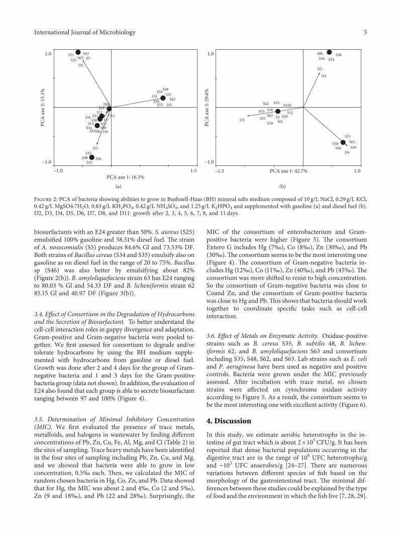
hydrocarbon and gut system bacteria, we first evaluate theability of microbial strains to degrade and/or to toleratehydrocarbons by using the BH medium supplemented withgasoline and diesel fuel hydrocarbons. *e results after14 days of incubation are shown in Figure 2. Among the 72isolates obtained, basing on gasoline hydrocarbons criteria,36.1% (26) of strains are able to degrade this type of fuel. S51,S25, S65, S5, and S53 are able to grow after 2 days. S31, S29,S55, S62, S58, S59, and S52 are able to grow after 3 days. S32,S48, S56, and S33 are able to grow after 5 days (Figure 2(a)).
For diesel fuel criteria, 29.1% (21) are able to degradeand/or to tolerate diesel fuel hydrocarbons. Most strainsincluding S62, S55, S41b, S38, S65, S51, S5, S20, S19, and S61grow after three days. S48 strain was able to grow after 5 daysbut S34, S46, and S8b were able to grow after 4 days. Somestrains have been grown after 6 days including S35, S58, S63,S46, and S39 (Figure 2(b)). Together with both criteria, 20.83
% (15) are able to degrade gasoline and diesel fuel hydro-carbons. *is includes S35, S34, S62, S52, S39, S65, S38, S5,S35, S55, S48, S62, S51, and S53 (Figures 2(a) and 2(b)).
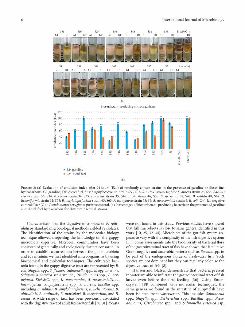
3.3. Ability of Bacteria to Produce and to Secrete Biosurfactantin Extracellular Area. To highlight how bacteria can grow inwastewater with more successful viability, we assessed theproduction of biosurfactants by conducting a qualitative testcalled emulsion index (E24), from inoculated precultures inflasks containing the nutrient broth. Incubation has beendone overnight at 37°C. As results illustrated in Figure 3(a),we showed that among the 34 bacterial strains selectedaccording to the profile of degradation and/or tolerance tohydrocarbons (gasoline and/or gas oil), 41.17 % (14) strainswere biosurfactant-producing including Bacillus sp.,Staphylococcus spp, and P. aeruginosa. Most strains produce
Table 1: Isolates identified in this work.
Number of isolates Identification method Strains
48
(i) Sporulation test(ii) Mossel medium
(iii) 3% KOH
Bacillus cereus (S2, S3, S4, S6, S7, S9, S10, S12, S13,S17, S18, S20, S21, S24, S28, S20, S56, S57, S58, S59)Bacillus spp. (S1, S11, S19, S22, S23, S27 S32, S43, S44,
S47, S50, S51)
(i) Sporulation test(ii) Mossel medium
(iii) 3% KOH(iv) 16S rRNA gene
Bacillus cereus (S34, S35, S64)Bacillus subtilis (S48)
Bacillus amyloliquefaciens (S63)Bacillus licheniformis (S62)Bacillus altitudinis (S42, S15)
Bacillus megaterium (S14, S32, S52, S45, S46)Bacillus marisflavi (S54, S55, S60)
Bacillus anthracis (S61)
6
(i) Chapman(ii) Medium(iii) 3% KOH
(iv) 16S rRNA gene
Staphylococcus spp. (S41b, S53)S. aureus (S16, S25, S30, S53)
16
(i) EMB(ii) SS
(iii) 3% KOH(iv) Enterosystem 18R
(v) Targeted genes: icsB (Shigella spp.) and invG(Salmonella spp.)
Pseudomonas aeruginosa (S65)Pseudomonas spp. (S70)
Salmonella enterica ssp.arizonae. (S66)Citrobacter sp.(S67)Enterobacter sp. (S68)
Escherichia coli (S8a, S39)Shigella flexneri (S8b, S31, S69)
Shigella sp. (S52)Salmonella sp. (S29, S33)
Klebsiella pneumoniae (S26)Klebsiella spp. (S37)
Enterobacter agglomerans (S36)
2(i) EMB
(ii) 3% KOH(iii) 16S rRNA gene
Acinetobacter nosocomialis (S5)Acinetobacter haemolyticus (S38)
Figure 1: Poecilia reticulata collected from wastewaters. Female (37.5mm SL).
4 International Journal of Microbiology
biosurfactants with an E24 greater than 50%. S. aureus (S25)emulsified 100% gasoline and 58.51% diesel fuel. *e strainof A. nosocomialis (S5) produces 84.6% Gl and 73.53% DF.Both strains of Bacillus cereus (S34 and S35) emulsify also ongasoline as on diesel fuel in the range of 20 to 75%. Bacillussp (S46) was also better by emulsifying about 82%(Figure 2(b)). B. amyloliquefaciens strain 63 has E24 rangingto 80.03 % Gl and 54.33 DF and B. licheniformis strain 6285.15 Gl and 40.97 DF (Figure 3(b)).
3.4. Effect of Consortium in the Degradation of Hydrocarbonsand the Secretion of Biosurfactant. To better understand thecell-cell interaction roles in guppy divergence and adaptation,Gram-positive and Gram-negative bacteria were pooled to-gether. We first assessed for consortium to degrade and/ortolerate hydrocarbons by using the BH medium supple-mented with hydrocarbons from gasoline or diesel fuel.Growth was done after 2 and 4 days for the group of Gram-negative bacteria and 1 and 3 days for the Gram-positivebacteria group (data not shown). In addition, the evaluation ofE24 also found that each group is able to secrete biosurfactantranging between 97 and 100% (Figure 4).
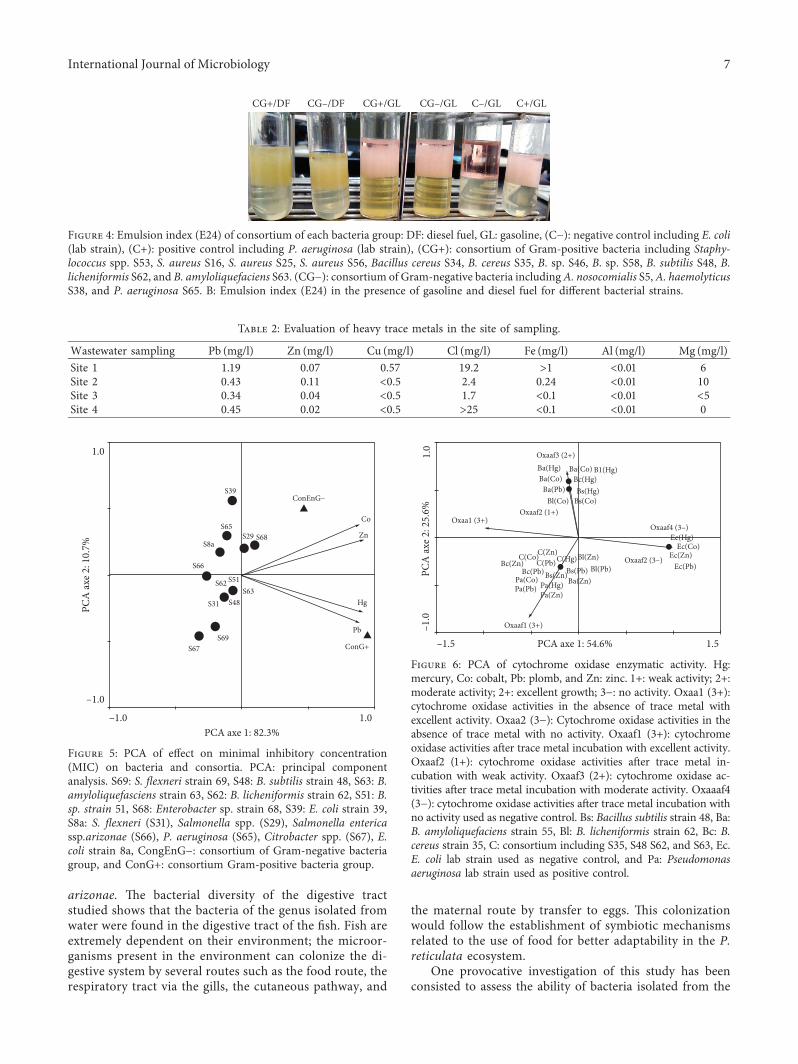
3.5. Determination of Minimal Inhibitory Concentration(MIC). We first evaluated the presence of trace metals,metalloids, and halogens in wastewater by finding differentconcentrations of Pb, Zn, Cu, Fe, Al, Mg, and Cl (Table 2) inthe sites of sampling. Trace heavymetals have been identifiedin the four sites of sampling including Pb, Zn, Cu, and Mg,and we showed that bacteria were able to grow in lowconcentration, 0.5‰ each. *en, we calculated the MIC ofrandom chosen bacteria in Hg, Co, Zn, and Pb. Data showedthat for Hg, the MIC was about 2 and 4‰, Co (2 and 5‰),Zn (9 and 18‰), and Pb (22 and 28‰). Surprisingly, the
MIC of the consortium of enterobacterium and Gram-positive bacteria were higher (Figure 5). *e consortiumEntero G includes Hg (7‰), Co (8‰), Zn (30‰), and Pb(30‰).*e consortium seems to be the most interesting one(Figure 4). *e consortium of Gram-negative bacteria in-cludes Hg (12‰), Co (11‰), Zn (40‰), and Pb (45‰). *econsortium was more shifted to resist to high concentration.So the consortium of Gram-negative bacteria was close toCoand Zn, and the consortium of Gram-positive bacteriawas close to Hg and Pb.*is shows that bacteria should worktogether to coordinate specific tasks such as cell-cellinteraction.
3.6. Effect of Metals on Enzymatic Activity. Oxidase-positivestrains such as B. cereus S35, B. subtilis 48, B. lichen-iformis 62, and B. amyloliquefaciens S63 and consortiumincluding S35, S48, S62, and S63. Lab strains such as E. coliand P. aeruginosa have been used as negative and positivecontrols. Bacteria were grown under the MIC previouslyassessed. After incubation with trace metal, no chosenstrains were affected on cytochrome oxidase activityaccording to Figure 5. As a result, the consortium seems tobe the most interesting one with excellent activity (Figure 6).
4. Discussion
In this study, we estimate aerobic heterotrophs in the in-testine of gut tract which is about 2×10⁵ CFU/g. It has beenreported that dense bacterial populations occurring in thedigestive tract are in the range of 108 UFC heterotrophs/gand ∼105 UFC anaerobes/g [24–27]. *ere are numerousvariations between different species of fish based on themorphology of the gastrointestinal tract. *e minimal dif-ferences between these studies could be explained by the typeof food and the environment in which the fish live [7, 28, 29].
S51
S59S31S29
S55
S58S52
S62
S25 S65
D2
D3S39
S12
S38S14
S34S35
S32
PCA
axe 2
: 15.
1%
PCA axe 1: 16.5%
D5
S48S33
–1.0–1.0
1.0
1.5
S56
S8a
S11D4 D11D7
S57S8b
D6
S53S5
DJ8
(a)
S56 S34S48
S38
S58S46
S35S63S39
S53S65
S51S19 S61
S5 S20S52
S41bS55
S8b
S62
D3
D5
D4
D6
PCA
axe 2
: 29.
6%
–1.0
1.0
PCA axe 1: 42.7%–1.5 1.0
(b)
Figure 2: PCA of bacteria showing abilities to grow in Bushnell-Haas (BH) mineral salts medium composed of 10 g/L NaCl, 0.29 g/L KCl,0.42 g/L MgSO4·7H2O, 0.83 g/L KH2PO4, 0.42 g/L NH4SO4, and 1.25 g/L K2HPO4 and supplemented with gasoline (a) and diesel fuel (b).D2, D3, D4, D5, D6, D7, D8, and D11: growth after 2, 3, 4, 5, 6, 7, 8, and 11 days.
International Journal of Microbiology 5
Characterization of the digestive microbiota of P. retic-ulata by standardmicrobiologicalmethods yielded 72 isolates.*e identification of the strains by the molecular biologytechnique allowed deepening the knowledge on the guppymicrobiota digestive. Microbial communities have beenconsisted of genetically and ecologically distinct consortia. Inorder to establish a correlation between the gut microbiotaand P. reticulata, we first identified microorganisms by usingbiochemical and molecular techniques. *e culturable bac-teria found in the guppy digestive tract are represented by: E.coli, Shigella spp., S. flexneri, Salmonella spp., E. agglomerans,Salmonella enterica ssp.arizonae., Pseudomonas spp., P. aer-uginosa, Klebsiella spp., K. pneumoniae, A. nosocomialis, A.haemolyticus, Staphylococcus spp., S. aureus, Bacillus spp.including B. subtilis, B. amyloliquefaciens, B. licheniformis, B.altitudinis, B. anthracis, B. marisflavi, B. megaterium, and B.cereus. A wide range of taxa has been previously associatedwith the digestive tract of adult freshwater fish [30, 31]. Yeasts
were not found in this study. Previous studies have showedthat fish microbiota is close to same genera identified in thiswork [10, 25, 32–34]. Microbiota of the gut fish system ap-pears to vary with the complexity of the fish digestive system[35]. Some assessments into the biodiversity of bacterial floraof the gastrointestinal tract of fish have shown that facultativeGram-negative and anaerobic bacteria such as Bacillus spp. tobe part of the endogenous florae of freshwater fish. Suchspecies are not dominant but they can regularly colonize thedigestive tract of fish [8].
Hansen and Olafsen demonstrate that bacteria presentin water are able to infiltrate the gastrointestinal tract of fishlarvae even before the first feeding [36]. Using Enter-osystem 18R combined with molecular techniques, thesame genera we found in the intestine of guppy fish havebeen isolated from wastewater. *is includes Salmonellaspp., Shigella spp., Escherichia spp., Bacillus spp., Pseu-domonas, Citrobacter spp., and Salmonella enterica ssp.
S53GI DF GI DF GI DF GI DF GI DF GI DF GI DF GI DF
GI DF GI DF GI DF GI DF GI DF GI DF GI DF GI DF
S16 S25 S56 S46 S34 S35 E. coli (C–)
S46 S58 S48 S62 S63 S65 S5 Paer (C+)
(a)
S530
20
40
60
80
100
120
Emul
sific
atio
n in
dex
(E24
)
Biosurfactant-producing microorganisms
S16
S25
S56
S34
S35
E24 gasolineE24-diesel fuel
S46
S58
S48
S62
S63
S65 S5
E. co
li (C
–)
Paer
(C+)
(b)
Figure 3: (a) Evaluation of emulsion index after 24 hours (E24) of randomly chosen strains in the presence of gasoline or diesel fuelhydrocarbons. Gl: gasoline; DF: diesel fuel. S53: Staphylococcus sp. strain S53; S16: S. aureus strain 16; S25: S. aureus strain 25; S56: Bacilluscereus strain 56; S34: B. cereus strain 34; S35: B. cereus strain 35; S46: B. sp. strain 46; S58: B. sp. strain 58; S48: B. subtilis 48; S62: B.licheniformis strain 62; S63: B. amyloliquefaciens strain 63; S65: P. aeruginosa strain 65; S5: A. nosocomialis strain 5; E. coli (C−): lab negativecontrol; Paer (C+): Pseudomonas aeruginosa positive control. (b) Percentages of biosurfactant-producing bacteria in the presence of gasolineand diesel fuel hydrocarbon for different bacterial strains.
6 International Journal of Microbiology
arizonae. *e bacterial diversity of the digestive tractstudied shows that the bacteria of the genus isolated fromwater were found in the digestive tract of the fish. Fish areextremely dependent on their environment; the microor-ganisms present in the environment can colonize the di-gestive system by several routes such as the food route, therespiratory tract via the gills, the cutaneous pathway, and
the maternal route by transfer to eggs. *is colonizationwould follow the establishment of symbiotic mechanismsrelated to the use of food for better adaptability in the P.reticulata ecosystem.
One provocative investigation of this study has beenconsisted to assess the ability of bacteria isolated from the
CG+/DF CG–/DF CG+/GL CG–/GL C–/GL C+/GL
Figure 4: Emulsion index (E24) of consortium of each bacteria group: DF: diesel fuel, GL: gasoline, (C−): negative control including E. coli(lab strain), (C+): positive control including P. aeruginosa (lab strain), (CG+): consortium of Gram-positive bacteria including Staphy-lococcus spp. S53, S. aureus S16, S. aureus S25, S. aureus S56, Bacillus cereus S34, B. cereus S35, B. sp. S46, B. sp. S58, B. subtilis S48, B.licheniformis S62, and B. amyloliquefaciens S63. (CG−): consortium of Gram-negative bacteria includingA. nosocomialis S5,A. haemolyticusS38, and P. aeruginosa S65. B: Emulsion index (E24) in the presence of gasoline and diesel fuel for different bacterial strains.
S39ConEnG–
ConG+
Co
Zn
Hg
Pb
S65
S8a
S66
S51S62
S31 S48
S69S67
S63
S29 S68
PCA
axe 2
: 10.
7%
–1.0
1.0
PCA axe 1: 82.3%–1.0 1.0
Figure 5: PCA of effect on minimal inhibitory concentration(MIC) on bacteria and consortia. PCA: principal componentanalysis. S69: S. flexneri strain 69, S48: B. subtilis strain 48, S63: B.amyloliquefasciens strain 63, S62: B. licheniformis strain 62, S51: B.sp. strain 51, S68: Enterobacter sp. strain 68, S39: E. coli strain 39,S8a: S. flexneri (S31), Salmonella spp. (S29), Salmonella entericassp.arizonae (S66), P. aeruginosa (S65), Citrobacter spp. (S67), E.coli strain 8a, CongEnG−: consortium of Gram-negative bacteriagroup, and ConG+: consortium Gram-positive bacteria group.
Oxaaf3 (2+)
Oxaaf2 (1+)
Oxaaf4 (3–)
Oxaaf2 (3–)
Oxaa1 (3+)
Oxaaf1 (3+)
Ba(Hg)Ba(Co)
Ba(Co)Bc(Hg)
C(Zn)C(Co)Bc(Zn)
Bc(Pb)C(Pb)C(Hg)
Pa(Co)Pa(Pb) Pa(Hg)
Pa(Zn)
Ba(Zn)
Bl(Zn)Bl(Pb)Bs(Pb)Bs(Zn)
Ee(Hg)Ec(Co)
Ec(Zn)Ec(Pb)
B1(Hg)
Bs(Hg)Bs(Co)
Ba(Pb)Bl(Co)
PCA
axe 2
: 25.
6%–1
.01.
0
PCA axe 1: 54.6%–1.5 1.5
Figure 6: PCA of cytochrome oxidase enzymatic activity. Hg:mercury, Co: cobalt, Pb: plomb, and Zn: zinc. 1+: weak activity; 2+:moderate activity; 2+: excellent growth; 3−: no activity. Oxaa1 (3+):cytochrome oxidase activities in the absence of trace metal withexcellent activity. Oxaa2 (3−): Cytochrome oxidase activities in theabsence of trace metal with no activity. Oxaaf1 (3+): cytochromeoxidase activities after trace metal incubation with excellent activity.Oxaaf2 (1+): cytochrome oxidase activities after trace metal in-cubation with weak activity. Oxaaf3 (2+): cytochrome oxidase ac-tivities after trace metal incubation with moderate activity. Oxaaaf4(3−): cytochrome oxidase activities after trace metal incubation withno activity used as negative control. Bs: Bacillus subtilis strain 48, Ba:B. amyloliquefaciens strain 55, Bl: B. licheniformis strain 62, Bc: B.cereus strain 35, C: consortium including S35, S48 S62, and S63, Ec.E. coli lab strain used as negative control, and Pa: Pseudomonasaeruginosa lab strain used as positive control.
Table 2: Evaluation of heavy trace metals in the site of sampling.
Wastewater sampling Pb (mg/l) Zn (mg/l) Cu (mg/l) Cl (mg/l) Fe (mg/l) Al (mg/l) Mg (mg/l)Site 1 1.19 0.07 0.57 19.2 >1 <0.01 6Site 2 0.43 0.11 <0.5 2.4 0.24 <0.01 10Site 3 0.34 0.04 <0.5 1.7 <0.1 <0.01 <5Site 4 0.45 0.02 <0.5 >25 <0.1 <0.01 0
International Journal of Microbiology 7
digestive track of guppy fish to biodegrade gasoline anddiesel fuel hydrocarbons. We showed that among 72 isolates,36.1% are able to degrade and/or tolerate gasoline hydro-carbons. For diesel fuel criteria, 29.1% are able to degradeand/or to tolerate diesel fuel hydrocarbons. We also showedthat the consortium of different groups of Gram-stainingbacteria can degrade and/or tolerate gasoline and diesel fuelhydrocarbons more after 2 days only. A couple of strainsincluding Bacillus spp, Pseudomonas spp., Staphylococcusspp., Shigella spp., E. coli, Salmonella spp., and Acinetobacterspp. have been identified to be able to grow on BH agarsupplemented with different gasoline and diesel fuel hy-drocarbons. *e complex chemical nature of the hydro-carbons present in diesel fuel could be easily degraded asshown [37–39].
In addition, we have showed that among isolates,41.17% strains are biosurfactant-producing. A special at-tention was carried out with Bacillus spp and Staphylo-coccus spp., and Pseudomonas spp and Acinetobacter spp.having a better E24 of hydrocarbons varying between 75%and 100%, respectively. In this work, we also showed thatthe E24 of enterobacterium consortium was about 99 %.*is consortium includes Gram-negative bacteria such asPseudomonas spp and Acinetobacter spp. *e consortiumof Gram-positive bacteria including Bacillus spp. andStaphilococcus spp. also showed E24 about 98%. Differentbacteria species could easily secrete at the same moment alandscape of biosurfactant encompassing rhamnolipid,surfactin, and lipopeptides [40–43] by empowering theguppy adaptability. Alone or together with consortium,bacteria strains could protect the guppy fish in the pollutedenvironments by producing biosurfactants in the digestivetract once the organic pollutants get in touch with thedigestive tissues. Recently, it has been confirmed thatbacterial strains of the genera of Staphylococcus spp. [44]and Bacillus spp. [45] play an important role in bio-remediation by degrading the hydrocarbons present in thepolluted areas and using them as the only carbon source[46]. *ese bacteria are able to produce biosurfactants withE24 up to 50%. Our results are particularly interestingregarding the ability of isolated strains to emulsify hy-drocarbons. *is could highlight to be a clear mechanismshowing relationship between emulsion activity, cell ad-hesion to hydrocarbon, and growth rate of isolates ongasoline and/or gas oil.
It has been previously demonstrated the capacity ofbacteria for surviving in toxic heavy-metal concentrations[14, 18, 19]. Here, we have shown that microbiota land-scape isolated from gut systems can tolerate Hg, Co, Zn,and Pb.*eMIC showed mercury (Hg) between 2 and 4‰,cobalt (Co) 2 and 5‰, zinc (Zn) 9 and 18‰, and plomb(Pb) 22 and 27‰. Zn and Pb were the trace metal for whichthe rate of tolerance was significantly higher. In addition,the consortium value of trace metal including Hg, Co, Zn,and Pb was higher compared with the strain tested alone.*is clearly reinforces the fact that the microbiota land-scape confers to guppy fish the ability to endure the se-lective pressures of their environment. Trace metal andhydrocarbon are present in many ecosystems. Associations
between trace metals and guppy, P. reticulata have beenpreviously illustrated. *e effects of some trace heavymetals have also shown that P. reticulata is able to adapt theecosystem contaminated with trace metals. *is finding isthe first one to demonstrate the direct involvement oflandscape microbiota in the digestive track of guppyfish [16].
We also showed that Hg, Co, Zn, and Pb had no visibleeffect on cytochrome c oxidase activity on B. cereus (S35),B. subtilis (S48), B. amyloliquefaciens (S55), and B.licheniformis (S62) growth. However, it is noteworthy thatbacteria could also utilize an alternative enzyme for hy-drocarbon degradation. Investigation in this way could beinteresting. *e cytochrome activity of consortia includingS35, S48, S62, and S55 has not been affected. *is ishighlighted to understand that bacteria communitiesshould work together to achieve their best. *ese findingsseem to be the first one by demonstrating the direct in-volvement of microbiota landscape in the digestive track ofguppy fish by highlighting that together with differentmicrobiota genera, guppies are able to adapt in wastewatercontaminated with hydrocarbon and trace metals. It is alsounwise and difficult to give this entire role to microbiota forthe mechanisms of P. reticulata adaptation in oil-contaminated waters including cell-cell interaction. Ad-ditional physiological factors may influence the adaptationof guppy fish. Response results in morphological, physi-ological, and even genetic differentiation, paralleling withmicrobiota growth [47]. In addition, the bacterial enzymessecretion machinery could increase the systematic degra-dation of gasoline and/or diesel fuel hydrocarbons or theundesirable products. Future studies should also deeplyassess relationship between microbiota and P. reticulatabased on the physiology aspects.
5. Conclusion
Our findings tried to illustrate adaptive mechanism abilitiesamong guppies in Brazzaville wastewaters. Evolution hasallowed these hydrocarbon- and heavy-metal-adaptedmicroorganisms not to simply survive, but also to growsuccessfully under the extreme conditions of hydrocarbonhabitats, through a variety of microbial aspects andphysiological adjustments in their genomes. Within P.reticulata is given the opportunity to explore the micro-biological related to biological strategies to adapt in vivocoping with high hydrocarbon concentration and heavymetal. Guppies have developed networks of adaptationmechanisms to protect against wild range of pollutantsincluding hydrocarbons and trace heavy metals. Cell-cellinteraction could be the most attractive way to keep oninvestigating by seeing different molecules involved in theadaptation.
Data Availability
*e Excel sheet including the data used to support thefindings of this study is available from the correspondingauthor upon request.
8 International Journal of Microbiology
Conflicts of Interest
*e authors declare that the research was conducted in theabsence of any intellectual commercial or financial relation-ships that could be construed as potential conflicts of interest.
Acknowledgments
We are grateful to Prof. Anne Botteaux (Laboratoire deBacteriologie Moleculaire, Batiment GE, 5eme Etage,Campus ERASME-Universite Libre de Bruxelles), Prof.Clobite Bouka Biona (Universite Marien NGOUABI), Prof.Arsene Lenga, and Dr. Bienvenu Dinga (Universite MarienNGOUABI) for their continuous encouragements andmaterial support.*is work was supported in part by InstitutNational de Recherche en Sciences Exactes et Naturelles(IRSEN).
References
[1] G. M. G. Sabatinelli, S. Blanchy, P. Fayaerts, and M. Papakay,Experimentation du Poisson Larvivore Poecilia reticulata dansla Lutte Contre le Paludisme en RFI des Comores, OrganisationMondiale de la Sante, WHO/MAL/901060 1931, Geneva,Switzerland, 1990.
[2] F. Bashey, “Cross-generational environmental effects and theevolution of offspring size in the Trinidadian guppy Poeciliareticulata,” Evolution, vol. 60, no. 2, pp. 348–361, 2006.
[3] T. L. A. Piersma, “Rapid reversible changes in organ size as acomponent of adaptive behaviour,” Trends in Ecology andEvolution, vol. 12, no. 4, pp. 134–138, 1997.
[4] G. V. Dussault and D. L. Kramer, “Food and feeding behaviorof the guppy, Poecilia reticulata (Pisces: Poeciliidae),” Ca-nadian Journal of Zoology, vol. 59, no. 4, pp. 684–701, 1981.
[5] A. Shyrilo’steen, J. Cullum, and F. B. Albert, “Rapid evolutionof escape ability in Trinidad guppies Poecilia reticulata,”Evolution, vol. 56, no. 4, pp. 776–784, 2002.
[6] E. Zandona, S. K. Auer, S. S. Kilham, and D. N. Reznick,“Contrasting population and diet influences on gut length of anomnivorous tropical fish, the Trinidadian guppy (Poeciliareticulata),” PLoS One, vol. 10, no. 9, Article ID e0136079, 2015.
[7] G. Yang, B. Bao, E. Peatman, H. Li, L. Huang, and D. Ren,“Analysis of the composition of the bacterial community inpuffer fish Takifugu obscurus,” Aquaculture, vol. 262, no. 2–4,pp. 183–191, 2007.
[8] E. Ringo and F. J. Gatesoupe, “Lactic and bacteria in fish: areview,” Aquaculture, vol. 160, no. 177, 1998.
[9] G. H. Hansen and J. A. Olafsen, “Bacterial interactions in earlylife stages of marine cold water fish,” Microbial Ecology,vol. 38, no. 1, pp. 1–26, 1999.
[10] K. E. Sullam, B. E. Rubin, C. M. Dalton, S. S. Kilham,A. S. Flecker, and J. A. Russell, “Divergence across diet, timeand populations rules out parallel evolution in the gutmicrobiomes of Trinidadian guppies,” Be ISME Journal,vol. 9, no. 7, pp. 1508–1522, 2015.
[11] K. E. Sullam, C. M. Dalton, J. A. Russell et al., “Changes indigestive traits and body nutritional composition accom-modate a trophic niche shift in Trinidadian guppies,” Oeco-logia, vol. 177, no. 1, pp. 245–257, 2014.
[12] P. J. Turnbaugh, R. E. Ley, M. A. Mahowald, V. Magrini,E. R. Mardis, and J. I. Gordon, “An obesity-associated gutmicrobiome with increased capacity for energy harvest,”Nature, vol. 444, no. 7122, pp. 1027–1031, 2006.
[13] F. Backhed, H. Ding, T. Wang et al., “*e gut microbiota as anenvironmental factor that regulates fat storage,” Proceedings ofthe National Academy of Sciences, vol. 101, no. 44,pp. 15718–15723, 2004.
[14] A. Bairagi, K. Sarkar Ghosh, S. K. Sen, and A. K. Ray,“Duckweed (Lemna polyrhiza) leaf meal as a source offeedstuff in formulated diets for rohu (Labeo rohita Ham.)fingerlings after fermentation with a fish intestinal bacte-rium,” Bioresource Technology, vol. 85, no. 1, pp. 17–24, 2002.
[15] B. Spanggaard, F. Jørgensen, L. Gram, and H. H. Huss,“Antibiotic resistance in bacteria isolated from three fresh-water fish farms and an unpolluted stream in Denmark,”Aquaculture, vol. 115, no. 3-4, pp. 195–207, 1993.
[16] B. Widianarko, C. A. M. Van Gestel, R. A. Verweij, andN. M. Van Straalen, “Associations between trace metals insediment, water, and guppy, Poecilia reticulata (Peters), fromurban streams of Semarang, Indonesia,” Ecotoxicology andEnvironmental Safety, vol. 46, no. 1, pp. 101–107, 2000.
[17] R. P. Khunyakari, V. Tare, and R. N. Sharma, “Effects of sometrace heavy metals on Poecilia reticulata (Peters),” Journal ofEnvironmental Biology, vol. 22, no. 2, pp. 141–144, 2001.
[18] M. Shuhaimi-Othman, N. Yakub, N.-A. Ramle, and A. Abas,“Comparative toxicity of eight metals on freshwater fish,”Toxicology and Industrial Health, vol. 31, no. 9, pp. 773–782,2013.
[19] Y.-J. Yi and S.-H. Zhang, “Heavy metal (Cd, Cr, Cu, Hg, Pb,Zn) concentrations in seven fish species in relation to fishsize and location along the Yangtze River,” EnvironmentalScience and Pollution Research, vol. 19, no. 9, pp. 3989–3996,2012.
[20] M. Drancourt, C. Bollet, A. Carlioz, R. Martelin, J. P. Gayral,and D. Raoult, “16S ribosomal DNA sequence analysis of alarge collection of environmental and clinical unidentifiablebacterial isolates,” Journal of Clinical Microbiology, vol. 38,no. 10, pp. 3623–3630, 2000.
[21] L. D. Bushnell and H. F. Haas, “*e utilization of certainhydrocarbons by microorganisms,” Journal of Bacteriology,vol. 41, no. 5, pp. 653–673, 1941.
[22] J. R. Fischer and C. P. Paukert, “Habitat relationships with fishassemblages in minimally disturbed Great Plains regions,”Ecology of Freshwater Fish, vol. 17, no. 4, pp. 597–609, 2008.
[23] C. J. F. Ter Braak and P. Smilauer, Canoco 4, CambridgeUniversity Press, New York, NY, USA, 2003.
[24] T. J. Trust and R. A. H. Sparrow, “*e bacterial flora in thealimentary tract of freshwater salmonid fishes,” CanadianJournal of Microbiology, vol. 20, no. 9, pp. 1219–1228, 1974.
[25] M. Yoshimizu and T. Kimura, “Study on the intestinal mi-croflora of salmonids,” Fish Pathology, vol. 10, no. 2,pp. 243–259, 1976.
[26] A. C. Campbell and J. A. Buswell, “*e intestinal microflora offarmed Dover sole (Solea solea) at different stages of fishdevelopment,” Journal of Applied Bacteriology, vol. 55, no. 2,pp. 215–223, 2008.
[27] Y. Yano, A. Nakayama, and K. Yoshida, “Population sizes andgrowth pressure responses of intestinal microfloras of deep-sea fish retrieved from the abyssal zone,” Applied and Envi-ronmental Microbiology, vol. 61, no. 12, pp. 4480–4483, 1995.
[28] R. Tanaka, M. Ootsubo, T. Sawabe, Y. Ezura, and K. Tajima,“Biodiversity and in situ abundance of gut microflora ofabalone (Haliotis discus hannai) determined by culture-independent techniques,” Aquaculture, vol. 241, no. 1–4,pp. 453–463, 2004.
[29] E. Ringo, S. Sperstad, R. Myklebust, T. M. Mayhew, andR. E. Olsen, “*e effect of dietary inulin on aerobic bacteria
International Journal of Microbiology 9
associated with hindgut of Arctic charr (Salvelinus alpinusL.),” Aquaculture Research, vol. 37, no. 9, pp. 891–897, 2006.
[30] B. Austin, “*e bacterial microflora of fish,” Be ScientificWorld Jourdnal, vol. 2, pp. 558–572, 2002.
[31] B. Austin, “*e bacterial microflora of fish, revised,” BeScientific World Journal, vol. 6, pp. 931–945, 2006.
[32] Y. Kamei, T. Sakata, and D. Kakimoto, “Microflora in thealimentary tract of Tilapia: characterization and distributionof anaerobic bacteria,” Be Journal of General and AppliedMicrobiology, vol. 31, no. 2, pp. 115–124, 1985.
[33] K. Apun, A.M. Yusof, and K. Jugang, “Distribution of bacteriain tropical freshwater fish and ponds,” International Journal ofEnvironmental Health Research, vol. 9, no. 4, pp. 285–292,1999.
[34] T. P. Nieto, A. E. Toranzo, and J. L. Barja, “Comparisonbetween the bacterial flora associated with fingerling rainbowtrout cultured in two different hatcheries in the north-west ofSpain,” Aquaculture, vol. 42, no. 3-4, pp. 193–206, 1984.
[35] M. M. Cahill, “Bacterial flora of fishes: a review,” MicrobialEcology, vol. 19, no. 1, pp. 21–41, 1990.
[36] G. H. Hansen and J. A. Olafsen, “Bacterial colonization of cod(Gadus morhua L.) and halibut (Hippoglossus hippoglossus)eggs in marine aquaculture,” Applied and EnvironmentalMicrobiology, vol. 55, pp. 1435–1446, 1989.
[37] H. I. Tazeena, G. Badal, M. Mehedi Hasan Magnet, F. Kaniz,A. Selina, andM. Rahim Khan, “Isolation and identification ofpetroleum degrading bacteria from oil contaminated soil andwater and assessment of their potentiality in bioremediation,”IOSR Journal of Environmental Science, Toxicology and FoodTechnology, vol. 5, no. 2, pp. 55–58, 2013.
[38] T. Wu, W. J. Xie, Y. L. Yi, X. B. Li, J. Wang, and X. M. Hu,“Isolation identification and characterization of halotolerantpetroleum-degrading bacteria,” Huan Jing Ke Xue, vol. 33,no. 11, pp. 3949–3955, 2012.
[39] M. Mahjoubi, A. Jaouani, A. Guesmi et al., “Hydro-carbonoclastic bacteria isolated from petroleum contaminatedsites in Tunisia: isolation, identification and characterization ofthe biotechnological potential,” New Biotechnology, vol. 30,no. 6, pp. 723–733, 2013.
[40] P. Singh, Y. Patil, and V. Rale, “Biosurfactant production:emerging trends and promising strategies,” Journal of AppliedMicrobiology, vol. 126, no. 1, pp. 2–13, 2018.
[41] H. Chong and Q. Li, “Microbial production of rhamnolipids:opportunities, challenges and strategies,” Microbial CellFactories, vol. 16, no. 1, p. 137, 2017.
[42] A. M. Abdel-Mawgoud, F. Lepine, and E. Deziel, “Rham-nolipids: diversity of structures, microbial origins and roles,”Applied Microbiology and Biotechnology, vol. 86, no. 5,pp. 1323–1336, 2010.
[43] M. Bouassida, N. Fourati, F. Krichen, R. Zouari, S. Ellouz-Chaabouni, and D. Ghribi, “Potential application of Bacillussubtilis SPB1 lipopeptides in toothpaste formulation,”Journal of Advanced Research, vol. 8, no. 4, pp. 425–433,2017.
[44] K. Eddouaouda, S. Mnif, A. Badis et al., “Characterization of anovel biosurfactant produced by Staphylococcus sp. strain 1Ewith potential application on hydrocarbon bioremediation,”Journal of Basic Microbiology, vol. 52, no. 4, pp. 408–418, 2011.
[45] A. Al-Sayegh, Y. Al-Wahaibi, S. Joshi, S. Al-Bahry, A. Elshafie,and A. Al-Bemani, “Draft genome sequence of Bacillussubtilis AS2, a heavy crude oil-degrading and biosurfactant-producing bacterium isolated from a soil sample,” GenomeAnnouncements, vol. 5, no. 39, 2017.
[46] S. Arunagiri and M. Sangeetha, “Polyaromatic Hydrocarbon(PAH) degraders from oil contaminated soil samples,” In-ternational Journal of Advanced Research, vol. 3, pp. 999–1006, 2015.
[47] R. F. Rosenzweig and J. Adams, “Microbial adaptation to achangeable environment: cell-cell interactions mediatephysiological and genetic differentiation,” BioEssays, vol. 16,no. 10, pp. 715–717, 1994.
10 International Journal of Microbiology

Hindawiwww.hindawi.com
International Journal of
Volume 2018
Zoology
Hindawiwww.hindawi.com Volume 2018
Anatomy Research International
PeptidesInternational Journal of
Hindawiwww.hindawi.com Volume 2018
Hindawiwww.hindawi.com Volume 2018
Journal of Parasitology Research
GenomicsInternational Journal of
Hindawiwww.hindawi.com Volume 2018
Hindawi Publishing Corporation http://www.hindawi.com Volume 2013Hindawiwww.hindawi.com
The Scientific World Journal
Volume 2018
Hindawiwww.hindawi.com Volume 2018
BioinformaticsAdvances in
Marine BiologyJournal of
Hindawiwww.hindawi.com Volume 2018
Hindawiwww.hindawi.com Volume 2018
Neuroscience Journal
Hindawiwww.hindawi.com Volume 2018
BioMed Research International
Cell BiologyInternational Journal of
Hindawiwww.hindawi.com Volume 2018
Hindawiwww.hindawi.com Volume 2018
Biochemistry Research International
ArchaeaHindawiwww.hindawi.com Volume 2018
Hindawiwww.hindawi.com Volume 2018
Genetics Research International
Hindawiwww.hindawi.com Volume 2018
Advances in
Virolog y Stem Cells International
Hindawiwww.hindawi.com Volume 2018
Hindawiwww.hindawi.com Volume 2018
Enzyme Research
Hindawiwww.hindawi.com Volume 2018
International Journal of
MicrobiologyHindawiwww.hindawi.com
Nucleic AcidsJournal of
Volume 2018
Submit your manuscripts atwww.hindawi.com