Imaging of the Basal Cerebral Arteries and Measurement of Blood ...
6
Takashi Tsuchiya 1 Masahiro Yasaka Takenori Yamaguchi Kazumi Kimura Tsuyoshi Omae Received June 25, 1990; revision requested September 10, 1990; revision received October 17, 1990; accepted November 7, 1990. ' All authors: Cerebrovascular Division, Depart- ment of Medicine, National Cardiovascular Center, 5-7-1 Fujishirodai, Suita Cit y, Osaka 565, Japan. Address reprint requests toT. Tsuchiya. 0195-61 08/91 /1203-0497 © American Society of Neuroradiology 497 Imaging of the Basal Cerebral Arteries and Measurement of Blood Velocity in Adults by Using Transcranial Real-Time Color Flow Doppler Sonography We report an attempt to obtain images of the basal cerebral arteries and to measure the quantitative flow velocity in these arteries from outside the skull via the color Doppler imaging method. In 25 healthy volunteers, 22-41 years old, the transducer was posi- tioned just superior to the zygomatic arch and anterior to the external ear canal. The absolute flow velocity was calculated by dividing the measured velocity by the cosine of the incident angle. The middle cerebral artery was readily identified in all subjects, but the anterior cerebral artery was seen in only 7/50, and the posterior cerebral artery in 27 ;so. The corrected mean velocity of the horizontal middle cerebral artery was 86 :t 17 cmfsec at an average incident angle of 49°. However, it was difficult to calculate the absolute flow velocity in the anterior and posterior cerebral arteries because the length of those arteries delineated was too short to read the incident angle. The reproducibility of the mean blood velocity in the middle cerebral artery was tested by two examiners on two different occasions, and showed a linear regression with a correlation coefficient of 0.93 (p < .001) when the correction with the incident angle was made. Transcranial real-time color-flow Doppler imaging permits more accurate noninvasive quantification of cerebral hemodynamic consequences than previous methods do. AJNR 12:497-502, MayfJune 1991; AJR 157:August 1991 In 1982, Aaslid et al. [1] demonstrated that signals could be obtained from the basal cerebral arteries with a 2-M Hz probe directed intracranially from the temporal bone just above the zygomatic arch. Assessment of blood velocity in the basal cerebral arteries provides valuable information with regard to the functional capacity of the circle of Willis. Transcranial Doppler techniques have recently been used to evaluate vasospasm following subarachnoid hemorrhage, intracranial arterial ste- nosis, and arteriovenous malformation [2-5). In addition, the patterns of collateral circulation in patients with extracranial carotid artery disease may be determined with this technique [6). However, for measuring blood velocity, Doppler imaging presents several problems. Precise placement of the sample volume as well as angle correction is not possible by means of the transcranial Doppler technique. Recently, in neonates and infants, the effectiveness of color Doppler imaging has been shown for visualizing intracranial vessels through the anterior fontanel , which serves as an acoustic window [7, 8) . But, in adults, imaging of the intracranial vessels from outside the skull has not been documented previously. In this study we chose a color Doppler technique (transcranial real-time color flow Doppler sonography) to visualize the intracranial vessels. The objectives were to investigate the possibility of obtaining images of the basal cerebral arteries with a noninvasive method and to measure more accurate flow velocity following angle correction than previously reported. Materials and Methods Subjects consisted of 25 healthy individuals, 11 men and 14 women, 21 to 41 years old (mean age, 28 years). Examinations were performed with a real -time 8-mode and color
Transcript of Imaging of the Basal Cerebral Arteries and Measurement of Blood ...
Takashi Tsuchiya1
Masahiro Yasaka Takenori Yamaguchi
Kazumi Kimura Tsuyoshi Omae
Received June 25, 1990; revision requested September 10, 1990; revision received October 17, 1990; accepted November 7, 1990.
' All authors: Cerebrovascular Division, Department of Medicine, National Cardiovascular Center, 5-7-1 Fujishirodai , Suita City, Osaka 565, Japan. Address reprint requests toT. Tsuchiya.
0195-61 08/91 /1203-0497 © American Society of Neuroradiology
497
Imaging of the Basal Cerebral Arteries and Measurement of Blood Velocity in Adults by Using Transcranial Real-Time Color Flow Doppler Sonography
We report an attempt to obtain images of the basal cerebral arteries and to measure the quantitative flow velocity in these arteries from outside the skull via the color Doppler imaging method. In 25 healthy volunteers, 22-41 years old, the transducer was positioned just superior to the zygomatic arch and anterior to the external ear canal. The absolute flow velocity was calculated by dividing the measured velocity by the cosine of the incident angle. The middle cerebral artery was readily identified in all subjects, but the anterior cerebral artery was seen in only 7/50, and the posterior cerebral artery in 27 ;so. The corrected mean velocity of the horizontal middle cerebral artery was 86 :t 17 cmfsec at an average incident angle of 49°. However, it was difficult to calculate the absolute flow velocity in the anterior and posterior cerebral arteries because the length of those arteries delineated was too short to read the incident angle. The reproducibility of the mean blood velocity in the middle cerebral artery was tested by two examiners on two different occasions, and showed a linear regression with a correlation coefficient of 0.93 (p < .001) when the correction with the incident angle was made.
Transcranial real-time color-flow Doppler imaging permits more accurate noninvasive quantification of cerebral hemodynamic consequences than previous methods do.
AJNR 12:497-502, MayfJune 1991; AJR 157:August 1991
In 1982, Aaslid et al. [1] demonstrated that signals could be obtained from the basal cerebral arteries with a 2-M Hz probe directed intracranially from the temporal bone just above the zygomatic arch. Assessment of blood velocity in the basal cerebral arteries provides valuable information with regard to the functional capacity of the circle of Willis. Transcranial Doppler techniques have recently been used to evaluate vasospasm following subarachnoid hemorrhage, intracranial arterial stenosis, and arteriovenous malformation [2-5). In addition, the patterns of collateral circulation in patients with extracranial carotid artery disease may be determined with this technique [6). However, for measuring blood velocity , Doppler imaging presents several problems. Precise placement of the sample volume as well as angle correction is not possible by means of the transcranial Doppler technique.
Recently, in neonates and infants, the effectiveness of color Doppler imaging has been shown for visualizing intracranial vessels through the anterior fontanel , which serves as an acoustic window [7, 8). But, in adults, imaging of the intracranial vessels from outside the skull has not been documented previously. In this study we chose a color Doppler technique (transcranial real-time color flow Doppler sonography) to visualize the intracranial vessels. The objectives were to investigate the possibility of obtaining images of the basal cerebral arteries with a noninvasive method and to measure more accurate flow velocity following angle correction than previously reported.
Materials and Methods
Subjects consisted of 25 healthy individuals, 11 men and 14 women, 21 to 41 years old (mean age, 28 years). Examinations were performed with a real-time 8-mode and color
4g8 TSUCHIYA ET AL. AJNR :12, MayfJune 1991
Doppler duplex scanner (SSH 160A; Toshiba, Inc. , Tokyo) . The transducer was operated at 2.5 MHz for both B-mode imaging and Doppler functions. This system permits rapid visualization of arterial flow patterns by superimposing a real-time color image showing regions of blood flow on a two-dimensional B-mode tissue image. This combination is made possible by high-speed, electric array processing of ultrasound signals. Flow toward the transducer was displayed in red , flow away from the transducer in blue.
The B-mode and Doppler information within the scan fields was updated six times per second for the approximately 40° field of view. The pulse repetition frequency was mainly 3.0 KHz, and low-pass filter was 300 Hz. Proper adjustment of velocity scale to match the flow velocity of the vessels was one of the important factors for obtaining a good image. A change in the velocity scales directly reflected a change in the pulse repetition frequency or the sampling rate. Doppler color gain was first turned down completely and then increased very gradually until the static background noise barely appeared.
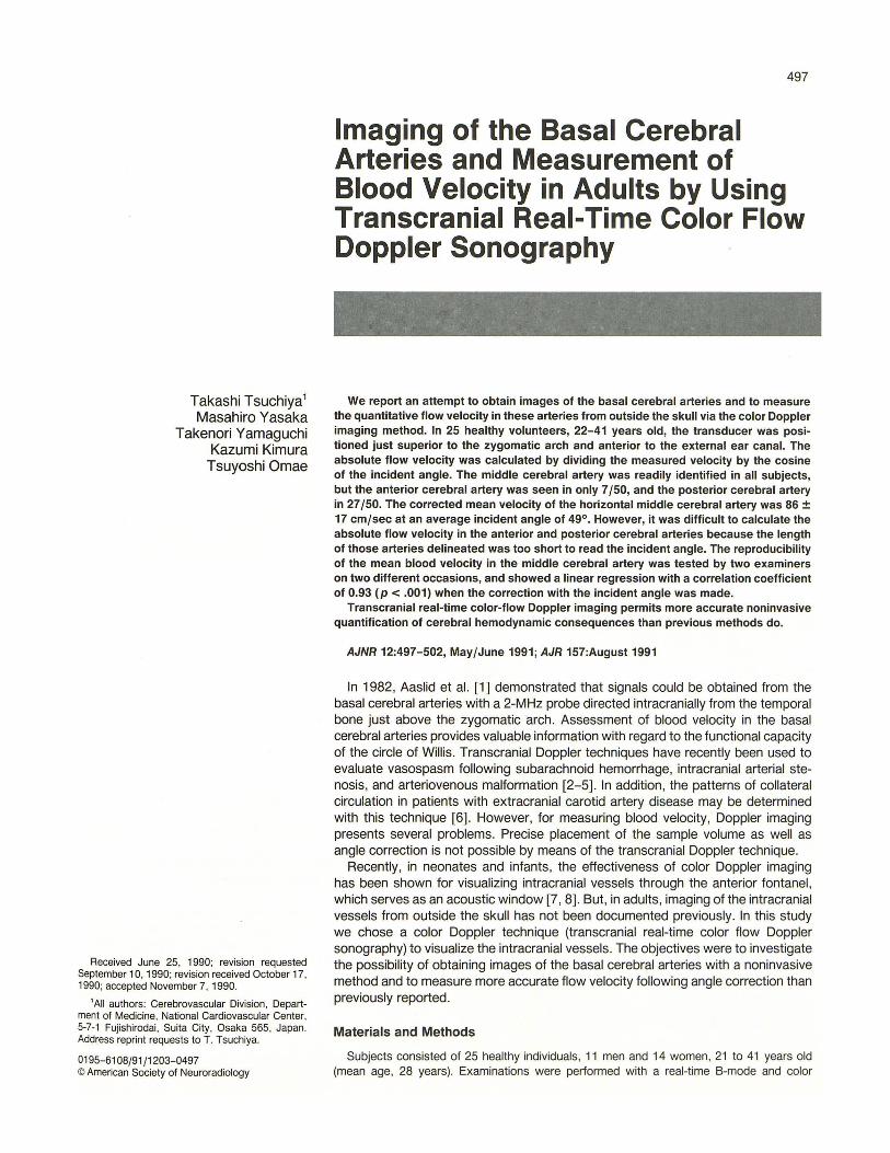
The subjects were examined first in the left lateral decubitus position and then in the right lateral decubitus position. The transducer was placed just superior to the zygomatic arch and anterior to the external ear canal (Fig. 1 ). Particular care was taken to obtain a long axis view of the desired vessel , especially of the horizontal portion of the middle cerebral artery (MCA), by means of til ting, rotating , or shifting the transducer.
For the purpose of identifying the color-flow images obtained by this system, we compared those images with the basal cerebral arteries displayed as flow-void in MR imaging in two of the subjects.
A range-gate pulsed Doppler sample volume, 5 mm in size, was used to measure both the blood flow velocity within the vessel and the angle of incidence (0) to the vessel of interest.
The reproducibility of the MCA flow velocity was investigated in all
Fig. 1.-Photograph of the equipment used and the location of the ultrasonic window.
subjects. Measurements of the MCA blood velocity were repeated within a 7 -day period by two examiners. The data of each examiner were not shown to the other until the second measurement had been made.
For the statistical evaluation, mean velocity was calculated from the time-averaged peak Doppler shift over one cardiac cycle. Then, the absolute velocity was calculated by dividing the measured velocity by the cosine 0. The correlations of the values measured by the two examiners were assessed by linear regression analysis .
Results
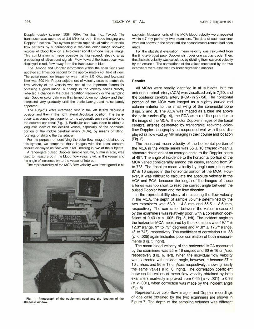
All MCAs were readily identified in all subjects, but the anterior cerebral artery (ACA) was visualized only in 7/50, and the posterior cerebral artery (PCA) in 27/50. The horizontal portion of the MCA was imaged as a slightly curved red column anterior to the small wing of the sphenoidal bone (Figs. 2 and 3). The ACA was imaged as a blue line above the sella turcica (Fig . 4), the PCA as a red line posterior to the image of the MCA. The color Doppler images of the basal cerebral arteries delineated by transcranial real-time colorflow Doppler sonography corresponded well with those displayed as flow void by MR imaging in their course and location (Fig. 2).
The measured mean velocity of the horizontal portion of the MCA in the whole series was 55 ± 16 cmjsec (mean ± standard deviation) at an average angle to the Doppler beam of 4go . The angle of incidence to the horizontal portion of the MCA varied considerably among the cases, ranging from go to 73°. The absolute mean velocity by angle correction was 87 ± 16 cmjsec in the horizontal portion of the MCA. However, it was difficult to calculate the absolute velocity in the ACA and PCA, because the length of the images of those arteries was too short to read the correct angle between the pulsed Doppler beam and the flow direction.
In the reproducibility study of measuring the flow velocity in the MCA, the depth of sample volume determined by the two examiners was 53.g ± 4.3 mm and 55.5 ± 3.6 mm, respectively . The correlation between the values measured by the examiners was relatively poor, with a correlation coefficient of 0.40 (p < .005; Fig. 5, left). The incident angle to the horizontal MCA measured by the examiners was 4g.1 o ± 12.3° (range, go to 73° degrees) and 41.8° ± 17.7° (range, 4° to 74°), respectively. The coefficient of correlation r = .38 (p < .005) again indicated poor correlation of both measurements (Fig . 5, right).
The mean blood velocity of the horizontal MCA measured by the examiners was 55 ± 16 cmjsec and 60 ± 16 cmjsec, respectively (Fig. 6, left). When the individual flow velocity was corrected with incident angle, however, it became 87 ± 16 cmjsec and 86 ± 13 cmjsec, respectively , showing nearly the same values (Fig. 6, right). The correlation coefficient between the values of mean flow velocity obtained by both examiners markedly improved from 0.65 (p < .001) to o.g3 (p < .001 ), when correction was made by the incident angle (Fig. 6).
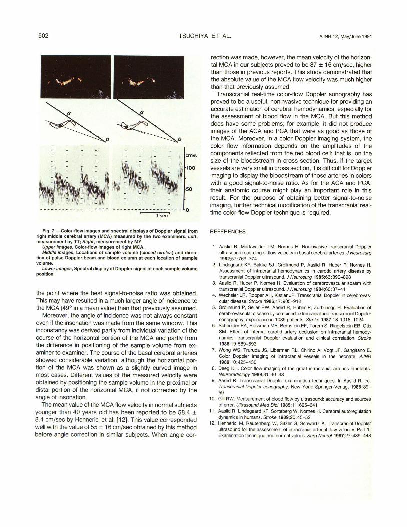
Representative color-flow images and Doppler recordings of one case obtained by the two examiners are shown in Figure 7. The depth of the sampling volumes was different
AJNR:12, May/June 1991 DOPPLER SONOGRAPHY OF CEREBRAL ARTERIES 499
Fig. 2.-Upper image, T2-weighted (2000/23) MR image and corresponding graphic representation. Horizontal portion of middle cerebral artery (MCA) is displayed with intraluminal flow void (closed arrows). Open arrows indicate location of ultrasonic window used.
Lower image, Transcranial real-time color-flow Doppler sonogram of MCA in same subject. Horizontal portion of MCA is clearly imaged as red column. Course and location of horizontal MCA coincides closely with that shown by MR.
for each examiner. The mean velocities measured by the examiners were 58 cmfsec and 93 cmfsec, respectively, which were considerably different. When correction according to incident angle was made, the flow velocities approximated more closely, with respective values of 91 cmfsec and 93 emf sec.
Discussion
Transcranial Doppler recordings of flow velocity by Aaslid et al. [1] in 1982 have been reported to be useful for evaluation of intracranial hemodynamic alterations from outside the skull [2, 3]. However, two important features of this method should be borne in mind. First, in their previous reports the insonation angle was ignored, because the angle of incidence to the MCA and ACA from the temporal window was assumed to be close to zero on the basis of the anatomic structure of the cerebral arteries. The error in the velocity measured by transcranial Doppler recordings would be less than 15%, if assuming the angle of incidence ranges from 0° to 30°. Second, the location of the sample volume used in transcranial Doppler
recordings is uncertain because of a blind technique. Therefore, the source of the information of artery identification depends on the spatial relation of the signal to other intracranial signals, the direction of flow, and the response of the signal to compression test [9].
In general, accurate measurement of flow velocity by the Doppler principle requires the identification of the location of sample volume and the estimation of both the angle of incidence and the Doppler shift. The Doppler methods are capable of good absolute accuracy with systemic errors of 6% or less when suitably designed equipment is used in appropriate situations [1 0].
The Doppler shift is given by the following equation:
Of = 2(v cos 8)fa/C
where Of = Doppler shift, v = magnitude of the scatterer's velocity, 8 =angle between ultrasonic beam and direction of motion, fa = frequency of ultrasound transmitted, and C = speed of propagation of ultrasound in blood.
Since fa and C are generally known, measurement of the Doppler shift allows calculation of the product (v cos 8). Any error in estimating the angle could directly affect the meas-
CM IS
125
100
75
50
.. , .. • I
• I 25 • I J
1·, · ----- l--l ·--- --- ----------------- --- --- - --- --- - ---- --- - - ------ 0
· · ! SEC. , , 0 , , • • , , , , • I , , , , ~ I , • • , , , 0 • , I •
L_j I SEC.
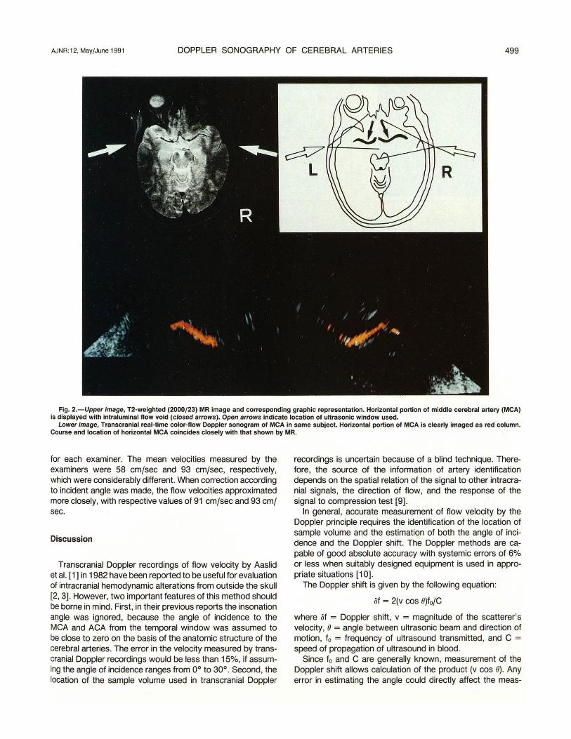
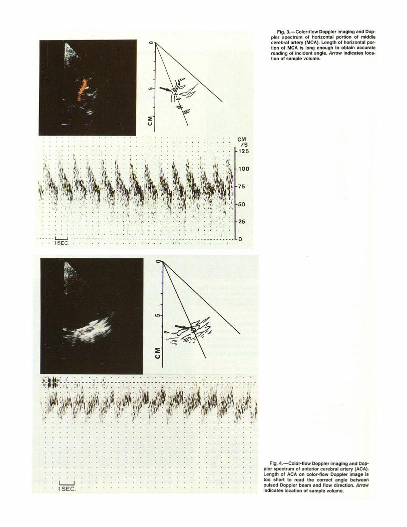
Fig. 3.-Color-flow Doppler imaging and Doppler spectrum of horizontal portion of middle cerebral artery (MCA). Length of horizontal portion of MCA is long enough to obtain accurate reading of incident angle. Arrow indicates location of sample volume.
Fig. 4.-Color-flow Doppler imaging and Doppler spectrum of anterior cerebral artery (ACA). Length of ACA on color-flow Doppler image is too short to read the correct angle between pulsed Doppler beam and flow direction. Arrow indicates location of sample volume.

urement of blood velocity. Moreover, when the angle e is large, cos e varies greatly with e. If, for example, the angle of incidence (8) is assumed to be 0 but is in fact 20°, the flow velocity will be underestimated by 6%. However, if the actual angle of insonation is 60° , blood velocity would be underestimated by 50%.
We succeeded in imaging the basal cerebral arteries and measuring their absolute flow velocities from outside the skull , using a real-time color-flow Doppler imaging system, which is commercially available mainly for cardiac use (SSH 160A). This instrument, which can display the bloodstream in colors superimposed on a real-time 8-mode image, makes it possible to measure the angle of incidence to the vessel of interest and to identify the location of the sample volume.
In all the previous reports it was assumed that for the horizontal portion of the MCA, the angle of insonation was probably small, so that measured values corresponded well to true velocity. However, our results demonstrated that the average value of the angle between the horizontal MCA and ultrasonic beam was 49°, which was much larger than that previously assumed by Aaslid et al. [1, 11]. Moreover, the measured values of the incident angle varied from subject to subject and from side to side.
Reproducibilities for the positioning of depth of sample volume and for the incident angle between the pulsed Doppler beam and blood flow column by the two examiners were relatively poor before correction by incident angle. These results are considered to be based on the difference in positionings of the echo window and sample volume by the examiners.
The location of the ultrasonic window is one of the very important factors that decides the incident angle to the vessels of interest. According to Aaslid et al. [9] , it is useful to discriminate between three different locations of the temporal window: the anterior, middle, and posterior temporal window. The anterior temporal window, which is located posterior to the frontal process of the zygomatic arch, allows the angle of insonation of almost 0° to the MCA. On the other hand, the posterior temporal window, which is located just anterior to the ear, is more suitable for insonation in elderly patients than the anterior temporal window is . From the posterior window, a somewhat more obtuse angle to the MCA might be expected. In this study, the anterior temporal window showed a poor signal-to-noise ratio in almost all subjects, and it was used in only rare cases. We used the posterior and middle temporal windows in the majority of cases in this study, at
502 TSUCHIYA ET AL. AJNR :12, MayfJune 1991
------- -- ----- -- --- ------------------- 0 1 sec
Fig. 7.-Color-flow images and spectral displays of Doppler signal from right middle cerebral artery (MCA) measured by the two examiners. Left, measurement by IT; Right, measurement by MY.
Upper images, Color-flow images of right MCA. Middle images, Locations of sample volume (closed circles) and direc
tion of pulse Doppler beam and blood column at each location of sample volume.
Lower images, Spectral display of Doppler signal at each sample volume position.
the point where the best signal-to-noise ratio was obtained. This may have resulted in a much larger angle of incidence to the MCA (49° in a mean value) than that previously assumed.
Moreover, the angle of incidence was not always constant even if the insonation was made from the same window. This inconstancy was derived partly from individual variation of the course of the horizontal portion of the MCA and partly from the difference in positioning of the sample volume from examiner to examiner. The course of the basal cerebral arteries showed considerable variation, although the horizontal portion of the MCA was shown as a slightly curved image in most cases. Different values of the measured velocity were obtained by positioning the sample volume in the proximal or distal portion of the horizontal MCA, if not corrected by the angle of insonation.
The mean value of the MCA flow velocity in normal subjects younger than 40 years old has been reported to be 58.4 ± 8.4 cmjsec by Hennerici et al. [12]. This value corresponded well with the value of 55 ± 16 cmjsec obtained by this method before angle correction in similar subjects. When angle cor-
rection was made, however, the mean velocity of the horizontal MCA in our subjects proved to be 87 ± 16 cmjsec, higher than those in previous reports. This study demonstrated that the absolute value of the MCA flow velocity was much higher than that previously assumed.
Transcranial real-time color-flow Doppler sonography has proved to be a useful, noninvasive technique for providing an accurate estimation of cerebral hemodynamics, especially for the assessment of blood flow in the MCA. But this method does have some problems; for example, it did not produce images of the ACA and PCA that were as good as those of the MCA. Moreover, in a color Doppler imaging system, the color flow information depends on the amplitudes of the components reflected from the red blood cell; that is, on the size of the bloodstream in cross section. Thus, if the target vessels are very small in cross section, it is difficult for Doppler imaging to display the bloodstream of those arteries in colors with a good signal-to-noise ratio. As for the ACA and PCA, their anatomic course might play an important role in this result. For the purpose of obtaining better signal-to-noise imaging, further technical modification of the transcranial realtime color-flow Doppler technique is required.
REFERENCES
1. Aaslid R, Markwalder TM , Nornes H. Noninvasive transcranial Doppler ultrasound recording of flow velocity in basal cerebral arteries. J Neurosurg 1982;57:769-774
2. lindegaard KF, Bakke SJ, Grolimund P, Aaslid R, Huber P, Nornes H. Assessment of intracranial hemodynamics in carotid artery disease by transcranial Doppler ultrasound. J Neurosurg 1985;63: 890-898
3. Aaslid R, Huber P, Nornes H. Evaluation of cerebrovascular spasm with transcranial Doppler ultrasound. J Neurosurg 1984;60:37-41
4. Wechsler LR, Ropper AH , Kistler JP. Transcranial Doppler in cerebrovascular disease. Stroke 1986;17:905-912
5. Grolimund P, Seiler RW. Aaslid R, Huber P, Zurbruegg H. Evaluation of cerebrovascular disease by combined extracranial and transcranial Doppler sonography: experience in 1039 patients. Stroke 1987;18: 1018-1024
6. Schneider PA, Rossman ME. Bernstein EF, Torem S. Ringelstein EB, Otis SM. Effect of internal carotid artery occlusion on intracranial hemodynamics: transcranial Doppler evaluation and clinical correlation. Stroke 1988;19:589-593
7. Wong WS, Truruda JS, Liberman RL, Chirino A, Vogt JF, Gangitano E. Color Doppler imaging of intracranial vessels in the neonate. AJNR 1989; 1 0:425-430
8. Deeg KH. Color flow imaging of the great intracranial arteries in infants. Neuroradiology 1989;31 :40-43
9. Aaslid R. Transcranial Doppler examination techniques. In Aaslid R, ed. Transcranial Doppler sonography. New York: Springer-Verlag, 1986 :39-59
10. Gill RW. Measurement of blood flow by ultrasound: accuracy and sources of error. Ultrasound Med Biol1985;11 :625-641
11 . Aaslid R, Lindegaard KF, Sorteberg W, Nornes H. Cerebral autoregulation dynamics in humans. Stroke 1989;20 :45-52
12. Hennerici M. Rautenberg W, Sitzer G, Schwartz A. Transcranial Doppler ultrasound for the assessment of intracranial arterial flow velocity. Part 1: Examination technique and normal values. Surg Neurol1987;27 :439-448


![A Casson Fluid Model for multiple Stenosed Artery in the ...e-jst.teiath.gr/issues/issue_42/Bali_42.pdf · non-Newtonian aspects of blood flow through stenosed arteries [18],flow](https://static.fdocument.pub/doc/165x107/60f1e291199db767cb7d41fe/a-casson-fluid-model-for-multiple-stenosed-artery-in-the-e-jst-non-newtonian.jpg)