Dasar - Dasar Penting Nervous Sleep - Neuro Sleep - Hipnosis (1)
© 2005 Nature Publishing Group
NATURE|Vol 437|27 October 2005|doi:10.1038/nature04284 INSIGHT REVIEW
1257
In 1916, Baron Constantin von Economo, a Viennese neurologist,began to see patients with a new type of encephalitis that specificallyattacked regions of the brain that regulate sleep and wakefulness1. Thisdisorder, which was eventually called encephalitis lethargica or vonEconomo’s sleeping sickness, swept through Europe and North Amer-ica during the second decade of the twentieth century; by the end ofthe following decade it had apparently disappeared, as only sporadicand unconvincing reports have appeared since. Although the virusthat caused it was never identified, von Economo was able to identifythe areas of the brain in which lesions caused specific alterations ofwake–sleep regulation (Fig. 1). Only during the past decade has theaccuracy of his observations come to be appreciated, as key compo-nents of the wake–sleep-regulatory system were found to reside at thesites that von Economo first identified.
In this review, we cover recent advances in understanding the braincircuitry that regulates sleep and produces wakefulness, including cellgroups in the brainstem, hypothalamus and basal forebrain (BF) thatare crucial for arousing the cerebral cortex and thalamus. These neu-rons are inhibited during sleep by a system of �-aminobutyric acid(GABA)-containing neurons, in which the ventrolateral preopticnucleus (VLPO) seems to have a key role. Mutual inhibition betweenthe arousal- and sleep-producing circuitry results in switching prop-erties that define discrete wake and sleep states, with sharp transitionsbetween them. We examine how this switch is stabilized by orexinneurons in the lateral hypothalamus (LHA), and why loss of these neu-rons results in narcolepsy. We also explore how basic drives affect thissystem, including the homeostatic need to sleep that accumulates as afunction of prolonged wakefulness, the circadian drive that moulds thedaily cycles of sleep–wakefulness, and allostatic influences that adaptsleep and wakefulness to external behavioural events. Interactionswith this circuitry might explain the effects of a range of drugs on sleepand wakefulness.
The ascending arousal system promotes wakefulnessThe majority of patients with von Economo’s encephalitis lethargicaslept excessively. Many slept for 20 or more hours per day, arising onlybriefly to eat and drink. Their cognitive function was reasonablyintact, but they would soon return to sleep — a cycle that lasted for
many weeks before recovery1. Von Economo found that these patientsinvariably had lesions at the junction of the midbrain and the dien-cephalon. He therefore proposed that there was an ascending arousalsystem originating in the brainstem that kept the forebrain awake (Fig. 1). During the years after the Second World War, investigators,such as Moruzzi and Magoun and their many colleagues, showed thatthis influence was mediated by an ascending arousal pathway thatbegins in the rostral pons and runs through the midbrain reticular for-
1Department of Neurology and Program in Neuroscience, Harvard Medical School, Beth Israel Deaconess Medical Center, Boston, Massachusetts, 02215, USA.
Hypothalamic regulation of sleep andcircadian rhythmsClifford B. Saper1, Thomas E. Scammell1 & Jun Lu1
A series of findings over the past decade has begun to identify the brain circuitry and neurotransmitters that regulate our daily cycles of sleep and wakefulness. The latter depends on a network of cell groups thatactivate the thalamus and the cerebral cortex. A key switch in the hypothalamus shuts off this arousalsystem during sleep. Other hypothalamic neurons stabilize the switch, and their absence results ininappropriate switching of behavioural states, such as occurs in narcolepsy. These findings explain howvarious drugs affect sleep and wakefulness, and provide the basis for a wide range of environmentalinfluences to shape wake–sleep cycles into the optimal pattern for survival.
Figure 1 | A drawing of the human brainstem, taken from von Economo’soriginal work. It illustrates the site of the lesion (diagonal hatching) at thejunction of the brainstem and forebrain that caused prolonged sleepiness,and the site of the lesion (horizontal hatching) in the anteriorhypothalamus that caused prolonged insomnia. The arrow points to aregion between the two, including the posterior lateral hypothalamus. VonEconomo suggested that narcolepsy was caused by lesions at this site.
02 Saper 05-11 19/10/05 11:52 AM Page 5
Nature Publishing Group© 2005
© 2005 Nature Publishing Group
INSIGHT REVIEW NATURE|Vol 437|27 October 2005
1258
mation2; hence, the concept of the ‘ascending reticular activating sys-tem’ achieved wide currency2,3.
Studies in the 1970s and 1980s clarified the nature of this pathway(Fig. 2). A key finding was that the ascending arousal system largelyoriginates from a series of well-defined cell groups with identified neu-rotransmitters4. This pathway has two major branches. The firstbranch is an ascending pathway to the thalamus that activates the thal-amic relay neurons that are crucial for transmission of information tothe cerebral cortex. The major source of upper brainstem input to thethalamic-relay nuclei, as well as to the reticular nucleus of the thala-mus, is a pair of acetylcholine-producing cell groups: the pedunculo-pontine and laterodorsal tegmental nuclei (PPT/LDT)5. The neuronsin the PPT/LDT fire most rapidly during wakefulness and rapid eyemovement (REM) sleep, which is the stage accompanied by corticalactivation, loss of muscle tone in the body and active dreams6. Thesecells are much less active during non-REM (NREM) sleep, when cor-tical activity is slow. Their input to the reticular nucleus is crucial, as itsits between the thalamic-relay nuclei and the cerebral cortex, actingas a gating mechanism that can block transmission between the thal-amus and cerebral cortex, which is important for wakefulness7. Otherinputs to the thalamic midline and intralaminar nuclei originate morebroadly in the upper brainstem, including the reticular formation, thePPT/LDT, the monoaminergic systems (see below) and theparabrachial nucleus8. The intralaminar and midline nuclei are alsobelieved to have a role in cortical arousal.
The second branch of the ascending arousal system bypasses thethalamus, instead activating neurons in the lateral hypothalamic areaand BF, and throughout the cerebral cortex4,9,10. This pathway origi-nates from monoaminergic neurons in the upper brainstem and cau-dal hypothalamus, including the noradrenergic locus coeruleus (LC),serotoninergic dorsal (DR) and median raphe nuclei, dopaminergicventral periaqueductal grey matter and histaminergic tuberomam-millary neurons. The input to the cerebral cortex is augmented by lateral hypothalamic peptidergic neurons (containing melanin-con-centrating hormone (MCH) or orexin/hypocretin), and BF neurons(containing acetylcholine or GABA). Lesions along this pathway, par-ticularly in the LHA and rostral midbrain, produce the most profoundand long-lasting forms of sleepiness or even coma11,12. Neurons in eachof the monoaminergic nuclei that contribute to this pathway have theproperty of firing fastest during wakefulness, slowing down duringNREM sleep and stopping altogether during REM sleep13–15. Orexinneurons in the LHA are, similarly, most active during wakefulness16–18,whereas MCH neurons are active during REM sleep19. Many BF neu-rons, including most cholinergic neurons, are active during both wakeand REM sleep20.
Given the anatomy and functions of these systems, it is not surpris-ing that von Economo found that lesions at the junction of the mid-brain and forebrain, which block both ascending pathways, produceprofound and long-lasting impairment of arousal.
The VLPO promotes sleepVon Economo’s second major observation was an opposite reponse ina small percentage of the victims of encephalitis lethargica1. Ratherthan being sleepy, they became insomniac and slept for only a fewhours each day. Typically, they were extremely tired, but found it diffi-
Brainstem
Cerebellum
Thalamus
Pons
Medulla
LC (NA)
Raphe(5-HT)
TMN(His) PPT (ACh)
LDT (ACh)
Hypothalamus
BF(ACh,
GABA)
vPAG(DA)LH
(ORX,MCH)
Brainstem
Cerebellum
Thalamus
Pons
Medulla
LC (NA)
Raphe(5-HT)
TMN(His)
PPT (ACh)
LDT (ACh)
Hypothalamus
vPAG(DA)VLPO
(GABA, Gal)
PeF(ORX)
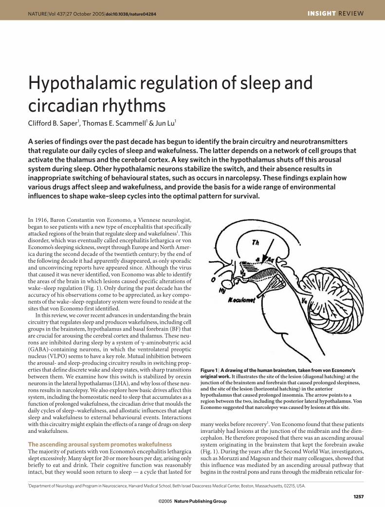
Figure 2 | A schematic drawing showing some key components of theascending arousal system. A major input to the relay and reticular nucleiof the thalamus (yellow pathway) originates from cholinergic (ACh) cellgroups in the upper pons, the pedunculopontine (PPT) and laterodorsaltegmental nuclei (LDT). These inputs facilitate thalamocorticaltransmission. A second pathway (red) activates the cerebral cortex tofacilitate the processing of inputs from the thalamus. This arises fromneurons in the monoaminergic cell groups, including thetuberomammillary nucleus (TMN) containing histamine (His), the A10cell group containing dopamine (DA), the dorsal and median raphe nucleicontaining serotonin (5-HT), and the locus coeruleus (LC) containingnoradrenaline (NA) . This pathway also receives contributions frompeptidergic neurons in the lateral hypothalamus (LHA) containing orexin(ORX) or melanin-concentrating hormone (MCH), and from basalforebrain (BF) neurons that contain �-aminobutyric acid (GABA) or ACh.Note that all of these ascending pathways traverse the region at the junctionof the brainstem and forebrain where von Economo noted that lesionscaused profound sleepiness.
Figure 3 | A schematic drawing to show the key projections of theventrolateral preoptic nucleus (VLPO) to the main components of theascending arousal system. It includes the monoaminergic cell groups (red)such as the tuberomammillary nucleus (TMN), the A10 cell group, theraphe cell groups and the locus coeruleus (LC). It also innervates neuronsin the lateral hypothalamus (LHA; green), including the perifornical (PeF)orexin (ORX) neurons, and interneurons in the cholinergic (ACh) cellgroups (yellow), the pedunculopontine (PPT) and laterodorsal tegmentalnuclei (LDT). Note that the VLPO neurons lie within the region outlinedby von Economo for the anterior hypothalamic lesion that causedinsomnia. 5-HT, serotonin; GABA, �-aminobutyric acid; gal, galanin; NA,noradrenaline; His, histamine.
02 Saper 05-11 19/10/05 11:52 AM Page 6
Nature Publishing Group© 2005
© 2005 Nature Publishing Group
NATURE|Vol 437|27 October 2005 INSIGHT REVIEW
1259
loss of neurons in the human equivalent of the VLPO24 with age-ing40–42.
Orexin/hypocretin neurons and state stabilityIn the years that followed the epidemic of encephalitis lethargica, sev-eral well-known clinicians, such as Wilson in London and Spiller inPhiladelphia, recorded a rash of cases of narcolepsy, a mysterious ill-ness in which the patients had attacks of irresistible sleepiness, as wellas episodes of cataplexy during which emotional situations wouldcause them to lose muscle tone and collapse to the floor43. Wilsonbelieved that at least some of the cases were post-encephalitic, and vonEconomo proposed that the responsible pathology involved the pos-terior hypothalamus1.
In 1998, two groups of investigators simultaneously discovered apair of closely related neuropeptides, named orexins by one group andhypocretins by the other44–46. These peptides are produced exclusivelyby a cluster of neurons in the posterior half of the LHA. One year later,two groups of investigators again simultaneously found that a lack oforexins or their type 2 receptor can cause symptoms of narcolepsy inexperimental animals46,47. The following year, it was reported thathumans who have narcolepsy with cataplexy have few orexin neurons
cult to fall asleep, could sleep for only a short time, and then awoke andwere unable to fall back asleep. These patients had lesions involving thebasal ganglia and adjacent anterior hypothalamus. Later experimentsin animals identified a hypothalamic site involving the lateral pre-optic area where lesions caused similar insomnia21,22.
During the 1980s and 1990s, investigators began to examine theinputs to the monoaminergic cell groups that might be responsible fortheir remarkable, stereotyped and coordinated changes in firing pat-terns associated with sleep. One such cell group, the VLPO, was foundto send outputs to all of the major cell groups in the hypothalamus andbrainstem that participate in arousal23 (Fig. 3). The VLPO neurons areprimarily active during sleep, and contain the inhibitory neurotrans-mitters, galanin and GABA24–26. These neurons form a dense cluster, aswell as a more diffuse extended part of the nucleus24,25.
These observations suggested that damage to the VLPO might havecaused the insomnia in von Economo’s patients. Experiments showedthat cell-specific lesions of the VLPO in animals reduced both NREMand REM sleep by more than 50% (refs 27, 28). Lesions of the VLPOcluster primarily reduced NREM sleep, whereas lesions of theextended VLPO mainly disrupted REM sleep. The extended VLPOneurons provide the main output from the VLPO to the LC and DR,which are thought to be important in gating REM sleep27,29. By con-trast, the VLPO cluster more heavily innervates the histaminergic neu-rons, which are closely linked to transitions between arousal andNREM sleep27,30,31.
The VLPO also receives afferents from each of the majormonoaminergic systems32. Both noradrenaline and serotonin inhibitVLPO neurons33. The latter do not have histamine receptors, but thetuberomammillary neurons also contain GABA34, which is inhibitoryto VLPO neurons35, as well as several other potentially inhibitory pep-tides, such as galanin and endomorphin36,37. Therefore, the VLPO canbe inhibited by the very arousal systems that it inhibits during sleep(Fig. 4).
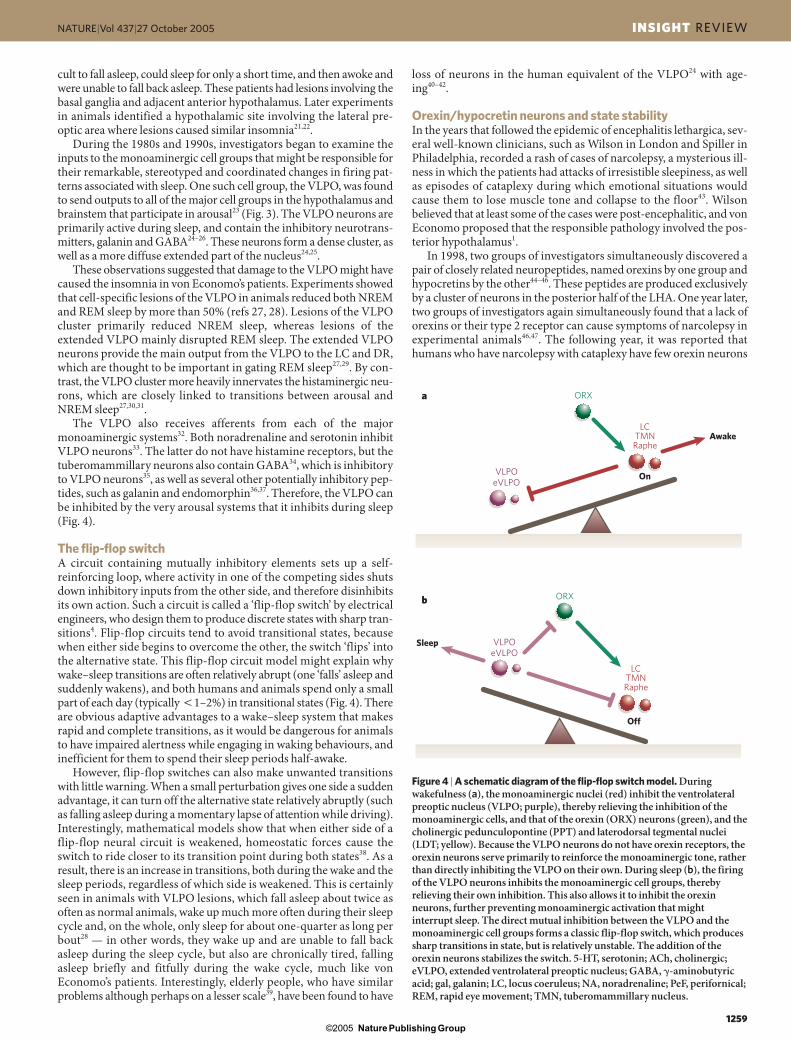
The flip-flop switchA circuit containing mutually inhibitory elements sets up a self-reinforcing loop, where activity in one of the competing sides shutsdown inhibitory inputs from the other side, and therefore disinhibitsits own action. Such a circuit is called a ‘flip-flop switch’ by electricalengineers, who design them to produce discrete states with sharp tran-sitions4. Flip-flop circuits tend to avoid transitional states, becausewhen either side begins to overcome the other, the switch ‘flips’ intothe alternative state. This flip-flop circuit model might explain whywake–sleep transitions are often relatively abrupt (one ‘falls’ asleep andsuddenly wakens), and both humans and animals spend only a smallpart of each day (typically �1–2%) in transitional states (Fig. 4). Thereare obvious adaptive advantages to a wake–sleep system that makesrapid and complete transitions, as it would be dangerous for animalsto have impaired alertness while engaging in waking behaviours, andinefficient for them to spend their sleep periods half-awake.
However, flip-flop switches can also make unwanted transitionswith little warning. When a small perturbation gives one side a suddenadvantage, it can turn off the alternative state relatively abruptly (suchas falling asleep during a momentary lapse of attention while driving).Interestingly, mathematical models show that when either side of aflip-flop neural circuit is weakened, homeostatic forces cause theswitch to ride closer to its transition point during both states38. As aresult, there is an increase in transitions, both during the wake and thesleep periods, regardless of which side is weakened. This is certainlyseen in animals with VLPO lesions, which fall asleep about twice asoften as normal animals, wake up much more often during their sleepcycle and, on the whole, only sleep for about one-quarter as long perbout28 — in other words, they wake up and are unable to fall backasleep during the sleep cycle, but also are chronically tired, fallingasleep briefly and fitfully during the wake cycle, much like vonEconomo’s patients. Interestingly, elderly people, who have similarproblems although perhaps on a lesser scale39, have been found to have
Awake
Sleep
On
Off
ORX
ORX
VLPOeVLPO
VLPOeVLPO
a
b
LCTMNRaphe
LCTMNRaphe
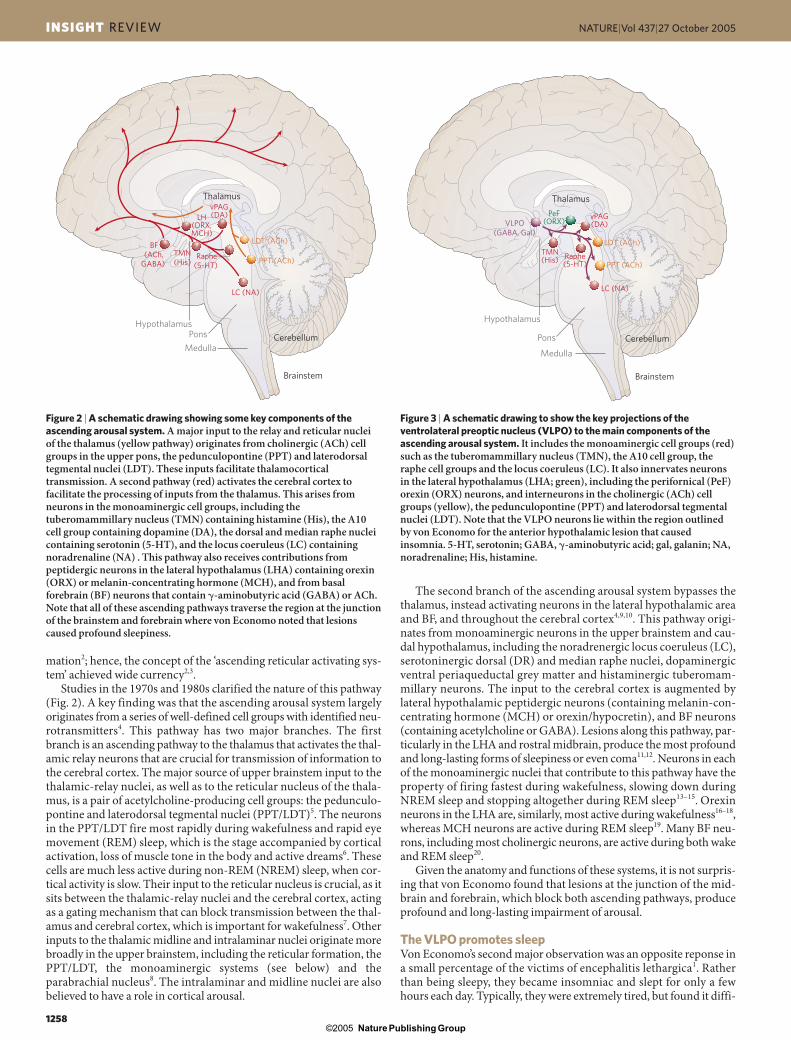
Figure 4 | A schematic diagram of the flip-flop switch model. Duringwakefulness (a), the monoaminergic nuclei (red) inhibit the ventrolateralpreoptic nucleus (VLPO; purple), thereby relieving the inhibition of themonoaminergic cells, and that of the orexin (ORX) neurons (green), and thecholinergic pedunculopontine (PPT) and laterodorsal tegmental nuclei(LDT; yellow). Because the VLPO neurons do not have orexin receptors, theorexin neurons serve primarily to reinforce the monoaminergic tone, ratherthan directly inhibiting the VLPO on their own. During sleep (b), the firingof the VLPO neurons inhibits the monoaminergic cell groups, therebyrelieving their own inhibition. This also allows it to inhibit the orexinneurons, further preventing monoaminergic activation that mightinterrupt sleep. The direct mutual inhibition between the VLPO and themonoaminergic cell groups forms a classic flip-flop switch, which producessharp transitions in state, but is relatively unstable. The addition of theorexin neurons stabilizes the switch. 5-HT, serotonin; ACh, cholinergic;eVLPO, extended ventrolateral preoptic nucleus; GABA, �-aminobutyricacid; gal, galanin; LC, locus coeruleus; NA, noradrenaline; PeF, perifornical;REM, rapid eye movement; TMN, tuberomammillary nucleus.
02 Saper 05-11 19/10/05 11:52 AM Page 7
Nature Publishing Group© 2005
© 2005 Nature Publishing Group
INSIGHT REVIEW NATURE|Vol 437|27 October 2005
1260
in the LHA and low orexin levels in the cerebrospinal fluid48–50. So,within a remarkably short period of time, the molecular basis of thismysterious illness was identified.
Interestingly, few people with narcolepsy have mutations in eitherthe orexin ligand or receptor genes49,51. In most narcoleptics, the dis-ease begins in the second or third decade of life, and the loss of orexinneurons is remarkably specific (producing no injury to the adjacentneurons that produce MCH)48,49. The cause is believed to be autoim-mune, although convincing evidence for this hypothesis is still lack-ing, and it might be a neurodegenerative condition51. Other patientswith lesions of the posterior LHA due to a range of causes, such asthose first reported by von Economo and Wilson, also acquired nar-colepsy.
The orexin neurons are mainly active during wakefulness and espe-cially during motor activity when animals actively explore their envi-ronment16–18. They have ascending projections to the cerebral cortex,as well as descending projections to all the monoaminergic and cholin-ergic cell groups of the arousal systems47,52. There are mutual projec-tions between the VLPO neurons and the orexin neurons32,53,54, but theformer do not express orexin receptors55. Hence, the orexin neuronsreinforce the arousal systems, but probably do not directly inhibit theVLPO (Fig. 4). This asymmetric relationship could help stabilize theflip-flop switch, like a ‘finger’ on the switch that might preventunwanted transitions into sleep. The increase in homeostatic sleepdrive due to consolidated wakefulness might, in turn, help produceconsolidated sleep. Narcoleptic people and animals lack this influenceand behave as if their sleep flip-flop switch has been destabilized51,56.They do not sleep more than normal individuals, but easily doze offduring the day and wake more often from sleep at night53, as the flip-flop model would predict.
Homeostatic regulation of sleepAlthough the purpose of sleep remains unknown, it clearly has arestorative effect on the brain. Like other homeostatic systems, such asthat governing body temperature, which seek to recover to a set pointwhen perturbed, sleep deprivation is followed by extra recovery sleepthat is proportional to the sleep loss. An influential model of sleep reg-ulation, proposed by Borbely and colleagues, includes both homeo-static and circadian drives for sleep57,58. The homeostatic influence isbelieved to be due to some structure or substance that accumulates‘need to sleep’ during prolonged wakefulness, and discharges this
homeostatic need during sleep. NREM and REM sleep probably haveseparate homeostatic mechanisms, and, after a period of sleep depri-vation, NREM sleep is typically repleted first.
The mechanisms for this homeostatic determinant remain unclear.The VLPO neurons, for example, do not accumulate a need to sleep, asthey remain at a low level of activity after a prolonged period of sleepdeprivation, until the animal actually falls asleep23,27. At that point,VLPO neurons fire about twice as fast as they do during normalsleep27, implying that they are under the influence of, but distinct from,the homeostatic factors that reflect sleep need.
Adenosine has been proposed as a homeostatic accumulator of theneed to sleep6,59,60. During prolonged wakefulness, the energy-pro-ducing systems in the brain run down, with exhaustion of brain glyco-gen reserves and depletion of ATP levels61,62. During prolongedwakefulness, as ATP is degraded to ADP, AMP and eventually adeno-sine, extracellular adenosine levels rise in some parts of the brain,including the BF63. Injection of adenosine or an adenosine A1 recep-tor agonist into the BF of cats6, or an adenosine A2a receptor agonistnear the VLPO in rats, causes sleep; the latter also results in expressionof Fos (a marker of neuronal activity) in VLPO neurons64. In addition,adenosine might disinhibit the VLPO via presynaptic A1 receptors byreducing inhibitory GABAergic inputs35. Hence, at least one mecha-nism for homeostatic sleep drive might be an accumulation of a sleep-promoting substance that enhances the activity of sleep-promotingcells and reduces the activity of wake-promoting neurons. In this way,adenosine, and perhaps other somnogens, can allow the activation ofthe VLPO that is necessary to trigger a sleep episode.
Circadian regulation of sleepBorbely also proposed a circadian influence on sleep and wakefulness(process C) that is distinct from the homeostatic drive (process S) forsleep57,58. Careful studies of humans in a forced desynchrony protocol,where they experience a 28-hour ‘day’ without any cues as to externaltime, have confirmed a strong 24-hour circadian rhythm in sleepdrive65. Recent studies in experimental animals have clarified how thisdrive is maintained.
The suprachiasmatic nucleus (SCN) serves as the brain’s ‘masterclock’. Neurons in the SCN fire in a 24-hour cycle that is driven by atranscriptional–translational loop, which persists even when the neu-rons are dissociated in cell culture66,67. Loss of the SCN abolishes thecircadian rhythms of a range of behaviours and physiological
Many of the drugs that are used to regulate sleep and wakefulness are nowthought to act directly on the wake and sleep systems that have beenoutlined here.
Wake-promoting drugs, such as amphetamines, methylphenidate(Ritalin) and modafinil (Provigil), all act on the dopamine (DA)-reuptaketransporter. Their potency is roughly proportional to their ability to blockDA reuptake, and mice in which the DA-transporter gene has been deletedfail to respond to this entire class of medications94. Modafinil might alsointerfere with noradrenaline reuptake, and this dual mode of action mightexplain why it can work without inducing other dopaminergic effects, suchas addiction95.
Some sleep-promoting drugs interfere with the actions of the ascendingarousal system. For example, antihistamines that cross the blood–brainbarrier, such as diphenhydramine (Benadryl), cause drowsiness by blockingthe arousing influence of the central histaminergic system. This side effectaccounts for the popularity of antihistamines that do not cross the blood-brain barrier and, hence, do not cause drowsiness. Centrally acting DAantagonists also cause drowsiness96. These drugs are used primarily totreat psychotic disorders, and this side effect is often a limiting factor indrug acceptability. Paradoxically, D2-agonist drugs can also causedrowsiness at low dosages96. The D2-presynaptic autoreceptor inhibits thefiring of dopaminergic neurons, and therefore results in reduceddopaminergic activity. Similarly, dexmedetomidine, an �-2 adrenergicagonist, reduces firing of noradrenergic neurons, and is thought to induceloss of wakefulness by disinhibiting the ventrolateral preoptic nucleus
(VLPO), which is ordinarily under noradrenergic inhibition duringwakefulness97 (Fig. 4).
The largest class of sleep-promoting drugs consists of those thatenhance the activity of �-aminobutyric acid-A (GABA-A) receptors,including barbiturates, benzodiazepines, chloral hydrate, ethanol andmost gaseous anaesthetics98. Few of these drugs are direct agonists(that is, few compete with GABA for binding); rather, most bind to othercomponents of the GABA-A receptor and enhance the action of GABA atthe receptor. At low dosages, these drugs might act on the targets of theVLPO, which also uses GABA as a neurotransmitter to silence thearousal system. At higher dosages, they suppress firing in much of thecentral nervous system. Benzodiazepines, such as diazepam (Valium),act on GABA-A receptors that contain a �-subunit and subunits of the �-1, �-2, �-3 or �-5 class98. Newer ‘non-benzodiazepines’, such aszolpidem (Ambien) or eszopiclone (Lunesta), also bind at �-� sites butdo not have the classic benzodiazepine structure and are more selectivefor the �1-containing receptors, which are important in sedation but notreduction of anxiety98. Gaboxadol binds to GABA-A receptors thatcontain the �-4 and � (but not �) subunits. These receptors, which are athighest concentration in the thalamus, limbic system and cerebralcortex, are not accessible to benzodiazepines, and hence represent adistinct drug target99. This might reduce the arousal drive from themedial temporal and prefrontal cortex that is seen during insomnia, asgaboxadol can activate Fos expression by neurons in the VLPO, andincreases the amount of slow-wave sleep100.
Box 1 | Drugs that affect sleep and wakefulness
02 Saper 05-11 19/10/05 11:52 AM Page 8
Nature Publishing Group© 2005
© 2005 Nature Publishing Group
NATURE|Vol 437|27 October 2005 INSIGHT REVIEW
1261
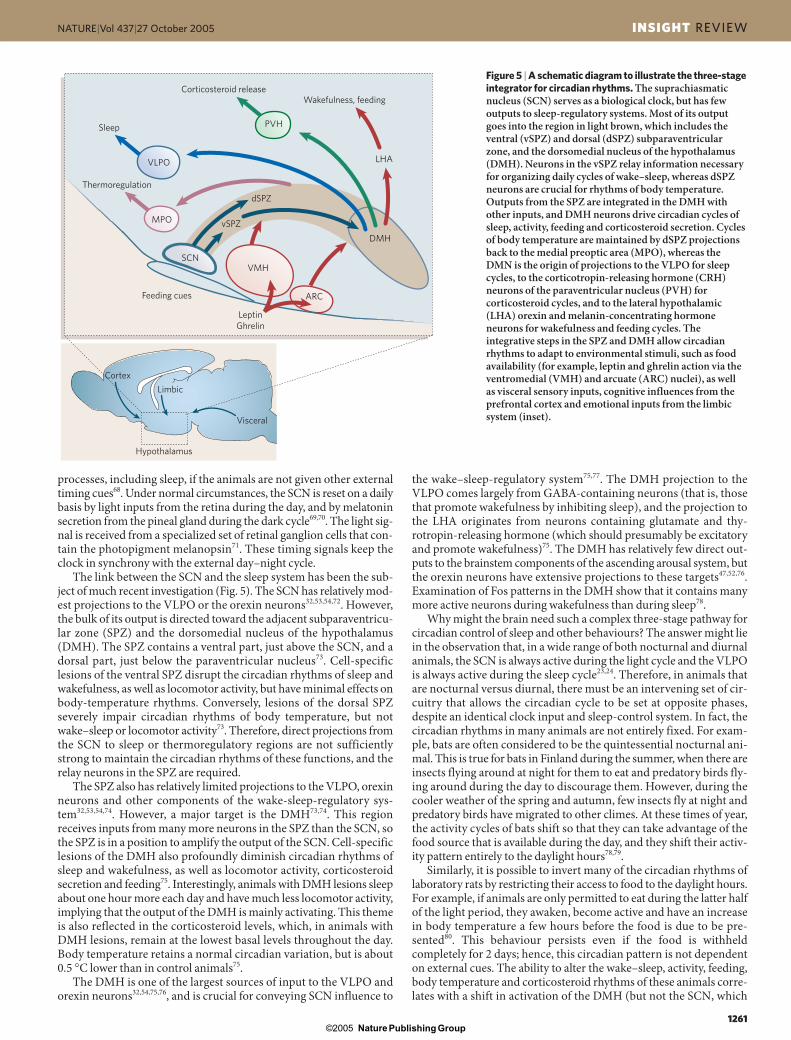
the wake–sleep-regulatory system75,77. The DMH projection to theVLPO comes largely from GABA-containing neurons (that is, thosethat promote wakefulness by inhibiting sleep), and the projection tothe LHA originates from neurons containing glutamate and thy-rotropin-releasing hormone (which should presumably be excitatoryand promote wakefulness)75. The DMH has relatively few direct out-puts to the brainstem components of the ascending arousal system, butthe orexin neurons have extensive projections to these targets47,52,76.Examination of Fos patterns in the DMH show that it contains manymore active neurons during wakefulness than during sleep78.
Why might the brain need such a complex three-stage pathway forcircadian control of sleep and other behaviours? The answer might liein the observation that, in a wide range of both nocturnal and diurnalanimals, the SCN is always active during the light cycle and the VLPOis always active during the sleep cycle23,24. Therefore, in animals thatare nocturnal versus diurnal, there must be an intervening set of cir-cuitry that allows the circadian cycle to be set at opposite phases,despite an identical clock input and sleep-control system. In fact, thecircadian rhythms in many animals are not entirely fixed. For exam-ple, bats are often considered to be the quintessential nocturnal ani-mal. This is true for bats in Finland during the summer, when there areinsects flying around at night for them to eat and predatory birds fly-ing around during the day to discourage them. However, during thecooler weather of the spring and autumn, few insects fly at night andpredatory birds have migrated to other climes. At these times of year,the activity cycles of bats shift so that they can take advantage of thefood source that is available during the day, and they shift their activ-ity pattern entirely to the daylight hours78,79.
Similarly, it is possible to invert many of the circadian rhythms oflaboratory rats by restricting their access to food to the daylight hours.For example, if animals are only permitted to eat during the latter halfof the light period, they awaken, become active and have an increasein body temperature a few hours before the food is due to be pre-sented80. This behaviour persists even if the food is withheld completely for 2 days; hence, this circadian pattern is not dependenton external cues. The ability to alter the wake–sleep, activity, feeding,body temperature and corticosteroid rhythms of these animals corre-lates with a shift in activation of the DMH (but not the SCN, which
processes, including sleep, if the animals are not given other externaltiming cues68. Under normal circumstances, the SCN is reset on a dailybasis by light inputs from the retina during the day, and by melatoninsecretion from the pineal gland during the dark cycle69,70. The light sig-nal is received from a specialized set of retinal ganglion cells that con-tain the photopigment melanopsin71. These timing signals keep theclock in synchrony with the external day–night cycle.
The link between the SCN and the sleep system has been the sub-ject of much recent investigation (Fig. 5). The SCN has relatively mod-est projections to the VLPO or the orexin neurons32,53,54,72. However,the bulk of its output is directed toward the adjacent subparaventricu-lar zone (SPZ) and the dorsomedial nucleus of the hypothalamus(DMH). The SPZ contains a ventral part, just above the SCN, and adorsal part, just below the paraventricular nucleus73. Cell-specificlesions of the ventral SPZ disrupt the circadian rhythms of sleep andwakefulness, as well as locomotor activity, but have minimal effects onbody-temperature rhythms. Conversely, lesions of the dorsal SPZseverely impair circadian rhythms of body temperature, but notwake–sleep or locomotor activity73. Therefore, direct projections fromthe SCN to sleep or thermoregulatory regions are not sufficientlystrong to maintain the circadian rhythms of these functions, and therelay neurons in the SPZ are required.
The SPZ also has relatively limited projections to the VLPO, orexinneurons and other components of the wake-sleep-regulatory sys-tem32,53,54,74. However, a major target is the DMH73,74. This regionreceives inputs from many more neurons in the SPZ than the SCN, sothe SPZ is in a position to amplify the output of the SCN. Cell-specificlesions of the DMH also profoundly diminish circadian rhythms ofsleep and wakefulness, as well as locomotor activity, corticosteroidsecretion and feeding75. Interestingly, animals with DMH lesions sleepabout one hour more each day and have much less locomotor activity,implying that the output of the DMH is mainly activating. This themeis also reflected in the corticosteroid levels, which, in animals withDMH lesions, remain at the lowest basal levels throughout the day.Body temperature retains a normal circadian variation, but is about 0.5 °C lower than in control animals75.
The DMH is one of the largest sources of input to the VLPO andorexin neurons32,54,75,76, and is crucial for conveying SCN influence to
Hypothalamus
Cortex
Limbic
Feeding cues
Visceral
Sleep
MPO
VLPO
PVH
LHA
dSPZ
DMH
vSPZ
VMH
ARC
SCN
Thermoregulation
Corticosteroid releaseWakefulness, feeding
LeptinGhrelin
Figure 5 | A schematic diagram to illustrate the three-stageintegrator for circadian rhythms. The suprachiasmaticnucleus (SCN) serves as a biological clock, but has fewoutputs to sleep-regulatory systems. Most of its outputgoes into the region in light brown, which includes theventral (vSPZ) and dorsal (dSPZ) subparaventricularzone, and the dorsomedial nucleus of the hypothalamus(DMH). Neurons in the vSPZ relay information necessaryfor organizing daily cycles of wake–sleep, whereas dSPZneurons are crucial for rhythms of body temperature.Outputs from the SPZ are integrated in the DMH withother inputs, and DMH neurons drive circadian cycles ofsleep, activity, feeding and corticosteroid secretion. Cyclesof body temperature are maintained by dSPZ projectionsback to the medial preoptic area (MPO), whereas theDMN is the origin of projections to the VLPO for sleepcycles, to the corticotropin-releasing hormone (CRH)neurons of the paraventricular nucleus (PVH) forcorticosteroid cycles, and to the lateral hypothalamic(LHA) orexin and melanin-concentrating hormoneneurons for wakefulness and feeding cycles. Theintegrative steps in the SPZ and DMH allow circadianrhythms to adapt to environmental stimuli, such as foodavailability (for example, leptin and ghrelin action via theventromedial (VMH) and arcuate (ARC) nuclei), as well as visceral sensory inputs, cognitive influences from theprefrontal cortex and emotional inputs from the limbicsystem (inset).
02 Saper 05-11 19/10/05 11:52 AM Page 9
Nature Publishing Group© 2005

© 2005 Nature Publishing Group
INSIGHT REVIEW NATURE|Vol 437|27 October 2005
1262
stays locked to the original light–dark cycle) to the new wake cycle,and lesions of the DMH prevent these behavioural shifts78 (J. J. Gooleyand C. B. Saper, unpublished observations). So, the DMH seems tointegrate clock information from the SCN and SPZ with feeding, tem-perature, social and other cues, providing animals with the flexibilityto adapt their behavioural and physiological cycles to the environment,thereby maximizing their chances of survival (Fig. 5).
Allostatic regulation of sleepThe rapid expansion of our knowledge about the substrates of the reg-ulation of sleep–wakefulness and circadian rhythms highlights thelack of knowledge about how these systems are controlled in a complexand ever-changing environment. Although we are beginning to under-stand the homeostatic drive for sleep, it is clear that sleep patterns andthe circadian cycle can be modified by external events, such as avail-ability of food or external temperatures. Similarly, homeostatic and cir-cadian drives for sleep can be overcome for brief periods whenexternal events demand an emergency response.
McEwen and Stellar, in 1993, proposed the term ‘allostatic’ drive81
for situations where “rather than maintaining constancy, the physi-ologic systems within the body fluctuate to meet demands fromexternal forces.” We know remarkably little about how these externalforces overcome the homeostatic and circadian systems or reset them(Fig. 5). There are clearly cues from visceral sensory systems andfeeding regulatory systems to the arousal systems54,82–85. Visceralinputs relayed through the nucleus of the solitary tract, such as gas-tric stretch, have a synchronizing sleep-inducing influence86,whereas absence of sufficient food causes an arousing influence87.However, we know little about the mechanisms by which cognitiveand emotional systems can act on the sleep or circadian control sys-tems. Recent studies have emphasized the inputs to the SCN82,VLPO32 and orexin neurons53,54 from corticolimbic sites, such as theinfralimbic cortex, ventral subiculum, lateral septum and bednucleus of the stria terminalis, as possible routes by which this influ-ence might be expressed.
Positron-emission tomography studies during sleep in humanswith insomnia also show increased activity in corticolimbic sites,including the medial prefrontal cortex and medial temporal lobe, com-pared with sleeping subjects without insomnia88. These inputs mightmaintain a hyperaroused state, which can be essential in an emergencysituation, such as when a doctor must stay awake to care for a sickpatient overnight. However, when the arousal systems override thehomeostatic and circadian regulation of sleep during periods of behav-ioural stress or depression, the result might be unwanted and debili-tating insomnia.
ConclusionsUnderstanding the interactions of these allostatic influences with thesleep and circadian systems is a major challenge for the next decade.However, learning to control our wake–sleep systems holds thepromise of improved health and cognitive performance (Box 1).Individuals who lose even small amounts of sleep on a daily basisshow progressive impairment of cognitive performance and eleva-tion of C-reactive protein, which predicts cardiovascular risk89–91.Because older individuals sleep about half an hour less per day, it ispossible that at least some of their cognitive decline and increase incardiovascular disease might be explained by sleep restriction92. Sim-ilarly, sleep loss might impair performance among adolescents whoarise early for school, shift workers, overnight long-haul truckers andeven medical personnel working in hospitals93. The public-healthimplications of sleep loss indicate that there is a great deal at stake inworking out the mechanisms that regulate our daily cycles of sleepand wakefulness. ■
1. Von Economo, C. Sleep as a problem of localization. J. Nerv. Ment. Dis. 71, 249–259(1930).
2. Moruzzi, G. & Magoun, H. W. Brain stem reticular formation and activation of the EEG.Electroencephalogr. Clin. Neurol. 1, 455–473 (1949).
3. Starzl, T. E., Taylor, C. W. & Magoun, H. W. Ascending conduction in reticular activatingsystem, with special reference to the diencephalon. J. Neurophysiol. 14, 461–477 (1951).
4. Saper, C. B., Chou, T. C. & Scammell, T. E. The sleep switch: hypothalamic control of sleepand wakefulness. Trends Neurosci. 24, 726–731 (2001).
5. Hallanger, A.H., Levey, A. I., Lee, H. J., Rye, D. B. & Wainer, B. H. The origins of cholinergicand other subcortical afferents to the thalamus in the rat. J. Comp. Neurol. 262, 104–124(1987).
6. Strecker, R. E. et al. Adenosinergic modulation of basal forebrain and preoptic/anteriorhypothalamic neuronal activity in the control of behavioral state. Behav. Brain Res. 115,183–204 (2000).
7. McCormick, D. A. Cholinergic and noradrenergic modulation of thalamocorticalprocessing. Trends Neurosci. 12, 215–220 (1989).
8. Krout, K. E., Belzer, R. E. & Loewy, A. D. Brainstem projections to midline and intralaminarthalamic nuclei of the rat. J. Comp. Neurol. 448, 53–101 (2002).
9. Saper, C. B. Organization of cerebral cortical afferent systems in the rat. II.Hypothalamocortical projections. J. Comp. Neurol. 237, 21 (1985).
10. Jones, B. E. Arousal systems. Front. Biosci. 8, S438–S451 (2003).11. Ranson, S. W. Somnolence caused by hypothalamic lesions in monkeys. Arch Neurol.
Psychiatr. 41, 1–23 (1939).12. Gerashchenko, D., Blanco-Centurion, C., Greco, M. A. & Shiromani, P. J. Effects of lateral
hypothalamic lesion with the neurotoxin hypocretin-2-saporin on sleep in Long-Evansrats. Neuroscience 116, 223–235 (2003).
13. Aston-Jones, G. & Bloom, F. E. Activity of norepinephrine-containing locus coeruleusneurons in behaving rats anticipates fluctuations in the sleep-waking cycle. J. Neurosci. 1,876–886 (1981).
14. Fornal, C., Auerbach, S. & Jacobs, B. L. Activity of serotonin-containing neurons innucleus raphe magnus in freely moving cats. Exp. Neurol. 88, 590–608 (1985).
15. Steininger, T. L., Alam, M. N., Gong, H., Szymusiak, R. & McGinty, D. Sleep-wakingdischarge of neurons in the posterior lateral hypothalamus of the albino rat. Brain Res.840, 138–147 (1999).
16. Estabrooke, I. V. et al. Fos expression in orexin neurons varies with behavioral state. J. Neurosci. 21, 1656–1662 (2001).
17. Mileykovskiy, B. Y., Kiyashchenko, L. I. & Siegel, J. M. Behavioral correlates of activity inidentified hypocretin/orexin neurons. Neuron 46, 787–798 (2005).
18. Lee, M. G, Hassani, O. K. & Jones, B. E. Discharge of identified orexin/hypocretin neuronsacross the wake-sleep cycle. J. Neurosci. 25, 6716–6720 (2005).
19. Verret, L. et al. A role of melanin-concentrating hormone producing neurons in the centralregulation of paradoxical sleep. BMC Neurosci. 4, 19 (2003).
20. Lee, M. G., Hassani, O. K., Alonso, A. & Jones, B. E. Cholinergic basal forebrain neuronsburst with theta during waking and paradoxical sleep. J. Neurosci. 25, 4365–4369 (2005).
21. Nauta, W. J. H. Hypothalamic regulation of sleep in rats. An experimental study. J. Neurophysiol. 9, 285–314 (1946).
22. McGinty, D. J. & Sterman, M. B. Sleep suppression after basal forebrain lesions in the cat.Science 160, 1253–1255 (1968).
23. Sherin, J. E., Shiromani, P. J., McCarley, R. W. & Saper, C. B. Activation of ventrolateralpreoptic neurons during sleep. Science 271, 216–219 (1996).
24. Gaus, S. E., Strecker, R. E., Tate, B. A., Parker, R. A. & Saper, C. B. Ventrolateral preopticnucleus contains sleep-active, galaninergic neurons in multiple mammalian species.Neuroscience 115, 285–294 (2002).
25. Sherin, J. E., Elmquist, J. K., Torrealba, F. & Saper, C. B. Innervation of histaminergictuberomammillary neurons by GABAergic and galaninergic neurons in the ventrolateralpreoptic nucleus of the rat. J. Neurosci. 18, 4705–4721 (1998).
26. Szymusiak, R., Alam, N., Steininger, T. L. & McGinty, D. Sleep-waking discharge patternsof ventrolateral preoptic/anterior hypothalamic neurons in rats. Brain Res. 803, 178–188(1998).
27. Lu, J. et al. Selective activation of the extended ventrolateral preoptic nucleus during rapideye movement sleep. J. Neurosci. 22, 4568–4576 (2002).
28. Lu, J., Greco, M. A., Shiromani, P. & Saper, C. B. Effect of lesions of the ventrolateralpreoptic nucleus on NREM and REM sleep. J. Neurosci. 20, 3830–3842 (2000).
29. Verret, L. et al. Localization of the neurones potentially responsible for the inhibition oflocus coeruleus noradrenergic neurones during paradoxical sleep in the rat. J. Comp.Neurol. (in the press).
30. Ko, E. M., Estabrooke, I. V., McCarthy, M. & Scammell, T. E. Wake-related activity oftuberomammillary neurons in rats. Brain Res. 992, 220–226 (2003).
31. John, J., Wu, M. F., Boehmer, L. N. & Siegel, J. M. Cataplexy-active neurons in thehypothalamus: implications for the role of histamine in sleep and waking behavior.Neuron 42, 619–634 (2004).
32. Chou, T. C. et al. Afferents to the ventrolateral preoptic nucleus. J. Neurosci. 22, 977–990(2002).
33. Gallopin, T. et al. Identification of sleep-promoting neurons in vitro. Nature 404, 992–995(2000).
34. Vincent, S. R., Hokfelt, T. & Wu, J.-Y. GABA neuron systems in hypothalamus and thepituitary gland. Neuroendocrinology 34, 117–125 (1982).
35. Chamberlin, N. L. et al. Effects of adenosine on GABAergic synaptic inputs to identifiedventrolateral preoptic neurons. Neuroscience 119, 913–918 (2003).
36. Martin-Schild, S., Gerall, A. A., Kastin, A. J. & Zadina, J. E. Differential distribution ofendomorphin 1- and endomorphin 2-like immunoreactivities in the CNS of the rodent. J. Comp Neurol. 405, 450–471 (1999).
37. Kohler, C. et al. Galanin immunoreactivity in hypothalamic neurons: further evidence formultiple chemical messengers in the tuberomammillary nucleus. J. Comp. Neurol. 250,58–64 (1986).
38. Chou, T. C. Regulation of Wake-Sleep Timing: Circadian Rhythms and Bistability of Sleep-Wake States, 82-99. Thesis, Harvard Univ. (2003).
39. Ancoli-Israel, S. & Kripke, D. F. Prevalent sleep problems in the aged. Biofeedback SelfRegul. 16, 349-359 (1991).
40.Hofman, M. A. & Swaab, D. F. The sexually dimorphic nucleus of the preoptic area in thehuman brain: a comparative morphometric study. J. Anat. 164, 55–72 (1989).
02 Saper 05-11 19/10/05 11:52 AM Page 10
Nature Publishing Group© 2005
© 2005 Nature Publishing Group
NATURE|Vol 437|27 October 2005 INSIGHT REVIEW
1263
74 Deurveilher, S. & Semba, K. Indirect projections from the suprachiasmatic nucleus tomajor arousal-promoting cell groups in rat: implications for the circadian control ofbehavioural state. Neuroscience 130,165–183 (2005).
75. Chou, T. C. et al. Critical role of dorsomedial hypothalamic nucleus in a wide range ofbehavioral circadian rhythms. J. Neurosci. 23, 10691–10702 (2003).
76. Thompson, R., Swanson, L. W. & Canteras, N. Organization of projections from thedorsomedial nucleus of the hypothalamus: a PHA-L study in the rat. J. Comp. Neurol. 376,143–173 (1997).
77. Aston-Jones, G., Chen, S., Zhu, Y. & Oshinsky, M. L. A neural circuit for circadianregulation of arousal. Nature Neurosci. 4, 732–738 (2001).
78. Saper, C. B., Lu, J., Chou, T. C. & Gooley, J. The hypothalamic integrator for circadianrhythms. Trends Neurosci. 28, 152–157 (2005).
79. Nyholm, H. Zur okologie von Myotis mystacinus (Leisl.) und M. daubentoni (Leisl.)(Chiroptera). Ann. Zool. Fenn. 2, 77–123 (1955).
80.Stephan, F. K. The “other” circadian system: food as a Zeitgeber. J. Biol. Rhythms 17,284–292 (2002).
81. McEwen, B. S. & Stellar, E. Stress and the individual. Mechanisms leading to disease. ArchIntern. Med. 153, 2093–2101 (1993).
82. Krout, K. E., Kawano, J., Mettenleiter, T. C. & Loewy, A. D. CNS inputs to thesuprachiasmatic nucleus of the rat. Neuroscience 110, 73–92 (2002).
83. Elmquist, J. K., Elias, C. F. & Saper, C. B. From lesions to leptin: hypothalamic control offood intake and body weight. Neuron 22, 221–232 (1999).
84.Elmquist, J. K., Ahima, R. S., Elias, C. F., Flier, J. S. & Saper, C. B. Leptin activates distinctprojections from the dorsomedial and ventromedial hypothalamic nuclei. Proc. Natl Acad.Sci. USA 95, 741–746 (1998).
85. Saper, C. B. in The Rat Nervous System (ed. Paxinos, G.) 761–796 (Elsevier Academic, SanDiego, 2004).
86. Schachter, S. C. & Saper, C. B. Vagus nerve stimulation. Epilepsia 39, 677–686 (1998).87. Yamanaka, A. et al. Hypothalamic orexin neurons regulate arousal according to energy
balance in mice. Neuron 38, 701–713 (2003).88. Nofzinger, E. A. et al. Functional neuroimaging evidence for hyperarousal in insomnia. Am.
J. Psychiatry 161, 2126-2128 (2004).89. Meier-Ewert, H. K. et al. Effect of sleep loss on C-reactive protein, an inflammatory
marker of cardiovascular risk. J. Am. Coll. Cardiol. 43, 678–683 (2004).90.Van Dongen, H. P., Maislin, G., Mullington, J. M. & Dinges, D. F. The cumulative cost of
additional wakefulness: dose-response effects on neurobehavioral functions and sleepphysiology from chronic sleep restriction and total sleep deprivation. Sleep 26, 117–126(2003).
91. Doran, S. M., Van Dongen, H. P. & Dinges, D. F. Sustained attention performance duringsleep deprivation: evidence of state instability. Arch. Ital. Biol. 139, 253–267 (2001).
92. Prinz, P. N. Age impairments in sleep, metabolic, and immune functions. Exp. Gerontol. 39,1739–1743 (2004).
93. Landrigan, C. P. et al. Effect of reducing interns’ work hours on serious medical errors inintensive care units. N. Engl. J. Med. 351, 1838–1848 (2004).
94. Wisor, J. et al. Dopaminergic role in stimulant-induced wakefulness. J. Neurosci. 21,1787–1895 (2001).
95. Saper, C. B. & Scammell, T. E. Modafinil: a drug in search of a mechanism. Sleep 27, 11–12(2004).
96. Roth, T. et al. Assessment of sleepiness and unintended sleep in Parkinson's diseasepatients taking dopamine agonists. Sleep Med. 4, 275–280 (2003).
97. Nelson, L. E. et al. The ±-2 adrenoreceptor agonist dexmedetomidine converges on anendogenous sleep-promoting pathway to exert its sedative effects. Anaesthesiology 98,428–436 (2003).
98. Rudolf, U. & Mohler, H. Analysis of GABAA receptor function and dissection of thepharmacology of benzodiazepines and general anesthetics through mouse genetics.Annu. Rev. Pharmacol. Toxicol. 44, 475–498 (2004).
99. Wisden, W., Laurie, D. J., Monyer, H. & Seeburg, P. The distribution of 13 GABAA receptorsubunit mRNAs in the rat brain. I. Telencephalon, diencephalons, mesencephalon. J. Neurosci. 12, 1040–1062 (1992).
100. Lancel, M., Wetter, T. C., Steiger, A. & Mathias, S. Effect of the GABAA agonistgaboxadol on nocturnal sleep and hormone secretion in healthy elderly subjects. Am. J.Physiol. Endocrinol. Metab. 281, E130–E137 (2001).
Acknowledgements This work was supported by United States Public HealthService grants.
Author Information Reprints and permissions information is available atnpg.nature.com/reprintsandpermissions. The authors declare competing financial interests: details accompany the paper onwww.nature.com/nature. Correspondence should be addressed to C.F.B.([email protected]).
41. Allen, L. S., Hines, M., Shryne, J. E. & Gorski, R. A. Two sexually dimorphic cell groups inthe human brain. J. Neurosci. 9, 497–506 (1989).
42. Byne, W. et al. The interstitial nuclei of the human anterior hypothalamus: aninvestigation of sexual variation in volume and cell size, number and density. Brain Res.856, 254–258 (2000).
43. Wilson, S. A. K. Modern Problems in Neurology (Edward Arnold, London, 1928).44.Sakurai, T. et al. Orexins and orexin receptors: a family of hypothalamic neuropeptides
and G protein-coupled receptors that regulate feeding behavior. Cell 92, 573–585 (1998).45. de Lecea, L. et al. The hypocretins: hypothalamus-specific peptides with neuroexcitatory
activity. Proc. Natl Acad. Sci. USA 95, 322–327 (1998).46.Lin, L. et al. The sleep disorder canine narcolepsy is caused by a mutation in the
hypocretin (orexin) receptor 2 gene. Cell 98, 365–376 (1999).47. Chemelli, R. M. et al. Narcolepsy in orexin knockout mice: molecular genetics of sleep
regulation. Cell 98, 437–451 (1999).48.Thannickal, T. C. et al. Reduced number of hypocretin neurons in human narcolepsy.
Neuron 27, 469–474 (2000).49. Peyron, C. et al. A mutation in a case of early onset narcolepsy and a generalized absence
of hypocretin peptides in human narcoleptic brains. Nature Med. 6, 991–997 (2000).50. Ripley, B. et al. CSF hypocretin/orexin levels in narcolepsy and other neurological
conditions. Neurology 57, 2253–2258 (2001).51. Scammell, T. E. The neurobiology, diagnosis, and treatment of narcolepsy. Ann. Neurol.
53, 154–166 (2003).52. Peyron, C. et al. Neurons containing hypocretin (orexin) project to multiple neuronal
systems. J. Neurosci. 18, 9996–10015 (1998).53. Sakurai, T. et al. Input of orexin/hypocretin neurons revealed by a genetically encoded
tracer in mice. Neuron 46, 297–308 (2005).54. Yoshida, K., McCormack, S., Espana, R. A., Crocker, A. D. & Scammell, T. E. Afferents to
the orexin neurons. J. Comp. Neurol. (in the press). 55. Marcus, J. N. et al. Differential expression of orexin receptors 1 and 2 in the rat brain.
J. Comp. Neurol. 435, 6–25 (2001).56 Mochizuki, T. et al. Behavioral state instability in orexin knock-out mice. J. Neurosci. 24,
6291–6300 (2004).57. Borbely, A. A. & Tobler, I. Brain Mechanisms of Sleep (eds McGinty, D. J. et al.) 35–44
(Raven, New York, 1985).58. Achermann, P. & Borbely, A. A. Mathematical models of sleep regulation. Front. Biosci. 8,
S683–S693 (2003).59. Radulovacki, M., Virus, R. M., Djuricic-Nedelson, M. & Green, R. D. Adenosine analogs
and sleep in rats. J. Pharmacol. Exp. Ther. 228, 268–274 (1984).60.Benington, J. H. & Heller, H. C. Restoration of brain energy metabolism as the function of
sleep. Prog. Neurobiol. 45, 347–360 (1995).61. Kong, J. et al. Brain glycogen decreases with increased periods of wakefulness:
implications for homeostatic drive to sleep. J. Neurosci. 22, 5581–5587 (2002).62. Shepel, P. N., Ramonet, D., Stevens, P. & Geiger, J. D. Purine level regulation during energy
depletion associated with graded excitatory stimulation in brain. Neurol. Res. 27, 139–148(2005).
63. Porkka-Heiskanen, T., Strecker, R. E. & McCarley, R. W. Brain site-specificity ofextracellular adenosine concentration changes during sleep deprivation andspontaneous sleep: an in vivo microdialysis study. Neuroscience 99, 507–517 (2000).
64.Scammell, T. E. et al. An adenosine A2a agonist increases sleep and induces Fos inventrolateral preoptic neurons. Neuroscience 107, 653–663 (2001).
65. Dijk, D. J. & Czeisler, C. A. Contribution of the circadian pacemaker and the sleephomeostat to sleep propensity, sleep structure, electroencephalographic slow waves,and sleep spindle activity in humans. J. Neurosci. 15, 3526–3538 (1995).
66. Reppert, S. M. & Weaver, D. R. Coordination of circadian timing in mammals. Nature 418,935-941 (2002).
67 Jin, X. et al. A molecular mechanism regulating rhythmic output from thesuprachiasmatic circadian clock. Cell 96, 1–20 (1999).
68. Moore, R. Y. & Eichler, V. B. Loss of a circadian adrenal corticosterone rhythm followingsuprachiasmatic lesions in the rat. Brain Res. 42, 201–206 (1972).
69. Johnson, R. F., Moore, R. Y. & Morin, L. P. Loss of entrainment and anatomical plasticityafter lesions of the hamster retinohypothalamic tract. Brain Res. 460, 297–313 (1988).
70. Cassone, V. M., Chesworth, M. J. & Armstrong, S. M. Entrainment of rat circadianrhythms by daily injection of melatonin depends upon the hypothalamic suprachiasmaticnuclei. Physiol. Behav. 36, 1111–1121 (1986).
71 Gooley, J. J., Lu, J., Fischer, D. & Saper, C. B. A broad role for melanopsin in nonvisualphotoreception. J. Neurosci. 23, 7093–7106 (2003).
72. Watts, A. G., Swanson, L. W. & Sanchez-Watts, G. Efferent projections of thesuprachiasmatic nucleus: I. Studies using anterograde transport of Phaseolus vulgarisleucoagglutinin in the rat. J. Comp. Neurol. 258, 204–229 (1987).
73. Lu, J. et al. Contrasting effects of ibotenate lesions of the paraventricular nucleus andsubparaventricular zone on sleep-wake cycle and temperature regulation. J. Neurosci. 21,4864–4874 (2001).
02 Saper 05-11 19/10/05 11:52 AM Page 11
Nature Publishing Group© 2005