
Frequent PVT1 Rearrangement and Novel Chimeric Genes...
10
Molecular and Cellular Pathobiology Frequent PVT1 Rearrangement and Novel Chimeric Genes PVT1-NBEA and PVT1-WWOX Occur in Multiple Myeloma with 8q24 Abnormality Hisao Nagoshi 1,2 , Tomohiko Taki 2 , Ichiro Hanamura 3 , Masakazu Nitta 3 , Takemi Otsuki 4 , Kazuhiro Nishida 1 , Keiko Okuda 2 , Natsumi Sakamoto 1 , Satoru Kobayashi 1 , Mio Yamamoto-Sugitani 1 , Yasuhiko Tsutsumi 1 , Tsutomu Kobayashi 1 , Yosuke Matsumoto 1 , Shigeo Horiike 1 , Junya Kuroda 1 , and Masafumi Taniwaki 1,2 Abstract Chromosome 8q24 rearrangements are occasionally found in multiple myeloma and are associated with tumor progression. The 8q24 rearrangements were detected by FISH in 12 of 54 patients with multiple myeloma (22.2%) and in 8 of 11 multiple myeloma cell lines (72.7%). The breakpoints of 8q24 in 10 patients with multiple myeloma and in all multiple myeloma cell lines were assigned to a 360 kb segment, which was divided into 4 regions: approximately 120 kb centromeric to MYC (5 0 side of MYC), the region centromerically adjacent to PVT1 ( 170 kb region, including MYC, of 5 0 side of PVT1), the PVT1 region, and the telomeric region to PVT1. PVT1 rearrangements were most common and found in 7 of 12 patients (58.3%) and 5 of 8 cell lines (62.5%) with 8q24 abnormalities. A combination of spectral karyo- typing (SKY), FISH, and oligonucleotide array identified several partner loci of PVT1 rearrangements, such as 4p16, 4q13, 13q13, 14q32, and 16q23-24. Two novel chimeric genes were identified: PVT1-NBEA in the AMU-MM1 cell line harboring t(8;13)(q24;q13) and PVT1-WWOX in RPMI8226 cell line harboring der(16)t(16;22)ins(16;8)(q23;q24). The PVT1-NBEA chimera in which PVT1 exon 1 was fused to NBEA exon 2 and the PVT1-WWOX in which PVT1 exon 1 was fused to WWOX exon 9 were associated with the expression of abnormal NBEA and WWOX lacking their N-terminus, respectively. These findings suggest that PVT1 rearrangements may represent a novel molecular paradigm underlying the pathology of 8q24 rearrangement– positive multiple myeloma. Cancer Res; 72(19); 4954–62. Ó2012 AACR. Introduction Genetic abnormalities play a crucial role in the pathogenesis of various malignancies, including multiple myeloma. The primary cytogenetic abnormalities associated with disease development are either nonrandom chromosomal gains known as hyperdiploid, which is characterized by trisomies of chromosomes 3, 5, 7, 9, 11, 15, 19, and 21, or structural rearrangements involving the immunoglobulin heavy chain gene (IGH) located at 14q32.33 (IGH translocation; refs. 1, 2). Secondary cytogenetic abnormalities implicated in disease progression include 8q24 rearrangements, gain of the long arm of chromosome 1 (1qþ), and loss of the short arm of chromosome 17 (17p; refs. 1, 3). The 8q24 rearrangements have been identified by conven- tional cytogenetic analysis in 3.5% to 5.0% of patients with multiple myeloma (4, 5), and by FISH and spectral karyotyping (SKY) in 9.5% to 20% (6–9). The 8q24 rearrangements are frequently associated with advanced disease in patients with multiple myeloma and multiple myeloma cell lines (10, 11). Ig chromosomal translocations, such as t(8;14)(q24;q32) and t(8;22)(q24;q11), occur in approximately 25% of multiple mye- lomas with 8q24 rearrangements, whereas non-Ig chromosom- al loci, including 1p13, 1p21-22, 6p21, 6q12-15, 13q14, and 16q22, in which no candidate genes have been delineated so far, have also been identified as translocation partners (7, 8, 12, 13). MYC has long been a possible candidate target gene for 8q24 rearrangements; however, many of the breakpoints with- in 8q24 have been assigned to various regions that encom- passed more than 2 Mb centromeric or telomeric to MYC (9, 11). In contrast to the typical Burkitt lymphoma translo- cation t(8;14) with breakpoints within the MYC gene (14), rearrangements of plasmacytoma variant translocation 1/Molo- ney leukemia virus integration-1 locus (PVT1), which is located 57 kb 3 0 of MYC, have been identified in variant Burkitt lymphoma translocations t(8;22) and t(2;8). In the latter Authors' Affiliations: Departments of 1 Hematology and Oncology and 2 Molecular Diagnostics and Therapeutics, Kyoto Prefectural University of Medicine Graduate School of Medical Science, Kyoto; 3 Division of Hema- tology, Department of Internal Medicine, Aichi Medical University, Naga- kute, Aichi; and 4 Department of Hygiene, Kawasaki Medical School, Kur- ashiki, Japan Note: Supplementary data for this article are available at Cancer Research Online (http://cancerres.aacrjournals.org/). Corresponding Author: Masafumi Taniwaki, Department of Hematology and Oncology, Kyoto Prefectural University of Medicine Graduate School of Medical Science, 465 Kajii-cho, Kawaramachi-Hirokoji, Kamigyo-ku, Kyoto 602-8566, Japan. Phone: 81-75-251-5740; Fax: 81-75-251-5743; E-mail: [email protected] doi: 10.1158/0008-5472.CAN-12-0213 Ó2012 American Association for Cancer Research. Cancer Research Cancer Res; 72(19) October 1, 2012 4954 on September 6, 2018. © 2012 American Association for Cancer Research. cancerres.aacrjournals.org Downloaded from Published OnlineFirst August 6, 2012; DOI: 10.1158/0008-5472.CAN-12-0213
Transcript of Frequent PVT1 Rearrangement and Novel Chimeric Genes...

Molecular and Cellular Pathobiology
Frequent PVT1 Rearrangement and Novel Chimeric GenesPVT1-NBEA and PVT1-WWOX Occur in Multiple Myelomawith 8q24 Abnormality
Hisao Nagoshi1,2, Tomohiko Taki2, Ichiro Hanamura3, Masakazu Nitta3, Takemi Otsuki4, Kazuhiro Nishida1,Keiko Okuda2, Natsumi Sakamoto1, Satoru Kobayashi1, Mio Yamamoto-Sugitani1, Yasuhiko Tsutsumi1,Tsutomu Kobayashi1, Yosuke Matsumoto1, Shigeo Horiike1, Junya Kuroda1, and Masafumi Taniwaki1,2
AbstractChromosome 8q24 rearrangements are occasionally found in multiple myeloma and are associated with
tumor progression. The 8q24 rearrangements were detected by FISH in 12 of 54 patients with multiplemyeloma (22.2%) and in 8 of 11 multiple myeloma cell lines (72.7%). The breakpoints of 8q24 in 10 patientswith multiple myeloma and in all multiple myeloma cell lines were assigned to a 360 kb segment, whichwas divided into 4 regions: approximately 120 kb centromeric to MYC (50 side of MYC), the regioncentromerically adjacent to PVT1 (� 170 kb region, including MYC, of 50 side of PVT1), the PVT1 region,and the telomeric region to PVT1. PVT1 rearrangements were most common and found in 7 of 12 patients(58.3%) and 5 of 8 cell lines (62.5%) with 8q24 abnormalities. A combination of spectral karyo-typing (SKY), FISH, and oligonucleotide array identified several partner loci of PVT1 rearrangements,such as 4p16, 4q13, 13q13, 14q32, and 16q23-24. Two novel chimeric genes were identified: PVT1-NBEAin the AMU-MM1 cell line harboring t(8;13)(q24;q13) and PVT1-WWOX in RPMI8226 cell line harboringder(16)t(16;22)ins(16;8)(q23;q24). The PVT1-NBEA chimera in which PVT1 exon 1 was fused to NBEA exon2 and the PVT1-WWOX in which PVT1 exon 1 was fused toWWOX exon 9 were associated with the expressionof abnormal NBEA and WWOX lacking their N-terminus, respectively. These findings suggest that PVT1rearrangements may represent a novel molecular paradigm underlying the pathology of 8q24 rearrangement–positive multiple myeloma. Cancer Res; 72(19); 4954–62. �2012 AACR.
IntroductionGenetic abnormalities play a crucial role in the pathogenesis
of various malignancies, including multiple myeloma. Theprimary cytogenetic abnormalities associated with diseasedevelopment are either nonrandom chromosomal gainsknown as hyperdiploid, which is characterized by trisomiesof chromosomes 3, 5, 7, 9, 11, 15, 19, and 21, or structuralrearrangements involving the immunoglobulin heavy chaingene (IGH) located at 14q32.33 (IGH translocation; refs. 1, 2).Secondary cytogenetic abnormalities implicated in disease
progression include 8q24 rearrangements, gain of the longarm of chromosome 1 (1qþ), and loss of the short arm ofchromosome 17 (17p; refs. 1, 3).
The 8q24 rearrangements have been identified by conven-tional cytogenetic analysis in 3.5% to 5.0% of patients withmultiple myeloma (4, 5), and by FISH and spectral karyotyping(SKY) in 9.5% to 20% (6–9). The 8q24 rearrangements arefrequently associated with advanced disease in patients withmultiple myeloma and multiple myeloma cell lines (10, 11). Igchromosomal translocations, such as t(8;14)(q24;q32) andt(8;22)(q24;q11), occur in approximately 25% of multiple mye-lomaswith 8q24 rearrangements, whereas non-Ig chromosom-al loci, including 1p13, 1p21-22, 6p21, 6q12-15, 13q14, and16q22, in which no candidate genes have been delineated sofar, have also been identified as translocation partners (7, 8, 12,13). MYC has long been a possible candidate target gene for8q24 rearrangements; however, many of the breakpoints with-in 8q24 have been assigned to various regions that encom-passed more than 2 Mb centromeric or telomeric to MYC(9, 11). In contrast to the typical Burkitt lymphoma translo-cation t(8;14) with breakpoints within the MYC gene (14),rearrangements of plasmacytoma variant translocation 1/Molo-ney leukemia virus integration-1 locus (PVT1), which is located57 kb 30 of MYC, have been identified in variant Burkittlymphoma translocations t(8;22) and t(2;8). In the latter
Authors' Affiliations: Departments of 1Hematology and Oncology and2Molecular Diagnostics and Therapeutics, Kyoto Prefectural University ofMedicine Graduate School of Medical Science, Kyoto; 3Division of Hema-tology, Department of Internal Medicine, Aichi Medical University, Naga-kute, Aichi; and 4Department of Hygiene, Kawasaki Medical School, Kur-ashiki, Japan
Note: Supplementary data for this article are available at Cancer ResearchOnline (http://cancerres.aacrjournals.org/).
Corresponding Author: Masafumi Taniwaki, Department of Hematologyand Oncology, Kyoto Prefectural University of Medicine Graduate Schoolof Medical Science, 465 Kajii-cho, Kawaramachi-Hirokoji, Kamigyo-ku,Kyoto 602-8566, Japan. Phone: 81-75-251-5740; Fax: 81-75-251-5743;E-mail: [email protected]
doi: 10.1158/0008-5472.CAN-12-0213
�2012 American Association for Cancer Research.
CancerResearch
Cancer Res; 72(19) October 1, 20124954
on September 6, 2018. © 2012 American Association for Cancer Research. cancerres.aacrjournals.org Downloaded from
Published OnlineFirst August 6, 2012; DOI: 10.1158/0008-5472.CAN-12-0213

translocations, fusion of the constant region of the IG g or kchain gene to PVT1 was detected, resulting in a lack of proteinproduction (15).In this study, the 8q24 rearrangements were analyzed in
patients withmultiplemyeloma and cell lines by FISH and SKYcombined with oligonucleotide arrays. Results showed fre-quent PVT1 rearrangements with several partners and novelPVT1-NBEA and PVT1-WWOX chimeric genes.
Materials and MethodsPatients and cell linesTheuse of clinical sampleswas approved by the Institutional
Review Board of Kyoto Prefectural University of Medicine(Kyoto, Japan). Informed consent was obtained from allpatients. Primary samples were obtained from the bone mar-row of 53 patients and the lymph node of 1 patient betweenApril 2005 and January 2011. Eleven multiple myeloma celllines, AMU-MM1, KMS-12-BM, KMS-18, KMS-20, KMS-28-PE,KMS-34, AMO1, IM9, LP-1, NCI-H929, and RPMI8226, were alsoanalyzed. AMU-MM1 was established at Aichi Medical Uni-versity (Aichi, Japan) from the tumor cells of the cerebrospinalfluid of a 72-year-old Japanese female patient with IgA-kmultiple myeloma (16).
FISH analysisThe FISH was conducted as described previously (17). To
assess the 8q24 rearrangement patterns and identify the genesinvolved, 3 sets of probes were used. The first set of probes wasthe 8q24 probe-LSI MYC Dual Color, Break Apart Rearrange-ment Probe. It consists of the SpectrumOrange-labeled 50 LSIMYC probe, which begins at 119 kb upstream of the 50 end ofMYC and extends 266 kb toward the centromere, and theSpectrumGreen–labeled 30 LSI MYC probe, which startsapproximately at 1.5 Mb 30 ofMYC and extends 407 kb towardthe telomere (Abbott Japan). The second set of probes wasdesigned to hybridize to both adjacent sides on the PVT1 gene,defined as PVT1-adjacent (PVT1-A) probe. The PVT1-A probeset was composed of 2 specific bacterial artificial chromosomeclonesCTD-3066D1, a fragment approximately 170 kb in lengthadjacent to the 50 end of PVT1, and RP11-628C14, a fragmentapproximately 190 kb in length adjacent to the 30 end of PVT1.The third set of probes was the PVT1-spanning (PVT1-S) probeconsisting of 2 bacterial artificial chromosome clones, CTD-22267H2, a fragment approximately 120 kb in length covering50 regions of PVT1, and RP11-164J24, a fragment approximately190 kb in length covering 30 regions of PVT1 (Fig. 1A). Forinterphase analysis, signals were evaluated in a minimum of100 nuclei with hybridization efficiency greater than 90%.
SKY analysis and SKY combined with FISH analysisThe SKY analysis was conducted as described previously
(18). For the SKY–FISH analysis, SKY and FISH probe mixturessimultaneously hybridized to chromosomes for 2 days at 37�C.Ten to 20 metaphase spreads were analyzed, and karyotypeswere defined according to ISCN 2009 (19). For complex abnor-malities with rearranged 8q24 locus, such as translocations orinsertions, which cannot be detected by either FISH or SKY, a
SKY–FISH procedure was used to detect chromosomal loca-tions involving MYC and PVT1.
Genome copy number analysisThe DNA gain and loss assay on the basis of high-density
oligonucleotide microarrays (GeneChip Human Mapping 50K,250K, or 6.0 single-nucleotide polymorphism (SNP) array,Affymetrix) was conducted with genomic DNA extracted fromcell lines and tumor specimens. Breakpoints in chromosomaltranslocations were identified by the means of genome copynumber analysis combined with SKY, and the SNP array datawere analyzed to determine total copy numbers using theCNAG3.0 or 3.3 programs (20).
Reverse-transcription PCR and sequencing analysisReverse-transcription PCR (RT-PCR) analysis was con-
ducted as described previously (21). The following primerswere used: P1S (forward primer in exon 1 of PVT1,NR_003367.1), 50-TTGCGGAAAGGATGTTGGCG-30, and N3A(reverse primer in exon 3 ofNBEA, NM_015678.3), 50-GCTCCA-TATTTCTGCTTGACA-30, for the detection of any chimericgene on der(8)t(8;13); N2S (forward primer in exon 2 of NBEA),50-CATACAGGTCGGAGAGGTC-30, and P2A (reverse primer inexon 2 of PVT1), 50-AGGGCTTCACCGGCTCAAT-30, or P3A(reverse primer in exon 3 of PVT1), 50-GGGTCTTACATTCCA-TAGGG-30, for the detection of any chimeric gene on der(13)t(8;13); and P1S and W9A (reverse primer in exon 9 of WWOX,NM_016373.2), 50-CAGGGAGATACGGAACCTAC-30, for thedetection of any chimeric gene on der(16)t(16;22)ins(16;8)(q23;q24). The nucleotide sequences of PCR products weredetermined with the fluorometric method (Dye TerminatorCycle Sequencing Kit, Applied Biosystems).
Real-time quantitative RT-PCRNBEA and WWOX mRNA levels were measured with speci-
fic primer probe sets from Assays-on-Demand (Applied Bio-systems) or SYBR Green method using the ABI Prism 7300system (Applied Biosystems) according to the manufacturer'sinstructions. Primers were Assays-on-Demand NBEA 2-3(Hs00995629_m1), NBEA 58-59 (Hs00995655_m1), W8S (for-ward primer in exon 8 of WWOX), 50-GCAACATCCTCTTCT-CCAACGA-30, WPA2(reverse primer in exon 9 of WWOX),50-TGGGACAGCAGCACAGTACA-30, W9S (forward primerin exon 9 of WWOX), 50-TGTACTGTGCTGCTGTCCCA-30,and WPA3(reverse primer in exon 9 of WWOX), 50-CCGTT-CTTGGATCAGCCTC-30. These primer sets were used to dis-tinguish abnormal chimeric NBEA or WWOX transcripts fromnormal transcripts: NBEA 2-3, spanning the breakpoint of 8q24in t(8;13)(q24;q14), can detect only the normalNBEA transcript,whereas NBEA 58-59 can detect both normal and chimerictranscripts, similarly, combination of W8S and W9A(2) candetect only the normalWWOX transcript andW9S andW9A(3)can detect both normal and chimeric WWOX transcripts.The b-actin mRNA level was measured as an internal con-trol. In addition to the 11 multiple myeloma cell lines,normal peripheral lymphocytes, an Epstein–Barr virus–transformed B-cell line derived from a normal healthy volun-teer, the erythroleukemia cell line K562, and the Burkitt
Recurrent PVT1 Rearrangement in Multiple Myeloma
www.aacrjournals.org Cancer Res; 72(19) October 1, 2012 4955
on September 6, 2018. © 2012 American Association for Cancer Research. cancerres.aacrjournals.org Downloaded from
Published OnlineFirst August 6, 2012; DOI: 10.1158/0008-5472.CAN-12-0213

lymphoma cell line, Daudi, were analyzed. Each assay wasdone in triplicate.
ResultsFrequent involvement of PVT1 locus in 8q24rearrangements
The 8q24 rearrangements were detected using 3 FISH probesets in 12 patients (22.2%) and 8 cell lines (72.7%). The break-points of 8q24 were assigned to a 360 kb segment containingtheMYC and PVT1 genes in 10 patients withmultiple myelomaand in all multiple myeloma cell lines, and 11 breakpoint typeswere identified (Fig. 1, Supplementary Fig. S1 and Supplemen-tary Table S1). Breakpoint regions at 8q24 could be divided into4 regions: approximately 120 kb centromeric toMYC (50 side ofMYC; region A), the region centromerically adjacent to PVT1(approximately 170 kb region, includingMYC, of 50 side ofPVT1;region B), the PVT1 region (region C), and the telomeric regionto PVT1 (30 side of PVT1; region D; Fig. 1). In patients withmultiple myeloma, region C was the most frequent (Fig. 1B),with breakpoints detected within the PVT1 gene in 5 patients(41.7%; Pt-1 to 4, and 7). Region B was identified in 4 patients(33.3%; Pt- 7 to 10), and both regions A and C in 2 patients(16.7%; Pt- 5 and 6). The remaining 2 patients (16.7%; Pt- 11 and
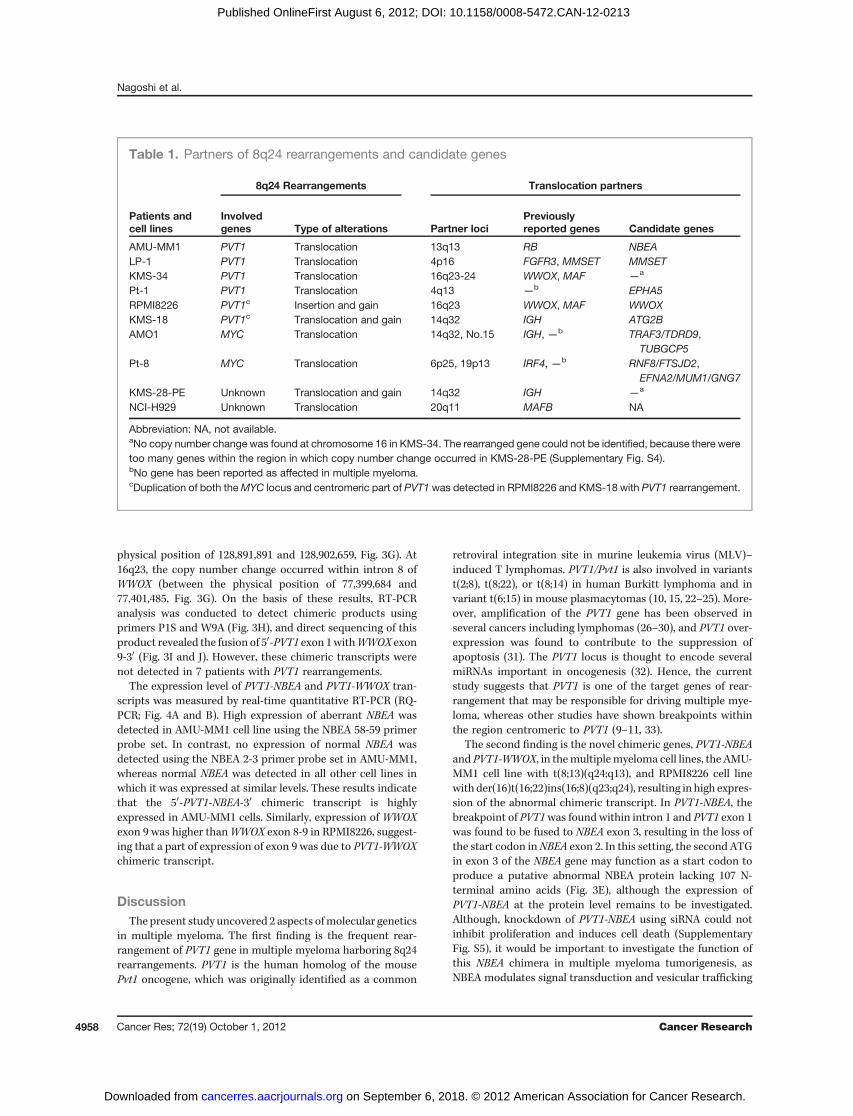
12) showed breakpoints outside of these 3 regions (region D).Among the 8 cell lines with 8q24 rearrangements, breakpointswere identified in the PVT1 (region C) in 3 cell lines (37.5%;AMU-MM1, LP-1, and KMS-34), in region B in 1 cell line (12.5%;AMO1), and in both regions A and C in 2 cell lines (25.0%;RPMI8226 and KMS-18). The breakpoints observed in KMS-28-PE and NCI-H929 were either centromeric or telomeric toMYCand PVT1. The SNP array analysis validated the interphaseFISH data by showing copy number gains at the regions ofinterest in KMS-18, RPMI8226, and KMS-28-PE (Supplemen-tary Fig. S2). In summary, breakpoints were assigned to thePVT1 gene in 7 of 54 patients (13.0%) and 5 of 11 cell lines(45.5%), and to theMYC gene in 4 patients (7.4%) and 1 cell line(9.1%).
Partners of 8q24 rearrangement detected by SKY–FISHand genome copy number analysis
Metaphase analysis of 2 patients (Pt- 1 and 8) and 8multiplemyeloma cell lines by SKY, SKY–FISH, and FISH identifiedvarious translocation/insertion partner loci for 8q24 rearran-gements (Fig. 2A and B and Supplementary Fig. S3). To detectthe candidate genes within partner loci of 8q24 rearrangement,the boundaries of copy number gains and losses in the regionsof interest were mapped using the SNP arrays (Table 1 and
Chromosome 8
6004002000-200-400-600 1500
Centromere TelomerePVT1MYC
~407kb~266kb119kb
8q24
kb
RP11-628C14CTD-3066D1PVT1-A
PVT1-S CTD-2267H2 RP11-164J24~120kb ~190kb
~170kb ~190kb
TP-1
TP-2
TM
DN-2
TN
DN-3
TP-3
DN-1
DP-2
DP-1
DP-3
Breakpoint types
DCBABreakpoint regions
BA
Patients and cell lines
Breakpoint types
Mapping of breakpoints and duplicated regions
AMU-MM1 TP-1
Pt-3 TP-1
Pt-4 TP-1
LP-1 TP-2
KMS-34 TP-3
Pt-1 TP-3
Pt-2 TP-3
Pt-7 TP-1 / TM
RPMI8226 DP-1
KMS-18 DP-2
Pt-5 DP-2
Pt-6 DP-3
KMS-28-PE DN-1
Pt-12 DN-2
NCI-H929 DN-3
AMO1 TM
Pt-8 TM
Pt-9 TM
Pt-10 TM
Pt-11 TN
Breakpoint regions A B C D
Frequency* 6 5 12 3
PVT1MYC
Figure 1. Identification of breakpoints region at 8q24 by FISH analysis. A, location of FISH probes and mapping of putative breakpoints at 8q24.FISH probes are depicted as color bars. Vertical arrows indicate breakpoints and horizontal double-headed arrows indicate the possible range of breakpointson the basis of interphase FISH analysis. The combination of the FISH analysis with 3 sets of probes reveals 4 breakpoint regions and 11 breakpointtypes as shown in B and Supplementary Fig. S1. B, mapping of breakpoints in patients with multiple myeloma and cell lines with 8q24 rearrangement.Dark gray boxes, the breakpoint regions; light gray boxes, duplicated regions. Pt, patient number. Breakpoint region A is the 120 kb length region ofcentromeric to MYC, B is the 170 kb length region centromerically adjacent to PVT1 including MYC, C is the PVT1 region, and D is the region of telomericto PVT1. �, frequency shows the case number for each of the breakpoint regions.
Nagoshi et al.
Cancer Res; 72(19) October 1, 2012 Cancer Research4956
on September 6, 2018. © 2012 American Association for Cancer Research. cancerres.aacrjournals.org Downloaded from
Published OnlineFirst August 6, 2012; DOI: 10.1158/0008-5472.CAN-12-0213
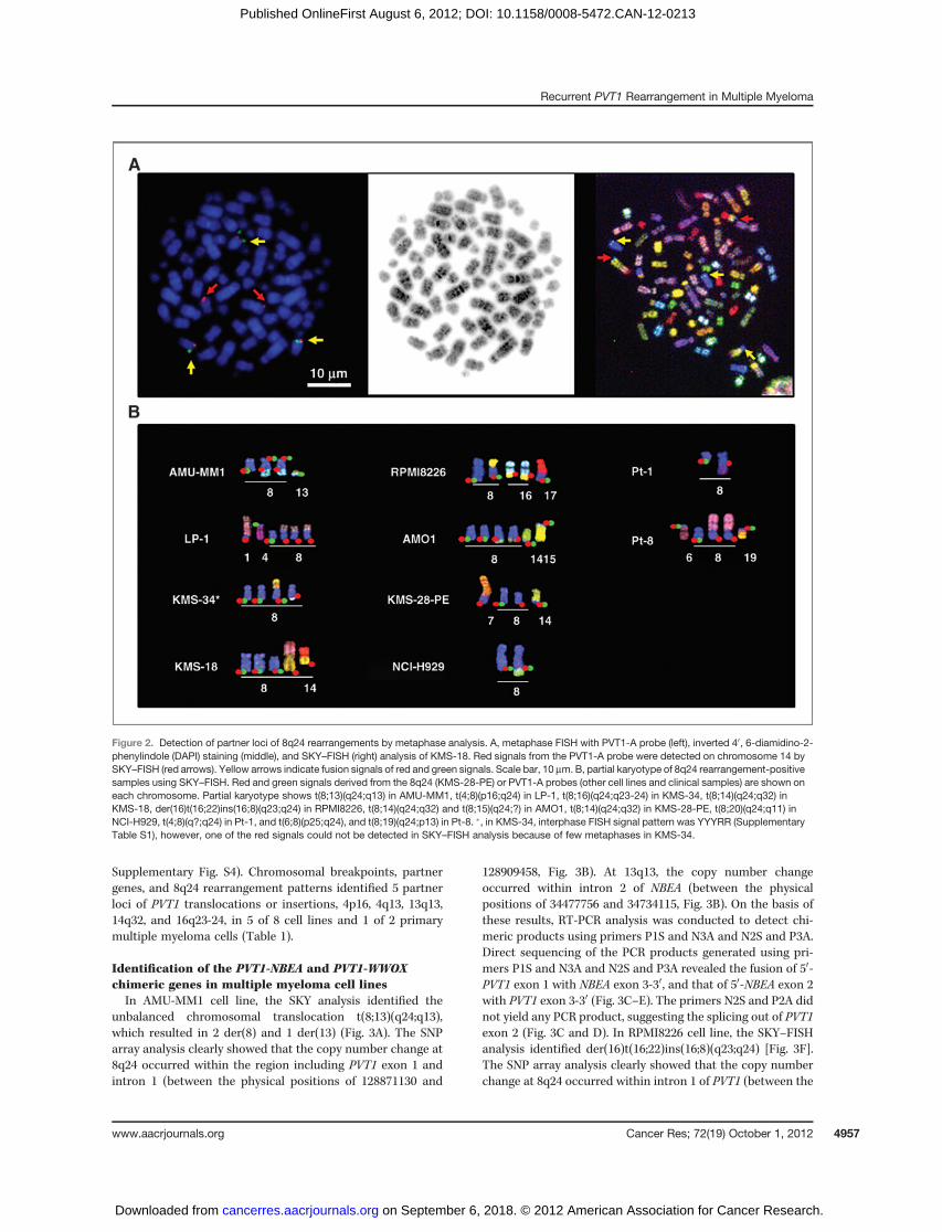
Supplementary Fig. S4). Chromosomal breakpoints, partnergenes, and 8q24 rearrangement patterns identified 5 partnerloci of PVT1 translocations or insertions, 4p16, 4q13, 13q13,14q32, and 16q23-24, in 5 of 8 cell lines and 1 of 2 primarymultiple myeloma cells (Table 1).
Identification of the PVT1-NBEA and PVT1-WWOXchimeric genes in multiple myeloma cell linesIn AMU-MM1 cell line, the SKY analysis identified the
unbalanced chromosomal translocation t(8;13)(q24;q13),which resulted in 2 der(8) and 1 der(13) (Fig. 3A). The SNParray analysis clearly showed that the copy number change at8q24 occurred within the region including PVT1 exon 1 andintron 1 (between the physical positions of 128871130 and
128909458, Fig. 3B). At 13q13, the copy number changeoccurred within intron 2 of NBEA (between the physicalpositions of 34477756 and 34734115, Fig. 3B). On the basis ofthese results, RT-PCR analysis was conducted to detect chi-meric products using primers P1S and N3A and N2S and P3A.Direct sequencing of the PCR products generated using pri-mers P1S and N3A and N2S and P3A revealed the fusion of 50-PVT1 exon 1 with NBEA exon 3-30, and that of 50-NBEA exon 2with PVT1 exon 3-30 (Fig. 3C–E). The primers N2S and P2A didnot yield any PCR product, suggesting the splicing out of PVT1exon 2 (Fig. 3C and D). In RPMI8226 cell line, the SKY–FISHanalysis identified der(16)t(16;22)ins(16;8)(q23;q24) [Fig. 3F].The SNP array analysis clearly showed that the copy numberchange at 8q24 occurred within intron 1 of PVT1 (between the
Figure 2. Detection of partner loci of 8q24 rearrangements by metaphase analysis. A, metaphase FISH with PVT1-A probe (left), inverted 40, 6-diamidino-2-phenylindole (DAPI) staining (middle), and SKY–FISH (right) analysis of KMS-18. Red signals from the PVT1-A probe were detected on chromosome 14 bySKY–FISH (red arrows). Yellow arrows indicate fusion signals of red and green signals. Scale bar, 10 mm. B, partial karyotype of 8q24 rearrangement-positivesamples using SKY–FISH. Red and green signals derived from the 8q24 (KMS-28-PE) or PVT1-A probes (other cell lines and clinical samples) are shown oneach chromosome. Partial karyotype shows t(8;13)(q24;q13) in AMU-MM1, t(4;8)(p16;q24) in LP-1, t(8;16)(q24;q23-24) in KMS-34, t(8;14)(q24;q32) inKMS-18, der(16)t(16;22)ins(16;8)(q23;q24) in RPMI8226, t(8;14)(q24;q32) and t(8;15)(q24;?) in AMO1, t(8;14)(q24;q32) in KMS-28-PE, t(8;20)(q24;q11) inNCI-H929, t(4;8)(q?;q24) in Pt-1, and t(6;8)(p25;q24), and t(8;19)(q24;p13) in Pt-8. �, in KMS-34, interphase FISH signal pattern was YYYRR (SupplementaryTable S1), however, one of the red signals could not be detected in SKY–FISH analysis because of few metaphases in KMS-34.
Recurrent PVT1 Rearrangement in Multiple Myeloma
www.aacrjournals.org Cancer Res; 72(19) October 1, 2012 4957
on September 6, 2018. © 2012 American Association for Cancer Research. cancerres.aacrjournals.org Downloaded from
Published OnlineFirst August 6, 2012; DOI: 10.1158/0008-5472.CAN-12-0213
physical position of 128,891,891 and 128,902,659, Fig. 3G). At16q23, the copy number change occurred within intron 8 ofWWOX (between the physical position of 77,399,684 and77,401,485, Fig. 3G). On the basis of these results, RT-PCRanalysis was conducted to detect chimeric products usingprimers P1S and W9A (Fig. 3H), and direct sequencing of thisproduct revealed the fusion of 50-PVT1 exon 1withWWOX exon9-30 (Fig. 3I and J). However, these chimeric transcripts werenot detected in 7 patients with PVT1 rearrangements.
The expression level of PVT1-NBEA and PVT1-WWOX tran-scripts was measured by real-time quantitative RT-PCR (RQ-PCR; Fig. 4A and B). High expression of aberrant NBEA wasdetected in AMU-MM1 cell line using the NBEA 58-59 primerprobe set. In contrast, no expression of normal NBEA wasdetected using the NBEA 2-3 primer probe set in AMU-MM1,whereas normal NBEA was detected in all other cell lines inwhich it was expressed at similar levels. These results indicatethat the 50-PVT1-NBEA-30 chimeric transcript is highlyexpressed in AMU-MM1 cells. Similarly, expression of WWOXexon 9 was higher thanWWOX exon 8-9 in RPMI8226, suggest-ing that a part of expression of exon 9 was due to PVT1-WWOXchimeric transcript.
DiscussionThe present study uncovered 2 aspects ofmolecular genetics
in multiple myeloma. The first finding is the frequent rear-rangement of PVT1 gene in multiple myeloma harboring 8q24rearrangements. PVT1 is the human homolog of the mousePvt1 oncogene, which was originally identified as a common
retroviral integration site in murine leukemia virus (MLV)–induced T lymphomas. PVT1/Pvt1 is also involved in variantst(2;8), t(8;22), or t(8;14) in human Burkitt lymphoma and invariant t(6;15) in mouse plasmacytomas (10, 15, 22–25). More-over, amplification of the PVT1 gene has been observed inseveral cancers including lymphomas (26–30), and PVT1 over-expression was found to contribute to the suppression ofapoptosis (31). The PVT1 locus is thought to encode severalmiRNAs important in oncogenesis (32). Hence, the currentstudy suggests that PVT1 is one of the target genes of rear-rangement that may be responsible for driving multiple mye-loma, whereas other studies have shown breakpoints withinthe region centromeric to PVT1 (9–11, 33).
The second finding is the novel chimeric genes, PVT1-NBEAand PVT1-WWOX, in themultiplemyeloma cell lines, the AMU-MM1 cell line with t(8;13)(q24;q13), and RPMI8226 cell linewith der(16)t(16;22)ins(16;8)(q23;q24), resulting in high expres-sion of the abnormal chimeric transcript. In PVT1-NBEA, thebreakpoint of PVT1was found within intron 1 and PVT1 exon 1was found to be fused to NBEA exon 3, resulting in the loss ofthe start codon inNBEA exon 2. In this setting, the second ATGin exon 3 of the NBEA gene may function as a start codon toproduce a putative abnormal NBEA protein lacking 107 N-terminal amino acids (Fig. 3E), although the expression ofPVT1-NBEA at the protein level remains to be investigated.Although, knockdown of PVT1-NBEA using siRNA could notinhibit proliferation and induces cell death (SupplementaryFig. S5), it would be important to investigate the function ofthis NBEA chimera in multiple myeloma tumorigenesis, asNBEA modulates signal transduction and vesicular trafficking
Table 1. Partners of 8q24 rearrangements and candidate genes
8q24 Rearrangements Translocation partners
Patients andcell lines
Involvedgenes Type of alterations Partner loci
Previouslyreported genes Candidate genes
AMU-MM1 PVT1 Translocation 13q13 RB NBEALP-1 PVT1 Translocation 4p16 FGFR3, MMSET MMSETKMS-34 PVT1 Translocation 16q23-24 WWOX, MAF —
a
Pt-1 PVT1 Translocation 4q13 —b EPHA5
RPMI8226 PVT1c Insertion and gain 16q23 WWOX, MAF WWOXKMS-18 PVT1c Translocation and gain 14q32 IGH ATG2BAMO1 MYC Translocation 14q32, No.15 IGH, —b TRAF3/TDRD9,
TUBGCP5Pt-8 MYC Translocation 6p25, 19p13 IRF4, —b RNF8/FTSJD2,
EFNA2/MUM1/GNG7KMS-28-PE Unknown Translocation and gain 14q32 IGH —
a
NCI-H929 Unknown Translocation 20q11 MAFB NA
Abbreviation: NA, not available.aNo copy number change was found at chromosome 16 in KMS-34. The rearranged gene could not be identified, because there weretoo many genes within the region in which copy number change occurred in KMS-28-PE (Supplementary Fig. S4).bNo gene has been reported as affected in multiple myeloma.cDuplication of both theMYC locus and centromeric part of PVT1was detected in RPMI8226 and KMS-18 with PVT1 rearrangement.
Nagoshi et al.
Cancer Res; 72(19) October 1, 2012 Cancer Research4958
on September 6, 2018. © 2012 American Association for Cancer Research. cancerres.aacrjournals.org Downloaded from
Published OnlineFirst August 6, 2012; DOI: 10.1158/0008-5472.CAN-12-0213

in neurons and other cells, and as gene abnormality andaberrant expression ofNBEA have been associatedwith plasmacell dyscrasias (34–36). In addition, the association betweenthe PVT1-NBEA fusion gene and the t(8;13) chromosomalabnormality, which has been reported in a small populationof multiple myeloma, remains to be verified (7, 11, 37, 38).Chromosome 13 is often deleted in multiple myeloma and thishas been linked to poorer prognosis. In such cases with loss ofchromosome 13, RB1 is thought to be amajor target and driver.However, a group has reported NBEA to also be a target ofrecurrent interstitial deletions at 13q13 and proposed thatNBEAmight be a tumor suppressor gene in multiple myeloma(36). WWOX is generally considered to be a candidate tumor
suppressor gene, and known to have a proapoptotic effect byparticipating in the TNF apoptotic pathway and via directphysical interaction with p53 and its homolog p73 (39). How-ever, immunohistochemistry revealed that WWOX proteinlevels were rather elevated in gastric and breast carcinoma(40). Therefore,WWOX did not seem to act as tumor suppres-sor gene simply. Interestingly, although bothNBEA andWWOXare located at common fragile site, usually contributing to geneinactivation, FRA13A and FRA16D, respectively, these geneshighly express via fusion toPVT1 (41, 42). It would be importantto further elucidate the function of NBEA and WWOX. Severaltranslocation/insertion partners of 8q24 rearrangements wereidentified in the remaining samples (Table 1). The genes
NBEA exon 3PVT1 exon 13’5’
der (8)
NBEA exon 2
3’5’
PVT1 exon 3
der (13)
PVT1 exon 1 2
8q24
NBEA exon 2
13q13
N3AN2S
3 3
P3AP2AP1SPrimers
MYC
128920K128830K 34500K34420K
AM
U-M
M1
Nor
mal
con
trol
Mar
ker
Wat
er
P1S-N3A (PVT1-NBEA)
N2S-P2A (NBEA-PVT1)
N2S-P3A (NBEA-PVT1)
β-actin
β-actin
300200
200100500300300200
bp
exon1 exon2ATG exon3ATGNBEA
exon3ATGPVT1 exon1PVT1-NBEA
*
*
PVT1 exon 1 2MYC
128920K128830K
WWOX exon 8 9
77600K77200K
P1SPrimers W9A
WWOX exon 9PVT1 exon 1 3’5’
10 μm
P1S-W9A (PVT1-WWOX )
RPM
I822
6
Nor
mal
con
trol
Mar
ker
Wat
er
500 600
300 200
bp
exon 8 exon 9WWOX
exon 9PVT1 exon1PVT1-WWOX
A
B
DC
FE
G
H I
J
Figure 3. Identification of PVT1-NBEA and PVT1-WWOX chimeric genes in multiple myeloma cell lines. A, SKY analysis of AMU-MM1 reveals a complexkaryotype including t(8;13)(q24;q13) as 46,X,-X,þder(1;19)(q10;p10),der(2)t(2;17)(q37;q11.2),der(7)(qter! q11.2::p15! q11.2::7?), t(8;13)(q24;q13),þder(8)t(8;13),del(12)(p11.2),-13,der(19)t(1;19)(q12;p13)�2,del(20)(p13). Arrow, breakpoint of der(8)t(8;13)(q24;q13); arrowhead, breakpoint of der(13)t(8;13)(q24;q13). B, copy number changes at 8q24 (PVT1) and 13q13 (NBEA) detected by SNP array. Primers used to detect chimeric transcript. The y-axesindicate the linear scale corresponding to genome copy number of each chromosome. However, copy number data have never been corrected for theinfluence of tumor cell percentage or real copy number of some chromosomes. C, detection of PVT1-NBEA and NBEA-PVT1 chimeric transcripts byRT-PCR. D, sequencing of chimeric junction of PVT1-NBEA and NBEA-PVT1 chimeric transcripts. E, putative structure of abnormal NBEA fusiontranscript. PVT1-NBEA lacks a start codon in NBEA exon 2; the second ATG in exon 3 might function as a start codon, resulting in an abnormal NBEAprotein lacking itsN-terminus. �, ATG indicates start codon. F, SKY–FISHanalysis ofRPMI8226 reveals a complex karyotype including der(16)t(16;22)ins(16;8)(q23;q24). Arrows indicate red signals of PVT1-A inserting to t(16;22). It is difficult to detect the 8q24 locus on der(16) using conventional cytogenetictechnique. Scale bar, 10 mm. G, copy number changes at 8q24 (PVT1) and 16q23 (WWOX) detected by SNP array. Primers used to detectchimeric transcript. The y-axes indicate genomecopynumber.H, detection ofPVT1-WWOX chimeric transcript byRT-PCR. I, sequencingof chimeric junctionof PVT1-WWOX chimeric transcript. J, putative structure of abnormal WWOX fusion transcript.
Recurrent PVT1 Rearrangement in Multiple Myeloma
www.aacrjournals.org Cancer Res; 72(19) October 1, 2012 4959
on September 6, 2018. © 2012 American Association for Cancer Research. cancerres.aacrjournals.org Downloaded from
Published OnlineFirst August 6, 2012; DOI: 10.1158/0008-5472.CAN-12-0213

located at translocation/insertion breakpoints, identified bycopy number analysis, have been frequently associated withcancer (43–49). No chimeric genes could be cloned in othersamples. In Pt-1, LP-1, and KMS-18, PVT1 was translocated tothe regions of EPHA5, MMSET, or ATG2B with opposite direc-tion of transcription, suggesting that these genes are notinvolved in the fusion with PVT1. In KMS-34, AMO1, Pt-8,KMS-28PE, andNCI-H929, genome copy number changes werenot identified within a single gene at 8q24 and/or partner loci(Supplementary Fig. S4). The translocation/insertion partner
breakpoints identified in the present study warrant furthermolecular analysis of the candidate genes.
The relationship between MYC and PVT1 in terms of mul-tiple myeloma development and progression is difficult toelucidate. RQ-PCR analysis revealed high expression of PVT1and MYC in most multiple myeloma cell lines regardless ofPVT1 or MYC rearrangement status (Supplementary Fig. S6).Results showed that 8q24 rearrangements included variouscomplex translocations with either deletion or insertion of apart of the 8q24 segment including MYC and the centromericsegment of PVT1. In addition, gains of chromosome 8, includ-ing PVT1 and MYC, are frequently identified in multiple mye-loma cell lines (Fig 1B, Supplementary Fig. S1B). These gainsare likely to contribute to the amplification of the PVT1 andMYC genes (28). Beyond chromosomal abnormalities, themolecular mechanisms underlying MYC overexpression inmultiple myeloma, such as interaction with PVT1 or deregu-lation of miRNAs, warrant further research (32, 50).
In conclusion, PVT1 is frequently involved with variouspartner loci in multiple myeloma with 8q24 abnormalities,and, PVT1-NBEA and PVT1-WWOX were identified as novel,highly expressed chimeric genes inwhichNBEA andWWOX arefused with PVT1 in multiple myeloma cell lines harboringt(8;13)(q24;q13) and der(16)t(16;22)ins(16;8)(q23;q24), respec-tively. These findings suggest that PVT1 rearrangements mayrepresent a novel molecular paradigm underlying the pathol-ogy of multiple myeloma with 8q24 rearrangements.
Disclosure of Potential Conflicts of InterestNo potential conflicts of interest were disclosed.
Authors' ContributionsConception and design: I. Hanamura, K. Nishida, J. Kuroda, M. TaniwakiDevelopment of methodology: H. Nagoshi, T. Otsuki, K. Nishida, S. HoriikeAcquisition of data (provided animals, acquired and managed patients,provided facilities, etc.): K. Nishida, S. Horiike, J. KurodaAnalysis and interpretation of data (e.g., statistical analysis, biostatistics,computational analysis): H. Nagoshi, T. Taki, I. Hanamura, K. Nishida, K.Okuda, N. Sakamoto, S. Kobayashi, M. Yamamoto-Sugitani, Y. Tsutsumi,T. Kobayashi, Y. Matsumoto, S. Horiike, J. Kuroda, M. TaniwakiWriting, review, and/or revision of the manuscript: H. Nagoshi, T. Taki, I.Hanamura, K. Nishida, S. Horiike, J. Kuroda, M. TaniwakiAdministrative, technical, or material support (i.e., reporting or orga-nizing data, constructing databases): H. Nagoshi, T. Taki, I. Hanamura, K.Nishida, S. HoriikeStudy supervision: T. Taki, M. Nitta, K. Nishida, S. Horiike, J. Kuroda, M.Taniwaki
AcknowledgmentsThe authors thank Kayoko Kurita and Akari Kazami for their expert technical
assistance.
Grant SupportThis work was supported by a Grant-in-Aid for Cancer Research from the
Ministry of Health, Labor and Welfare of Japan, by a Grant-in-aid for ScientificResearch (B) and (C) from theMinistry of Education, Culture, Sports, Science andTechnology of Japan, and by the National Cancer Center Research and Devel-opment Fund.
The costs of publication of this article were defrayed in part by the payment ofpage charges. This article must therefore be hereby marked advertisement inaccordance with 18 U.S.C. Section 1734 solely to indicate this fact.
Received January 24, 2012; revised May 29, 2012; accepted June 18, 2012;published OnlineFirst August 6, 2012.
A
B
0
0.5
1
1.5
2
2.5
3
3.5
4
AM
U-M
M1
LP
-1
KM
S-3
4
RP
MI8
22
6
KM
S-1
8
AM
O1
KM
S-2
8-P
E
NC
I-H929
IM9
KM
S-1
2-B
M
KM
S-2
0
PB
L
EB
V-B
K5
62
Dau
di
WWOX exon 8 - 9 WWOX exon 9
WW
OX
/ β-
Act
in (
fold
ch
an
ge
)
MM cell lines
0
0.5
1
1.5
2
2.5
3
3.5
4
4.5
5
AM
U-M
M1
LP
-1
KM
S-3
4
RP
MI8
22
6
KM
S-1
8
AM
O1
KM
S-2
8-P
E
NC
I-H9
29
IM9
KM
S-1
2-B
M
KM
S-2
0
PB
L
EB
V-B
K5
62
Da
ud
i
NBEA exon 2 - 3
NBEA exon 58 - 59
MM cell lines
NB
EA
/ β-
Act
in (
fold
ch
an
ge)
Figure4. ExpressionofNBEA andWWOX in cell lines. A,RQ-PCRanalysisshowing that the abnormal NBEA transcript is highly expressed in theAMU-MM1 cell line as compared with other multiple myeloma cell lines,normal lymphocytes, an Epstein–Barr virus–transformed B-cell line, aleukemia cell line (K562), and a Burkitt lymphoma cell line (Daudi). Dottedand black bars indicate expression levels using the NBEA 2-3 and NBEA58-59 primer sets, respectively. B, RQ-PCR analysis showing that theabnormal WWOX transcript is relatively highly expressed in theRPMI8226 cell line as compared with other cell lines. Dotted and blackbars indicate expression levels using theWWOX 8-9 andWWOX9 primersets, respectively.
Nagoshi et al.
Cancer Res; 72(19) October 1, 2012 Cancer Research4960
on September 6, 2018. © 2012 American Association for Cancer Research. cancerres.aacrjournals.org Downloaded from
Published OnlineFirst August 6, 2012; DOI: 10.1158/0008-5472.CAN-12-0213

References1. Fonseca R, Bergsagel PL, Drach J, Shaughnessy J, Gutierrez N,
Stewart AK, et al. International MyelomaWorking Group. InternationalMyelomaWorkingGroupmolecular classification ofmultiplemyeloma:spotlight review. Leukemia 2009;23:2210–21.
2. ChenKC,BevanPC,MatthewsJG.Analysis ofGbandedkaryotypes inmyeloma cells. J Clin Pathol 1986;39:260–6.
3. Hanamura I, Stewart JP, Huang Y, Zhan F, Santra M, Sawyer JR, et al.Frequent gain of chromosome band 1q21 in plasma-cell dyscrasiasdetected by fluorescence in situ hybridization: incidence increasesfrom MGUS to relapsed myeloma and is related to prognosis anddisease progression following tandem stem-cell transplantation.Blood 2006;108:1724–32.
4. Gould J, Alexanian R, Goodacre A, Pathak S, Hecht B, Barlogie B.Plasma cell karyotype in multiple myeloma. Blood 1988;71:453–6.
5. Sawyer JR,Waldron JA, JagannathS, BarlogieB.Cytogenetic findingsin 200 patients with multiple myeloma. Cancer Genet Cytogenet1995;82:41–9.
6. Nishida K, Tamura A, Nakazawa N, Ueda Y, Abe T, Matsuda F, et al.The Ig heavy chain gene is frequently involved in chromosomaltranslocations in multiple myeloma and plasma cell leukemia asdetected by in situ hybridization. Blood 1997;90:526–34.
7. Avet-LoiseauH, Gerson F,Magrangeas F,Minvielle S, Harousseau JL,Bataille R, et al. Intergroupe Francophone du My�elome.Rearrange-ments of the c-myc oncogene are present in 15% of primary humanmultiple myeloma tumors. Blood 2001;98:3082–6.
8. Sawyer JR, Lukacs JL, Thomas EL, Swanson CM, Goosen LS, Sam-martino G, et al. Multicolour spectral karyotyping identifies new trans-locations and a recurring pathway for chromosome loss in multiplemyeloma. Br J Haematol 2001;112:167–74.
9. Fabris S, Storlazzi CT, Baldini L, Nobili L, Lombardi L, Maiolo AT, et al.Heterogeneous pattern of chromosomal breakpoints involving theMYC locus in multiple myeloma. Genes Chromosomes Cancer2003;37:261–9.
10. Shou Y, Martelli ML, Gabrea A, Qi Y, Brents LA, Roschke A, et al.Diverse karyotypic abnormalities of the c-myc locus associatedwith c-myc dysregulation and tumor progression in multiple myeloma. ProcNatl Acad Sci U S A 2000;97:228–33.
11. Dib A, Gabrea A, Glebov OK, Bergsagel PL, Kuehl WM. Characteri-zation of MYC translocations in multiple myeloma cell lines. J NatlCancer Inst Monogr 2008;39:25–31.
12. Avet-Loiseau H, Daviet A, Brigaudeau C, Callet-Bauchu E, Terr�eC, Lafage-Pochitaloff M, et al. Cytogenetic, interphase, and mul-ticolor fluorescence in situ hybridization analyses in primaryplasma cell leukemia: a study of 40 patients at diagnosis, onbehalf of the Intergroupe Francophone du My�elome and theGroupe Francais de Cytog�en�etique H�ematologique. Blood 2001;97:822–5.
13. Mohamed AN, Bentley G, Bonnett ML, Zonder J, Al-Katib A. Chro-mosome aberrations in a series of 120 multiple myeloma cases withabnormal karyotypes. Am J Hematol 2007;82:1080–7.
14. Shiramizu B, Magrath I. Localization of breakpoints by polymerasechain reactions in Burkitt's lymphoma with 8;14 translocations. Blood1990;75:1848–52.
15. Shtivelman E, Bishop JM. Effects of translocations on transcriptionfrom PVT. Mol Cell Biol 1990;10:1835–9.
16. Hanamura I, Goto M, Nagoshi H, Taki T, Imai N, Suganuma K, et al.Establishment and characterization of a novel human myeloma cellline, AMU-MM1, from a multiple myeloma patient involving centralnerve system after treatment with bortezomib. Blood 2010;116:(abstract [4991]).
17. Taniwaki M, Sliverman GA, Nishida K, Horiike S, Misawa S, ShimazakiC, et al. Translocations and amplification of the BCL2 gene aredetected in interphase nuclei of non-Hodgkin's lymphoma by in situhybridization with yeast artificial chromosome clones. Blood 1995;86:1481–6.
18. KakazuN, TaniwakiM,Horiike S,NishidaK, TatekawaT,NagaiM, et al.Combined spectral karyotyping and DAPI banding analysis of chro-mosome abnormalities inmyelodysplastic syndrome. GenesChromo-somes Cancer 1999;26:336–45.
19. Shaffer LG, Slovak ML, Campbell LJ, editors. ISCN: an internationalsystem of human cytogenetic nomenclature. Basel, Switzerland: Kar-ger; 2009.
20. Nannya Y, Sanada M, Nakazaki K, Hosoya N, Wang L, Hangaishi A,et al. A robust algorithm for copy number detection using high-densityoligonucleotide single nucleotide polymorphism genotyping arrays.Cancer Res 2005;65:6071–9.
21. Chinen Y, Taki T, Nishida K, Shimizu D, Okuda T, Yoshida N, et al.Identification of the novel AML1 fusion partner gene, LAF4, a fusionpartner ofMLL, in childhood T-cell acute lymphoblastic leukemiawith t(2;21)(q11;q22) by bubble PCRmethod for cDNA. Oncogene 2008;27:2249–56.
22. Henglein B, Synovzik H, Groitl P, Bornkamm GW, Hartl P, Lipp M.Three breakpoints of variant t(2;8) translocations inBurkitt's lymphomacells fall within a region 140 kilobases distal from c-myc. Mol Cell Biol1989;9:2105–13.
23. Rack KA, Delabesse E, Radford-Weiss I, Bourquelot P, Le Guyader G,Vekemans M, et al. Simultaneous detection of MYC, BVR1, and PVT1translocations in lymphoidmalignanciesbyfluorescence in situhybrid-ization. Genes Chromosomes Cancer 1998;23:220–6.
24. Villeneuve L, Rassart E, Jolicoeur P, Graham M, Adams JM. Proviralintegration site Mis-1 in rat thymomas corresponds to the pvt-1translocation breakpoint in murine plasmacytomas. Mol Cell Biol1986;6:1834–7.
25. Huppi K, Siwarski D. Chimeric transcripts with an open reading frameare generated as a result of translocation to the Pvt-1 region in mouseB-cell tumors. Int J Cancer 1994;59:848–51.
26. Asker C, Mareni C, Coviello D, Ingvarsson S, SessaregoM, Origone P,et al. Amplification of c-myc and pvt-1 homologous sequences inacute nonlymphatic leukemia. Leuk Res 1988;12:523–7.
27. ShtivelmanE, Bishop JM. The PVT gene frequently amplifieswithMYCin tumor cells. Mol Cell Biol 1989;9:1148–54.
28. Bakkus MH, Brakel-van Peer KM, Michiels JJ, van't Veer MB, BennerR. Amplification of the c-myc and the pvt-like region in humanmultiplemyeloma. Oncogene 1990;5:1359–64.
29. Costinean S, Zanesi N, Pekarsky Y, Tili E, Volinia S, Heerema N, et al.Pre-B cell proliferation and lymphoblastic leukemia/high-grade lym-phoma in E(mu)-miR155 transgenic mice. Proc Natl Acad Sci U S A2006;103:7024–9.
30. Carramusa L, Contino F, Ferro A, Minafra L, Perconti G, Giallongo A,et al. ThePVT-1oncogene is aMycprotein target that is overexpressedin transformed cells. J Cell Physiol 2007;213:511–8.
31. Guan Y, Kuo WL, Stilwell JL, Takano H, Lapuk AV, Fridlyand J, et al.Amplification of PVT1 contributes to the pathophysiology of ovarianand breast cancer. Clin Cancer Res 2007;13:5745–55.
32. Huppi K, Volfovsky N, Runfola T, Jones TL, Mackiewicz M, MartinSE, et al. The identification of microRNAs in a genomically unstableregion of human chromosome 8q24. Mol Cancer Res 2008;6:212–21.
33. Palumbo AP, Boccadoro M, Battaglio S, Corradini P, Tsichlis PN,Huebner K, et al. Human homologue of Moloney leukemia virusintegration-4 locus (MLVI-4), located 20 kilobases 30 of the mycgene, is rearranged in multiple myelomas. Cancer Res 1990;50:6478–82.
34. Wang X, Herberg FW, Laue MM, Wullner C, Hu B, Petrasch-Parwez E,et al. Neurobeachin: a protein kinase A-anchoring, beige/Chediak-higashi protein homolog implicated in neuronal membrane traffic. JNeurosci 2000;20:8551–65.
35. Zhan F, Barlogie B, Arzoumanian V, Huang Y, Williams DR, Hollmig K,et al. Gene-expression signature of benign monoclonal gammopathyevident in multiple myeloma is linked to good prognosis. Blood2007;109:1692–700.
36. O'Neal J, Gao F, Hassan A, Monahan R, Barrios S, Kilimann MW, et al.Neurobeachin (NBEA) is a target of recurrent interstitial deletions at13q13 in patients with MGUS and multiple myeloma. Exp Hematol2009;37:234–44.
37. S�aez B, Martín-Subero JI, Largo C, Martín MC, Odero MD, Prosper F,et al. Identification of recurrent chromosomal breakpoints in multiplemyeloma with complex karyotypes by combined G-banding, spectral
Recurrent PVT1 Rearrangement in Multiple Myeloma
www.aacrjournals.org Cancer Res; 72(19) October 1, 2012 4961
on September 6, 2018. © 2012 American Association for Cancer Research. cancerres.aacrjournals.org Downloaded from
Published OnlineFirst August 6, 2012; DOI: 10.1158/0008-5472.CAN-12-0213

karyotyping, and fluorescence in situ hybridization analyses. CancerGenet Cytogenet 2006;169:143–9.
38. Chiecchio L, Dagrada GP, Protheroe RK, Stockley DM, Smith AG,Orchard KH, et al. UKMyeloma Forum. Loss of 1p and rearrangementof MYC are associated with progression of smouldering myeloma tomyeloma: sequential analysis of a single case. Haematologica2009;94:1024–8.
39. Aqeilan RI, Pekarsky Y, Herrero JJ, Palamarchuk A, Letofsky J, DruckT, et al. Functional association between Wwox tumor suppressorprotein and p73, a p53 homolog. Proc Natl Acad Sci U S A2004;101:4401–6.
40. Watanabe A, Hippo Y, Taniguchi H, Iwanari H, Yashiro M, Hirakawa K,et al. An opposing view on WWOX protein function as a tumorsuppressor. Cancer Res 2003;63:8629–33.
41. Savelyeva L, Sagulenko E, Schmitt JG, Schwab M. The neurobeachingene spans the common fragile site FRA13A. Hum Genet2006;118:551–8.
42. Bednarek AK, Laflin KJ, Daniel RL, Liao Q, Hawkins KA, Aldaz CM.WWOX, a novel WW domain-containing protein mapping to humanchromosome 16q23.3-24.1, a region frequently affected in breastcancer. Cancer Res 2000;60:2140–5.
43. FuDY,WangZM,WangBL, Chen L, YangWT, Shen ZZ, et al. Frequentepigenetic inactivation of the receptor tyrosine kinase EphA5 bypromoter methylation in human breast cancer. Hum Pathol2010;41:48–58.
44. Annunziata CM, Davis RE, Demchenko Y, Bellamy W, Gabrea A, ZhanF, et al. Frequent engagement of the classical and alternative NF-
kappaB pathways by diverse genetic abnormalities in multiple mye-loma. Cancer Cell 2007;12:115–30.
45. Keats JJ, Fonseca R, Chesi M, Schop R, Baker A, Chng WJ, et al.Promiscuous mutations activate the noncanonical NF-kappaB path-way in multiple myeloma. Cancer Cell 2007;12:131–44.
46. Braggio E, Keats JJ, Leleu X, VanWier S, Jimenez-Zepeda VH, ValdezR, et al. Identification of copy number abnormalities and inactivatingmutations in twonegative regulators of nuclear factorkappaB signalingpathways in Waldenstroms macroglobulinemia. Cancer Res 2009;69:3579–88.
47. Giefing M, Arnemann J, Martin-Subero JI, Niel€ander I, Bug S, Hart-mann S, et al. Identification of candidate tumour suppressor gene locifor Hodgkin and Reed–Sternberg cells by characterisation of homo-zygous deletions in classical Hodgkin lymphoma cell lines. Br JHaematol 2008;142:916–24.
48. Ohta M, Mimori K, Fukuyoshi Y, Kita Y, Motoyama K, Yamashita K,et al. Clinical significance of the reduced expression of G proteingamma 7 (GNG7) in oesophageal cancer. Br J Cancer 2008;98:410–7.
49. Shibata K, Mori M, Tanaka S, Kitano S, Akiyoshi T. Identification andcloning of human G-protein gamma 7, down-regulated in pancreaticcancer. Biochem Biophys Res Commun 1998;246:205–9.
50. Sasaki N, Kuroda J, Nagoshi H, Yamamoto M, Kobayashi S, TsutsumiY, et al. Bcl-2 is a better therapeutic target than c-Myc, but attackingboth could be amore effective treatment strategy for B-cell lymphomawith concurrent Bcl-2 and c-Myc overexpression. Exp Hematol2011;39:817–28.
Nagoshi et al.
Cancer Res; 72(19) October 1, 2012 Cancer Research4962
on September 6, 2018. © 2012 American Association for Cancer Research. cancerres.aacrjournals.org Downloaded from
Published OnlineFirst August 6, 2012; DOI: 10.1158/0008-5472.CAN-12-0213

2012;72:4954-4962. Published OnlineFirst August 6, 2012.Cancer Res Hisao Nagoshi, Tomohiko Taki, Ichiro Hanamura, et al. Abnormality
Occur in Multiple Myeloma with 8q24PVT1-WWOX and PVT1-NBEA Rearrangement and Novel Chimeric Genes PVT1Frequent
Updated version
10.1158/0008-5472.CAN-12-0213doi:
Access the most recent version of this article at:
Material
Supplementary
http://cancerres.aacrjournals.org/content/suppl/2012/08/06/0008-5472.CAN-12-0213.DC1
Access the most recent supplemental material at:
Cited articles
http://cancerres.aacrjournals.org/content/72/19/4954.full#ref-list-1
This article cites 48 articles, 25 of which you can access for free at:
Citing articles
http://cancerres.aacrjournals.org/content/72/19/4954.full#related-urls
This article has been cited by 4 HighWire-hosted articles. Access the articles at:
E-mail alerts related to this article or journal.Sign up to receive free email-alerts
Subscriptions
Reprints and
To order reprints of this article or to subscribe to the journal, contact the AACR Publications Department at
Permissions
Rightslink site. Click on "Request Permissions" which will take you to the Copyright Clearance Center's (CCC)
.http://cancerres.aacrjournals.org/content/72/19/4954To request permission to re-use all or part of this article, use this link
on September 6, 2018. © 2012 American Association for Cancer Research. cancerres.aacrjournals.org Downloaded from
Published OnlineFirst August 6, 2012; DOI: 10.1158/0008-5472.CAN-12-0213


![سامى عبد الشكور... · 134 [1973BCSJ3625] Synthesis and rearrangement of oxanilic esters arylhydrazones. Shawali, A. Sami; Ahmad, M. Kamal. Fac. Sci., Univ. Cairo, Giza,](https://static.fdocument.pub/doc/165x107/5f5ff49368fbf70cf43cd86f/-f-134-1973bcsj3625-synthesis-and-rearrangement.jpg)


