Explain homeostasis (remember homeodynamics) oflpc1.clpccd.cc.ca.us/lpc/jgallagher/Physio/Chapter 20...
23
Ch 20: Integrative Physiology II Fluid & Electrolyte Balance Explain homeostasis (remember homeodynamics) of 1. Water Balance (ECF/ICF volumes) 2. Electrolyte Balance (Na + and K +) 3. Acid-Base Balance (pH) Objectives Developed by John Gallagher, MS, DVM
Transcript of Explain homeostasis (remember homeodynamics) oflpc1.clpccd.cc.ca.us/lpc/jgallagher/Physio/Chapter 20...

Ch 20: Integrative Physiology II
Fluid & Electrolyte Balance
Explain homeostasis (remember
homeodynamics) of
1. Water Balance (ECF/ICF volumes)
2. Electrolyte Balance (Na+ and K+)
3. Acid-Base Balance (pH)
Objectives
Developed by
John Gallagher, MS, DVM

Fig 20-18
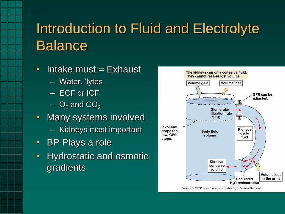
Introduction to Fluid and Electrolyte
Balance
• Intake must = Exhaust
– Water, ‘lytes
– ECF or ICF
– O2 and CO2
• Many systems involved
– Kidneys most important
• BP Plays a role
• Hydrostatic and osmotic
gradients

Kidneys maintain H2O balance by
regulating urine concentration
• Daily H2O intake balanced
by H2O excretion (ins
and outs)
• Kidneys react to changes
in osmolarity, volume,
and blood pressure
Fig 20-1
Fig 20-2

Urine Concentration
Established by LOH, CD
and vasa recta
reabsorption of varying
amounts of H2O and Na+
Key player: ADH (=
Vasopressin)
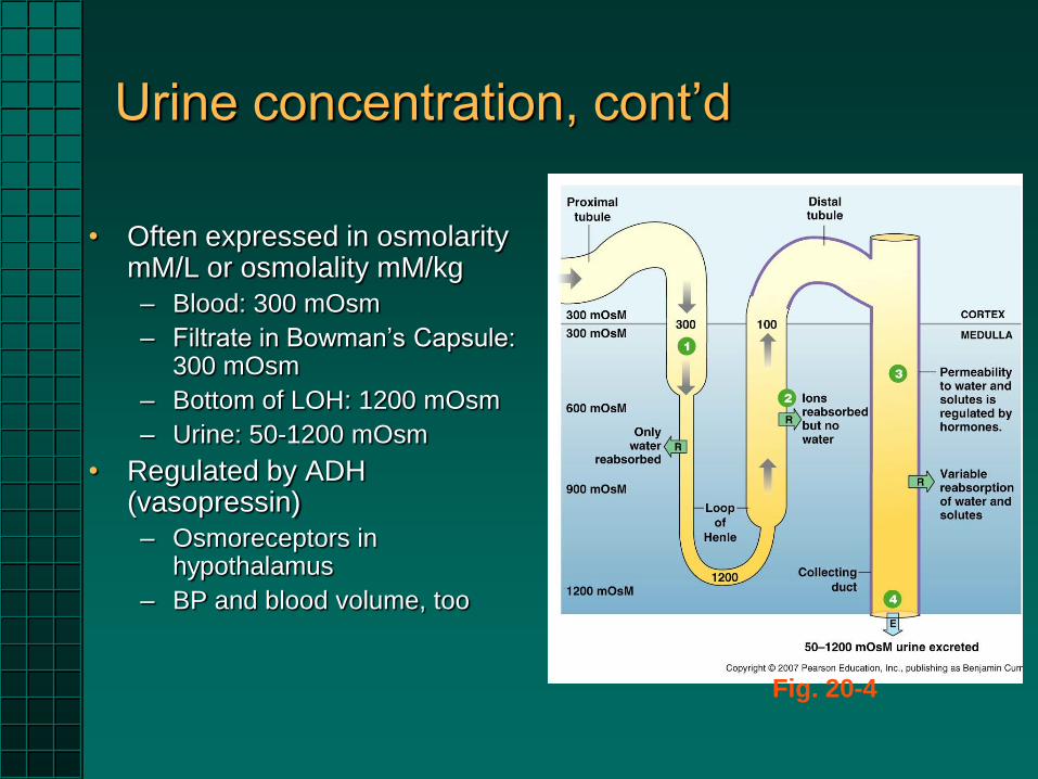
Urine concentration, cont’d
• Often expressed in osmolarity mM/L or osmolality mM/kg
– Blood: 300 mOsm
– Filtrate in Bowman’s Capsule: 300 mOsm
– Bottom of LOH: 1200 mOsm
– Urine: 50-1200 mOsm
• Regulated by ADH (vasopressin)
– Osmoreceptors in hypothalamus
– BP and blood volume, too
Fig. 20-4
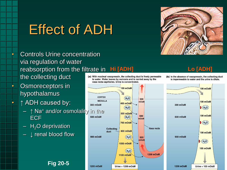
Effect of ADH
Hi [ADH] Lo [ADH]
Fig 20-5
• Controls Urine concentration
via regulation of water
reabsorption from the filtrate in
the collecting duct
• Osmoreceptors in
hypothalamus
• ↑ ADH caused by:
– ↑ Na+ and/or osmolality in the
ECF
– H2O deprivation
– ↓ renal blood flow
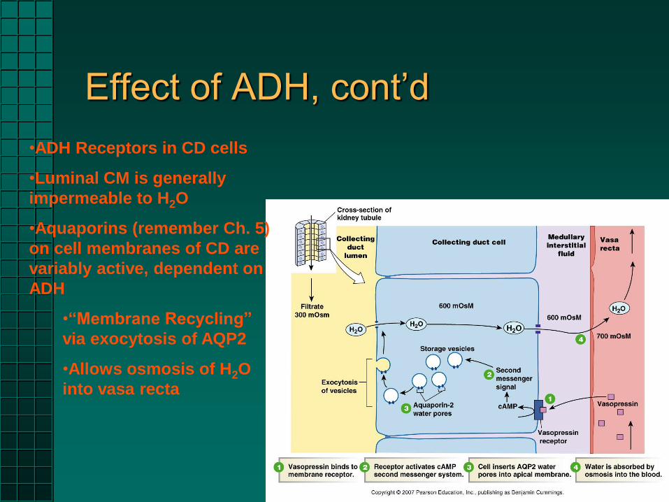
Effect of ADH, cont’d
•ADH Receptors in CD cells
•Luminal CM is generally
impermeable to H2O
•Aquaporins (remember Ch. 5)
on cell membranes of CD are
variably active, dependent on
ADH
•“Membrane Recycling”
via exocytosis of AQP2
•Allows osmosis of H2O
into vasa recta

Troubles with ADH?
•Diabetes insipidus
•Central
•Nephrogenic
•Nocturnal enuresis
ADH deficiency:
ADH Excess:
•AKA Inappropriate ADH secretion
•XS H2O retention
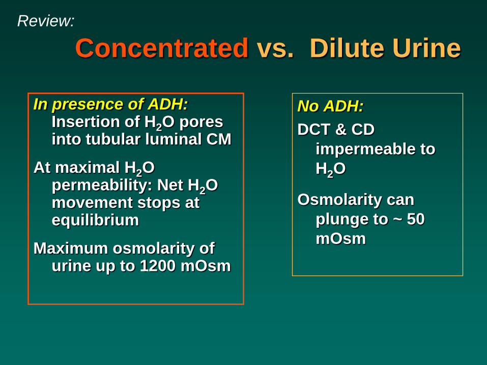
Concentrated vs. Dilute Urine
In presence of ADH: Insertion of H2O pores into tubular luminal CM
At maximal H2O permeability: Net H2O movement stops at equilibrium
Maximum osmolarity of urine up to 1200 mOsm
No ADH:
DCT & CD
impermeable to
H2O
Osmolarity can
plunge to ~ 50
mOsm
Review:
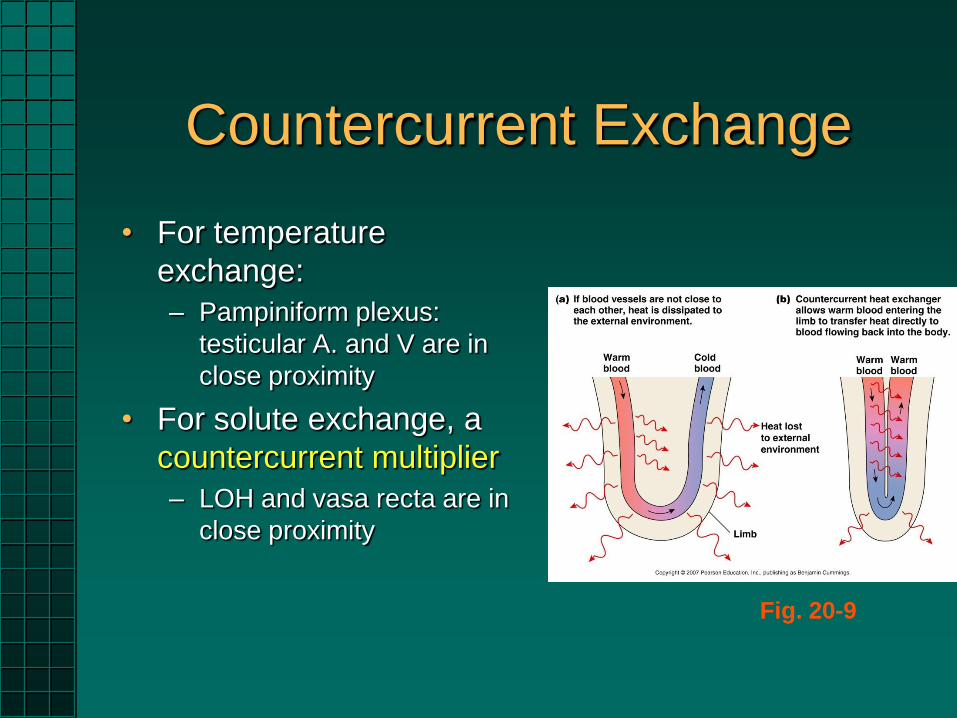
Countercurrent Exchange
• For temperature
exchange:
– Pampiniform plexus:
testicular A. and V are in
close proximity
• For solute exchange, a
countercurrent multiplier
– LOH and vasa recta are in
close proximity
Fig. 20-9
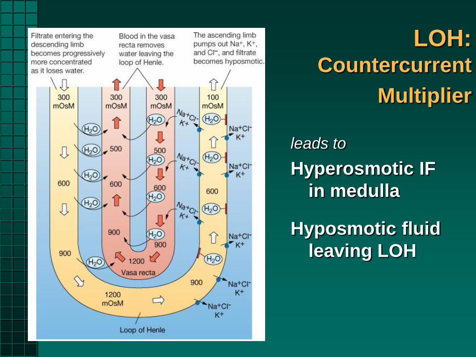
leads to
Hyperosmotic IF
in medulla
Hyposmotic fluid
leaving LOH
LOH: Countercurrent
Multiplier
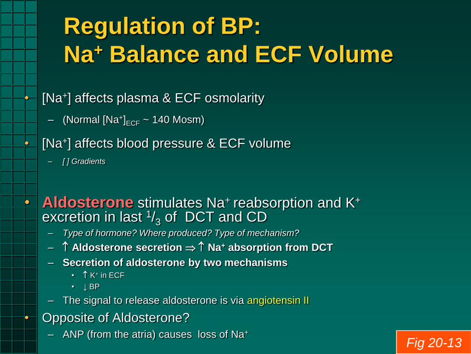
Regulation of BP:
Na+ Balance and ECF Volume
• [Na+] affects plasma & ECF osmolarity
– (Normal [Na+]ECF ~ 140 Mosm)
• [Na+] affects blood pressure & ECF volume
– [ ] Gradients
• Aldosterone stimulates Na+ reabsorption and K+ excretion in last 1/3 of DCT and CD
– Type of hormone? Where produced? Type of mechanism?
– Aldosterone secretion Na+ absorption from DCT
– Secretion of aldosterone by two mechanisms • K+ in ECF
• ↓ BP
– The signal to release aldosterone is via angiotensin II
• Opposite of Aldosterone?
– ANP (from the atria) causes loss of Na+
Fig 20-13
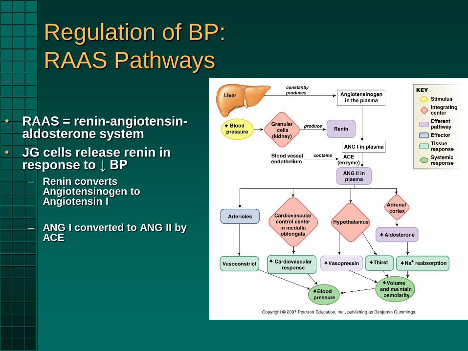
Regulation of BP:
RAAS Pathways
• RAAS = renin-angiotensin-aldosterone system
• JG cells release renin in response to ↓ BP
– Renin converts Angiotensinogen to Angiotensin I
– ANG I converted to ANG II by ACE

RAAS Pathways, cont’d
ANG II causes ↑ BP via ↑ ADH Secretion
Thirst
Vasoconstriction
Sympathetic stimulation of heart ↑ HR and CO
ACE inhibitors will ↓ BP

Potassium
• Recall that
– 2% of K+ is in ECF
– Major contributor to resting membrane potential
• Hypokalemia
– MP more negative (weakness)
• Hyperkalemia
– MP more positive (poor AP and cardiac
arrhythmias
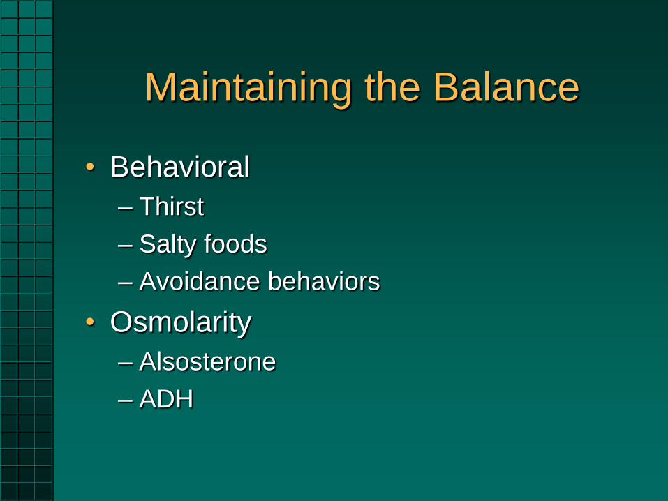
Maintaining the Balance
• Behavioral
– Thirst
– Salty foods
– Avoidance behaviors
• Osmolarity
– Alsosterone
– ADH
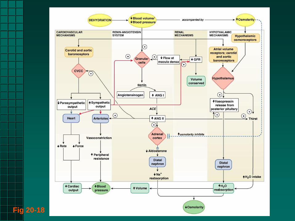
Fig 20-18
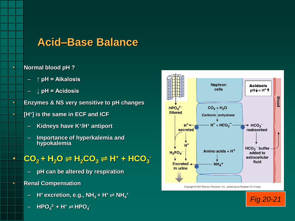
Acid–Base Balance
• Normal blood pH ?
– ↑ pH = Alkalosis
– ↓ pH = Acidosis
• Enzymes & NS very sensitive to pH changes
• [H+] is the same in ECF and ICF
– Kidneys have K+/H+ antiport
– Importance of hyperkalemia and hypokalemia
• CO2 + H2O ⇌ H2CO3 ⇌ H+ + HCO3-
– pH can be altered by respiration
• Renal Compensation
– H+ excretion, e.g., NH3 + H+ ⇌ NH4
+
– HPO42- + H+ ⇌ HPO4
-
Fig 20-21
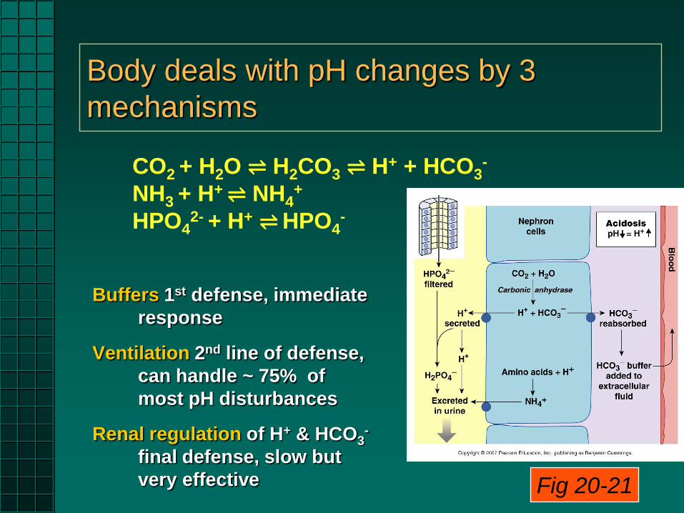
Body deals with pH changes by 3
mechanisms
Buffers 1st defense, immediate
response
Ventilation 2nd line of defense,
can handle ~ 75% of
most pH disturbances
Renal regulation of H+ & HCO3-
final defense, slow but
very effective Fig 20-21
CO2 + H2O ⇌ H2CO3 ⇌ H+ + HCO3-
NH3 + H+ ⇌ NH4
+
HPO42- + H+ ⇌ HPO4
-

Acidosis
Respiratory acidosis due to alveolar hypoventilation (accumulation of CO2)
Possible causes: Respiratory depression, increased airway resistance (?), impaired gas exchange (emphysema, fibrosis, muscular dystrophy, pneumonia)
Metabolic acidosis due to gain of fixed acid or loss of bicarbonate
Possible causes: lactic acidosis, ketoacidosis, diarrhea
Buffer capabilities exceeded once pH change appears in plasma. Options for compensation?
Alkalosis
Respiratory alkalosis due to alveolar hyperventilation (excessive loss of CO2)
Possible causes: Anxiety, excessive artificial ventilation, aspirin toxicosis, fever, high altitude
Metabolic alkalosis due to loss of H+ ions or shift of H+ into the intracellular space. Alkali administration.
Possible causes: Vomiting or nasogastric (NG) suction; hypokalemia; antacid overdose
Buffer capabilities exceeded once pH change appears in plasma. Options for compensation?