Distinct Roles of Protein Disulfide Isomerase and P5 ... · domain and was efficiently targeted...
15
Distinct Roles of Protein Disulfide Isomerase and P5 Sulfhydryl Oxidoreductases in Multiple Pathways for Oxidation of Structurally Diverse Storage Proteins in Rice W OA Yayoi Onda, a,1 Ai Nagamine, a,1 Mutsumi Sakurai, a Toshihiro Kumamaru, b Masahiro Ogawa, c and Yasushi Kawagoe a,2 a Division of Plant Sciences, National Institute of Agrobiological Sciences, Tsukuba, Ibaraki 305-8602, Japan b Institute of Genetic Resources, Faculty of Agriculture, Kyushu University, Hakozaki, Fukuoka 812-8581, Japan c Faculty of Life Science, Yamaguchi Prefectural University, Sakurabatake, Yamaguchi 753-8502, Japan In the rice (Oryza sativa) endosperm, storage proteins are synthesized on the rough endoplasmic reticulum (ER), in which prolamins are sorted to protein bodies (PBs) called type-I PB (PB-I). Protein disulfide isomerase (PDI) family oxidoreductase PDIL2;3, an ortholog of human P5, contains a conserved structural disulfide in the redox-inactive thioredoxin-like (TRX) domain and was efficiently targeted to the surface of PB-I in a redox active site–dependent manner, whereas PDIL1;1, an ortholog of human PDI, was localized in the ER lumen. Complementation analyses using PDIL1;1 knockout esp2 mutant indicated that the a and a9 TRX domains of PDIL1;1 exhibited similar redox activities and that PDIL2;3 was unable to perform the PDIL1;1 functions. PDIL2;3 knockdown inhibited the accumulation of Cys-rich 10-kD prolamin (crP10) in the core of PB-I. Conversely, crP10 knockdown dispersed PDIL2;3 into the ER lumen. Glutathione S-transferase-PDIL2;3 formed a stable tetramer when it was expressed in Escherichia coli, and the recombinant PDIL2;3 tetramer facilitated a-globulin(C79F) mutant protein to form nonnative intermolecular disulfide bonds in vitro. These results indicate that PDIL2;3 and PDIL1;1 are not functionally redundant in sulfhydryl oxidations of structurally diverse storage proteins and play distinct roles in PB development. We discuss PDIL2;3-dependent and PDIL2;3-independent oxidation pathways that sustain disulfide bonds of crP10 in PB-I. INTRODUCTION During seed development, rice (Oryza sativa) endosperm cells synthesize large amounts of proteins, including three major groups of storage proteins with distinct solubility profiles (Shewry et al., 1995): acid- and alkaline-soluble glutelins (11S globulin homologs; 60% of the total proteins), alcohol-soluble prolamins (;20%), and saline-soluble a-globulin (;10%). These storage proteins are synthesized on the rough endoplasmic reticulum (ER) and are deposited into two distinct types of protein bodies (PBs): the ER-derived type-I PB (PB-I) and the protein storage vacuole type-II PB (PB-II) (Tanaka et al., 1980; Yamagata et al., 1982). Glutelin precursors (proglutelins) and a-globulin acquire intramolecular disulfide bonds and then exit the ER via the Golgi body to PB-II, in which proglutelins are processed by vacuolar processing enzyme (Wang et al., 2009b; Kumamaru et al., 2010) and form the crystalloid (crystal-like blocks in which lattice structures are often formed), and a-globulin is sequestered to the matrix surrounding the crystalloid (Krishnan et al., 1992; Kawagoe et al., 2005). PB-I contains prolamins, which are classified to Cys-rich (10-, 14-, and 16-kD) and Cys-poor (13- kD) prolamins (Ogawa et al., 1987). The Cys-rich 10-kD prolamin (crP10) and Cys-poor 13-kD prolamin (cpP13) are segregated to the central core and outer layer of PB-I, respectively (Kumamaru et al., 2007). Although these results have suggested that sulfhy- dryl oxidations of crP10 are involved in the development of PB-I, how crP10 is oxidized in the ER has remained obscure. Protein disulfide isomerase (PDI), a ubiquitous sulfhydryl ox- idoreductase found in all eukaryotic cells, is multifunctional enzyme, which catalyzes a wide range of thiol-disulfide ex- change reactions, including oxidation, reduction, and isomeri- zation, and also displays chaperone activity (Hatahet and Ruddock, 2009). The PDI family oxidoreductases contain both redox-active and redox-inactive thioredoxin (TRX)-like domains and show wide variation in the number and arrangement of the TRX-like domains (Ferrari and So ¨ ling, 1999). For example, yeast (Saccharomyces cerevisiae) Pdi1p is a U-shaped molecule in which two redox-active TRX-like domains (a and a9, each contains a redox-active Cys- X-X-Cys motif) and two noncatalytic TRX-like domains (b and b9) are arranged in the order abb9a9 (Tian et al., 2006). Human PDI and ERp57 also have the abb9a9 domain organization (Appenzeller-Herzog and Ellgaard, 2008). Another subfamily of PDI, including rice PDIL2;3 and human P5 (hereafter referred to as the P5 subfamily), contains two redox-active TRX-like domains in tandem followed by a single redox-inactive domain 1 These authors contributed equally to this work. 2 Address correspondence to [email protected]. The author responsible for distribution of materials integral to the findings presented in this article in accordance with the policy described in the Instructions for Authors (www.plantcell.org) is: Yasushi Kawagoe ([email protected]). W Online version contains Web-only data. OA Open Access articles can be viewed online without a subscription. www.plantcell.org/cgi/doi/10.1105/tpc.110.079509 The Plant Cell, Vol. 23: 210–223, January 2011, www.plantcell.org ã 2011 American Society of Plant Biologists
Transcript of Distinct Roles of Protein Disulfide Isomerase and P5 ... · domain and was efficiently targeted...
Distinct Roles of Protein Disulfide Isomerase and P5 SulfhydrylOxidoreductases in Multiple Pathways for Oxidation ofStructurally Diverse Storage Proteins in Rice W OA
Yayoi Onda,a,1 Ai Nagamine,a,1 Mutsumi Sakurai,a Toshihiro Kumamaru,b Masahiro Ogawa,c
and Yasushi Kawagoea,2
a Division of Plant Sciences, National Institute of Agrobiological Sciences, Tsukuba, Ibaraki 305-8602, Japanb Institute of Genetic Resources, Faculty of Agriculture, Kyushu University, Hakozaki, Fukuoka 812-8581, Japanc Faculty of Life Science, Yamaguchi Prefectural University, Sakurabatake, Yamaguchi 753-8502, Japan
In the rice (Oryza sativa) endosperm, storage proteins are synthesized on the rough endoplasmic reticulum (ER), in which
prolamins are sorted to protein bodies (PBs) called type-I PB (PB-I). Protein disulfide isomerase (PDI) family oxidoreductase
PDIL2;3, an ortholog of human P5, contains a conserved structural disulfide in the redox-inactive thioredoxin-like (TRX)
domain and was efficiently targeted to the surface of PB-I in a redox active site–dependent manner, whereas PDIL1;1, an
ortholog of human PDI, was localized in the ER lumen. Complementation analyses using PDIL1;1 knockout esp2 mutant
indicated that the a and a9 TRX domains of PDIL1;1 exhibited similar redox activities and that PDIL2;3 was unable to perform
the PDIL1;1 functions. PDIL2;3 knockdown inhibited the accumulation of Cys-rich 10-kD prolamin (crP10) in the core of PB-I.
Conversely, crP10 knockdown dispersed PDIL2;3 into the ER lumen. Glutathione S-transferase-PDIL2;3 formed a stable
tetramer when it was expressed in Escherichia coli, and the recombinant PDIL2;3 tetramer facilitated a-globulin(C79F)
mutant protein to form nonnative intermolecular disulfide bonds in vitro. These results indicate that PDIL2;3 and PDIL1;1 are
not functionally redundant in sulfhydryl oxidations of structurally diverse storage proteins and play distinct roles in PB
development. We discuss PDIL2;3-dependent and PDIL2;3-independent oxidation pathways that sustain disulfide bonds of
crP10 in PB-I.
INTRODUCTION
During seed development, rice (Oryza sativa) endosperm cells
synthesize large amounts of proteins, including three major
groups of storage proteinswith distinct solubility profiles (Shewry
et al., 1995): acid- and alkaline-soluble glutelins (11S globulin
homologs; 60% of the total proteins), alcohol-soluble prolamins
(;20%), and saline-soluble a-globulin (;10%). These storage
proteins are synthesized on the rough endoplasmic reticulum
(ER) and are deposited into two distinct types of protein bodies
(PBs): the ER-derived type-I PB (PB-I) and the protein storage
vacuole type-II PB (PB-II) (Tanaka et al., 1980; Yamagata et al.,
1982). Glutelin precursors (proglutelins) and a-globulin acquire
intramolecular disulfide bonds and then exit the ER via the Golgi
body to PB-II, in which proglutelins are processed by vacuolar
processing enzyme (Wang et al., 2009b; Kumamaru et al., 2010)
and form the crystalloid (crystal-like blocks in which lattice
structures are often formed), and a-globulin is sequestered to
the matrix surrounding the crystalloid (Krishnan et al., 1992;
Kawagoe et al., 2005). PB-I contains prolamins, which are
classified to Cys-rich (10-, 14-, and 16-kD) and Cys-poor (13-
kD) prolamins (Ogawa et al., 1987). The Cys-rich 10-kD prolamin
(crP10) and Cys-poor 13-kD prolamin (cpP13) are segregated to
the central core and outer layer of PB-I, respectively (Kumamaru
et al., 2007). Although these results have suggested that sulfhy-
dryl oxidations of crP10 are involved in the development of PB-I,
how crP10 is oxidized in the ER has remained obscure.
Protein disulfide isomerase (PDI), a ubiquitous sulfhydryl ox-
idoreductase found in all eukaryotic cells, is multifunctional
enzyme, which catalyzes a wide range of thiol-disulfide ex-
change reactions, including oxidation, reduction, and isomeri-
zation, andalsodisplays chaperoneactivity (Hatahet andRuddock,
2009). The PDI family oxidoreductases contain both redox-active
and redox-inactive thioredoxin (TRX)-like domains and show wide
variation in the number and arrangement of the TRX-like domains
(Ferrari and Soling, 1999). For example, yeast (Saccharomyces
cerevisiae) Pdi1p is aU-shapedmolecule inwhich two redox-active
TRX-like domains (a and a9, each contains a redox-active Cys-
X-X-Cys motif) and two noncatalytic TRX-like domains (b and
b9) are arranged in the order abb9a9 (Tian et al., 2006). Human
PDI and ERp57 also have the abb9a9 domain organization
(Appenzeller-Herzog and Ellgaard, 2008). Another subfamily
of PDI, including rice PDIL2;3 and human P5 (hereafter referred
to as the P5 subfamily), contains two redox-active TRX-like
domains in tandem followed by a single redox-inactive domain
1 These authors contributed equally to this work.2 Address correspondence to [email protected] author responsible for distribution of materials integral to thefindings presented in this article in accordance with the policy describedin the Instructions for Authors (www.plantcell.org) is: Yasushi Kawagoe([email protected]).WOnline version contains Web-only data.OAOpen Access articles can be viewed online without a subscription.www.plantcell.org/cgi/doi/10.1105/tpc.110.079509
The Plant Cell, Vol. 23: 210–223, January 2011, www.plantcell.org ã 2011 American Society of Plant Biologists
(aa9b) (Kikuchi et al., 2002). The genomes of rice, Arabidopsis
thaliana, and maize (Zea mays) each encode at least seven
members of the PDI family with two redox-active sites (see
Supplemental Figure 1 online): PDIL1;121;4 (abb9a9) and
PDIL2;122;3 (aa9b) (Houston et al., 2005; Lu and Christopher,
2008). The essential function and broad substrate specificity of
the PDI family oxidoreductases complicate the identification of
redundant and specific functions of each member of the PDI
family in vivo. Yeast Pdi1p is essential for cell viability, but the
functions of Pdi1p can be performed by other PDI family oxido-
reductases, such as Mpd1p, when overexpressed (Tachikawa
et al., 1995; Nørgaard et al., 2001). In human, extensive studies
have elucidated some specific functions of the PDI family oxi-
doreductases in different cells. For example, PDI prevents neu-
rotoxicity associated with ER stress and with protein misfolding
in neurodegenerative diseases, including Parkinson’s disease
(Uehara et al., 2006), whereas P5 enables shedding of tumor-
associated NKG2D ligands by reducing disulfide bonds on the
surface of tumor cells (Kaiser et al., 2007).
Specific functions of plant PDI family oxidoreductases have
been postulated with accumulating evidence. In the maize en-
dosperm, the level of PDIL1;1 (an ortholog of rice PDIL1;1; see
Supplemental Figure 1 online) is upregulated in the floury-2
mutant, which contains an abnormally processed a-zein (Li
and Larkins, 1996). In the soybean (Glycine max) cotyledon, P5
subfamily oxidoreductase PDIM associates noncovalently with
proglycinin, 11S globulin-type storage protein (Wadahama et al.,
2008). In Arabidopsis, PDI5 (PDIL1;1) regulates the timing of
programmed cell death in endothelial cells (Andeme Ondzighi
et al., 2008), andPDIL2;1 plays a role in embryo sac development
(Wang et al., 2008). In Arabidopsis liquid culture, the PDI family
oxidoreductases respond differently to various types of stresses
(Lu and Christopher, 2008). These results have suggested that
some of the PDI family oxidoreductases play distinct role in
specific cell types in plants. However, the redundancy or spec-
ificity for the PDI family oxidoreductases, when expressed in the
same cell, has been difficult to characterize and has thus
remained an open question. The rice endosperm provides an
ideal system for studying specific functions of the PDI family
oxidoreductases. A PDIL1;1 knockout mutant esp2 accumulates
large protein aggregates containing proglutelins through inter-
molecular disulfide bonds in the ER (Takemoto et al., 2002; Onda
et al., 2009; Satoh-Cruz et al., 2010). These studies leave little
room for doubt that PDIL1;1 facilitates oxidative folding of
proglutelins and that its activity is indispensable when the rate
of proglutelin synthesis exceeds a critical rate. We revealed that
the ER membrane-localized sulfhydryl oxidase Ero1 promotes
the formation of native intramolecular disulfide bonds in proglu-
telins in the peripheral region of endosperm, also known as
subaleurone cells, and that ERO1 knockdown results in a mark-
edly decreased production of PDIL1;1, increased production of
PDIL2;3, and unaltered production of PDIL1;4 (a paralog of
PDIL1;1; see Supplemental Figure 1 online) (Onda et al., 2009),
which together suggested that these PDI oxidoreductases play
specific roles in the rice endosperm.
In this study, we examined oxidative folding of storage pro-
teins and development of PBs in the rice endosperm, focusing on
the role of PDIL1;1 and PDIL2;3. We demonstrated that PDIL1;1
and PDIL2;3 exhibited distinct localizations in the ER lumen of
the same cell; PDIL1;1 was uniformly distributed in the ER lumen,
whereas PDIL2;3 was localized mainly on the surface of PB-I in
the ER. PDIL2;3 knockdown caused aberrant accumulation of
prolamins in PB-I, whereas the oxidative folding of vacuole-
targeted proteins, such as proglutelins and a-globulin, was
hardly affected. Based on these results, we discuss the distinct
roles of PDIL1;1 and PDIL2;3 in sulfhydryl oxidations of struc-
turally diverse storage proteins, which are tightly linked to PB
development in the rice endosperm.
RESULTS
Localization and Role of PDIL2;3 in the ER of
Endosperm Cells
In the endosperm of rice esp2, a knockout mutant of PDIL1;1,
the protein level of PDIL2;3 is significantly upregulated,
whereas that of PDIL1;4 is relatively constant (Onda et al.,
2009). To gain insights into specific roles of PDIL1;1 and
PDIL2;3 in the oxidative folding of storage proteins and devel-
opment of PBs, we compared the subcellular localizations
of PDIL1;1 and PDIL2;3 by producing transgenic rice plants
coexpressing two chimeric genes encoding PDIL1;1 and
PDIL2;3, fused to Discosoma species red fluorescent protein
(DsRed-Monomer) and green fluorescent protein (GFP), re-
spectively, which were both modified to contain a signal pep-
tide (sp) at the N terminus. DsRed-PDIL1;1 was uniformly
dispersed in the luminal space (Figure 1A, middle panel).
Intriguingly, GFP-PDIL2;3 was also detected in the ER lumen,
and strong fluorescence signals were detected in rings within
the ER (Figure 1A, left panel). Note that the ring-like signals of
GFP-PDIL2;3 overlapped with the Rhodamine-stained PB-Is
(Figure 1B), indicating that GFP-PDIL2;3 was targetedmainly to
the surface of PB-I in the ER lumen.
Because GFP-PDIL2;3 showed characteristic localizations on
the PB-I surface, we hypothesized that PDIL2;3 assists the
development of PB-I in the ER lumen. We analyzed the effects
of knockdown of PDIL2;3 on the accumulations of prolamins in
PB-I. We coexpressed crP10 and cpP13, fused to spGFP and
spDsRed, respectively, in the developing endosperm of wild-
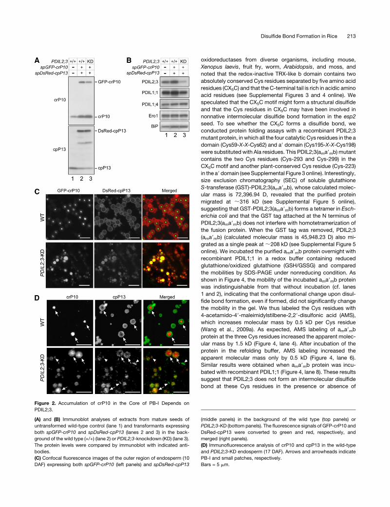
type or PDIL2;3-knockdown (PDIL2;3-KD) plants (Figure 2A).
Lowering the expression level of PDIL2;3 did not upregulate the
protein levels of PDIL1;1, PDIL1;4, Ero1, or BiP (Figure 2B). We
found that GFP-crP10 was concentrated in the core of PB-I, and
DsRed-cpP13 was targeted to the peripheral layers of PB-I in the
wild-type endosperm cells (Figure 2C, panel WT), which indi-
cated that GFP or DsRed tagging to crP10 and cpP13, respec-
tively, does not alter their localizations in PB-I. By contrast, in the
endosperm of PDIL2;3-KD, GFP-crP10 was not concentrated in
the core of PB-I, and the size of PB-I was more heterogeneous
compared with the wild type (Figure 2C, panel PDIL2;3-KD).
Similar results were obtained by immunocytochemical analyses
(Figure 2D). We confirmed the alterations in the distributions of
crP10 and cpP13 in PB-I between the wild type and PDIL2;3-KD
by immunocytochemical electron microscopy (see Supplemen-
tal Figure 2 online).
Disulfide Bond Formation in Rice 211
Oxidative Folding of Storage Proteins and PB-I
Development in the esp2 Seed
The esp2 endosperm cells do not produce typical PBs but form
abnormal small particles, which are uniformly stained with Rho-
damine (Onda et al., 2009). To see if this is caused in part by
the aberrant accumulation of different classes of storage pro-
teins in the small particles, we coexpressed spGFP-crP10 and
spDsRed-cpP13 in the esp2 endosperm. As we reported previ-
ously (Onda et al., 2009), the protein levels of PDIL2;3, Ero1, and
BiP were upregulated in the esp2 seed (Figure 3A, cf. lanes 1 and
2). Expressing spGFP-crP10 and spDsRed-cpP13 in the esp2
seed further upregulated PDIL2;3 (Figure 3A, lane 3). When esp2
seed proteins are extracted and fractionated by centrifugation
under nonreducing condition, the pellet (P) contains a large
amount of proglutelins in complexes through intermolecular
disulfide bonds (Onda et al., 2009). Almost all crP10 was frac-
tionated into the P fraction when proteins were extracted from
the wild-type seed, whereas a small amount of crP10 was
detected in the S fraction when extracted from the esp2 seed
(Figure 3B, panel crP10, lane 3). Other storage proteins, such as
a-globulin and RA17, which both contain ABC domains con-
served in the 2S albumin superfamily (Nakase et al., 1996;
Kawagoe et al., 2005), were mostly in the S fraction when
extracted from the wild-type seed (Figure 3B, lane 1), whereas
significant amounts of the proteins were fractionated into the P
fraction when extracted from the esp2 seed (Figure 3B, lane 4).
By contrast, cpP13, which does not contain Cys residue in the
mature polypeptide, was fractionated into the S fraction when
extracted from the seeds of the wild type and esp2 (Figure 3B,
panel cpP13), which supports the notion that the proteins frac-
tionated into the P fraction form large complexes through inter-
molecular disulfide bonds (Onda et al., 2009). When the
expression level of PDIL2;3 was lowered to a level comparable
to the wild type, the S/P ratio of proglutelins was not noticeably
altered from that of esp2 (Figure 3B, lanes 7 and 8). We then
analyzed the localizations of GFP-crP10 and DsRed-cpP13 in
the esp2 endosperm. A large fraction ofGFP-crP10 accumulated
and formed particles of different sizes (Figure 3C, left panel).
Interestingly, DsRed-cpP13 did not strongly associate with the
particles containing GFP-crP10, but it was spread in the cell,
most likely the ER network (Figure 3C, middle panel). When the
PDIL2;3 level was significantly lowered in the esp2 seed, GFP-
crP10 wasmostly localized in PB-I–like particles, but GFP-crP10
was not accumulated in the core of these particles (Figure 3D, left
panel). The localization of DsRed-cpP13 in the PDIL2;3-KD
endosperm of esp2 showed extensive overlaps with that of
GFP-crP10 (Figure 3D, middle panel). These results indicate that
the expression level of PDIL2;3 influences the localizations of
GFP-crP10 and DsRed-cpP13 in both wild-type and esp2 en-
dosperm.
AConserved Structural Disulfide in the bDomain of PDIL2;3
We noted that a significant fraction of PDIL2;3 was fractionated
into the P fraction when extracted from esp2 seeds (Figure 3B,
panel PDIL2;3, lane 4), which suggested that PDIL2;3 in the P
fraction was contained in large complexes through intermolec-
ular disulfide bonds. To gain insights into how PDIL2;3 formed
nonnative intermolecular disulfides in the esp2 endosperm, we
compared the amino acid sequences of the P5 subfamily
Figure 1. PDIL2;3 Localizes on the Surface of PB-I in the Endosperm.
(A) Confocal fluorescence images of the outer region of endosperm (10 d after flowing [DAF]) expressing both spGFP-PDIL2;3 and spDsRed-PDIL1;1.
The fluorescence signals of GFP-PDIL2;3 (left panel) and DsRed-PDIL1;1 (middle panel) were converted to green and red, respectively, and merged
(right panel). Arrowheads indicate PB-I in the ER lumen.
(B) PB-I (arrowheads) and PB-II (arrows) were visualized by Rhodamine staining. The fluorescence signals of GFP-PDIL2;3 (left panel) and Rhodamine
(middle panel) were converted to green and red, respectively, and merged (right panel). The fluorescence signals of DsRed-PDIL1;1 were not visible
under the conditions for obtaining Rhodamine signals at the appropriate intensity.
Bars = 5 mm.
212 The Plant Cell
oxidoreductases from diverse organisms, including mouse,
Xenopus laevis, fruit fry, worm, Arabidopsis, and moss, and
noted that the redox-inactive TRX-like b domain contains two
absolutely conserved Cys residues separated by five amino acid
residues (CX5C) and that the C-terminal tail is rich in acidic amino
acid residues (see Supplemental Figures 3 and 4 online). We
speculated that the CX5C motif might form a structural disulfide
and that the Cys residues in CX5C may have been involved in
nonnative intermolecular disulfide bond formation in the esp2
seed. To see whether the CX5C forms a disulfide bond, we
conducted protein folding assays with a recombinant PDIL2;3
mutant protein, in which all the four catalytic Cys residues in the a
domain (Cys59-X-X-Cys62) and a9 domain (Cys195-X-X-Cys198)
were substituted with Ala residues. This PDIL2;3(ama9mb) mutant
contains the two Cys residues (Cys-293 and Cys-299) in the
CX5C motif and another plant-conserved Cys residue (Cys-223)
in the a9 domain (see Supplemental Figure 3 online). Interestingly,
size exclusion chromatography (SEC) of soluble glutathione
S-transferase (GST)-PDIL2;3(ama9mb), whose calculated molec-
ular mass is 72,396.94 D, revealed that the purified protein
migrated at ;316 kD (see Supplemental Figure 5 online),
suggesting that GST-PDIL2;3(ama9mb) forms a tetramer in Esch-
erichia coli and that the GST tag attached at the N terminus of
PDIL2;3(ama9mb) does not interfere with homotetramerization of
the fusion protein. When the GST tag was removed, PDIL2;3
(ama9mb) (calculated molecular mass is 45,948.23 D) also mi-
grated as a single peak at;208 kD (see Supplemental Figure 5
online). We incubated the purified ama9mb protein overnight with
recombinant PDIL1;1 in a redox buffer containing reduced
glutathione/oxidized glutathione (GSH/GSSG) and compared
the mobilities by SDS-PAGE under nonreducing condition. As
shown in Figure 4, the mobility of the incubated ama9mb protein
was indistinguishable from that without incubation (cf. lanes
1 and 2), indicating that the conformational change upon disul-
fide bond formation, even if formed, did not significantly change
the mobility in the gel. We thus labeled the Cys residues with
4-acetamido-49-maleimidylstilbene-2,29-disulfonic acid (AMS),
which increases molecular mass by 0.5 kD per Cys residue
(Wang et al., 2009a). As expected, AMS labeling of ama9mbprotein at the three Cys residues increased the apparent molec-
ular mass by 1.5 kD (Figure 4, lane 4). After incubation of the
protein in the refolding buffer, AMS labeling increased the
apparent molecular mass only by 0.5 kD (Figure 4, lane 6).
Similar results were obtained when ama9mb protein was incu-
bated with recombinant PDIL1;1 (Figure 4, lane 8). These results
suggest that PDIL2;3 does not form an intermolecular disulfide
bond at these Cys residues in the presence or absence of
Figure 2. Accumulation of crP10 in the Core of PB-I Depends on
PDIL2;3.
(A) and (B) Immunoblot analyses of extracts from mature seeds of
untransformed wild-type control (lane 1) and transformants expressing
both spGFP-crP10 and spDsRed-cpP13 (lanes 2 and 3) in the back-
ground of the wild type (+/+) (lane 2) or PDIL2;3-knockdown (KD) (lane 3).
The protein levels were compared by immunoblot with indicated anti-
bodies.
(C) Confocal fluorescence images of the outer region of endosperm (10
DAF) expressing both spGFP-crP10 (left panels) and spDsRed-cpP13
(middle panels) in the background of the wild type (top panels) or
PDIL2;3-KD (bottom panels). The fluorescence signals of GFP-crP10 and
DsRed-cpP13 were converted to green and red, respectively, and
merged (right panels).
(D) Immunofluorescence analysis of crP10 and cpP13 in the wild-type
and PDIL2;3-KD endosperm (17 DAF). Arrows and arrowheads indicate
PB-I and small patches, respectively.
Bars = 5 mm.
Disulfide Bond Formation in Rice 213
PDIL1;1. It is thus probable that the conserved CX5C motif in the
b domain forms an intramolecular, structural disulfide bond.
We asked a question whether the CX5C motif influences the
function of PDIL2;3. Mutating the two Cys residues (Cys-293 and
Cys-299) in the CX5C structural disulfide to Ala residues (AX5A)
did not affect the retention time in SEC (see Supplemental Figure
5 online), indicating that the CX5C motif is not necessary for
tetramer formation in E. coli and that the stability of the tetramer
in vitro does not depend on the CX5C structural disulfide. We
next examined whether the CX5C motif affects the redox activity
of PDIL2;3 using reduced, denatured ribonuclease A (rRNase)
as substrate. The wild-type PDIL2;3 and mutant PDIL2;3(AX5A),
in which Cys-293 and Cys-299 in the CX5C motif were sub-
stituted with Ala residues, were preincubated in a redox buffer
containing GSH/GSSG to facilitate the formation of the struc-
tural disulfide of the CX5C motif and then mixed with the
substrate rRNase. The mutant protein exhibited 95.1%6 1.4%
activity (mean 6 SD of duplicates) of the wild-type protein,
indicating that the CX5C motif in the b domain does not
significantly modulate the refolding activity. We then examined
the effect of Cys-to-Ala substitution in the CX5C motif on
the localization of GFP-PDIL2;3 in the developing endosperm
and found that the GFP-PDIL2;3(AX5A) mutant protein was
Figure 3. Aberrant Localizations of Prolamins in the PDIL1;1-Knockout
esp2 Endosperm.
(A) Immunoblot analyses of extracts from mature seeds with indicated
antibodies. Total seed proteins were extracted under reducing conditions
from untransformed wild-type (+/+) control (lane 1), PDL1;1-knockout
(�/�) esp2 (lane 2), esp2 expressing both spGFP-crP10 and spDsRed-
cpP13 (lanes 3), and esp2 expressing both spGFP-crP10 and spDsRed-
cpP13 under the background of PDIL2;3-knockdown (KD) (lane 4).
(B) Immunoblot analyses of seed proteins. Seed proteins were fraction-
ated into the supernatant (S) and pellet (P) fractions by centrifugation
under nonreducing conditions (see Methods for details). pGT, proglute-
lins; Glb, a-globulin; RA17, a-amylase inhibitor.
(C) Confocal fluorescence images of the outer region of endosperm (10
DAF) expressing both spGFP-crP10 and spDsRed-cpP13 in the back-
ground of esp2 (pdil1;1). Arrowheads indicate PB-I–like particles con-
taining GFP-crP10. Note that DsRed-cpP13 did not accumulate in these
PB-I-like particles. The fluorescence signals of GFP-crP10 (left panel)
and DsRed-cpP13 (middle panel) were converted to green and red,
respectively, and merged (right panel). Bars = 5 mm.
(D) Confocal fluorescence images of the PDIL2;3-knockdown (KD) endo-
sperm cells (10 DAF) expressing both spGFP-crP10 and spDsRed-cpP13
in the background of esp2 (pdil1;1). The fluorescence signals of GFP-
crP10 (left panel) and DsRed-cpP13 (middle panel) were converted to
green and red, respectively, and merged (right panel). Bars = 5 mm.
Figure 4. Structural Disulfide CX5C in the b Domain of PDIL2;3.
(A) Purified PDIL2;3 (ama9mb, Cys59/62/195/198Ala) was incubated with
or without PDIL1;1 in a GSH/GSSG redox buffer overnight (O/N) or mixed
immediately with the SDS sample buffer after starting the reaction. The
denatured proteins were immunoblotted with anti-PDIL2;3 antibody.
Alkylation at three Cys residues with AMS shifted the protein by;1.5 kD
(cf. lanes 3 and 4), indicating that the oxidation state of PDIL2;3 protein
purified from E. coli was predominantly reduced. By contrast, the
oxidized protein, which was presumably alkylated at Cys-223, was
shifted by ;0.5 kD (lanes 6 and 8). Note that that PDIL1;1 assisted the
oxidation of PDIL2;3 (cf. lanes 7 and 8).
(B) Confocal fluorescence images of the outer region of endosperm (7
DAF) expressing spGFP-PDIL2;3(AX5A). PB-I (arrowheads) were visual-
ized by Rhodamine staining. The fluorescence signals of GFP-PDIL2;3
(AX5A) (left panel) and Rhodamine (middle panel) were converted to
green and red, respectively, and merged (right panel). Bars = 2 mm.
214 The Plant Cell
predominantly on the PB-I surface (Figure 4B), indicating that
the CX5C structural disulfide is not required for targeting GFP-
PDIL2;3 to the PB-I surface.
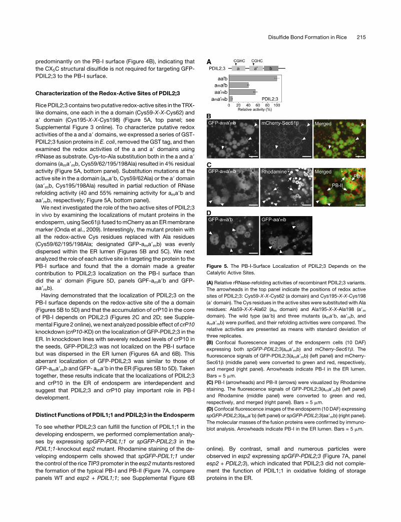
Characterization of the Redox-Active Sites of PDIL2;3
Rice PDIL2;3 contains twoputative redox-active sites in the TRX-
like domains, one each in the a domain (Cys59-X-X-Cys62) and
a9 domain (Cys195-X-X-Cys198) (Figure 5A, top panel; see
Supplemental Figure 3 online). To characterize putative redox
activities of the a and a9 domains, we expressed a series of GST-
PDIL2;3 fusion proteins in E. coli, removed the GST tag, and then
examined the redox activities of the a and a9 domains using
rRNase as substrate. Cys-to-Ala substitution both in the a and a9domains (ama9mb, Cys59/62/195/198Ala) resulted in 4% residual
activity (Figure 5A, bottom panel). Substitution mutations at the
active site in the a domain (ama9b, Cys59/62Ala) or the a9 domain
(aa9mb, Cys195/198Ala) resulted in partial reduction of RNase
refolding activity (40 and 55% remaining activity for ama9b and
aa9mb, respectively; Figure 5A, bottom panel).
We next investigated the role of the two active sites of PDIL2;3
in vivo by examining the localizations of mutant proteins in the
endosperm, using Sec61b fused tomCherry as anERmembrane
marker (Onda et al., 2009). Interestingly, the mutant protein with
all the redox-active Cys residues replaced with Ala residues
(Cys59/62/195/198Ala; designated GFP-ama9mb) was evenly
dispersed within the ER lumen (Figures 5B and 5C). We next
analyzed the role of each active site in targeting the protein to the
PB-I surface and found that the a domain made a greater
contribution to PDIL2;3 localization on the PB-I surface than
did the a9 domain (Figure 5D, panels GPF-ama9b and GFP-
aa9mb).Having demonstrated that the localization of PDIL2;3 on the
PB-I surface depends on the redox-active site of the a domain
(Figures 5B to 5D) and that the accumulation of crP10 in the core
of PB-I depends on PDIL2;3 (Figures 2C and 2D; see Supple-
mental Figure 2 online), we next analyzed possible effect of crP10
knockdown (crP10-KD) on the localization of GFP-PDIL2;3 in the
ER. In knockdown lines with severely reduced levels of crP10 in
the seeds, GFP-PDIL2;3 was not localized on the PB-I surface
but was dispersed in the ER lumen (Figures 6A and 6B). This
aberrant localization of GFP-PDIL2;3 was similar to those of
GFP-ama9mb and GFP- ama9b in the ER (Figures 5B to 5D). Taken
together, these results indicate that the localizations of PDIL2;3
and crP10 in the ER of endosperm are interdependent and
suggest that PDIL2;3 and crP10 play important role in PB-I
development.
Distinct Functions of PDIL1;1 andPDIL2;3 in the Endosperm
To see whether PDIL2;3 can fulfill the function of PDIL1;1 in the
developing endosperm, we performed complementation analy-
ses by expressing spGFP-PDIL1;1 or spGFP-PDIL2;3 in the
PDIL1;1-knockout esp2 mutant. Rhodamine staining of the de-
veloping endosperm cells showed that spGFP-PDIL1;1 under
the control of the rice TIP3 promoter in the esp2mutants restored
the formation of the typical PB-I and PB-II (Figure 7A, compare
panels WT and esp2 + PDIL1;1; see Supplemental Figure 6B
online). By contrast, small and numerous particles were
observed in esp2 expressing spGFP-PDIL2;3 (Figure 7A, panel
esp2 + PDIL2;3), which indicated that PDIL2;3 did not comple-
ment the function of PDIL1;1 in oxidative folding of storage
proteins in the ER.
Figure 5. The PB-I-Surface Localization of PDIL2;3 Depends on the
Catalytic Active Sites.
(A) Relative rRNase-refolding activities of recombinant PDIL2;3 variants.
The arrowheads in the top panel indicate the positions of redox active
sites of PDIL2;3: Cys59-X-X-Cys62 (a domain) and Cys195-X-X-Cys198
(a9 domain). The Cys residues in the active sites were substituted with Ala
residues: Ala59-X-X-Ala62 (am domain) and Ala195-X-X-Ala198 (a9mdomain). The wild type (aa9b) and three mutants (ama9b, aa9mb, and
ama9mb) were purified, and their refolding activities were compared. The
relative activities are presented as means with standard deviation of
three replicates.
(B) Confocal fluorescence images of the endosperm cells (10 DAF)
expressing both spGFP-PDIL2;3(ama9mb) and mCherry-Sec61b. The
fluorescence signals of GFP-PDIL2;3(ama9mb) (left panel) and mCherry-
Sec61b (middle panel) were converted to green and red, respectively,
and merged (right panel). Arrowheads indicate PB-I in the ER lumen.
Bars = 5 mm.
(C) PB-I (arrowheads) and PB-II (arrows) were visualized by Rhodamine
staining. The fluorescence signals of GFP-PDIL2;3(ama9mb) (left panel)
and Rhodamine (middle panel) were converted to green and red,
respectively, and merged (right panel). Bars = 5 mm.
(D)Confocal fluorescence images of the endosperm (10 DAF) expressing
spGFP-PDIL2;3(ama9b) (left panel) or spGFP-PDIL2;3(aa9mb) (right panel).
The molecular masses of the fusion proteins were confirmed by immuno-
blot analysis. Arrowheads indicate PB-I in the ER lumen. Bars = 5 mm.
Disulfide Bond Formation in Rice 215
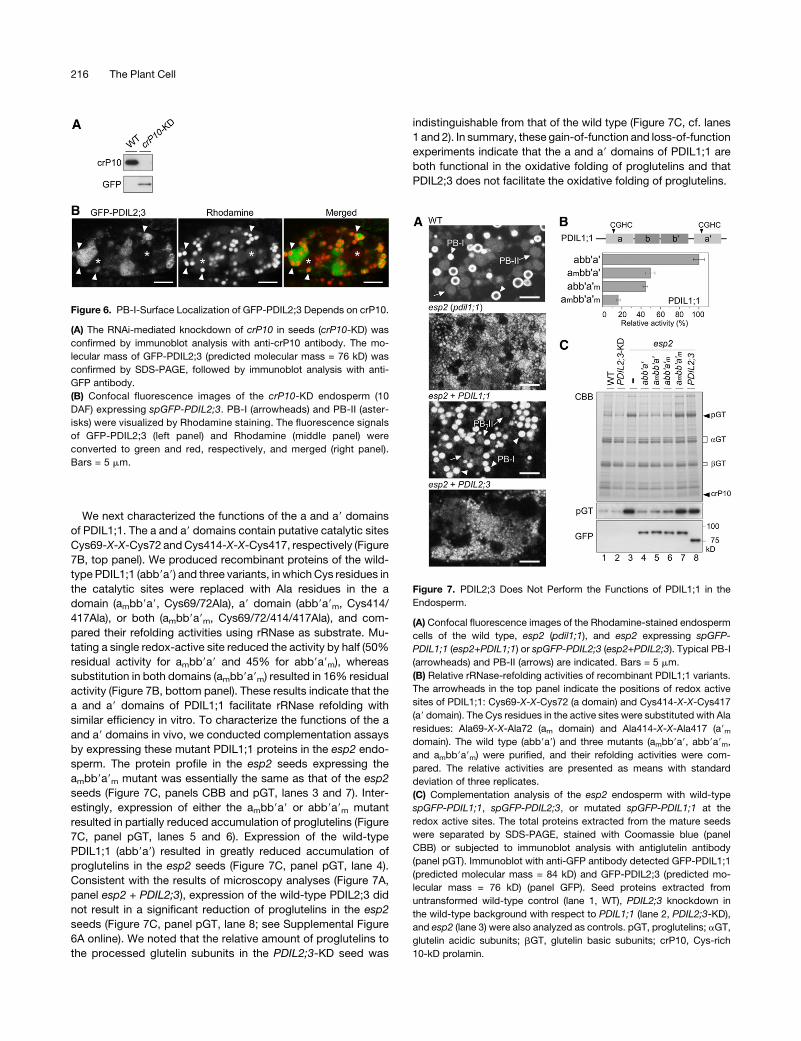
We next characterized the functions of the a and a9 domains
of PDIL1;1. The a and a9 domains contain putative catalytic sites
Cys69-X-X-Cys72 andCys414-X-X-Cys417, respectively (Figure
7B, top panel). We produced recombinant proteins of the wild-
type PDIL1;1 (abb9a9) and three variants, in which Cys residues in
the catalytic sites were replaced with Ala residues in the a
domain (ambb9a9, Cys69/72Ala), a9 domain (abb9a9m, Cys414/417Ala), or both (ambb9a9m, Cys69/72/414/417Ala), and com-
pared their refolding activities using rRNase as substrate. Mu-
tating a single redox-active site reduced the activity by half (50%
residual activity for ambb9a9 and 45% for abb9a9m), whereas
substitution in both domains (ambb9a9m) resulted in 16% residual
activity (Figure 7B, bottom panel). These results indicate that the
a and a9 domains of PDIL1;1 facilitate rRNase refolding with
similar efficiency in vitro. To characterize the functions of the a
and a9 domains in vivo, we conducted complementation assays
by expressing these mutant PDIL1;1 proteins in the esp2 endo-
sperm. The protein profile in the esp2 seeds expressing the
ambb9a9m mutant was essentially the same as that of the esp2
seeds (Figure 7C, panels CBB and pGT, lanes 3 and 7). Inter-
estingly, expression of either the ambb9a9 or abb9a9m mutant
resulted in partially reduced accumulation of proglutelins (Figure
7C, panel pGT, lanes 5 and 6). Expression of the wild-type
PDIL1;1 (abb9a9) resulted in greatly reduced accumulation of
proglutelins in the esp2 seeds (Figure 7C, panel pGT, lane 4).
Consistent with the results of microscopy analyses (Figure 7A,
panel esp2 + PDIL2;3), expression of the wild-type PDIL2;3 did
not result in a significant reduction of proglutelins in the esp2
seeds (Figure 7C, panel pGT, lane 8; see Supplemental Figure
6A online). We noted that the relative amount of proglutelins to
the processed glutelin subunits in the PDIL2;3-KD seed was
indistinguishable from that of the wild type (Figure 7C, cf. lanes
1 and 2). In summary, these gain-of-function and loss-of-function
experiments indicate that the a and a9 domains of PDIL1;1 are
both functional in the oxidative folding of proglutelins and that
PDIL2;3 does not facilitate the oxidative folding of proglutelins.
Figure 6. PB-I-Surface Localization of GFP-PDIL2;3 Depends on crP10.
(A) The RNAi-mediated knockdown of crP10 in seeds (crP10-KD) was
confirmed by immunoblot analysis with anti-crP10 antibody. The mo-
lecular mass of GFP-PDIL2;3 (predicted molecular mass = 76 kD) was
confirmed by SDS-PAGE, followed by immunoblot analysis with anti-
GFP antibody.
(B) Confocal fluorescence images of the crP10-KD endosperm (10
DAF) expressing spGFP-PDIL2;3. PB-I (arrowheads) and PB-II (aster-
isks) were visualized by Rhodamine staining. The fluorescence signals
of GFP-PDIL2;3 (left panel) and Rhodamine (middle panel) were
converted to green and red, respectively, and merged (right panel).
Bars = 5 mm.
Figure 7. PDIL2;3 Does Not Perform the Functions of PDIL1;1 in the
Endosperm.
(A) Confocal fluorescence images of the Rhodamine-stained endosperm
cells of the wild type, esp2 (pdil1;1), and esp2 expressing spGFP-
PDIL1;1 (esp2+PDIL1;1) or spGFP-PDIL2;3 (esp2+PDIL2;3). Typical PB-I
(arrowheads) and PB-II (arrows) are indicated. Bars = 5 mm.
(B) Relative rRNase-refolding activities of recombinant PDIL1;1 variants.
The arrowheads in the top panel indicate the positions of redox active
sites of PDIL1;1: Cys69-X-X-Cys72 (a domain) and Cys414-X-X-Cys417
(a9 domain). The Cys residues in the active sites were substituted with Ala
residues: Ala69-X-X-Ala72 (am domain) and Ala414-X-X-Ala417 (a9mdomain). The wild type (abb9a9) and three mutants (ambb9a9, abb9a9m,
and ambb9a9m) were purified, and their refolding activities were com-
pared. The relative activities are presented as means with standard
deviation of three replicates.
(C) Complementation analysis of the esp2 endosperm with wild-type
spGFP-PDIL1;1, spGFP-PDIL2;3, or mutated spGFP-PDIL1;1 at the
redox active sites. The total proteins extracted from the mature seeds
were separated by SDS-PAGE, stained with Coomassie blue (panel
CBB) or subjected to immunoblot analysis with antiglutelin antibody
(panel pGT). Immunoblot with anti-GFP antibody detected GFP-PDIL1;1
(predicted molecular mass = 84 kD) and GFP-PDIL2;3 (predicted mo-
lecular mass = 76 kD) (panel GFP). Seed proteins extracted from
untransformed wild-type control (lane 1, WT), PDIL2;3 knockdown in
the wild-type background with respect to PDIL1;1 (lane 2, PDIL2;3-KD),
and esp2 (lane 3) were also analyzed as controls. pGT, proglutelins; aGT,
glutelin acidic subunits; bGT, glutelin basic subunits; crP10, Cys-rich
10-kD prolamin.
216 The Plant Cell
Redox Activities of PDIL1;1 and PDIL2;3 in Vitro
We previously showed that how sulfhydryls of a-globulin are
oxidized plays a critical role in sorting a-globulin in the endo-
sperm (Kawagoe et al., 2005). In this study, we found that almost
half of a-globulin was fractionated into the P fraction when
proteins were extracted from the esp2 seed under nonreducing
conditions, whereas nearly all a-globulin produced in the wild-
type seed was fractionated into the S fraction (Figure 3B). These
results suggested that PDIL1;1 facilitates the oxidative folding of
not only proglutelins but also a-globulin. To investigate whether
PDIL2;3 also promotes the oxidative folding of a-globulin, we
extracted and fractionateda-globulin from thePDIL2;3-KD seed.
We found that a-globulin extracted from the PDIL2;3-KD seed
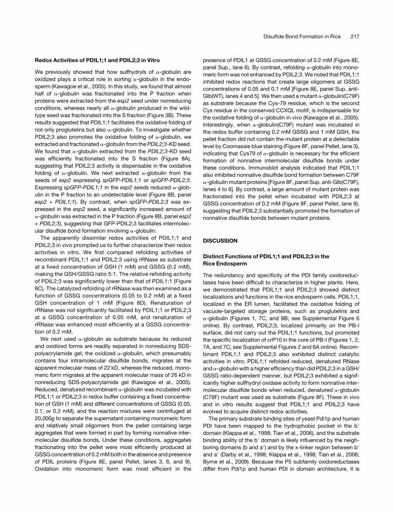
was efficiently fractionated into the S fraction (Figure 8A),
suggesting that PDIL2;3 activity is dispensable in the oxidative
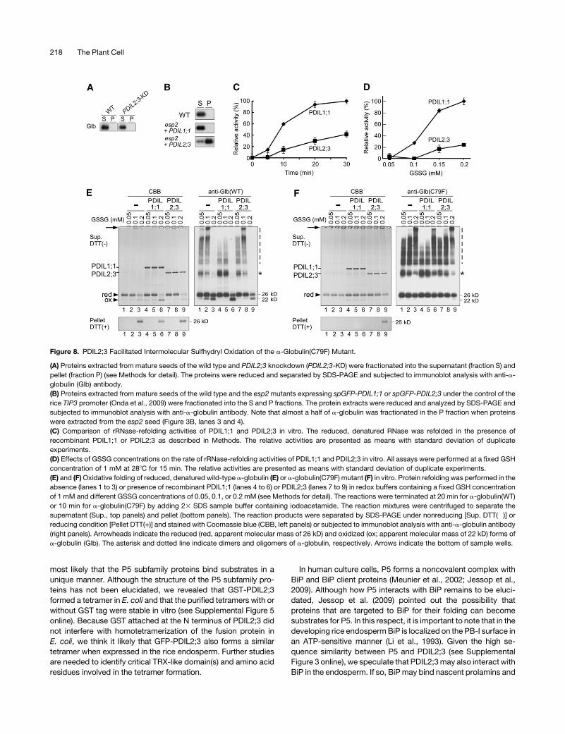
folding of a-globulin. We next extracted a-globulin from the
seeds of esp2 expressing spGFP-PDIL1;1 or spGFP-PDIL2;3.
Expressing spGFP-PDIL1;1 in the esp2 seeds reduced a-glob-
ulin in the P fraction to an undetectable level (Figure 8B, panel
esp2 + PDIL1;1). By contrast, when spGFP-PDIL2;3 was ex-
pressed in the esp2 seed, a significantly increased amount of
a-globulin was extracted in the P fraction (Figure 8B, panel esp2
+ PDIL2;3), suggesting that GFP-PDIL2;3 facilitates intermolec-
ular disulfide bond formation involving a-globulin.
The apparently dissimilar redox activities of PDIL1;1 and
PDIL2;3 in vivo prompted us to further characterize their redox
activities in vitro. We first compared refolding activities of
recombinant PDIL1;1 and PDIL2;3 using rRNase as substrate
at a fixed concentration of GSH (1 mM) and GSSG (0.2 mM),
making the GSH/GSSG ratio 5:1. The relative refolding activity
of PDIL2;3 was significantly lower than that of PDIL1;1 (Figure
8C). The catalyzed refolding of rRNase was then examined as a
function of GSSG concentrations (0.05 to 0.2 mM) at a fixed
GSH concentration of 1 mM (Figure 8D). Renaturation of
rRNase was not significantly facilitated by PDIL1;1 or PDIL2;3
at a GSSG concentration of 0.05 mM, and renaturation of
rRNase was enhanced most efficiently at a GSSG concentra-
tion of 0.2 mM.
We next used a-globulin as substrate because its reduced
and oxidized forms are readily separated in nonreducing SDS-
polyacrylamide gel; the oxidized a-globulin, which presumably
contains four intramolecular disulfide bonds, migrates at the
apparent molecular mass of 22 kD, whereas the reduced, mono-
meric form migrates at the apparent molecular mass of 26 kD in
nonreducing SDS-polyacrylamide gel (Kawagoe et al., 2005).
Reduced, denatured recombinant a-globulin was incubated with
PDIL1;1 or PDIL2;3 in redox buffer containing a fixed concentra-
tion of GSH (1 mM) and different concentrations of GSSG (0.05,
0.1, or 0.2 mM), and the reaction mixtures were centrifuged at
20,000g to separate the supernatant containing monomeric form
and relatively small oligomers from the pellet containing large
aggregates that were formed in part by forming nonnative inter-
molecular disulfide bonds. Under these conditions, aggregates
fractionating into the pellet were most efficiently produced at
GSSGconcentration of 0.2mMboth in the absence andpresence
of PDIL proteins (Figure 8E, panel Pellet, lanes 3, 6, and 9).
Oxidation into monomeric form was most efficient in the
presence of PDIL1 at GSSG concentration of 0.2 mM (Figure 8E,
panel Sup., lane 6). By contrast, refolding a-globulin into mono-
meric formwas not enhanced by PDIL2;3.We noted that PDIL1;1
inhibited redox reactions that create large oligomers at GSSG
concentrations of 0.05 and 0.1 mM [Figure 8E, panel Sup, anti-
Glb(WT), lanes 4 and 5]. We then used amutant a-globulin(C79F)
as substrate because the Cys-79 residue, which is the second
Cys residue in the conserved CCXQL motif, is indispensable for
the oxidative folding of a-globulin in vivo (Kawagoe et al., 2005).
Interestingly, when a-globulin(C79F) mutant was incubated in
the redox buffer containing 0.2 mM GSSG and 1 mM GSH, the
pellet fraction did not contain the mutant protein at a detectable
level by Coomassie blue staining (Figure 8F, panel Pellet, lane 3),
indicating that Cys79 of a-globulin is necessary for the efficient
formation of nonnative intermolecular disulfide bonds under
these conditions. Immunoblot analysis indicated that PDIL1;1
also inhibited nonnative disulfide bond formation between C79F
a-globulinmutant proteins [Figure 8F, panel Sup. anti-Glb(C79F),
lanes 4 to 6]. By contrast, a large amount of mutant protein was
fractionated into the pellet when incubated with PDIL2;3 at
GSSG concentration of 0.2 mM (Figure 8F, panel Pellet, lane 9),
suggesting that PDIL2;3 substantially promoted the formation of
nonnative disulfide bonds between mutant proteins.
DISCUSSION
Distinct Functions of PDIL1;1 and PDIL2;3 in the
Rice Endosperm
The redundancy and specificity of the PDI family oxidoreduc-
tases have been difficult to characterize in higher plants. Here,
we demonstrated that PDIL1;1 and PDIL2;3 showed distinct
localizations and functions in the rice endosperm cells. PDIL1;1,
localized in the ER lumen, facilitated the oxidative folding of
vacuole-targeted storage proteins, such as proglutelins and
a-globulin (Figures 1, 7C, and 8B; see Supplemental Figure 6
online). By contrast, PDIL2;3, localized primarily on the PB-I
surface, did not carry out the PDIL1;1 functions, but promoted
the specific localization of crP10 in the core of PB-I (Figures 1, 2,
7A, and 7C; see Supplemental Figures 2 and 6A online). Recom-
binant PDIL1;1 and PDIL2;3 also exhibited distinct catalytic
activities in vitro; PDIL1;1 refolded reduced, denatured RNase
anda-globulinwith a higher efficiency than did PDIL2;3 in aGSH/
GSSG ratio-dependent manner, but PDIL2;3 exhibited a signif-
icantly higher sulfhydryl oxidase activity to form nonnative inter-
molecular disulfide bonds when reduced, denatured a-globulin
(C79F) mutant was used as substrate (Figure 8F). These in vivo
and in vitro results suggest that PDIL1;1 and PDIL2;3 have
evolved to acquire distinct redox activities.
The primary substrate binding sites of yeast Pdi1p and human
PDI have been mapped to the hydrophobic pocket in the b9domain (Klappa et al., 1998; Tian et al., 2006), and the substrate
binding ability of the b9 domain is likely influenced by the neigh-
boring domains (b and a9) and by the x-linker region between b9and a9 (Darby et al., 1998; Klappa et al., 1998; Tian et al., 2006;
Byrne et al., 2009). Because the P5 subfamily oxidoreductases
differ from Pdi1p and human PDI in domain architecture, it is
Disulfide Bond Formation in Rice 217
most likely that the P5 subfamily proteins bind substrates in a
unique manner. Although the structure of the P5 subfamily pro-
teins has not been elucidated, we revealed that GST-PDIL2;3
formed a tetramer in E. coli and that the purified tetramers with or
without GST tag were stable in vitro (see Supplemental Figure 5
online). Because GST attached at the N terminus of PDIL2;3 did
not interfere with homotetramerization of the fusion protein in
E. coli, we think it likely that GFP-PDIL2;3 also forms a similar
tetramer when expressed in the rice endosperm. Further studies
are needed to identify critical TRX-like domain(s) and amino acid
residues involved in the tetramer formation.
In human culture cells, P5 forms a noncovalent complex with
BiP and BiP client proteins (Meunier et al., 2002; Jessop et al.,
2009). Although how P5 interacts with BiP remains to be eluci-
dated, Jessop et al. (2009) pointed out the possibility that
proteins that are targeted to BiP for their folding can become
substrates for P5. In this respect, it is important to note that in the
developing rice endospermBiP is localized on the PB-I surface in
an ATP-sensitive manner (Li et al., 1993). Given the high se-
quence similarity between P5 and PDIL2;3 (see Supplemental
Figure 3 online), we speculate that PDIL2;3may also interact with
BiP in the endosperm. If so, BiPmay bind nascent prolamins and
Figure 8. PDIL2;3 Facilitated Intermolecular Sulfhydryl Oxidation of the a-Globulin(C79F) Mutant.
(A) Proteins extracted from mature seeds of the wild type and PDIL2;3 knockdown (PDIL2;3-KD) were fractionated into the supernatant (fraction S) and
pellet (fraction P) (see Methods for detail). The proteins were reduced and separated by SDS-PAGE and subjected to immunoblot analysis with anti-a-
globulin (Glb) antibody.
(B) Proteins extracted from mature seeds of the wild type and the esp2mutants expressing spGFP-PDIL1;1 or spGFP-PDIL2;3 under the control of the
rice TIP3 promoter (Onda et al., 2009) were fractionated into the S and P fractions. The protein extracts were reduced and analyzed by SDS-PAGE and
subjected to immunoblot analysis with anti-a-globulin antibody. Note that almost a half of a-globulin was fractionated in the P fraction when proteins
were extracted from the esp2 seed (Figure 3B, lanes 3 and 4).
(C) Comparison of rRNase-refolding activities of PDIL1;1 and PDIL2;3 in vitro. The reduced, denatured RNase was refolded in the presence of
recombinant PDIL1;1 or PDIL2;3 as described in Methods. The relative activities are presented as means with standard deviation of duplicate
experiments.
(D) Effects of GSSG concentrations on the rate of rRNase-refolding activities of PDIL1;1 and PDIL2;3 in vitro. All assays were performed at a fixed GSH
concentration of 1 mM at 288C for 15 min. The relative activities are presented as means with standard deviation of duplicate experiments.
(E) and (F)Oxidative folding of reduced, denatured wild-type a-globulin (E) or a-globulin(C79F) mutant (F) in vitro. Protein refolding was performed in the
absence (lanes 1 to 3) or presence of recombinant PDIL1;1 (lanes 4 to 6) or PDIL2;3 (lanes 7 to 9) in redox buffers containing a fixed GSH concentration
of 1 mM and different GSSG concentrations of 0.05, 0.1, or 0.2 mM (see Methods for detail). The reactions were terminated at 20 min for a-globulin(WT)
or 10 min for a-globulin(C79F) by adding 23 SDS sample buffer containing iodoacetamide. The reaction mixtures were centrifuged to separate the
supernatant (Sup., top panels) and pellet (bottom panels). The reaction products were separated by SDS-PAGE under nonreducing [Sup. DTT(�)] or
reducing condition [Pellet DTT(+)] and stained with Coomassie blue (CBB, left panels) or subjected to immunoblot analysis with anti-a-globulin antibody
(right panels). Arrowheads indicate the reduced (red, apparent molecular mass of 26 kD) and oxidized (ox; apparent molecular mass of 22 kD) forms of
a-globulin (Glb). The asterisk and dotted line indicate dimers and oligomers of a-globulin, respectively. Arrows indicate the bottom of sample wells.
218 The Plant Cell
channel them to PDIL2;3, and the whole complex may be
targeted to PB-I in the ER.
The a domain of yeast Pdi1p and Mpd1p contains CX6C and
CX8C structural disulfide, respectively (Wilkinson et al., 2005;
Vitu et al., 2008). The CX6C structural disulfide in the a domain of
Pdi1p destabilizes the active site in the same TRX-like domain
(Wilkinson et al., 2005). We revealed that the redox activity of
PDIL2;3(AX5A) was not significantly different from that of wild-
type PDIL2;3 when the rRNase was used as substrate and that
the CX5C motif of PDIL2;3 was not necessary for homotetra-
merization when produced in E. coli. Furthermore, mutating the
CX5C motif to AX5A did not significantly alter the localization of
GFP-PDIL2;3 in vivo (Figure 4B). However, it is notable that
almost all PDIL2;3 proteins were in the S fraction when proteins
were extracted from wild-type seed, whereas a substantial
amount of PDIL2;3 was in the P fraction when proteins were
extracted from the esp2 seed (Figure 3B, panel PDIL2;3). It is
possible that in the esp2 endosperm cells, the Cys residues (Cys-
293/Cys-299) in the CX5C motif of PDIL2;3 may have formed
nonnative intermolecular disulfide bonds with storage proteins,
such as proglutelins and a-globulin, which also accumulated in
the P fraction. If so, these results suggest that PDIL1;1 facilitates
the oxidative folding of the CX5C structural disulfide; hence,
PDIL1;1 may regulate the function of PDIL2;3 in the ER.
Multiple Disulfide Bond Formation Pathways in the
Rice Endosperm
Disulfide bond formation plays a dominant role in sorting storage
proteins to the two types of PBs in the rice endosperm (Kawagoe
et al., 2005), suggesting that sulfhydryl oxidases and the PDI
family oxidoreductases are involved in protein sorting in the ER. It
is notable that the specific localization of GFP-PDIL2;3 depended
primarily on the active site of the a domain (Figures 5B to 5D).
Furthermore, crP10 knockdown similarly inhibited the targeting
of GFP-PDIL2;3 to the PB-I surface (Figure 6). Conversely, crP10
did not accumulate in the core of PB-I in thePDIL2;3-knockdown
seed (Figures 2C and 2D; see Supplemental Figure 2 online).
These results together suggest that thiol-disulfide exchange
reactions between crP10 and PDIL2;3 occur in PB-I: the accu-
mulation of crP10 in the core of PB-I involves sulfhydryl oxida-
tions of crP10, and the reduced form of PDIL2;3 dissociates from
PB-I. Interestingly, in the esp2 endosperm cells, DsRed-cpP13
did not accumulate in PB-I-like particles in the ERbutwas spread
in the cell, most likely in the ER (Figure 3C). It is conceivable that
abnormally elevated amounts of unfolded proteins, including
proglutelins, a-globulin, and RA17, titrated PDIL2;3 and/or BiP in
the ER of esp2 endosperm. We speculate that the aberrant
environment in the esp2 ER hindered interactions of PDIL2;3
and/or BiP with otherwise preferred substrates, such as prola-
mins. These results indicate that PDIL1;1 facilitates the oxidative
folding of PB-II–targeted proteins, such as proglutelins and
a-globulin, and thereby assists indirectly targeting prolamins to
PB-I in the ER.
In a recent study, we demonstrated that ER membrane-local-
ized sulfhydryl oxidase Ero1 facilitates the formation of native
disulfide bonds in proglutelins and that Ero1, PDIL2;3, and BiP
are produced at markedly higher levels in the esp2 seed (Onda
et al., 2009). Considering that crP10 forms polymers in the core of
PB-I and that PDIL2;3 was concentrated on the PB-I surface
(Figures 1 and 2), we speculate that either the a or a9 domain of
PDIL2;3 is oxidized by Ero1 or other sulfhydryl oxidases, such as
QSOX (Fass, 2008; Sevier, 2010), and that the oxidized form of
PDIL2;3 then transfers oxidizing equivalents to prolamins in PB-I.
After having said so, we point out that crP10 was predominantly
fractionated into the P fraction when proteins were extracted
from the PDIL2;3-knockdown seed (Figure 3B), suggesting that
crP10 forms large protein complexes through intermolecular
disulfide bonds.We thus speculate that crP10 oxidation in PB-I is
sustained by PDIL2:3-dependent and PDIL2;3-independent ox-
idation pathways.
A recent study has revealed the existence of Ero1-indepen-
dent mechanisms to generate disulfide bonds in mammalian
cells (Zito et al., 2010). In this respect, it is important to note that
ERO1 knockdown in the rice endosperm inhibits the oxidative
folding of proglutelins, but proglutelins nonetheless form nonna-
tive intermolecular disulfide bonds (Onda et al., 2009), suggest-
ing that sulfhydryl oxidases other than Ero1 accept electrons
from the PDI family oxidoreductases. Comprehensive gene
expression analyses indeed revealed that rice QSOX (accession
number AK121660) is expressed in the developing endosperm
(Sato et al., 2010); thus, QSOX may also play a role in sulfhydryl
oxidation of storage proteins in rice. Further studies are needed
to elucidate relative contributions of Ero1 and other sulfhydryl
oxidases, such as QSOX, in oxidation of prolamins in PB-I and
oxidative folding of PB-II–targeted proteins in the ER lumen.
Interestingly, human Ero1-a preferentially oxidizes the redox-
active Cys pair in the a9 domain of PDI (Wang et al., 2009a),
whereas it is the a domain of Pdi1p that yeast Ero1p predom-
inantly oxidizes (Vitu et al., 2010). These results indicate that
oxidizing equivalents flow from Ero1 to structurally diverse
substrates through a subset of redox-active TRX-like domains
of the PDI family oxidoreductases. Although it is unknown which
redox-active TRX-like domains of the PDI family are preferentially
oxidized by Ero1 in plants, it is surprising that expressing PDIL1;1
(ambb9a9) or PDIL1;1(abb9a9m) in the esp2 endosperm signifi-
cantly reduced the level of proglutelins to a similar extent (Figure
7C). These results strongly suggest that oxidoreductases other
than PDIL1;1 supply oxidizing equivalents to nascent proglute-
lins and that the a and a9 redox-active TRX-like domains of
PDIL1;1 assist independently the oxidative folding of proglutelins
by reduction and/or isomerization reactions. Consistently, the a
and a9 redox-active TRX-like domains of PDIL1;1 refolded
rRNase at a similar rate in vitro (Figure 7B). In summary, we
revealed that the PDI family oxidoreductases play distinct roles in
multiple pathways of sulfhydryl oxidations of structurally diverse
storage proteins in rice.
METHODS
Materials
Oryza sativa (cv Kinmaze) and an N-methyl-N-nitrosourea–induced mu-
tant, esp2 (cv Kinmaze), were grown in the field. The full-length cDNA
clones for PDIL2;3 (AK062254), PDIL1;1 (AK068268), crP10 (AK108254),
and cpP13 (AK242260) were obtained from the National Institute of
Disulfide Bond Formation in Rice 219
Agrobiological Sciences (Tsukuba, Japan). Antibodies against PDIL1;1,
glutelins, a-globulin, and BiP were prepared as described previously
(Takemoto et al., 2002; Kawagoe et al., 2005). Anti-GFP antibody was
purchased from MBL. Anti-PDIL2;3 antibody was raised in rabbit by
injecting purified PDIL2;3 (ama9b) used for the in vitro rRNase refolding
assay. Anti-crP10, anti-cpP13, and anti-RA17 antibodies used for
immunoblot analysis were prepared as follows. A PCR fragment en-
coding crP10 (Ile25–Cys134) was amplified from the full-length cDNA
(AK108254) with primers 59-ATCACCACTATGCAGTAT-39 and 59-TATC-
TCGAGACAACAACCACAGGAAGA-39. A PCR fragment encoding cpP13
(Ala19–Leu150) was similarly amplified from the full-length cDNA
(AK242260) with primers 59-ATACCATGGCGCAGTTTGATGTTTTA-39
and 59-ATACTCGAGCAAGACACCGCCAAGGGT-39. The PCR frag-
ments were cloned in appropriate sites in pET23d (Novagen). A PCR
fragment encoding RA17 (Asp28-His163) was amplified from the
genomic DNA isolated from the seedling (Nipponbare) with primers
59-ATACATATGGACCACCACCAAGTCTAC-39 and 59-TATCTCGAGGT-
GACCGGTTCTTGGGGT-39 and inserted into pET22b (Novagen) at the
appropriate sites. The plasmids were transferred to Rosetta2(DE3)
(Novagen), and protein production was induced with 0.4 mM isopropyl-
b-D(2)-thiogalactopyranoside (IPTG) at 378C for 3 h. The recombinant
proteins in inclusion bodies were solubilized in a denaturing buffer (8 M
urea, 100mMNaH2PO4, 20mM2-mercaptoethanol, and 10mMTris·HCl,
pH 8.0) and were purified with a Ni-NTA agarose column (Qiagen)
according to the manufacturer’s instructions. The purified protein was
injected into rat (crP10 and RA17) and rabbit (cpP13) for antibody
production.
Vector Construction and Rice Transformation
The binary vectors for rice transformation were constructed from
modified plasmids derived from the Gateway Entry and Destination
vectors (Invitrogen). For the construction of spGFP-PDIL2;3, a fragment
encoding Ser19–Leu441 of PDIL2;3 was amplified from the full-length
cDNA (AK062254) with primers 59-CGCGGATCCTCGCCGGTTTCC-39
and 59-CCGCTCGAGCGGTCACAACTCGTCATTTACTGGA-39 and in-
serted into PTIP3-spGFP-TGT1 (Onda et al., 2009) at the appropriate
sites. For the construction of spDsRed-PDIL1;1, a fragment encoding
Glu26–Leu512 of PDIL1;1 was amplified from the full-length cDNA
(AK068268) with primers 59-CGCGGATCCGAGGAGGCGGCGGCT-39
and 59-CCGCTCGAGCGGTTAGAGCTCATCCTTGAGAGGC-39 and
inserted into PTIP3-spDsRed(monomer)-TGT1 (Onda et al., 2009) at
the appropriate sites. The vector containing spGFP-PDIL1;1 was de-
scribed previously (Onda et al., 2009). For the construction of spGFP-
crP10, a fragment encoding Ile25–Cys134 of crP10 was amplified from
the full-length cDNA (AK108254) with primers 59-CATCACCACTATG-
CAGTAT-39 and 59-TATCTCGAGTCAACAACAACCACAGG-39 and in-
serted into the spGFP under the control of maize (Zea mays) Ubiquitin1
promoter (Onda et al., 2009). A fragment encoding Gln20–Leu150 of
cpP13 was similarly amplified from the full-length cDNA (AK242260)
with primers 59-CCAGTTTGATGTTTTAGGT-39 and 59-TATCTCGAGTTT-
CACATGTCACATA-39 and inserted into spDsRed-Monomer (Onda et al.,
2009) to generate spDsRed-cpP13 under the control of maize Ubiquitin1
promoter. The PDIL2;3 RNA interference (RNAi) construct contained a
461-bp linker of the chloramphenicol resistance gene (CmR), flanked
by inverted repeats of the 682-bp PDIL2;3 fragment (amplified with
primers 59-TATGGTACCATAATAGCAGCTCAAGGTAC-39 and 59-TAT-
GAATTCGCAGGAATATGGAATAAGAG-39), under the control of the
APS2b promoter and the GT1 terminator (Onda et al., 2009). The crP10
RNAi construct contained the CmR linker flanked by inverted repeats of
the 429-bp BamHI/XhoI fragment from the full-length cDNA (AK108254),
under the control of the a-globulin promoter and the NOS terminator
(Onda et al., 2009). Cys-to-Ala substitutions at the active sites of the a and
a9 domains of PDIL1;1 and PDIL2;3 and at the CX5Cmotif of PDIL2;3 were
performed using mutagenic primers and the QuikChange Lightning site-
directed mutagenesis kit (Stratagene) according to the manufacturer’s
instructions. The mutations were confirmed by DNA sequencing. The
primer sequences and procedures are described in Supplemental Table
1 online.
The binary vectors were transferred into Agrobacterium tumefaciens
strain EHA105 by electroporation and were used to transform rice (cv
Yukihikari and Kinmaze) as described previously (Kawagoe et al.,
2005). For complementation analysis of esp2 mutants, calli were
induced from the esp2 seeds and used for Agrobacterium-mediated
transformation.
Protein Extraction fromMature Rice Seeds
Total seed proteins were extracted by the following method unless
otherwise indicated. Mature seeds were powdered in liquid nitrogen, and
total seed proteins were extracted in buffer (10% [v/v] glycerol, 4% [w/v]
SDS, 6 M urea, 100 mM dithiothreitol (DTT), and 50 mM Tris·HCl, pH 6.8;
200 mL per 10 mg of mature seed) by vigorous shaking for 2.5 h at 258C.
The homogenate was centrifuged at 20,400g for 5 min at 258C, and the
resulting supernatant was subjected to SDS-PAGE.
Protein fractionation under nonreducing conditions was performed as
described previously (Onda et al., 2009). For protein fractionation into the
S and P fractions, seed proteins were first extracted from the fine
powders of mature seeds in nonreducing buffer (20% [v/v] glycerol, 4%
[w/v] SDS, 8 M urea, and 50 mM Tris·HCl, pH 6.8; 320 mL per 10 mg of
seed] by shaking for 3 h at room temperature. The homogenate was
centrifuged at 10,000g for 5 min, and the resulting supernatant was
reduced with 100 mM DTT (fraction S). The pellet was washed twice with
the nonreducing buffer and then homogenized in reducing buffer (20%
[v/v] glycerol, 4% [w/v] SDS, 8Murea, 100mMDTT, and 50mMTris·HCl,
pH 6.8) at the same final volume as the S fractions overnight at room
temperature. The soluble fractions were collected by centrifugation at
10,000g for 5 min (fraction P).
Alkylation of Recombinant PDIL2;3 with AMS
The oxidation state of PDIL2;3 in vitro was examined with the purified
recombinant protein (ama9mb, Cys59/62/195/198Ala), which contains
three Cys residues, by alkylation of free thiols with AMS (Jessop and
Bulleid, 2004). The protein (0.5 mM) was incubated at 288C in a redox
buffer (0.1 M Tris·HCl, pH 8.0, 2 mM EDTA, 1 mM GSH, and 0.2 mM
GSSG) with or without purified recombinant PDIL1;1 (0.5 mM). Disulfide
exchange reactions were stopped either immediately or after overnight
incubation by adding trichloroacetic acid to a final concentration of 10%
(w/v). The proteins were precipitated at 2308C for 30 min, followed by
centrifugation at 15,000g for 20 min at 48C. The pellet was washed with
ethanol/ether (1:1 v/v), air-dried, and suspended in SDS sample buffer
consisting of 62.5 mM Tris·HCl, pH 6.8, 10% (v/v) glycerol, and 2% (w/v)
SDSwith orwithout 100mMAMS (Invitrogen). Alkylation of free thiols with
AMS was conducted on ice overnight in the dark. The denatured proteins
were separated on SDS-polyacrylamide gel (5 to 20% acrylamide gradi-
ent; ATTO) under nonreducing condition, blotted onto a polyvinylidene
difluoride membrane, and immunodetected with anti-PDIL2;3 antibody
and Immobilon Western (Millipore).
Refolding Assays of Reduced, Denatured RNase in Vitro
Recombinant GST proteins fusedwith rice PDIL1;1 (Glu26–Leu512), rice
PDIL2;3 (Ser19–Leu441), or their variants were constructed using
pGEX-2TK or pGEX-6P-3 (GE Healthcare) and expressed in Rosetta2
(DE3) (Novagen) at 378C for 3 h with 0.4 mM IPTG induction. Cells were
harvested and lysed with lysozyme and DNase, and the lysate was
centrifuged at 11,000g for 10 min at 48C. The fusion proteins in the
220 The Plant Cell
supernatant were purified with Glutathione Sepharose 4B (MicroSpin
GST Purification Module; GE Healthcare) and cleaved with protease
according to the manufacturer’s instructions. SEC of GST-PDIL2;3
(ama9mb), PDIL2;3(ama9mb), and PDIL2;3(AX5A) was performed using a
Superdex 200 column with 20 mM Tris·HCl buffer, pH 7.0, containing
200 mMNaCl at a flow rate of 1 mL/min. Ribonuclease A (Sigma-Aldrich
R5125) was reduced and denatured by incubation of the native enzyme
overnight at room temperature in reducing/denaturing buffer (6 M
guanidine hydrochloride, 140 mM DTT, 100 mM Tris·HCl, pH 8.0, and
2 mM EDTA). The reduced and denatured RNase A was then separated
from excess DTT and guanidine hydrochloride by centrifugal gel filtra-
tion in a Micro Bio-Spin 6 column (Bio-Rad), which was prewashed with
0.1% acetic acid in water for buffer exchange. RNase A refolding assays
were essentially conducted by the method of Lyles and Gilbert (1991) in
buffer (0.1 M Tris·HCl, pH 8.0, 4.5 mM cyclic CMP [cCMP], 2 mM EDTA,
1 mM GSH, 0.2 mM GSSG, 10 mM reduced, denatured RNase A
[rRNase], and 1 mM PDIL) at 378C for 13 min for PDIL1;1 and 30 min for
PDIL2;3. Kinetic assays and effects of GSSG concentrations on rRNase
refolding activities were conducted by following the method of Rancy
and Thorpe (2008). Briefly, rRNase was incubated at 288C in buffer (0.1
M Tris·HCl, pH 8.0, 2 mM EDTA, 1 mM GSH, 0.2 mM or otherwise
indicated concentrations of GSSG, 10 mM rRNase, and 1 mMPDIL). The
reactions were stopped by mixing with water containing N-ethylmalei-
mide to give a final concentration of 2 mM N-ethylmaleimide. The
samples were then diluted with an equal volume of Tris buffer containing
2 mM cCMP. As for the oxidation analysis of the CX5C motif of PDIL2;3,
the wild-type PDIL2;3 and PDIL2;3(AX5A) mutant were preincubated
in the reaction buffer without rRNase and cCMP at 288C for 3 h prior to
the refolding assay. The hydrolysis of cCMP was tested by measuring the
absorbance at 296 nm. The data were converted to a percentage of the
activity of the wild-type PDIL proteins.
Refolding Assays of Reduced, Denatured a-Globulin in Vitro
The sequences encoding the wild-type and C79F a-globulin (Glu23–
Tyr186), which lacks the signal peptide, were amplified by PCR with
primers 59-ATACCATGGAGTCGGAGATGAGGTTCAGG-39 and 59-TAT-
GAGCTCTCAGTGGTGGTGGTGGTGGTGGTACTGGCCGGCGGCGAA-
GAT-39 (the His tag is underlined) and cloned into appropriate sites in
pET23d vector (Novagen). Construction of a-globulin C79F mutant was
described previously (Kawagoe et al., 2005). The wild-type and C79F
a-globulin were expressed in Rosetta2(DE3) (Novagen) with IPTG induc-
tion, yielding inclusion bodies. The polypeptides in the inclusion bodies
were purified with a Ni column under denaturing conditions and eluted in
elution buffer (8 M urea, pH 4.5, 100 mM NaH2PO4, and 10 mM Tris·HCl).
The purified polypeptides were reduced in the presence of DTT (final
concentration 140 mM) at room temperature for 3 h. The reduced
polypeptides were separated from excess DTT and urea by centrifugal
gel filtration in a Micro Bio-Spin 6 column (Bio-Rad), which was pre-
washed with 0.1% acetic acid in water. The refolding reaction was
initiated by adding the wild type or C79F a-globulin (final concentration of
10 mM) and recombinant PDIL proteins (final concentration of 2.2 mM) to
the refolding buffer (150 mMNaCl [50 mMNaCl for C79F a-globulin], 100
mM Tris·HCl, pH 8.0, 2 mM EDTA, 1 mM GSH, and three concentrations
[0.05, 0.1, or 0.2 mM] of GSSG). The samples were incubated at 288C for
20 min for the wild type and 10 min for C79F a-globulin. The reactions
were stopped by adding equal volume of nonreducing 23 SDS sample
buffer (20% [v/v] glycerol, 4% [w/v] SDS, and 125 mM Tris·HCl, pH 6.8)
containing 70 mM iodoacetamide. The samples were incubated at 48C
overnight and centrifuged at 20,000g for 5 min. The supernatant was
directly subjected to nonreducing SDS-PAGE (10 to 20% acrylamide
gradient). The pellets were washed with nonreducing 13 SDS sample
buffer and were then resuspended in 13 SDS sample buffer containing
100 mM DTT. The resulting samples were centrifuged and the superna-
tant was subjected to SDS-PAGE (10 to 20% acrylamide gradient). The
gels were either stained with Coomassie Brilliant Blue or subjected to
immunoblot analysis using anti-a-globulin antibody as described previ-
ously (Kawagoe et al., 2005).
Immunofluorescence Microscopy and
Immunocytochemical Analysis
Rice prolamins were purified from mature seeds as described elsewhere
(Takemoto et al., 2002). cpP13 (pI 6.65) was purified by isoelectric fo-
cusing from Cys-poor prolamins that have been extracted into 60% (v/v)
n-propanol containing 1 mM EDTA; the purified cpP13 was subjected to
preparative SDS-PAGE. crP10 was purified by preparative SDS-PAGE.
Antibodies against crP10 and cpP13 were raised in rabbit and mouse,
respectively. Rhodamine-conjugated anti-rabbit and FITC-conjugated
anti-mouse antibodies were purchased from Funakoshi Chemical. The
fluorescence images of the developing endosperm were analyzed with a
fluorescence microscope (AX80; Olympus) as described previously
(Takemoto et al., 2002).
Confocal Laser Scanning Microscopy
Sections (100 mm) of the endosperm were prepared from developing
seeds of rice plants. The seed sections were incubated in a Rhodamine
solution to stain protein bodies PB-I and PB-II at different intensities as
described previously (Onda et al., 2009). The fluorescence images of the
peripheral region of endosperm, also called subaleurone cells, were
analyzed with a confocal laser scanning microscope (TCS SP2 AOBS;
Leica) at 488 and 543 nm as described previously (Onda et al., 2009). The
data were processed in Adobe Photoshop CS3.
Accession Numbers
Sequence data from this article can be found in the Arabidopsis Genome
Initiative or GenBank/EMBL databases under the following accession
numbers:ERO1 (CB654132),a-Globulin (Glb) (D50643),PDIL1;1 (AK068268),
PDIL1;4 (AK071514), PDIL2;3 (AK062254), crP10 (AK108254), cpP13
(AK242260), and RA17 (AK242340).
Supplemental Data
The following materials are available in the online version of this article.
Supplemental Figure 1. Phylogenetic Tree of PDI Proteins.
Supplemental Figure 2. Immunolabeling of crP10 and cpP13 in PB-I
of the Wild-Type and PDIL2;3-KD Endosperm Cells.
Supplemental Figure 3. Sequence Alignment of Human P5 and Rice
PDIL2;3.
Supplemental Figure 4. Sequence Alignment of the b Domain of the
P5 Subfamily Oxidoreductases.
Supplemental Figure 5. Size Exclusion Chromatography of GST-
PDIL2;3(ama9mb), PDIL2;3(ama9mb), and PDIL2;3(AX5A).
Supplemental Figure 6. Complementation Analysis of esp2, a Null
Mutant of PDIL1;1.
Supplemental Table 1. List of PCR Primers for PDIL1;1 and PDIL2;3
Mutagenesis.
Supplemental Data Set 1. Text File of Alignment Corresponding to
Phylogenetic Analysis in Supplemental Figure 1.
Disulfide Bond Formation in Rice 221
ACKNOWLEDGMENTS
We thank Z. Fujimoto for assisting SEC analysis and Chiemi Takaboshi
and Mariko Shibahara for technical assistance. This work was sup-
ported by the Research and Development Program for New Bio-Industry
Initiatives from the Bio-Oriented Technology Research Advanced Insti-
tution, by a grant from the Ministry of Agriculture, Forestry, and Fisheries
of Japan (Genomics for Agricultural Innovation, IPG-0023), and by a
Grant-in-Aid for Scientific Research from the Japan Society for the
Promotion of Science (21380008).
Received September 13, 2010; revised December 9, 2010; accepted
January 3, 2011; published January 28, 2011.
REFERENCES
Andeme Ondzighi, C., Christopher, D.A., Cho, E.J., Chang, S.C., and
Staehelin, L.A. (2008). Arabidopsis protein disulfide isomerase-5
inhibits cysteine proteases during trafficking to vacuoles before
programmed cell death of the endothelium in developing seeds. Plant
Cell 20: 2205–2220.
Appenzeller-Herzog, C., and Ellgaard, L. (2008). The human PDI
family: Versatility packed into a single fold. Biochim. Biophys. Acta
1783: 535–548.
Byrne, L.J., Sidhu, A., Wallis, A.K., Ruddock, L.W., Freedman, R.B.,
Howard, M.J., and Williamson, R.A. (2009). Mapping of the ligand-
binding site on the b’ domain of human PDI: Interaction with peptide
ligands and the x-linker region. Biochem. J. 423: 209–217.
Darby, N.J., Penka, E., and Vincentelli, R. (1998). The multi-domain
structure of protein disulfide isomerase is essential for high catalytic
efficiency. J. Mol. Biol. 276: 239–247.
Fass, D. (2008). The Erv family of sulfhydryl oxidases. Biochim. Biophys.
Acta 1783: 557–566.
Ferrari, D.M., and Soling, H.D. (1999). The protein disulphide-isomer-
ase family: Unravelling a string of folds. Biochem. J. 339: 1–10.
Hatahet, F., and Ruddock, L.W. (2009). Protein disulfide isomerase: A
critical evaluation of its function in disulfide bond formation. Antioxid.
Redox Signal. 11: 2807–2850.Erratum. Antioxid. Redox Signal. 12: 322.
Houston, N.L., Fan, C.Z., Xiang, J.Q., Schulze, J.M., Jung, R., and
Boston, R.S. (2005). Phylogenetic analyses identify 10 classes of the
protein disulfide isomerase family in plants, including single-domain
protein disulfide isomerase-related proteins. Plant Physiol. 137:
762–778.
Jessop, C.E., and Bulleid, N.J. (2004). Glutathione directly reduces an
oxidoreductase in the endoplasmic reticulum of mammalian cells. J.
Biol. Chem. 279: 55341–55347.
Jessop, C.E., Watkins, R.H., Simmons, J.J., Tasab, M., and Bulleid,
N.J. (2009). Protein disulphide isomerase family members show
distinct substrate specificity: P5 is targeted to BiP client proteins. J.
Cell Sci. 122: 4287–4295.
Kaiser, B.K., Yim, D., Chow, I.T., Gonzalez, S., Dai, Z., Mann, H.H.,
Strong, R.K., Groh, V., and Spies, T. (2007). Disulphide-isomerase-
enabled shedding of tumour-associated NKG2D ligands. Nature 447:
482–486.
Kawagoe, Y., Suzuki, K., Tasaki, M., Yasuda, H., Akagi, K., Katoh, E.,
Nishizawa, N.K., Ogawa, M., and Takaiwa, F. (2005). The critical
role of disulfide bond formation in protein sorting in the endosperm of
rice. Plant Cell 17: 1141–1153.
Kikuchi, M., Doi, E., Tsujimoto, I., Horibe, T., and Tsujimoto, Y.
(2002). Functional analysis of human P5, a protein disulfide isomerase
homologue. J. Biochem. 132: 451–455.
Klappa, P., Ruddock, L.W., Darby, N.J., and Freedman, R.B. (1998).
The b’ domain provides the principal peptide-binding site of protein
disulfide isomerase but all domains contribute to binding of misfolded
proteins. EMBO J. 17: 927–935.
Krishnan, H.B., White, J.A., and Pueppke, S.G. (1992). Characteriza-
tion and localization of rice (Oryza sativa L) seed globulins. Plant Sci.
81: 1–11.
Kumamaru, T., Ogawa, M., Satoh, H., and Okita, T.W. (2007). Protein
body biogenesis in cereal endosperm. In Endosperm: Plant Cell
Monographs (8), O.A. Olsen, ed (Heidelberg, Germany: Springer), pp.
141–158.
Kumamaru, T., Uemura, Y., Inoue, Y., Takemoto, Y., Siddiqui, S.U.,
Ogawa, M., Hara-Nishimura, I., and Satoh, H. (2010). Vacuolar
processing enzyme plays an essential role in the crystalline structure
of glutelin in rice seed. Plant Cell Physiol. 51: 38–46.
Li, C.P., and Larkins, B.A. (1996). Expression of protein disulfide
isomerase is elevated in the endosperm of the maize floury-2 mutant.
Plant Mol. Biol. 30: 873–882.
Li, X.X., Wu, Y.J., Zhang, D.Z., Gillikin, J.W., Boston, R.S., Franceschi,
V.R., and Okita, T.W. (1993). Rice prolamine protein body biogenesis:
a BiP-mediated process. Science 262: 1054–1056.
Lu, D.P., and Christopher, D.A. (2008). Endoplasmic reticulum stress
activates the expression of a sub-group of protein disulfide isomerase
genes and AtbZIP60 modulates the response in Arabidopsis thaliana.
Mol. Genet. Genomics 280: 199–210.
Lyles, M.M., and Gilbert, H.F. (1991). Catalysis of the oxidative folding
of ribonuclease A by protein disulfide isomerase: Dependence of the
rate on the composition of the redox buffer. Biochemistry 30: 613–619.
Meunier, L., Usherwood, Y.K., Chung, K.T., and Hendershot, L.M.
(2002). A subset of chaperones and folding enzymes form multiprotein
complexes in endoplasmic reticulum to bind nascent proteins. Mol.
Biol. Cell 13: 4456–4469.
Nakase, M., Adachi, T., Urisu, A., Miyashita, T., Alvarez, A.M.,
Nagasaka, S., Aoki, N., Nakamura, R., and Matsuda, T. (1996).
Rice (Oryza sativa L) a-amylase inhibitors of 14–16 kDa are potential
allergens and products of a multigene family. J. Agric. Food Chem.
44: 2624–2628.
Nørgaard, P., Westphal, V., Tachibana, C., Alsøe, L., Holst, B., and
Winther, J.R. (2001). Functional differences in yeast protein disulfide
isomerases. J. Cell Biol. 152: 553–562.
Ogawa, M., Kumamaru, T., Satoh, H., Iwata, N., Omura, T., Kasai, Z.,
and Tanaka, K. (1987). Purification of protein body-I of rice seed and
its polypeptide composition. Plant Cell Physiol. 28: 1517–1527.
Onda, Y., Kumamaru, T., and Kawagoe, Y. (2009). ER membrane-
localized oxidoreductase Ero1 is required for disulfide bond formation
in the rice endosperm. Proc. Natl. Acad. Sci. USA 106: 14156–14161.
Rancy, P.C., and Thorpe, C. (2008). Oxidative protein folding in vitro: a
study of the cooperation between quiescin-sulfhydryl oxidase and
protein disulfide isomerase. Biochemistry 47: 12047–12056.
Sato, Y., Antonio, B.A., Namiki, N., Takehisa, H., Minami, H.,
Kamatsuki, K., Sugimoto, K., Shimizu, Y., Hirochika, H., and
Nagamura, Y. (2010). RiceXPro: A platform for monitoring gene
expression in japonica rice grown under natural field conditions.
Nucleic Acids Res. 39: D1141–D1148.
Satoh-Cruz, M., Crofts, A.J., Takemoto-Kuno, Y., Sugino, A., Washida,
H., Crofts, N., Okita, T.W., Ogawa, M., Satoh, H., and Kumamaru, T.
(2010). Protein disulfide isomerase like 1-1 participates in the maturation
of proglutelin within the endoplasmic reticulum in rice endosperm. Plant
Cell Physiol. 51: 1581–1593.
Sevier, C.S. (2010). New insights into oxidative folding. J. Cell Biol. 188:
757–758.
Shewry, P.R., Napier, J.A., and Tatham, A.S. (1995). Seed storage
proteins: Structures and biosynthesis. Plant Cell 7: 945–956.
222 The Plant Cell
Tachikawa, H., Takeuchi, Y., Funahashi, W., Miura, T., Gao, X.D.,
Fujimoto, D., Mizunaga, T., and Onodera, K. (1995). Isolation and
characterization of a yeast gene, MPD1, the overexpression of which
suppresses inviability caused by protein disulfide isomerase deple-
tion. FEBS Lett. 369: 212–216.
Takemoto, Y., Coughlan, S.J., Okita, T.W., Satoh, H., Ogawa, M.,
and Kumamaru, T. (2002). The rice mutant esp2 greatly accumulates
the glutelin precursor and deletes the protein disulfide isomerase.
Plant Physiol. 128: 1212–1222.
Tanaka, K., Sugimoto, T., Ogawa, M., and Kasai, Z. (1980). Isolation
and characterization of 2-types of protein bodies in the rice endo-
sperm. Agric. Biol. Chem. 44: 1633–1639.
Tian, G., Xiang, S., Noiva, R., Lennarz, W.J., and Schindelin, H.
(2006). The crystal structure of yeast protein disulfide isomerase
suggests cooperativity between its active sites. Cell 124: 61–73.
Uehara, T., Nakamura, T., Yao, D.D., Shi, Z.Q., Gu, Z.Z., Ma, Y.L.,
Masliah, E., Nomura, Y., and Lipton, S.A. (2006). S-nitrosylated
protein-disulphide isomerase links protein misfolding to neurodegen-
eration. Nature 441: 513–517.
Vitu, E., Gross, E., Greenblatt, H.M., Sevier, C.S., Kaiser, C.A., and
Fass, D. (2008). Yeast Mpd1p reveals the structural diversity of the
protein disulfide isomerase family. J. Mol. Biol. 384: 631–640.
Vitu, E., Kim, S., Sevier, C.S., Lutzky, O., Heldman, N., Bentzur, M.,
Unger, T., Yona, M., Kaiser, C.A., and Fass, D. (2010). Oxidative activity
of yeast Ero1p on protein disulfide isomerase and related oxidoreduc-
tases of the endoplasmic reticulum. J. Biol. Chem. 285: 18155–18165.
Wadahama, H., Kamauchi, S., Nakamoto, Y., Nishizawa, K., Ishimoto,
M., Kawada, T., and Urade, R. (2008). A novel plant protein disulfide
isomerase family homologous to animal P5 - molecular cloning and
characterization as a functional protein for folding of soybean seed-
storage proteins. FEBS J. 275: 399–410.
Wang, H.Z., Boavida, L.C., Ron, M., and McCormick, S. (2008).
Truncation of a protein disulfide isomerase, PDIL2-1, delays embryo
sac maturation and disrupts pollen tube guidance in Arabidopsis
thaliana. Plant Cell 20: 3300–3311.
Wang, L., Li, S.J., Sidhu, A., Zhu, L., Liang, Y., Freedman, R.B., and
Wang, C.C. (2009a). Reconstitution of human Ero1-Lalpha/protein-
disulfide isomerase oxidative folding pathway in vitro. Position-
dependent differences in role between the a and a’ domains of
protein-disulfide isomerase. J. Biol. Chem. 284: 199–206.
Wang, Y.H., Zhu, S.S., Liu, S.J., Jiang, L., Chen, L.M., Ren, Y.L., Han,
X.H., Liu, F., Ji, S.L., Liu, X., and Wan, J.M. (2009b). The vacuolar
processing enzyme OsVPE1 is required for efficient glutelin process-
ing in rice. Plant J. 58: 606–617.
Wilkinson, B., Xiao, R.Y., and Gilbert, H.F. (2005). A structural disulfide
of yeast protein-disulfide isomerase destabilizes the active site disul-
fide of the N-terminal thioredoxin domain. J. Biol. Chem. 280: 11483–
11487.
Yamagata, H., Sugimoto, T., Tanaka, K., and Kasai, Z. (1982).
Biosynthesis of storage proteins in developing rice seeds. Plant
Physiol. 70: 1094–1100.
Zito, E., Chin, K.T., Blais, J., Harding, H.P., and Ron, D. (2010). ERO1-b,
a pancreas-specific disulfide oxidase, promotes insulin biogenesis and
glucose homeostasis. J. Cell Biol. 188: 821–832.
Disulfide Bond Formation in Rice 223
DOI 10.1105/tpc.110.079509; originally published online January 28, 2011; 2011;23;210-223Plant Cell
KawagoeYayoi Onda, Ai Nagamine, Mutsumi Sakurai, Toshihiro Kumamaru, Masahiro Ogawa and Yasushi
Pathways for Oxidation of Structurally Diverse Storage Proteins in RiceDistinct Roles of Protein Disulfide Isomerase and P5 Sulfhydryl Oxidoreductases in Multiple
This information is current as of August 29, 2018
Supplemental Data /content/suppl/2011/01/13/tpc.110.079509.DC1.html
References /content/23/1/210.full.html#ref-list-1
This article cites 44 articles, 21 of which can be accessed free at:
Permissions https://www.copyright.com/ccc/openurl.do?sid=pd_hw1532298X&issn=1532298X&WT.mc_id=pd_hw1532298X
eTOCs http://www.plantcell.org/cgi/alerts/ctmain
Sign up for eTOCs at:
CiteTrack Alerts http://www.plantcell.org/cgi/alerts/ctmain
Sign up for CiteTrack Alerts at:
Subscription Information http://www.aspb.org/publications/subscriptions.cfm
is available at:Plant Physiology and The Plant CellSubscription Information for
ADVANCING THE SCIENCE OF PLANT BIOLOGY © American Society of Plant Biologists


![[VJSC] Business Manner 2015 buổi 1.pdf](https://static.fdocument.pub/doc/165x107/5695cff21a28ab9b02903f95/vjsc-business-manner-2015-buoi-1pdf.jpg)


