![c Copyright 2011 [please consult the authors] Notice …eprints.qut.edu.au/48802/2/48802.pdfschools should take to ensure the development of a numerate society who can effectively](https://static.fdocument.pub/doc/165x107/5af776117f8b9aac248be1fc/c-copyright-2011-please-consult-the-authors-notice-should-take-to-ensure-the.jpg)
c Copyright 2012 2013 WILEY-VCH Verlag GmbH & …eprints.qut.edu.au/55501/2/55501.pdf · (2013)...
18
This is the author’s version of a work that was submitted/accepted for pub- lication in the following source: Woodruff, Maria A., Lange, Claudia, Chen, Fulin, Fratzl, Peter, & Hut- macher, Dietmar (2013) Nano- to macroscale remodeling of functional tissue-engineered bone. Advanced Healthcare Materials, 2 (4), pp. 546-551. This file was downloaded from: https://eprints.qut.edu.au/55501/ c Copyright 2012 2013 WILEY-VCH Verlag GmbH & Co. KGaA, Wein- heim Notice: Changes introduced as a result of publishing processes such as copy-editing and formatting may not be reflected in this document. For a definitive version of this work, please refer to the published source: https://doi.org/10.1002/adhm.201200289
Transcript of c Copyright 2012 2013 WILEY-VCH Verlag GmbH & …eprints.qut.edu.au/55501/2/55501.pdf · (2013)...

This is the author’s version of a work that was submitted/accepted for pub-lication in the following source:
Woodruff, Maria A., Lange, Claudia, Chen, Fulin, Fratzl, Peter, & Hut-macher, Dietmar(2013)Nano- to macroscale remodeling of functional tissue-engineered bone.Advanced Healthcare Materials, 2(4), pp. 546-551.
This file was downloaded from: https://eprints.qut.edu.au/55501/
c© Copyright 2012 2013 WILEY-VCH Verlag GmbH & Co. KGaA, Wein-heim
Notice: Changes introduced as a result of publishing processes such ascopy-editing and formatting may not be reflected in this document. For adefinitive version of this work, please refer to the published source:
https://doi.org/10.1002/adhm.201200289
Submitted to
1
DOI: 10.1002/adhm.
Nano to Macro-Scale Remodeling of Functional Tissue-Engineered Bone
Maria Ann Woodruff, Claudia Lange, Fulin Chen, Peter Fratzl, Dietmar Werner Hutmacher*
Corresponding Author* Dr. M.A. Woodruff Institute for Health and Biomedical Innovation, Queensland University of Technology, 60 Musk Avenue, Kelvin Grove 4059, Australia. [email protected] Dr. C. Lange Max Planck Institute of Colloids and Interfaces, Department of Biomaterials, Potsdam, Germany. [email protected] Prof. F. Chen Departments of Oral and Maxillofacial Surgery, Fourth Military Medical University, Shaanxi, People's Republic of China. [email protected] Prof. P. Fratzl Max Planck Institute of Colloids and Interfaces, Department of Biomaterials, Potsdam, Germany. [email protected] Prof. D.W. Hutmacher Institute for Health and Biomedical Innovation, Queensland University of Technology, 60 Musk Avenue, Kelvin Grove 4059, Australia. [email protected] Keywords: composite scaffold; polycaprolactone; bone material characterization; tissue engineering; additive manufacturing
Submitted to
2
The drive to develop bone grafts for the regeneration of large gaps in the skeletal
structure, whilst circumventing the need to use permanent implants, has led to a major research
thrust towards developing biodegradable scaffolds for bone tissue engineering. Forerunners,
Langer and Vacanti led the birth of the tissue engineering field with their seminal 1993
Science paper which remains one of the most influential and cited works in the field of
regenerative medicine. [1] The application of the principles of biology and engineering towards
development of functional grafts for diseased or traumatized tissue has seen laboratories
worldwide forging impressive multi-disciplinary teams to focus on restoring, maintaining or
improving the function of a wide range of human tissues. [2-6] Unfortunately, the promise of
tissue engineering which was so vibrant a decade ago has so far failed to deliver the
anticipated results, from a clinical perspective. One reason is the virtual absence of long-term
in-vivo studies which are tantamount to robust data collection associated with taking tissue
engineering therapies from bench to bedside. [7-9]
The basic concept fundamental to bone tissue engineering is to combine a scaffold
with living cells, decellularized extracellular matrix, and/or recombinantly produced growth
factors such as bone morphogenic protein (BMP), platelet derived growth factor (PDGF), and
vascular endothelial growth factor (VEGF) to form a “tissue engineering construct” (TEC)
which aims to promote the repair and/or regeneration of tissues. [9, 10]
A well engineered scaffold for bone tissue engineering combines inspired design,
technical innovation and precise craftsmanship. Original thinking in the field endorsed
scaffold degradation to occur as soon new tissue started to form. [6, 8] In contrast, we
emphasize the importance of the scaffolds remaining intact as newly formed tissue matures
within the porous and fully interconnected scaffold architecture and that the onset of
degradation should only occur after the regenerated tissue has remodeled at least once in the
Submitted to
3
natural remodeling cycle. [11] The scaffold plays a significant role in tissue development by
providing a spatial and temporal support for cells to attach and migrate whilst enabling cell
differentiation and stimulating production of different ECM components in this
microenvironment. The design of these scaffolds also needs to consider physico-chemical
properties, morphology and degradation kinetics. External size and shape of the construct are
also of importance, particularly if the construct is customized for an individual patient. Most
importantly, clinically successful bone constructs should stimulate and support both the onset
and the continuance of bone in-growth as well as subsequent remodeling and maturation by
providing optimal stiffness and external and internal geometrical shapes. Therefore, scaffolds
must provide sufficient initial mechanical strength and stiffness to substitute for the loss of
mechanical function of the bone. Hence, mechanical properties in the lower range of
cancellous bone are sufficient for low-load-bearing locations such as the skull and mid-face
whereas for high-load-bearing bones such as the tibia and femur, additional internal or
external fixation is needed . [11]
Continuous cell and tissue remodeling is important for achieving stable
biomechanical conditions which are essential for angiogenesis and subsequently
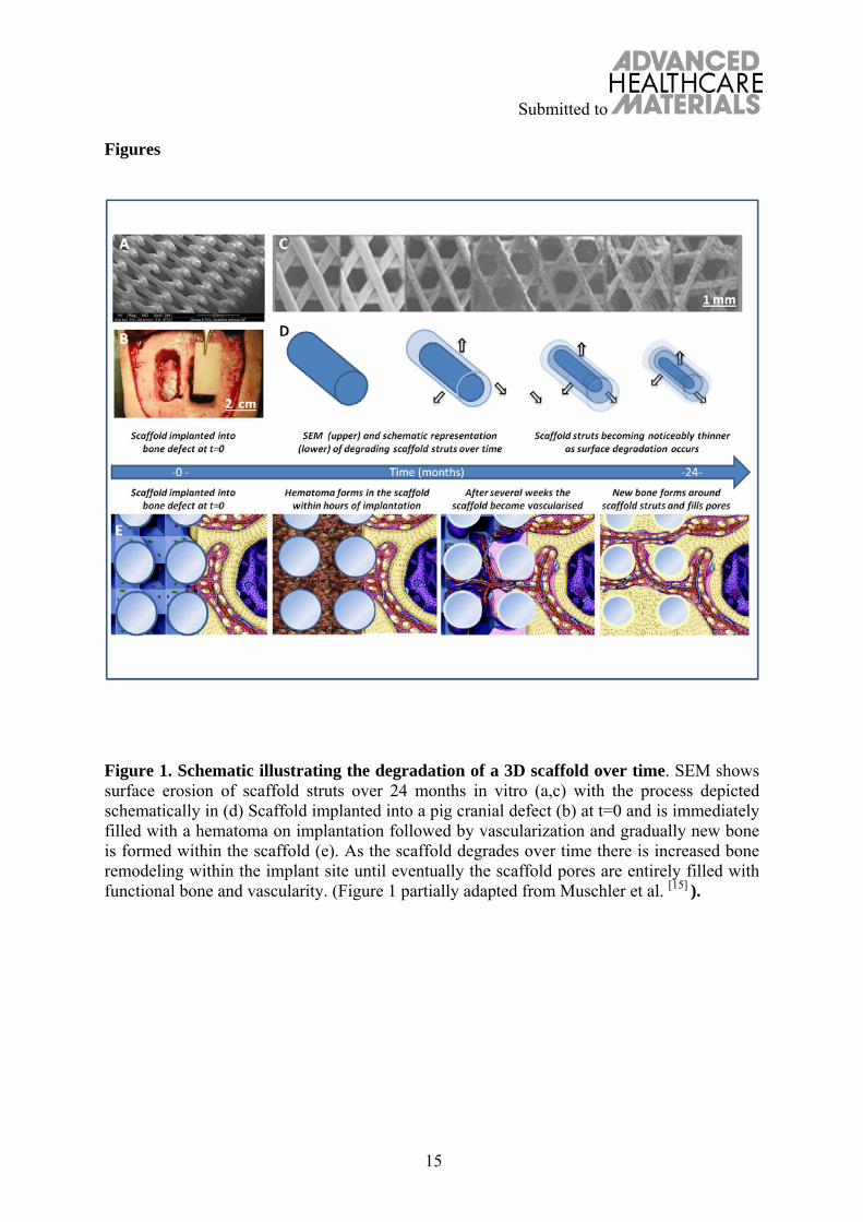
vascularization at the host site; a process depicted schematically in Figure 1. Part (a) shows a
scanning electron microscopy (SEM) image of a medical grade composite scaffold implanted
into a pig cranial defect (b). Over 24 months the scaffold degrades slowly via surface erosion
(in phosphate buffered saline solution (c)) with clear erosion of the scaffold struts shown
using SEM; this process is depicted schematically in part (d). At the same time, the scaffold
becomes gradually vascularised in vivo via the migration and proliferation of endothelial
progenitor cells through the scaffold-entrapped hematoma. Subsequently, osteoblastic
Submitted to
4
progenitor cells invade the scaffold pores and proliferate and differentiate to form new bone
(e).
The degree of tissue turnover and maturation within the scaffold depends on the tissue
itself (e.g. muscle 6-8 weeks, bone 12-18 months), and its host anatomy and physiology. In
addition to these essentials of mechanics and geometry, a suitable construct will (i) possess a
3D and highly porous interconnected pore network with surface properties which are
optimized for the attachment, migration, proliferation and differentiation of cell types of
interest (depending on the targeted tissue) and enable flow transport of nutrients and
metabolic waste, and (ii) be biocompatible and biodegradable with a controllable rate to
compliment cell/tissue growth and maturation. [11]
It is absolutely essential to understand and control the different stages of the scaffold
degradation process to achieve not only the initially successful tissue formation but also
subsequent remodeling, and maturation at the defect site which occurs later. Initial thinking
in the field of tissue engineering advocated that scaffolds should degrade and vanish at the
same time as the tissue is growing. [6,8] Yet, tissue in-growth and maturation differs
temporally from tissue to tissue and, furthermore, tissue in-growth does not necessarily
equate to tissue maturation and remodeling, hence we should not consider a defect filled with
immature tissue as being “regenerated”. For this reason, many scaffold-based strategies have
failed in the past as the scaffold degradation was more rapid than tissue remodeling and/or
maturation. [12,13]
The long-term characteristics of bone growth and remodeling within TECs are not
well known. Warnke et al. introduced an innovative bone engineering concept into the clinic.
A titanium mesh cage was filled with bone mineral blocks and infiltrated with 7 mg
recombinant human bone morphogenetic protein 7 (BMP-7) and 20 mL of the patient's own
Submitted to
5
bone marrow. The transplant was then implanted into the latissimus dorsi muscle and 7
weeks later transplanted as a free bone-muscle flap to repair the mandibular defect.
Postoperatively the patient had an improved degree of mastication and aesthetics, but
unfortunately the graft failed from a long-term perspective. [9] This highlights the imperative
to study long-term bone regeneration and remodeling to gain optimum insight into the
clinical success of such procedures, and this can only be achieved using the most
sophisticated techniques. Here we use a suite of advanced analytical techniques to assess the
material properties of the tissue-engineered bone generated during long-term in vivo studies,
proposing that the onset of degradation should only occur after the regenerated tissue within
the scaffold has remodeled at least once in the natural remodeling cycle. This paradigm shift
is particularly relevant for higher load bearing tissues, such as bone. Original hypotheses
promoted scaffold degradation to onset immediately as new tissue starts to form. In contrast,
we underline the importance of the scaffold remaining intact as the tissue matures in the
scaffold pores with bulk degradation occurring later. We illustrate this process schematically
in Figure 1 whereby the scaffold struts slowly degrade over time as the newly formed bone
matures within the pores.
In the work described in this paper, we demonstrate that this rationale leads to
structural and functional bone regeneration in a large critical-sized defect model. Bone
regeneration is influenced by a complex interplay of biochemical and biomechanical
processes at the tissue, cellular and molecular levels. [14] This integrated hierarchical system
cannot be fully understood using qualitative or intuitive approaches only, nor by focusing on
single spatial or temporal considerations in isolation. [15] Hence, here we report a long-term
bone engineering study which supports the new paradigm which leads to superior bone
regeneration within slowly degrading composite scaffolds. We detail the post explantation
Submitted to
6
analysis techniques which enable bone quantity and quality to be assessed on the macro,
micro and nano-scale. To our knowledge, this is the first time such detailed examination of
long-term regenerated bone has been undertaken using the comprehensive techniques
presented here, which include micro-computed tomography (µCT), advanced mineralized
hard-tissue resin histology, scanning electron microscopy and small angle x-ray scattering.
This provides key insight for the first time into the cellular and extracellular matrix function
and organization pertaining to long-term bone remodeling behavior within clinically relevant
defect sites which are treated with a clinically proven tissue-engineered bone strategy.
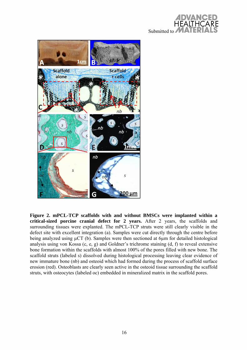
Medical grade polycaprolactone-tricalcium phosphate scaffolds (mPCL-TCP) (4cm x
2 cm x 1 cm, volume of 8 cubic centimeters) were implanted into bilateral porcine critical-
sized cranial defects, and 6 scaffolds were seeded intra-operatively with bone marrow stromal
cells (BMSCs) and implanted for a duration of 2 years. Scaffolds and the surrounding tissue
were explanted after 2 years and it was observed that extensive bone regeneration had
occurred within the defect sites containing mPCL-TCP scaffolds both with and without
BMSC addition (Figure 2). The scaffolds pores were filled with regenerated mineralized bone
with extensive bone remodeling evident around scaffold struts (labeled s) and clear evidence
of surface degradation of the composite scaffold (Figure 2d-g). The fully interconnected
scaffold architecture was still evident within the defect sites after 2 years of implantation,
albeit with a reduction in strut diameter owing to some surface degradation of the composite
material and the pores were seen to be completely filled with tissue, mostly identified as
mineralized bone.
Micro-computed tomography was performed to determine the bone volume fraction
and bone mineral density and three-dimensional (3D) rendered images were generated to
Submitted to
7
demonstrate the extent of mineralization throughout the entire scaffold (Figure 2b).
Histological assessment (Figure 2c-g) using von Kossa staining with Macneal’s tetrachrome
counter stain highlights the black mineralized tissue reflecting near complete mineralization
within the scaffold pores (Figure 2e,g). It can clearly be seen from histological sections that
the scaffold has undergone surface erosion with scaffolds’ struts having decreased in
diameter; however, as designed and predicted the overall scaffold morphology remained
largely intact. The slow process of surface erosion and degradation of the scaffold struts is
also evident along with remodeling taking place within the pores (Figure 2d,e). Goldner’s
trichrome staining (Figure 2d,f) confirms bone maturation via positive staining for osteocytes
(black arrows) embedded within the mineralized matrix and clear osteoid formation (actively
mineralizing bone front) around the scaffold struts (stained red in Figure 2d,f) as well as
osteoblasts (labeled ob) encompassing the struts (Figure 2g). There is notable osteoid
formation around the scaffold struts demonstrating a tissue remodeling and maturation
occurring as the scaffold gradually degrades via surface erosion enabling new bone to
progressively replace the mPCL-TCP scaffold itself as it slowly erodes and its by-products
are metabolized via the Krebs-cycle without causing any inflammation. We next
quantitatively compared scaffolds implanted with and without BMSCs to study the effect of
autologous cell implantation on bone regeneration. Scaffold groups without cells
demonstrated bone regenerative capabilities, yet the quantity of new bone formed throughout
the entire scaffold was significantly lower than those scaffolds loaded with BMSCs as
depicted in Figure 3k. Fully mineralized tissue, from histology sections (von Kossa staining)
were measured within the defect and values were reported as a percentage of bone volume
per tissue volume (% BV/TV). Positive pixel areas were divided by total tissue available for
growth (defect area minus the strut area of the scaffold) and revealed significantly higher
Submitted to
8
(p<0.05) mineralization (75%) for scaffolds implanted with BMSCs compared to scaffold
alone (49%).
The µCT and histology trend corresponded for all samples containing adjacent cell and cell-
free scaffolds (Figure 3 a-f) with bone regeneration occurring in all cases but statistically
more bone was formed from the addition of the BMSC cells (Figure 3k). The calcium content
of both the tissue-engineered bone and the native bone in adjoining regions of the skull was
also visualized by environmental scanning electron microscopy (ESEM) in backscattered
electron (BSE) mode, as shown in Figure 3, indicating that bone had been forming all around
and in-between the scaffold struts (Figure 3g-j). The areas corresponding to native calvarial
bone show some porosity surrounded by bone material often with a lower mineral content
(Figure 3i). This indicated high remodeling activity, since lower mineral content usually
means younger bone. Interestingly, the bone material surrounding the struts of the scaffold
does not have a lower mineral density (Figure 3h, j). A possible explanation could be that the
pores in the native calvaria gradually decrease by new bone formation at the inside of the
pore, while tissue-engineered bone starts to grow on the surface of the struts and expands
from there. As a consequence, the bone matrix around pore spaces in the skull is the
youngest, while around the struts of the scaffold it is the oldest compared to the surroundings.
We utilized small angle x-ray scattering (SAXS) to determine thickness and
orientation of the mineral particles in the newly generated bone tissue and compared this with
the native, host bone. Areas of interest for SAXS analysis were chosen in the vicinity of pores
and struts. Figure 3h-j shows a characteristic difference in the bone ultrastructure between
normal and tissue-engineered bone. The mineral particles in native bone show no preferential
orientation (as revealed by nearly spherical SAXS patterns, Figure 3i), in agreement with
earlier observations that elongated mineral particles are mostly oriented within the plane of

Submitted to
9
the calvaria, but with no preferential alignment within this plane. [16,17] In contrast to this,
mineral particles are preferentially orientated along the circumference of the scaffold struts in
tissue-engineered bone, as shown by the eccentricity of the SAXS pattern (Figure 3h, j). This
is also very different from the earliest mineralized tissue found in the callus during fracture
healing, which is much less organized (woven) with nearly isotropic fibril and mineral
orientation. [18, 19] Indeed, it has been found in scaffold-free fracture healing that lamellar bone
is deposited only once this disordered substrate is generated, which acts as an “endogenous
scaffold”. [19] Similar observations have also been made in deer antler, a bone which re-grows
every year. [20] Most interestingly, no traces of primary woven bone are found in the present
study next to the scaffold struts, which seems to indicate that lamellar bone is forming
directly on the internal surfaces of the scaffold thus short-circuiting the need for the
intermediate framework.
Importantly this direct comparison shows significantly faster bone formation without
alteration of the ultrastructural patterns, when the implanted scaffolds contain BMSCs, as
revealed by µCT and histological staining (Figure 3a-f, k), as well as ESEM and SAXS
analysis (Figure 3g-j) which demonstrated similar orientation of mineral particles around the
scaffold struts for cell-containing and cell-free scaffolds, compared with native bone.
Slow degrading composite scaffolds seeded with BMSCs regenerated the entire defect
and showed extensive bone remodeling inside the fully interconnected pore architecture.
Osteoid formation around the scaffold struts and viable vascular network demonstrated a
tissue remodeling and maturation occurring as the scaffold material gradually degraded via
surface erosion. This enabled new bone to progressively replace the mPCL-TCP itself as it
slowly degraded without the over-production of detrimental acidic by-products which may
lead to inflammation. The cell-free scaffold group demonstrated bone regenerative

Submitted to
10
capabilities and good osseointegration into the host bone due to the osteoconductive
properties of the mPCL-TCP surface. However, the quantity of new bone formed throughout
the entire architecture was significant lower than those scaffolds loaded with BMSC’s.
Hence, our results demonstrate the principle challenges to clinical translation of bone
engineering of clinically relevant defect regeneration need to be addressed by the
combination of a well-designed composite scaffold with a cell source which has strong
osteogenic potential.
In conclusion, we have spent the last decade translating bone tissue engineering
concepts from the bench to the bedside. After a series of in vitro and small animal studies
reviewed in detail by Woodruff and Hutmacher 11, we have designed and executed a long-
term, pre-clinical study to regenerate clinically relevant critical-sized cranial defects and have
successfully demonstrated not only extensive bone regeneration but also remodeling over a
period of two years within these defects implanted with a scaffold/BMSC construct. The
importance of long-term implantation studies followed by in depth analysis at different orders
of magnitude, using sophisticated methods prove highly organized and functional regenerated
bone is crucial to future development and optimization of TEC’s. These spatial and temporal
considerations collectively enforce the vision which was so vivid a decade ago and remains a
key target in regenerative medicine - to advance these promising tissue engineering therapies
into the clinic.
Submitted to
11
Materials and Methods
BMSCs were attained from the same animals under general anesthesia using routine bone
marrow extraction and cultured based on published protocols. 21 On reaching 80%
confluency, BMSC’s were treated with osteoinductive media for 4 weeks prior to seeding
onto mPCL-TCP scaffolds using fibrin glue (Baxter) during cranial implantation. Scaffolds
comprising medical grade ε-poly-caprolactone incorporating 20% β-tricalcium phosphate
(mPCL–TCP) were produced by fused deposition modeling (FDM) as described previously
(Osteopore International, Singapore; www.osteopore.com.sg). 11 The structural parameters of
the scaffolds, tailored by computer aided design, were 100% pore interconnectivity within a
range of 350–450 μm size, 70% scaffold porosity, and a 0/90° lay down pattern. mPCL-TCP
scaffolds were implanted into the defect sites either alone or in combination with BMSC cells
which were immobilized inside the scaffold by using fibrin glue. Eight defect sites were
implanted with TEC’s, eight defect sites were implanted with scaffold alone. Animals were
euthanized after 2 years with an overdose of barbiturates.
Scaffolds and the entire surrounding skull were explanted and immediately fixed in 4%
paraformaldehyde for 2 weeks and then transferred to 70% ethanol. For quantitative
evaluation of bone formation, the bones were then scanned in a micro-computed tomography
scanner (microCT 40, Scanco Medical, Brüttisellen, Switzerland) based on published
protocols. 21,22 After completion of the embedding process, the resin blocks were trimmed and
cut in half, directly through the center of the scaffolds, to enable histology sections to be
taken from the center outward based on published protocols. 21,22 Resin sections were stained
using Macneals/von kossa and Goldner’s Trichrome stains and were measured semi-
Submitted to
12
quantitatively using Bioquant Image Analysis® software (Nashville, TN, USA). Fully
mineralized tissue was measured by applying a fixed threshold to select for the positive stain
(black von Kossa staining) within the defect and calculating the positive pixel area per stain
divided by the total area available for bone in growth.
Polished sample blocks with the embedded scaffold and the entire skull were investigated
with an ESEM (FEI-Company, Oregon, USA) in low vacuum using backscattered electron
(BSE) as described by Fratzl et al.16,23 The two-dimensional SAXS patterns were analyzed
for mean mineral crystal thickness, T-parameter, and the degree of alignment of the mineral
crystals, Rho-parameter, within each sample.
Received: ((will be filled in by the editorial staff)) Revised: ((will be filled in by the editorial staff))
Published online: ((will be filled in by the editorial staff))
Submitted to
13
References [1] R. Langer, J.P. Vacanti, Science 1993, 260, 920.
[2] J.D. Kretlow, S. Young, L. Klouda, M. Wong, A.G. Mikos, Adv Mater 2009, 21,
3368.
[3] J. Yang, M. Yamato, H, Sekine, S. Sekiya, Y. Tsuda, K. Ohashi, T. Shimizu, T.
Okano, Adv Mater 2009, 21, 3404.
[4] L.E. Freed, G.C. Engelmayr Jr., J.T. Borenstein, F.T. Moutos, F. Guilak, Adv Mater
2009, 21, 3410.
[5] S.J. Hollister, Adv Mater 2009, 21, 3330.
[6] A. Khademhosseini, J.P. Vacanti, R. Langer, Sci Am 2009, 300, 64.
[7] D. Grafahrend, Nat Mater 2010, 10, 67.
[8] D.E. Discher, D.J. Mooney, P.W. Zandstra, Science 2009, 324, 1673.
[9] P.H. Warnke, J. Wiltfang, I. Springer, Y. Acil, H. Bolte, M. Kosmahl, P.A.J. Russo,
E. Sherry, U. Lützen, S. Wolfart, H. Terheyden Biomaterials 2006, 27, 3163.
[10] D.W. Hutmacher, Biomaterials 2000, 21, 2529.
[11] M.A. Woodruff, D.W. Hutmacher, Prog Polym Sci 2010, 35, 1217.
[12] F.T. Moutos, L.E. Freed, F. Guilak, Nat Mater 2007, 6, 162.
[13] M. Vallet-Regí, E. Ruiz-Hernández, Adv Mater 2011, 23, 5177.
[14] J.O. Hollinger, J.C. Kleinschmidt, J Craniofac Surg 1990, 1, 60.
[15] G.E. Muschler, C. Nakamoto, L.G. Griffith, J Bone Joint Surg Am 2004, 86A, 1541.
[16] P. Fratzl, N. Fratzlzelman, K. Klaushofer, G. Vogl, K. Koller, Calcified Tiss Int 1991,
48, 407.
[17] P. Fratzl, H.S. Gupta, E.P. Paschalis, P. Roschger, J Mater Chem 2004, 14, 2115.
Submitted to
14
[18] Y.F. Liu, I. Manjubala, H. Schell, D.R. Epari, P. Roschger, G.N. Duda, P. Fratzl, J.
Bone Miner Res 2010, 25, 2029.
[19] M. Kerschnitzki, W.Wagermaier, Y. Liu, P. Roschger, G.N. Duda, P. Fratzl. Cells
Tissues Organs 2011, 194, 119.
[20] S. Krauss, W. Wagermaier, J.A. Estevez, J.D. Currey, P. Fratzl, P. J Struct Biol 2011,
175, 457.
[21] J.C. Reichert, A. Cipitria, D.R. Epari, S. Saifzadeh, P. Krishnakanth, A. Berner, M.A.
Woodruff, H. Schell, M. Mehta, M.A. Schuetz, G.N. Duda, and D.W. Hutmacher.
Science Translational Medicine 2012, 4, 141ra93.
[22] A. Berner, J.D. Boerckel, S. Saifzadeh, R. Steck, J. Ren, C. Vaquette, Q. Zhang, M.
Nerlich, R.E. Guldberg, D.W. Hutmacher, M.A. Woodruff. Cells and Tissue Research
2012, 347, 603.
[23] S. Rinnerthaler, P. Roschger, H.F. Jakob, A. Nader, K. Klaushofer, P. Fratzl,
Calcified Tissue Int 1999, 64, 422.
Submitted to
15
Figures
Figure 1. Schematic illustrating the degradation of a 3D scaffold over time. SEM shows surface erosion of scaffold struts over 24 months in vitro (a,c) with the process depicted schematically in (d) Scaffold implanted into a pig cranial defect (b) at t=0 and is immediately filled with a hematoma on implantation followed by vascularization and gradually new bone is formed within the scaffold (e). As the scaffold degrades over time there is increased bone remodeling within the implant site until eventually the scaffold pores are entirely filled with functional bone and vascularity. (Figure 1 partially adapted from Muschler et al. [15] ).
Submitted to
16
Figure 2. mPCL-TCP scaffolds with and without BMSCs were implanted within a critical-sized porcine cranial defect for 2 years. After 2 years, the scaffolds and surrounding tissues were explanted. The mPCL-TCP struts were still clearly visible in the defect site with excellent integration (a). Samples were cut directly through the centre before being analyzed using µCT (b). Samples were then sectioned at 6µm for detailed histological analysis using von Kossa (c, e, g) and Goldner’s trichrome staining (d, f) to reveal extensive bone formation within the scaffolds with almost 100% of the pores filled with new bone. The scaffold struts (labeled s) dissolved during histological processing leaving clear evidence of new immature bone (nb) and osteoid which had formed during the process of scaffold surface erosion (red). Osteoblasts are clearly seen active in the osteoid tissue surrounding the scaffold struts, with osteocytes (labeled oc) embedded in mineralized matrix in the scaffold pores.
Submitted to
17
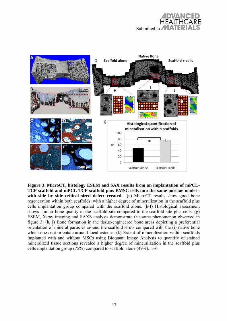
Figure 3. MicroCT, histology ESEM and SAX results from an implantation of mPCL-TCP scaffold and mPCL-TCP scaffold plus BMSC cells into the same porcine model - with side by side critical sized defect created. (a) MicroCT results show good bone regeneration within both scaffolds, with a higher degree of mineralization in the scaffold plus cells implantation group compared with the scaffold alone. (b-f) Histological assessment shows similar bone quality in the scaffold site compared to the scaffold site plus cells. (g) ESEM, X-ray imaging and SAXS analysis demonstrate the same phenomenon observed in figure 3. (h, j) Bone formation in the tissue-engineered bone areas depicting a preferential orientation of mineral particles around the scaffold struts compared with the (i) native bone which does not orientate around local osteons. (k) Extent of mineralization within scaffolds implanted with and without MSCs using Bioquant Image Analysis to quantify of stained mineralized tissue sections revealed a higher degree of mineralization in the scaffold plus cells implantation group (75%) compared to scaffold alone (49%). n=6.