Bacillus subtilis 5′-nucleotidases with various functions ...
13
RESEARCH ARTICLE Open Access Bacillus subtilis 5′-nucleotidases with various functions and substrate specificities Ayako Terakawa 1 , Ayane Natsume 1 , Atsushi Okada 1 , Shogo Nishihata 1 , Junko Kuse 1 , Kosei Tanaka 2 , Shinji Takenaka 1,2 , Shu Ishikawa 3 and Ken-ichi Yoshida 2,3* Abstract Background: In Escherichia coli, nagD, yrfG, yjjG, yieH, yigL, surE, and yfbR encode 5′-nucleotidases that hydrolyze the phosphate group of 5′-nucleotides. In Bacillus subtilis, genes encoding 5′-nucleotidase have remained to be identified. Results: We found that B. subtilis ycsE, araL, yutF, ysaA, and yqeG show suggestive similarities to nagD. Here, we expressed them in E. coli to purify the respective His 6 -tagged proteins. YcsE exhibited significant 5′-nucleotidase activity with a broader specificity, whereas the other four enzymes had rather weak but suggestive activities with various capacities and substrate specificities. In contrast, B. subtilis yktC shares high similarity with E. coli suhB encoding an inositol monophosphatase. YktC exhibited inositol monophosphatase activity as well as 5′-nucleotidase activity preferential for GMP and IMP. The ycsE, yktC, and yqeG genes are induced by oxidative stress and were dispensable, although yqeG was required to maintain normal growth on solid medium. In the presence of diamide, only mutants lacking yktC exhibited enhanced growth defects, whereas the other mutants without ycsE or yqeG did not. Conclusions: Accordingly, in B. subtilis, at least YcsE and YktC acted as major 5′-nucleotidases and the four minor enzymes might function when the intracellular concentrations of substrates are sufficiently high. In addition, YktC is involved in resistance to oxidative stress caused by diamide, while YqeG is necessary for normal colony formation on solid medium. Keywords: Bacillus subtilis, Haloacid dehalogenase superfamily, Inositol monophosphatase, Inositol phosphate, Nucleoside/nucleotide metabolism, 5′-nucleotidase, Oxidative stress, Phosphatase, Protein motif Background The pool sizes of nucleotides and nucleosides are bal- anced to enable the efficient synthesis of DNA and RNA [1]. Numerous enzymes involved in the biosynthesis and catabolism of nucleic acids are controlled to regulate the appropriate pool size of each compound. For example, 5′-nucleotidases hydrolyze 5′-nucleotides to generate nucleosides and inorganic phosphate, which participate in the regulatory mechanism that opposes the generation of nucleotides with the phosphorylation of nucleosides catalyzed by kinases. 5′-Nucleotidases are ubiquitous among species and reside in different subcellular locations. Extracellular 5′- nucleotidases are produced by certain bacteria. For ex- ample, Vibrio parahaemolyticus NutA is a 5′-nucleotidase bound to the membrane by a lipid anchor [2]. Escherichia coli UshA is a periplasmic enzyme with 5′-nucleotidase and UDP-sugar hydrolase activities [3]. These 5′-nucleo- tidases degrade extracellular nucleotides to satisfy the cell’ s nutritional requirements. In contrast, bacteria produce numerous intracellular 5′-nucleotidases that belong to various enzyme families. In E. coli, the substrates of NagD are UMP, GMP, AMP, and CMP [4]. NagD belongs to the haloacid dehalogenase superfamily (HADSF) characterized by a specific protein motif [5]. The HADSF family comprises numerous pro- teins in organisms ranging from prokaryotes to higher eu- karyotes, including humans. The vast majority of enzymes * Correspondence: [email protected] 2 Organization of Advanced Science and Technology, Kobe University, 1-1 Rokkodai, Nada, Kobe, Hyogo 657-8501, Japan 3 Department of Science, Technology and Innovation, Kobe University, 1-1 Rokkodai, Nada, Kobe, Hyogo 657-8501, Japan Full list of author information is available at the end of the article © The Author(s). 2016 Open Access This article is distributed under the terms of the Creative Commons Attribution 4.0 International License (http://creativecommons.org/licenses/by/4.0/), which permits unrestricted use, distribution, and reproduction in any medium, provided you give appropriate credit to the original author(s) and the source, provide a link to the Creative Commons license, and indicate if changes were made. The Creative Commons Public Domain Dedication waiver (http://creativecommons.org/publicdomain/zero/1.0/) applies to the data made available in this article, unless otherwise stated. Terakawa et al. BMC Microbiology (2016) 16:249 DOI 10.1186/s12866-016-0866-5
Transcript of Bacillus subtilis 5′-nucleotidases with various functions ...
RESEARCH ARTICLE Open Access
Bacillus subtilis 5′-nucleotidases with variousfunctions and substrate specificitiesAyako Terakawa1, Ayane Natsume1, Atsushi Okada1, Shogo Nishihata1, Junko Kuse1, Kosei Tanaka2,Shinji Takenaka1,2, Shu Ishikawa3 and Ken-ichi Yoshida2,3*
Abstract
Background: In Escherichia coli, nagD, yrfG, yjjG, yieH, yigL, surE, and yfbR encode 5′-nucleotidases that hydrolyze thephosphate group of 5′-nucleotides. In Bacillus subtilis, genes encoding 5′-nucleotidase have remained to beidentified.
Results: We found that B. subtilis ycsE, araL, yutF, ysaA, and yqeG show suggestive similarities to nagD. Here, weexpressed them in E. coli to purify the respective His6-tagged proteins. YcsE exhibited significant 5′-nucleotidaseactivity with a broader specificity, whereas the other four enzymes had rather weak but suggestive activities withvarious capacities and substrate specificities. In contrast, B. subtilis yktC shares high similarity with E. coli suhBencoding an inositol monophosphatase. YktC exhibited inositol monophosphatase activity as well as 5′-nucleotidaseactivity preferential for GMP and IMP. The ycsE, yktC, and yqeG genes are induced by oxidative stress and weredispensable, although yqeG was required to maintain normal growth on solid medium. In the presence of diamide,only mutants lacking yktC exhibited enhanced growth defects, whereas the other mutants without ycsE or yqeGdid not.
Conclusions: Accordingly, in B. subtilis, at least YcsE and YktC acted as major 5′-nucleotidases and the fourminor enzymes might function when the intracellular concentrations of substrates are sufficiently high. Inaddition, YktC is involved in resistance to oxidative stress caused by diamide, while YqeG is necessary fornormal colony formation on solid medium.
Keywords: Bacillus subtilis, Haloacid dehalogenase superfamily, Inositol monophosphatase, Inositol phosphate,Nucleoside/nucleotide metabolism, 5′-nucleotidase, Oxidative stress, Phosphatase, Protein motif
BackgroundThe pool sizes of nucleotides and nucleosides are bal-anced to enable the efficient synthesis of DNA and RNA[1]. Numerous enzymes involved in the biosynthesis andcatabolism of nucleic acids are controlled to regulate theappropriate pool size of each compound. For example,5′-nucleotidases hydrolyze 5′-nucleotides to generatenucleosides and inorganic phosphate, which participatein the regulatory mechanism that opposes the generationof nucleotides with the phosphorylation of nucleosidescatalyzed by kinases.
5′-Nucleotidases are ubiquitous among species andreside in different subcellular locations. Extracellular 5′-nucleotidases are produced by certain bacteria. For ex-ample,Vibrio parahaemolyticus NutA is a 5′-nucleotidasebound to the membrane by a lipid anchor [2]. Escherichiacoli UshA is a periplasmic enzyme with 5′-nucleotidaseand UDP-sugar hydrolase activities [3]. These 5′-nucleo-tidases degrade extracellular nucleotides to satisfy the cell’snutritional requirements.In contrast, bacteria produce numerous intracellular
5′-nucleotidases that belong to various enzyme families.In E. coli, the substrates of NagD are UMP, GMP, AMP,and CMP [4]. NagD belongs to the haloacid dehalogenasesuperfamily (HADSF) characterized by a specific proteinmotif [5]. The HADSF family comprises numerous pro-teins in organisms ranging from prokaryotes to higher eu-karyotes, including humans. The vast majority of enzymes
* Correspondence: [email protected] of Advanced Science and Technology, Kobe University, 1-1Rokkodai, Nada, Kobe, Hyogo 657-8501, Japan3Department of Science, Technology and Innovation, Kobe University, 1-1Rokkodai, Nada, Kobe, Hyogo 657-8501, JapanFull list of author information is available at the end of the article
© The Author(s). 2016 Open Access This article is distributed under the terms of the Creative Commons Attribution 4.0International License (http://creativecommons.org/licenses/by/4.0/), which permits unrestricted use, distribution, andreproduction in any medium, provided you give appropriate credit to the original author(s) and the source, provide a link tothe Creative Commons license, and indicate if changes were made. The Creative Commons Public Domain Dedication waiver(http://creativecommons.org/publicdomain/zero/1.0/) applies to the data made available in this article, unless otherwise stated.
Terakawa et al. BMC Microbiology (2016) 16:249 DOI 10.1186/s12866-016-0866-5
of the HADSF family are phosphoryl transferases, al-though the superfamily was named after 2-haloacid deha-logenase, because it is the first structurally characterizedmember. When the amino acid sequences of their entirecoding regions are compared, similarities among theHADSF members are not usually very high (15–30 %identical), although their central regions involved in cata-lytic activity are relatively conserved. Further, correlationbetween structure and catalytic activity is frequently ob-served and interrelates with similarities among the struc-tures of substrates [6–8]. E. coli produces other HADSF-family enzymes such as YrfG, YjjG, YieH, and YigL, andeach exhibits 5′-nucleotide phosphatase activity [9, 10].In addition, E. coli produces SurE and YfbR, which
exhibit 5′-nucleotidase activity and do not belong tothe HADSF family [10]. SurE shows broad substratespecificity, by dephosphorylating 5′-nucleotides as wellas 3′-nucleotides, with highest affinity for 3′-AMP.SurE hydrolyzes polyphosphate with preference forshort-chain substrates. Homologs of E. coli surE arepresent in numerous eubacteria and archaea, and SurErepresents a family of metal-dependent phosphatases[11]. YfbR belongs to the HD domain superfamily ofmetal-dependent phosphatases and phosphodiesterases,and the HD domain was named after a protein motifwith predicted catalytic residues containing the conserveddoublet His–Asp [12]. YfbR specifically hydrolyzes 5′-deoxyribonucleotides.Inositol monophosphatase denotes phosphatases
that liberate inorganic phosphate from myo-inositol 1-monophosphate (MIMP). The mammalian and plantenzymes are involved in the metabolism of inositolphospholipids, including the generation and degrad-ation of inositol phosphates and phosphatidylinositolsthat mediate cellular signal transduction [13]. Inositolmonophosphatases are present in diverse organismssuch as bacteria and higher eukaryotes, and evolvedfrom a common ancestral gene [14, 15]. Mammalianinositol monophosphatases exhibit relatively broadsubstrate specificity for phosphate-containing compounds[16, 17]. For example, the enzyme isolated from rat testishydrolyzes adenosine 2′-monophosphate [16] and thebovine brain enzyme hydrolyzes β-glycerophosphateand adenosine 2′-monophosphate [17]. E. coli suhB en-codes a protein homologous to eukaryotic inositolmonophosphatases with equivalent activities [18].However, phosphatidylinositol is not present in E. coli
usually [19], and its physiological function in cells is un-known. In contrast, E. coli SuhB exhibits wider substratespecificity and hydrolyzes β-glycerophosphate and ad-enosine 2′-monophosphate as well [18]. Thus, we hy-pothesized that bacterial inositol monophosphatasesmight also function as 5′-nucleotidase. Moreover, variousE. coli enzymes likely dephosphorylate 5′-nucleotides with
different specificities and functions and may therefore acttogether to maintain the sizes of the intracellular pools ofnucleotides and nucleosides.In Bacillus subtilis, the partial nucleotide limitation in-
duces 5′-nucleotidase activity [20], and B. subtilis is usedfor fermentative inosine production, which may involvethe dephosphorylation of IMP catalyzed by an unidenti-fied 5′-nucleotidase [21]. However, B. subtilis genes en-coding 5′-nucleotidase have remained to be identified.Here we selected B. subtilis genes potentially encoding
5′-nucleotidases [22] and expressed them in E. coli tocharacterize their function. We identified two major andfour minor genes encoding 5′-nucleotidases, with vari-ous functions and substrate specificities. A major 5′-nu-cleotidase was involved in resistance to oxidative stress,and a minor enzyme was required for normal growth onsolid medium.
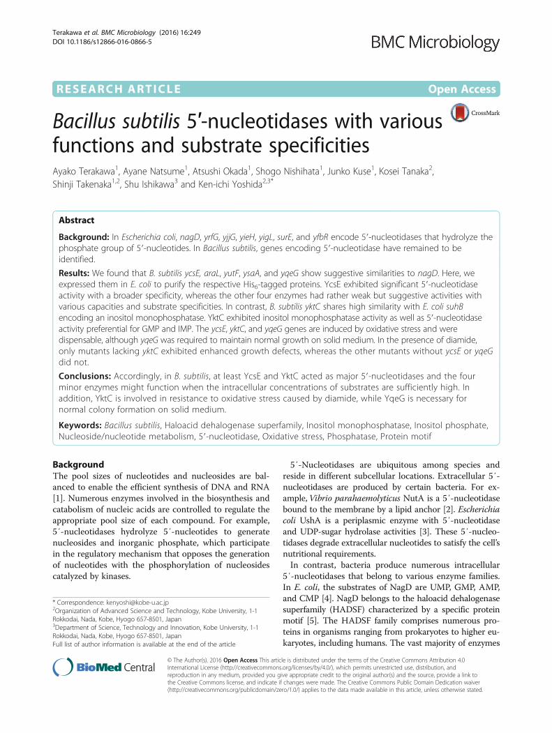
ResultsSelection of B. subtilis genes similar to E. coli genesencoding 5′-nucleotidaseIn E. coli, at least nagD, yrfG, yjjG, yieH, yigL, surE, andyfbR encode 5′-nucleotidases, and their gene productsare classified in the families as follows: HADSF (NagD,YrfG, YjjG, YieH, and YigL), SurE, and HD domain(YfbR). In the B. subtilis genome, no gene shares signifi-cant homology with surE or yfbR. However, gene prod-ucts of araL, yutF, yqeG, ysaA, ycsE, gapB, ftsA, and hprPshare some suggestive similarities with that of E. colinagD (Fig. 1); AraL (65.8 % similarity/272 aa overlap),
Fig. 1 Similarities among the amino acid sequences of the proteinsencoded by E. coli nagD homologs in the B. subtilis genome. Theamino acid sequences of the proteins encoded by B. subtilis araL,yutF, yqeG, ysaA, ycsE, gapB, ftsA, and hprP, and E. coli nagD wereanalyzed using CLUSTALW (default fast/approximate settings [http://www.genome.jp/tools/clustalw/]) to generate the phylogenic tree
Terakawa et al. BMC Microbiology (2016) 16:249 Page 2 of 13
YutF (73.0 %/256 aa), YqeG (15.7 %/172 aa), YsaA(16.9 %/260 aa), YcsE (51.4 %/249 aa), GapB (6.8 %/340aa), FtsA (9.5 %/440 aa), and HprP (15.7 %/216 aa).Among them, AraL, YutF, YqeG, YsaA, YcsE, and HprPwere supposed to belong to HADSF, since their amino acidsequences contain the HADSF motif (http://pfam.xfam.org/family/PF00702.24).The araL gene resides within the L-arabinose operon,
which encodes enzymes required for the degradation ofL-arabinose and is under the control of AraR repressorto be induced in the presence of L-arabinose [23]. AraLwas predicted to be a phosphatase that hydrolyzes cer-tain sugar-phosphates [23].The yutF gene encodes a protein that was predicted to
be involved in N-acetyl-glucosamine catabolism and simi-lar to 4-nitrophenyl phosphatase [24]. The condition-dependent transcriptome analysis revealed that yutF wasalmost constitutively expressed at low levels [25].The yqeG, ysaA, and ycsE genes encode a putative
HADSF-family enzyme of unknown function. The yqeGgene is the first gene of the long operon comprising yqeG,yqeH, aroD, yqeI, nadD, yqeK, yqeL, and yqeM. This op-eron encodes essential genes [26], which are expressedconstitutively at significant levels that increase during ger-mination and under conditions of oxidative stress [25].The ysaA and ycsE genes are constitutively expressed andare induced, respectively, during the nutritional shift frommalate to malate plus glucose and in the presence of etha-nol or diamide [25].The products of gapB, ftsA, and hprP show relatively
lower similarities to NagD (Fig. 1). GapB is NADP-dependent glyceraldehyde-3-phosphate dehydrogenase,which is involved in gluconeogenesis [27]. FtsA is anactin-like ATPase involved in cell division [28]. AndHprP is a P-Ser-HPr phosphatase involved in carbon ca-tabolite repression [29].
5′-Nucleotidase activity of B. subtilis enzymes sharingsome similarities with NagDOf the eight B. subtilis genes that share some similaritieswith nagD as described above, gapB, ftsA, and hprP haverespective and specific functions other than as 5′-nucle-otidases [27–29]. Therefore, these genes were excludedfrom the present study. We cloned and expressed araL,yutF, yqeG, ysaA, and ycsE in E. coli as His6-tag fusionproteins.The His6-tag fusion proteins were purified to form re-
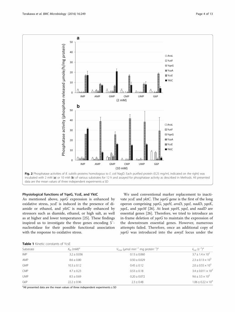
spective single bands in sodium dodecyl sulfate poly-acrylamide gel electrophoresis (SDS-PAGE, data notshown), and were subjected to phosphatase assays usingthe substrates as follows: AMP, CMP, GMP, IMP, UMP,and glucose 6-phosphate (G6P) (Fig. 2). G6P was includedbecause it is the substrate of certain HADSF enzymes [9].YcsE appeared to be the most efficient phosphatase acting
on all six substrates at a lower concentration (2 mM), indi-cating its broad substrate specificity with preference forIMP, CMP, and G6P. Its activity increased in the presenceof 10 mM substrates, particularly AMP, GMP, and UMP.The other four enzymes were relatively less active even atthe higher substrate concentration. All 5′-nucleotidesserved almost equally as substrates for AraL at lower andhigher concentrations, and AraL activity was enhancedparticularly against G6P at the higher concentration. YutFexhibited purine 5′-nucleotidase activity with the sub-strates GMP and IMP, but only at the higher concentra-tion, and did not hydrolyze pyrimidine nucleotides andG6P. YqeG hydrolyzed GMP and G6P only at the higherconcentration. YsaA activity was weakest with purine-nucleotide substrates at the higher concentration.The catalytic properties of YcsE are shown in Table 1.
The substrate specificity of YcsE was broad with higherKM values, suggesting that it efficiently dephosphory-lated these substrates at higher concentrations. In con-trast, the properties of the other four enzymes were notdetermined because of their lower activities. Neverthe-less, the data do not exclude their physiological functionas 5′-nucleotidases, because the activities of two of the5′-nucleotidases YieH and YigL identified previously inE. coli were too low to determine their enzymatic prop-erties [9].
5′-Nucleotidase activity of SuhB homolog in B. subtilisAs described above, E. coli SuhB is an inositol monopho-sphatase, which exhibits wider substrate specificity, as ithydrolyzes β-glycerophosphate and adenosine 2′-mono-phosphate as well [18]. Thus, we hypothesized that bac-terial inositol monophosphatases might also function as5′-nucleotidase.The B. subtilis yktC gene, which is the homolog of E.
coli suhB (31.7 % similarity of 265 overlapping aminoacid residues), was cloned and expressed as a His6-tagged protein in E. coli. His6-tagged YktC was purifiedto form a single band in SDS-PAGE (data not shown)and was subjected to phosphatase assays in the presenceof various substrates (Fig. 2, Table 2). The inositolmonophosphatase activity of YktC was indeed demon-strated by its efficient ability to hydrolyze MIMP, and italso turned out to exhibit 5′-nucleotidase activitypreferentially against IMP and GMP as substrates. Toour knowledge, this is the first identification of an in-ositol monophosphatase with 5′-nucleotidase activity.Compared with E. coli SuhB, YktC hydrolyzed G6Pless efficiently, while β-glycerophosphate more effi-ciently [18]. Expression of yktC is mainly constitutiveand is markedly enhanced by stressors such as diamide,ethanol, or high salt as well as at higher and lower temper-atures [25].
Terakawa et al. BMC Microbiology (2016) 16:249 Page 3 of 13
Physiological functions of YqeG, YcsE, and YktCAs mentioned above, yqeG expression is enhanced byoxidative stress, ycsE is induced in the presence of di-amide or ethanol, and yktC is markedly enhanced bystressors such as diamide, ethanol, or high salt, as wellas at higher and lower temperatures [25]. These findingsinspired us to investigate the three genes encoding 5′-nucleotidase for their possible functional associationwith the response to oxidative stress.
We used conventional marker replacement to inacti-vate ycsE and yktC. The yqeG gene is the first of the longoperon comprising yqeG, yqeH, aroD, yqeI, nadD, yqeK,yqeL, and yqeM [26]. At least yqeH, yqeI, and nadD areessential genes [26]. Therefore, we tried to introduce anin-frame deletion of yqeG to maintain the expression ofthe downstream essential genes. However, numerousattempts failed. Therefore, once an additional copy ofyqeG was introduced into the amyE locus under the
Table 1 Kinetic constants of YcsE
Substrate KM (mM)a Vmax (μmol min−1 mg protein−1)a kcat (s−1)a
IMP 3.2 ± 0.036 0.13 ± 0.060 3.7 ± 1.4 × 10²
AMP 8.6 ± 0.80 0.50 ± 0.029 2.3 ± 0.13 × 10³
GMP 10.3 ± 0.12 0.45 ± 0.12 2.0 ± 0.55 × 10³
CMP 4.7 ± 0.23 0.53 ± 0.18 3.4 ± 0.011 × 10³
UMP 8.5 ± 0.69 0.20 ± 0.072 9.6 ± 3.5 × 10²
G6P 22.2 ± 0.96 2.3 ± 0.48 1.06 ± 0.22 × 10⁴
aAll presented data are the mean values of three independent experiments ± SD
Fig. 2 Phosphatase activities of B. subtilis proteins homologous to E. coli NagD. Each purified protein (0.25 mg/ml, indicated on the right) wasincubated with 2 mM (a) or 10 mM (b) of various substrates for 12 h and assayed for phosphatase activity as described in Methods. All presenteddata are the mean values of three independent experiments ± SD
Terakawa et al. BMC Microbiology (2016) 16:249 Page 4 of 13
control of spac promoter, which is conditionally inducedby IPTG. And then, in the presence of IPTG, the existingyqeG gene was deleted using marker replacement so thatthe genes downstream were constitutively expressed byread-through from the kanamycin-resistance cassette(strain NON05). Regardless of the presence and absenceof IPTG, strain NON05 grew in liquid medium (Fig. 3),suggesting that yqeG might be dispensable in planktonicgrowth. However, the spac promoter is somewhat leaky; asignificant basal level of expression still exists in the ab-sence of IPTG [30]. Therefore, in the absence of IPTG,there might be some production of YqeG, and this mightbe sufficient for planktonic growth. Nevertheless, NON05formed fewer colonies in the absence of IPTG (Fig. 4),suggesting that yqeG was required to maintain normalgrowth particularly on solid medium. Similarly, less effi-cient colony-formation was exhibited by the triple mutantNON06 in the absence of IPTG, and colony formationwas slightly decreased when ycsE and yktC were inacti-vated additionally.The addition of diamide to liquid medium arrested the
growth of the parental strain 168 (Fig. 3). The growthcharacteristics of the mutant NON01 lacking ycsE werecomparable to those of the parental strain in the pres-ence of diamide. Conversely, the mutants NON02 andNON03 lacking yktC exhibited the enhanced growth de-fect in the presence of the higher concentration of di-amide (4 mM), as the optical density for cells decreasedsignificantly possibly because of cell lysis. The resultssuggest that yktC, but not ycsE, contributed to resistanceto oxidative stress induced by diamide. In contrast,NON05 did not exhibit significantly increased sensitivityto diamide, depending on the repression of yqeG in theabsence of IPTG (Fig. 3). The triple-mutant NON06 ex-hibited the growth defect similar to that of NON02 orNON03 in the presence of diamide (Fig. 3), and thegrowth of NON06 did not depend on IPTG. Therefore,the conditional expression of yqeG was not associatedwith the susceptibility to diamide. Taken together, thedata indicate that only YktC was involved in the resist-ance to oxidative stress.
DiscussionIn eukaryotes, multiple nucleotidases and nucleosidekinases control the intracellular concentration of nucle-otides [31–33]. Nucleotidases play a role in the main-tenance of a constant nucleoside supply, which isrequired to maintain RNA metabolism and biosynthesisof cell wall [1]. In bacteria, numerous nucleotidaseswith different substrate specificities control the intra-cellular nucleotide pool [34], and it is believed that theaccumulation of intracellular nucleotides is potentiallytoxic and therefore must be prevented [31–33].Here we show that B. subtilis YktC and YcsE func-
tioned as 5′-nucleotidases, suggesting that they may rep-resent two of the main enzymes involved in intracellularnucleoside/nucleotide metabolism. The other enzymessimilar to E. coli NagD, which belong to the HADSFfamily (YutF, YsaA, YqeG, and AlaL), exhibited lower ac-tivity, although the data suggest they may function asminor 5′-nucleotidases. HADSF phosphatases often havebroader substrate specificity and hydrolyze various phos-phorylated compounds [5]. Thus, 5′-nucleotidases withlower substrate affinity might contribute to the balanceof intracellular nucleotide concentrations as well as thatof other phosphorylated metabolites. The KM values ofE. coli NagD for UMP, CMP, GMP, AMP, and G6P are0.16 ± 0.038, 1.47 ± 0.044, 0.40 ± 0.130, 0.84 ± 0.250, and5.90 ± 0.750 mM, respectively [4], which are smaller thanthose of YcsE and YktC (Tables 1 and 2, respectively).Therefore, B. subtilis YktC and YcsE might be less effi-cient as 5′-nucleotidase than NagD, but it is still sug-gestive that they may be able to function as two of the5′-nucleotidases that are the first to respond to the ac-cumulation of nucleotides, and the other enzymes mayfunction when the concentration of nucleotides is in farexcess.Some of the genes encoding 5′-nucleotidases might be
induced upon partial nucleotide limitation as previouslyreported [20]. Interestingly, the expression of yqeG isenhanced under oxidative stress, that of ycsE is inducedby diamide or ethanol, and that of yktC is markedly en-hanced in the presence of the various stressors, including
Table 2 Kinetic constants of YktC
Substrate KM (mM)a Vmax (μmol min−1 mg protein−1)a kcat (s−1)a
MIMP 0.076 ± 0.006 0.82 ± 0.16 2.9 ± 0.33 × 10³
IMP 1.1 ± 0.083 0.078 ± 0.0084 1.8 ± 0.59 × 10²
AMP 1.8 ± 0.13 0.030 ± 0.00022 5.3 ± 0.039 × 101
GMP 1.9 ± 0.026 0.169 ± 0.028 3.0 ± 0.34 × 10²
CMP 1.6 ± 0.017 0.049 ± 0.014 1.5 ± 0.52 × 10²
UMP 2.4 ± 0.052 0.11 ± 0.0099 9.8 ± 4.6 × 10²
G6P 2.6 ± 0.51 0.021 ± 0.010 3.7 ± 1.3 × 101
β-glycerophosphate 0.49 ± 0.035 1.2 ± 0.32 5.7 ± 1.2 × 10²
aAll presented data are the mean values of three independent experiments ± SD
Terakawa et al. BMC Microbiology (2016) 16:249 Page 5 of 13
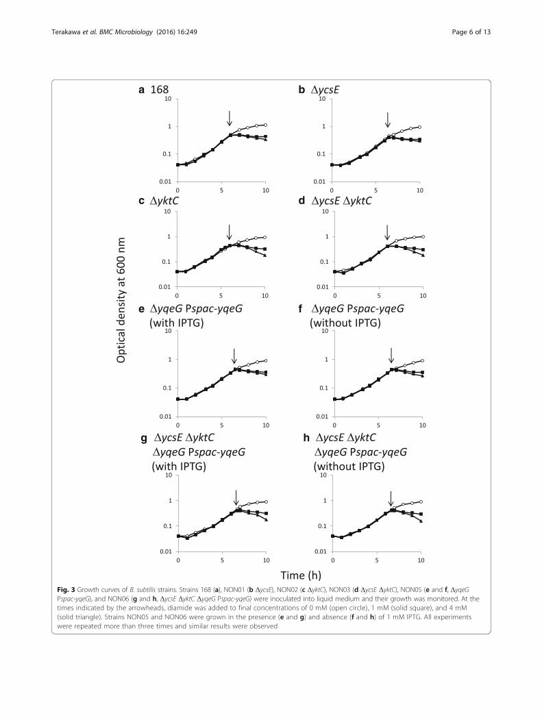
Fig. 3 Growth curves of B. subtilis strains. Strains 168 (a), NON01 (b ΔycsE), NON02 (c ΔyktC), NON03 (d ΔycsE ΔyktC), NON05 (e and f, ΔyqeGPspac-yqeG), and NON06 (g and h, ΔycsE ΔyktC ΔyqeG Pspac-yqeG) were inoculated into liquid medium and their growth was monitored. At thetimes indicated by the arrowheads, diamide was added to final concentrations of 0 mM (open circle), 1 mM (solid square), and 4 mM(solid triangle). Strains NON05 and NON06 were grown in the presence (e and g) and absence (f and h) of 1 mM IPTG. All experimentswere repeated more than three times and similar results were observed
Terakawa et al. BMC Microbiology (2016) 16:249 Page 6 of 13
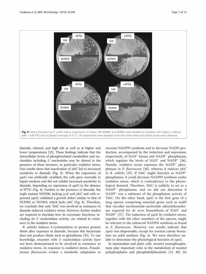
diamide, ethanol, and high salt as well as at higher andlower temperatures [25]. These findings indicate that theintracellular levels of phosphorylated metabolites and nu-cleotides including 5′-nucleotides may be altered in thepresence of these stressors, in particular oxidative stress.Our results show that inactivation of yktC led to increasedsensitivity to diamide (Fig. 4). When the expression ofyqeG was artificially modified, the cells grew normally inliquid medium and did not exhibit increased sensitivity todiamide, depending on repression of yqeG in the absenceof IPTG (Fig. 4). Further, in the presence of diamide, thetriple mutant NON06, lacking ycsE and yktC and with re-pressed yqeG, exhibited a growth defect similar to that ofNON02 or NON03, which lacks yktC (Fig. 4). Therefore,we conclude that only YktC was involved in resistance todiamide-induced oxidative stress. However, further studiesare required to elucidate how its enzymatic functions, in-cluding its 5′-nucleotidase activity, are related to resist-ance to the oxidative stress.B. subtilis induces S-cysteinylation to protect protein
thiols after exposure to diamide, because this bacteriumdoes not produce thiols such as glutathione [35]. To ourknowledge, enzymes with 5′-nucleotidase activity havenot been demonstrated to be involved in resistance tooxidative stress. In response to oxidative stress, Pseudo-monas fluorescens evokes a metabolic adaptation to
increase NADPH synthesis and to decrease NADH pro-duction, accompanied by the induction and repression,respectively, of NAD+ kinase and NADP+ phosphatase,which regulate the levels of NAD+ and NADP+ [36].Namely, oxidative stress represses the NADP+ phos-phatase in P. fluorescens [36], whereas it induces yktCin B. subtilis [25]. If YktC might function as NADP+
phosphatase, it could decrease NADPH synthesis underoxidative stress, which is contradictory to the physio-logical demand. Therefore, YktC is unlikely to act as aNADP+ phosphatase, and we did not determine ifNADP+ was a substrate of the phosphatase activity ofYktC. On the other hand, yqeG is the first gene of along operon comprising essential genes such as nadDthat encodes nicotinamide-nucleotide adenylyltransfer-ase required for de novo biosynthesis of NAD+ andNADP+ [37]. The induction of yqeG by oxidative stress,together with the other members of the operon, mightbe relevant to the enhanced NADPH synthesis as foundin P. fluorescens. However, our results indicate thatyqeG was dispensable, except for normal colony forma-tion on solid medium (Fig. 4). We were therefore un-able to determine the physiological function of yqeG.In mammalian and plant cells, inositol monophospha-
tases play important roles in the metabolism of inositolpolyphosphates and phosphatidylinositols [13, 38]. De
Fig. 4 Colony formation by B. subtilis strains. Suspensions of strains 168, NON05, and NON06 were streaked on LB plates with (right) or without(left) 1 mM IPTG and incubated overnight at 37 °C. All experiments were repeated more than three times and similar results were observed
Terakawa et al. BMC Microbiology (2016) 16:249 Page 7 of 13
novo phosphatidylinositol synthesis in mammals andSaccharomyces cerevisiae involves a reaction that com-bines CDP-diacylglycerol with myo-inositol. myo-Inositolis produced through the isomerization of glucose 6-phosphate to form MIMP [39] and the subsequent de-phosphorylation of MIMP by inositol monophosphatase[16]. Further, MIMP is produced by the dephosphorylationof the second messenger inositol polyphosphates [40]. InE. coli and B. subtilis, however, phosphatidylinositols arenot components of phospholipids usually present in thecell membrane [41, 42], and inositol 1-phosphate synthasehas not been identified. Therefore, it is unlikely thatMIMP occurs naturally in these bacteria and might serveas the physiological substrate of E. coli SuhB and B. subti-lis YktC. In E. coli, a decrease in intracellular SuhB levelscompensated the defects in secY and rpoH [43, 44]. ThesecY gene encodes a membrane component involved inprotein secretion [45], and rpoH encodes σ32, a compo-nent of the RNA polymerase involved in heat-shock in-duction [46]. Therefore, defects in secY and rpoH impairsecretion and the heat-shock response, respectively, whichare involved in posttranslational quality control of proteins[18]. Therefore, SuhB may control the rates of peptidechain elongation and protein folding [42]. In contrast, theSuhB homolog of P. aeruginosa plays an important role inpathogenesis to control the genes required for acute andchronic infection [47]. Burkholderia cepacia SuhB is re-quired for the secretion of proteins associated with mo-tility and biofilm formation [48], suggesting that thephysiological functions of SuhB homologs are complex,which may be true for B. subtilis YktC. Nevertheless, itis intriguing that MIMP is the best substrate for theseenzymes. Kozloff and the colleagues detected a smallamount of phosphatidylinositol from E. coli cells, im-plying the possibility of a weak phosphatidylinositolbiosynthesis under some specific conditions [49]. E. coliSuhB and B. subtilis YktC might be involved in bacterialmetabolism of phosphatidylinositol, which has not beencharacterized yet.The broad substrate specificity of YktC revealed here
implies the association of this enzyme with sugar-phosphate stress. Although sugars serve as energy andcarbon sources, sugar phosphates are produced during theirmetabolism. Excess accumulation of sugar phosphates im-pairs cell growth [50] and may trigger cell death [51, 52]through an unknown mechanism. Similarly, accumulationof nucleotides is detrimental to the cell [34]. Therefore, byhydrolyzing sugar phosphates as well as nucleotides, YktCmay play a role in correcting imbalances of metabolites. Forexample, we found that the KM value of YktC for G6Pwas 2.6 ± 0.51 mM, which is greater than the reportedintracellular concentrations of glucose 6-phosphate inE. coli (0.8–2.0 mM) [53]. However, under certain condi-tions, the intracellular concentration of G6P in Lactococcus
lactis reaches 20–50 mM [54], indicating that G6P accumu-lates to higher levels in bacterial cells. Accordingly, in thepresence of excess intracellular concentrations of G6P, YktCmay hydrolyze it to intervene in glucose metabolism. Thismay be true as well for YcsE, because it hydrolyzed G6P invitro more efficiently than YktC (Fig. 2). Similarly, YktCmay hydrolyze β-glycerophosphate, which is a biosyntheticprecursor of phospholipids [55], to interfere with the main-tenance of the cell membrane. Therefore, their phosphataseactivities with broad-specificity may exert pleiotropic effectson metabolism and other cellular functions. It would beworthwhile to perform metabolomic analyses on the mu-tants lacking yktC and ycsE to determine concentrations ofthe various metabolites, including 5′-nucleotides and othersugar phosphates, which would reinforce the hypothesisconcerning the role of these enzymes.Together with sodium glutamate, IMP and GMP are
generally used as food additives to enhance taste. Indus-trial production of IMP is achieved using enzymatic con-version of inosine into IMP. B. subtilis strains producinglarge amounts of inosine in the fermentation mediumwere generated [56–60], and 5′-nucleotidase activity ofthese strains is remarkably increased, which may convertIMP into inosine [59]. Thus, optimizing the expressionof genes encoding 5′-nucleotidases such as those studiedhere may further improve the fermentation of inosineproduction.
ConclusionsB. subtilis ycsE, araL, yutF, ysaA, and yqeG show sug-gestive similarities to E. coli nagD encoding 5′-nucleo-tidase. Among the five, only YcsE exhibited significant5′-nucleotidase activity with a broader specificity, whereasthe other four enzymes had rather weak but suggestive ac-tivities with various capacities and substrate specificities.Interestingly, YqeG was required to maintain normalgrowth on solid medium. On the other hand, B. subtilisyktC shares high similarity with E. coli suhB encoding in-ositol monophosphatase, the gene product of which had5′-nucleotidase activity preferential for GMP and IMP,and turned out to be involved in resistance to oxidativestress.
MethodsBacterial strains, plasmids, oligonucleotide primers, andculture conditionsBacterial strains and plasmids are listed in Table 3. Bac-terial strains were usually grown aerobically at 37 °Cand maintained in Luria-Bertani (LB) medium (BectonDickinson, NJ, USA) containing 50 mg/l ampicillin and50 mg/l kanamycin as required. Oligonucleotide primersare listed in Table 4. To assess the resistance to oxidativestress of B. subtilis strains, cells were inoculated into LBand allowed to grow. The oxidizing agent diamide was
Terakawa et al. BMC Microbiology (2016) 16:249 Page 8 of 13
added to the exponentially growing culture at various con-centrations, and growth was continuously monitored.
Construction of bacterial strainsE. coli strains expressing B. subtilis genes were con-structed as follows: DNA fragments corresponding tothe open reading frames of genes of interest were PCR-amplified using B. subtilis strain 168 chromosomal DNA(Table 3) as template and pairs of respective oligo-nucleotide primers with generation of unique restrictionsites at their 5′-termini (araL-F/araL-R for araL, ycsE-F/ycsE-R for ycsE, yktC-F/yktC-R for yktC, yqeG-F/yqeG-R for yqeG, ysaA-F/ysaA-R for ysaA, and yutF-F/yutF-Rfor yutF) (Table 4). Each fragment was cloned into thepMD20-T plasmid using the Mighty TA-cloning kit(Takara Bio, Otsu, Japan) to yield pMD20-araL, pMD20-
ycsE, pMD20-yktC, pMD20-yqeG, pMD20-ysaA, andpMD20-yutF, which were used to transform E. coli strainDH5α. After performing nucleotide sequencing to con-firm the constructs, each recombinant plasmid wasdigested with the appropriate restriction enzyme(s) toprepare a DNA fragment corresponding to its respectiveopen reading frame and then ligated to the arms ofpET28b (Takara Bio) digested with the same enzyme(s).The plasmids pET28b-araL, pET28B-ycsE, pET28B-yktC,pET28B-yqeG, pET28B-ysaA, and pET28B-yutF wereconstructed to express each protein as a C-terminal His6-fusion, and expression was controlled by the T7 promoterin E. coli strain BL21(DE3) (Takara Bio).Mutant strains of B. subtilis lacking ycsE and yktC
were constructed using conventional marker replace-ment as follows (Additional file 1: Figure S1): To delete
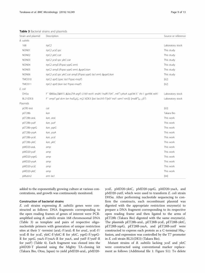
Table 3 Bacterial strains and plasmids
Strain and plasmid Description Source or reference
B. subtilis
168 trpC2 Laboratory stock
NON01 trpC2 ycsE::spc This study
NON02 trpC2 yktC::cat This study
NON03 trpC2 ycsE::spc yktC::cat This study
NON04 trpC2 amyE::(Pspac-yqeG erm) This study
NON05 trpC2 amyE::(Pspac-yqeG erm) ΔyqeG::kan This study
NON06 trpC2 ycsE::spc yktC::cat amyE::(Pspac-yqeG lacI erm) ΔyqeG::kan This study
TMO310 trpC2 aprE::(spec lacI Pspac-mazF) [62]
TMO311 trpC2 aprE::(kan lacI Pspac-mazF) [62]
E. coli
DH5α F− Φ80lacZΔM15 Δ(lacZYA-argF) U169 recA1 endA1 hsdR17(rK−, mK+) phoA supE44 λ− thi-1 gyrA96 relA1 Laboratory stock
BL21(DE3) F− ompT gal dcm lon hsdSB(rB−, mB
−) λ(DE3 [lacI lacUV5-T7p07 ind1 sam7 nin5]) [malB+]K-12(λS) Laboratory stock
Plasmids
pCRE-test cat [63]
pET28b kan Takara Bio
pET28b-araL kan, araL This work
pET28b-yutF kan, yutF This work
pET28b-yqeG kan, yqeG This work
pET28b-ysaA kan, ysaA This work
pET28b-ycsE kan, ycsE This work
pET28b-yktC kan, yktC This work
pMD20-araL amp This work
pMD20-yutF amp This work
pMD20-yqeG amp This work
pMD20-ysaA amp This work
pMD20-ycsE amp This work
pMD20-yktC amp This work
pMutin2 erm lacI [64]
Terakawa et al. BMC Microbiology (2016) 16:249 Page 9 of 13
ycsE, two PCR fragments (each approximately 700-bp)corresponding to the upstream and downstream regionsof ycsE were amplified from the DNA of B. subtilis strain
168 using the primer pairs ycsE-1/ycsE-2 and ycsE-5/ycsE-6, respectively (Table 4). A PCR fragment containingthe spectinomycin-resistance gene cassette was amplified
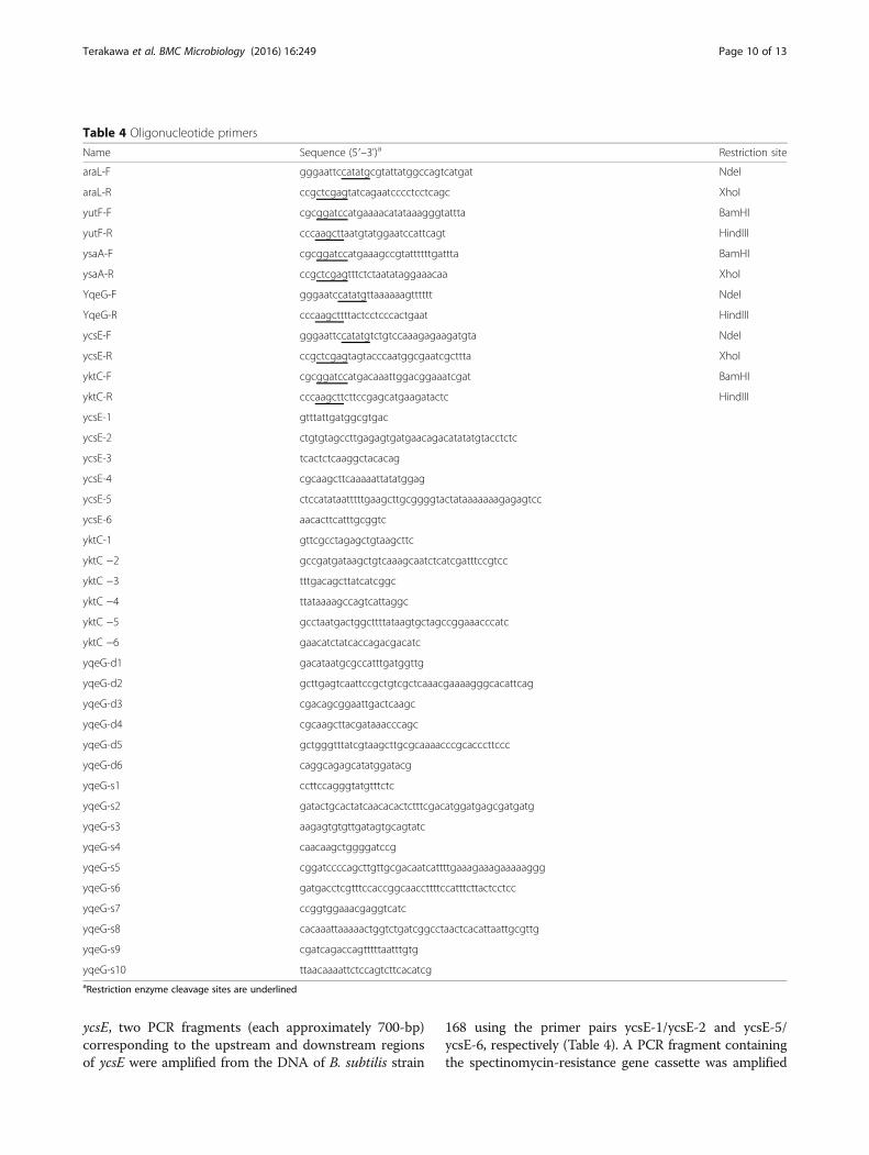
Table 4 Oligonucleotide primers
Name Sequence (5′–3')a Restriction site
araL-F gggaattccatatgcgtattatggccagtcatgat NdeI
araL-R ccgctcgagtatcagaatcccctcctcagc XhoI
yutF-F cgcggatccatgaaaacatataaagggtattta BamHI
yutF-R cccaagcttaatgtatggaatccattcagt HindIII
ysaA-F cgcggatccatgaaagccgtattttttgattta BamHI
ysaA-R ccgctcgagtttctctaatataggaaacaa XhoI
YqeG-F gggaatccatatgttaaaaaagtttttt NdeI
YqeG-R cccaagcttttactcctcccactgaat HindIII
ycsE-F gggaattccatatgtctgtccaaagagaagatgta NdeI
ycsE-R ccgctcgagtagtacccaatggcgaatcgcttta XhoI
yktC-F cgcggatccatgacaaattggacggaaatcgat BamHI
yktC-R cccaagcttcttccgagcatgaagatactc HindIII
ycsE-1 gtttattgatggcgtgac
ycsE-2 ctgtgtagccttgagagtgatgaacagacatatatgtacctctc
ycsE-3 tcactctcaaggctacacag
ycsE-4 cgcaagcttcaaaaattatatggag
ycsE-5 ctccatataatttttgaagcttgcggggtactataaaaaaagagagtcc
ycsE-6 aacacttcatttgcggtc
yktC-1 gttcgcctagagctgtaagcttc
yktC −2 gccgatgataagctgtcaaagcaatctcatcgatttccgtcc
yktC −3 tttgacagcttatcatcggc
yktC −4 ttataaaagccagtcattaggc
yktC −5 gcctaatgactggcttttataagtgctagccggaaacccatc
yktC −6 gaacatctatcaccagacgacatc
yqeG-d1 gacataatgcgccatttgatggttg
yqeG-d2 gcttgagtcaattccgctgtcgctcaaacgaaaagggcacattcag
yqeG-d3 cgacagcggaattgactcaagc
yqeG-d4 cgcaagcttacgataaacccagc
yqeG-d5 gctgggtttatcgtaagcttgcgcaaaacccgcacccttccc
yqeG-d6 caggcagagcatatggatacg
yqeG-s1 ccttccagggtatgtttctc
yqeG-s2 gatactgcactatcaacacactctttcgacatggatgagcgatgatg
yqeG-s3 aagagtgtgttgatagtgcagtatc
yqeG-s4 caacaagctggggatccg
yqeG-s5 cggatccccagcttgttgcgacaatcattttgaaagaaagaaaaaggg
yqeG-s6 gatgacctcgtttccaccggcaaccttttccatttcttactcctcc
yqeG-s7 ccggtggaaacgaggtcatc
yqeG-s8 cacaaattaaaaactggtctgatcggcctaactcacattaattgcgttg
yqeG-s9 cgatcagaccagtttttaatttgtg
yqeG-s10 ttaacaaaattctccagtcttcacatcgaRestriction enzyme cleavage sites are underlined
Terakawa et al. BMC Microbiology (2016) 16:249 Page 10 of 13
from the DNA of strain TMO310 (Table 3) using theprimer pair ycsE-3/ycsE-4 (Table 4). The three PCRfragments were ligated using recombinant PCR withthe primer pair ycsE-1/ycsE-6 to generate a single PCRfragment comprising the spectinomycin-resistance genecassette flanked by the upstream and downstream re-gions of ycsE. B. subtilis strain 168 was transformedwith the recombinant PCR fragment to confer resist-ance to spectinomycin, and a transformant with correctmarker replacement was selected and designated strainNON01 (Table 3). To delete yktC, upstream and down-stream regions of yktC were amplified using the primerpairs yktC-1/yktC-2 and yktC-5/yktC-6, respectively(Table 4). The chloramphenicol-resistance cassette wasamplified from pCRE-test using the primer pair yktC-3/yktC-4 (Table 4). The three PCR fragments were li-gated using recombinant PCR with the primer pair ofyktC-1/yktC-6 to generate a single PCR fragment, whichwas integrated into chromosome of B. subtilis strain 168to yield strain NON02 (Table 3). Strain NON01 was fur-ther transformed with NON02 DNA to confer resistanceto chloramphenicol and generate strain NON03 lackingboth ycsE and yktC (Table 3).Another mutant that conditionally expressed yqeG was
constructed as follows (Additional file 1: Figure S1). FiveDNA fragments, corresponding to the C-terminal regionof amyE, the stretch containing the erythromycin-resistance cassette and the spac promoter of pMutin2(Table 3), the coding region of yqeG, the lacI gene ofpMutin2, and the N-terminal region of amyE, werePCR-amplified using the primer pairs yqeG-s1/yqeG-s2,yqeG-s3/yqeG-s4, yqeG-s5/yqeG-s6, yqeG-s7/yqeG-s8,and yqeG-s9/yqeG-s10, respectively (Table 4). Using re-combinant PCR with the primer pair yqeG-s1/yqeG-s10,all these five fragments were ligated in the order as de-scribed above and then integrated into the amyE locusof strain 168 to generate the erythromycin-resistantstrain NON04 harboring an additional IPTG-induciblecopy of yqeG.Next, the endogenous yqeG gene of NON04 was de-
leted using marker replacement. Two DNA fragmentscomprising the upstream and downstream regions ofyqeG (approximately 700 bp each) were amplified usingthe primer pairs yqeG-d1/yqeG-d2 and yqeG-d5/yqeG-d6, respectively (Table 4). The kanamycin-resistance cas-sette was amplified from the DNA of strain TMO311using the primer pair yqeG-d3/yqeG-d4 (Table 4). Re-combinant PCR was used to ligate together the threefragments using the primer pair of yqeG-d1/yqeG-d6t,and the ligation product was integrated into thechromosome of NON04 to yield strain NON05 in whichexpression of the additional yqeG was induced with1 mM IPTG (Table 3). In NON05, yqeG was replacedwith the kanamycin-resistance cassette so that the rest
of operon comprising yqeH, aroD, yqeI, nadD, yqeK,yqeL, and yqeM, which reside downstream of yqeG, areconstitutively expressed by read-through from thekanamycin-resistance gene. NON05 was transformedfurther with NON03 DNA to confer resistance to bothchloramphenicol and spectinomycin inactivating yktCand ycsE, respectively (strain NON06).Chromosomal DNAs of mutant strains of B. subtilis
were subjected to diagnostic PCR experiments with therespective primer pairs used for recombinant PCR, andtheir correct construction was confirmed by appearanceof the respective PCR fragments with altered length(Additional file 1: Figure S1).
Purification of gene products produced in E. coliE. coli strain BL21(DE3) bearing the pET28b derivativeswere inoculated into 600 ml of LB medium containingkanamycin and were cultured at 37 °C with shaking at180 rpm. When the optical density of the culturesreached 0.4–0.6, IPTG was added to a final concentra-tion of 1 mM, the cells were cultivated further for 5 h,harvested using centrifugation, and resuspended into10.8 ml of NP buffer (50 mM NaH2PO4, pH 8.0, and300 mM NaCl). The suspension was chilled in an icebath, mixed with 1.2 ml of 10 mg/ml lysozyme in NP buffer,incubated for 30 min, and sonicated to disrupt the cells.After centrifugation, the supernatant was subjected to His6-tag affinity purification using TALON Metal Affinity Resins(Takara Bio). The His6-tagged proteins were purified in areaction buffer containing 1 mM NH4Cl, 250 mM KCl,10 mM MgCl2, and 50 mM Tris–HCl (pH 7.8), and sub-jected to SDS-PAGE followed by Coomassie brilliant bluestaining.
Enzyme assayThe phosphatase assay was performed as previously de-scribed [61]. Each purified enzyme (0.25 mg/ml) was incu-bated with substrate in 50 μl of reaction buffer containing1 mM NH4Cl, 250 mM KCl, 10 mM MgCl2, and 50 mMTris–HCl (pH 7.8) at 37 °C, and the reaction was termi-nated using 50 μl of 10 % trichloroacetic acid. After centri-fugation, the supernatant was mixed with 20 μl of solutionA (10 % ascorbic acid in 2.25 M H2SO4) and 20 μl of solu-tion B (2.4 % ammonium molybdate and 1 mg/ml potas-sium antimonyl tartrate in 2.25 M H2SO4). The mixturewas incubated at room temperature for 10 min, and ab-sorbance at 820 nm was measured to calculate the con-centration of the released phosphate ion with reference toa standard curve made with various concentrations ofinorganic phosphate. Trace of phosphate released inthe reaction mixtures in the absence of purified enzymewas subtracted as background.
Terakawa et al. BMC Microbiology (2016) 16:249 Page 11 of 13
Additional file
Additional file 1: Figure S1. Genetic organization of the alteredchromosomal loci; ΔycsE::spc(A), ΔyktC::cat(B), ΔyqeG::kan(C), andamyE::(Pspac-yqeG lacI erm)(D). Genes and primers are indicatedschematically by thick arrows and arrowheads, respectively. (PDF 144 kb)
AbbreviationsG6P: Glucose 6-phosphate; HADSF: Haloalkane acid dehalogenasesuperfamily; IPTG: Isopropyl β-D-1-thiogalactopyranoside; MIMP: Myo-inositol1-monophosphate; SDS-PAGE: Sodium dodecyl sulfate polyacrylamide gelelectrophoresis
AcknowledgementsWe thank Mr. Kentaro Kojima for his technical contributions in the earlierstage of this work.
FundingThis work was supported by the Ministry of Education, Culture, Sports,Science and Technology, Japan and in part by the Special CoordinationFunds for Promoting Science and Technology, Creation of Innovative Centersfor Advanced Interdisciplinary Research Areas; the Advanced Low-CarbonTechnology Research and Development Program; KAKENHI (26660067); andGrants-in-Aid from the NC-CARP project.
Availability of data and materialsThe datasets during and/or analyzed during the current study are availablefrom the corresponding author on reasonable request. The phylogenetictree, sequence data, and alignments used to produce the results displayed inFig. 1 are deposited in TreeBASE (http://purl.org/phylo/treebase/phylows/study/TB2:S19999).
Authors’ contributionsAT conducted most of the experiments and analyzed the results. Theenzymatic experiments were performed under the supervision of KT and ST.KT, AN, AO, SN, and JK conducted experiments with the mutant strains of B.subtilis. KY conceived the idea for the project and wrote the final manuscriptwith SI. All authors read and approved the final manuscript.
Competing interestsThe authors declare that the research was conducted in the absence of anycommercial or financial relationships that could be construed as a potentialconflict of interest.
Consent for publicationNot applicable.
Ethics approval and consent to participateNot applicable.
Author details1Department of Agrobioscience, Kobe University, 1-1 Rokkodai, Nada, Kobe,Hyogo 657-8501, Japan. 2Organization of Advanced Science and Technology,Kobe University, 1-1 Rokkodai, Nada, Kobe, Hyogo 657-8501, Japan.3Department of Science, Technology and Innovation, Kobe University, 1-1Rokkodai, Nada, Kobe, Hyogo 657-8501, Japan.
Received: 8 September 2016 Accepted: 15 October 2016
References1. Rampazzo C, Ferraro P, Pontarin G, Fabris S, Reichard P, Bianchi V.
Mitochondrial deoxyribonucleotides, pool sizes, synthesis, and regulation.J Biol Chem. 2004;279:17019–26.
2. Tamao Y, Noguchi K, Sakai-Tomita Y, Hama H, Shimamoto T, Kanazawa H,Tsuda M, Tsuchiya T. Sequence analysis of nutA gene encoding membrane-bound Cl(−)-dependent 5′-nucleotidase of Vibrio parahaemolyticus. JBiochem. 1991;109:24–9.
3. Burns DM, Beacham IR. Nucleotide sequence and transcriptional analysisof the E. coli ushA gene, encoding periplasmic UDP-sugar hydrolase
(5′-nucleotidase): regulation of the ushA gene, and the signal sequenceof its encoded protein product. Nucleic Acids Res. 1986;14:4325–42.
4. Tremblay LW, Dunaway-Mariano D, Allen KN. Structure and activity analyses ofEscherichia coli K-12 NagD provide insight into the evolution of biochemicalfunction in the haloalkanoic acid dehalogenase superfamily. Biochemistry.2006;45:1183–93.
5. Koonin EV, Tatusov RL. Computer analysis of bacterial haloacid dehalogenasesdefines a large superfamily of hydrolases with diverse specificity: Application ofan iterative approach to database search. J Mol Biol. 1994;244:125–32.
6. Allen KN, Dunaway-Mariano D. Phosphoryl group transfer: evolution of acatalytic scaffold. Trends Biochem Sci. 2004;29:495–503.
7. Lu Z, Dunaway-Mariano D, Allen KN. HAD superfamily phosphotransferasesubstrate diversification: structure and function analysis of HAD subclass IIBsugar phosphatase BT4131. Biochemistry. 2005;44:8684–96.
8. Shin DH, Roberts A, Jancarik J, Yokota H, Kim R, Wemmer DE, Kim SH.Crystal structure of a phosphatase with a unique substrate binding domainfrom Thermotoga maritime. Protein Sci. 2003;12:1464–72.
9. Kuznetsova E, Proudfoot M, Gonzalez CF, Brown G, Omelchenko MV,Borozan I, Carmel L, Wolf YI, Mori H, Savchenko AV, Arrowsmith CH, KooninEV, Edwards AM, Yakunin AF. Genome-wide analysis of substrate specificitiesof the Escherichia coli haloacid dehalogenase-like phosphatase family. J BiolChem. 2006;281:36149–61.
10. Proudfoot M, Kuznetsova E, Brown G, Rao NN, Kitagawa M, Mori H,Savchenko A, Yakunin AF. General enzymatic screens identify three newnucleotidases in Escherichia coli. Biochemical characterization of SurE, YfbR,and YjjG. J Biol Chem. 2004;279:54687–94.
11. Lee JY, Kwak JE, Moon J, Eom SH, Liong EC, Pedelacq JD, Berendzen J, SuhSW. Crystal structure and functional analysis of the SurE protein identify anovel phosphatase family. Nat Struct Biol. 2001;8:789–94.
12. Aravind L, Koonin EV. The HD domain defines a new superfamily of metal-dependent phosphohydrolases. Trends Biochem Sci. 1998;23:469–72.
13. Drøbak BK. The plant phosphoinositide system. Biochem J. 1992;288:697–712.14. Gläser HU, Thomas D, Gaxiola R, Montrichard F, Surdin-Kerjan Y, Serrano R.
Salt tolerance and methionine biosynthesis in Saccharomyces cerevisiaeinvolve a putative phosphatase gene. EMBO J. 1993;12:3105–10.
15. Neuwald AF, York JD, Majerus PW. Diverse proteins homologous to inositolmonophosphatase. FEBS Lett. 1991;294:16–8.
16. Eisenberg Jr F. D-Myoinositol 1-phosphate as product of cyclization ofglucose 6-phosphate and substrate for a specific phosphatase in rat testis. JBiol Chem. 1967;242:1375–82.
17. Gee NS, Ragan CI, Watling KJ, Aspley S, Jackson RG, Reid GG, Gani D, ShuteJK. The purification and properties of myo-inositol monophosphatase frombovine brain. Biochemistry. 1988;249:883–9.
18. Matsuhisa A, Suzuki N, Noda T, Shiba K. Inositol monophosphatase activityfrom the Escherichia coli suhB gene product. J Bacteriol. 1995;177:200–5.
19. Kates M. Bacterial lipids. Adv Lipid Res. 1964;2:17–90.20. Dhariwal KR, Vasantha N, Freese E. Partial nucleotide limitation induces
phosphodiesterase I and 5′-nucleotidase in Bacillus subtilis. J Bacteriol.1982;149:1146–9.
21. Asahara T, Mori Y, Zakataeva NP, Livshits VA, Yoshida K, Matsuno K.Accumulation of gene-targeted Bacillus subtilis mutations that enhancefermentative inosine production. Appl Microbiol Biotechnol. 2010;87:2195–207.
22. Kunst F, Ogasawara N, Moszer I, Albertini AM, Alloni G, Azevedo V, BerteroMG, Bessières P, Bolotin A, Borchert S, Borriss R, Boursier L, Brans A, Braun M,Brignell SC, Bron S, Brouillet S, Bruschi CV, Caldwell B, Capuano V, CarterNM, Choi S-K, Codani JJ, Connerton IF, Cummings NJ, Daniel RA, Denizot F,Devine KM, Düsterhöft A, Ehrlich SD, Emmerson PT, Entian KD, Errington J,Fabret C, Ferrari E, Foulger D, Fritz C, Fujita M, Fujita Y, Fuma S, Galizzi A,Galleron N, Ghim S-Y, Glaser P, Goffeau A, Golightly EJ, Grandi G, GuiseppiG, Guy BJ, Haga K, Haiech J, Harwood CR, Hénaut A, Hilbert H, Holsappel S,Hosono S, Hullo M-F, Itaya M, Jones L, Joris B, Karamata D, Kasahara Y,Klaerr-Blanchard M, Klein C, Kobayashi Y, Koetter P, Koningstein G, Krogh S,Kumano M, Kurita K, Lapidus A, Lardinois S, Lauber J, Lazarevic V, Lee S-M,Levine A, Liu H, Masuda S, Mauël C, Médigue C, Medina N, Mellado RP,Mizuno M, Moestl D, Nakai S, Noback M, Noone D, O’Reilly M, Ogawa K,Ogiwara A, Oudega B, Park S-H, Parro V, Pohl TM, Portetelle D, Porwollik S,Prescott AM, Presecan E, Pujic P, Purnelle B, Rapoport G, Rey M, Reynolds S,Rieger M, Rivolta C, Rocha E, Roche B, Rose M, Sadaie Y, Sato T, Scanlan E,Schleich S, Schroeter R, Scoffone F, Sekiguchi J, Sekowska A, Seror SJ, Serror P,Shin B-S, Soldo B, Sorokin A, Tacconi E, Takagi T, Takahashi H, Takemaru K,Takeuchi M, Tamakoshi A, Tanaka T, Terpstra P, Tognoni A, Tosato V, Uchiyama
Terakawa et al. BMC Microbiology (2016) 16:249 Page 12 of 13
S, Vandenbol M, Vannier F, Vassarotti A, Viari A, Wambutt R, Wedler E, Wedler H,Weitzenegger T, Winters P, Wipat A, Yamamoto H, Yamane K, Yasumoto K, YataK, Yoshida K, Yoshikawa H-F, Zumstein E, Yoshikawa H, Danchin A. Thecomplete genome sequence of the Gram-positive bacterium Bacillussubtilis. Nature. 1997;390:249–56.
23. Sa-Nogueira I, Nogueira TV, Soares S, de Lencastre H. The Bacillus subtilisL-arabinose (ara) operon: nucleotide sequence, genetic organization andexpression. Microbiology. 1997;143:957–69.
24. Guo Z, Wang F, Shen T, Huang J, Wang Y, Ji C. Crystal structure ofthermostable p-nitrophenylphosphatase from Bacillus Stearothermophilus(Bs-TpNPPase). Protein Pept Lett. 2014;21:483–9.
25. Nicolas P, Mäder U, Dervyn E, Rochat T, Leduc A, Pigeonneau N, Bidnenko E,Marchadier E, Hoebeke M, Aymerich S, Becher D, Bisicchia P, Botella E,Delumeau O, Doherty G, Denham EL, Fogg MJ, Fromion V, Goelzer A, HansenA, Härtig E, Harwood CR, Homuth G, Jarmer H, Jules M, Klipp E, Le Chat L,Lecointe F, Lewis P, Liebermeister W, March A, Mars RA, Nannapaneni P, NooneD, Pohl S, Rinn B, Rügheimer F, Sappa PK, Samson F, Schaffer M, Schwikowski B,Steil L, Stülke J, Wiegert T, Devine KM, Wilkinson AJ, van Dijl JM, Hecker M,Völker U, Bessières P, Noirot P. Condition-dependent transcriptome revealshigh-level regulatory architecture in Bacillus subtilis. Science. 2012;335:1103–6.
26. Morimoto T, Loh PC, Hirai T, Asai K, Kobayashi K, Moriya S, Ogasawara N. SixGTP-binding proteins of the Era/Obg family are essential for cell growth inBacillus subtilis. Microbiology. 2002;148:3539–52.
27. Fillinger S, Boschi-Muller S, Azza S, Dervyn E, Branlant G, Aymerich S. Twoglyceraldehyde-3-phosphate dehydrogenases with opposite physiologicalroles in a nonphotosynthetic bacterium. J Biol Chem. 2000;275:14031–7.
28. Feucht A, Lucet I, Yudkin MD, Errington J. Cytological and biochemicalcharacterization of the FtsA cell division protein of Bacillus subtilis. MolMicrobiol. 2001;40:115–25.
29. Galinier A, Kravanja M, Engelmann R, Hengstenberg W, Kilhoffer MC,Deutscher J, Haiech J. New protein kinase and protein phosphatase familiesmediate signal transduction in bacterial catabolite repression. Proc NatlAcad Sci U S A. 1998;95:1823–8.
30. Yansura DG, Henner DJ. Use of the Escherichia coli lac repressor andoperator to control gene expression in Bacillus subtilis. Proc Natl Acad Sci US A. 1984;81:439–43.
31. Bianchi V, Spychala J. Mammalian 5′-nucleotidases. J Biol Chem.2003;278:46195–8.
32. Koshland Jr DE. Control of enzyme activity and metabolic pathways. TrendsBiochem Sci. 1984;9:155–9.
33. Newsholme EA, Challiss RAJ, Crabtree B. Substrate cycles: their role inimproving sensitivity in metabolic control. Trends Biochem Sci. 1984;9:277–80.
34. Breton A, Pinson B, Coulpier F, Giraud MF, Dautant A, Daignan-Fornier B.Lethal Accumulation of Guanylic Nucleotides in Saccharomyces cerevisiaeHPT1-Deregulated Mutants. Genetics. 2008;178:815–24.
35. Pöther DC, Liebeke M, Hochgräfe F, Antelmann H, Becher D, Lalk M,Lindequist U, Borovok I, Cohen G, Aharonowitz Y, Hecker M. Diamidetriggers mainly S thiolations in the cytoplasmic proteomes of Bacillus subtilisand Staphylococcus aureus. J Bacteriol. 2009;191:7520–30.
36. Singh R, Mailloux RJ, Puiseux-Dao S, Appanna VD. Oxidative stress evokes ametabolic adaptation that favors increased NADPH synthesis and decreasedNADH production in Pseudomonas fluorescens. J Bacteriol. 2007;189:6665–75.
37. Kobayashi K, Ehrlich SD, Albertini A, Amati G, Andersen KK, Arnaud M, AsaiK, Ashikaga S, Aymerich S, Bessieres P, Boland F, Brignell SC, Bron S, Bunai K,Chapuis J, Christiansen LC, Danchin A, Débarbouille M, Dervyn E, DeuerlingE, Devine K, Devine SK, Dreesen O, Errington J, Fillinger S, Foster SJ, Fujita Y,Galizzi A, Gardan R, Eschevins C, Fukushima T, Haga K, Harwood CR, HeckerM, Hosoya D, Hullo MF, Kakeshita H, Karamata D, Kasahara Y, Kawamura F,Koga K, Koski P, Kuwana R, Imamura D, Ishimaru M, Ishikawa S, Ishio I, LeCoq D, Masson A, Mauël C, Meima R, Mellado RP, Moir A, Moriya S,Nagakawa E, Nanamiya H, Nakai S, Nygaard P, Ogura M, Ohanan T, O’ReillyM, O’Rourke M, Pragai Z, Pooley HM, Rapoport G, Rawlins JP, Rivas LA,Rivolta C, Sadaie A, Sadaie Y, Sarvas M, Sato T, Saxild HH, Scanlan E, SchumannW, Seegers JF, Sekiguchi J, Sekowska A, Séror SJ, Simon M, Stragier P, Studer R,Takamatsu H, Tanaka T, Takeuchi M, Thomaides HB, Vagner V, van Dijl JM,Watabe K, Wipat A, Yamamoto H, Yamamoto M, Yamamoto Y, Yamane K, YataK, Yoshida K, Yoshikawa H, Zuber U, Ogasawara N. Essential Bacillus subtilisgenes. Proc Natl Acad Sci U S A. 2003;100:4678–83.
38. Hawkins AR, Lamb HK, Smith M, Keyte JW, Roberts CF. Molecularorganisation of the quinic acid utilization (QUT) gene cluster in Aspergillusnidulans. Mol Gen Genet. 1988;214:224–31.
39. Chen IW, Charalampous FC. A soluble enzyme system from yeast whichcatalyzes the biosynthesis of inositol from glucose. Biochem Biophys ResCommun. 1963;12:62–7.
40. Majerus PW. Inositol phosphate biochemistry. Annu Rev Biochem.1992;61:225–50.
41. Cronan JE. Bacterial membrane lipids: where do we stand? Annu RevMicrobiol. 2003;57:203–24.
42. den Kamp JA, Redai I, van Deenen LL. Phospholipid composition of Bacillussubtilis. J Bacteriol. 1969;99:298–303.
43. Shiba K, Ito K, Yura T. Mutation that suppresses the protein export defect ofthe secY mutation and causes cold-sensitive growth of Escherichia coli. JBacteriol. 1984;160:696–701.
44. Yano R, Nagai H, Shiba K, Yura T. A mutation that enhances synthesis ofsigma 32 and suppresses temperature-sensitive growth of the rpoH15mutant of Escherichia coli. J Bacteriol. 1990;172:2124–30.
45. Ito K, Wittekind M, Nomura M, Shiba K, Yura T, Miura A, Nashimoto H. Atemperature-sensitive mutant of E. coli exhibiting slow processing ofexported proteins. Cell. 1983;32:789–97.
46. Grossman AD, Erickson JW, Gross CA. The htpR gene product of E. coli is asigma factor for heat-shock promoters. Cell. 1984;38:383–90.
47. Li K, Xu C, Jin Y, Sun Z, Liu C, Shi J, Chen G, Chen R, Jin S, Wu W. SuhB is aregulator of multiple virulence genes and essential for pathogenesis ofPseudomonas aeruginosa. MBio. 2013;4:e00419–13.
48. Rosales-Reyes R, Saldías MS, Aubert DF, El-Halfawy OM, Valvano MA. ThesuhB gene of Burkholderia cenocepacia is required for protein secretion,biofilm formation, motility and polymyxin B resistance. Microbiology.2012;158:2315–24.
49. Kozloff LM, Turner MA, Arellano F, Lute M. Phosphatidylinositol, aphospholipid of ice-nucleating bacteria. J Bacteriol. 1991;173:2053–60.
50. Englesberg E, Anderson RL, Weinberg R, Lee N, Hoffee P, Huttenhauer G,Boyer H. L-Arabinose-sensitive, L-ribulose 5-phosphate 4-epimerase-deficientmutants of Escherichia coli. J Bacteriol. 1962;84:137–46.
51. Irani MH, Maitra PK. Properties of Escherichia coli mutants deficient inenzymes of glycolysis. J Bacteriol. 1977;132:398–410.
52. Yarmolinsky MB, Wiesmeyer H, Kalckar HM, Jordan E. Hereditary defects ingalactose metabolism in Escherichia coli mutants. II. Galactose-inducedsensitivity. Proc Natl Acad Sci U S A. 1959;45:1786–91.
53. Lowry OH, Carter J, Ward JB, Glaser L. The effect of carbon and nitrogensources on the level of metabolic intermediates in Escherichia coli. J BiolChem. 1971;246:6511–21.
54. Papagianni M, Avramidis N, Filiousis G. Glycolysis and the regulation ofglucose transport in Lactococcus lactis spp. lactis in batch and fed-batchculture. Microb Cell Fact. 2007;6:16.
55. Paoletti L, Lu YJ, Schujman GE, de Mendoza D, Rock CO. Coupling offatty acid and phospholipid synthesis in Bacillus subtilis. J Bacteriol.2007;189:5816–24.
56. Ishii K, Shiio I. Improved inosine production and derepression of purinenucleotide biosynthetic enzymes in 8-azaguanine resistant mutants ofBacillus subtilis. Agric Biol Chem. 1972;36:1511–22.
57. Matsui H, Sato K, Enei H, Hirose Y. Mutation of an inosine producing strainof Bacillus subtilis to DL-methionine sulfoxide resistant for guanosineproduction. Appl Environ Microbiol. 1977;34:337–41.
58. Kotani Y, Yamaguchi K, Kato F, Furuya A. Inosine accumulation by mutantsof Brevibacterium ammoniagenes: strain improvement and cultureconditions. Agric Biol Chem. 1978;42:399–405.
59. Matsui H, Sato K, Enei H, Takinami K. 5′-Nucleotidase activity in improvedinosine-producing mutants of Bacillus subtilis. Agric Biol Chem. 1982;46:2347–52.
60. Chen S, Chu J, Zhuang Y, Zhang S. Enhancement of inosine production byBacillus subtilis through suppression of carbon overflow by sodium citrate.Biotechnol Lett. 2005;27:689–92.
61. Barnett JE, Brice RE, Corina DL. A colorimetric determination of inositolmonophosphates as an assay for D-glucose 6-phosphate-1 L-myoinositol1-phosphate cyclase. Biochem J. 1970;119:183–6.
62. Morimoto T, Ara K, Ozaki K, Ogasawara N. A new simple method tointroduce marker-free deletions in the Bacillus subtilis genome. Genes GenetSyst. 2009;84:315–8.
63. Miwa Y, Nakata A, Ogiwara A, Yamamoto M, Fujita Y. Evaluation andcharacterization of catabolite-responsive elements (cre) of Bacillus subtilis.Nucleic Acids Res. 2000;28:1206–10.
64. Vagner V, Dervyn E, Ehrlich SD. A vector for systematic gene inactivation inBacillus subtilis. Microbiology. 1998;144:3097–104.
Terakawa et al. BMC Microbiology (2016) 16:249 Page 13 of 13