Redalyc.Histochemistry and morphoanatomy study …Histochemistry and morphoanatomy study on guava...
9
Ciência e Tecnologia de Alimentos ISSN: 0101-2061 [email protected] Sociedade Brasileira de Ciência e Tecnologia de Alimentos Brasil de ABREU, José Renato; Donizete dos SANTOS, Custódio; Patto de ABREU, Celeste Maria; de CASTRO, Evaristo Mauro Histochemistry and morphoanatomy study on guava fruit during ripening Ciência e Tecnologia de Alimentos, vol. 32, núm. 1, abril-junio, 2012, pp. 179-186 Sociedade Brasileira de Ciência e Tecnologia de Alimentos Campinas, Brasil Available in: http://www.redalyc.org/articulo.oa?id=395940112027 How to cite Complete issue More information about this article Journal's homepage in redalyc.org Scientific Information System Network of Scientific Journals from Latin America, the Caribbean, Spain and Portugal Non-profit academic project, developed under the open access initiative
Transcript of Redalyc.Histochemistry and morphoanatomy study …Histochemistry and morphoanatomy study on guava...
Ciência e Tecnologia de Alimentos
ISSN: 0101-2061
Sociedade Brasileira de Ciência e
Tecnologia de Alimentos
Brasil
de ABREU, José Renato; Donizete dos SANTOS, Custódio; Patto de ABREU, Celeste
Maria; de CASTRO, Evaristo Mauro
Histochemistry and morphoanatomy study on guava fruit during ripening
Ciência e Tecnologia de Alimentos, vol. 32, núm. 1, abril-junio, 2012, pp. 179-186
Sociedade Brasileira de Ciência e Tecnologia de Alimentos
Campinas, Brasil
Available in: http://www.redalyc.org/articulo.oa?id=395940112027
How to cite
Complete issue
More information about this article
Journal's homepage in redalyc.org
Scientific Information System
Network of Scientific Journals from Latin America, the Caribbean, Spain and Portugal
Non-profit academic project, developed under the open access initiative
Ciênc. Tecnol. Aliment., Campinas, 32(1): 179-186, jan.-mar. 2012 179
Original
Ciência e Tecnologia de Alimentos ISSN 0101-2061
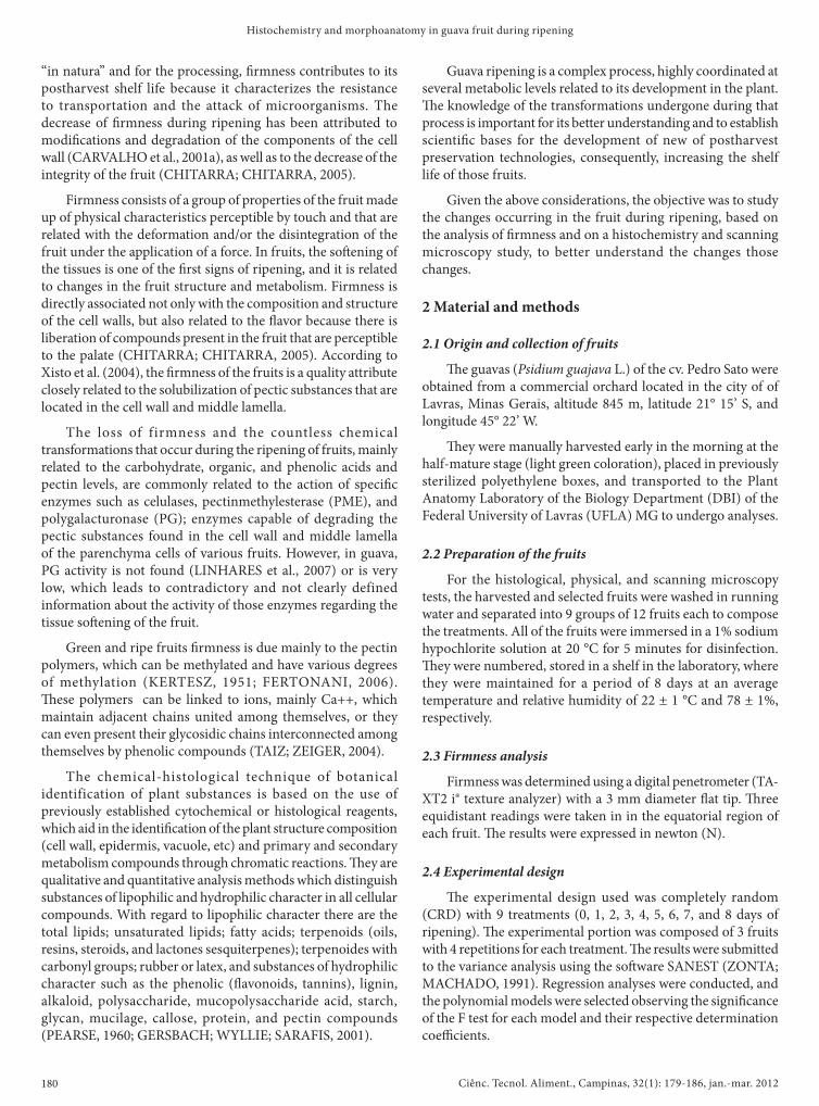
Received 9/9/2010Accepted 27/9/2011 (005039)1 Departamento de Química, Universidade Federal de Lavras – UFLA, CP 3037, CEP 37200-000, Lavras, MG, Brasil, e-mail: [email protected]*Corresponding author
Histochemistry and morphoanatomy study on guava fruit during ripeningHistoquímica e morfoanatomia em frutos de goiaba durante amadurecimento
José Renato de ABREU1*, Custódio Donizete dos SANTOS1, Celeste Maria Patto de ABREU1, Evaristo Mauro de CASTRO1
1 Introduction�e guava tree (Psidium guajava L.) is a bush or small tree
(2 to 7 m heigh) of the Mirtáceae family, which is made up of more than 70 genera and 2,800 species native to the tropical and subtropical areas of America. It is cultivated in Brazil from the state of Rio Grande do Sul up to Maranhão state (MANICA et al., 2000). Brazil is the world’s largest producer of red guavas, and the export of guava by-products is about 700 t (MENDONÇA et al., 2007). In 2006, the production was of approximately 328,255 t covering an area of 14,982 ha. �e production is concentrated mainly in the Southeast and Northeast regions, and the states of São Paulo and Pernambuco are the largest producers (INSTITUTO..., 2008).
Guava presents high acceptance for “in natura” consumption or in the processed form. It is rich in nutrients and is considered one of the best vitamin C sources, with values 6 to 7 times higher than those of the citric fruits. It also presents vitamins A and B,
such as thiamine and niacin, besides containing good levels of P, Fe, and Ca (MANICA et al., 2000).
Guava is a fruit with high respiration rates and very short postharvest shelf life, a result of the loss of pulp �rmness, which limits transportation and storage period. �is aspect that makes it di�cult or even impossible to send the fruits to distant consumer markets (XISTO et al., 2004). Highly perishable, due to its intense metabolism during ripening, guava has a shelf life of about 3 to 5 days at room temperature (CARVALHO, 1994; DURIGAN, 1997; GONGATTI NETO; GARCIA; ARDITO, 1996; AZZOLINI, 2005).
The ripening of the fruits corresponds to a series of physiological, biochemical, and structural changes, which make the fruit attractive for consumption, and among all of the changes, �rmness is the most important attribute because in addition to de�ning the quality of the fruit for consumption
Resumo A goiaba (Psidium guajava L.) é um fruto altamente perecível devido ao seu intenso metabolismo durante o amadurecimento. As informações sobre a atividade das enzimas que degradam substâncias pécticas, bem como a quantidade de pectina, são bem contraditórias e não claramente de�nidas. Assim, objetivou-se monitorar as mudanças ocorridas no fruto durante o amadurecimento, por meio de processos histoquímicos, físico e de microscopia de varredura. Foram colhidas goiabas no estágio “de vez” e armazenadas por 9 dias a uma temperatura de 22 ± 1 °C e UR de 78% ± 1%. As análises realizadas no dia da colheita (dia 0) e a cada dia do armazenamento (1, 2, 3, 4, 5, 6, 7 e 8 dias) foram: �rmeza, análises histoquímicas (cloreto férrico, lugol, comassie blue, vanilina clorídrica e vermelho de rutênio) observadas em microscópio ótico e análise em microscópio eletrônico de varredura. O vermelho de rutênio mostrou grande quantidade de pectina na parede celular no dia zero e a diminuição desta, da parede, no decorrer do amadurecimento e seu acúmulo na região central da célula. A microscopia de varredura mostrou a perda da estrutura celular com o amadurecimento. Essas observações sugerem que a pectina é o principal polímero responsável pela manutenção da �rmeza na goiaba.Palavras-chave: pectina; �rmeza; Psidium guajava L.
Abstract Guava (Psidium guajava L.) is a highly perishable fruit due to its intense metabolism during ripening. Information on the enzyme activities that degrade pectic substances, as well as the amount of pectin, is very contradictory and not clearly de�ned. �us, this study aimed to monitor the changes occurred in the fruit during ripening through histochemical, physical, and scanning microscopy processes. Guavas were picked at the half-mature stage and stored for 9 days at 22 ± 1 °C and 78 ± 1% RH. �e analyses conducted on the day of harvest (0) and each day of storage (1, 2, 3, 4, 5, 6, 7, and 8 days) were: �rmness and histochemical analyses (ferric chloride, lugol, comassie blue, vanillin hydrochloric, and ruthenium red) observed under an optic microscope and a scanning electron microscope. Ruthenium red showed a high amount of pectin in the cell wall on day zero as well as its decrease in the wall during ripening and its accumulation in the central area of the cell. Scanning microscopy showed loss of the cell structure during ripening. �ose observations suggest that the pectin is the main polymer responsible for �rmness maintenance in the guava fruit.Keywords: pectin; �rmness; Psidium guajava L.
DOD http//dx.doi.org/10.1590/S0101-20612012005000019
Ciênc. Tecnol. Aliment., Campinas, 32(1): 179-186, jan.-mar. 2012180
Histochemistry and morphoanatomy in guava fruit during ripening
Guava ripening is a complex process, highly coordinated at several metabolic levels related to its development in the plant. �e knowledge of the transformations undergone during that process is important for its better understanding and to establish scienti�c bases for the development of new of postharvest preservation technologies, consequently, increasing the shelf life of those fruits.
Given the above considerations, the objective was to study the changes occurring in the fruit during ripening, based on the analysis of �rmness and on a histochemistry and scanning microscopy study, to better understand the changes those changes.
2 Material and methods
2.1 Origin and collection of fruits
�e guavas (Psidium guajava L.) of the cv. Pedro Sato were obtained from a commercial orchard located in the city of of Lavras, Minas Gerais, altitude 845 m, latitude 21° 15’ S, and longitude 45° 22’ W.
�ey were manually harvested early in the morning at the half-mature stage (light green coloration), placed in previously sterilized polyethylene boxes, and transported to the Plant Anatomy Laboratory of the Biology Department (DBI) of the Federal University of Lavras (UFLA) MG to undergo analyses.
2.2 Preparation of the fruits
For the histological, physical, and scanning microscopy tests, the harvested and selected fruits were washed in running water and separated into 9 groups of 12 fruits each to compose the treatments. All of the fruits were immersed in a 1% sodium hypochlorite solution at 20 °C for 5 minutes for disinfection. �ey were numbered, stored in a shelf in the laboratory, where they were maintained for a period of 8 days at an average temperature and relative humidity of 22 ± 1 °C and 78 ± 1%, respectively.
2.3 Firmness analysis
Firmness was determined using a digital penetrometer (TA-XT2 i® texture analyzer) with a 3 mm diameter �at tip. �ree equidistant readings were taken in in the equatorial region of each fruit. �e results were expressed in newton (N).
2.4 Experimental design
�e experimental design used was completely random (CRD) with 9 treatments (0, 1, 2, 3, 4, 5, 6, 7, and 8 days of ripening). �e experimental portion was composed of 3 fruits with 4 repetitions for each treatment. �e results were submitted to the variance analysis using the so�ware SANEST (ZONTA; MACHADO, 1991). Regression analyses were conducted, and the polynomial models were selected observing the signi�cance of the F test for each model and their respective determination coe�cients.
“in natura” and for the processing, �rmness contributes to its postharvest shelf life because it characterizes the resistance to transportation and the attack of microorganisms. The decrease of �rmness during ripening has been attributed to modi�cations and degradation of the components of the cell wall (CARVALHO et al., 2001a), as well as to the decrease of the integrity of the fruit (CHITARRA; CHITARRA, 2005).
Firmness consists of a group of properties of the fruit made up of physical characteristics perceptible by touch and that are related with the deformation and/or the disintegration of the fruit under the application of a force. In fruits, the so�ening of the tissues is one of the �rst signs of ripening, and it is related to changes in the fruit structure and metabolism. Firmness is directly associated not only with the composition and structure of the cell walls, but also related to the �avor because there is liberation of compounds present in the fruit that are perceptible to the palate (CHITARRA; CHITARRA, 2005). According to Xisto et al. (2004), the �rmness of the fruits is a quality attribute closely related to the solubilization of pectic substances that are located in the cell wall and middle lamella.
The loss of firmness and the countless chemical transformations that occur during the ripening of fruits, mainly related to the carbohydrate, organic, and phenolic acids and pectin levels, are commonly related to the action of speci�c enzymes such as celulases, pectinmethylesterase (PME), and polygalacturonase (PG); enzymes capable of degrading the pectic substances found in the cell wall and middle lamella of the parenchyma cells of various fruits. However, in guava, PG activity is not found (LINHARES et al., 2007) or is very low, which leads to contradictory and not clearly defined information about the activity of those enzymes regarding the tissue so�ening of the fruit.
Green and ripe fruits �rmness is due mainly to the pectin polymers, which can be methylated and have various degrees of methylation (KERTESZ, 1951; FERTONANI, 2006). �ese polymers can be linked to ions, mainly Ca++, which maintain adjacent chains united among themselves, or they can even present their glycosidic chains interconnected among themselves by phenolic compounds (TAIZ; ZEIGER, 2004).
The chemical-histological technique of botanical identification of plant substances is based on the use of previously established cytochemical or histological reagents, which aid in the identi�cation of the plant structure composition (cell wall, epidermis, vacuole, etc) and primary and secondary metabolism compounds through chromatic reactions. �ey are qualitative and quantitative analysis methods which distinguish substances of lipophilic and hydrophilic character in all cellular compounds. With regard to lipophilic character there are the total lipids; unsaturated lipids; fatty acids; terpenoids (oils, resins, steroids, and lactones sesquiterpenes); terpenoides with carbonyl groups; rubber or latex, and substances of hydrophilic character such as the phenolic (�avonoids, tannins), lignin, alkaloid, polysaccharide, mucopolysaccharide acid, starch, glycan, mucilage, callose, protein, and pectin compounds (PEARSE, 1960; GERSBACH; WYLLIE; SARAFIS, 2001).
Ciênc. Tecnol. Aliment., Campinas, 32(1): 179-186, jan.-mar. 2012 181
Abreu et al.
similar results (LIMA, 2004; XISTO et al., 2004); however, the results presented in the literature for other cultivars of guava also show some similar results, with variation of 75% for guavas ‘Cortibel 1’ and ‘Cortibel 4’ (MENDONÇA et al., 2007), 83% in guavas cv. Allahabad Safeda (SINGH; PAL, 2008), and very di�erent results too. Firmness variations of 34% were found between the 3rd and 7th day of ripening for the cultivar Media China (MERCADO-SILVA; BAUTISTA; GARCIA-VELASCO, 1998), while the cultivar Kumagai showed only a 25% variations between the 1st and 10th day of ripening (CARVALHO et al., 2001b). It should be pointed out that this cultivar is considered one of long shelf life.
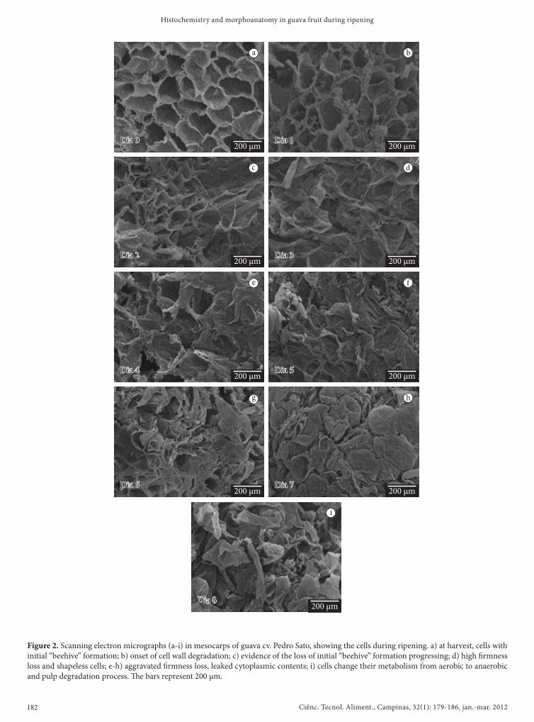
�e scanning micrographs (Figure 2) of the guava cv. Pedro Sato mesocarp during ripening show, as in the texture analyses, an accentuated �rmness decrease. On day zero (harvest day), the cells presented a beehive shape and a preserved cell (Figure 2a). On the subsequent days of ripening, it was possible to observe the gradual deformation of the beehive structure and the formation of an uneven cell mass (Figure 2b to i).
�e loss of �rmness during the ripening of the fruits is due to the activity of hydrolytic enzymes that promotes intense solubilization of the cell wall pectin constituents, mainly the pectinmethylesterase (PME) and polygalacturonase (PG) activity (TUCKER, 1993; JAIN et al., 2001; OLIVEIRA et al., 2006). Considering that the guava cv. Pedro Sato does not present polygalacturonase activity (LINHARES et al., 2007), the mechanism for the fast �rmness decrease still remains inexplicable. �e existence of high esterase activity in the cell membrane/cell wall of guava endocarp (LINHARES et al., 2007) can suggest that the fast �rmness decrease occurs due to other processes di�erent from the hydrolysis of the polygalacturonic acids.
�e histochemical analyses, conducted through histological sections of the mesocarp of guava cv. Pedro Sato during ripening, submitted to the speci�c staining for identi�cation of the cell wall constituents showed, through an optical
2.5 Histochemical analysis
For the conduction of the histochemical tests, cross sections in the mesocarp of the fruits were made manually using razor blades. �e sections were submitted to clari�cation with 50% sodium hypochlorite and washed in distilled water, neutralized in 1% acetic water, and mounted in 50% glycerin. A�erwards, they were stained with speci�c stains, according to methods described in the literature. As a result, semi-permanent slides were made, which were photographed under a Hen-A-Vision TT18 optical microscope coupled to a Canon Power Shot A630 digital camera.
�e cross sections were submitted to tests with: ferric chloride, for identi�cation of general phenolic compounds according to Gabe (1968); Lugol, according to Jensen (1962), for identi�cation of starch; comassie blue, according to Gahan (1984), for identi�cation of proteins; vanillin-hydrochloric acid, according to Mace and Howell (1974), for identi�cation of tannins and ruthenium red, according to Johansen (1940), for identi�cation of pectins.
2.6 Preparation of fruits for scanning electron microscopy (SEM)
�e fruits, harvested and stored as previously described, were also used for the preparation and observation under a scanning electron microscope conducted in the Electronic Microscopy and Ultrastrutural Analysis Laboratory (LME), in the Phytopathology Department of of the Federal University of Lavras. A�er collection, the pulp of the fruits (representative of the respective days of ripening) was cut into 1 cm3 fragments and immersed in Karnovisky (pH 7.2) �xing solution for a period of 24 hours. A�er that period, the samples were transferred to a cacodylate bu�er and, soon a�erwards, those fragments were transferred to an osmium tetroxide solution (1%) for 1 hour and, subsequently, dehydrated in an acetone series (25, 50, 75, 90, and 100%) for 10 minutes under each concentration. At the concentration of 100%, the process was repeated 3 times. A�er that procedure, the fragments were taken to a critical point drying apparatus, and mounted on aluminum stubs using carbon tape placed over aluminum foil, sputter-covered with gold, and observed under a LEO EVO 40 scanning electron microscope (ALVES, 2005).
3 Results and discussion�e so�ening or loss of �rmness of the fruit, a�er the
alteration of the color, represents the most important change that occurs in its ripening process (AWAD, 1993; OLIVEIRA et al., 2006).
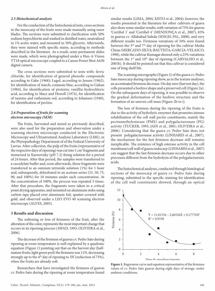
�e decrease of the �rmness of guavas cv. Pedro Sato during ripening at room temperature is well explained by a quadratic equation (Figure 1) pointing out that on the harvest day (half-mature fruits, light green peel) the �rmness was 11N, decreasing strongly up to the 4th day of ripening to 3N (reduction of 73%), when the fruits are already so�.
Researchers that have investigated the �rmness of guavas cv. Pedro Sato during the ripening at room temperature found
Figure 1. Regression curve and equation representative of the �rmness values of cv. Pedro Sato guavas during eight days of storage, under ambient conditions.
Ciênc. Tecnol. Aliment., Campinas, 32(1): 179-186, jan.-mar. 2012182
Histochemistry and morphoanatomy in guava fruit during ripening
Figure 2. Scanning electron micrographs (a-i) in mesocarps of guava cv. Pedro Sato, showing the cells during ripening. a) at harvest, cells with initial “beehive” formation; b) onset of cell wall degradation; c) evidence of the loss of initial “beehive” formation progressing; d) high �rmness loss and shapeless cells; e-h) aggravated �rmness loss, leaked cytoplasmic contents; i) cells change their metabolism from aerobic to anaerobic and pulp degradation process. �e bars represent 200 µm.
a
c
e
b
d
f
g h
i
Ciênc. Tecnol. Aliment., Campinas, 32(1): 179-186, jan.-mar. 2012 183
Abreu et al.
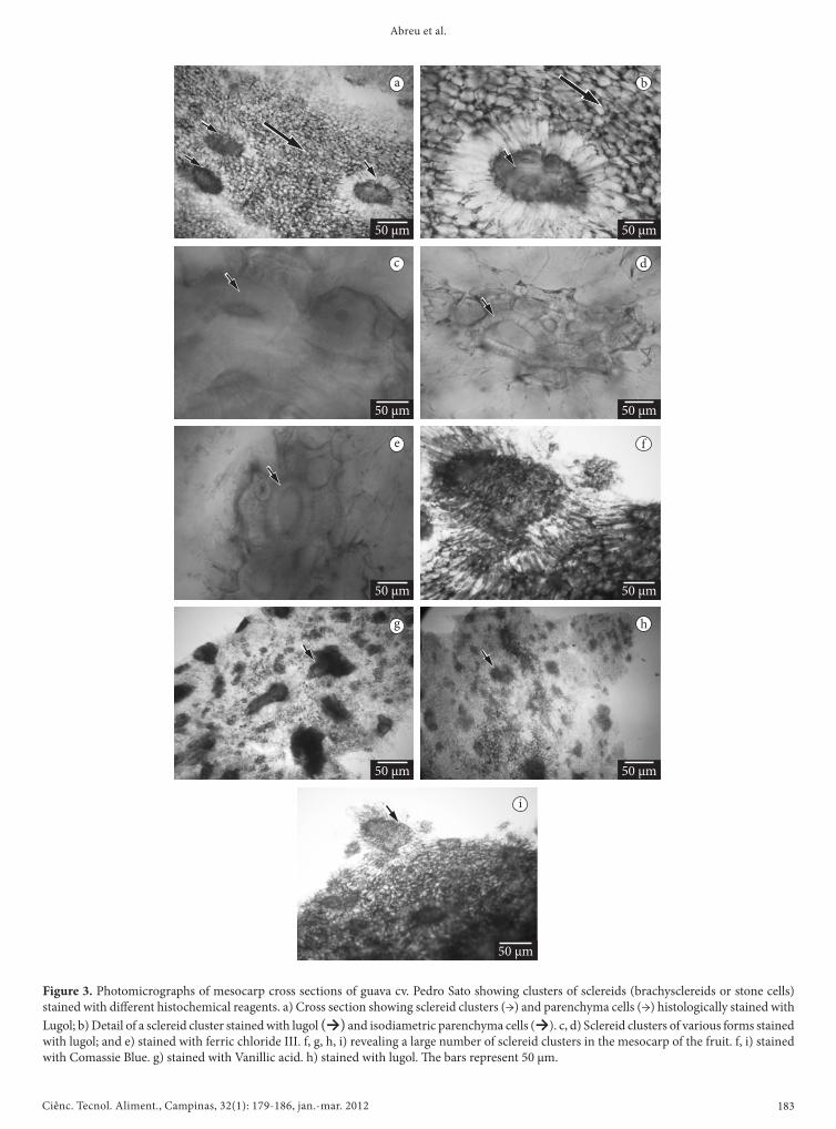
Figure 3. Photomicrographs of mesocarp cross sections of guava cv. Pedro Sato showing clusters of sclereids (brachysclereids or stone cells) stained with di�erent histochemical reagents. a) Cross section showing sclereid clusters (→) and parenchyma cells (→) histologically stained with Lugol; b) Detail of a sclereid cluster stained with lugol (→) and isodiametric parenchyma cells (→). c, d) Sclereid clusters of various forms stained with lugol; and e) stained with ferric chloride III. f, g, h, i) revealing a large number of sclereid clusters in the mesocarp of the fruit. f, i) stained with Comassie Blue. g) stained with Vanillic acid. h) stained with lugol. �e bars represent 50 µm.
a
c
e
g
b
d
f
h
i
Ciênc. Tecnol. Aliment., Campinas, 32(1): 179-186, jan.-mar. 2012184
Histochemistry and morphoanatomy in guava fruit during ripening
a b
c d
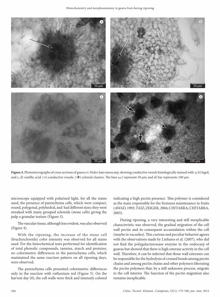
Figure 4. Photomicrographs of cross sections of guava cv. Pedro Sato mesocarp, showing conductive vessels histologically stained with: a, b) lugol; and c, d) vanillic acid. (→) conductive vessels. (→) sclereid clusters. �e bars a,c) represent 50 µm; and d) bar represents 100 µm.
microscope equipped with polarized light, for all the stains used, the presence of parenchyma cells, which were compact, round, polygonal, polyhedral, and had di�erent sizes; they were streaked with many grouped sclereids (stone cells) giving the pulp a granular texture (Figure 3).
�e vascular tissue, although less evident, was also observed (Figure 4).
With the ripening, the increase of the stone cell (brachysclereids) color intensity was observed for all stains used. For the histochemical tests performed for identi�cation of total phenolic compounds, tannins, starch and proteins, no colorimetric di�erences in the parenchyma cells, which maintained the same reaction pattern on all ripening days, were observed.
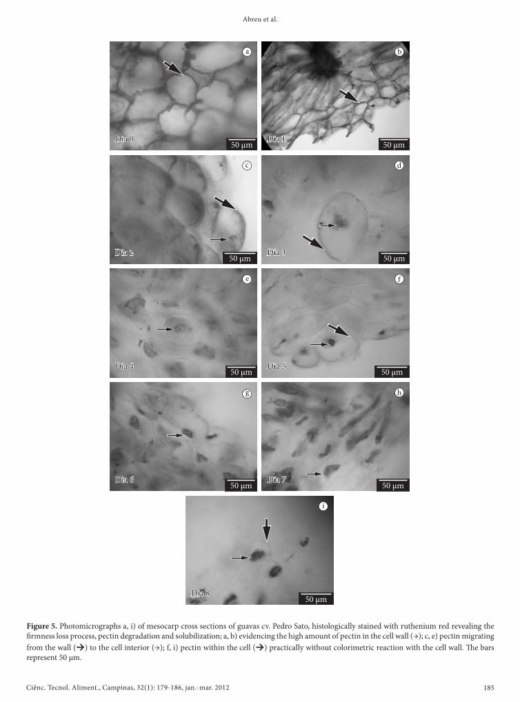
�e parenchyma cells presented colorimetric di�erences only in the reaction with ruthenium red (Figure 5). On the harvest day (0), the cell walls were thick and intensely colored
indicating a high pectin presence. �is polymer is considered as the main responsible for the �rmness maintenance in fruits (AWAD, 1993; TAIZ; ZEIGER, 2004; CHITARRA; CHITARRA, 2005).
During ripening, a very interesting and still inexplicable characteristic was observed, the gradual migration of the cell wall pectin and its consequent accumulation within the cell (maybe in vacuoles). �is curious and peculiar behavior agrees with the observations made by Linhares et al. (2007), who did not �nd the polygalacturonase enzyme in the endocarp of guavas but showed that there is high esterase activity in the cell wall. �erefore, it can be inferred that those wall esterases can be responsible for the hydrolysis of crossed bonds among pectin chains and among pectin chains and other polymers liberating the pectin polymers that, by a still unknown process, migrate to the cell interior. �e function of the pectin migration also remains inexplicable.
Ciênc. Tecnol. Aliment., Campinas, 32(1): 179-186, jan.-mar. 2012 185
Abreu et al.
Figure 5. Photomicrographs a, i) of mesocarp cross sections of guavas cv. Pedro Sato, histologically stained with ruthenium red revealing the �rmness loss process, pectin degradation and solubilization; a, b) evidencing the high amount of pectin in the cell wall (→); c, e) pectin migrating from the wall (→) to the cell interior (→); f, i) pectin within the cell (→) practically without colorimetric reaction with the cell wall. �e bars represent 50 µm.
a
c
e
g
b
d
f
h
i
Ciênc. Tecnol. Aliment., Campinas, 32(1): 179-186, jan.-mar. 2012186
Histochemistry and morphoanatomy in guava fruit during ripening
INSTITUTO BRASILEIRO DE GEOGRAFIA E ESTATÍSTICA -IBGE. Levantamento Sistemático da Produção Agrícola.IBGE, 2008. Disponível em: <http://www1.ibge.gov.br/home/estatistica/indicadores/agropecuaria/lspa/defaulttab.shtm>. Acesso em: 20 jun. 2008.
JAIN, N. et al. Compositional and enzymatic changes in guava (Psidium guajava L.) fruits during ripening. Acta Physiologiae Plantarum, v. 23, n. 3, p. 357-362, 2001. http://dx.doi.org/10.1007/s11738-001-0044-7
JENSEN, W. A. Botanical histochemistry: principles and practice. San Francisco: W. H. Freeman, 1962.
JOHANSEN, D. A. Plant microtechinique. New York: McGraw-Hill, 1940.
KERTESZ, Z. I. �e pectic substances. New York: Interscience, 1951.LIMA, A. V. Qualidade pós-colheita da goiaba “Pedro Sato” tratada
com Ca ‘Cl IND.2’ e 1-MCP em condições ambiente. 2004. 67 f. Dissertação (Mestrado em Ciência dos Alimentos)- Universidade Federal de Lavras, Lavras, 2004
LINHARES, L. A. et al. Transformações químicas, físicas e enzimáticas de goiabas “Pedro Sato” tratadas na pós-colheita com cloreto de cálcio e 1-metilciclopropeno e armazenadas sob refrigeração.Ciência e Agrotecnologia, v. 31, n. 3, p. 829-841, 2007. http://dx.doi.org/10.1590/S1413-70542007000300033
MACE, M. E.; HOWELL, C. R. Histochemistry and identi�cation of condensed tannin precursor in roots of cotton seedlings. Phytopathology, v. 64, p. 1297-1302, 1974. http://dx.doi.org/10.1094/Phyto-64-1297
MENDONÇA, R. D. et al. Características físicas e químicas de goiabas ‘cortibel 1’ e ‘cortibel 4’ armazenadas em condições ambientais. Bragantia, Campinas, v. 66, n. 4, p. 685-692, 2007.
MERCADO-SILVA, E.; BAUTISTA, P. B.; GARCIA-VELASCO, M. A. Fruit development, harvest index ripening changes of guavas produced in central Mexico. Postharvest Biology and Technology, v. 13, n. 2, p. 143-150, 1998. http://dx.doi.org/10.1016/S0925-5214(98)00003-9
MANICA, I. et al. Goiaba. Porto Alegre: Cinco Continentes, 2000. 374p.OLIVEIRA, A. C. G. et al. Conservação pós-colheita de goiaba
branca kumagai por irradiação gama: aspectos físicos, químicos e sensoriais. Boletim do Centro de Pesquisa de Processamento de Alimentos, Curitiba, v. 24, n. 2, p. 375-396, 2006.
PEARSE, A. G. E. Histochemistry, theoretical and applied. London: Churchill, 1960. 998 p.
SINGH, S. P.; PAL, R. K. Response of climacteric-type guava (Psidium guajava L.) to postharvest treatment with 1-MCP. Postharvest Biology and Technology, v. 47, n. 3, p. 307-314, 2008. http://dx.doi.org/10.1016/j.postharvbio.2007.08.010
TAIZ, L.; ZEIGER, E. Fisiologia Vegetal. 3. ed. Porto Alegre: Artmed. 2004. 719 p.
TUCKER, G. A. Introduction. In: SEYMOUR, G. B.; TAYLOR, J. E.; TUCKER, G. A. Biochemestry of fruit ripening. London: Chapmal & Hall, 1993. cap. 1, p. 2-51.
XISTO, A. L. R. P. et al. Textura de goiabas ‘Pedro Sato’ submetidas à aplicação de cloreto de cálcio. Ciências e Agrotecnologia, v. 28, n. 1, p. 113-118, 2004. http://dx.doi.org/10.1590/S1413-70542004000100015
ZONTA, E. P.; MACHADO, A. A. Manual do saneste: sistema de análise estatística para microcomputadores. Pelotas: UFPel, 1991. 102 p.
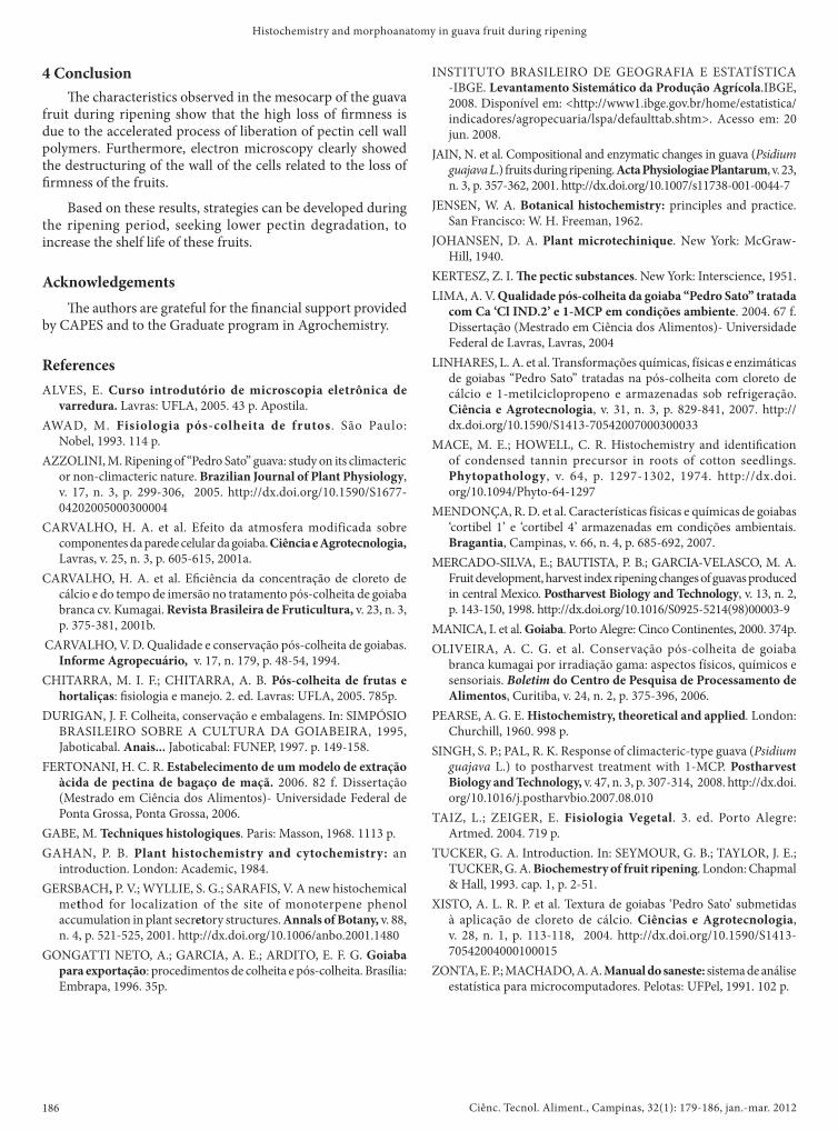
4 Conclusion�e characteristics observed in the mesocarp of the guava
fruit during ripening show that the high loss of �rmness is due to the accelerated process of liberation of pectin cell wall polymers. Furthermore, electron microscopy clearly showed the destructuring of the wall of the cells related to the loss of �rmness of the fruits.
Based on these results, strategies can be developed during the ripening period, seeking lower pectin degradation, to increase the shelf life of these fruits.
Acknowledgements�e authors are grateful for the �nancial support provided
by CAPES and to the Graduate program in Agrochemistry.
ReferencesALVES, E. Curso introdutório de microscopia eletrônica de
varredura. Lavras: UFLA, 2005. 43 p. Apostila.AWAD, M. Fisiologia pós-colheita de frutos. São Paulo:
Nobel, 1993. 114 p.AZZOLINI, M. Ripening of “Pedro Sato” guava: study on its climacteric
or non-climacteric nature. Brazilian Journal of Plant Physiology, v. 17, n. 3, p. 299-306, 2005. http://dx.doi.org/10.1590/S1677-04202005000300004
CARVALHO, H. A. et al. Efeito da atmosfera modificada sobre componentes da parede celular da goiaba. Ciência e Agrotecnologia,Lavras, v. 25, n. 3, p. 605-615, 2001a.
CARVALHO, H. A. et al. E�ciência da concentração de cloreto de cálcio e do tempo de imersão no tratamento pós-colheita de goiaba branca cv. Kumagai. Revista Brasileira de Fruticultura, v. 23, n. 3, p. 375-381, 2001b.
CARVALHO, V. D. Qualidade e conservação pós-colheita de goiabas. Informe Agropecuário, v. 17, n. 179, p. 48-54, 1994.
CHITARRA, M. I. F.; CHITARRA, A. B. Pós-colheita de frutas e hortaliças: �siologia e manejo. 2. ed. Lavras: UFLA, 2005. 785p.
DURIGAN, J. F. Colheita, conservação e embalagens. In: SIMPÓSIO BRASILEIRO SOBRE A CULTURA DA GOIABEIRA, 1995, Jaboticabal. Anais... Jaboticabal: FUNEP, 1997. p. 149-158.
FERTONANI, H. C. R. Estabelecimento de um modelo de extração àcida de pectina de bagaço de maçã. 2006. 82 f. Dissertação (Mestrado em Ciência dos Alimentos)- Universidade Federal de Ponta Grossa, Ponta Grossa, 2006.
GABE, M. Techniques histologiques. Paris: Masson, 1968. 1113 p.GAHAN, P. B. Plant histochemistry and cytochemistry: an
introduction. London: Academic, 1984.GERSBACH, P. V.; WYLLIE, S. G.; SARAFIS, V. A new histochemical
method for localization of the site of monoterpene phenol accumulation in plant secretory structures. Annals of Botany, v. 88, n. 4, p. 521-525, 2001. http://dx.doi.org/10.1006/anbo.2001.1480
GONGATTI NETO, A.; GARCIA, A. E.; ARDITO, E. F. G. Goiaba para exportação: procedimentos de colheita e pós-colheita. Brasília: Embrapa, 1996. 35p.