Abdominal tenderness to touch with irradiating pain into...
38
Abdominal tenderness to touch with irradiating pain into the lower extremity Neurology Department 2013. Jelen esetbemutatás kizárólag oktatási célokat szolgál, a diák , videók bármilyen felhasználása kizárólag a Debreceni Egyetem OEC írásos engedélyével történhet.
Transcript of Abdominal tenderness to touch with irradiating pain into...

Abdominal tenderness to touch with
irradiating pain into the lower extremity
Neurology Department
2013.
Jelen esetbemutatás kizárólag oktatási célokat szolgál, a diák , videók bármilyen felhasználása kizárólag a Debreceni Egyetem OEC írásos engedélyével történhet.

The present complains
• 28 yrs. old female patient had severe pain, which
irradiated into her left leg and into her right ham,
started 2 days ago, she was not able to walk.
• The day before she got tolperison and painkiller iv.
from her family doctor, had nausea, vomited, but the
pain .
• At night the pain increased, got meloxicam and
tolperison, pain more , she was sent to our Clinic.
The patient

Course of the disease
Allergy
4 years ago
During the night the pain
increased, got meloxicam,
tolperison, pain ↓ + had a
temperature
1 day before
Pain which irradiated into the
left leg and into the right ham
•tolperison
•vomited, but the pain↓
28 years old
1 day before Day of
admission

Status
Alert
RR: 110/70 Hgmm
P: 68/ min
EKG: SR
T: 38,1 fok
Lasegue sign positive at 50 degree
on the left side!
Valleix’ point: painful at pressure
on the left side!
Left sided decreased patella and
Achilles reflexes correlated to the
other side!
Both sided abdominal
tenderness to touch!
Missed abdominal reflexes!
Cross or sensory
abnormalities Ǿ

The possible diagnosis is always
questionable, in case of low back pain,
always have to think on primary and
secundary causes!

What could be the possible diagnosis?
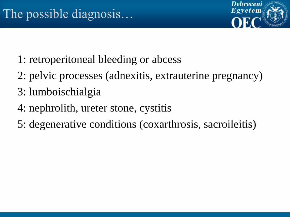
The possible diagnosis…
1: retroperitoneal bleeding or abcess
2: pelvic processes (adnexitis, extrauterine pregnancy)
3: lumboischialgia
4: nephrolith, ureter stone, cystitis
5: degenerative conditions (coxarthrosis, sacroileitis)

The explanation of the differential
diagnosis
1: retroperitoneal bleeding or abcess: could be, abdominal
US must be done
2: pelvic processes: could be, gynaecological examination
is needed
3: lumboischialgia: could be
4: nephrolith, ureter stone, cystitis: the complains are not
characteristics for urological processes
5: degenerative conditions: in this age, it’s not typical

How to start the examination?

Results of the examinations
Laboratory deviations:
Wbc: 12,71 Giga/L
Neut%: 79,1 %
Lymp%: 4,0 %
CRP: 68 mg/L
Liquor glucose: 5,1 mmol/L
Cell analysis was not performed.

Results of examinations
Abdominal US: (abdominal tenderness to touch, elevated
sedimentation/inflammatory parameters and had temperature):
no pathological, but the pelvic region couln’t be examined surely
Gynacological examination: Sine morbo gynec.
Because of the menstruation, pregnancy test couldn ‘t be performed,
HCG laboratory test was suggested.
HCG:< 0,1 U/l (negative)

Results of examinations:
Lumbosacral spine X-ray:
No pathological abnormalities.
Lumbosacral spine CT:
- On the left mediolaterally part a half
cm large hernia, which fills the recessus
lateralis, on the left side L.5.radical
compression.
- height of the L.III. discus is normal. In
this level on the left side laterally 3 mm
large protrusion could be seen .

The explanation of the differential
diagnosis
1: retroperitonial bleeding or abcess: NO, because on the
abdominal US there was nothing pathological
2: Pelvical processes: NO, bacause the gynecological
examination excluded the possibility of this
3: lumboischialgia: YES
4: nephrolith, ureter stone, cystitis:NO, because the
symptoms were not typical for urogenital processes
5: degenerative conditions: NO, in this age it’s not typical
and the X-ray was also negative

Diagnostics – How to take on the
anamnesis?
• Pain, paraesthesia, sensory deficit: the localisation of the pain?
What kind of pain is it? How long does it take? How does it start (sudden
movement, injury)? Any worsening? Does abdominal squeezing provoke pain?
During coughing, sneezing what happens? Where does the irradiating pain
spread?Does the pain decreased/disappeared? Sensory deficit/ sensory loss/
paraesthesia?
• Weakness of the extremities: clumsy movements during dressing, not
able to elevate the arm, not able to do stepping, drop his leg? Muscle atrophyt?
• Vegetative: Incontinence? Feel the stool /urine? Not able the evacuate the
urinary bladder? Impotence?
Diagnostics – Physical examination
• Spinal colomn: patting is painful? Antalgic gait? Tone of the
parevertebral muscles? Examination of the hip joints, the rotation is
painful?
• Sensory symptoms: subjective (without stimuli, for example:
spontaneous paraesthesia), objective (tactile, algetic, temperature,
vibration, dermolexia, joint position); localisation is typical for
dermatome, peripheral nerve, the whole extremity?
• Motoric movement, reflexes, pyramidal signs:able to
stand on heels and toes, foot slapping, able to cower and stand up, fingers
and toes works proximally well?
• Vegetative: incontinence, retention?
Imaging
Imaging examinations:
- bilateral X-ray
- Spinal CT scan 99% is enough
- Spinal MR scan
- Myelographia, myelo CT (rarely)
Electrophysiological examinations:
− ENG: F-wave, H-reflex: root laesion? Before imaging, it’s
apparent!
− EMG: to differentiate neurogen and myogen damages! To
localise the level of the spinal laesion ! SSEP (upper or lower
extremity), MEP-examination could be useful
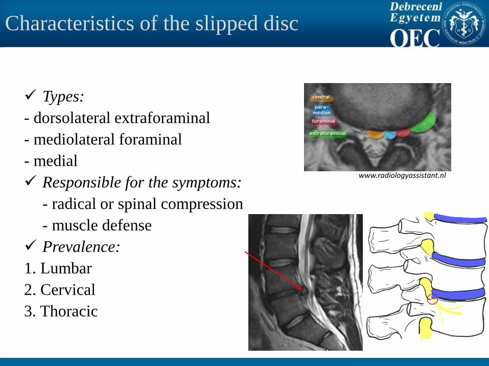
Characteristics of the slipped disc
Types:
- dorsolateral extraforaminal
- mediolateral foraminal
- medial
Responsible for the symptoms:
- radical or spinal compression
- muscle defense
Prevalence:
1. Lumbar
2. Cervical
3. Thoracic
www.radiologyassistant.nl

Disc prolapse
The process of the development
Provoking factors: - congenital deformities (spina bifida) - mechanical causes (sitting work) - injuries - metabolic osteopathies - inflammations (spondylitis, spondylodiscitis) - genetics, obesity

Localisation: cervical spine
Cause: Most common levels affected are C5/C6 and C6/C7.
Cervicocephalic syndrome: the impairment of the plexuses
causes nuchal or occipital headache.
Cervicobrachialgia: pain in the neck radiating into the arm.
Symptoms:
Pain in the neck,which could radiate into the neck, arms, shoulders or the head
Paracervical defense
In the case of medially progression: paraparesis, tetraparesis with increased deep
reflexes and pyramidal signs
Diagnostics:
Spurling-sign: it involves turning the patient's head to the affected side and
applying downward pressure to the top of the patient's head, if radicular pain is
elicited, this is called a positive Spurling's!

Clinical presentation of cervical
radiculopathies: Nerve
root
Sensory disturbance Motor weakness Muscle
movement,
which could be
affected
Reflex
absent/
decreased
C5 Shoulder, lateral arm m. deltoideus Abduction of the
arm
radialis
C6 Lateral forearm,
thumb and index
finger
m. biceps brachii, m.
brachioradialis
Flexion of the
elbow
biceps and
radialis
C7 Posterior arm,
dorsum, forearm,
middle finger
m. triceps brachii, m.
extensor carpi
radialis longus et
brevis
triceps
C8 Shoulder medial
forearm, ring and
little fingers
hypothenar, m. flexor
carpi ulnaris
Flexion of the
fingers

Cervical spine
Differential diagnosis: - cervical spondylosis
- spinalis tumor, which cause radicular compression like meningeoma, neurofibroma
- Thoracic outlet syndrom
- Pancoast tumor which infiltrates the plexus brachialis
- Tunnel syndromes (carpal/ulnar tunnel syndromes).
- Periarthritis humeroscapularis
C5
C6
C7
C8

Thoracic spine
Cause: the most stable part of the spinal colomn, disc prolapses are rare! In this localisation malignancy have to be think on always!
Symptoms:
Pain,which irradiates like lunar shape
If Th 5-12 is affected, abdominal reflexes could be weaker!
Differential diagnosis:
- Intercostal neuralgia
- Tumor
- Herpes zoster
- Dissection of the aorta
- MS, neuromyelitis optica

Lumbar spine:
Cause: L5/S1 disc and L4/5 disc prolapses account for>95%, older ages the stenosis
of the canalis spinalis is common!
Symptoms:
Irradiating pain from the lumbar region
Lumbar lordosis will be straighten, the mass of the paralumbar muscles will
increase, defense could be seen
Antalgic position, which spares the painful side
In severe cases paresis, vegetative symptoms
Lumbago: the pain is locally, which is characteristics for the radix. No irradiating
pain, no reflex abnormalities, no vegetative symptoms!
Lumboischialgia: irradiating pain could be detected. The clinical symptoms do NOT
localise the radix with the help of the dermatome or the reflex abnormalities.
Imaging could help!
Hernia dicsi intervertebralis: dermatome and also reflex abnormality could be seen.
Anamnesis, physical examinations could help, imaging confirms!

Lumbar spine- Diagnostic:
Lasegue-sign: (extend the ischiadic nerve); positive in case of L4, L5, S1 radices
Bragard-sign: the same manoeuvre, but with dorsalflexion of the leg
Inverz Lasegue-sign: (extend the femoral nerve); positive in case of L3. L4 radices
Valleix-points: points are painful which are at the radiation of the gluteofemoral part of the ischiadic nerve
www.umn.edu

Clinical presentation of lumbar radiculopathies:
Nerve root Motor weakness Muscle movements
could be damaged
Plus examination Reflex absent/
decreased
L3 m. psoas, m.
quadriceps
Hip flexion,
adduction of the leg
Inverz Lasegue sign
positive
No abnormalities
L4 m. tibialis anterior,
m. quadriceps
femoris
Femoral extension
(walking on stairs)
Lasegue and inverz
Lasegue signs are
positive
Patella reflex
L5 m. peroneus longus
and m. tibialis
anterior, m. extensor
hallucis longus
Dorsalflexion, (stand
on heels)
Lasegue sign positive No reflex
abnormalities
S1 m. gastrocnemius plantarflexion (stand
on toes)
Lasegue-sign
positive, Valleix
points are painful
Achilles-reflex
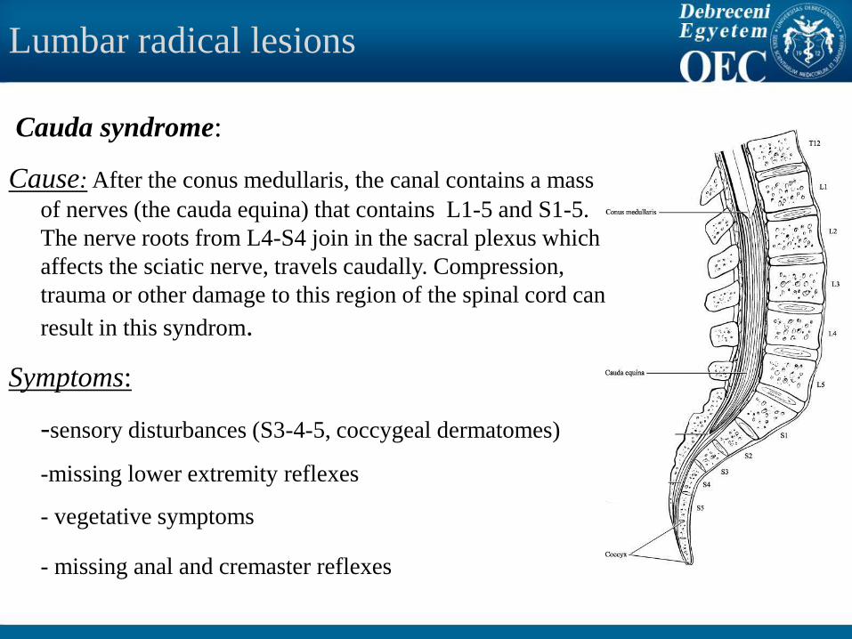
Lumbar radical lesions
Cauda syndrome:
Cause: After the conus medullaris, the canal contains a mass
of nerves (the cauda equina) that contains L1-5 and S1-5.
The nerve roots from L4-S4 join in the sacral plexus which
affects the sciatic nerve, travels caudally. Compression,
trauma or other damage to this region of the spinal cord can
result in this syndrom.
Symptoms:
-sensory disturbances (S3-4-5, coccygeal dermatomes)
-missing lower extremity reflexes
- vegetative symptoms
- missing anal and cremaster reflexes

Conus-syndrome, spinal canal stenosis
Conus-syndrome:
Cause: the lesion is at the level of L1, the conus medullaris and the cauda equina
are damaged!
Symptoms: -sensory loss
- L3-S2 radical damage could associate
- urinary and/or faecal incontinence
- deep reflexes of the lower extremities could be evoked
Spinal canal stenosis:
Cause: narrowing of the spinal canal causing root compression, most common
L4/5 and L 3/4
Symptoms: Lumbar:usually bilaterally but may affect only one leg, leg numbness
or paraesthesia
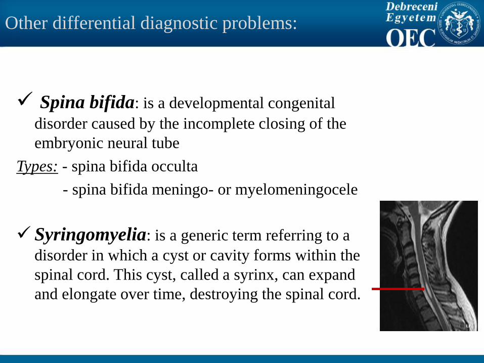
Other differential diagnostic problems:
Spina bifida: is a developmental congenital
disorder caused by the incomplete closing of the
embryonic neural tube
Types: - spina bifida occulta
- spina bifida meningo- or myelomeningocele
Syringomyelia: is a generic term referring to a
disorder in which a cyst or cavity forms within the
spinal cord. This cyst, called a syrinx, can expand
and elongate over time, destroying the spinal cord.

Other differential diagnostic problems:
Neuromyelitis optica /Devic (3 vertebral segments< lesion)
The white matter is damaged in more
segments ( often the opticus nerve as
well)
Oligoclonal gammopathy is uncommun
in the CSF
Antibodies against the Aquaporin-4
antigen could be detected

Other differential diagnostic problems
Spinal tumors: extradural
intradural extramedullaris (juxtamedullar
intramedullar
- Most of them have metastatic origin and take place in the vertebras (80% the primary sources: lung, breast, prostate, GI, malignant melanomes)
- The pain is typical for the extraduralis processes, that is why they could turn out early
- Most of the intradural tumors are meningeomas or spinal neurinomas (Schwannoma)
- Intramedullary astrocytomas infiltrates the spinal cord diffuse way
Conservative treatment:
In case of acut pain (<4 weeks)
muscle relaxants
non steroid anti-inflammation drugs (NSAID)
painkiller gel, unguent, suppository
Lidocain infiltration (Lange)
initial bed rest, early mobilisation
physiotherapy
In case of chronic pain (>3 months)
tricyclic antidepressants, SSRI, carbamazepine
lumbal corset, physiotherapy

Note!
72% of patients had herniations at presentation:
one-third of these herniations regressed in size or disappeared
at 6-week follow-up,
and two-thirds regressed or disappeared at 6- month follow-
up.
15% of herniations in the LBP group and 35% of herniations
in the radiculopathy group were reduced or had disappeared at
6-week followup. Modic MT, Ross JS, Obuchowski NA, Browning KH, Cianflocco AJ, Mazanec DJ. Contrastenhanced MR imaging in acute lumbar
radiculopathy: a pilot study of the natural history. Radiology 1995;195(2):4
Indications for surgery:
• Absolute surgical indication:
- paresis
- vegetative symptoms
- cauda syndrome
• Relative surgical indication: after 4-6 weeks conservative
treatment there is no improvement
!! Failed back surgery syndrome: the surgery doesn’t decrease
the complains of the patient, could be chronic!!
!!Behind lumbago could be an atipical discus hernia, if it doesn’t
recover imaging must be done!!
Further examinations are needed:
Severe pain not responding to conservative measures
Chronic recidive pain
Chronic pain and symptoms of radicular compression (paresis)
Compression of the spinal cord

Indications for surgery:
Severe pain not responding to conservative measures
Chronic recidive pain
Chronic pain and progressive motor deficit (paresis)
Symptoms of the compression of the spinal cord, which could be confirmed radiologically also (size, localisation) , the symptoms and the results of the examination have to be fit!

Expert video
Dr. Csiba László
Literature:
Csiba László és munkatársai (Debreceni Egyetemi Kiadó,
2010.): Fejezetek a neurológiából
Szirmai Imre (Medicina, 2000.): Neurológia
Komoly Sámuel és Palkovits Miklós (Medicina, 2010.):
Gyakorlati neurológia és neuroanatómia
Oxford Handbook of Neurology (Oxford University Press,
2011.)