2011
27
2011 09 08 Spinal cord 부부 1. Pain modulation 부 부부 부부 . Pain modulation 부 substania gelatinosa (Spinal cord 부 Lamina II)부부 부부부부부 부부 부부부부부. 부부 부부 descending excitatory fibers 부 enkephalinergic interneurons 부부 activate 부부, postsynaptic inhibitory control 부 부부 pain impulse 부 부부부부 부부부부 fiber 부부부 부부부 부부. 부부, descending inhibitory fibers 부 부부 spinothalamic projection neurons 부 direct contact 부 부부 pain 부 modulate 부부 부부부 부부. 부 부부부 부부부 Gate control theory of pain 부 부부, 부부 부 pain 부 small c fiber 부 부부 부부부 부 부 부부부 부부 fiber 부 touch 부부부 부부 부부부부. 부부부부 touch 부부부 neuron 부 collateral axon 부 s. gelatinosa 부 부부 interneuron 부 부부부 부부부 부 interneuron 부 inhibitory effects 부 부부 부부부 부부. (*부부부부부부) 2. Syringomyelia 부 부부부 부부부 부부 부부부 부부부부부 부 부부부 부부부부 ? Syringomyelia 부 cavitation of the central regions of spinal cord 부 부부 부부부부, 부부부 frequent 부부 anterior white commissure 부 crossing 부부 fiber 부부부 부부부 부부. 부 fiber bundle 부 opposite side ALS (anterolateral syst.)부 enter 부부부 fiber 부부 posterior horn 부 부부 midline 부 cross 부부 fiber 부부 부부부부부부. 부부부부 부 부부부 lesion 부 부부부, 부부부 부부부 부부 fiber 부부부 부부부 부부부부. 부부부부 bilateral 부부 부 부부 spinal cord level 부 부부, loss of pain and thermal sensations 부 부부부부부부 since ALS 부 부부부 pain 부 thermal sense 부 convey 부부. 부부부부 부부 부부부 cervical region 부부 syringomyelia 부 부부부, cape 부 부부 부 부부 mid and lower cervical level 부 부부부부 부부부 부부부부 thermal sense 부 pain sensation 부 deficit 부 부부부부 부부부. (* 부 syrinx 부 ant. horn 부부부 부부부 부부부 부부 부부 ant.horn 부 부부부 부 level 부 부부부부 extremity 부 ipsilat. weakness 부 부부부부부부. 부부 부부부 ant.horn 부 부부부 bilat. weakness 부 부.) 부부부 syringomyelia 부 central canal 부 부부부 부부 부부부 hydromyelia 부부. 3. Functional hemisection of the spinal cord (=Brown-Sequard Synd.) spinal cord 부 부부부 부부부부 부 부부부부 부부부부부 부부부부 , 부부부 부부부 tract 부 부부부부 ?
-
Upload
brigittekang -
Category
Documents
-
view
60 -
download
0
Transcript of 2011
2011 09 08
Spinal cord 부분
1. Pain modulation 에 대해 쓰기 .
Pain modulation 은 substania gelatinosa (Spinal cord 의 Lamina II)에서 이루어지는 걸로 알려져있다. 예를 들어 descending excitatory fibers 는 enkephalinergic interneurons 들을 activate 하여, postsynaptic inhibitory control 을 하여 pain impulse 를 전달하는 들어오는 fiber
들에게 영향을 준다. 또한, descending inhibitory fibers 가 직접 spinothalamic projection
neurons 와 direct contact 를 하여 pain 을 modulate 하는 경우도 있다.
또 하나의 이론은 Gate control theory of pain 에 의해, 아플 때 pain 이 small c fiber 을 타고 들어올 때 더 강하고 굵은 fiber 을 touch 감각이 타고 들어온다. 이럴경우 touch 감각의 neuron 의 collateral axon 이 s. gelatinosa 에 있는 interneuron 에 신호를 보내어 이 interneuron 이 inhibitory effects 를 내게 할수도 있다. (*그림그려보기)
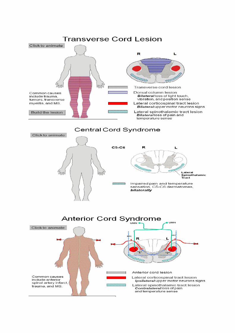
2. Syringomyelia 가 생기면 통증과 온도 감각이 소실되는데 그 이유는 무엇인가 ?
Syringomyelia 는 cavitation of the central regions of spinal cord 에 의해 생기는데, 이것은 frequent 하게 anterior white commissure 을 crossing 하는 fiber 들에게 손상을 준다. 이 fiber
bundle 은 opposite side ALS (anterolateral syst.)에 enter 하려는 fiber 들로 posterior horn 을 지나 midline 을 cross 하는 fiber 들로 이루어져있다. 그러므로 이 구조에 lesion 이 생기면, 여기를 지나는 양쪽 fiber 들에게 손상이 갈것이다. 그러므로 bilateral 하게 그 해당 spinal cord level 을 따라, loss of pain and thermal sensations 가 나타날것이다 since ALS 의 기능은 pain 과 thermal sense 를 convey 해줌.
예를들어 중간 아래쪽 cervical region 에서 syringomyelia 가 발생시, cape 를 입은 것 처럼 mid
and lower cervical level 에 해당하는 어깨와 팔주위가 thermal sense 와 pain sensation 에 deficit 이 생길생길 것이다. (* 큰 syrinx 는 ant. horn 까지도 영향을 주므로 만약 한쪽 ant.horn 이 눌리면 그 level 에 해당하는 extremity 에 ipsilat. weakness 를 가져올것이다. 만약 양쪽의 ant.horn 이 눌리면 bilat. weakness 가 옴.) 그리고 syringomyelia 는 central canal 의 확장이 아님 이것은 hydromyelia 라함.
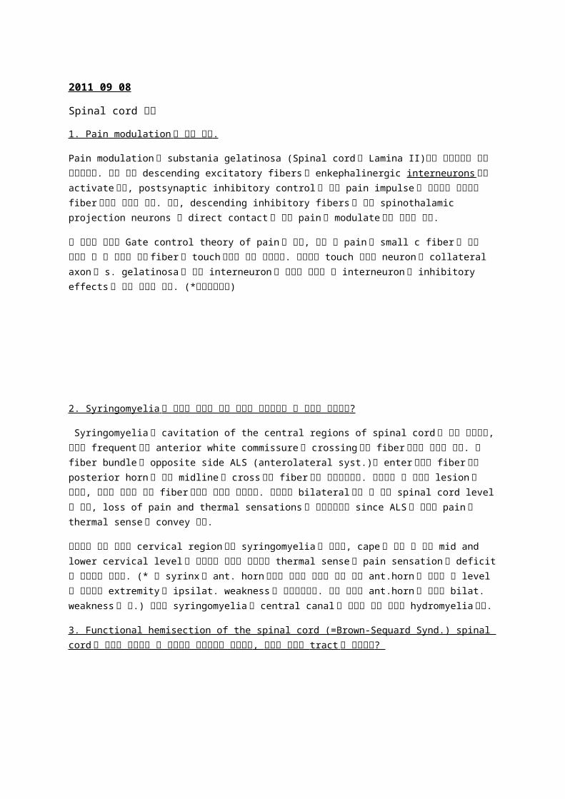
3. Functional hemisection of the spinal cord (=Brown-Sequard Synd.) spinal cord 의 절반이 날라갔을 때 일어나는 임상증례가 무엇인지 , 그것과 관련된 tract 는 무엇인지 ?


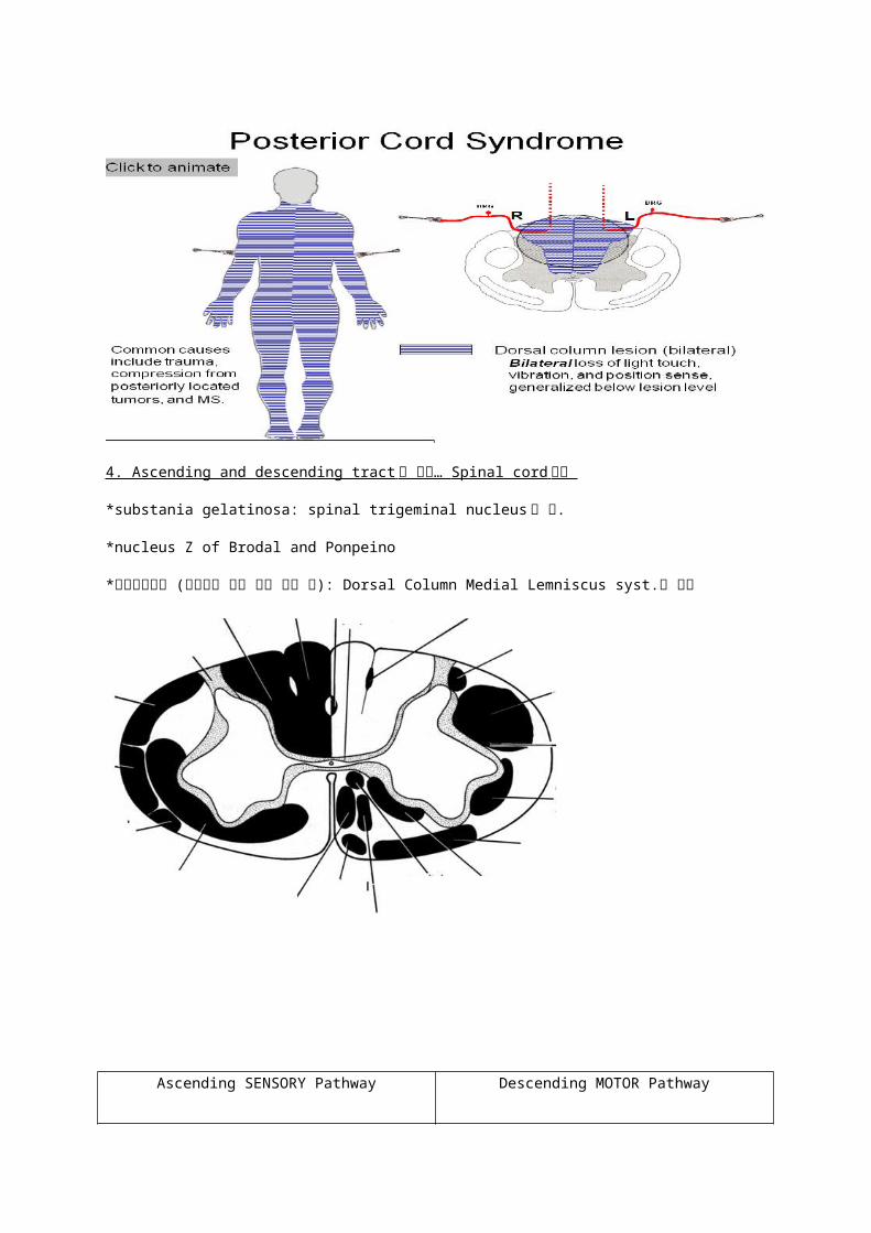
4. Ascending and descending tract 에 대해… Spinal cord 단면
*substania gelatinosa: spinal trigeminal nucleus 가 됨.
*nucleus Z of Brodal and Ponpeino
*형태인지불능 (주머니속 동전 구분 못할 때): Dorsal Column Medial Lemniscus syst.에 문제
Ascending SENSORY Pathway Descending MOTOR Pathway
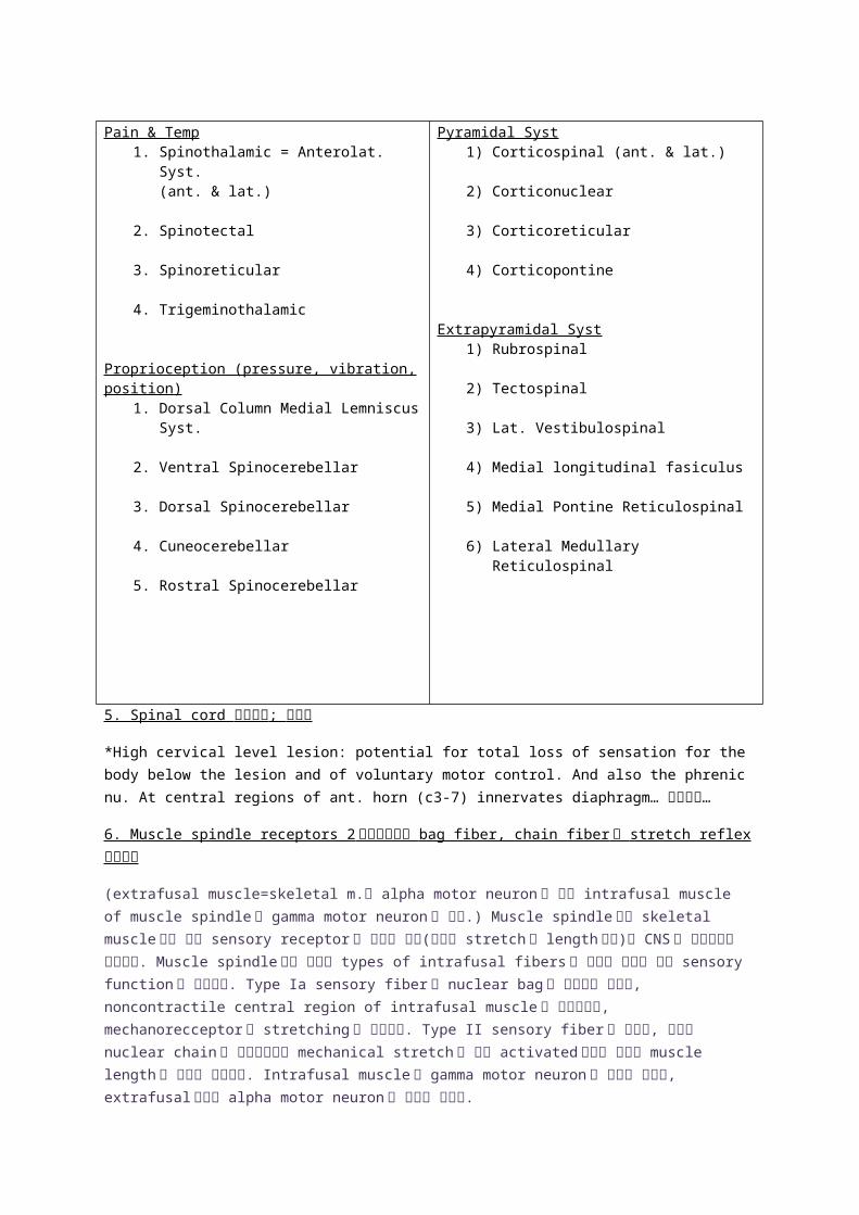
Pain & Temp1. Spinothalamic = Anterolat. Syst.
(ant. & lat.)
2. Spinotectal
3. Spinoreticular
4. Trigeminothalamic
Proprioception (pressure, vibration, position)
1. Dorsal Column Medial Lemniscus Syst.
2. Ventral Spinocerebellar
3. Dorsal Spinocerebellar
4. Cuneocerebellar
5. Rostral Spinocerebellar
Pyramidal Syst1) Corticospinal (ant. & lat.)
2) Corticonuclear
3) Corticoreticular
4) Corticopontine
Extrapyramidal Syst1) Rubrospinal
2) Tectospinal
3) Lat. Vestibulospinal
4) Medial longitudinal fasiculus
5) Medial Pontine Reticulospinal
6) Lateral Medullary Reticulospinal
5. Spinal cord 임상증례 ; 표참고
*High cervical level lesion: potential for total loss of sensation for the body below the lesion and of voluntary motor control. And also the phrenic nu. At central regions of ant.
horn (c3-7) innervates diaphragm… 사망으로…
6. Muscle spindle receptors 2 가지있는것과 bag fiber, chain fiber 랑 stretch reflex 설명하기
(extrafusal muscle=skeletal m.은 alpha motor neuron 이 지배 intrafusal muscle of muscle
spindle 은 gamma motor neuron 이 지배.) Muscle spindle 이란 skeletal muscle 안에 있는 sensory receptor 로 근육의 정보(근육의 stretch 나 length 정보)를 CNS 로 전달해주는 구조이다.
Muscle spindle 에는 두가지 types of intrafusal fibers 가 있으며 그것은 다른 sensory function
을 갖고있다. Type Ia sensory fiber 은 nuclear bag 과 연관되어 있으며, noncontractile central
region of intrafusal muscle 를 감고있으며, mechanorecceptor 로 stretching 을 감지한다. Type
II sensory fiber 이 있으며, 이것은 nuclear chain 과 연관되어있고 mechanical stretch 에 의해 activated 되지만 이것은 muscle length 의 차이를 감지한다. Intrafusal muscle 은 gamma
motor neuron 의 지배를 받으며, extrafusal 부분은 alpha motor neuron 의 지배를 받는다.
Stretch(Extensor) Reflex 란 감각신경원이 운동신경원에 직접 연결되는 monosynaptic reflex 로,
stretch 에 의하여 intrafusal muscle 이 자극되어 afferent fiber 가 흥분된다. 이 afferent fiber 의 흥분파는 척수내로 들어와 motoneuron 에 전달되며, motoneuron 의 efferent fiber 를 통하여 근육에 흥분파가 전달되어 수축이 일어남. 예를 들면, Deep tendon reflex 가 있는데, patellar
tendon 을 살짝 tap 하면 primary sensory endings in the muscle spindles in the quadriceps
femoris muscle 가 stretch 한다-> 이것은 type Ia fiber 을 통해 DRG 로 impulse 를 보내며 dl
afferent axons 는 anterior horn 에 있는 QFm 을 지배하는 motor neuron 에 바로 synapse 한다.
그러므로 sudden contraction of QF occurs and an extension of the leg at knees occur.
그외 Flexor(Avoid) Reflex; Sensory 와 motor neuron 사이에 interneuron 이 들어가는 polysynaptic reflex 임 Receptor : 피부의 free nerve ending
Gamma Reflex loop: 상위중추(뇌)로부터 척수반사 muscle tone 을 조절, 뇌로부터 내려오는 하행로들은 gamma moto neurons 의 기능을 억제함. Descending tracts→ Gamma motor fibers → Intrafusal muscle fibers (in muscle spindle)→ Sensory afferent fibers →Alpha motor neurons → Extrafusal muscle fibers
7. Upper motor neuron lesion and Lower motor neuron lesion
Upper motor neuron 이란 cerebral cortex 에 위치하는 neuron 의 soma 와 brainstem or
anterior horn 의 somatic motor neuron 으로 투사되는 n. fiber 말단가지까지를 UMN 이라한다. (ex: corticospinal corticonuclear rubrospinal reticulospinal)
Lower motor neuron 은 brainstem or spinal cord 의 ant. horn 의 motor neuron 의 soma 와 effector 와 neuromuscular junct.을 형성하는 n. fiber 의 말단까지를 LMN 이라한다.
Symptom UMNL LMNLparalysis Spastic 강직성 마비 Flaccid 이완성 마비M tone 서있을 때 + _M atrophyDecrease in mass
no yes
Deep tendon reflex ++ -Pathologic reflex +(대표적: 바빈스키 사인) -
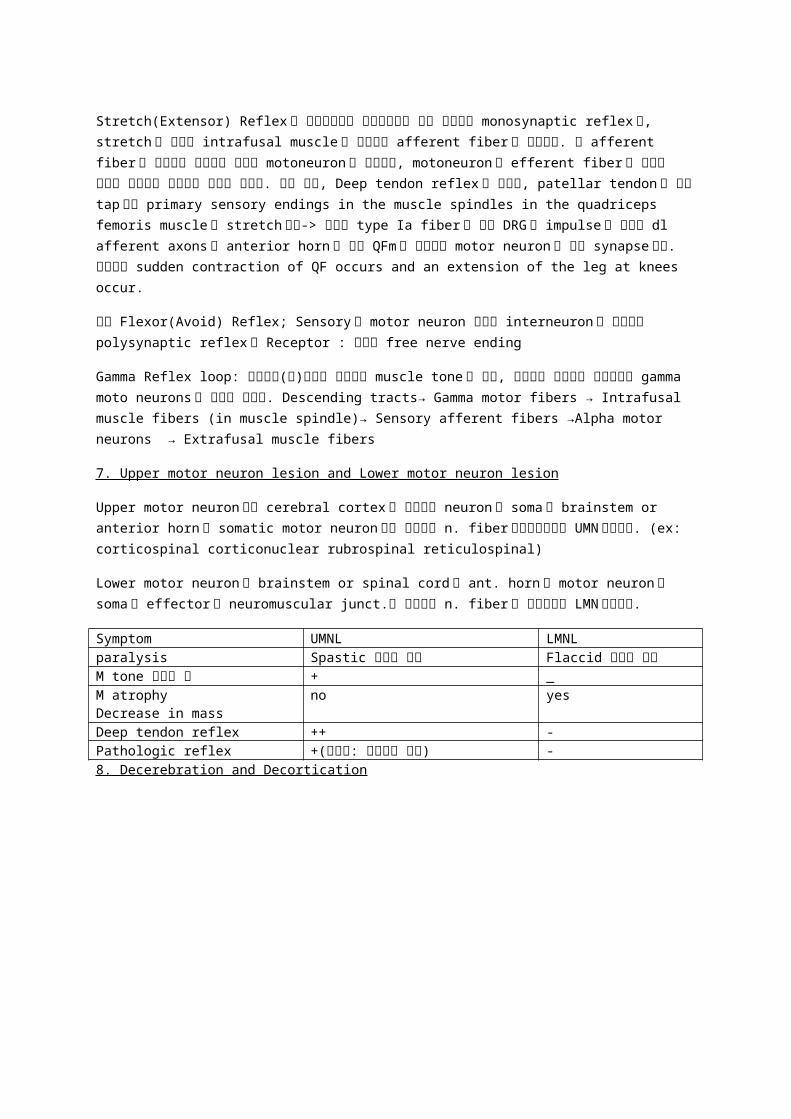
8. Decerebration and Decortication
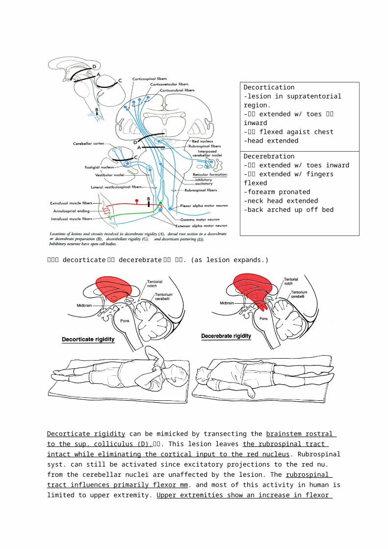
환자가 decorticate 에서 decerebrate 될수 있음. (as lesion expands.)
Decorticate rigidity can be mimicked by transecting the brainstem rostral to the sup.
colliculus (D).상태. This lesion leaves the rubrospinal tract intact while eliminating the cortical input to the red nucleus. Rubrospinal syst. can still be activated since excitatory projections to the red nu. from the cerebellar nuclei are unaffected by the lesion. The rubrospinal tract influences primarily flexor mm. and most of this activity in human is limited to upper extremity. Upper extremities show an increase in flexor tone due to the intact rubrospinal syst. However the lower extremities exhibit extensor hypertonus for the same reasons as in decerebration.
Decortication-lesion in supratentorial region.-하지 extended w/ toes 약간 inward-상지 flexed agaist chest-head extended
Decerebration-하지 extended w/ toes inward-상지 extended w/ fingers flexed-forearm pronated-neck head extended-back arched up off bed
Decerebrate condition(A-complete transaction btw. Sup and inf colliculus this resembles supratentorial lesion that causes herniation of midbrain downward thru the tentorial notch-central herniation). All descending cortical systems are interrupted (corticospinal,
corticorubral, corticoreticular) and rubrospinal tract 도 transected 되었지만, but excitatory and inhibitory components of the reticular formation are intact. Also the ascending somatosensory input to the reticular formation thru the ALS and reticulospinal syst. Flexor muscles are inactive due to the loss of descending corticospinal and corticorubrospinal input to flexor motor neurons. Conversely, extensor motor neurons are unaffected by the loss of descending cortical fibers b/c they are activated by descending reticulospinal and vestibulospinal inputs that are not involved by the decerebration lesion; thus these tracts remain intact.
8. Spinal cord 의 분화과정 및 4 layer 에 대해 쓰기 .
Neural tube 가 closed 된다.
세포분열을 계속하여, neuroepithelial layer 을 형성한다.
Neural tube 의 내강면을 따라 groove 가 형성된다 (sulcus limitans).
Neuroblasts 가 출현한다.
유사분열을 계속하여 neuroepithelial layer 주변에 mantle layer 을 형성한다. (mantle layer: gray mt).
Ant. and Post. Thickening 이 일어난다. 그러므로 basal and alar laminae 가 형성된다.
Basal lamina 의 thickening 으로 ventral horn 을 형성하고, immature nerve cells 는 vent. root
이됨.
Alar lamina 가 thickening 되고 dorsal horn 을 형성.
Mantle layer 의 neuroblast 에서 나온 nerve fiber 은 marginal layer 을 형성한다. 그리고 이 fiber
이 myelination 되어 white mt.을 형성한다.
Spinal ganglion 에서 나온 n. fiber 이 marginal layer 로 연결된다.
마지막으로 doral and ventral median fissure 이 생긴다.
(* Sulcus limitans 는 신경관의 외측벽의 중간에 형성되는 longitudinal groove. 신경학적 의의:
mantle layer 을 alar plate (dorsal)과 basal plate(ventral)로 구분한다.)
<Neural tube 의 layers>
3 개 (안->밖): 1) ependymal layer(lining lumen, germinal cells, future vent canal)
2) mantle layer(layer that differentiates into gray matter, neuroblasts->neuron, spongioblasts->neuroglia)
3) marginal layer(consists of n fibers projecting from the cells in mantle layer and
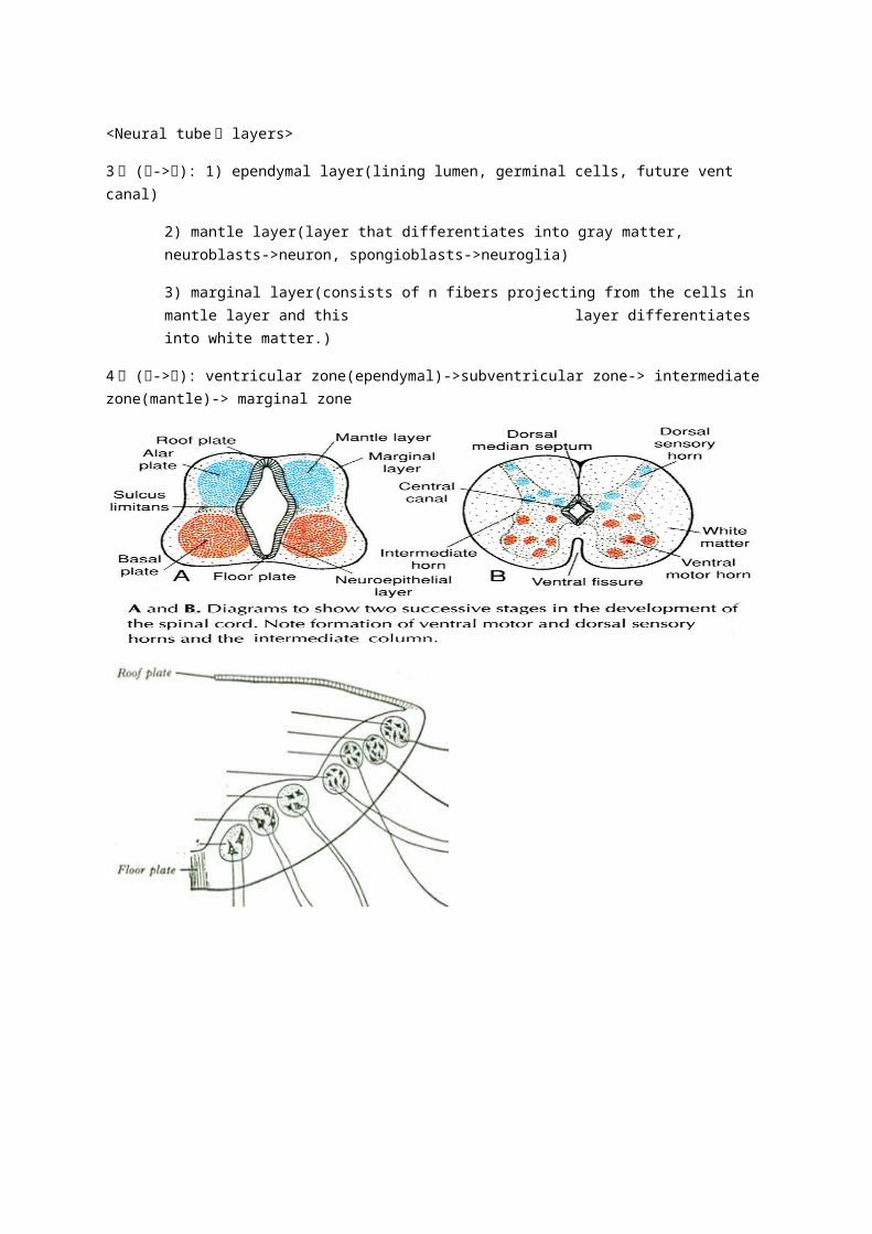
this layer differentiates into
white matter.)
4 개 (안->밖): ventricular zone(ependymal)->subventricular zone-> intermediate zone(mantle)-> marginal zone
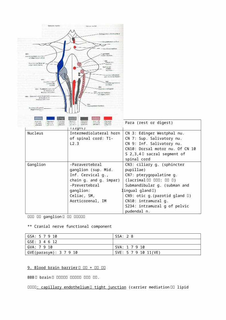
Sympathetic (fight or flight)
Para (rest or digest)
Nucleus Intermediolateral horn of spinal cord: T1-L2.3
CN 3: Edinger Westphal nu.CN 7: Sup. Salivatory nu.CN 9: Inf. Salivatory nu.CN10: Dorsal motor nu. Of CN 10S 2,3,4 에 sacral segment of spinal cord
Ganglion -Paravertebral ganglion (sup. Mid. Inf. Cervical g., chain g. and g. impar)-Prevertebral ganglion: Celiac, SM, Aorticorenal, IM
CN3: ciliary g. (sphincter pupillae)CN7: pterygopalatine g. (lacrimal 눈물 코점막: 콧물 침)
Submandibular g. (subman and lingual gland 침)
CN9: otic g.(parotid gland 침)CN10: intramural g.S234: intramural g of pelvic pudendal n.
그외의 핵과 ganglion 은 다른 프린트보기
** Cranial nerve functional component
GSA: 5 7 9 10 SSA: 2 8GSE: 3 4 6 12GVA: 7 9 10 SVA: 1 7 9 10GVE(parasym): 3 7 9 10 SVE: 5 7 9 10 11(VE)
9. Blood brain barrier 에 대해 + 없는 지역
BBB 란 brain 의 신경조직과 혈관사이에 형성된 장벽.
구성요소: capillary endothelium 의 tight junction (carrier mediation 이나 lipid mediation
으로 들어갈수있음.) + 신경조직의 basement memb. + Astrocytic process (glial perivascular feet)
기능: brain 내 fluid compartment 에서 chemical substance 의 transport 을 조절.
**BBB 가 없는 지역: Circumventricular organs (POS MAN)
Pineal body (melatonin 분비해야함), OVLT (organum vasculosum of lamina terminalis or supra optic crest), Subfornical organ and subcommissural organ, Median eminence of
hypothalamus (pituitary gland 로 이어져있어서), Area postrema (obex of MO 의 deep
portion), Neurohypophysis (H 분비하니까~)

10. Common vascular disease(Haines 124 and 25)
-Aneurysm: comes from Greek aneurusma dilation. It is a localized, blood-filled balloon-like bulge in the wall of a BV. When the size of an aneurysm increases, there is a
significant risk of rupture, resulting in severe hemorrhage(출혈). Aneurysm will cause the
wall of the blood vessel to weaken. **잘일어나는 지역: 주로 bifurcation 이 일어나는 지역 circle of Willis (Acom 과 ACA 만나는, basilar a.가 bifurcate 하여 PCA 를 만드는 부분 etc.)
*Haines: is the dilation of a vessel wall, usually in an artery, that extends from the lumen to the vessel surface and includes all layers of the vessel wall. Ex) cerebral aneurysm are frequently located at branch points of vessels or at points where the vessels may make an abnormally sharp abrupt turn on their course. (clip stalk off of aneurysm)
-Cerebral embolism: is the occlusion of a cerebral vessel by some extraneous material. This occlusion leads to ischemia and if prolonged to infarction of the area served by the vessel.
-Ischemia(허혈): insufficient blood supply due to vascular occlusion. Infarction(경색): a localized vascular insufficiency resulting in necrosis.
**ex) 작은 embolisms small cerebral vessels 를 occlude 하여 TIA 를 일으킬수 있다. TIA: a sudden loss of neurologic function that usually resolves within 24 hours.
TIA 는 3 개월이후로 15%가 attack 이 옴. => ischemic stroke (뇌졸중)
Embolic infarct(경색막힘): plaque 조각이 떨어져 나가서 조각이 혈관에가서 막힘/ ACUTE.
Thrombotic infart: 서서히 일어남, thrombus 가 생겨서 막힘/ CHRONIC
-Transient ischemic attack (TIA): is referred to as mini stroke, it is a transient episode of neurologic dysfunction caused by ischemia (loss of blood flow) – either focal brain, spinal cord or retinal – without acute infarction (tissue death). TIAs share the same underlying cause as strokes: a disruption of cerebral blood flow (CBF). TIAs and strokes cause the same symptoms, such as contralateral paralysis (opposite side of body from affected brain hemisphere) or sudden weakness or numbness. A TIA may cause sudden dimming or loss of vision, aphasia, slurred speech and mental confusion. But unlike a stroke, the symptoms of a TIA can resolve within a few minutes or 24 hours. Brain injury may still occur in a TIA lasting only a few minutes. Having a TIA is a risk factor for eventually having a stroke or a silent stroke.
-Arteriovenous malformation: 자체적으로 a.와 v.이 malformation 으로 서로 꼬여있음, rupture
이 생길수 있음. AVM results when the communications btw. Major aa. And veins do not develop normally.
11. Cavernous sinus 에서의 Internal Carotid A. 의 위치관계
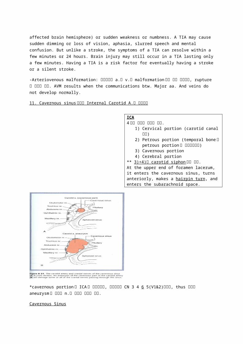
*cavernous portion 에 ICA 가 위치하는데, 이주변에는 CN 3 4 6 5(V1&2)가위치, thus 여기서 aneurysm 이 생기면 n.에 이상이 갈수도 있다.
Cavernous Sinus
The large irregular spaces lies on each side of the sella turcica, body of the sphenoid bone and pituitary gland that extends from the sup. orbital fissure to the petrous portionof the temporal bone. It is composed of network of intercommunicating venous
channels enclosing neurovascular structures. (lat. wall: CN 3 4 5-1 6 ICA 가 있음.) It makes communications with ophthalmic v, IJV, pterygoid plexus, pharyngeal plexus, basilar venous plexus.
*Danger Triangle 눈썹사이 upper lip: veins in this area communicate with the sup. and inf. ophthalmic veins which drain into the cavernous venous sinuses of the dura mater.
그러므로 이지역에서 inflammation 시, pterygoid plexus 로-> cavernous sinus->바로뇌로 pathogen 이 진입가능.
12. Brain 의 Blood Supply 에 대해 (Also 막혔을때 어떤일이 생기는지도… .)
ICA (ant. circulation) Vertebral a. (post. Circulation)A.Ophthalmic arteries (central a. of retina) A. Anterior spinal artery
ICA4 개의 부위로 나눌수 있음.
1) Cervical portion (carotid canal 통해)2) Petrous portion (temporal bone 의
petrous portion 을 휘어서올라감)3) Cavernous portion4) Cerebral portion
** 3)+4) 를 carotid siphon 이라 한다. At the upper end of foramen lacerum, it enters the cavernous sinus, turns anteriorly, makes a hairpin turn, and enters the subarachnoid space.
B. Anterior cerebral arteries (ACA)
C. Anterior communicating arteries (Acom)
D. Middle cerebral arteries (MCA)
B. Posterior spinal arteries
C. Posterior inferior cerebellar arteries (PICA)
D. Basilar artery (BA)
1) Anterior inferior cerebellar arteries (AICA)
2) Pontine arteries
3) Superior cerebellar arteries
E. Posterior cerebral artery (PCA)
F. Posterior communicating arteries (Pcom)
Brain 의 blood supply 강의록 간단정리
1.Internal Carotid Artery 의 cerebral portion (OPA, AM)
-Major branches of ICA: Ophthalmic a (optic n. 와 비슷하게 주행하고 retina 에 b supply).,
Pcom (carotid siphon 의 dorsal aspect 에서 나옴), Anterior choroidal a. (optic tract 와 같이 backward 로 run 하고 temporal lobe 쪽 choroid plexus 로), Ant. cerebral a. & Middle cerebral a. (terminal branches of ICA).
1) Anterior choroidal a.는 proximal portion of ICA 또는 MCA 에서 나오며, supplies choroid plexus in the temporal horn of the lat. ventricle, and medial side of the temporal lobe and
ventral part of the cerebrum thus supplies lat. ventricle(inf. Horn-temporal 부분)의 주변 구조물들~ a. to choroid plexus, optic tract, uncus, amygdale, hippocampus, globus pallidus, ventral part of internal capsule, LGB(lat. geniculate body), subthalamus, ventral thalamus, rostral part of midbrain.
** The lateral geniculate nucleus (LGN) is the primary relay center for visual information received from the retina of the eye. The LGN is found inside the thalamus of the brain.
***Ant. choroidal a. Synd.
:임상에서 ant. choroidal a. occlusion 시 combination of visual deficits(optic tract) and weakness of the opposite upper and lower extremities (inf. Portion of the post. limb of int. capsule).
: 혈전(thrombosis)이 잘생긴다. Proximal portion 의 surgical occlusion: Parkinsonian tremor
& rigidity 치료
: also this a. is long and slender thus easy to develop a thrombosis-> degeneration of the
hippocampus or globus pallidus resulting from impaired circulation. (Binswanger’s synd: loss of memory and intellectual function and by changes in mood.)
2) Anterior cerebral a. (FORCP): Brain 의 Medial aspect 에서 위쪽까지 supply
-optic chiasm 바로 앞에서 양쪽 ACA 는 Acom 에 의해 join 됨.->both aa. 는 median cerebral
fissure 로 들어간다.
-BR: frontopolar br, orbital br, recurrent a. of Heubner (med. Striate a.), callosomarginal a
(pericall a. 주변), pericallosal a.(바로 cor call 위)
- 임상 : (homonculus 생각하기 !!)
(A) 한쪽 ACA occlusion 시: direct lower limb
-contralateral m. weakness (primary motor cortex 에서 corticospinal tract 로 signal 보낼때 피가 원활히 돌지 않으면 힘들것임 -> m.에 weakness 가 생김. 특히 다리 b/c ACA 는 med. side of
brain 공급 and homonculus 따르면 ACA 가 가장 잘 닿는데가 leg and foot lower limb 있는데임.)
-contralateral sensory loss (anesthesia or paresthesia): discriminative touch,
proprioception, pain, temp, light touch (primary sensory cortex 로 spinothalamic tract-
pain temp 와 dorsal column medial lemniscus syst.-proprioception 의 정보가 들어와야하는데 여기가 b supply 가 제대로 안이루어지면 받아드리는데도 힘들것임.
(B) 양쪽 ACA occlusion 시: Bilateral paralysis “특히 lower limb”
3) Middle cerebral a. (LAO-PRAPP): supplies lat. view of cortex
-enters into lat. cerebral fissure and funs posteriorly w/in fissure=> fanlike fashion over lat. convexity of brain.
-BR: Lenticulostriate aa. (lat. striate a.), ant. temporal a., orbitofrontal a., pre-Rolandic a., Rolandic a., ant. parietal brs, post. Parietal brs, post. Temporal br.
**MCA 임상: 여기에 이상시, nonfluent aphasia (Lt-Broca’s area), B ganglia(반대팔 motor
안됨), Hearing 에도 문제 (Heschl area 도 b supply 하니까)

MCA occlusion 시
-contralateral hemiplegia: upper extremity and face
-contralateral sensory loss (cortical type): position, discriminative sensory loss
-aphasia: dominant hemisphere (Lt) Broca’s expressive aphasia/ Wernicke’s receptive aphasia
2. Major branches of Vertebral a.
: Subclavian a.에서 기원, cervical vertebrae 의 transverse foramina 지나서 foramen
magnuum 을 통해 post. cranial fossa 에 진입-> MO 를 따라 올라가면 Pontomedullary junct.
에서 양쪽 vertebral aa.가 만나서 Basilar a.를 만든다.
: 4 portion 으로 나뉨 (Spinal aa.- ant. and post. vertebral aa., Muscular brs, Post. inf cerebellar a, Basilar a.)
1)Spinal arteries: vertebral a.에서 얇은 가지를 각각 하나씩 내어 ant. aspect 에서 합쳐져서 내려오는 ant. vertebral a.(1 개-sc 의 ant. median fissure 을지남)와 vertebral a. 또는 가끔은 PICA 에서 나오는 post. vertebral aa. (2 개가 나옴)
2)Post. Inf. Cerebellar a. (PICA): 나중에 PICA, AICA, Sup. cerebellar a.가 만나 cerebellum
공급.
3)Basilar artery (Ant.-Lab-Pon-Sup) ant lab 에서 먹는 pon soup
-ant. inf. Cerebellar a.
-labyrinthine br. : facial n. 와 vestibulocochlear n.를 따라 internal acoustic meatus 를 통해 귀로~
-pontine aa.
-sup. cerebellar a.
-공급: cervical seg of spinal cord, MO, Pons, Cerebellum, MB, thalamus 의 post. part, post. and inf. Parts of temporal and occipital lobe, and body and membranous labyrinth
-Basilar a.는 마지막에 bifurcates into Posterior cerebral a.
4)Post. cerebral a.
-at pons and midbrain junction the basilar a. bifurcates in the interpeduncular cistern and gives rise to the post. cerebral aa.
-sends branches to midbrain, thalamus, ventral and medial surfaces of the temporal and occipital lobes as far as the level of the parieto-occipital sulcus
-basilar bifurcation 과 Pcom 사이에서 작은 가지를 내는데, 이것이 “Quadrigeminal and
thalamoperforating aa.”이다. => med. & lat. post. choroidal & thalamogeniculate aa. & branches to midbrain=> temporal br., parieto-occipital a., calcarine a.
** 임상
-Occlusion of PCA: Contralateral homonymous 같은쪽 hemianopsia 반맹증 (visual field defects)
:hemianopic visual loss on the same side of both eyes. Hemianopsia occurs b/e the fight 1/2 of brain has visual pathways for eh left hemifield of both eyes and the left 1/2 of brain
has v parts for the rt. Hemifield of eye. Lt. HH 시 보이는 모습
***CIRCLE OF WILLIS*** (7)
-Circle of Willis is an arterial wreath encircling the optic chiasm, interpeduncular retion
and tuber cinerum. (ICA-ACA-Acom-MCA-Pcom 까지 ICA 의 brs-PCA-Basilar)
1) ICA 2) Ant. cerebral a. 3) Ant. communicating a. 4) Middle cerebral a. 5) Post. Communicating a. 6) Post. Cerebral a. 7) Basilar a.
** 중요한 지표 : Posterior Cerebral a. 와 Superior Cerebellar a. 사이에 CN3 가 위치한다 .
-Circle of Willis 의 BR: central or ganglionic branches, choroidal aa., cortical aa. that spring off proximal portion of main aa. Of circle.
Central/ Ganglionic BR (4 groups)
A) anteromedial group: ACA & Acom 에서 기원, enter ant. perforated substance, supply ant., preoptic, supraoptic hypothalamic regions
B) anterolateral group: striated aa from MCA and ACA., recurrent a. of Heubner-caudate & putamen, lenticulostriate a.-putamen & globus pallidus, A. of Charcot=A. of Hemorrhage : : the largest of the lat. striate aa.=> easily ruptured in hypertension
patients. 이 a. 는 internal capsule 에 blood supply 를 한다 . 문제시 hemiplegia
** AAs that supply the Basal Ganglia: recurrent a. of Heubner (ACA), lenticulostriate a. (MCA), Ant. choroidal a.(cerebral portion of ICA).
C) posteromedial group (=thalamoperforating a): PCA & Pcom 기원, tuber cinerum,
mammillary body, interpeduncular fossa 주변 공급 ex: hypophysis, infundibulum, hypothal, thalamus, mammillary body, subthal region, med. Nu. Of thalamus, midbrain (tegmentum), crus cerebri
D) posterolateral group (=thalamogeniculate a.): PCA 의 lat.쪽에서 기원, enter lat.
geniculate body on post. Aspect of thalamus. 공급: caudal 1/2 of thalamus, LGB, Pulvinar, Lat. nuclear group of thal, vent. Thalamic nuclear tier
문제시: Occlusion of thalamogeniculate a.- Paralysis, Paresthesia, Pain and hyperpathia in
the contralateral face, body or extremity. 다 contralat.인 이유는… Decussation level 이전에 lesion 되서!
Choroidal aa. (ant. and post. choroidal aa.)
a) Ant. choroidal aa.위에서 했음
b) Post. choroidal a. : PCA 의 prox. part origin, med. & lat. 로 나뉨, tectum (MB 의 sup.-
시청각반사의 reflex center and inf-auditory relay nu. Colliculi), lat. ventricle 의 choroid
plexus 에 분포
Cortical aa. (ACA, MCA, PCA)
13. Watershed Zone 고혈압에 의한 brain damage zone (border zone infarction)=> cerebral cortex, int. capsule, basal ganglia.
수업시간: 이부위에 많은 branches 들이 겹쳐져서, 혈압이 낮아지면, ischemia 가 먼저 일어나는지역. sudden hypotension(저혈압), hypoperfusion(decreased blood flow) of the
distal vascular bed of a major cerebral a.에 의해 생기는 zone ex) ACA-MCA: ant. watershed infarct (contralat. Hemi paresis of lower extrm, expressive language deficits or behavioral changes), MCA-PCA: post. watershed infarct (parial visual loss, variety of language loss)
Blood Supply of Cerebellum/Brainstem/Spinal Cord + 임상 ~~
-Cerebellum 과 Brainstem 은 post. circulation 을 담당하는 Vertebral a.의 branches 들을 통해 공급을 받는다. Ex) PICA, Ant. Spinal a, Post. Spinal aa., Basilar a., AICA, Pontine aa., Sup. Cbll a.,
1) Post. Inf. Cerebellar A. (basilar a.의 분지일수두…있음)
-inferolat.하게 주행, MO 에 posterolat.하게 분지, Cerebellum 의 posteroinferior and med.하게 분지를 내며 끝남.
-분포지역: MO (fascicule & nuclei of cuneatus & gracilis), caudal and dorsal portions of inf.
cerebellar peduncle, NTS 의 fasciculus, Vagal motor nu., Spinal trigeminal nu., nu. Ambiguus, area postrema
-Anastomoses: AICA and post. spinal aa.(post. spinal aa 가 없을 때 PICA 가 대신 공급해줌.)
**임상 생각하기전~
무조건 motor decussation 전에 잘리면 즉 MO 전에: contralateral 하게 나오고
Cranial n.와 관련된건 다 ipsilateral 하게 나오고…
Sensory 는 좀 생각해보기~!
-임상

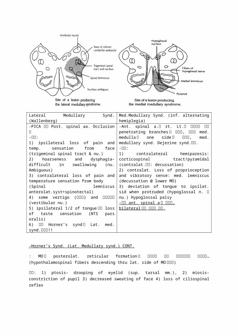
Lateral Medullary Synd. (Wallenberg) Med.Medullary Synd. (inf. alternating hemiplegia)
-PICA 혹은 Post. spinal aa. Occlusion 시-증상: 1) ipsilateral loss of pain and temp. sensation from face (trigeminal spinal tract & nu.)2) hoarseness and dysphagia- difficult in swallowing (nu. Ambiguous)3) contralateral loss of pain and temperature sensation from body (Spinal lemniscus anterolat.syst+spinotectal)4) some vertigo (현기증) and 안구진탕증 (vestibular nu.)5) ipsilateral 1/2 of tongue 에서 loss of taste sensation (NTS pars oralis)6) 밑의 Horner’s synd 도 Lat. med. synd.일부임!!
-Ant. spinal a.는 rt. Lt.를 교차하며 가는 penetrating branches 가 있으며, 이것이 med. medulla 의 one side 로 눌릴시, med. medullary synd. Dejerine synd.라함. -증상:1) contralateral hemiparesis: corticospinal tract/pyramidal (contralat.이유: decussation)2) contralat. Loss of proprioception and vibratory sense: med. lemniscus (decussation @ lower MO)3) deviation of tongue to ipsilat. sid when protruded (hypoglossal n. 나 nu.) Hypoglossal palsy- 완전 ant. spinal a 가 막히면 , bilateral 하게 증상이 생김 .
-Horner’s Synd. (Lat. Medullary synd.) CONT.
: MO 의 posterolat. reticular formation 을 통과하는 하행 교감신경로의 손상으로… (hypothalamospinal fibers descending thru lat. side of MO 이상시)
증상: 1) ptosis- drooping of eyelid (sup. tarsal mm.), 2) miosis- constriction of pupil 3) decreased sweating of face 4) loss of ciliospinal reflex
2) Basilar a.
-pontomedullary junct.에서… groove for basilar a.를 따라 상행=> pontomesencephalic
junct.에서 Lt Rt Posterior cerebral a.로 나뉘어짐.
-Branches
(1) Ant. Inf. Cerebellar Artery (AICA)
-inferolat. Pontomedullary junct. 감고 분지내고 cerebellum 의 inferior 쪽 lateral 쪽으로 가서 분지.
-분포지역: rostral MO 쪽, pyramid, ML, MLF, 추 12 nu, NTS, nu of CN10.
-임상:
<Middle Alternating Hemiplegia=Med. pontine synd.>
-Pons level 에서 Basilar a.와 Pontine a.이 눌릴 때
Ex) 오른쪽 이상시, 오른쪽 눈: abducens n. 이상=> med. adduction/ 왼쪽: hemiplegia
(2) Pontine a.: basilar a.의 branches on pons.
(3) Sup. Cerebellar artery
-pontomesencephalic junct.근처에서 brainstem 을 감소 cerebellum 의 상면으로 가서 분지.
**CN 3 이 sup. cerebellar a.와 post. cerebral a. 사이에 있다는 것~! Skull base surgery 시 조심!
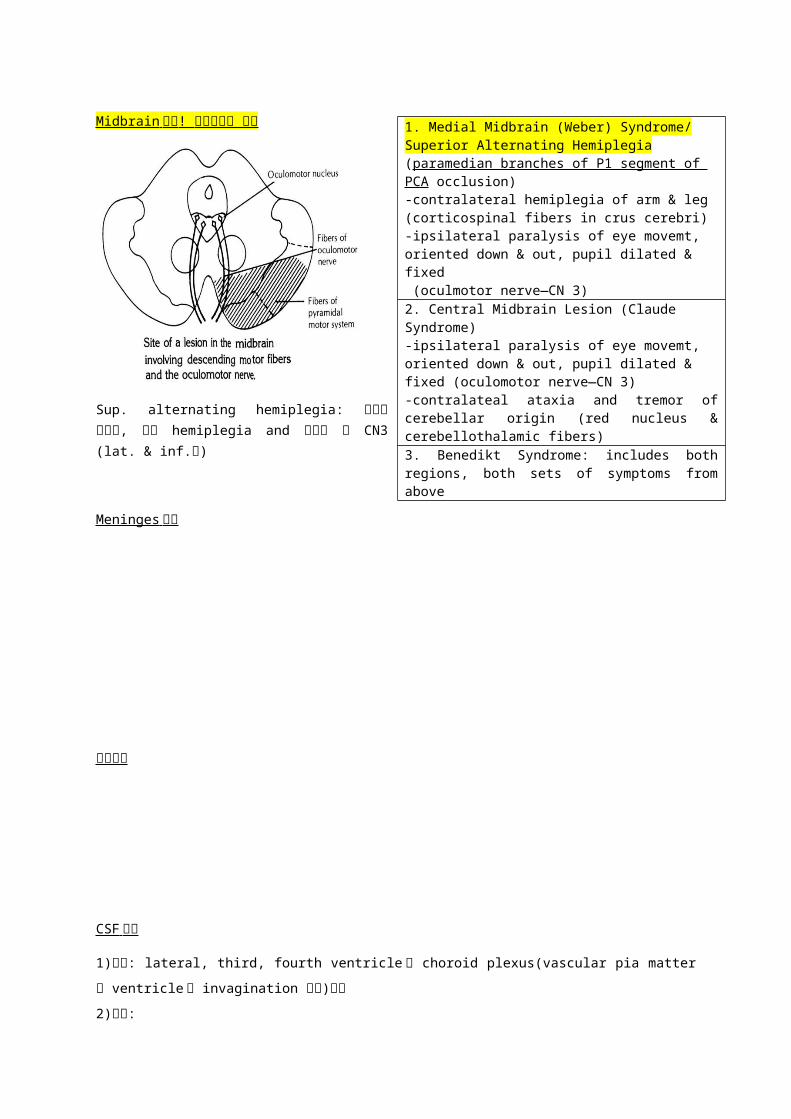
Midbrain 에서 ! 임상적으로 중요
-contralateral hemiplegia of arm & leg (corticospinal fibers in basilar pons)-contralateral loss/decrease of proprioception, vibration, discriminative touch (medial lemniscus)-ipsilateral lateral rectus muscle paralysis (abducens nerve fibers or nucleus—CN 6)-paralysis of conjugate gaze toward side of lesion (paramedian pontine reticular formation/pontine gaze center)
1. Medial Midbrain (Weber) Syndrome/ Superior Alternating Hemiplegia (paramedian branches of P1 segment of PCA occlusion) -contralateral hemiplegia of arm & leg (corticospinal fibers in crus cerebri)-ipsilateral paralysis of eye movemt, oriented down & out, pupil dilated & fixed (oculmotor nerve—CN 3)2. Central Midbrain Lesion (Claude Syndrome)-ipsilateral paralysis of eye movemt, oriented down & out, pupil dilated & fixed (oculomotor nerve—CN 3)-contralateal ataxia and tremor of cerebellar origin (red nucleus & cerebellothalamic fibers)3. Benedikt Syndrome: includes both regions, both sets of symptoms from above

Sup. alternating hemiplegia: 오른쪽 이상시, 왼쪽 hemiplegia and 오른쪽 눈 CN3 (lat. & inf.
로)
Meninges 정리
용어정리
CSF 순환
1)생성: lateral, third, fourth ventricle 의 choroid plexus(vascular pia matter 가 ventricle 로
invagination 된것)에서
2)순환:
3)흡수: dural venous sinus(특히, sup. saggital sinus & venous lacunae)로 돌출된 arachnoid
villi 를 통해 C.S.F 를 venous blood 로 흡수 *arachnoid villi 가 과도하게 커졌을 때 cranial bone
을 파고들어 자국을 남긴다(=> 이런 것을 arachnoid graulation 이라함)
Ventricle 형성과정
-neural tube 의 lumen 이 남아있는 구조
lateral ventricle -> 양쪽에 하나씩 2 개, cerebral hemisphere 에 존재
third ventricle -> 간뇌(diencephalon)에 존재
cerebral aqueduct(실비우스관) -> mid brain 을 관통하는 위치
fourth ventricle -> medulla, pons, cerebellum 에 둘러쌓여 있음
medulla oblongata 의 아래부분(spinal cord)에서는 ventricle 이 좁은 관으로 남아 central canal
이 됨
Hemorrhages
Herniations
발생학 brain 표
1。 Vesicle 2。 vesicle mature brain
prosencephalon
diencephalonthalamus, epithalamus,
hypothalamus, subthalamus
telecephalon
cerebral hemispheres, olfactory
system, corpus stratum, cb cortex,
white matter
mesencephalonmesencephalo
n
midbrain, consisting of tectum &
cerebral peduncle
rhombencephal
on
myelencephalo
nmedulla oblongata
metencephalo
npons & cerebellum
Neural crest origin 세포들
DRG/ Sensory ganglia of CN 5 7 9 10 (semilunar, geniculate, sup. and inf. ganglia of cn9, sup. & inf. ganglia of cn10)/ Autonomic ganglia (sym. Chain g, parasym. G of CN 3 7 9 10: ciliary, pterygopalatine, subman., otic, intramural-auerbach meissner’s plexus)/ Intramural g of pelvic pudendal n. (s234)/ Neuralemmal cell of PNS Schwann cell/ Stellate cell/ Melanocyte of skin/ Ganglionic cell in the adrenal medulla (sym)/ Enterochromaffin cell (ADUP cell)/ Leptomeninges/ Part of skull (related to face)
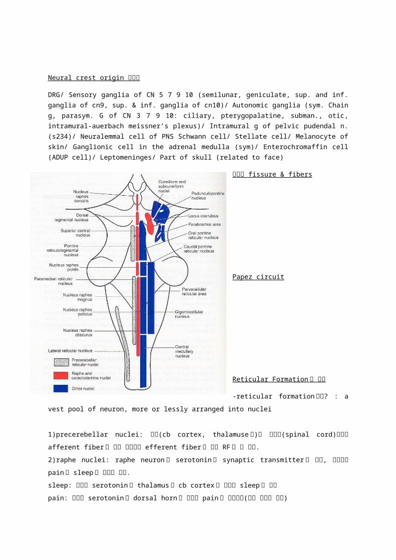
소뇌의 fissure & fibers
Papez circuit
Reticular Formation 에 대해
-reticular formation 이란? : a vest
pool of neuron, more or lessly
arranged into nuclei
1)precerebellar nuclei: 위쪽(cb
cortex, thalamuse등)과 아래쪽
(spinal cord)로부터 afferent fiber 를
받아 소뇌로만 efferent fiber 를 내는
RF 의 한 핵군.
2)raphe nuclei: raphe neuron 은
serotonin 을 synaptic transmitter
로 분비, 이물질이 pain 과 sleep 에
영향을 미침.
sleep: 분비된 serotonin 이 thalamus 와 cb cortex 를 억제해 sleep 을 유도
pain: 분비된 serotonin 이 dorsal horn 을 억제해 pain 을 억제시킴(자극 전도의 억제)
3)central group of nuclei
-inspiratory center 가 존재(central group nuclei 중 gigantocellular nucleus 에 있음)
-motor control 의 기능함
-의식을 각성시키는 기능함
(이 핵에서 나온 efferent fiber 가 intralaminar thalamic nuclei 로 유입 -> intralaminar
thalamic nuclei 에서 cb cortex 의 전 부분으로 자극이 퍼져 각성의 효과 나타냄)
=>이것을 ARAS(Ascending Reticular Activating System) 이라함
4)cholinergic neuron
-efferent fiber 중 일부가 basal ganglia 로 유입
=>일정한 패턴(sterotyped)을 갖는 운동을 할수 있게하는 기능 함 (ex; locomotion)
5)catecholamine nuceli
-locus coeruleus
-modulation of synapse between other neuron 의 기능
-spinal reflex 와 alterness 에 excitory 한 영향 미침.
6)lateral parvocellular reticular area
-expiratory center 가 존재함(심장의 acceleration, arterial blood pressure 의 증가시키는
기능함)
7)parabrachial area
-pneumotaxic center 가 존재
8)superficial medullary reticular neuron
-cardiovascular, respiratory regulation 의 기능함
CORPUS STRIATUM 의 분류와 명칭
1. corpus striatum = lentiform and caudate nuclri
2. lentiform nucleus = putamen and globus pallidus (external div. & internal division 으로
나뉜다.)
3. striatum = neostriatum = putamen and caudate nucleus
4. pallidum = paleostriatum = globus pallidus
5. basal ganglia(traditional anatomical usage) = caudate and lentiform nuclei, amygdala,
claustrum
6. basal ganglia(clinical and physiological usage) = corpus striatum, substantia nigra,
subthalamic nucleus.
뇌층 archicortex n etc.
Subiculum
외부구조 : area posterma gag?, tuberculum cinerum trigeminal,