財團法人新光吳火獅紀念醫院 放射診斷科 Wai-Yip Law Multidetector CT (MDCT)...
36
財財財財財財財財財財財財財 財財財財財 Wai-Yip La w Multidetector CT (MDCT) 財財財財財財財財財財
-
Upload
deirdre-cooper -
Category
Documents
-
view
309 -
download
0
Transcript of 財團法人新光吳火獅紀念醫院 放射診斷科 Wai-Yip Law Multidetector CT (MDCT)...

財團法人新光吳火獅紀念醫院 放射診斷科 Wai-Yip Law
Multidetector CT (MDCT) 在心臟血管方面的運用

coronary artery lumen
Plaques
LV function
LV perfusion

Left posterior aortic sinus
Left main artery (LM) -- 1- 2 cm
Left anterior descending (LAD)
1. Septal branches
2. Diagonal branches
Left circumflex artery (LCx)
1. Marginal branches
Intermedial artery


Left posterior aortic sinus
Left main artery (LM) -- 1- 2 cmcoronary
anatomy
Left circumflex artery (LCx)
1. Marginal branches
Intermedial artery Anterior aortic sinsus
Right coronary artery (RCA)
1. Marginal branches
Left anterior descending (LAD)
1. Septal branches
2. Diagonal branches

coronary anatomy
Right dominant (80%)
Left dominant()
Balanced type
1. Crux
2. Posterior descending artery (PDA)
3. Posterior lateral branch (PL)

coronary anatomy
1
2
3
4 PDA
5
PL
67
89
10
11
12
13 1415
Seg. Vessel
010203040506070809101112131415
RCARCARCAPDALMLADLADLADD1D2CxMoCXMOPL
Ca-X
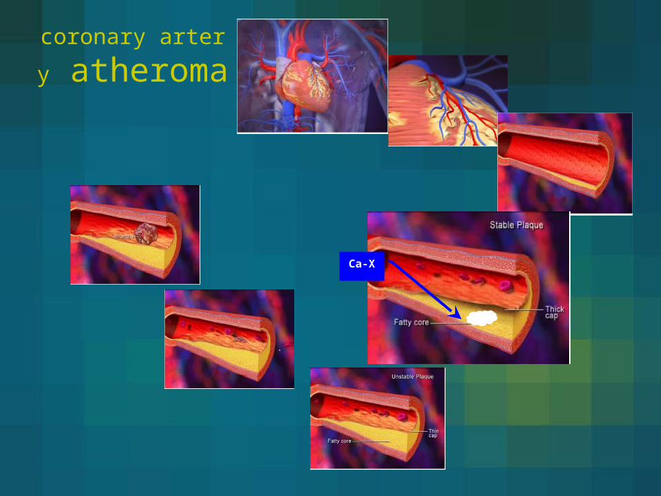
coronary artery atheroma

MSCT freatures of plaques (Modified AHA calcification)
Modified AHA
Calcified nodules
Fibrocalcified plaque
Fibreous cap atheroma
Thin fibrous cap atheroma
Trombus
DensityMDCT
>150 High High/Low
50-100 intermediate
20-50 low
<20 very low
Pictrure
coronary artery atheroma
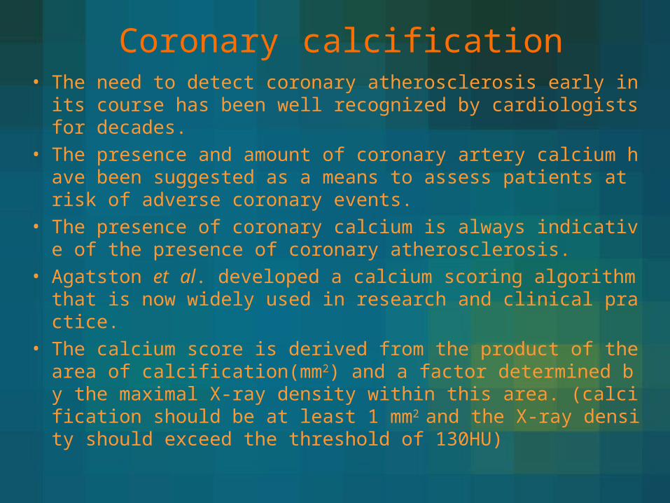
Coronary calcification• The need to detect coronary atherosclerosis early in its cours
e has been well recognized by cardiologists for decades.• The presence and amount of coronary artery calcium have b
een suggested as a means to assess patients at risk of adverse coronary events.
• The presence of coronary calcium is always indicative of the presence of coronary atherosclerosis.
• Agatston et al. developed a calcium scoring algorithm that is now widely used in research and clinical practice.
• The calcium score is derived from the product of the area of calcification(mm2) and a factor determined by the maximal X-ray density within this area. (calcification should be at least 1 mm2 and the X-ray density should exceed the threshold of 130HU)
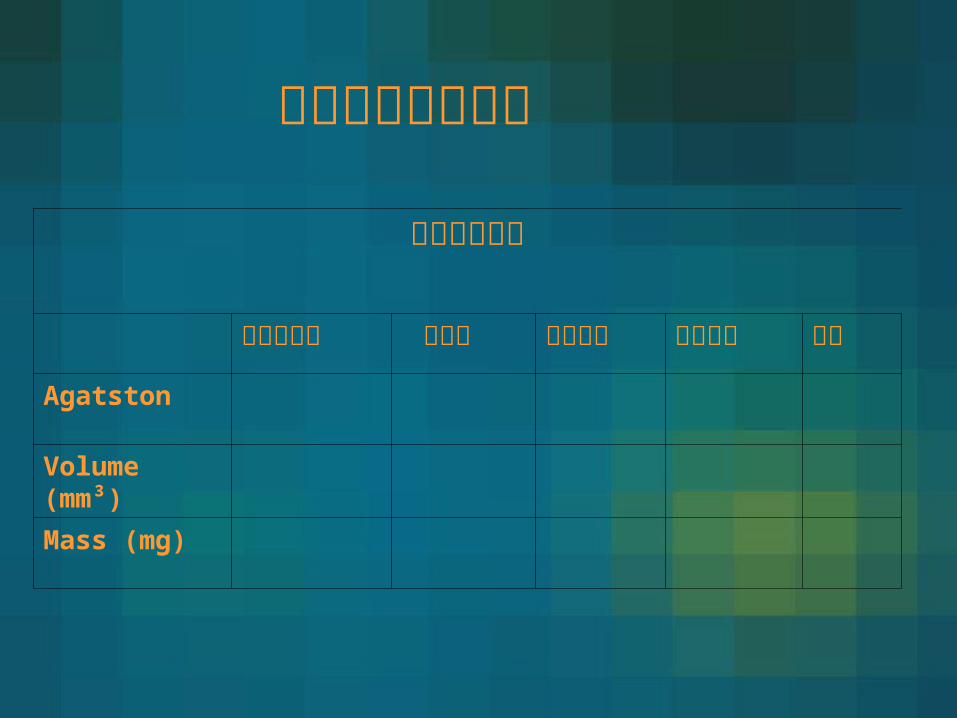
冠狀動脈鈣化評分
冠狀動脈鈣化
右冠狀動脈 左主幹 左迴旋枝 左前降枝 總合
Agatston
Volume (mm³)
Mass (mg)

Area of plaque × weighting factor
= Lesion score
∑ Lesion scores = Vessels score
∑ Vessels scores = Total calcium score (Agatston Score)

” 冠狀動脈鈣化評分” (the Agatston Calcium Score) 之說明
鈣化評分
粥狀硬化斑塊承載量Plaque burden
有意義冠狀血管阻塞的可能性Significant CAD
心血管之風險
CV risk
0 無動脈粥狀硬化斑塊 可能性極低 ( < 5% ) 極低
1-10極輕度的動脈粥狀硬化斑塊 可能性極低 ( < 10% ) 低
11-100輕微的動脈粥狀硬化斑塊
具輕微或極輕度冠狀動脈狹窄的可能性 中等
101-400中等的動脈粥狀硬化斑塊
高度有冠狀動脈狹窄存在的可能性 中高等
> 400高量的動脈粥狀硬化斑塊
極有可能有冠狀動脈狹窄的存在 高等
鈣化分數越高心血管疾病發生機率越高
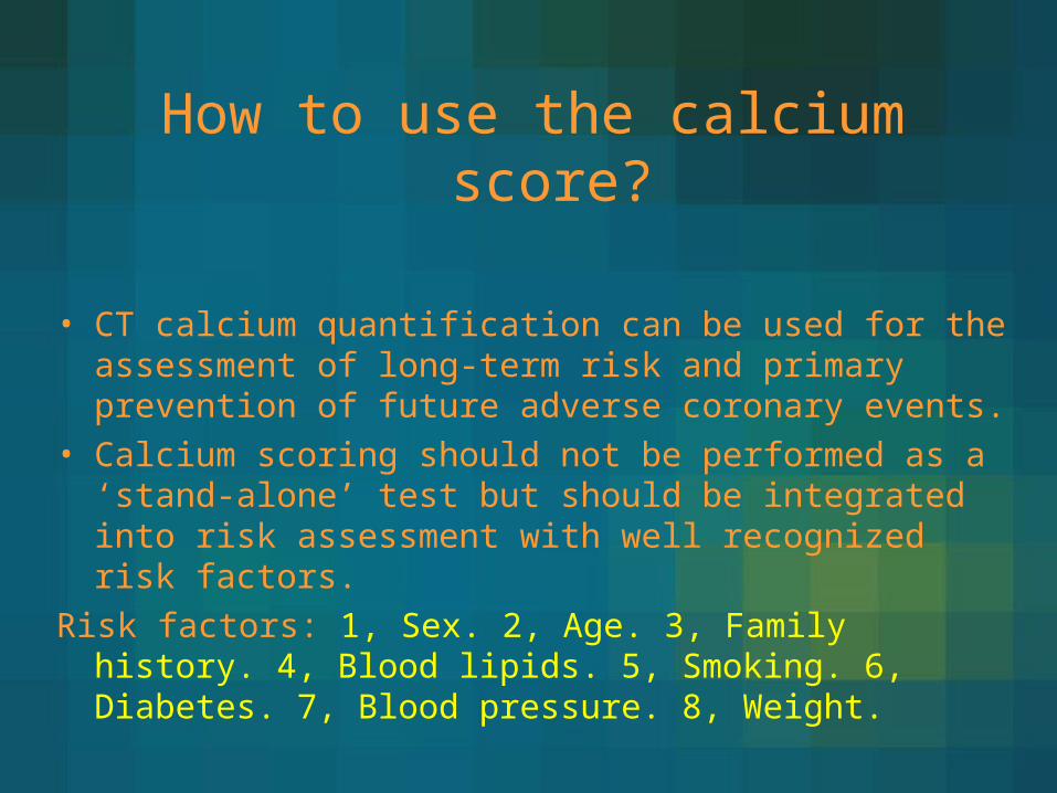
How to use the calcium score?
• CT calcium quantification can be used for the assessment of long-term risk and primary prevention of future adverse coronary events.
• Calcium scoring should not be performed as a ‘stand-alone’ test but should be integrated into risk assessment with well recognized risk factors.
Risk factors: 1, Sex. 2, Age. 3, Family history. 4, Blood lipids. 5, Smoking. 6, Diabetes. 7, Blood pressure. 8, Weight.
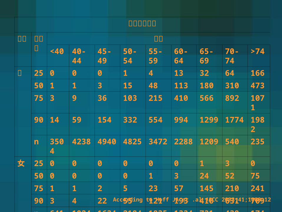
冠狀動脈鈣化性別
百分比
年紀<40 40-
4445-49
50-54
55-59
60-64
65-69
70-74
>74
男 25 0 0 0 1 4 13 32 64 166
50 1 1 3 15 48 113 180 310 473
75 3 9 36 103 215 410 566 892 1071
90 14 59 154 332 554 994 1299 1774 1982
n 3504
4238 4940 4825 3472 2288 1209 540 235
女 25 0 0 0 0 0 0 1 3 0
50 0 0 0 0 1 3 24 52 75
75 1 1 2 5 23 57 145 210 241
90 3 4 22 55 121 193 410 631 709
n 641 1024 1634 2184 1835 1334 731 438 174According to Hoff JA et .al JACC 2003;41;1008-12

Intermediate(1)Calcium score < 80 Lower risk(2)Calcium score > 80 Higher risk
Level description Low risk
0-1 risk factor
Intermediate
2 risk factor
High risk
>3 risk factor
Risk of event within 10 yrs
<10% 10-20% >20%
Estimated prevalence adults
35% 40% 25%
Ca. test useful No Yes No
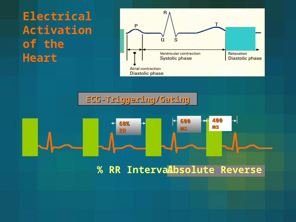
Electrical Activation of the Heart
ECG-Triggering/GatingECG-Triggering/Gating
600 ms600 ms
Absolute Time
400 ms400 ms
Absolute Reverse
60% RR60% RR
% RR Interval

“3D” Image Data
R R R R
DelayR
econ
Rec
on
Rec
on
Rec
on
Continuous
Spiral Scan & Feed
z -
Pos
itio
n
Time
Retrospective ECG Gating
加圖

Challenges in MSCT coronary imaging
• size of coronary arteries are small
• LCA = 4.4 mm
• LAD = 3.6 mm distally = 0.8 mm
• CX = 3.4 mm distally = 1.5 mm
• RCA = 3.0 mm distally = 1.0 mm
• movement of mid RCA up to 45 mm/sec
small vessel

64 dectector array
Flat Panel
16 dectector array
small vessel
4s 16s 64s
Rotation 0.5 0.42/0.37 0.33
Temp resolution 0.25 0.21 0.165
Spatial resolution 1.0 0.75 0.35
Data per rotation 4 16 64
Scan time >40s 20s 14s
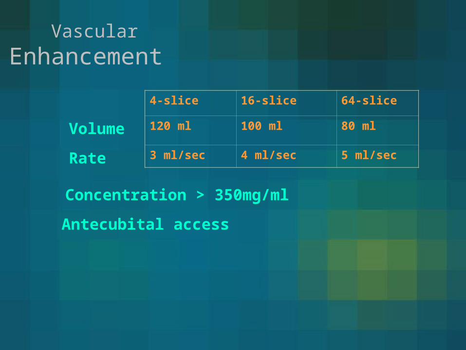
Vascular
Enhancement
Rate
4-slice 16-slice 64-slice
120 ml 100 ml 80 ml
3 ml/sec 4 ml/sec 5 ml/sec
Volume
Concentration > 350mg/ml
Antecubital access

Patient SelectionPatient Selection
Small coronary arteries
Fast heart rate >75 bpm
Persistent irregular heart rhythm
Respiratory impairment and related motion artifacts
Diffuse and calcified coronary atherosclerosis
Coronary stents
Metal objects
Renal dysfunction and contrast medium allergy

motion artifacts
60 NTG Diastolic
60-80 Beta-blocker
80 Systolic
β- blocking agent
- Metoprolol (Beloc ®) 100 mg 1h prior to exam. p.o.
- Propranolol(Dociton ®) 80 mg 1h prior to exam. p.o.
- i.v. Metoprolol 5 mg @1mg/min i.v.
check heart rate @ 5 minutes
repeat @ 5 mg (15 mg max.)
NTG

Patient education 1. Breath holding
2. CM burning sensation
64 dectector array : >9 sec (12)
16 dectector array : >15 sec (20)

coronary anomaly
1%

coronary
anomaly
bridge
Absent right coronary

Left Anterior Descending stenosis
coronary artery lumen
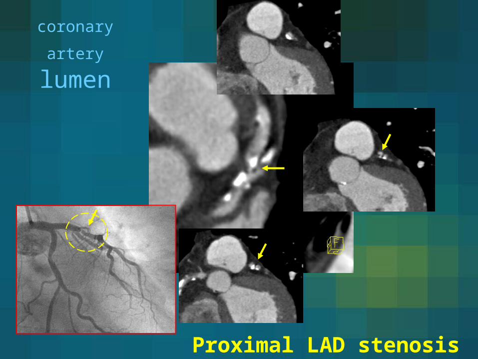
Proximal LAD stenosis
coronary artery lumen
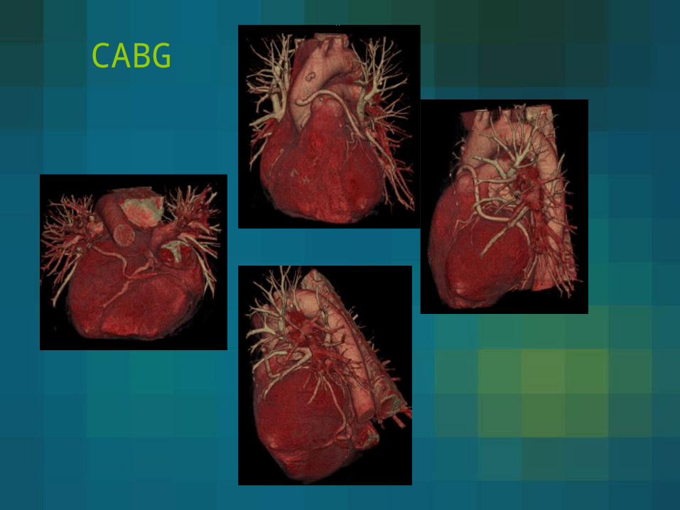
CABG
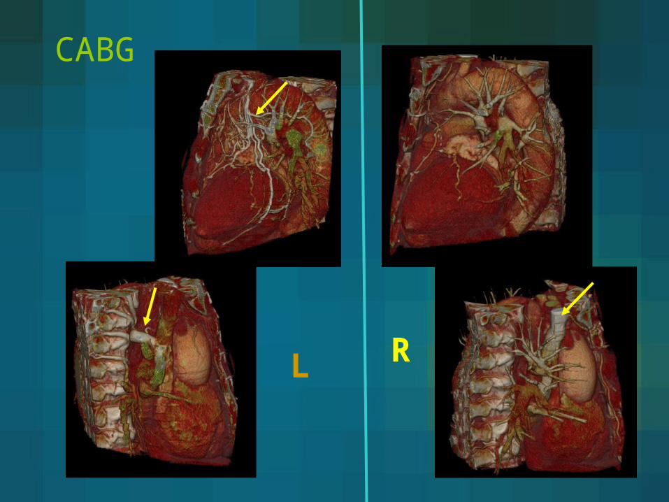
CABG
RL

CABG
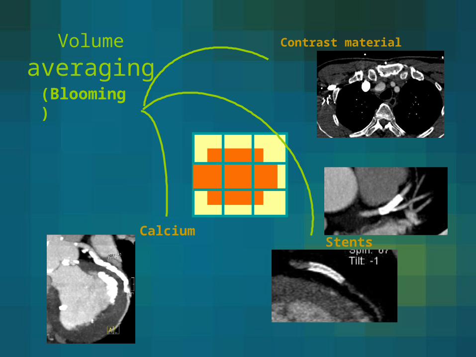
(Blooming)
Volume averaging
Contrast material
StentsCalcium

Bean hardening
(Streak artifacts)
stents
Contrast material
Pacemaker wires
Prosthetic valve
Surgical clips
Calcium
Effective energy is shifted to higher value as the X-rays pass through an object
Sternal wires

Resolution
Furture
small vessel(spatial resolution)
motion artifacts(temporal resolution)
Calcium/stent
obstruction estimation
Radiation exposure


Thanks for you attention!