


Languages
Pages
Legal

Tick-borne Diseases
Tanya Bobo, MPHApplied Epidemiology Fellow
Division of Zoonotic and Environmental Epidemiology (DZEE)
Virginia Department of Health
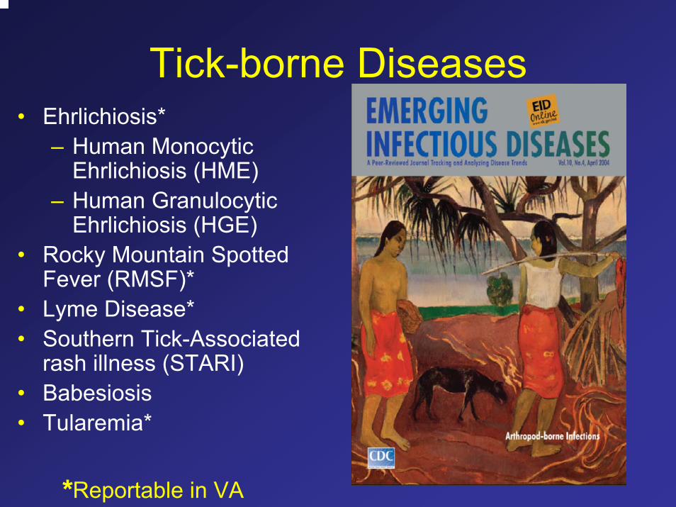
Tick-borne Diseases• Ehrlichiosis*
– Human Monocytic Ehrlichiosis (HME)
– Human Granulocytic Ehrlichiosis (HGE)
• Rocky Mountain Spotted Fever (RMSF)*
• Lyme Disease*• Southern Tick-Associated
rash illness (STARI)• Babesiosis• Tularemia*
*Reportable in VA

Ehrlichiosis• Obligate intracellular, gram-negative cocci• Two different bacteria causing similar
syndromes• Human Monocytic Ehrlichiosis (HME)
– Caused by Ehrlichia chaffeensis– Most frequently infects monocytes and
macrophages• Human Granulocytic Ehrlichiosis (HGE)
– Caused by Anaplasma phagocytophila(formerly E. phagocytophila)
– Most frequently infects granulocytes (neutrophils and rarely eosinophils)
Ehrlichiosis - US Epidemiology
• 80-90% of all ehrlichiosis cases occur between April and September
• HME cases most frequently reported in Southeastern and Midwestern states
• Most HGE cases occur in states with high incidence of Lyme Disease
• Increased age – risk factor for disease• Case-fatality rate: 2 - 3%
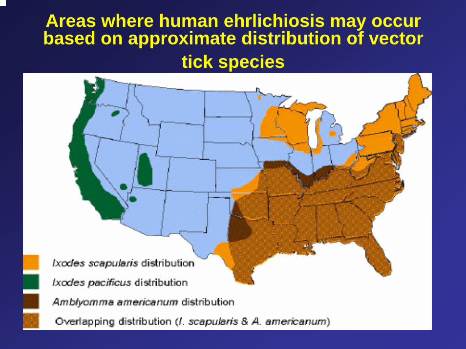
Areas where human ehrlichiosis may occur based on approximate distribution of vector
tick species
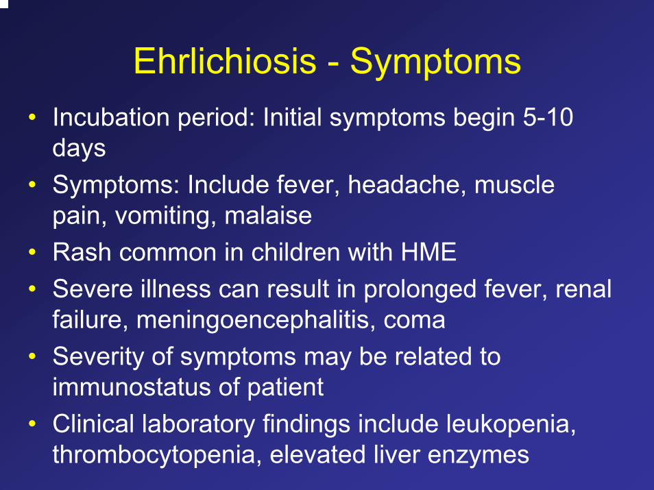
Ehrlichiosis - Symptoms• Incubation period: Initial symptoms begin 5-10
days • Symptoms: Include fever, headache, muscle
pain, vomiting, malaise• Rash common in children with HME• Severe illness can result in prolonged fever, renal
failure, meningoencephalitis, coma• Severity of symptoms may be related to
immunostatus of patient• Clinical laboratory findings include leukopenia,
thrombocytopenia, elevated liver enzymes

Ehrlichiosis - Diagnosis• Diagnosis:
Serology: Indirect Immunofluorescence Antibody (IFA)- 4x change in antibody titer between paired serum samples
– Detection of E. chaffeensisOR A. phagocytophilaDNA by Polymerase Chain Reaction (PCR)
– Direct Isolation of agent –Cell Culture
– Immunostaining of E. chaffeensis OR A. phagocytophila antigen
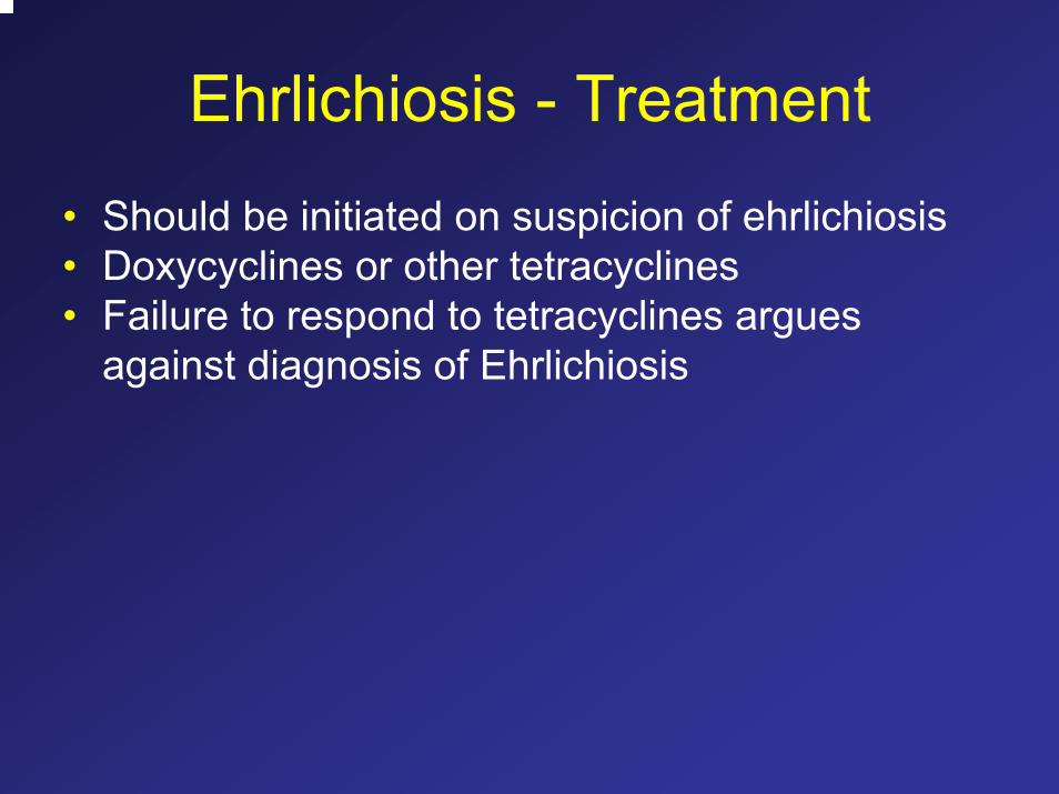
Ehrlichiosis - Treatment
• Should be initiated on suspicion of ehrlichiosis • Doxycyclines or other tetracyclines• Failure to respond to tetracyclines argues
against diagnosis of Ehrlichiosis
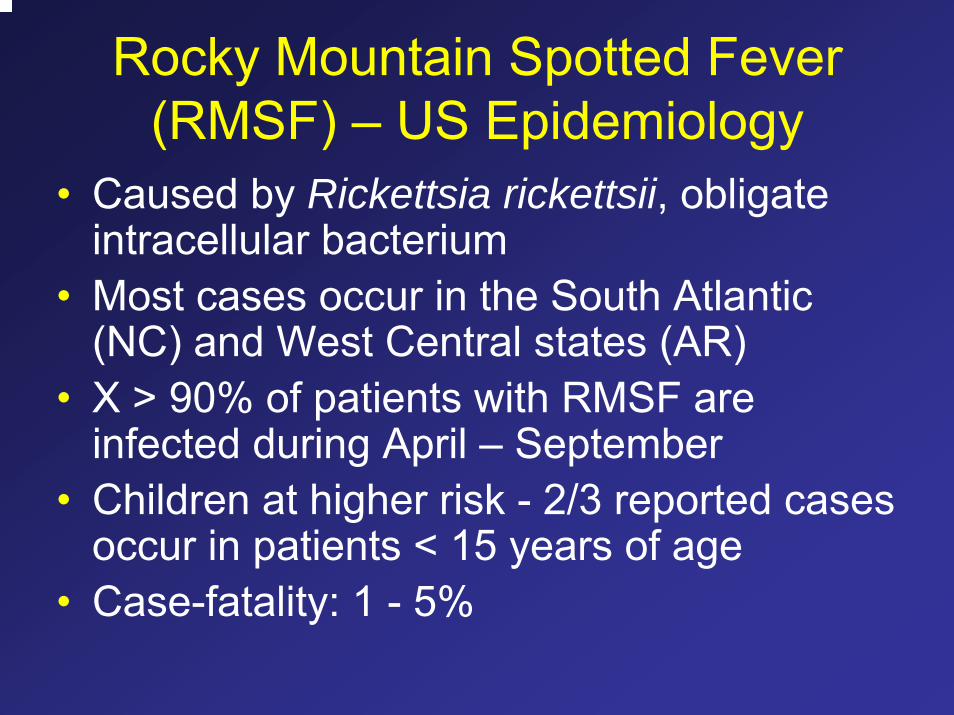
Rocky Mountain Spotted Fever (RMSF) – US Epidemiology
• Caused by Rickettsia rickettsii, obligate intracellular bacterium
• Most cases occur in the South Atlantic (NC) and West Central states (AR)
• X > 90% of patients with RMSF are infected during April – September
• Children at higher risk - 2/3 reported cases occur in patients < 15 years of age
• Case-fatality: 1 - 5%
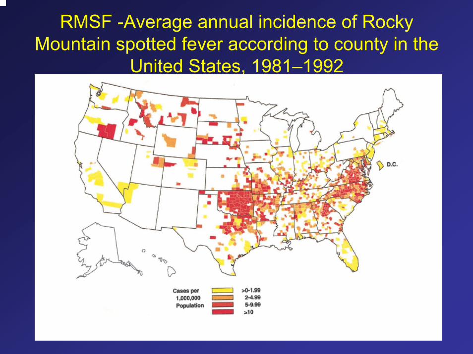
RMSF -Average annual incidence of Rocky Mountain spotted fever according to county in the
United States, 1981–1992
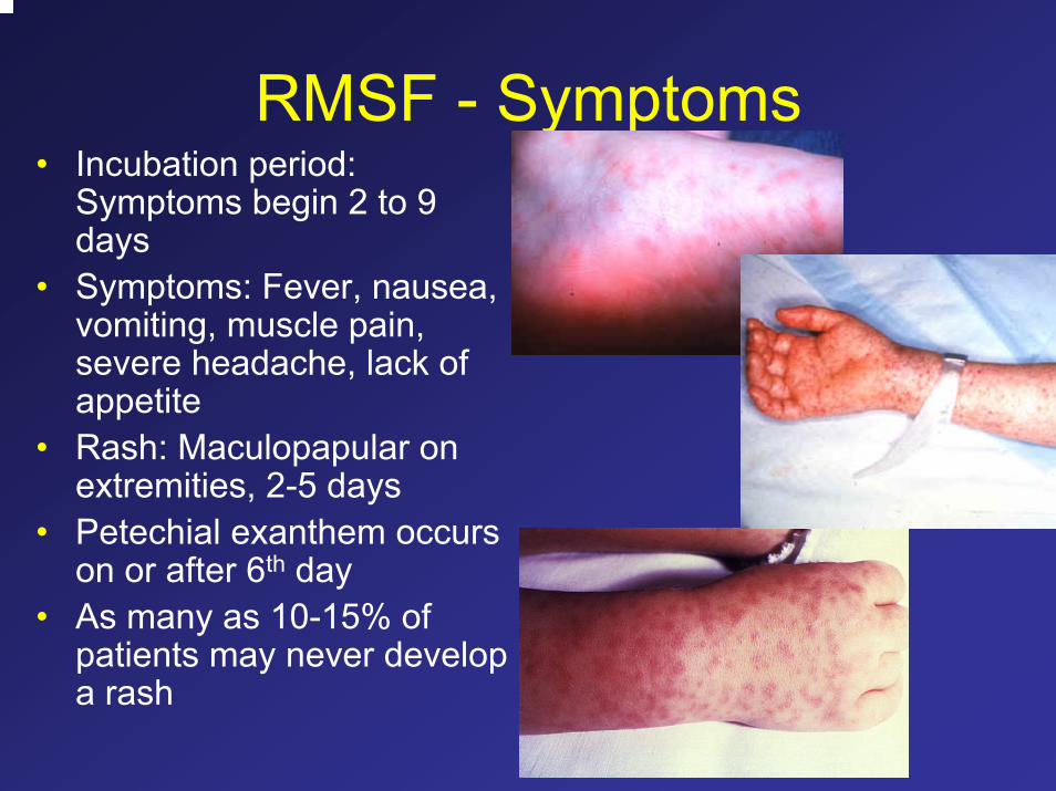
RMSF - Symptoms• Incubation period:
Symptoms begin 2 to 9 days
• Symptoms: Fever, nausea, vomiting, muscle pain, severe headache, lack of appetite
• Rash: Maculopapular on extremities, 2-5 days
• Petechial exanthem occurs on or after 6th day
• As many as 10-15% of patients may never develop a rash
RMSF-Diagnosis & Treatment
• DiagnosisSerological evidence of significant change in serum antibody titer reactive to R. rickettsii
– Detection of R. rickettsii DNA by PCR– Direct Isolation of agent – Cell Culture– Immunostaining of R. rickettsii antigen
• Treatment: – Initiated upon suspicion of RMSF– Doxycycline– Failure to respond to tetracyclines argues against
diagnosis of RMSF– Chloramphenicol - alternate drug
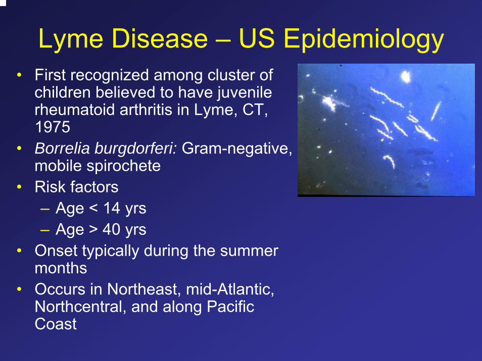
Lyme Disease – US Epidemiology• First recognized among cluster of
children believed to have juvenile rheumatoid arthritis in Lyme, CT, 1975
• Borrelia burgdorferi: Gram-negative, mobile spirochete
• Risk factors– Age < 14 yrs – Age > 40 yrs
• Onset typically during the summer months
• Occurs in Northeast, mid-Atlantic, Northcentral, and along Pacific Coast
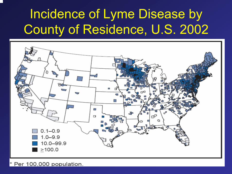
Incidence of Lyme Disease by County of Residence, U.S. 2002
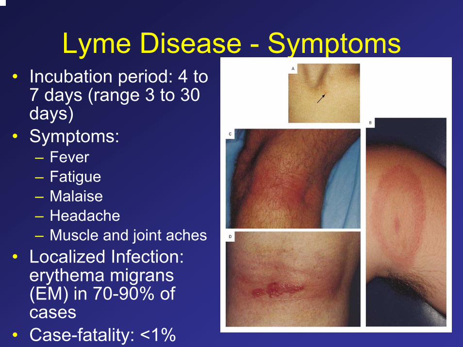
Lyme Disease - Symptoms• Incubation period: 4 to
7 days (range 3 to 30 days)
• Symptoms: – Fever– Fatigue– Malaise– Headache– Muscle and joint aches
• Localized Infection: erythema migrans (EM) in 70-90% of cases
• Case-fatality: <1%
Lyme Disease – Later Stages• Early disseminated infection – Several weeks to
months after onset– Neuro: Meningitis, cranial neuropathy (includes facial
palsy) – Cardio: myocarditis, heart block– Arthritis: migratory joint and muscle pains
• Late disseminated infection – Months to years after onset– Neuro: Encephalopathy, cognitive disorders – Arthritis: Chronic arthritis, prolonged arthritis attacks
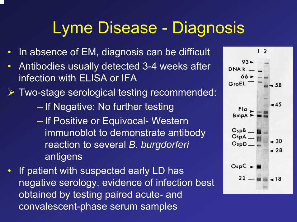
Lyme Disease - Diagnosis• In absence of EM, diagnosis can be difficult• Antibodies usually detected 3-4 weeks after
infection with ELISA or IFATwo-stage serological testing recommended:
– If Negative: No further testing– If Positive or Equivocal- Western
immunoblot to demonstrate antibody reaction to several B. burgdorferiantigens
• If patient with suspected early LD has negative serology, evidence of infection best obtained by testing paired acute- and convalescent-phase serum samples
Lyme Disease - Treatment• Treatment: Appropriate to treat patients early based
on clinical findings• Several antibiotics including doxycycline,
amoxicillin, penicillin, ceftriaxone, cefotaxime, or cefuroxime axetil
• Intravenous antibiotics for central nervous system involvement and recurrent arthritis
• Complications– Treatment-resistant Lyme arthritis associated with HLA-
DRB1*0401, 0101 and other related alleles– Overtreatment: patients that have no evidence of Lyme
disease are treated, report more adverse drug reactions
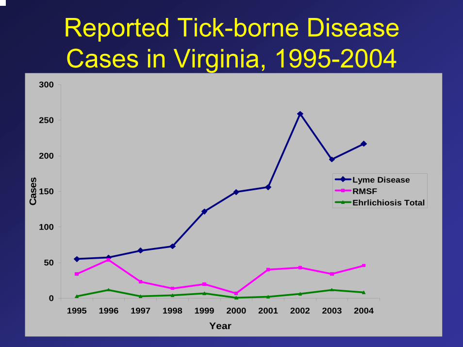
Reported Tick-borne Disease Cases in Virginia, 1995-2004
0
50
100
150
200
250
300
1995 1996 1997 1998 1999 2000 2001 2002 2003 2004
Year
Cas
es Lyme DiseaseRMSFEhrlichiosis Total
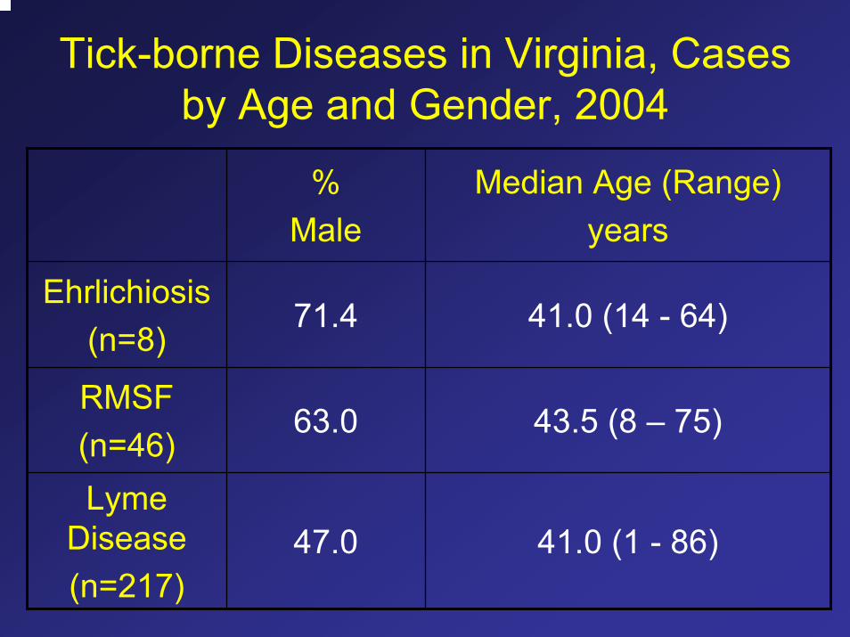
Tick-borne Diseases in Virginia, Cases by Age and Gender, 2004
% Male
Median Age (Range)years
Ehrlichiosis(n=8)
71.4 41.0 (14 - 64)
RMSF(n=46)
63.0 43.5 (8 – 75)
Lyme Disease(n=217)
47.0 41.0 (1 - 86)
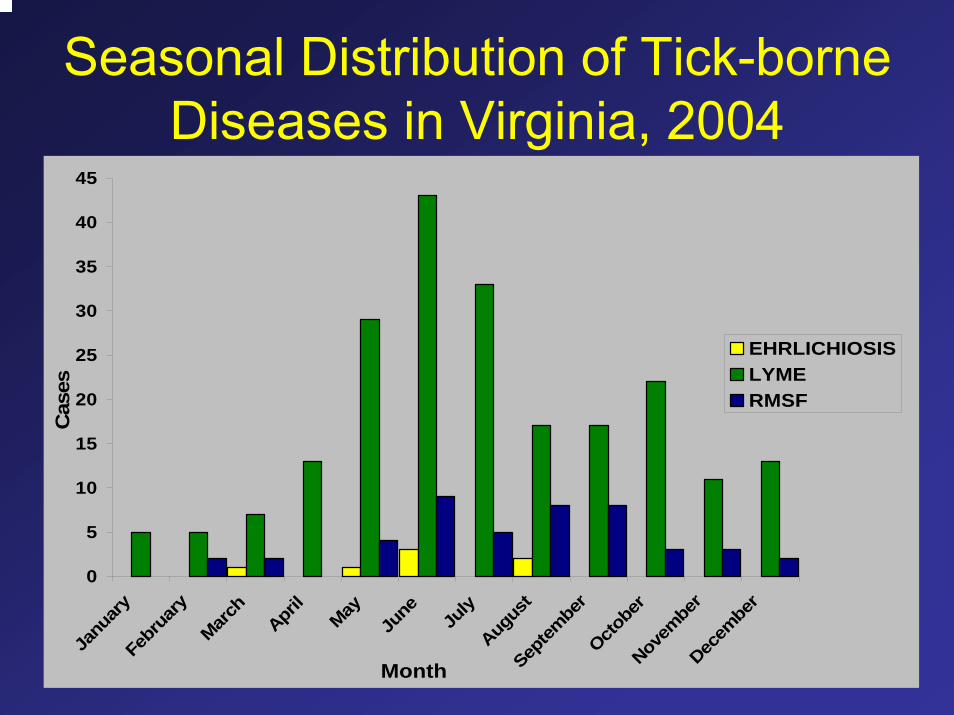
Seasonal Distribution of Tick-borne Diseases in Virginia, 2004
0
5
10
15
20
25
30
35
40
45
Janu
ary
Febr
uary
March
April
May
June July
Augus
tSep
tembe
rOcto
ber
Novem
ber
Decem
ber
Month
Cas
es
EHRLICHIOSISLYMERMSF
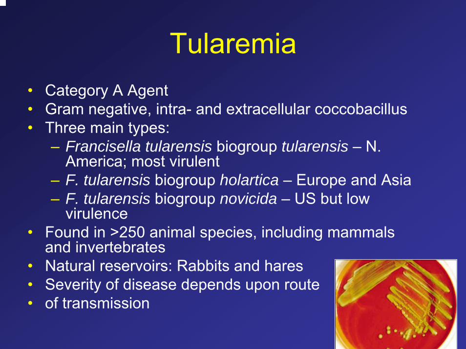
Tularemia• Category A Agent• Gram negative, intra- and extracellular coccobacillus• Three main types:
– Francisella tularensis biogroup tularensis – N. America; most virulent
– F. tularensis biogroup holartica – Europe and Asia– F. tularensis biogroup novicida – US but low
virulence• Found in >250 animal species, including mammals
and invertebrates• Natural reservoirs: Rabbits and hares • Severity of disease depends upon route • of transmission

Tularemia - Routes of transmission
– Aerosol droplets– Animal bites– Arthropods: ticks, deerfly, various mosquito
species– Contaminated water– Direct contact with animal product/infected
dead animals (skinning rabbits)– Eating undercooked infected meat– Inhaling contaminated dust
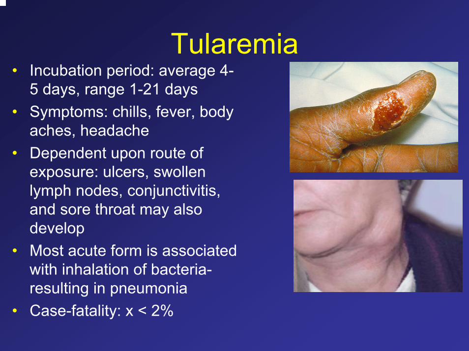
Tularemia• Incubation period: average 4-
5 days, range 1-21 days• Symptoms: chills, fever, body
aches, headache• Dependent upon route of
exposure: ulcers, swollen lymph nodes, conjunctivitis, and sore throat may also develop
• Most acute form is associated with inhalation of bacteria-resulting in pneumonia
• Case-fatality: x < 2%
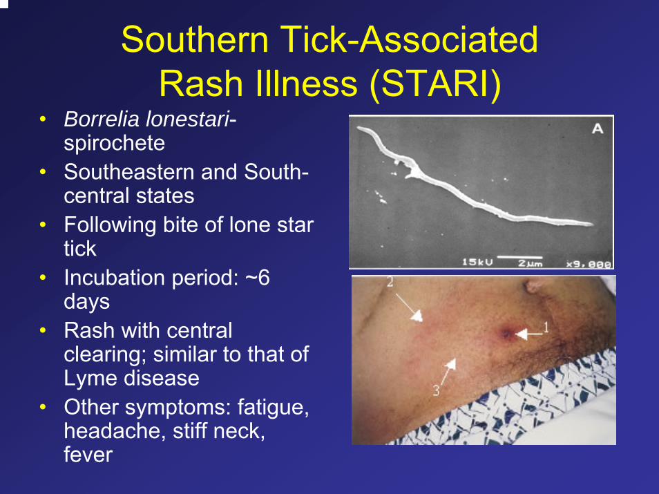
Southern Tick-Associated Rash Illness (STARI)
• Borrelia lonestari-spirochete
• Southeastern and South-central states
• Following bite of lone star tick
• Incubation period: ~6 days
• Rash with central clearing; similar to that of Lyme disease
• Other symptoms: fatigue, headache, stiff neck, fever
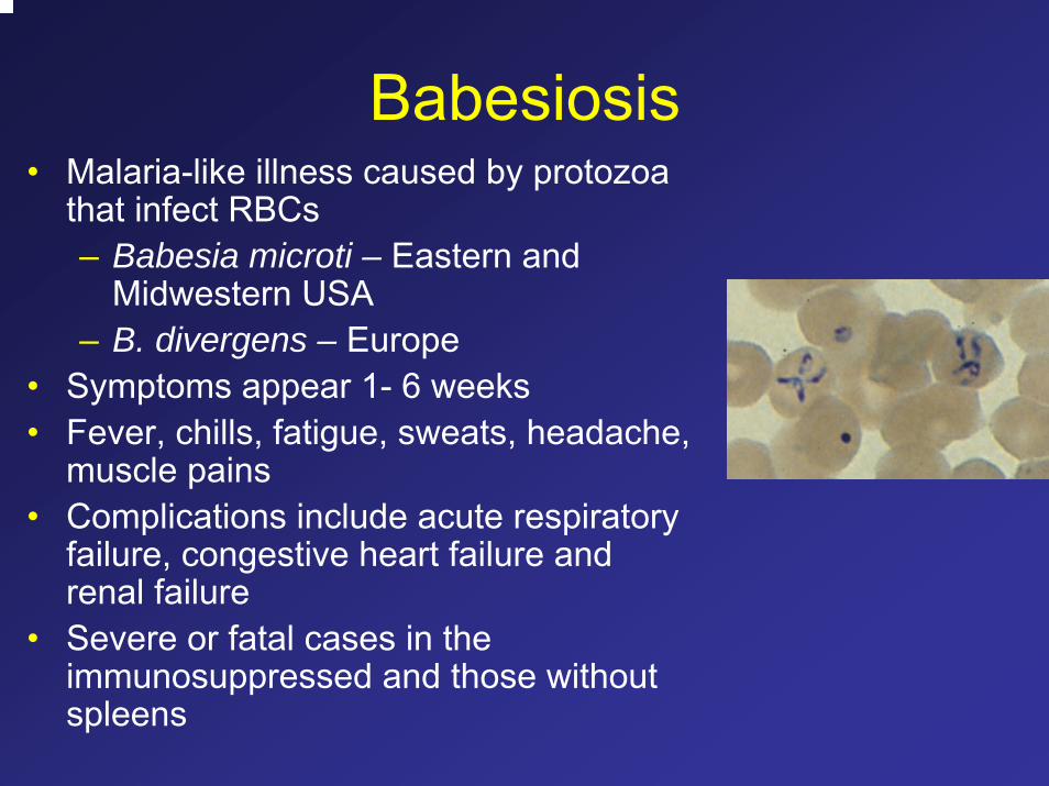
Babesiosis• Malaria-like illness caused by protozoa
that infect RBCs– Babesia microti – Eastern and
Midwestern USA– B. divergens – Europe
• Symptoms appear 1- 6 weeks • Fever, chills, fatigue, sweats, headache,
muscle pains• Complications include acute respiratory
failure, congestive heart failure and renal failure
• Severe or fatal cases in the immunosuppressed and those without spleens
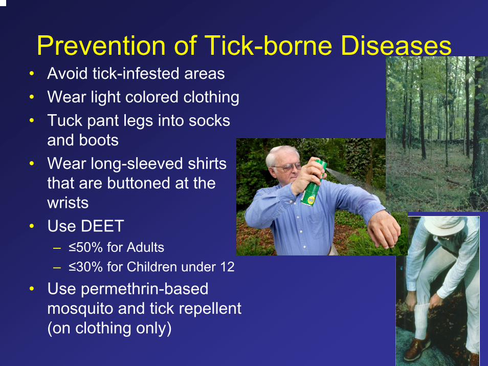
Prevention of Tick-borne Diseases• Avoid tick-infested areas• Wear light colored clothing • Tuck pant legs into socks
and boots• Wear long-sleeved shirts
that are buttoned at the wrists
• Use DEET– ≤50% for Adults– ≤30% for Children under 12
• Use permethrin-based mosquito and tick repellent (on clothing only)
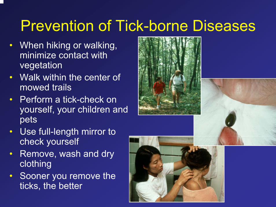
Prevention of Tick-borne Diseases• When hiking or walking,
minimize contact with vegetation
• Walk within the center of mowed trails
• Perform a tick-check on yourself, your children and pets
• Use full-length mirror to check yourself
• Remove, wash and dry clothing
• Sooner you remove the ticks, the better
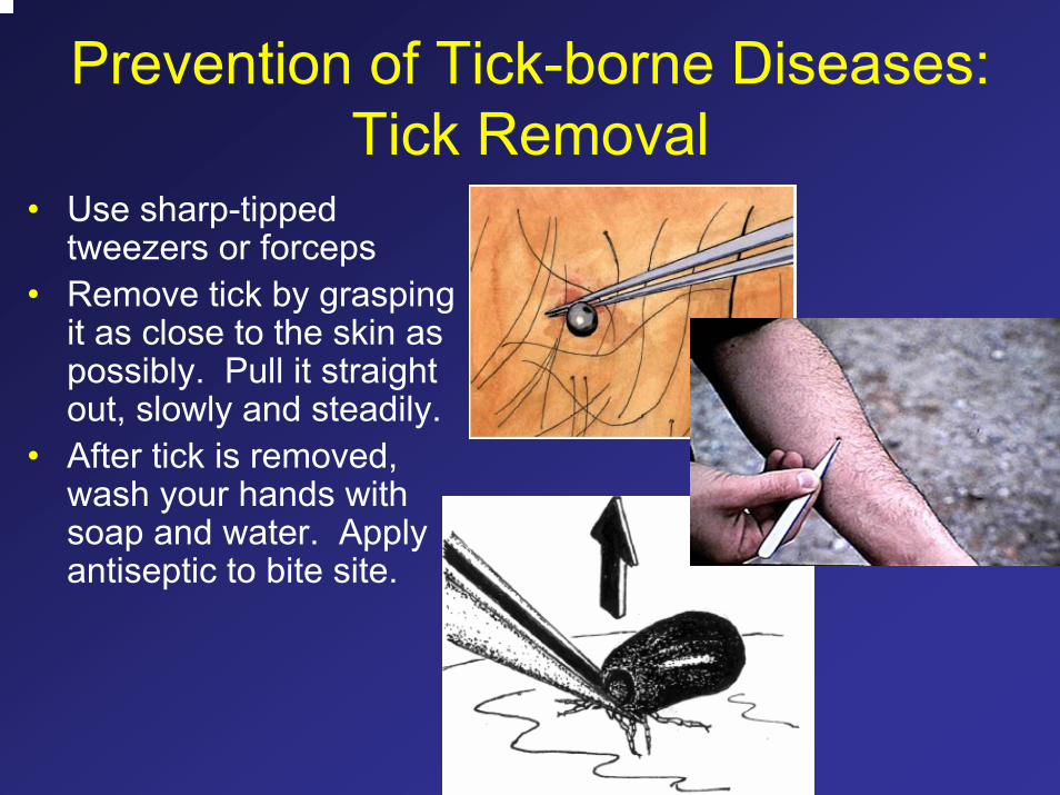
Prevention of Tick-borne Diseases: Tick Removal
• Use sharp-tipped tweezers or forceps
• Remove tick by grasping it as close to the skin as possibly. Pull it straight out, slowly and steadily.
• After tick is removed, wash your hands with soap and water. Apply antiseptic to bite site.
References and Photo Credits1. Homer MJ, Aguilar-Delfin I, Telford SR, 3rd, Krause PJ, Persing DH.
Babesiosis. Clin Microbiol Rev 2000;13:451-69.2. Krause PJ. Babesiosis diagnosis and treatment. Vector Borne Zoonotic
Dis 2003;3:45-51.3. Bryant KA, Marshall GS. Clinical manifestations of tick-borne infections in
children. Clin Diagn Lab Immunol 2000;7:523-7.4. Steere AC, Coburn J, Glickstein L. The emergence of Lyme disease. J
Clin Invest 2004;113:1093-101.5. Reed KD. Laboratory testing for Lyme disease: possibilities and
practicalities. J Clin Microbiol 2002;40:319-24.6. Rich SM, Armstrong PM, Smith RD, Telford SR, 3rd. Lone star tick-
infecting borreliae are most closely related to the agent of bovine borreliosis. J Clin Microbiol 2001;39:494-7.
7. Steere AC. Lyme disease. N Engl J Med 2001;345:115-25.
More References and Photo Credits
8. Thorner AR, Walker DH, Petri WA, Jr. Rocky mountain spotted fever. Clin Infect Dis 1998;27:1353-9; quiz 1360.
9. Stanford III KC. Tick Management Handbook: The Connecticut Agricultural Experiment Station, 2004:72.
10.Parola P, Raoult D. Ticks and tickborne bacterial diseases in humans: an emerging infectious threat. Clin Infect Dis 2001;32:897-928.
11.Ellis J, Oyston PC, Green M, Titball RW. Tularemia. Clin Microbiol Rev 2002;15:631-46.
12.Dennis DT, Inglesby TV, Henderson DA, et al. Tularemia as a biological weapon: medical and public health management. Jama 2001;285:2763-73.
13. CDC Emerging Infectious Diseases Journal:http://www.cdc.gov/ncidod/EID/index.htm
14. Public Health Image Library, Centers for Disease Control and Prevention: http://phil.cdc.gov/phil/home.asp

Next, the Ticks . . .
Top Related