


Languages
Pages
Legal


Diabetology lecture notes
for medical students
Editor: Prof. Dr. István Wittmann
Lector: Prof. Dr. György Paragh
Language lector: Dr. Andrew C. Rouse
„AZ ÉLETTUDOMÁNYI -KLINIKAI FELSŐOKTATÁS GYAKORLATORIENTÁLT
ÉS HALLGATÓBARÁT KORSZER ŰSÍTÉSE A VIDÉKI KÉPZŐHELYEK NEMZETKÖZI
VERSENYKÉPESSÉGÉNEK ERŐSÍTÉSÉRE.” TÁMOP-4.1.1.C-13/1/KONV-2014-0001

2
Table of Contents
Chapter No. Title of the chapter Pages
1. Introduction and pathophysiology Dr. István Wittmann 4
2. Types, diagnosis and epidemiology of diabetes mellitus Dr. István Wittmann
14
3. Pregnancy and diabetes mellitus: Gestational diabetes Dr. András Szilágyi
17
4. Therapeutic plan and targets in diabetes mellitus Dr. István Wittmann
21
5. The basics of the non-pharmacological therapy of diabetes mellitus Dr. József Rinfel 25
6. Non-insulin-like antidiabetic agents Dr. István Wittmann 32
7. Insulin treatment Dr. István Wittmann 42
8. Insulin pump therapy Dr. Gergő A. Molnár
48
9. Pancreas-kidney transplantation Dr. Tibor Kovács
51
10. Diabetes care and education Dr. Gábor Fülöp
53
11. Self-monitoring of blood glucose, continuous glucose monitoring Dr. Gergő A. Molnár
57
12. Acute complications of diabetes mellitus and its management Dr. István Wittmann 60
13. Cardiovascular complications in diabetes mellitus Dr. Gergő A. Molnár 66
14. Cardiological issues in diabetes Dr. Attila Cziráki 71
15. Diabetic neuropathy Dr. Richard Halmai
73
16. Diabetic nephropathy Dr. István Wittmann
79
17. Microvascular complications of diabetes: diabetic eye problems Dr. Zsolt Biró
87
18. Hypertension and diabetes mellitus Dr. István Wittmann
90
19. Management of dyslipiademia in diabetes mellitus Dr. Gábor Fülöp 95
20. Rehabilitation in diabetes mellitus Dr. Gábor Fülöp 99
21. Perioperative management of patients with diabetes mellitus Dr. Botond Csiky 101
3
Authors in alphabetical order
• Dr. Biró, Zsolt Department of Ophthalmology Faculty of Medicine, University of Pécs
• Dr. Cziráki, Attila Heart Institute Faculty of Medicine, University of Pécs
• Dr. Csiky, Botond 2nd Department of Medicine and Nephrological Center Faculty of Medicine, University of Pécs
• Dr. Fülöp, Gábor 2nd Department of Medicine and Nephrological Center Faculty of Medicine, University of Pécs
• Dr. Halmai, Richard B. Braun Avitum 13. Dialysis Center Dunaújváros
• Dr. Kovács, Tibor 2nd Department of Medicine and Nephrological Center Faculty of Medicine, University of Pécs
• Dr. Molnár, Gergő Attila 2nd Department of Medicine and Nephrological Center Faculty of Medicine, University of Pécs
• Dr. Rinfel, József 2nd Department of Medicine and Nephrological Center Faculty of Medicine, University of Pécs
• Dr. Sebők, Judit 2nd Department of Medicine and Nephrological Center Faculty of Medicine, University of Pécs
• Dr. Szilágyi, András Kaposi Mór Educational County Hospital Somogy County, Kaposvár
• Dr. Wittmann, István 2nd Department of Medicine and Nephrological Center Faculty of Medicine, University of Pécs
Translation by: Dr. Botond Csiky: Chapter 21; Dr. Richard Halmai: Chapter 15; Dr. Tibor Kovács: Chapter 9; Dr. Gergő A. Molnár: Chapter 1, 2, 4, 5, 8, 11, 13, 16, Dr. Judit Sebők: Chapter 3, 6, 7, 10, 12, 14, 17, 18, 19, 20
4
Chapter 1. Introduction and pathophysiology
Dr. István Wittmann
The frequency of diabetes, its outstanding importance in the development of
cardiovascular and tumour-associated morbidity and mortality, and the medical intervention
of the field in the last years, make it necessary that we provide an easy-to-upgrade, electronic
format teaching material to support the learning of the students.
Changing perspectives in diabetology
Studies over the last years indicate that one of the major causal factors related to type
2 diabetes mellitus, insulin resistance, is not the only hormone resistance that can characterize
this type of diabetes. This field of medicine is related by several common-onset hormone
resistances to endocrinology; the immunological mechanisms present in type 1 diabetes relate
it to immunology, and through complications it is connected to obesitology, lipidology,
hypertension, cardiology, neurology, angiology, ophthalmology, nephrology and so on.
Diabetes leads to systemic alterations, thereby damaging all parts of the body. Thus
the study of diabetes promotes the learning of a holistic approach. In order to do this, let us
familiarize ourselves with relationships between hormonal resistances and cardiovascular
complications.
Hormone resistance and cardiovascular diseases
Insulin resistance plays a major role in the development of type 2 diabetes mellitus;
however, it is frequently already present in obesity without diabetes and in impaired glucose
tolerance. Moreover, it can also be detected in non-obese smokers, which suggests that insulin
resistance may also occur without any increase in abdominal fat mass. As a result, insulin
resistant patients may be divided into two groups, those with and those without obesity.
Obesity-associated insulin resistance
According to a widely accepted perspective, the major cause in obese patients for the
pathomechanism of insulin resistance is subclinical inflammation. According to the
hypothesis of subclinical inflammation, in obesity the abdominal fat cells undergo a
phenotype change, and produce among other elements cytokines (e.g. TNF-alpha) that flood
the circulation. The cytokines will be able to activate the NAD(P)H oxidase enzyme by
5
binding to cytokine receptors of endothelial and parenchymatous cells, while the enzyme will
in turn overproduce superoxide. The overproduction of superoxide leads to intracellular
oxidative stress, which leads to an altered phosphorylation of an important factor in the
signalling of insulin, namely insulin receptor substrate 1 and 2 (IRS1, IRS2). Exactly what
happens is that the inhibitory serine phosphorylation will outweigh the activating tyrosine
phosphorylation of IRS-1 and -2. As a consequence, insulin signalling via the IRS-pathway
decreases. It is important to emphasize that resistin, which is well-known for inducing insulin
resistance, and which originates from the abdominal fatty tissue, can lead to insulin resistance
via the same pathomechanism.
As the insulin signalling is able to run through the other pathways in an undisturbed
manner (insulin resistance is selective), and hyperinsulinaemia develops in the circulation
because of the loss of metabolic effects, there will be an increased insulin influence over other
pathways. This is the way, an increased vasoconstriction (increased endothelin-1 secretion)
and mitogenity develop in insulin resistance, along with in impaired glucose-lowering and
vasodilatory effect. This mechanism may also play a role - via increased mitogen activity - in
the development of increased risk of cancer in diabetes and obesity.
One important question to be asked is where the phenotype change of the abdominal
fat cells originates from. Why does the abdominal fat cell differ from the subcutaneous fat
cell? One possible explanation is that the gut flora changes in obesity and type 2 diabetes
mellitus, and that those types of bacteria will overgrow in the colon, the lipopolysaccharide of
which is able to pass through the bowel wall and reach the abdominal fat cells, driving them
to phenotype change. This is underlined by the fact that a transient antibiotic therapy is able to
influence insulin resistance. According to another explanation, the abdominal fat cells
produce aldosterone, which is able to transform the cells in an autocrine manner. A third
explanation is that the fat cells produce an aldosterone (or mineralocorticoid) releasing factor
that is able to promote aldosterone production in the adrenal gland. For the mechanism of
action of aldosterone, see below.
As a consequence, we can say that insulin resistance in obesity is mainly due to
oxidative stress developed due to cytokines, resistin and aldosterone.
Insulin resistance in the non-obese
Common thinking in the field focuses nearly exclusively on the insulin resistance of
obese persons, and far fewer data are available regarding insulin resistance in persons with a
normal body mass.
6
From this group, a hormonal disease that is well-known not to be associated with
obesity arises, hyperthyreodism. In hyperthyroidism, interleukin-6 overproduces in the
subcutaneous fatty tissue, and the elevated levels of circulating tumour necrosis factor alpha
and higher interleukin-18 are made responsible for the development of insulin resistance. All
three cytokines could lead to insulin resistance through the abovementioned mechanism,
through the production of superoxide free radical. The effect of hyperthyroidism on blood
glucose is attenuated by the hyperkinetic circulation.
While obesity-associated increased aldosterone production surely contributes to
insulin resistance (see above), in primary hyperaldosteronism there is no correlation of plasma
aldosterone levels and body mass index, i.e. a lean patient can also be insulin resistant. The
explanation for this is that aldosterone is able by binding to its mineralocorticoid receptor to
activate the NAD(P)H oxidase enzyme. The superoxide free radical that is formed can have
an effect on insulin signalling similar to that of cytokines. Here, we also have to emphasize
that angiotensin II, by activating the same mechanism, decreases the insulin response of the
cells.
Smoking markedly increases the risk of insulin resistance, type 2 diabetes and
metabolic syndrome. In this case it is not the obesity, but other components of the metabolic
syndrome that make up the diagnosis of the syndrome. Some water-soluble component of
cigarette smoke is able to inhibit insulin-signalling via the IRS-1. According to our own in
vitro data, cigarette smoke decreases the activating phosphorylation of Akt (protein kinase B),
which may lead to the development of insulin resistance. As the effect can be avoided using
antioxidants, a free radical mechanism can be hypothesized.
In consequence we can say that the same intracellular effects can lead to insulin
resistance in the non-obese as in the obese.
Insulin resistance of the beta cells
The proper efficacy of insulin is required for the insulin secretory effects of the beta
cells of the pancreatic islands. When activated under normal circumstances, the insulin
receptors on the surface of the beta cells ensure the resistance of the beta cells against
apoptosis. Glucagon-like peptide-1 (GLP-1) has a synergistic effect on these processes. In the
case of insulin resistance, insulin also exerts less influence over the beta cells, meaning that
insulin secretion will also decrease. As we will see later on, in such cases the beta cells also
become resistant towards GLP-1.
7
We can draw the conclusion that in the case of the beta cells, insulin resistance goes
hand-in-hand with lower rates of insulin secretion.
The breakthrough phenomenon
The “breakthrough phenomenon” is well-known. The principle is that in type 2
diabetes mellitus, an intensive insulin therapy of 2-4 weeks, using either the conservative
method or an insulin pump, is able to break through insulin resistance. Here the insulin
resistance of the beta cells also disappears, and insulin secretion also improves due to the
breakthrough. Conclusions from the breakthrough phenomenon are:
1. Insulin resistance is reversible, i.e. it is not due to irreversible DNA-damage.
2. We need a therapy of 2-4 weeks to achieve the breakthrough, i.e. it is not effective
instantaneously.
3. The success of a breakthrough of a couple of weeks may indicate that we can find
reversion of some sort of protein damage in the background.
Insulin resistance and mortality
According to a study in non-diabetics, the greater the degree of insulin resistance, that
is, the higher the value of the HOMAIR, the higher the risk of total mortality. From time to
time the possibility arises that in type 2 diabetes the endogenous hyperinsulinaemia or the
high exogenous insulin dose may increase cardiovascular risk; however, there is no real proof
of this. In diabetes it is hard to diagnose, as it is virtually impossible to distinguish the effect
of a large insulin dose from that of the effect of insulin resistance.
In conclusion we can say that according to studies in non-diabetics, it may be more
likely that insulin resistance, or even more likely the subcellular processes lying behind it, are
directly connected with increased mortality.
Insulin resistance is at the same time incretin resistance
In human preliminary studies it was observed that GLP-1 infusion given to healthy
persons (reaching a 46 pmol/l plasma level) could increase the insulin plasma levels at 120
minutes to 4000 pmol/l. Conversely, in patients with type 2 diabetes the plasma GLP-1-level
of 41 pmol/l led to an insulin level below 500 pmol/l. It can be concluded that in insulin
resistant type 2 diabetics GLP-1 is also less effective, i.e. there is a GLP-1-resistance. When a
dose of GLP-1 three times higher (leading to a plasma level of 126 pmol/l) was given to the

8
same patients, insulin production rose, reaching a value of around 4000 pmol/l observed in
healthy persons.
In conclusion we can say that in type 2 diabetes GLP-1 resistance is present besides
insulin resistance.
The breakthrough of incretin resistance
In the abovementioned population, a follow-up study was also carried out, within
which the blood glucose of the patients with type 2 diabetes was normalized using a four-
week intensified insulin therapy, i.e. a classic breakthrough was performed. The efficacy of
GLP-1 before and after the insulin treatment was tested. It was found that after the four weeks
of breakthrough the effect of GLP-1 on the increase of insulin secretion and glucagon
secretion improved significantly. This suggests that a close to euglycaemic state over duration
of four weeks decreases the GLP-1 resistance of alpha- and beta cells.
In conclusion we can say that a near-euglycaemic state due to intensified insulin
therapy can lead to a breakthrough of GLP-1-resistance of the alpha- and beta-cells.
The insulin-resistant are also erythropoietin-resistant
In adults, the erythropoietin required for erythropoiesis is produced in the tubulo-
interstitial fibroblasts of the kidney. Where there is renal damage, the production decreases;
therefore, nephrologic patients frequently require erythropoietin therapy. Our observations
show that red blood cell formation in diabetic patients with renal disease is lower than in
patients with renal disease without diabetes, but with similarly impaired kidney function and
erythropoietin levels. Others have found out that in dialyzed patients with or without diabetes,
the erythropoietin dose (obviously reflecting the extent of erythropoietin resistance) shows a
close connection with the value of the HOMAIR index, i.e. the higher the HOMAIR is, the
higher was the erythropoietin requirement of the dialyzed patients. According to an interesting
observation, the IRS-2-Akt pathway – the impairment of which is responsible for the
development of insulin resistance – also plays a role in the intracellular signalling of
erythropoietin.
In conclusion we can say that among diabetic and non-diabetic dialyzed patients,
insulin-resistant patients are also erythropoietin-resistant.
Erythropoietin resistance and mortality
A connection has been found between the erythropoietin dose and mortality. As a
result, a new tendency has evolved in nephrology to use the smallest possible dose of

9
erythropoietin. However, similarly to the case of insulin, with erythropoietin the question also
arises whether erythropoietin itself is harmful, or the source of the increased risk rather the
hormone resistance behind high doses of erythropoietin? Investigations are yet to be carried
out, but on the analogy of insulin, and also because of the same signaling pathways, it seems
more probable that cellular changes behind hormone resistance play a role.
It may be hypothesized that it is rather the alterations in the background of
erythropoietin resistance and not the higher erythropoietin dose itself that is responsible for
the increase in mortality.
The insulin resistant are also leptin resistant
Basic scientific research has shown that in the IRS-Akt pathway the phosphorylation
of the IRS component is changed by oxidative stress and is responsible for insulin resistance
and is common in the signalling of insulin and leptin. Probably this can also lead to leptin
resistance based on the abovementioned facts. Indeed, plasma insulin and leptin levels show a
tight correlation in healthy persons and in patients with polycystic ovary syndrome.
In conclusion we can say that the insulin-resistant are also leptin-resistant.
Leptin resistance increases mortality
According to a clinical study, in non-diabetic persons, the risk of cardiovascular
morbidity and mortality is higher in elderly (mean age=79 years) patients with leptin levels
above the median than in patients with leptin levels below the median. Again, the question
could be raised whether it is the higher hormone level or the hormone resistance behind the
higher hormone levels that account for the increase in risk?
We can assume the same as it is more or less proven for insulin resistance, namely
that maybe not the high leptin level itself, but the subcellular alterations behind the hormone
resistance could be the common cause.
The insulin resistant can also be acethylcholine resistant
It was also discovered that the same signalling (IRS-Akt) is also of importance in the
vasodilatory effect of acetylcholine, thus also acetylcholine resistance can be present in
insulin resistance, and both can lead to the elevation of blood pressure and to tissue ischemia.
Based on studies carried out in patients with hypertension, the possible beneficial
effect of inhibitors of the renin-angiotensin-aldosterone system on the risk of diabetes rises
again and again. These antihypertensive medications decrease the intracellular production of
10
superoxide and hydroxyl free radicals by decreasing the angiotensin-II- and aldosterone-
triggered activation of the intracellular NAD(P)H oxidase. This is then able to lead to a
decrease in blood pressure (increase in the efficacy of acetylcholine and insulin) and an
improvement of metabolism (increase in the efficacy of insulin and leptin).
As a counterargument it is often presented that in a part of clinical trials
(corresponding to the principles of evidence-based medicine) the RAAS-inhibition did not
have an effect on the development of diabetes mellitus. The cause for that can be, that patients
with a low risk (and thus a low level of activation of the RAAS) were included into the
negative trials, thus the efficacy of RAAS-inhibition was also low. On the contrary, if patient
with a high chance of developing diabetes were studied, the inhibition of the RAAS decreased
the risk of diabetes markedly.
In conclusion we can say that insulin resistance may be concomitted by acetylcholine
resistance.
The 'root' of the metabolic syndrome
In the light of the abovementioned data, we may assume that the cause for the
association of haemodinamic (hypertension) and metabolic (carbohydrate and lipid
metabolism) disorders may be damage to the abovementioned common IRS-Akt signalling
pathway. This is present in both obese and non-obese patients with metabolic syndrome. Thus
it may not be insulin resistance that is the cause, but rather one of the symptoms of the
metabolic syndrome. The real cause lies deeper, in ntracellular signalling, and principally
damage of the IRS-Akt signalling due to oxidative stress. Thus the increase in cardiovascular
risk present in the metabolic syndrome is in part a consequence of damage due to the
components of the metabolic syndrome (obesity, hypertension, rise in triglyceride levels,
decrease in HDL-cholesterol levels and carbohydrate metabolism-disorder), while on the
other hand the intracellular signalling damage (which is also the cause of the metabolic
syndrome) directly damages the blood vessels, the kidneys and the heart. In blood vessels, a
decrease in vasodilation develops due to this signalling malfunction.
We may assume that the metabolic syndrome has its roots in the signalling disorder of
the IRS-Akt pathway, that insulin resistance is only a sign of that, and that the signalling
malfunction behind the metabolic syndrome may therefore be one important cause of the
association of the metabolic syndrome and cardiovascular diseases.

11
The significance of the first phase of insulin secretion
The first phase of insulin secretion starts to decrease in obese patients already in the
phase of normal glucose tolerance (Figure). The consequence is the rise in plasma glucose in
the early postprandial phase, which may lead to an increase in the second phase of insulin
secretion. This disproportional rise of the second phase may be so great that it can lead to
hypoglycaemia. In some cases, type 2 diabetes is recognized upon this late postprandial
hypoglycaemia.
Because of the increased workload, the second-phase hypersecretion of insulin goes
along with an increased secretion of the amylin hormone, as insulin and amylin can be found
in the same secretion granula. This overproduction of amylin causes a local amyloidosis in the
islet cells of the pancreas and thus destroys the beta cells; moreover, in the later phase, the
alpha cells as well. Thus in advanced type 2 diabetes not only will insulin (and amylin)
production be impaired, but the secretion of contrainsular glucagon will be lost as well, and
this will lead to a worsening in the counter-regulation to hypoglycaemia.
Figure: Changes in the first and second phases of insulin secretion as a function of diabetes duration in the phase of normal glucose tolerance (NGT), impaired glucose tolerance (IGT) and diabetes mellitus (DM). Shematic image.
The role of organ and tissue damage in the development of type 2 diabetes mellitus
Adipose tissue
Visceral adipose tissue is more insulin resistant than subcutaneous tissue. In the
background, the gut flora is altered, and lipopolysacharid is able to translocate through the

12
bowel walls, reaching the visceral fat cells by binding to their surface receptors, activating the
NAD(P)H oxidase enzyme and bringing them to a phenotype change. Then the visceral fat
cells will produce cytokines, growth factors and the earlier mentioned mineralocorticoid-
releasing factor.
The visceral fat tissue becomes insulin resistant, initiating lipolysis, an important
contributor to diabetic lipotoxicity.
Furthermore, these processes are accompanied by the deposition of ectopic fat tissue;
that is, fat cumulates around the blood vessels and the heart, between muscle fibres, in the
liver, etc. This fatty tissue damages the organ it has appeared in, directly in loco through the
cytokines produced.
Liver
An increased hepatic glucose production and efflux can be observed in the liver in
diabetes. This plays an important role in the development of hyperglycaemia, and especially
in the rise of the fasting blood glucose.
Another important process in the liver is the fat accumulation known as non-alcoholic
fatty liver disease (NAFLD), which is the most important cause of cryptogenic cirrhosis.
NAFLD shows also a tight connection with insulin resistance.
Skeletal muscles
The glucose uptake of skeletal muscles is regulated by insulin at two levels. The first
level is the nutritive praecapillary arterioles. The skeletal muscle is not always equally
perfused. Insulin causes these nutritive praecapillary arterioles to open, and in this way insulin
is able to reach the skeletal muscles. In cases of insulin resistance, this process does not take
place, there is an insufficient perfusion, added to which the glucose transporter of the muscles,
GLUT4, is not translocated to the cell membrane from the intracellular pools, and so the
glucose uptake of the muscles will be lower.
Kidney
Gluconeogenesis
Two organs share fasting glucose production: the liver and the kidneys. The hepatic
glucose efflux comes in part from glycogenolysis, and in part from gluconeogenesis.
Gluconeogenesis, however, is the sole he source of renal glucose efflux (Table.). This
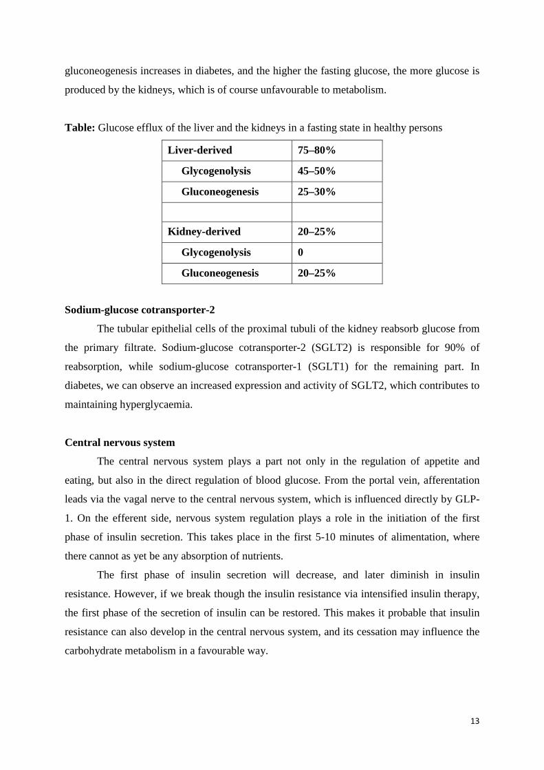
13
gluconeogenesis increases in diabetes, and the higher the fasting glucose, the more glucose is
produced by the kidneys, which is of course unfavourable to metabolism.
Table: Glucose efflux of the liver and the kidneys in a fasting state in healthy persons
Liver-derived 75–80%
Glycogenolysis 45–50%
Gluconeogenesis 25–30%
Kidney-derived 20–25%
Glycogenolysis 0
Gluconeogenesis 20–25%
Sodium-glucose cotransporter-2
The tubular epithelial cells of the proximal tubuli of the kidney reabsorb glucose from
the primary filtrate. Sodium-glucose cotransporter-2 (SGLT2) is responsible for 90% of
reabsorption, while sodium-glucose cotransporter-1 (SGLT1) for the remaining part. In
diabetes, we can observe an increased expression and activity of SGLT2, which contributes to
maintaining hyperglycaemia.
Central nervous system
The central nervous system plays a part not only in the regulation of appetite and
eating, but also in the direct regulation of blood glucose. From the portal vein, afferentation
leads via the vagal nerve to the central nervous system, which is influenced directly by GLP-
1. On the efferent side, nervous system regulation plays a role in the initiation of the first
phase of insulin secretion. This takes place in the first 5-10 minutes of alimentation, where
there cannot as yet be any absorption of nutrients.
The first phase of insulin secretion will decrease, and later diminish in insulin
resistance. However, if we break though the insulin resistance via intensified insulin therapy,
the first phase of the secretion of insulin can be restored. This makes it probable that insulin
resistance can also develop in the central nervous system, and its cessation may influence the
carbohydrate metabolism in a favourable way.

14
Chapter 2. Types, diagnosis and epidemiology of diabetes mellitus
Dr. István Wittmann
According to the present-day classification of diabetes the following types can be
differentiated:
1. Type 1 diabetes mellitus
With autoimmune mechanism
Idiopathic
2. Type 2 diabetes mellitus
3. Other, specific types
Genetic dysfunctions of the beta cells
Genetic defects of insulin function
Forms related to diseases of the exocrine part of the pancreas
Endocrinopathies
Types induced by drugs and chemicals
Infection-related
Unusual forms of the immunopathogenic diabetes
Genetic syndromes associated with diabetes
4. Gestational diabetes
Prediabetes (high risk of diabetes) and the diagnosis of diabetes
The major components of the diabetes mellitus syndrome are the following:
First, one has to mention the osmotic diuresis-induced polyuria due to osmotic diuresis. Being
a small molecular weight substance, glucose is able to pass freely through the barriers in the
renal glomeruli; and because of its high serum level, the tubular reabsorption system is not
able to reabsorb it completely, and so it will appear in the urine. The polyuria will lead to
polydypsia. On the other hand, glucosuria will lead to energy wasting and in this way to
weight loss. The compensatory reaction to weight loss is polyphagia.
Glucose is a reducing sugar that can set up Schiff-base bonds with free amino groups
of amino acids and proteins. This bond is stabilized due to a re-arrangement process, and
eventually leads to the formation of advanced glycation end-products. The process is called
non-enzymatic glycation. Glycation impairs protein functions, and in this way functions of the
antibodies as well. At the same time, in hyperglycaemia lesion of the cells responsible for

15
immunocompetence also develops. These two processes lead to immunosupression in
diabetes, increasing the risk of infections.
Diabetes may lead pruritus, i.e. itching of the skin, in part directly through non-
enzymatic glycation of components of the skin, and in part indirectly, through diabetic
neuropathy.
The symptoms mainly developing in type 1 diabetes mellitus are abdominal pain and
Kussmaul breathing, the cause of which is diabetic ketoacidosis.
Thus the most important diabetic symptoms are:
1. Polyuria-polydypsia
2. Weight loss - polyphagia
3. Risk of infection
4. Pruritus
5. Abdominal pain and Kussmaul breathing (in Type 1)
The diagnosis of praediabetic states (also called states with a high risk of diabetes) and
diabetes mellitus is based on fasting and 2 hours venous plasma glucose values (in an oral
glucose tolerance test, OGTT). According to present regulations, this has to happen using
validated measurements of central laboratories. A further method used mainly in the United
States is based on haemoglobin A1c (HbA1c) measurement. According to this, we can diagnose
diabetes by a value above 6.5%.
The diagnostic criteria are summarized in the following table. It is important to
emphasize that in the absence of the abovementioned symptoms, all measurements have to be
repeated in order to ensure accuracy of diagnosis.
Table: Diagnosis of praediabetes (high risk of diabetes) and diabetes mellitus
Venous plasma glucose concentration,
(mmol/l, laboratory measurement)
Normal glucose tolerance: Fasting blood glucose and 2-hour value of OGTT
≤ 6.0 < 7.8
Impaired Fasting Glucose, IFG: Fasting blood glucose and 2-hour value of OGTT
6.1-6.9 < 7.8
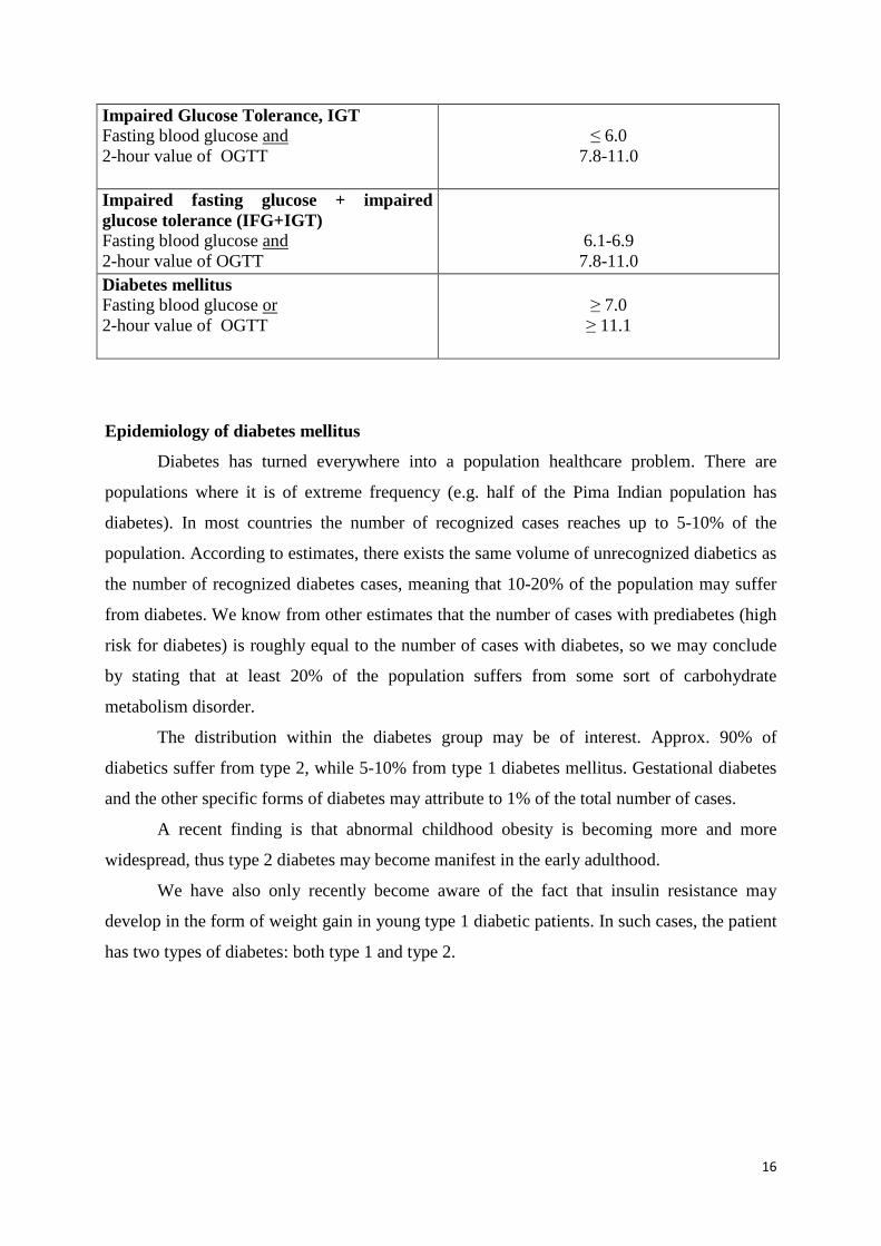
16
Impaired Glucose Tolerance, IGT Fasting blood glucose and 2-hour value of OGTT
≤ 6.0
7.8-11.0
Impaired fasting glucose + impaired glucose tolerance (IFG+IGT) Fasting blood glucose and 2-hour value of OGTT
6.1-6.9 7.8-11.0
Diabetes mellitus Fasting blood glucose or 2-hour value of OGTT
≥ 7.0 ≥ 11.1
Epidemiology of diabetes mellitus
Diabetes has turned everywhere into a population healthcare problem. There are
populations where it is of extreme frequency (e.g. half of the Pima Indian population has
diabetes). In most countries the number of recognized cases reaches up to 5-10% of the
population. According to estimates, there exists the same volume of unrecognized diabetics as
the number of recognized diabetes cases, meaning that 10-20% of the population may suffer
from diabetes. We know from other estimates that the number of cases with prediabetes (high
risk for diabetes) is roughly equal to the number of cases with diabetes, so we may conclude
by stating that at least 20% of the population suffers from some sort of carbohydrate
metabolism disorder.
The distribution within the diabetes group may be of interest. Approx. 90% of
diabetics suffer from type 2, while 5-10% from type 1 diabetes mellitus. Gestational diabetes
and the other specific forms of diabetes may attribute to 1% of the total number of cases.
A recent finding is that abnormal childhood obesity is becoming more and more
widespread, thus type 2 diabetes may become manifest in the early adulthood.
We have also only recently become aware of the fact that insulin resistance may
develop in the form of weight gain in young type 1 diabetic patients. In such cases, the patient
has two types of diabetes: both type 1 and type 2.
17
Chapter 3. Pregnancy and diabetes mellitus: Gestational diabetes
Dr. András Szilágyi
Definition
Pregnancy can be complicated by three types of diabetes: type 1 and type 2 diabetes
mellitus and gestational diabetes. Pregestational diabetes is defined as diabetes mellitus that
existed before pregnancy. To this group belong type 1 and type 2 diabetes, incidence of which
has shown an increase in recent times.
Gestational diabetes (GDM) is defined as glucose intolerance diagnosed during pregnancy,
regardless of whether it is treated with insulin or purely with diet or whether this condition
maintains or resolves after delivery.
Epidemiology (See also in related chapter)
While there is a trend in delayed childbearing, the association between type 2 diabetes
and pregnancy is expected to be more frequent. Prevalence of type 1 diabetes among women
of childbearing age is estimated to be approximately 0.3%. GDM is more common, the
prevalence being about 3-6 % according to Hungarian data, although this does depend upon
screening methods.
Etiology, pathogenesis
Insulin resistance increases by 30-90% in pregnancy. Anti-insulin, diabetogenic
hormones secreted by the placenta play important roles in this process, among which the role
of human placental lactogen (HPL) has to be highlighted. Maternal insulin production
increases to compensate for insulin resistance. If this increase is insufficient, the
compensation will be inadequate, resulting in GDM.
The insulin metabolising effect of the placenta also contributes to the emerging insulin
need.
Maternal and foetal complications in diabetes and pregnancy
Maternal complications: Hypo-and hyperglycaemia, diabetic ketoacidosis, higher rate
of infections, preeclampsia, polyhydramnion, ophtalmologic complications.
Foetal complications: malformations, miscarriage, intrauterine death, preterm birth,
macrosomia, placental insufficiency, intrauterine growth retardation, hypertrophyc
18
cardiomyopathy, polyhydramnion, birth injury, infections, delayed foetal lung maturity,
polycytaemia, and postnatal hypoglycaemia, hyperbilirubinemia and hypocalcemia.
Diagnostic evaluation
Screening for and diagnosis of GDM
There is no worldwide accepted unified approach to screening and diagnosing GDM.
In Europe the standard OGTT (2- h 75 g oral glucose tolerance test) recommended by WHO
is the applicable test to determine GDM.
Plasma glucose measurements before OGTT (fasting plasma glucose or zero minute)
and the 2 hour values are recommended for screening and diagnostic purposes, according to
WHO recommendations.
Fasting plasma glucose of 7 mmol/l or higher (≥126 mg/dl) supports the diagnosis of
GDM, and in this case OGTT is not recommended.
Normal fasting glucose with 2-hour plasma glucose over 7.8 mmol/l (˃ 140 mg/dl) also
indicates GDM.
GDM is also diagnosed when a random plasma glucose is 11.1 mmol/l or higher (≥200
mg/dl), confirmed through a second test, or when HbA1c is >6.5%.
In 2010 the International Association of the Diabetes and Pregnancy Study Groups
((IADPSG) recommended new criteria relating to 75 g OGTT for screening GDM ( FPG ≥5,1
mmol/l, 1-hour ≥10,0 mmol/l, 2-hour ≥ 8.5mmol/l) although these have not been widespread
as yet. According to these criteria the prevalence of GDM is higher.
All pregnant women should be screened for gestational diabetes at 24-28 weeks'
gestation.
Women who are at very high risk should undergo early testing (OGTT) at 12-16
weeks’ gestation. The criteria for very high risk are as follows: obesity, family history of
diabetes, hypertension, presence of glucosuria, previous delivery of an LGA (large for
gestational age) infant, previous delivery of an infant with malformation, previous intrauterine
foetal death, habitual abortion.
Management
Type 1 diabetes mellitus and pregnancy

19
The goal of insulin therapy is to achieve normoglycaemia and to prevent such
complications as foetopathy. Insulin therapy must be continuously corrected because insulin
needs increases by 2-3 times during pregnancy.
In late gestation (after the 36 weeks’), insulin need usually diminishes, although
excessive reduction in insulin need might mean a poor prognostic sign referring to placental
insufficiency. After delivery, insulin needs decreases markedly within hours.
Besides insulin treatment, dietary therapy is of great importance in maintaining
normoglycaemia and ensuring optimal conditions for foetal growth.
In addition to maintaining normoglycaemia, intrauterine monitoring of the foetus is
indispensable during prenatal care in order to achieve a good perinatal outcome.
From 26-28 weeks of gestation tests for evaluating foetus and foetal well being are
necessary (ultrasonography, Doppler flowmetry, Nonsterss test, Oxytocin stress test etc)
Type 2 dabetes mellitus and pregnancy
Normoglycaemic metabolic management must be initiated before conception with type
2 diabetic women as well. It is also important that oral antidiabetic agents should be
discontinued before pregnancy begins, although this issue is controversial in the case of
insulin sensitizers (metformin) which are permitted in early pregnancy.
In early pregnancy some type 2 diabetic patients can be controlled solely with a diet
regime. More than 50% of these women require insulin treatment as their pregnancy
advances.
Management of GDM
Implementation of an optimal diet is the first step in the management of GDM,
although even in obese patients extreme caloric restriction is not recommended.
Daily caloric intake should reach 1600 kcal with a 150-160g carbohydrate content
distributed among 5 meals throughout the day. Blood sugars should be maintained between
3.5-7 mmol/l.
An inappropriate diet results primarily in elevated postprandial glucose levels which
exceed 7.0 mmol/l. In such cases, the administration of short–acting insulin products before
meals (1-3 shots/day) might help to achieve normoglycaemia.

20
When over-high fasting plasma glucose is the primary alteration the treatment should
be complemented with bedtime NPH insulin. Twice daily doses of intermediate-acting insulin
might also be required. The daily insulin need is approximately 0.7-1.0 U/ kg.
The administration of rapid acting insulin analogues seems to be safe during
pregnancy. There is no sufficient experience regarding use of long-acting insulin analogues
(although detemir can be given).
Inadequately treated GDM is associated with higher perinatal morbidity and
mortality. Both the achievement of normoglycaemia and an intensive monitoring of foetus
and placental function during pregnancy and delivery play important roles in prevention of
this high morbidity and mortality. Initiation and frequency of use of intrauterine foetal
diagnostic tests (nonstress test, oxytocin challenge test, flowmetry of foetal vessels etc.)
depend on the severity of diabetes and the existence of possible complications.
Prognosis and care after delivery
In most cases, GDM will resolve itself soon after delivery. These patients have a
significantly higher risk of developing type 2 diabetes during their lifetime. Incidence rate can
reach as much as 50% over a period of 7 years. For this reason, postpartum follow up will be
of great importance. In this, the first step is the reclassification of maternal glycaemic status at
6 weeks after delivery or following the end of breastfeeding. This is also true for determining
whether the definitive diagnosis was purely GDM or pregestational diabetes which had
existed prior to, but recognised only during, pregnancy.
21
Chapter 4. Therapeutic plan and targets in diabetes mellitus
Dr. István Wittmann
After diagnosis, diabetes mellitus should be classified, i.e. it has to be decided which
type of diabetes is the patient is suffering from. Where there is a possibility of monogenic
inheritance, a centre should be contacted which has a professional approach to and experience
in the diagnosis and therapy of such cases.
It must be established what complications have already developed. We should not
forget that according to estimations the onset of type 2 diabetes is between 5-10 years prior to
the time of diagnosis. It is of equal importance that micro- and macrovascular complications
may already develop at the IGT phase. We may also then state that they are not diabetes-
associated, but carbohydrate dysmetabolism-associated micro-and macrovascular
complications.
At the time of diagnosis and at the initiation of a new follow-up one should consider
the following:
Case history
1. Total internistic case history
2. Age and diabetes duration
3. Eating habits, physical activity, body weight
4. Complications and concomitant diseases
5. Diabetes education
6. Initial or previous HbA1c value(s), data from the blood glucose 'diary'
7. Previous antidiabetic, antihypertensive therapy
8. Hypoglycaemic history (also hypoglycaemia awareness)
Physical examination to be carried out:
A total internistic physical examination, including measurement of height, body
weight, abdominal circumference, blood pressure, signs of orthostasis, and a thorough
examination of the feet, should be carried out.
22
Laboratory tests to be carried out:
HbA1c, lipid profile (total cholesterol, LDL-, HDL-cholesterol, triglyceride), liver
function tests, routine urine test, estimated GFR and in type 1 also TSH.
Consultations to be carried out
Dietetitian, ophthalmologist, maybe a dentist.
Target range instead of target value
Nowadays we consider setting target ranges rather than target values as the appropriate
approach. This consideration comes from the observation that both blood glucose and HbA1c
show a biphasic relation with mortality, that is, there exists an ideal range where mortality is
smallest. Underlying and beyond that, one has to face an increase in mortality rate. This range
may be individually different regarding following influencing factors:
1) Susceptibility to and history of hypoglycaemia, treatment predisposing to
hypoglycaemia
2) Age, duration of diabetes, life expectancy
3) Presence of micro- and macrovascular complications
4) Accompanying diseases
5) Motivation and medical adherence of the patient
6) Social and financial status of the patient
Susceptibility to and history of hypoglycaemia, treatment predisposing to
hypoglycaemia
With an increase in the duration of diabetes, the susceptibility to hypoglycaemia will
increase due to a loss of secretion of glucagon (due to destruction of the alpha cells), and due
to sympathetic neuropathy and nephropathy. The presence of severe hypoglycaemia in the
past history of the patient increases the chance of hypoglycaemia. Therapy using
sulphonylureas and insulin or overtight glycemic control increases the risk of hypoglycaemia.
Age, duration of diabetes, life expectancy
According to the above, an advanced age and longer duration of diabetes brings on a
predisposition to hypoglycaemia. On the other hand, with the decrease in life expectancy, a
very tight control is not justified.

23
Micro- and macrovascular complications
Accordingly, both sympathetic neuropathy and nephropathy predispose to
hypoglycaemia. On the other hand, an overfast and complete normalization of the
carbohydrate metabolism in a patient with high average glucose levels may worsen the
diabetic retinopathy. Furthermore, hypoglycaemia may lead to further damage to the cardiac
status in a patient with an already established ischemic disease; moreover, it may lead to
provocation of arrhythmias and thus sudden death.
Accompanying diseases
In the case of an actual, severe, ongoing infection or an operation with general
anaesthesia and the opening of a body cavity, one has to switch to insulin therapy and has to
halt other antidiabetics, including metformin.
Motivation and medical adherence of the patient
In the chronic phase of diabetes therapy it is not the doctor who treats the patient, but
the patient who is treating himself, in best cases following medical advice. Therefore it is an
important question as to whose advice the patient is adhering. Unfortunately, he does not want
to follow the advices of his own doctor, as the doctor can 'only' state the present-day stance of
science. On the other hand, unofficial 'advisers' say what the patient wants to hear, and so the
patient is of course more willing to accept their advice.
One important problem in cases of a relatively mild initiation into the onset of (type 2)
diabetes is that the patient does not feel any special complaint. As a result, his state of
motivation is low, despite our therapeutic options being most effective in this phase. In cases
of more advanced diabetes, the complications will bring on the motivation of the patient, but
our therapeutic options have already become pretty impaired by this phase of the disease.
According to our own surprising data, 30-40% of diabetics have already discontinued
their medication only 1 year after its initiation.
Social and financial status of the patient
Diabetes means a psychic and financial load upon the whole family of the patient.
Accepting this is a great medical achievement. It is of great importance to find out who does
the cooking in the family. The education of this person has to run parallelly with that of the
patient. Where there is deterioration of carbohydrate metabolism, besides the usual causes
(infection, changes in working habits or lifestyle etc.) one also has to consider family

24
problems. I have observed that even if the patient indicates another cause, this is most
frequently the real one.
Glycaemic markers and target ranges
HbA1c is the most important marker in the follow-up to diabetes. It shows glycated
haemoglobin, thereby indicating the average glycaemia of the past three months. Limitations
to the value of HbA1c are as follow: anaemia, iron deficiency, blood transfusion, dialysis and
haemoglobinopathies.
Fructosamine displays the value of glycated albumin. Accordingly, it represents the
average glycaemia of the past two weeks. It can be used in any type of diabetes, but is of
special importance in cases of pregnancy. It cannot be evaluated properly in cases of protein
loss (e.g. abnormal albuminuria, protein-losing enteropathies) and an accelerated protein
turnover (e.g. hyperthyroidism).
Glycaemic target ranges are as follow:
HbA1c in type 1: 6.5-7.5%
HbA1c in type 2: 6.0-8.0%
Praeprandial plasma glucose: 4.4-7.2 mmol/l
Postprandial plasma glucose: <10.0 mmol/l
25
Chapter 5. The basics of the non-pharmacological therapy of diabetes mellitus
Dr. József Rinfel
The non-pharmacological therapy of diabetes mellitus is referred to as lifestyle
modifications that have two major parts, namely dietetotherapy and therapy based on physical
activity. In a wider sense, psycho-social support and patient education can also be mentioned
here. The fulfilment of these is not only fundamental to successful patient care, but also a
marker of good cooperation between physician and patient. Putting these parts of patient care
into daily practice often exceeds the boundaries of conventional medical practice, hence the
cooperation of different specialists (dietetitian, physiotherapist, educator etc.), but knowing
the basics is inevitable for persons with a general medical degree.
Lifestyle is regarded as a determinant in the development of type 2 diabetes mellitus,
the worldwide rapid spread of the disease showing a connection with obesity and a sedentary
lifestyle: changing this lifestyle may be a key to prevention, as well. Several epidemiological
studies have provided proof of this. Efficient and consequent lifestyle changes, if initiated in
time, may prevent approx. 50-60% of type 2 diabetes cases.
Once developed, treatment of the disease also requires a proper diet and daily physical
activity. The elements of this should be tailored to the patient, taking into consideration the
properties and level of cooperation of the patient and adjusted to the actual pharmacological
therapy. However, changthese in lifestyle can only be successful with the cooperationof the
patient! It is of high importance that the patient understands what they should do, and how
and why they should do it.
Besides providing a good metabolic control, keeping a proper diet and doing regular
physical activity is also needed for body weight control and for the quality of life. Data also
prove that it may also delay or prevent the development of complications.
Dietetotherapy
The aim of prescribing a diet is to provide optimal nutrition to the individual. This has
general rules, but the unique properties of each patient, concomitant diseases, and related
pharmacologic therapy make it necessary to make individual recommendations to the
particular patient.
Diabetes mellitus is a complex metabolic disorder; and so several conditions have to
be taken into account simultaneously when planning the diet. Alterations in the background of

26
type 2 diabetes mellitus include the delay of the early prandial phase of insulin secretion,
insulin resistance and the frequently-observed obesity, all of which represent problems to be
solved. With insulin therapy, the effect of preparations employed on blood glucose levels also
makes regular timing of meals necessary.
It is therefore absolutely necessary that the patients should be able to properly plan his
own diet, determining besides daily calorie intake the ratio of major components of food
(carbohydrates, proteins, fat), and choosing food by taking into account regarding quality
criteria, timing of meals and the carbohydrate content of individual meals.
Differences in the ability of carbohydrates to raise blood glucose levels should also be
emphasized. Knowing the glycaemic index and application of it while planning the diet may
aid in reaching good metabolic control. This may only be reached when using a regular,
structured patient education (see the chapter on patient care and education in diabetes).
It is of fundamental importance that the patient should understand and accept the
sometimes large modifications of their previous dietary habits. These may sometimes be hard
to fit into the usual daily routine, and putting it into daily practice may also be problematic.
To overcome these, the involvement of specialists may be useful, and in this regard the
participation of dieticians and educators may assist, too, as they may help the patient
understand the details of lifestyle prescriptions. Additionally, the involvement of other
persons in helping the patient may be useful: the participation of family members in keeping
to a diet may improve the rate of success.
The dietetotherapy of diabetics should cover the following:
Energy content of the diet
A diabetic patient with normal body weight needs a calorie intake that is determined
by age, height, markers of metabolic controls, and the type, intensity and duration of physical
activity. This is around 25-35 kcal/bodyweight kg/day, that would generally come to a daily
intake of 1800-2500 kcal (7.6-10.5 MJ).
In the case of insulin resistance, a modest, 5-7 % weight loss may aid the efficacy of
insulin. However, restriction of energy intake is usually not sufficient to attain long-term
weight reduction and improvement of glycaemic control: for that, regular, daily physical
activity is also required. We also have to consider that energy requirement of the elderly is
lower than at a younger age.

27
Composition of the diet
Despite occasional changes in recommendations regarding the composition of the diet,
a diet with low fat content and a defined carbohydrate content that is mainly carbohydrate-
based is actually widely accepted. (Extreme diets should be avoided; diets preferring extreme
proportions may especially carry risks. Unfortunately, doctors also succumb to these despite
all the facts at their fingertips!
Accordingly, carbohydrate content should cover approx. 50-60% of the total energy
intake, and a protein intake of 0.8-1 g/bodyweight kg/day (approx. 20% of total calories) is
suggested, while the remaining calories (approx. 30% of total calories) go to fat.
(Thus, in the case of a middle-aged person of 70 kg, normal BMI and abdominal
circumference with intermediate level of physical activity
- the daily energy requirement would be 25x70 ~ 1800 kcal/day
- protein intake: 70 g x 4 kcal/g ~ 300 kcal (1500 kcal/day remain)
- carbohydrate intake: 1800x0,55 = 1000 kcal (: 4 kcal/g) = 250 g/day
- fat intake: 1800 - (300+1000) = 500 kcal (: 9 kcal/g) = 60 g/day
Thus the daily diet is: 1800 kcal; 70 g protein; 250 g carbohydrates; 60 g fat)
It is also important to know the glycaemic index, and consumption of complex
carbohydrates leading to less of a rise in blood glucose levels is recommended, within which
one should aim for foods with high fibre content (30 g dietary fibres/day). In this regard, the
consumption of vegetables, fruits and whole grains is recommended.
Regarding fat intake, a proportion of saturated fatty acids should be < 10% (but if the
LDL-cholesterol level is > 2.5 mmol/l, then a lower proportion of < 7% is suggested).
According to the guidelines, the proportion of polyunsaturated fatty acids should be ~ 10 %,
with monosaturated between 10-12%. It is also important to decrease the intake of trans fats
(fatty acids), as more components of the atherogenic dyslipidaemia (LDL ↓, HDL ↑) may be
ameliorated using this approach.
The protein intake is planned in the usual way. In the case of a nephropathy, the
amount and composition of protein intake may require changes usually prescribed by the
nephrologist.
Frequency of meals
In general, division of the total calorie intake to 5-6x smaller meals is suggested, (3
major meals with intermediate meals, in some cases with a bedtime snack), but with certain

28
therapies (e.g. prandial glucose regulators, analogue basal/bolus insulin therapy) 3 major
meals may be sufficient.
Limiting the carbohydrate content of individual meals in cases of patients with type 2
diabetes mellitus is important, the cause for this is to decrease the "carbohydrate load" related
to a meal (see postalimentary hyperglycaemia). On the other hand, in cases of certain
secretagogue therapies and insulin substitution we should also try to avoid a pronounced fall
in blood glucose, which could lead to the risk of hypoglycaemia.
Other dietary considerations related to diabetes
The diet of diabetic patients should be rich in vitamins and mineral salts and trace
elements, but an extra supplementation is not necessary. The use of vitamin complexes is not
proven, and in the case of certain antioxidants a large intake is rather risky and harmful!
So-called "dietary products" should be used with caution. Sweeteners should also be
used with caution. As a rule, the use of artificial – non-caloric – sweeteners (saccharin,
cyclamate, acesulphame-K, aspartam etc.) should be preferred, but temperance is also
required with their use. Besides individual sensitivity, thermal stability should also be taken
into account, an important consideration in baking and cooking.
It should also be noted that aspartam is a photosensitive substance, decomposing
quickly beyond its date of consumption (foods lose their sweet taste in this case), the
degradation products contain phenylalanine, and may therefore carry risk for patients with
known phenylketonuria
No potential side effects or risks considering non-caloric sweeteners have as yet been
proven, thus when used according to prescriptions, a significant health risk related to their use
does not need to be considered.
With sugar-replacement caloric sweeteners (fructose, sorbitol, xylitol, stevia) and food
products manufactured with their use, the calorie and carbohydrate content should be
considered and calculated into the daily intake.
The consumption of alcohol and other “drugs” should also be considered. In the case
of alcohol, besides the physiologic affect of alcohol attention should also be turned to the
relative high caloric intake (~7 kcal/g). The consumption of alcoholic drinks with sugar
should be definitely avoided. The alcohol intake should be 1 unit for women and 2 units for

29
men (1 unit = 1-1.5 dl of wine, 3 dl of beer, or 2-3 cl spirits, which corresponds to the
consumption of approx. 15 g).
Regarding the consumption of a daily amount of 1-2 dl of dry red wine, protective
cardiovascular effects have been observed in the studies.
Of the beverages containing coffeine, a daily quantity of 1-3 espresso and 2-4 cups of
tea may be consumed. The stimulants coffeine and tannin do not lead to a rise in blood
glucose.
Here we do not cover detailed special dietary considerations related to diabetes
(diabetic nephropathy, celiac disease more common in type 1 diabetes, lactose intolerance
with an increase in prevalence with ageing) but refer to them only.
Physical activity
As previously referred to, regular physical activity is a necessary part of lifestyle
interventions that may not only play a part in the prevention of diabetes melitus, but which is
also an integral part of the care of already developed diabetes.
However, the physical activity should be suited to the capacity, fitness, age,
accompanying diseases, and pharmacological therapy of the individual patient. The intensity,
duration, form and frequency of physical activity should be tailored to the individual.
The diabetic patient should consult his treating physician and a specialist (e.g.
physiotherapist, trainer, games master) before the initiation of such activity.
Properly planned and constructed, regular physical activity may lead to a complete
improvement of metabolic parameters, and besides blood glucose, lipid parameters and uric
acid may also show positive changes. This is inevitable as regards body weight control, the
long-term maintenance of which is not possible in the majority of cases without physical
activity. Studies have shown that it improves cardio-pulmonal performance and status. The
quality of life improves, the burden of the psychological load can be attenuated, it may be
beneficial in preventing stress and anxiety-depression disorders. Regular physical training is
also a pledge of active senescence, providing a tool that not only maintains physical functions
as well as possible, but also slows down mental decline.
Prior to the initiation of a regime of physical activity, a thorough check-up is
recommemnded which should cover general status, especially the cardiopulmonal loadability,
and diabetes-specific complications (neuro- nephro- retinopathy, osteoarthropathy).
30
The patient should recognize those physiological states where training may even be
harmful: acute metabolic deterioration (blood glucose over 15 mmol/l, the danger of
hypoglycaemic episode, ketoacidosis etc.), febrile states, infections, the presence of an
autonomic neuropathy, severe renal impairment, proliferative retinopathy, the onset of acute
cardiac symptoms etc. In these cases it is required that the exercise be postponed and an
extraordinary medical consultation undergone!
On planning an exercise programme, dynamic exercises that move large muscle
groups and mainly induce an aerobic load (walking, fast walking, bicycle racing, swimming,
aquatic work-up etc.) should be given chief emphasis. We should bear in mind what types of
movements the condition of the patient enables, and what they would like to do on a daily,
regular basis.
Static exercises which improve muscle force and condition may also be included
into the training programme, but they should only be carried out when tailored to the
individual, under stricter control.
Resistance training aims at the increase of muscle force and changing body
composition by increasing the basal metabolic rate; it leads to increased energy consumption.
Stretching may also be advisable, as well as those daily activities (walking, working
around the house, stair walking) that we carry out regularly. Arterious vessel exercises may be
beneficial to patients with atherosclerosis.
In general, a 3-5 times weekly exercise session of medium-intensity of 15-20 minutes
duration is suggested, which can be raised until the patient finds it too difficult.
We should draw attention to the replacement of fluid lost during exercise. During/after
exercise a stricter blood glucose control is suggested with an appropriate correction.
Summarizing, the objective of non-pharmacological therapy is to change the lifestyle
of the patient. Appropriate dieting and physical activity are major tools in the prevention of
type 2 diabetes mellitus, and it should be possible to prevent ~50% of cases of the diseases. In
therapy of the metabolic disease that has already developed, lifestyle interventions are also a
determining factor, and a successful therapy can only be provided using these approaches, as
well.

31
It is important that dietary changes and suggestions regarding physical activity should
be made together and in agreement with the patient. Putting these into practice can be
achieved using by educating and continuously motivating the patient.
The level of change in lifestyle mirrors the cooperation between doctor and patient!
32
Chapter 6. Non-insulin-like antidiabetic agents
Dr. István Wittmann
These agents are divided into 2 groups:
Oral non-insulin-like antidiabetic agents
Parenteral non-insulin-like antidiabetic agents
Oral non-insulin-like antidiabetic agents can be further classified as having effects that are:
Primarily insulin-dependent
Primarily insulin-independent
There are 2 subgroups of primarily insulin-dependent oral, non-insulin-like antidiabetic
agents:
Agents that enhanceenhance the effect of insulin
Agents that increase insulin secretion from the pancreas (insulin secretagogues)
Primarily insulin-dependent, oral, non-insulin-like insulin sensitising agents are as follows:
Biguanides: metformin (buformin is not recommended any more)
Alfa-glucosidase inhibitors: acarbose
Thiazolidinedions (PPAR-gamma-agonists, glitazones): pioglitazon
Primarily insulin-dependent, oral, non-insulin-like agents that increase insulin secretion
(insulin secretagogues):
Sulphonylureas: gliclazide, glimepiride, glipizide, gliquidone (glibenclamid/glyburide
is now regarded as an obsolete drug)
Prandial glucose regulators (glinides): nateglinide, repaglinide
Dipeptidyl-peptidase-4 (DPP4) –inhibitors (gliptins): alogliptin, linagliptin,
saxagliptin, sitagliptin, vildagliptin,
Primarily insulin-independent, oral, non-insulin-like agents:
Sodium/glucose cotrantsporter-2 (SGLT-2) inhibitors (glycoseurics): canagliflozin,
dapagliflozin, empagliflozin

33
Parenteral (injectable) non-insulin-like antidiabetic agents:
Amylin-analogues: pramlintide
Incretin mimetics:
GLP-1-analogues: liraglutide
GLP-1-receptor agonists: exenatide, lixisenatide
Table summarises oral antidiabetic agents in synoptic form. This classification does not
involve those human insulin and insulin analogues which are found in Chapter. 7

34
Table: Non-insulin-like antidiabetic agents
1. Oral non-insulin-like antidiabetic agents
i. Primarily insulin-dependent
1. Agents that enhance the effect of insulin
a. Biguanids: metformin
b. Alfa-glucozidase-inhibitors: acarbose
c. Thiazolidinedions (insulinsensitisers, PPAR-gamma-
agonists): pioglitazone
2. Agents that increase insulin secretion (insulin-secretagogues)
a. Sulphonylureas: gliclazide, glimepiride, glipizide,
gliquidone
b. Prandial glucose regulators: nateglinide, repaglinide
c. Dipeptidyl-peptidase-4-inhibitors: alogliptin, linagliptin,
saxagliptin, sitagliptin, vildaglitin,
ii. Primarily insulin independent
a. Sodium/glucose cotransporter-2 (SGLT2) inhibitors:
canagliflozin, dapagliflozin, empagliflozin
2. Parenteral non-insulin-like antidiabetc agents
a. Amylin-analogues: pramlintide
b. Incretin-mimetics:
i. GLP-1-analogue: liraglutide
ii. GLP-1-receptor-agonists: exenatide, lixisenatide
Primarily insulin–dependent, oral non insulin–like agents that enhance insulin effect
(metformin)
Mechanism of action: Metformin decreases hepatic glucose production, enhances insulin
sensitivity (decreases insulin resistance), influences intestinal glucose absorption and activates
incretin system. Behind these actions, at least in part, is the drug’s AMP kinase activating
35
effect which is not only favourable to metabolism but also decreases tumor mitogenesis
through mTOR inhibition.
Advantages: It does not cause hypoglycemia or weight gain. It is also favourable as regards
non-alcoholic fatty liver disease (NAFLD). It decreases cardiovascular- and cancer risks.
Indication : Used for treating all patients with type 2 diabetes who are not intolerant, or when
there is no contraindication.
Dosage: Should be started with low dose (500 mg once or twice a day). Dosage increases
should be made over 2-3 week periods up to the maximal dose of 2550 mg per day (850 mg
three times a day). Higher doses are unjustified as efficacy does not increase, but side effects
may develop. Extended release formulation is also available, which is tolerated well up to a
maximal daily dose of 2000 mg once a day.
Combination: Can be used in monotherapy or in combination with all types of antidiabetic
agents.
Contraindications and adverse effects: Metformin is contraindicated in patients with renal
impairment, in patients with congestive heart failure or respiratory failure who are at risk of
hypoxaemia, in severe liver failure, in pancreatitis and in pregnancy. It should be discontinued
in patients undergoing radiologic examinations with iodinated contrast material.
A rare but serious side effect of metformin is lactic acidosis, which is often fatal even today.
Metformin intolerance means a serious adverse reaction, usually with gastrointestinal side
effects (bloating, flatulence, diarrhoea), which leads to discontinuation of the drug.
Primarily insulin–dependent, oral, non insulin–like agent that enhances the effect of
insulin (acarbose):
Mechanism of action: Alpha-glucoside hydrolase enzyme inhibitor. The inhibition of alpha-
glucosidase enzymes leads to decreased intestinal glucose absorption and subsequently to
diminished glucotoxicity as well. It enhances the effect of incretin via an as yet unknown
mechanism.
Advantages: Does not cause hypoglycemia, is associated with weight loss, and decreases
cardiovascular risk.
Indications: It is recommended for obese patients for lowering postprandial hyperglycemia.
Dosage: Should be started with a low dose such as 50 mg once or twice a day, with gradual
titration up to a maximal dose of 400mg (100 mg 4 times a day).
Combination: Can be used in monotherapy as well as in combination with all types of
antidiabetic agents.

36
Contraindications and adverse effects: It is rarely used because of common gastrointestinal
side effects (bloating, flatulence, diarrhoea).
Primarily insulin–dependent, oral, non insulin–like agent that enhances insulin effect
(pioglitazone)
Mechanism of action: It is an agonist of the peroxisome proliferator-activated receptor
gamma (PPAR-gamma).
Advantages: The drug is associated with relevant A1C-reduction. It might be beneficial in
NAFLD and can also be used in renal impairment
Indications: It is recommended for obese, significantly insulin resistant patients.
Dosage: Dose up-titration is usually not required.
Combination with other antidiabetic agents: Can be used alone or in combination with
other antidiabetics except for insulin or some SGLT2-inhibitors.
Contraindications and adverse effects: It is contraindicated in congestive heart failure. An
increased incidence of bone fracture has been reported in women who take pioglitazone,
therefore it is not recommended for postmenopausal women. Weight gain has also been
observed, partly due to fluid retention, but subcutaneous fat accumulation may also occur.
Primarily insulin–dependent, oral, non insulin–like agents that increase insulin secretion
(secretagogues): sulphonylureas (gliclazide, glymepiride, glipizide, gliquidone)
Mechanism of action: Sulphonylureas close ATP-sensitive K-channels in the beta-cell
plasma membrane, resulting in membrane depolarisation. This opens voltage-gated Ca2+
channels and the Ca2+ influx leads to insulin release.
Advantage: The drug leads to significant A1C reduction.
Indications: Sulphonylurea monotherapy is only recommended when metformin intolerance
or contraindication occur. It is commonly used in combination with other antidiabetic agents.
Dosage: The most commonly used modified (extended) release form of gliclazide and
glymepiride is given once a day, while glipizide and gliquidon are given in two divided doses
per day.
Combination with other antidiabetic agents: It can be combined with all other antidiabetic
agents.
Contraindications and adverse effects: Sulphonylurea efficacy declines relatively rapidly
(after a few years of treatment), because these agents are associated with a progressive decline
in β-cell (and also alpha cell) function and their action is beta cell dependent. The beta cell
37
“burnout” is partly a consequence of concomitant amylin secretion, which leads to local
amyloidosis causing cell destruction.
Sulphonylureas may induce hypoglycemia and may inhibit hypoglycemic counterregulatory
responses via alpha cell destruction. They cause weight gain. Except for gliclazide they also
increase cardiovascular and cancer risk. They bind to albumin in circulation and so they may
interact with Vitamin K antagonists and NSAIDs. Their metabolisation via citokrom P4502C9
pathway also leads to numerous drug interactions.
Primarily insulin–dependent, oral, non insulin–like agents that increase insulin
secretion: prandial glucose regulators (nateglinide, repaglinide)
Mechanism of action: The same as in sulphonylurea.
Advantage: They specifically enhance early-phase prandial insulin secretion (prandial insulin
releasers) in contrast with sulphonylureas.
Indications: The same as in sulphonylurea. They might be suitable for lifestyles where meals
are unpredictable or missed.
Dosage and Combination: They are taken immediately before a meal. If a meal is skipped
the medication should also be skipped. It can be combined with all other antidiabetic agents.
Primarily insulin-dependent, oral, non-insulin-like agents that increase insulin secretion
dipeptidyl-peptidase-4-inhibitors (alogliptine, linagliptine, saxagliptine, sitagliptine,
vildagliptine)
Mechanism of action: The dipeptidyl-peptidase-4 enzyme splits GLP-1 and GIP, making
these hormones metabolically inactive. Inhibition of DPP-4 enzyme increases the plasma
level and duration of action of these hormones. GLP-1 stimulates insulin secretion in a
glucose dependent manner as well as decreasing glucagon release. GIP rather induces insulin
secretion alone.
Advantages: They are weight neutral, and are associated with low risk of hypoglycaemia.
Their antihyperglycemic effect remains permanent (there is no beta cell “burnout”). They can
also be used where there is kidney impairment and seem to be cardiovascularly safe.
Indications: DPP-4 inhibitors can be used in monotherapy and in combination with almost all
other antidiabetic agents. They are also effective in kidney failure.
Dosage: Dose up-titration is not required. The dose of some preparations has to be reduced
according to the kidney impairment.

38
Combination with other antidiabetic agents: Some preparation can be applied in
monothreapy and all preparation can be used in combination with one or two antidiabetic
agents. All the drugs are now available in a combined form with metformin.
Contraindications and adverse effects: Some of them are not recommended in liver disease.
If they are added to sulfonylureas hypoglycemia might occur, which requires sulfonylurea
dosage reduction. Their side-effect profile is usually comparable to placebo.
Primarily insulin independent oral, non-insulin lik e agents: SGLT2-inhibitors
(canagliflozin, dapagliflozin, empagliflozin)
Mechanism of action: SGLT-2 inhibitors promote glucose and sodium loss in the urine by
blocking sodium-glucose cotrasporter 2 in the renal proximal tubular epithelial cells.
Canagliflozin inhibits SGLT-1 in intestinal mucosa as well.
Advantages: SGLT-2 inhibitors can cause weight loss and also lower blood pressure. They
are associated with low risk for hypoglycaemia. They moderate glucotoxicity and are
cardiovasculary safe.
Indications: Can be used in monotherapy as well as in dual or triple combination therapy.
Dosage: A single daily dose is recommended, gradual dose titration is not required.
Combination with other antidiabetic agents: Can be used in monotherapy and also in
combination therapy.
Contraindications and adverse effects: Dapagliflosin is not recommended for use in patients
receiving loop diuretics or pioglitason. SGLT-2 inhibitors increase the incidence rate of
genital infections thus they are not recommended for patients with a previous history of
genital infections. The prevalence of urinary tract infections is comparable to placebo.
Parenteral non-insulin-like antidiabetic agents: incretin mimetics (exenatide, liraglutide,
lixisenatide)
Mechanism of action: Incretin mimetics bind to the GLP-1 receptor, stimulate glucose-
dependent insulin secretion (such as DPP-4 inhibitors) and decrease glucagon release.
According to the most recent classification, these drugs might be classified either as prandial
or non-prandial agents.
Prandial agents (exenatide, lixisenatide) have a more notable influence on postprandial blood
glucose rise and may slow gastric emptying more significantly. Non-prandial agents like

39
liraglutid and exenatide LAR (a long acting release (LAR) exenatide formulation with once-
weekly dosing) show greater reduction in fasting blood glucose.
Advantages: Incretin mimetics are considerably effective antihyperglycemic agents
associated with low risk of hypoglycaemia, and have a favourable impact on weight and blood
pressure. Cardiovascular outcome trials performed thus far have confirmed cardiovascular
safety.
Indications: Incretin mimetics are recommended as a second or third line therapy (in
combination with other antidiabetic agents) for patients with type-2 DM and obesity.
Dosage: Gradual titration is required. Exenatid LAR reaches its maximum effectiveness after
several weeks.
Combination with other antidiabetic agents: Can be used in combination with one or two
antidiabetic agents and even with insulin.
Contraindications and adverse effects: Nausea may occur, which is usually self limiting,
and resolves or at least lessens within a few weeks. GLP-1 agonists are not recommended in
severe renal impairment.
Algorithm for the treatment of type 2 diabetes mellitus
Figure shows a treatment algorithm for type 2 DM. Green marking refers to preferred
agents, whereas yellow marking refers to agents that can be appropriate for selected patients
(acceptable alternatives).
In newly diagnosed type 2 diabetic patients with ketoacidosis, hyperosmolar hyperglycaemic
state or markedly elevated blood glucose levels (fasting plasma glucose >13.9 mmol/l, or
random plasma glucose > 16.7 mmol/l or A1C >10%), insulin therapy is recommended from
the outset.
After initial management of the “acute state”, further chronic therapeutic regimens (e.g.: oral
therapy, initial combination therapy regimens etc.) can be considered.
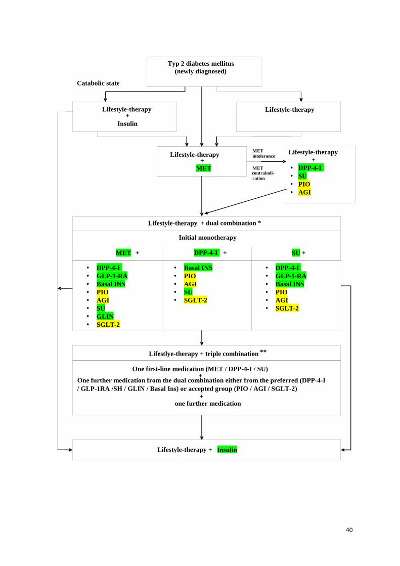
40
Typ 2 diabetes mellitus (newly diagnosed)
Lifestyle-therapy + Insulin
Lifestyle-therapy
Lifestyle-therapy + MET
Lifestyle-therapy +
• DPP-4-I • SU • PIO • AGI
Lifestyle-therapy + dual combination*
Lifestlye-therapy + triple combination **
Lifestyle-therapy + Insulin
Catabolic state
MET intolerance
METcontraindi- cation
Initial monotherapy
MET + DPP-4-I + SU +
• DPP-4-I • GLP -1-RA • Basal INS • PIO • AGI • SU • GLIN • SGLT -2
• Basal INS • PIO • AGI • SU • SGLT -2
• • GLP -1-RA • Basal INS • PIO • AGI • SGLT -2
One first-line medication (MET / DPP-4-I / SU) +
One further medication from the dual combination either from the preferred (DPP-4-I / GLP-1RA /SH / GLIN / Basal Ins) or accepted group (PIO / AGI / SGLT-2)
+
one further medication
DPP-4-I

41
Figure: Algorithm for management of type 2 diabetes mellitus. Abbreviations: MET=metformin, DPP-4-I=dipeptidyl-peptidase-4-inhibitor, SU=sulphonylurea, PIO=pioglitazon, AGI=alpha-glukozidase-inhibitor, GLP-1-RA=GLP-1-receptor-agonist (includink all GLP-1 mimetics), INZ=insulin, SGLT2=sodium-glucose-cotransporter-2-inhibitor, GLIN=glinides (prandial glucose regulators). When “acute” insulin treatment is not necessary, lifestyle modification and metformin
treatment are recommended as first line therapy. The algorithm opens the door to lifestyle
modifications alone as an initial therapy, and metformin is considered for use only when
lifestyle interventions fail to control blood glucose (A1C is above target after 3 months).
When considering the favourable effects of metformin on cardiovascular- and cancer risk in
diabetic population, early metformin therapy is usually preferable. When metformin
intolerance or contraindication occurs, DPP-4-I, SU, AGI, or PIO can be used.
If pharmacological monotherapy proves to be insufficient within a relatively short period (3
month) combination therapy becomes necessary.
The recommended combination regimens are listed in Figure.
According to the currently recommended diabetes management guidelines, three-drug
combination therapy is required in patients who do not reach their individualised glycemic
target within about a 3-6 month period. Some drugs (such as metformin or acarbose) require a
gradual up-titration which may last for weeks or months. In this case the lag period before
adding other antihyperglycemic agent(s) can be extended.
The main goal of this intensive management approach is to avoid hyperglycaemic
periods (A1C above target) lasting for years, which result in severe complications.
It should be noted that according to the changes in patients needs it is possible to raise
or reduce drug doses, or even to use step-down therapy (discontinue one or two oral agents or
even insulin after weight loss) or to replace antidiabetic agents if new indications or
contraindications occur.
Many patients with long-standing-type 2 diabetes eventually require insulin therapy.
All types of insulin (human as well as analogue insulin) can be used, such as bedtime insulin,
a bi-daily regimen with premix insulin, or intensive insulin therapy (basal bolus regimen) etc.
It is important that metformin should be continued unless it has not been become
contraindicated. The recommendation to use metformin is based on its (previously mentioned)
possible favourable effects on cardiovascular and cancer risk in diabetes. Furthermore, its role
in reducing insulin resistance, which results in lower insulin dose requirements, is also
beneficial.

42
Chapter 7. Insulin treatment
Dr. István Wittmann
Insulin treatment is among the most impressive examples of individually tailored
therapies. There are as many insulin regimens as there are patients! For this reason,
knowledge of the pharmacodynamic characteristics of various types of insulin is of the utmost
importance. Dosing and timing of insulin treatment is recommended according to these
pharmacodymanic parameters.
Table: Pharmacodynamic parameters of insulins
Type of insulin Time of onset (mins)
Time of max. effect (hours)
Duration of action (hours)
Human insulin
Regular, short-acting human insulin
30 2 5-7
Intermediate (NPH) insulin
60-120 4-6 12-16
Rapid-acting analogues
Insulin Aspart 10-20 1 2-5
Insulin Glulisine 10-20 1 2-5
Insulin Lispro 10-20 1 2-5
Long-acting analogues
Insulin Detemir 60-120 - 20-30
Insulin Glargine 60-120 - Max. 24
Premix insulins
Human premix insulin
30-60 4-6 12-16
Analogue premix insulin
10-20 4-6 12-16
Data are for orientation. Onset, peaktime and duration depend on dose, site of administration, physical activity and temperature.
The use of most varied combinations is possible, although in many countries financial
limitations hinder the use of some regimens. In other words, everything is possible, and this
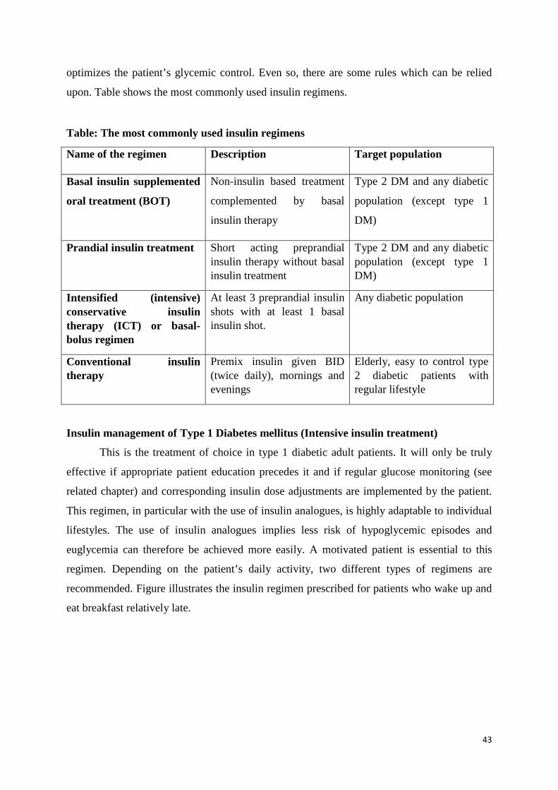
43
optimizes the patient’s glycemic control. Even so, there are some rules which can be relied
upon. Table shows the most commonly used insulin regimens.
Table: The most commonly used insulin regimens
Name of the regimen Description Target population
Basal insulin supplemented
oral treatment (BOT)
Non-insulin based treatment
complemented by basal
insulin therapy
Type 2 DM and any diabetic
population (except type 1
DM)
Prandial insulin treatment Short acting preprandial insulin therapy without basal insulin treatment
Type 2 DM and any diabetic population (except type 1 DM)
Intensified (intensive) conservative insulin therapy (ICT) or basal-bolus regimen
At least 3 preprandial insulin shots with at least 1 basal insulin shot.
Any diabetic population
Conventional insulin therapy
Premix insulin given BID (twice daily), mornings and evenings
Elderly, easy to control type 2 diabetic patients with regular lifestyle
Insulin management of Type 1 Diabetes mellitus (Intensive insulin treatment)
This is the treatment of choice in type 1 diabetic adult patients. It will only be truly
effective if appropriate patient education precedes it and if regular glucose monitoring (see
related chapter) and corresponding insulin dose adjustments are implemented by the patient.
This regimen, in particular with the use of insulin analogues, is highly adaptable to individual
lifestyles. The use of insulin analogues implies less risk of hypoglycemic episodes and
euglycemia can therefore be achieved more easily. A motivated patient is essential to this
regimen. Depending on the patient’s daily activity, two different types of regimens are
recommended. Figure illustrates the insulin regimen prescribed for patients who wake up and
eat breakfast relatively late.
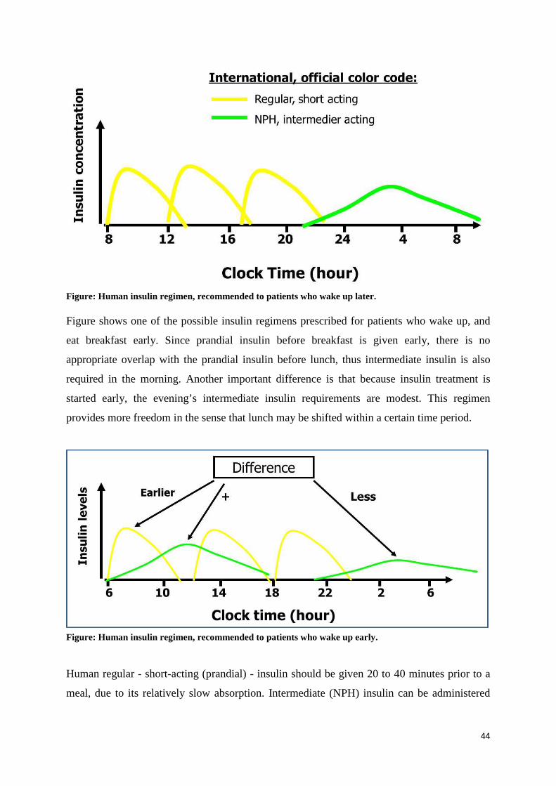
44
Figure: Human insulin regimen, recommended to patients who wake up later.
Figure shows one of the possible insulin regimens prescribed for patients who wake up, and
eat breakfast early. Since prandial insulin before breakfast is given early, there is no
appropriate overlap with the prandial insulin before lunch, thus intermediate insulin is also
required in the morning. Another important difference is that because insulin treatment is
started early, the evening’s intermediate insulin requirements are modest. This regimen
provides more freedom in the sense that lunch may be shifted within a certain time period.
Figure: Human insulin regimen, recommended to patients who wake up early.
Human regular - short-acting (prandial) - insulin should be given 20 to 40 minutes prior to a
meal, due to its relatively slow absorption. Intermediate (NPH) insulin can be administered

45
independently of meals although it is practical to give with the prandial one at least in the
morning.
In human insulin regimens regular snacks at mid-morning, mid-afternoon, and usually mid-
evening are necessary to prevent hypoglycaemic episodes.
Insulin regimens using rapid-acting analogues with intermediate insulin difficulty provide an
appropriate basal insulin level; therefore use of a long-acting analogue (eg Detemir) will
usually be desired. (Figure). The combination of intermediate insulin and rapid-acting insulin-
analogue demands practically three times a day NPH administration (to maintain an adequate
basal insulin level) which leads to interactions and hypoglycaemia, all of which has to be
avoided.
Figure: Rapid-acting and long-acting (Detemir) insulin analogue
Rapid-acting analogues can also be used in combination with long-acting analogue glargin
(Figure). Glargin is administered once a day, but the timing varies from morning to bedtime
according to the patient’s needs.
A small dose of glargin is effective for a little under 24 hours, so the timing of its
administration has to coincide with the duration of one of the rapid acting analogues to avoid
basal insulin deficit prior to giving the glargin shots.
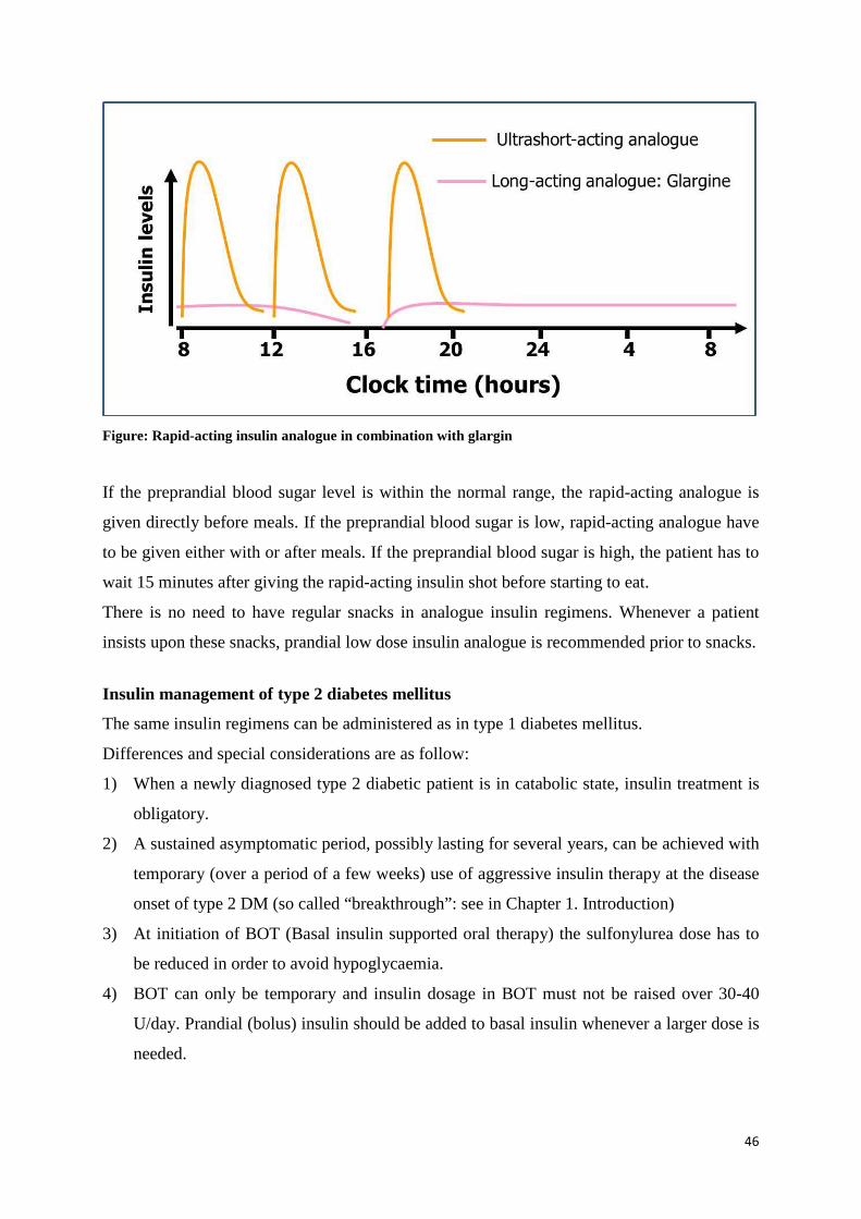
46
Figure: Rapid-acting insulin analogue in combination with glargin
If the preprandial blood sugar level is within the normal range, the rapid-acting analogue is
given directly before meals. If the preprandial blood sugar is low, rapid-acting analogue have
to be given either with or after meals. If the preprandial blood sugar is high, the patient has to
wait 15 minutes after giving the rapid-acting insulin shot before starting to eat.
There is no need to have regular snacks in analogue insulin regimens. Whenever a patient
insists upon these snacks, prandial low dose insulin analogue is recommended prior to snacks.
Insulin management of type 2 diabetes mellitus
The same insulin regimens can be administered as in type 1 diabetes mellitus.
Differences and special considerations are as follow:
1) When a newly diagnosed type 2 diabetic patient is in catabolic state, insulin treatment is
obligatory.
2) A sustained asymptomatic period, possibly lasting for several years, can be achieved with
temporary (over a period of a few weeks) use of aggressive insulin therapy at the disease
onset of type 2 DM (so called “breakthrough”: see in Chapter 1. Introduction)
3) At initiation of BOT (Basal insulin supported oral therapy) the sulfonylurea dose has to
be reduced in order to avoid hypoglycaemia.
4) BOT can only be temporary and insulin dosage in BOT must not be raised over 30-40
U/day. Prandial (bolus) insulin should be added to basal insulin whenever a larger dose is
needed.
47
5) BOT is usually used for 3-4 years. It is not sufficient for maintaining adequate glycaemic
control after that period.
6) In human ICT (Intensive Conservative Insulin Therapy) a single daily intermediate shot
in the evening is usually sufficient, because endogenous insulin production is maintained
at the early period of type 2 DM.
7) When detemir insulin is used in ICT, it is enough to be given once a day in the evening.
In this case weight gain also usually decreases.
8) Metformin should be continued after the patient starts insulin therapy because it reduces
cardiovascular- and cancer risk and is also associated with decreased weight gain and a
lower insulin dosage.
9) The ideal dosage ratio between a rapid-acting (prandial) and long-acting insulin dose is
approximately 50-50% in analogous regimens of type 2 diabetic patients
10) Analogue insulin treatment improves the quality of life and reduces the risk of
hypoglycaemia.
11) PPT (Prandial Premix Insulin Therapy) might also be a therapeutic option when tight
glycaemic control is aimed for, but for some reason ICT cannot be accomplished. In a
PPT regimen a biphasic (premix) insulin analogue is given before breakfast, dinner and
supper.

48
Chapter 8. Insulin pump therapy Dr. Gergő A. Molnár
With intensified insulin therapy, the insulin may be administered via a special insulin
administering pen or via an insulin pump.
The insulin needs of our body can be divided into two larger parts. On the one hand
one requires insulin to prevent blood glucose elevations resulting from meals, while on the
other we also need insulin independent of meals, for instance to inhibit gluconeogenesis of
the liver. The former is provided by bolus insulin, the latter by basal insulin.
The insulin pump system consists of several major parts: the pump itself, the
subcutaneous inserted Teflon catheter and the infusion set connecting the two (Figure).
Figure: Shematic representation of the structure of the insulin pump. Modified after Valla et al, Experimental Diabetes Research (2010), Article ID 178372, 14 pages, doi:10.1155/2010/178372 (Copyright © 2010 Vasiliki Valla, Creative Commons Licence)
The insulin pump itself is a device that is able to provide continuous, slow infusion of insulin,
in this way covering basal insulin need; it is also capable of administering the bolus insulin
doses. For that, a rod driven by small electromotor pumps insulin out of the insulin-filled
storage syringe inserted into the device.
Insertion devices may provide help in the setting of the canulla, by the help of which
the needle can be inserted quickly and in a relative painless way. Upon filling and assembly of
the system the most important thing is airtight sealing and avoidance of air bubbles.
49
The basal insulin is administered upon a pre-specified program, the setting enabling
the speed of the pump to be altered every hour in 0.05-0.10 units/hour steps. Once the
program is set, the device doses the basal insulin automatically, without external influence.
It is also possible to pre-program different basal profiles, e.g. for daytime and night-
time shifts; for weekdays and weekends; for the period before or during menstruation or for
days with and without exercise.
Moreover, it is also possible to change the basal rate temporarily, for couple of hours,
e.g. to raise it transiently up by 100-120-130% or to decrease it to 90-80-70%... in cases of
prolonged hypoglycaemia or for the periods of intense activity, such as sport.
The bolus doses have to be set by the patient upon the carbohydrates being consumed
or prior to consumption, the time of the day, the type of meals etc. The pre-specified amount
of bolus insulin is administered upon pressing a knob; that is, this also does not require a
separate needle stitch.
The advantages of insulin pump therapy include the possibility of a very fine
regulation of insulin doses, which has a special significance especially in cases where there is
a very small insulin need, let's consider a small child with a daily insulin need of 10 units,
which has to be divided into basal insulin and into bolus insulin administered alongside
meals. This procedure can be carried out in a more even and a better regulable way. Besides
that, because of the fine, hourly setting, the transient changes to the base amount, and the
possibility of providing different basal profiles, the glucose excursions can be lowered more
efficiently than with insulin administered via a pen.
Moreover, basal insulin administered by a pen will act even when it is not needed, so
(e.g.) on occasions when unplanned physical activity takes place after the administration of a
large dose of basal insulin even severe hypoglycaemia can develop. When using the pump,
hypoglycaemia can be prevented by transiently decreasing or even stopping the administration
of the basal insulin in such a case.
The insulin pump can also help in the prevention of the dawn phenomenon, meaning
that high fasting blood glucose can be lowered.
A “carbohydrate-free day” can assist in setting the proper basal rate, as then the
patient consumes only meals containing no or only a small amount of carbohydrates, and
measures their blood glucose frequently (hourly - two hourly); using this technique the blood
glucose-influencing effect of meals and bolus insulin can be ruled out, and we can really gain
an hour-by-hour picture of whether and where the basal insulin rate should be increased or
decreased.

50
The disadvantage of the therapy is that the patient is bound to a device, has to wear the
pump constantly, and due to the nature of the device there is the chance of a technical failure.
Based on the abovementioned, it is obvious that the pump therapy is recommended
mainly to diabetic patients who are well educated, is familiar with the disease, aware of
carbohydrate counting, insulin kinetics, the signs and proper correction of hypoglycaemia etc.
In cases of patients with bad glycaemic control and poor compliance the insulin pump would
not provide a good result. Conversely, in cases where the patient has been carefully selected
and in the presence of proper indications, the number of hypo- and hyperglycemias can
decrease, and carbohydrate metabolism, e.g. the value of HbA1c can be expected to decrease.
The current direction in insulin pump development is that the device should be able to
communicate with a continuous glucose sensor and to dose insulin automatically upon the
actual and previous glucose values, with minimal or no external influence. These devices,
functioning in part on a negative feedback are called 'closed loop' insulin pumps. A “bionic
pancreas” also exists, where two pumps can dose insulin and (to prevent hypoglycaemia)
glucagon independently of each other. These devices are currently in the phase of clinical
development.
Real indications of insulin therapy in type 1 diabetes in adults are:
- diabetes duration of at least 3 years
- a repeated HbA1c value of >7.0%
- in cases of praeconceptional therapy >6.5%, or
- large glucose variability (>=10.0 mmol/l), or
- dawn phenomenon which can be verified (fasting morning glucose repeatedly >8.0
mmol/l), or
- min. 3 times/month clinically overt hypoglycaemia, or
- documented hypoglycaemia-unawareness, or
- severe hypoglycaemia (blood glucose <3.0 mmol/l) developing min. once in 6 months
The insulin pump therapy is not recommended:
- in cases of severe disability (problems at sensing, absence of basic hand skills, coagulation disorders etc.), - in cases of psychic resistance: denial of diabetes, inability or rejection of self-monitoring, - poor or insufficient compliance between patient and the treatment team, - in cases of accompanying severe psychiatric disorders, - instability of social support (family, friends), in cases of illiteracy.

51
Chapter 9. Pancreas-kidney transplantation
Dr. Tibor Kovács
In type 1 diabetes the replacement of destroyed beta cells can be managed by the
transplantation of the entire pancreas (I) or isolated beta islet cells (II.). Although the latter
method is still rather at the experimental stage, its results are not particularly impressive. In
type 2 diabetes, beta cell exhaustion and destruction are secondary processes, and so
pancreas transplantation is not an option in 2DM. In obese type 1 diabetic patients, the
efficacy of pancreas transplantation is also problematic due to insulin resistance (especially if
the daily insulin requirement exceeds 60 IU).
I. Entire pancreas transplants (from cadaver donors) are today used in 3 different
ways (distribution of operations in US are indicated):
1. With type 1 diabetic patients with end-stage kidney failure, 75 % of pancreas
transplants are implanted at the same time as cadaver renal transplants
(simultaneous pancreas kidney – SPK). The advantage here is that both
entities will be implanted in one surgery, and foreign HLA antigens are
identical. The one-year graft survival is 86% and the 10-years graft survival is
54%.
2. 15% of pancreas transplants are implanted into patients with a renal transplant
(pancreas after kidney - PAK ). Disadvantage: 2 surgery; where there is a
stable graft function the immunosuppressive treatment must be reintensified
and the organism has to encounter two different alien HLA antigens. This
method may be important for patients with a living donated renal transplant.
The one-year graft survival is 79% and the ten-years graft survival is 29%
3. Pancreas transplantation into patients with a proper renal function: it is
indicated by recurring life-threatening hypoglycaemic episodes and is applied
in Brittle diabetes (pancreas transplantation alone - PTA) Disadvantage:
requires immunosuppressive therapy. One-year graft survival of 80% and the
10-years graft survival is 27%.
Pancreas transplantation (PAK, PTA) is not yet available in Hungary for the latter two
reasons.

52
Based on literature data, the survival rate of transplanted pancreases is highest in cases of
simultaneous pancreas-kidney transplantation. In other countries, SPK is the most commonly
carried out method of pancreas transplantation.
Benefits of pancreas-kidney transplantation performed in type 1 diabetic patients with chronic renal failure:
1. improvement to quality of life 2. termination of exogenous insulin therapy claims 3. normalization of carbohydrate metabolism, HbA1c 4. dietary freedom 5. With late diabetic complications, stabilization or possible improvement, but
established retinopathy /blindness or severe neuropathy are already irreversible processes. The progression of these alterations slows down or may stop.
In successful pancreas-kidney transplantations – due to the normalization of carbohydrate
metabolism – the repeated development of diabetic nephropathy rarely occurs as compared to
those who have only undergone kidney transplant. In the latter cases, diabetic nephropathy is
expected to re-occur after 5-10 years after transplantation in the transplanted kidney. There
are data about the cardioprotective role of the normoglycaemic state in patients with SPK.
The decrease in the left ventricular mass and slowdown of coronary calcification are more
significant in patients with SPK compared to that in diabetic patients with a kidney graft only.
A description of the surgical implantation of the pancreas exceeds the scope of this
note, but it should not be ignored that the exocrine secretion of the transplanted organ can
also be provided (flow of pancreatic juice into intestine).
The immunosuppressive treatment of patients with a pancreas graft is no different
from patients who have undergone only kidney transplantation.
II. Isolated pancreatic cell transplantation could be beneficial in that it places a much
smaller surgical strain on the patient and there is no need to provide the exocrine
pancreatic function. The islet cells are isolated from the cadaver pancreas using
special techniques for tissue digestion. The islet cells are infused into the recipients
after the puncture of the portal vein transhepatically using the micro-invasive
technique. Up to now the efficiency of this method has been poor, more than 90%
of the patients treated in this way needing insulin therapy a year after the
procedure.
53
Chapter 10. Diabetes care and education
Dr. Gábor Fülöp
Since diabetes is a chronic disease which, if glycaemic state is uncontrolled, may lead
to serious complications such as early disability and death, continuous medical care and
education for individuals with diabetes is fundamental.
Regular care and therapy revision or modification is necessary in both type 1 and type 2
diabetes. Type 1 diabetes requires this either because intensive insulin therapy is used or
because absolute insulin deficiency may lead to unstable glucose control, while in type 2
diabetes ongoing worsening blood glucose is anticipated due to the progressive nature of the
disease. Individuals with diabetes need continuous and complex medical care. Between the
medical visits continuous patient self management is required.
A chronic care model in diabetes as a complex process includes prevention, screening
and diagnostic actions, classification, therapy, regular controls, education and rehabilitation.
There is a coordinated partnership between health care systems and patients in
diabetes care, each having their rights and responsibilities. High-quality care is based on
evidence-based clinical guidelines but it requires an individual adaptation to the patient.
The chronic care model includes 6 core components: (www.improvingchroniccare.org):
1. Delivery system design (moving to proactive care where planned visits are
coordinated)
2. Self management support
3. Decision support
4. Clinical information systems
5. Community resources and policies (to support healthy lifestyle or smoking
cessation programs)
6. Health systems (to create quality oriented culture)
There has been a steady improvement in the proportion of diabetic patients achieving
recommended HbA1c, blood pressure and LDL-cholesterol in the last 10 years.
The average HbA1c was 7. 6% in 1999-2002 whereas it declined to 7. 2% in 2007-2010. (9).
However, 33-49% of diabetic patients do not meet target levels for HbA1c, blood
pressure or cholesterol and only 14% of them achieve all three targets and non-smoking
status.

54
Diabetes self-management education (DSME) is an integral element of diabetes care
since diabetes management is primarily accomplished by the patients (or their family
members), and depends upon how their blood glucose is self-monitored, their meals, physical
activity and living conditions under the guidance of heath care professionals. Patients with
diabetes need regular education and practice to achieve and maintain this knowledge and
related skills. Motivation of patients can be maintained or raised by achieving previously-set
goals or by providing psychosocial support.
Basic education includes principles of lifestyle therapy (diet and physical activity), use
of diabetic equipment and supplies (insulin pens, blood glucose monitors, insulin pump) and
recognition, prevention and treatment of possible complications (especially hypoglycaemia).
Diabetes care is teamwork. Essentially, most patients with diabetes should be treated
by the primary care physician and a specialist consultant is needed only in case of therapy
modifications, complications, or other emerging problems. Patients with type 1 diabetes or
type 2 diabetes with complications should rather be cared by specialist in collaboration with
primary care physician.
A collaborative multidisciplinary integrated team may include a diabetologist and
physicians from related specialities such as ophthalmology, angiology, vascular surgery,
neurology, cardiology, and nephrology, in addition to nurses, physician assistants, dietitians,
diabetes educators, mental health professionals, podiatrists and so on.
Recommendations for diabetes care and patient education are provided by the National
Diabetes Education Study Group (www.betterdiabetescare.nih.gov), in the US and by the
Diabetes Education Study Group (DESG) (www.desg.org) of EASD in Europe.
High-quality education (Diabetes self-management education, DSME) has been shown
to improve patient self management, satisfaction and glucose control.
Taking the holistic view, the management of patients with diabetes includes not only
glucose control but also the prevention and recognition of cardiovascular risk factors and
other comorbid conditions.
Diabetes management concludes possible maintenance of adequate glucose control
performed by proactive care. This can be achieved by regular monitoring, and it is
recommended that HbA1C is determined quarterly. Target goals should be defined
individually, but within the ranges of 6-8%. Blood lipid controls are recommended yearly,
although more frequent controls may be necessary in patients with dyslipidaemia or on
atilipaemic treatment.

55
Patient Education
Education is the cornerstone of diabetes care and prevention for patients with diabetes
as well as for health care professionals and even the population at large.
Unless adequate education is provided, patients with diabetes and their family members
cannot cope with the burden of the disease and with the essential changes in life style.
Acquisition of the essential knowledge related to one’s disease is the right and
obligation of all individuals with diabetes.
Therapeutic patient education as an “added value” associated with patient education can
be laid out as follows:
• As a result of ongoing education and study integrated in the health care system the
patient will be able to manage his or her disease in an optimal way.
• The patient-oriented education approach includes screening of individuals with
diabetes, provision of adequate information, learning of self-monitoring, psychosocial
support, medical guidelines and organizing hospital and out-patient care. It also
provides information about health-and-illness behaviour and outlines the possible
prospects of rehabilitation.
Successful patient education requires ongoing self-instruction together with retraining
from the instructors.
The most effective diabetes education is delivered either individually or in small groups.
Education which is matched to the patient’s current knowledge and experience is
interactive and involves both verbal and visual means (pictures, brochures, video films, even
educational learning games - for instance, how to choose an appropriate diet). For further
comprehension other written materials such as dietary booklets, brochures about foot care,
diet- or hypoglycaemia diaries or web pages, prints and books can be recommended.
Repetition and asking for confirmation (“loop technique) are fundamental in patient
education.
An adequate adaptation of lifestyle interventions is an essential element of well
controlled diabetes. Adequate lifestyle interventions include medical nutrition therapy and
regular (in many cases also controlled) physical activity, which have to be coordinated with
each other and also with diabetic pharmacotherapy. All this knowledge and accompanying
skills have to be learned during the process of therapeutic patient education. Ideally,
therapeutic patient education should be based on healthy literacy which has already begun to

56
be acquired at school. One of the important missions of the health care system is related to
this: namely, to provide information and health education to a wide range of populations.

57
Chapter 11. Self-monitoring of blood glucose, continuous glucose monitoring
Dr. Gergő A. Molnár We also have to evaluate the efficacy of treatment of patients suffering from diabetes
mellitus (DM). For that, there are a variety of options, for example laboratory tests such as the
haemoglobin A1c (HbA1c) or fructosamine level, where the former provides information on
average blood glucose levels of the last 3 months, the latter about the last 2 weeks. HbA1c
indicates glycation of haemoglobin of red blood cells, while fructosamine indicates glycation
of plasma proteins (mainly albumin).
Each method requires blood drawing and a laboratory background, and has its own
problems: the HbA1c value is dependant both on the average blood glucose value and on e.g.
the lifespan of the red blood cells, and thus may be influenced by anaemia, blood transfusion,
or bone marrow disease. Similarly, in the case of fructosamine, the turnover time of plasma
proteins may be a disturbing factor, and so the obtained value may underestimate the average
blood glucose level in cases of marked proteinuria or hyperthyroidism.
The combined evaluation of HbA1c and fructosamine may provide information on the
temporal changes of blood glucose values, as we find out what the average blood glucose
value was in the preceding 3 months, and within that period, in the last 2 weeks. A less
elevated fructosamine besides a high HbA1c may therefore indicate that the patient’s recent
glycaemic control was inferior to what it had been in the past. On the other hand, a high
fructosamine value with an acceptable HbA1c may signify that the patient has experienced
worse glycaemic control in recent weeks or days, due perhaps to a current, severe infection.
When evaluating HbA1c and fructosamine in combination, a discrepancy between the
results may also draw our attention to the possibility that a confounding factor (e.g. anaemia,
proteinuria, hyperthyroidism) should be sought.
A further problem with HbA1c and fructosamine is that they only show the average
blood glucose level, not the fluctuations. Thus, a patient with a completely properly controlled
diabetes can have an HbA1c value similar to that of a patient under bad control and
experiencing both high blood glucose values and frequent hypoglycaemias.
The self-monitoring of blood glucose may have an important role in detecting blood
glucose fluctuations. When taking the case history of a patient, whether in the outpatient unit
or on the ward, one also has to establish if the patient has a glucometer at home, and if he
58
regularly controls his blood glucose values. If so, how frequently and at what times of day
does he measure his blood sugar?
Multiple daily blood glucose measurement is also a useful tool in finding the right way
to control a patient on multiple daily insulin injection therapy. In our practice, we usually
suggest pairwise measurements, prior to and after meals, where the postprandial value is
measured 1.5 hours after the meals; for this, target values are provided by guidelines. We
usually also ask for daily profiles of six or more measurement points. For example, if the aim
is to differentiate Somogyi effect from dawn phenomenon resulting from high morning
fasting glucose values, night-time measurement is also suggested (depending on the time of
administration of intermediate insulin, at midnight or at 3 a.m. Clinical studies have also
verified that HbA1c can change in a more beneficial way in patients who perform self-
monitoring in combination with insulin dose adjustments.
Patients can keep the recorded values in the form of a glucose 'diary', but with many
glucometers it is now possible to upload the values onto a computer or via a computer onto
the internet, where we may obtain, besides tendencies, statistical evaluation (mean, standard
deviation).
If we desire a more detailed daily glucose profile, there is also the possibility of
continuous glucose monitoring (CGM). CGM systems usually comprise a sensor, a
transmitting system and a monitoring device. Sensors may be minimally invasive, obtaining a
signal from the interstitium, for instance via implantable glucose electrodes, using sensors
based on microdialysis or microperfusion. A non-invasive measurement is enabled by sensors
based on microporation, or transdermal or optical sensors.
As regards the transmission of the signal, besides wired devices, increasingly
frequently today wireless devices are also available, and the miniaturisation of monitors
enables devices to be directly connected to the sensor (mounted on the sensor).
The monitor is able to store the signals, and in some cases already converts them into
glucose values. Some monitor systems are able to work in real-time mode, thus displaying
actual values, graphs and trends to the patient. Another type of system does not provide
results during monitoring, the information only being given to the patient and doctor after
downloading the measurements onto a computer.
A major advantage of glucose monitors is that they show glucose fluctuations in a
more sensitive way than do values obtained from the self-monitoring of blood glucose
(SMBG, see Figure):
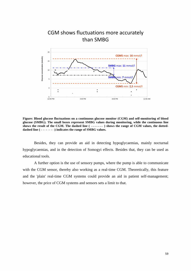
59
-5
0
5
10
15
20
12:00 PM 4:00 PM 8:00 PM 12:00 AM
Glu
cose
conc
entr
atio
n(m
mol
/L)
CGMS max: 16 mmol/l
CGMS min: 2,5 mmol/l
SMBG max: 11 mmol/l
SMBG min: 7 mmol/l
CGM shows fluctuations more accurately
than SMBG
Figure: Blood glucose fluctuations on a continuous glucose monitor (CGM) and self-monitoring of blood glucose (SMBG). The small boxes represent SMBG values during monitoring, while the continuous line shows the result of the CGM. The dashed line ( ) shows the range of CGM values, the dotted-dashed line ( ) indicates the range of SMBG values.
Besides, they can provide an aid in detecting hypoglycaemias, mainly nocturnal
hypoglycaemias, and in the detection of Somogyi effects. Besides that, they can be used as
educational tools.
A further option is the use of sensory pumps, where the pump is able to communicate
with the CGM sensor, thereby also working as a real-time CGM. Theoretically, this feature
and the 'plain' real-time CGM systems could provide an aid in patient self-management;
however, the price of CGM systems and sensors sets a limit to that.

60
Chapter 12. Acute complications of diabetes mellitus and its management
Dr. István Wittmann
Hypoglycaemia and its treatment
Hypoglycaemia is characterised by abnormally low blood glucose concentration. The
threshold glucose level is usually 3 mmol/l but the glucose level at which an individual
becomes symptomatic is highly variable. Symptoms of hypoglycaemia can typically occur
with a plasma glucose concentration of greater than 3 mmol/l, or can be absent with lower
plasma glucose concentrations (especially after long-term diabetes and in diabetic
neuropathy).
Hypoglycaemia can be mild, moderate or severe (if the patient requires assistance of
another person). The symptoms and signs of hypoglycaemia are as follow (Table):
Table: Signs and symptoms of hypoglycemia
Autonomic Neuroglycopenic General
Sweating Confusion Nausea
Palpitations Difficulty in speaking Headache
Trembling Dizziness Lack of coordination
Hunger Atypical behaviour
Vision changes
Perioral paraesthesia
Causes of hypoglycaemia (common):
1) Insulin treatment
2) Oral antidiabetic agents:
i) Sulphonylurea
ii) Glinides (prandial glucose regulators)
iii) Incretin therapy ( seldom)
3) Advanced age (>75 years)
4) Alcohol
5) Kidney failure
6) Liver disease

61
7) Vigorous physical activity (without adjusting medications and diet)
8) Drug interactions (at the level of albumin binding and metabolism)
9) Drugs that mask symptoms and signs of hypoglycaemia and which inhibit
counterregulation (e.g. beta blockers)
10) Inadequate diet
11) Lack of knowledge on the part of the patient (inappropriate patient education)
Mild and moderate hypoglycaemia can be treated by the patient. Ingestion of quick-
acting carbohydrates (simple carbohydrates or 15-20 g glucose, either in liquid or in
granulated form) is recommended, followed by the intake of long-acting complex
carbohydrates. After 15 minutes, it is recommended that blood glucose is measured. If
hypoglycaemia continues, the treatment should be repeated.
Hypoglycaemia may be extremely frightening to some patients with diabetes. These
patients ingest glucose after the detection of the mildest hypoglycaemia, causing reactive
hyperglycaemia. When the possibility of this reactive hyperglycaemia is not taken into
account, the consequence may be an increase in insulin (or other antidiabetic) dosage and a
“vicious circle” of hypoglycaemias and hyperglycemias may develop. Careful assessment of
SMBG data is required in order to recognise and avoid this.
In severe hypoglycaemia, oral glucose intake has to be avoided due to the risk of
aspiration. Intravenous glucose administration is required and hospitalisation may also be
necessary. After relatively long-lasting hypoglycaemia, elderly patients may only regain their
consciousness slowly, even with administration i.v. glucose infusion, thus frequent blood
sugar measurements are required.
Glucagon should be prescribed for patients with type 1 diabetes and family members
of these individuals should be instructed on its administration.
If hypoglycaemia is caused by sulfonylurea, especially in patients with renal
impairment, prolonged or recurrent hypoglycaemia can be expected and so at least 48-72
hours of observation/hospitalisation and ongoing glucose infusion may be required. This
scenario usually occurs in elderly patients treated with sulfonylurea and suffering from acute
prerenal kidney injury and thus GFR decline due to intercurrent infections (diarrhoea, fever
e.t.c) and/or inadequate fluid intake.
It is important to remember that a relatively significant number of hypoglycaemias,
especially the nocturnal ones, remain asymptomatic and thus unrecognised. Another
important point is that the recurrence rate is high after a severe hypoglycaemia, hence patients

62
may benefit from more permissive glycemic targets for a few weeks. Hypoglycemia in
patients on alfa-glucosidase inhibitor therapy can be treated only with glucose, since this type
of treatment inhibits degradation of di- and polysaccharides.
Diabetic ketoacidosis (DKA), and Hyperosmolar Hyperglycemic State (HHS) and their
treatment
DKA happens predominantly in patients with type 1 diabetes. It may occur in milder
form combined with hyperosmolar hyperglycaemic state in patients with type 2 diabetes,
especially after long-lasting diabetes when endogen insulin secretion is significantly
decreased. The clinical picture of both acute metabolic complications is dominated by
dehydration and complicated by ion disturbances. Complaints and signs of patients are
polyuria, polydipsia hypotension, tachycardia, prerenal acut kidney injury, muscle cramps and
abnormal mental status. Signs and symptoms of possible underlying intercurrent infection or
thromboembolic complications are also common. Patients with DKA often develop
Kussmaul’s breathing, and abdominal pain can also occur.
Both of these metabolic derangements are serious, life threatening conditions requiring
hospitalisation.
The most common precipitating factors are as follows:
1) Infections
2) Cardiovascular events
i) Myocardial infarction
ii) Stroke
3) Gastrointestinal diseases
i) Vomiting
ii) Diarrhoea
4) Iatrogen factors, Drugs
i) Diuretic abuse
ii) Corticosteroids
5) Poor- or non-compliance on the part of the patient
i) Inadequate diet
ii) Discontinuation of or inadequate insulin /oral antidiabetic therapy
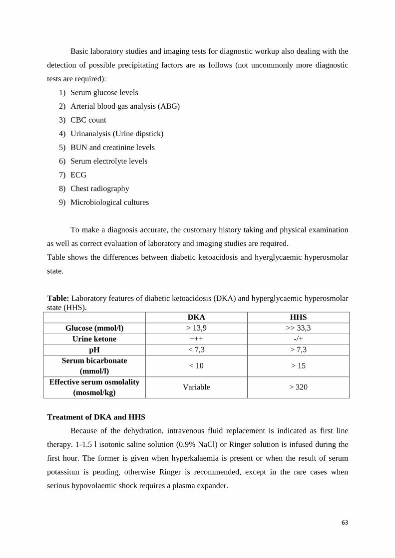
63
Basic laboratory studies and imaging tests for diagnostic workup also dealing with the
detection of possible precipitating factors are as follows (not uncommonly more diagnostic
tests are required):
1) Serum glucose levels
2) Arterial blood gas analysis (ABG)
3) CBC count
4) Urinanalysis (Urine dipstick)
5) BUN and creatinine levels
6) Serum electrolyte levels
7) ECG
8) Chest radiography
9) Microbiological cultures
To make a diagnosis accurate, the customary history taking and physical examination
as well as correct evaluation of laboratory and imaging studies are required.
Table shows the differences between diabetic ketoacidosis and hyerglycaemic hyperosmolar
state.
Table: Laboratory features of diabetic ketoacidosis (DKA) and hyperglycaemic hyperosmolar state (HHS). DKA HHS
Glucose (mmol/l) > 13,9 >> 33,3 Urine ketone +++ -/+
pH < 7,3 > 7,3 Serum bicarbonate
(mmol/l) < 10 > 15
Effective serum osmolality (mosmol/kg)
Variable > 320
Treatment of DKA and HHS
Because of the dehydration, intravenous fluid replacement is indicated as first line
therapy. 1-1.5 l isotonic saline solution (0.9% NaCl) or Ringer solution is infused during the
first hour. The former is given when hyperkalaemia is present or when the result of serum
potassium is pending, otherwise Ringer is recommended, except in the rare cases when
serious hypovolaemic shock requires a plasma expander.
64
Intravenous treatment should be continued until the hyperglycaemic crisis is resolved and the
patient’s condition stabilises.
In severe mental status alteration or coma airway management, intravenous access, bladder
catheterisation, initiation of antibiotic and antithrombotic medications are required; and
admission to an intensive care unit is necessary.
Insulin therapy should be initiated after the first hour of treatment because early
insulin administration may be associated with hypoglycaemia after circulation is restored due
to the sudden rise in the insulin level. The insulin infusion dose has to be adjusted to the
actual blood glucose level. It is usually recommended that an insulin pump be used (at a rate
of 0.1 U/kg/h), but a mixture of short–acting insulin and isotonic sodium chloride solution
mixture can also be used.
In DKA, blood glucose concentration should be maintained between 8-11 mmol/l, although it
has been recommended by some that a higher level up to 14 mmol/l may also be reasonable as
“ketone bodies burn in the fire of carbohydrates”.
Intravenous insulin treatment is typically associated with a decrease in potassium
concentration, thus potassium should be rigorously monitored and replaced if necessary.
A successful treatment requires correction of electrolyte imbalances along with
rehydration therapy. Potassium correction has to be initiated in the first hour of the treatment,
followed by management of magnesium and phosphate disturbances.
The following laboratory parameters should be monitored every 2-4 hours, depending
on the patient’s condition and the severity of laboratory disturbance.
1) Serum elecrolytes (Na+, K+)
2) Serum glucose
3) Arterial blood gas analysis (ABG)
4) Urine ketone
5) Serum creatinine
Bicarbonate is only infused if severe, life-threatening acidosis, where pH<7.0, is
present; and after initiated it is only indicated until the pH is 7.0-7.1. Bicarbonate treatment
has been correlated with cerebral edema; thus, if the pH is 7.0 or higher, no bicarbonate
therapy is recommended. During bicarbonate infusion strict potassium control is required
because bicarbonate administration leads to hypokalaemia.

65
Following the resolution to the hyperglycemic crisis and successful elimination of the
predisposing factor (such as infection), transition to previous antidiabetic therapy can be
initiated with an overlap of 4-8 hours of parenteral therapy.

66
Chapter 13. Cardiovascular complications in diabetes mellitus
Dr. Gergő A. Molnár
Diabetes mellitus (DM) is a population disease, which exerts a heavy financial load on
developed societies, not only directly through treatment of the disease itself, but also through
treatment of its complications. Of the latter, cardiovascular (CV) diseases are of special
importance. While a couple of decades ago, diabetes-related death was mainly in connection
with acute complications, nowadays it is rather related to long-term macrovascular (mainly
CV) and microvascular complications and malignancies. In DM, on the one hand diabetes
itself, while on the other hand, other accompanying factors (e.g. hypertension, dyslipidaemia,
obesity, renal failure) represent a risk factor for the development of CV diseases. In diabetes,
the function and/or structure of the blood vessel wall can be damaged among other ways via
hyperglycaemia, the polyol pathway, advanced glycation end-products, inflammatory
cytokines and free radicals.
In characterizing diabetes we frequently regard the CV-risk as a high risk, as if the patient
had already suffered a myocardial infarct. Accordingly, the CV-risk of patients with diabetes
is approx. 2-4 times as high as in the non-diabetic population.
The effect of the treatment of diabetes mellitus on CV risk in general
On planning the therapy of a patient, one has to consider differences between patients
with type 1 and type 2 diabetes mellitus. Other risk factors, found in type 2 diabetes mellitus
and accompanying the disease, also have to be taken into consideration. We believe that
glycaemic control is of fundamental importance; however, its significance is different in type
1 or type 2 diabetes. Based on data from the literature on large studies, such as the DCCT
trial, a milestone study dealing with type 1 diabetic patients, in the original observation period
of 10 years, the reduction of macrovascular risk was not significant, although during the
extension of the study to a further 20 years of follow-up, the DCCT-EDIC trial showed a clear
advantage of prior intensive treatment with regard to CV risk. It also raised the possibility of a
'metabolic memory'. This would mean that prolonged good or bad glycaemic control may
have an impact on CV risk even years later, i.e. the body 'remembers' its state of 10 years
earlier.
In patients with type 2 diabetes, in the 15 years of observation in the classical UKPDS
study, with regard to macrovascular endpoints no overall significant difference could be

67
observed between patients treated in an intensive and in a non-intensive manner, although
tendencies seemed to favour intensive therapy. The extended, 25-year follow-up of the
UKPDS has however shown a significantly better survival rate regarding macrovascular
complications with the original intensive treatment. Based on that, we believe that glycaemic
control is important in decreasing CV risk.
Several large studies over recent years have raised the possibility that too tight a
glycaemic control may lead not to a decrease, but rather an increase in CV risk, mainly in
elder patients. Taking this into consideration, today we do not recommend a uniform HbA1c
target value, but rather a target range tailored individually to age and accompanying diseases
for the treatment.
We believe that complex therapy of the patients is important, i.e. lifestyle changes,
application of glycaemic control and treatment of other accompanying risk factors.
„Silent ischemia”
We should not forget about a special characteristic of patients suffering from diabetes
and coronary heart disease (CHD), namely the 'silent ischemia', where patients may not be
aware of pain related to ischemia as a consequence of neuropathy. Because of that, screening
of high risk, asymptomatic patients may be important above the age of 50 or in the presence
of other risk factors. A part of the assessment of cardiovascular risk is blood pressure
measurement on each visit by the patient, added to which an ECG, lipid-control measurement
of the ankle-brachial index should be performed at least annually.
Types of antidiabetics and cardiovascular risk
Besides the target range of antidiabetic treatment, the choice of antidiabetic type to be used
also influences CV risk. Let us observe that in greater detail:
Sulphonylureas
When antidiabetics, i.e. sulphonylureas, are used for the longest time one important
consideration is pancreas selectivity, as sulphonylurea receptors can be found not only in
beta-cells of the pancreas, but also on cardiomyocytes and cells of the blood vessel walls. For
this reason, non-cardioselective sulphonylureas may for instance worsen ischemic
preconditioning, thereby increasing the likelihood of mortality. They may moreover also
worsen CV risk via hypoglycaemia-triggered arrythmias. Therefore, on a theoretical basis it is
important that we should choose a cardioselective sulphonylurea that leads to less
68
hypoglycaemia, and which has a more stable effect. Upon meta-analysis, sulphonylureas have
been associated with higher CV risk as compared to metformin, the only exception being
gliclazide in patients with a previous CV-event, as well as in patients without a prior CV
event. Taking the abovementioned into consideration, a differentiated sulphonylurea therapy
is preferable where medication is selected on the basis of its favourable and non-favourable
properties.
Metformin
In the extension of UKPDS, metformin has been more efficacious than conventional
therapy as regards CV risk. The results of meta-analyses are not entirely consistent: the data
from many studies have shown metformin to be more effective than other antidiabetics, but a
smaller portion of the studies has been unable verify this (although disadvantageous effects
could not be observed here, either).
Acarbose
In a study carried out using acarbose, the use of acarbose has led to significantly lower
CV risk than placebo, however, due to the small number of CVevents, the result is of
restricted value. The result of one meta-analysis indicated that acarbose was CV-protective;
here the statistical power was already appropriate.
Thiazolidine-diones (glitazones)
The CV safety of antidiabetics is of especial interest, since in 2007, according to the
data from a meta-analysis, the long marketed rosiglitazon was shown to increase CV risk. The
meta-analysis was highly criticized by many; however, precisely because of this potential CV
risk, the marketing of the substance was limited in many countries, while in others its use was
temporarily cancelled. As a result, drug authorities have set tight CV safety criteria. However,
data concerning rosiglitazone are not unambiguous; and so we do not consider its use as safe.
According to several studies, pioglitazon is safer than rosiglitazon, something underlined by
prospective double-blind studies, as well as by meta-analyses.
Glinides (postprandial glucose regulators)
According to studies and retrospective analyses, this group of medications can be
regarded as neutral from the aspect of CV.
69
DPP-4 inhibitors
Results of for the most part retrospective analyses of non-CV-endpoint designed,
efficacy studies now suggest a CV safety regarding both the group and the individual
substances. In the study carried out using saxagliptin, saxagliptin turned out to be neutral in
general; however, it increased the risk of heart failure. The study carried out using alogliptin
provided a neutral result. We await new results of these agents and also results of CV-targeted
studies from other agents from this group. Sitagliptin proved to be CV-neutral.
GLP-1-receptor agonists
For GLP-1 receptor agonists, results of both retrospective analyses and retrospective
meta-analyses of non-cardiovascular designed studies are available, based upon which this
group can be regarded as cardiovascularly safe. Results of prospective, targeted CV studies
are also awaited for this group.
SGLT-2 inhibitors (gliflozins)
Few data are also available regarding gliflozins, as they have only been marketed and
appearing in clinical studies for a short time. Given the CV safety data needed for their
approval, these agents can be regarded as safe overall from the CV aspect, and they have not
led to any statistically significant increase in risk of individual complications. Moreover,
empagliflozin decreased the risk for CV-death.
Insulin
In a Swedish registry study related to insulin, insulin – mainly in combination with
sulphonylureas – led to an increase in CV risk that could be attenuated by the addition of
metformin. According to a Danish meta-analysis, in the comparison of insulin monotherapy
vs. insulin + metformin, a non-significant increase in CV-risk was found; however, the
statistical value of this study can be strongly called into questiondue to the low number (2 vs.
4) of events.
In conclusion, we can say that correct glycemic control is protective, but overtight
metabolic control may be risky, especially in the case of patients of higher age and established
vascular disease. For this reason, a target range, and within that an individual target value
based on age and co-morbidities is recommended. Of the antidiabetics, metformin and
empagliflozin can be regarded as safe from a CV point of view, and may be beneficial not
only in lowering insulin-relaetd increased risk of malignancies, but also in lowering potential
70
insulin-related CV risk. Therefore, in type 2 diabetes mellitus – unless contraindicated
because of accompanying diseases – we may recommend the furthre use of metformin in
combination with insulin therapy. The sulphonylureas are not beneficial from the aspect of
CV, and a differentiated use may be suggested. Acarbose seems to be cardiovascularly
protective from the CV point of view. Of the thiazolidine-diones, the CV safety factor of
roziglitazon is at the least questionable, while pioglitazon seems to be CV-safe. The data on
glinides show that they can be administered safely. According to data related to DPP-4
inhibitors, GLP-1-receptor agonists and SGLT-2 inhibitors, no CV-risk has been
demonstrated, thus they may be regarded as safe.

71
Chapter 14. Cardiological issues in diabetes
Dr. Attila Cziráki
Type 2 diabetes is a metabolic disease of civilisation which develops insidiously over
the years and is associated with several cardiovascular complications.
The term “cardiometabolic syndrome” is often used in medical literature to refer to the
mutual pathophysiological nature of two diseases, such as endothelial dysfunction, vascular
remodelling, oxidative stress and vascular inflammation.
Diabetes Mellitus and Coronary Artery Disease Patients with diabetes are 2-5 times more
likely to experience myocardial infarction moreover mortality rate is higher after acute
coronary syndrome and infarction. Symptoms of angina and angina equivalents like dyspnoea
have to be carefully searched for in diabetic patients. Pain sensation is often impaired due to
autonomic neuropathy which results in silent or painless angina.
ST and T alterations may not be detected on a standard 12-lead ECG, and so it is
necessary to use 24-hour ECG recording (Holter) and/or ambulatory ECG monitoring with an
event-loop recording (ELR) device over several weeks.
To detect myocardial ischemia, stress tests have to be performed in good time. The Bruce
protocol treadmill exercise test, dipyridamole and dobutamine echocardiography (stress
echocardiography), and isotope myocardial perfusion scintigraphy (SPECT) are the tests of
choice.
The combined use of SPECT and PET examinations could represent a major advancement
in detecting the presence of cardiovascular autonomic neuropathy.
CT coronary angiography or coronarography demonstrates diffuse coronary artery disease
(significant stenosis of more than one, and usually all three coronaries) and the serious
involvement of small vessels, all characteristic of epicardial vessel alterations of diabetic
patients
Therapeutic options and success rates of percutaneous transluminal coronary angioplasty
(PTCA) or coronary bypass procedures (CABG) might be diminished due to such alternatives.
When stenting has been decided upon, drug eluting stents (DES) are usually
recommended. In a complex three-vessel disease CABG is the treatment of choice. Medical
therapy after revascularisation does not differ from therapeutic options used in the non-
diabetic population.
72
Diabetes and Heart Failure According to the Framingham Heart Study, type 2 diabetes is a
major and independent risk factor in heart failure. There is a significant increase in patients
above 60 years of age. Some studies have found left ventricular systolic dysfunction in 26%
and diastolic relaxation disturbances in 25% of this population, respectively.
2 D echocardiography can help detect left ventricular dilatation or regional wall motion
abnormalities also associated with normal cavity sizes in the majority of diabetic patients.
Doppler echocardiography allows identification of diastolic dysfunction, which is usually
isolated, indicating a functional disturbance caused by a metabolic disorder. (Evaluation of
Mitral Inflow signal)
The Echocardiographic patterns of diabetic myocardial disease also supported by
histological abnormalities (diabetic cardiomyopathy) are as follows: Diastolic (relaxation or
compliance-abnormalities decreased coronary reserve) and systolic (wall motion
abnormalities, abnormal circumferential fibre shortening, decreased ejection fraction)
dysfunctions not associated with other conditions.
Currently tissue Doppler imaging (TDI) echocardiography allows us to detect the
condition of the differently oriented left ventricular myocardial fibres
In this patient population severe left ventricular asynchrony is common, and treated with a
specialized type of pacemaker therapy (cardiac resynchronization therapy, CRT) in addition
to medical therapy (RAS inhibitors, beta receptor blockers, mineralocorticoid antagonists).
Diabetes and Cardiac Arrhythmias Reduced heart-rate variability associated with a
significant rise in cardiovascular risk is well known in diabetes mellitus.
The electrical activity and conduction pathways of the heart are often damaged, leading to
supraventricular arrhythmias. Atrial fibrillation occurs in about 13% of diabetic patients,
which along with the previously described structural alterations results in a significantly high
risk of a stroke. For this reason it is of major importance that arrhythmias are detected and
treated in this patient population. Invasive electrophysiological studies, catheter ablation
therapy of rhythm disturbances and pacemaker implantations are commonly used procedures.
Diabetes and Sudden Death Sudden death is the most common death cause in diabetes
mellitus. Predisposing factors are as follows: microvascular disease, autonomic neuropathy,
myocardial fibrosis, serious plasma glucose fluctuations and ion disturbances. Arrhythmias
must be detected using 24-hour Holter recording or ambulatory ECG monitoring with an
event-loop recording (ELR). When antiarrhythmic drugs are proven to be unsuccessful,
implantable cardioverter defibrillator (ICD) therapy is indicated.

73
Chapter 15. Diabetic neuropathy Dr. Richard Halmai
Neuropathy is one of the microvascular late complications of diabetes mellitus or
impaired glucose tolerance (IGT). Diabetic neuropathy represents either a functional or a
structural impairment of the peripherial and/or autonomic nerve system, pathophysiologic
processes are both demyelination and axon injury.
In the patomechanism there are two main processes: first the not well-controlled
carbohydrate status leads to hyperglycemia, which provokes metabolic changes – first of all
oxidative stress –, that directly harm the nerves, on the other hand the injury of the „vasa
nervorum” i.e. the microvasculature of the nerves – which is practically the own blood supply
of the nerves – causes an indirect nerve injury. The most important link between these two
components is probably the endothelial dysfunction.
There are also other factors that significantly contribute to the initiation and progression of
neuropathy: smoking, nephropathy, components of the metabolic syndrome, e.g. obesity,
pathologic lipid metabolism and arterial hypertension.
On the basis of epidemiological studies the prevalence of neuropathy is app. 10-50%, the
different types of diabetes and the different manifestations of neuropathies influence mainly
the statistics.
In diabetes either the symmetric or asymmetric injury of the autonomic-, sensory-, or
motor nerves can occur, and the beginning is either slow or rapid, furthermore it can be both
painful but also painless. The different types of neuropathy are summarized in a separate
table.
Table: Types of diabetic neuropathy
autonomic neuropathy (AN) sensory neuropathy motor neuropathy less common types of neuropathy
1.cardiovascular AN „positive symptoms” muscle atrophy, muscle weakness
acute painful neuropathy
2.orthostatic hypotension „negative symptoms” hammer toe; clumsiness of the
hands
mononeuropathy; mononeuropathy
multiplex 3.hypoglycemia unawareness uncertain walk,
mobility-impairment diabetic amyotrophy
4.erectile dysfunction radiculopathy 5.gastrointestinal AN 6.urinary retention
7.sudomotor dysfunction

74
Types of autonomic neuropathy
Autonomic neuropathy (AN) affect the most organs and it leads to the most various and
partly also to the most severe complications.
First the parasympathic then the sympathic nerves are damaged, the former is responsible
for the tachycardia at rest, which augments the risk of atrial fibrillation and that of
cardiovascular events. The damage of the innervation of the heart can lead to diastolic and
systolic left ventricular dysfunction, to left ventricular hypertrophy, to long QT-syndrome, to
diminished heart rate variability, to malignant arrhytmias, to the so called silent angina, and
to silent myocardial infarction, or also to sudden cardiac death.
With the pathologic innervation of the vessels the normal daily rhythm of blood pressure
disappears (so called non-dipper profile), because of damage of the sympathic nerve system
an orthostatic hypotension will develop, but in a lying position and by effort a pathologic
hypertension can emerge.
Due to AN a hypoglycemia unawareness and a reduced counter regulation to
hypoglycemia can develop, which can lead to more frequent and more severe hypoglycemia.
Erectile dysfunction is a type of AN. It causes a sexual dysfunction, furthermore it is also
a prognostic factor of macrovascular complications, like myocardial infarction. Erectile
dysfunction raises per se the risk of cardiovascular mortality, and worsens also the depression
of these patients with chronic metabolic disease.
All parts of the gastrointestinal tract can be involved in neuropathy: esophageal
dysmotility and dysfunction of the esophageal sphincter muscle lead to gastroesophageal
reflux; „gastroparesis diabeticorum” causes rapid bloatedness after meals, nausea, vomiting
and the prolonged absorption of carbohydrates can be partly responsible for the frequent
hypoglycemias; the modified motor and secretory function of the gastrointestinal system can
lead to obstipation or diarrhoea and even to fecal incontinence.
The diminished motility and contraction of the gall bladder favours the formation of gall
stones. Disturbances in the innervation of the urinary bladder will lead to enlargement of the
bladder, less frequent emptying of the bladder, later on also to „ischuria paradoxa”, and also
because of the later developed residuum in the urinary bladder more frequent urinary tract
infections will be present. Nota bene the inflammatory sign-, and symptom-unawareness –
because of AN – will increase the possibility of the development of more severe infections
even the occurrence of sepsis.

75
The damage of the sympathic innervation of the sweat glands begins on the foot, through
the lack of fluid the skin will be dry and also fissures will emerge. In contrast, on the upper
body a compensatory hyperhydrosis, i.e. unpleasing overperspiration will develop.
Sensory and motor neuropathy and less common types of neuropathy
The sensory polyneuropathy leads to the so called classic symptoms of diabetic
neuropathy. On the one hand pathological sensations develop („positive symptoms”): because
of the longer nerve fibers, first the distal parts of the lower extremities are symmetrical in
„stocking-glove” pattern affected. Mainly during the night, at rest suffers the patient of
paresthesia (tingling, crawling).
„Allodynia” means, that a typically painless stimulus (e.g. contact with the blanket)
provokes a burning, keen, or cramping pain.
On the other hand, the loss of certain sensation properties, like: touch, proprioception,
cold-, or warm sensation, sensation of vibration or pain will be summarized as „negative
symptoms”: therefore the patient will not feel the ground properly, consequently pathologic
pressure points and callosities will develop on the sole of the foot. Perception of traumatic
injuries will fail, painless ulcera develop, the walk will be uncertain and the risk of fall will
rise.
On the lower extremities - because of the inequal innervation - the motor neuropathy leads
to dominance of the flexor muscles, and e.g. a hammer toe can develop. The muscle atrophy
and the diminished reflexes cause deformity of the foot, disturbed walk and in more severe
cases even mobility-impairment. The atrophy of the smalle muscles of the hand can also end
in clumsiness of the hands. The classic types of the sensory and autonomic neuropathy are
progressive, they develop slowly during many years, on both extremities and in both gender
equally, and the progression depends on the (failure of the) control of the carbohydrate state.
In most of cases other late complications of diabetes are also present.
The less common types of neuropathy are rather for men typical, they can develop acutely,
and also earlier already at the beginning of the „diabetes career”. Typically there is no
connection between the degree of severity of neuropathy and the (success of) control of the
carbohydrate state, and these types react in general better to the adequate therapies. The
sudden beginning of the „positive symptoms” of the upper detailed classical sensory
neuropathy is typical for the acute painful neuropathy. Mononeuropathy and mononeuropathy
multiplex mean the painful damage of one or more – of each other far apart localized –
peripherial or cranial nerves. In case of diabetic amyotrophy components of the motor

76
neuropathy can be seen: muscle atrophy, pain, muscle twitching or even cachexia.
Radiculopathy refers to a one-sided segmental pain, typically by older patients, besides
peripheral neuropathy is also present.
Internal diagnostic procedure of diabetic neuropathy
The diagnosis of neuropathy will be confirmed with the help of patient’s history, physical
examination and by autonomic and sensory forms partly also with instrumental examinations.
For the screening of AN there are two simple test available: on the one hand neuropad (a
blue teststrip wich contains cobalt, that will be placed on the plantar surface and the sweat
will change the colour to pink if the sudomotor function is normal), on the other hand the
presence of the respiratory arrhythmia (with naked eye clearly visible change on the ECG
during a deep inspiration) make a severe AN unlikely. The Ewing-tests, as the cardiovascular
reflex tests, give numerical results of parasympathic and sympathic neuropathies by the
registration of changes in heart frequency and blood pressure due to different stimuli
(respiration maneuvers, change in body position, stretch of the hand muscles), therefore these
are suitable also for the follow-up of the therapeutic success. The latter investigations are not
informative or contraindicated if arrhytmias (atrial fibrillation, frequent extrasystoles),
uncontrolled hypertension, acute respiratory-, or heart failure, or even severe retinopathy are
present.
The Rydel-Seiffer graduated tuning fork and the Semmes-Weinstein monofilment are
suitable for the screening of the sensory neuropathy. By the former investigation the
vibration sensation of the nerve fibers will be tested: the striked tuning fork is placed on the
end of the big toe, on the inner ancle and on the extensor surface of the tarsometatarsal joint
of the second toe; vibration sensation ≤5/8 means a clearly pathological sensation. With the
help of the Semmes-Weinstin monofilament the protection-sensation is tested: the curved
monofilament – which gives a constant pressure – is placed on different points of the sole and
the patient gives a sign if he perceives the pressure. The invasive and the electrophysiologic
investigations are performed by the neurologists.
Differential diagnosis of diabetic neuropathy
Neuropathy can originate from many causes: most of the causes are parts of the internal
medicine, other diseases, toxic harms, etc. The most frequent causes are listed in.

77
Table: Most frequent causes of neuropathy
Internal medicine Other causes diabetes mellitus chronic alcohol consume
hyper-/hypothyreoidism paraneoplastic syndrome chronic alcoholic and non-alcoholic
(e.g.: chronic hepatitis C virus infection) liver diseases
intoxications – heavy metals; carbone monoxide;
medications e.g.: sulfonamid, metronidazol, gentamycin, etc.
chronic kidney disease, uraemia infections (e.g.: lyme-disease, HIV, varicella zoster, sepsis, etc.)
hematological diseases, porphyria direct harm of the peripheral nerves (e.g.: tuberculosis)
chronic inflammatory bowel diseases, malabsorption – vitamin B deficiency /
folic acid deficiency
allergies (e.g.: after tetanus vaccination, medicines, etc.)
systemic autoimmune diseases (e.g.: SLE, vasculitis)
genetic causes
In Hungary the most frequent cause of polyneuropathy is definitely diabetes mellitus.
The painful form of neuropathy has to be differentiated first of all from the obliterative
peripheral arterial disease – e.g. from a macroangiopathic complication of diabetes. For
diabetic neuropathy the following features are typical: pain at rest, disturbed sensation,
diminished tendon reflex, warm and dry foot with trophic disorders on the pressure points,
well palpable pulse and positive tests for sensory/motor/sudomotor dysfunction. In contrast, in
case of macroangiopathy: the foot is cold, the pulse is not palpable, the pain becomes worse
during walking, the neuropathic test results are negative, and necrosis develops rather on the
top of the toes/fingers. Neuropathy and obliterative peripheral arterial disease are often
parallel present, therefore it is no surprise that also in our homeland the diabetic foot is the
most frequent cause of the non-traumatic amputations.
Therapy of the diabetic neuropathy
In the therapeutic plan of all types of diabetic neuropathy the following primary aspects
have to be considered: well controlled carbohydrate metabolism, and the therapy of other
factors, that have a main influence on the progression.
Both by AN and also by sensomotor neuropathy the non-pharmacological therapy is of
great importance: by orthostatic hypotension the use of compression stockings, enough fluid
intake, avoidance of the rapid change in body position, stop taking peripheral vasodilator
drugs and diuretics (because they aggravate hypotension); by dry and insensible foot
moisturize the limbs, avoid traumatic foot lesion and regular self-control of the feet is
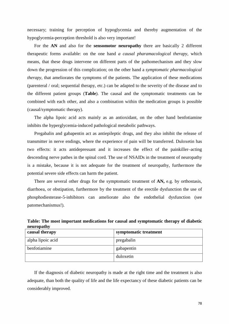
78
necessary; training for perception of hypoglycemia and thereby augmentation of the
hypoglycemia-perception threshold is also very important!
For the AN and also for the sensomotor neuropathy there are basically 2 different
therapeutic forms available: on the one hand a causal pharamacological therapy, which
means, that these drugs intervene on different parts of the pathomechanism and they slow
down the progression of this complication; on the other hand a symptomatic pharmacological
therapy, that ameliorates the symptoms of the patients. The application of these medications
(parenteral / oral; sequential therapy, etc.) can be adapted to the severity of the disease and to
the different patient groups (Table). The causal and the symptomatic treatments can be
combined with each other, and also a combination within the medication groups is possible
(causal/symptomatic therapy).
The alpha lipoic acid acts mainly as an antioxidant, on the other hand benfotiamine
inhibits the hyperglycemia-induced pathological metabolic pathways.
Pregabalin and gabapentin act as antiepileptic drugs, and they also inhibit the release of
transmitter in nerve endings, where the experience of pain will be transferred. Duloxetin has
two effects: it acts antidepressant and it increases the effect of the painkiller–acting
descending nerve pathes in the spinal cord. The use of NSAIDs in the treatment of neuropathy
is a mistake, because it is not adequate for the treatment of neuropathy, furthermore the
potential severe side effects can harm the patient.
There are several other drugs for the symptomatic treatment of AN, e.g. by orthostasis,
diarrhoea, or obstipation, furthermore by the treatment of the erectile dysfunction the use of
phosphodiesterase-5-inhibitors can ameliorate also the endothelial dysfunction (see
patomechanismus!).
Table: The most important medications for causal and symptomatic therapy of diabetic neuropathy causal therapy symptomatic treatment
alpha lipoic acid pregabalin
benfotiamine gabapentin
duloxetin
If the diagnosis of diabetic neuropathy is made at the right time and the treatment is also
adequate, than both the quality of life and the life expectancy of these diabetic patients can be
considerably improved.

79
Chapter 16. Diabetic nephropathy Dr. István Wittmann
Definition
Those diabetic patients should be regarded as having diabetic nephropathy, in whom
there is an acceleration of loss of renal function, and parallel to that they show either stage of
proteinuria (microalbuminuria, macroalbuminuria, proteinuria, nephritic syndrome); or they
show a decline in renal function, in the presence of normalbuminuria if the patient is on a
prolonged and effective renin-angiotensin-aldosterone system (RAAS) inhibition therapy and
another renal disease is not suspected; or if the renal histological examination verified signs
indicating diabetic nephropathy.
Epidemiology
Approximately 20-40% of diabetic patients have diabetic nephropathy, and in
countries with a western style of living, diabetic nephropathy is the most common cause of
end-stage renal disease.
Pathogenesis
Pathogenesis: Genetic predisposition: It seems that polymorphisms of the RAAS may
play a role in the development and progression of diabetic nephropathy. Those diabetic
patients, that concomitantly carry certain variants of aldose reductase and GLUT1, have a 9-
times elevated risk regarding diabetic nephropathy. Expression of eNOS variants may
promote the development of diabetic nephropathy, which effect may be independent of blood
pressure. According to human studies, a certain polymorphism of eNOS, in interaction with a
polymorphism of methylene tetrahydropholate reductase, may increase risk of
microalbuminuria. Latter polymorphism is related to homocystein metabolism, which is
related to the field of oxidative stress. According to a currently published meta-analysis one of
the polymorphisms of SOD2 decreases the risk of diabetic nephropathy by 20%. Carriers of
apoliprotein E4 have a 2.25-fold risk as compared to other E alleles.
Pathogenesis: Epigenetic approach: In the background of epigenetic changes,
acetylation/deacetylation of the lysine amino acid residue, or methylation/demethylation of
arginin may be found. Both directions (acetylation/deacetylation and
methylation/demethylation) of both processes (acetylation and methylation) are catalyzed by
80
different enzymes. Methylation is a modification that lasts longer and is more stable, but both
acetylation and methylation result in activation of affected genes. Not only histone, but also
DNA may be methylated, and in diabetes and chronic kidney disease there is a
hypomethylation of the DNA. Hyperglycaemia may lead to epigenetic changes through
‘metabolic memory’ to the development of diabetic complications among them to diabetic
nephropathy.
Pathogenesis: Hemodynamic aspects: The evaluation of the most important factor of
the hemodynamic approach, namely hyperfiltration is impaired by methodologic problems,
mainly uncertainties in the determination of the GFR. The relative kinetics of renal
enlargement and hyperfiltration is uncertain, but both are characteristic for the renal affection
in diabetes. Possible causes of hyperfiltration include oxidative stress, an increased secretion
of VEGF, the effect of insulin and an increased expression of SGLT2. As we do not have a
clinical study that would be long enough, large enough, and well enough documenting GFR
on the outcomes of GFR or albuminuria, at the moment no specific therapy is required to
hyperfiltration. It may be assumed that this is also beneficially influenced by RAAS
inhibition, such as in case of albuminuria, but further studies are needed to be able to say this.
Pathogenesis: Metabolic aspects: One important component of the metabolic
dysfunction in diabetes, hyperglycaemia is able to evoke intracellular glucotoxicity, which on
the one hand directly damages the cells, and on the other hand is able to induce insulin
resistance. Insulin resistance is able to alter the function of the podocytes (that play an
important role as a filtration barrier) in a way that many alterations that are well-known in
diabetes and are characteristic for diabetic nephropathy, may be be related to it. However, also
the possibility rises that the podocyte-effect of insulin resistance may play a role in other
pathologies not considered as diabetic nephropathy (e.g. obesity-related renal damages,
maybe secondary focal segmental glomerulosclerosis), or the worsening of other renal
diseases (eg. progression of IgA nephropathy).
Pathogenesis: Oxidative stress approach: All types of cells are affected by free radical
damage and the injury of redox regulation. It may launch different pathologic processes,
depending on the regenerating effect of the given cell. Diabetes mellitus is a typical example
for disorders in redox regulations, in the background of which we can mainly find the
reducing property of glucose, by which an unpaired spin electron can be transferred to various
molecules, thereby increasing their reactivity to an extreme value and this way damaging
cells. Free radical-derived and other effects can lead in diabetes to a tubulointerstitial hypoxia
in the kidney, thereby leading to early vitamin D and erythropoietin deficiency. Activation of

81
the RAAS, effects of cytokines and AGEs, all lead to free radical processes on a subcellular
level. Therefore, inhibition of the RAAS, the aim to achieve a good glycaemic control does
not mean anything else at a subcellular level then normalizing the redox balance.
Pathogenesis: Non-enzymatic glycation: The non-enzymatic glycation leads to
abnormal proteinuria and a decline in GFR by damaging all parts of the kidney. This is one of
the leading pathophysiological factors of the development of diabetic nephropathy.
Unfortunately, at the moment there are no real tools to its direct inhibition that could be used
in clinical practice.
Pathogenesis: Cytokines: An increased production of pro-inflammatory cytokines can
be observed in the background of subclinical inflammation. Most important roles are played
in diabetic nephropathy by TNF-alpha and the profibrotic TGF-beta.
Pathogenesis: Renin-angiotensin-aldosterone system: At the organ-tissue level the
pathogenesis of diabetic nephropathy is dominated by the activation of RAAS. In the
background of insulin resistance, oxidative stress, non-enzymatic glycation, hypoxia,
hyperfiltration, cytokine effects, the activation of RAAS can always be found.
Hyperglycaemia and AGEs are able to activate the RAAS by themselves.
Histology
No renal biopsy is carried out in the likelihood of diabetic nephropathy, the diagnosis
must be set clinically. Glomerular damages related to diabetic nephropathy are thickening of
the GBM, mesangial expansion, the Kimmelstiel-Wilson-type nodular glomerulosclerotic
lesion and a marked glomerulosclerosis. This may be complemented by tubulointerstitial and
vascular lesions.
Diagnosis
The diagnosis of diabetic nephropathy is based on three pillars: detection of diabetes
mellitus, microalbuminuria/proteinuria and GFR-loss. As provided in the definition, in the
absence of histology, a rule-out diagnosis will be set. As it shall be highlighted at the detailed
description of clinical course, the absence of microalbuminuria/proteinuria does not exclude
today the diagnosis of diabetic nephropathy. The diagnosis may be underlined by (but its
absence does not exclude the diagnosis) if we are able to observe hyperfiltration or with an
ultrasonography enlargement of the kidneys in a diabetic patient.

82
Differential diagnosis
Besides the general consideration, carrying out a renal biopsy is suggested in diabetes
mellitus (especially in type 2 diabetes), if the patient presents with glomerular-type hematuria,
or has no or only slight diabetic retinopathy (as compared to the level of renal damage), or an
early onset (within 5 years of diagnosis of diabetes) a sever (nephrotic range) proteinuria is
present, or if we see an early onset and fast decline in kidney function. Haematuria may be
caused by frequently coinciding urinary tract infection, malignancy, arterial or venous
thrombosis/embolism and papillary necrosis. In these cases, however, a haematuria with
normal morphology of the red blood cells is observed in the urine sediment, and no renal
biopsy is needed.
Clinical presentation, stages and prognosis
Table: Stages of DNP DNP stage European staging GFR-based staging
(ml/min/1,73m2)
Stage 1. Normoalbuminuria, hyperfiltration
>90
Stage 2. Normoalbuminuria, decreasing filtration
60-89
Stage 3. Abnormal (30-300 mg/day) albuminuria, decreasing filtration
30-59
Stage 4. Abnormal (>300 mg/day) albuminuria, decreasing filtration
15-29
Stage 5. End-stage renal disease <15
New phenomena in the clinical course of diabetic nephropathy: The threshold of
hyperfiltration (125, 130, 135 ml/min) should be corrected because of the approx. 1
ml/min/1.73m2/year decline in the GFR above 40 years as: 125(130,135) - (age - 40).
Currently we have no data on the prognosis of patients who are normalbuminuric due
to RAAS-inhibition and statin therapy but with impaired or normal renal function. Although
this question is always of higher clinical relevance, as though we earlier meant that there can
be no nephropathy in diabetics without abnormal albumin- and proteinuria, now we can
observe that this condition is always becoming more frequent due to the wide use of RAAS-
inhibitors and statins.

83
Screening for abnormal albuminuria: One measurement of albuminuria is not enough,
as the intraindividual variability is large, therefore, we can talk about an abnormal
albuminuria, if 2 out of 3 measurements are positive. There are also causes for transient
positivity, the diagnosis of abnormal albuminuria can only be set or declined, after these are
ruled out. If the diabetic patient has no abnormal albuminuria, annual control is suggested. In
patients with type 1 diabetes, 5 years after the diagnosis of diabetes, in type 2 immediately at
the diagnosis a screening for albuminuria is suggested. Normoalbuminuria means an albumin
amount < 30 mg in the 24-hour collected urine, or if the albumin/creatinine ratio in a non-
collected urine specimen is < 3.0 mg/mmol. If the patient performs no urine collection, the
albumin/creatinine ratio should be used.
Development of abnormal proteinuria and albuminuria: Structures in the kidney that
should prevent protein loss are endothelium, the glomerular basement membrane, the
podocyte, and the slit membrane that is stretched between the foot processes of the podocytes.
Protein can only get into the urine, if the reabsorptive capacity of proximal tubular cells are
exhausted.
Significance of abnormal proteinuria and albuminuria: In case of a patient with
abonormal albuminuria, besides renal disease also dyslipidaemia, obesity, hypertension,
micro- and macrovascular complications should be screened.
Measurment of albuminuria: Nowadays albuminuria is detected using routine
immunological methods, such widespread methods are immunonephelometry and
immunoturbidimetry.
Factors influencing albuminuria measurement: Positional that is postural, so-called
orthostatic proteinuria can occur. The clinical significance of this entity is debated. Clinically
overt urinary tract infections, most inflammatory diseases, acute febrile states, physical
exercise, heart failure, dietary protein load may induce transient proteinuria. In case of stored
urine samples – even at -80°C storage – we have to face the possibility of obtaining a lower
result.
Factors determining progression of albuminuria: Higher albuminuria, higher HbA1c,
the mean arterial pressure (MAP) value, cardiovascular disease in the family history,
hypertension, smoking, body mass and treatment of the patient seen to be predominant in
terms of determining progression of albuminuria.
Connection between blood pressure and diabetic nephropathy: While earlier guidelines
set the target of less than 130/80 mmHg in case of a proteinuria < 1 g/day, and suggested the
range below 125/75 mmHg in case of a proteinuria > 1g/day, now more permissive are
84
declared targets (< 140/90 mmHg is suggested)in the absence of data. Maybe it is more
correct if we talk about a target blood pressure range, knowing that a too large decrease in
systolic and diastolic blood pressure can increase mortality. Thus the systolic blood pressure
should not be lower than 100 mmHg, and the diastolic should not go below 60 mmHg.
Probably here, as in all fields of medicine, an individualized therapy should be emphasized,
and the target blood pressure should be determined by taking into account co-morbidities, age,
gender, the way of living, life expectancies of the patients. In order to reach the target, in
general 2-4 antihypertensive medications need to be combined.
Recurrence of diabetic nephropathy after transplantation: If the diabetic nephropathy
recurs in the transplanted graft kidney, then less time is needed to develop the mild damage
than in case of the native kidney. .
Connections between albuminuria and cardiovascular diseases: Renal disease and
cardiovascular disease progress together in diabetic patients. The explanation of this may be
the following: the same risk factors (smoking, components of the metabolic syndrome) lead to
an abnormal albuminuria and renal damage, as to the development of cardiovascular diseases.
Common evaluation of GFR and albuminuria: To elaborate the risk of cardiovascular
mortality and development of renal failure, albuminuria and the GFR value should be used in
combination. This method has a high predictive value, is widely available and cheap.
The therapy of diabetic nephropathy
The glycaemic control of diabetes: A common statement of guidelines is that the
intention to reach normoglycaemia can delay development of abnormal albuminuria in both
type 1 diabetic and type 2 diabetic patients. It seems that in an established nephropathy the
gylcemic control may not be as effective to slow down the progression. While intenting
euglycaemia, we should not forget that an ideal HbA1c range can be set up for mortality, as
mortality increases both above and underneath this range. We have to try to reach values in
the lower part of this range in order to decrease the risk of diabetic nephropathy.
The use of oral antidiabetic medications should be tailored individually to the GFR,
while we know about gliquidone, pioglitazone, gliptins and insulin can be given at any stage
of renal disease.
Inhibition of the renin-angiotensin-aldosterone system (RAAS): According to
guidelines, diabetic patients with albuminuria (in females excluding time of pregnancy) an
ACEI or ARB is recommended. In case of patients with type 1 diabetes mellitus ACE may be
preferred, while in patients with type 2 diabetes, hypertension and abnormal albuminuria,
85
ACEI and ARB may be equally good. If these patients also exhibit a decline in GFR, ARB
therapy is preferred. In case of intolerance to ACEI or ARB, an agent from the other class
should be chosen. After starting ACEI and ARB therapy, serum creatinine and potassium
measurements are eligible. Normalization of proteinuria is an important consideration in the
therapy of a diabetic patient. The RAAS-inhibitor therapy should not be discontinued when
the renal function becomes impaired, as it is further needed to prevent cardiovascular damage
in these diabetic patients.
The order of use of antihypertensive drugs in diabetic nephropathy: The following order
may be set up between antihypertensive drugs in diabetic nephropathy (numbers indicate
order of choice):
1. RAAS inhibitors 2. Diuretics and/or calcium channel blockers 3. Beta-blockers (cardio selective, metabolic neutral, neutral to peripheric arterial disease; if
myocardial infarct or heart failure is found in the past medical history, then use with RAAS inhibitors in the first line)
4. Central nervous system-acting or alpha1-blocker 5. Direct vasodilator
Lipid-lowering therapy: Statins (except for rosuvastatin) may decrease albuminuria,
proteinuria, some studies even described a beneficial effect in GFR-decline. Fenofibrate may
significantly decrease albuminuria both in micro- and macroalbuminuria, this effect is most
evident among patients with hypertriglyceridaemia, and fenofibrate also lead to a slowing
down of progression of albuminuria. Moreover, it seems favourable concerning GFR loss.
The role of diet: In CKD stages 1-4, sodium intake should be below 2.3 g/day, total fat
intake less than 30% of the total energy intake, saturated fatty acids less than 10% of total
calory intake, cholesterol intake should be below 200 mg/day, carbohydrate intake should
cover 50-60% of total energy intake. Protein intake in CKD 1-4 stages should be 0.8 g/day.
The role of weight loss: Weight loss could lead to a decrease in proteinuria in obese
diabetic and non-diabetic patients, meanwhile the GFR decreased (if they were hyperfiltering)
or remained constant.
Cessation of smoking: Although no randomized, controlled study was carried out because
of ethical reasons, the available data suggest that cessation of smoking may provide
significant benefit in regards of the development and progression of the diabetic nephropathy.
New treatment modalities verified as effective by human studies: From vitamin D and its
analogues, paricalcitol proved to decrease the rate of albuminuria and does not cause
significant adverse effects. In a meta-analysis, glitazones have shown to decrease
86
albuminuria. In randomized, controlled trials, pentoxyphyllin lead to a significant decrease of
albuminuria in patients with abnormal albuminuria (>300mg/day) (9 studies), but proves
inefficient in case of slighter (30-300 mg/day) albuminuria (4 studies). Aldose reductase
inhibitors are known and studied for 40 years, however their effect on albuminuria was only
studied oin one clinical study in patients with type 1 diabetes and abnormal albuminuria, there
a significant improvement was observed. Endothelin-inhibitors, given on top of RAAS-
inhibitors are able to decrease proteinuria, unfortunately they increase the chance of oedema
formation and one of them may lead to higher rate of heart failure. Further studies are
required to weigh risks and benefits.
87
Chapter 17. Microvascular complications of diabetes: diabetic eye problems
Dr. Zsolt Biró
Ocular complications of diabetes is not synonymous with diabetic retinopathy, for
practically all structures of the eye can be affected by the disease. Ocular complications can
be divided into extraocular and intraocular complications. Extraocular complications include
inflammation of the eyelid (blepharitis), yellowish deposits of fat underneath the skin
(xanthelasma) extraocular muscle palsy, especially involving the III (oculomotor nerve) and
VI (abducense nerve) cranial nerves, and microaneurysms of the conjunctiva. To the ocular
complications belongs reduced corneal sensitivity resulting in recurrent corneal epithelial
damage, glaucoma and disturbed visual function. Among the ocular complications mention
must be made of alterations of lens leading to myopia and early development of cataracts, as
well as diabetic retinopathy and anterior ischemic optic neuropathy (AION).
Let us look at this in greater detail.
Conjunctival bacterial infections occur in 80-90 % of patients with diabetes, resulting
in a decreased number of goblet cells which may lead to keratosis. Microaneurysms in the
bulbar conjunctiva are more common in diabetic individuals. There is a change in tear film
composition, leading to reduced corneal sensation, which results in increased risk of corneal
damage.
Tear break up time, which is a minimum of 20 seconds in healthy individuals,
significantly diminishes as a result of damage to tear film disposition and stability in the
corneal surface.
Superficial corneal inflammation (punctate keratitis) and recurrent corneal erosions
have also been linked to decreased corneal sensitivity and poor adhesion between epithelial
cells and the basal membrane.
Diabetic individuals may find it difficult to use contact lenses, as the cornea may
become oedematous and epithel damage may occur.
One of the most serious complications of diabetes is the neovascularisation of the iris.
This alteration is usually first observed at the pupillary margin but it can involve the entire iris
surface and filtration angle. Neovascularisation arises from hypoxia caused by retinal
capillary damage. It is reported up to 60 % of proliferative retinopathies.
88
In diabetic patients the pupils may be more narrow (miotic) and may have weaker
reaction to topical mydriatics originating in diminished innervations of pupil dilatator muscle
due to diabetic neuropathy.
The occurrence of Uveitis (inflammation of the iris or ciliary body) is also more
common in diabetes.
Neovascularisation of the iris results in neovascular glaucoma. The newly growing
vessels block the drainage of aqueous fluid through the anterior chamber angle, causing
elevation of intraocular pressure. The pupil may become irregularly shaped; the eyes become
red and painful leading to loss of visual acuity if not treated (e.g. by normalising intraocular
pressure).
Refractive changes are common in diabetes because significant plasma glucose
fluctuations (in uncontrolled diabetes) induce shifts in glucose concentration of aqueous
humour which leads to swelling of the lens. Glycosylation of lens proteins results in a
decrease in the lens’s transparency.
Patients with diabetes have a 2 to 4 times greater risk of developing cataracts than do
non-diabetic people. Cataracts typically tend to develop earlier and the risk increases with
age, duration of diabetes, uncontrolled diabetes (high HbA1c) and kidney disease. The only
effective treatment of a matured cataract is surgery.
Macular edema, which is actually the swelling or thickening of the central retina, is
often (in 7%) found while examining diabetic patients. Epidemiological data indicate that it is
the major cause of vision loss in diabetic individuals. Treatment of diabetic macular edema
includes Argon laser photocoagulation of the fundus, as well as the proper management of
systemic risk factors such as hyperglycaemia, hypertension and hyperlipidaemia.
Extensive laser photocoagulation may result in complications like central and
paracentral scotoma (visual field loss), colour vision changes or loss, or occasionally
secondary retinal neovascularisation. In these cases anti VEGF treatments are recommended,
such as intravitreal administration of bevacizumab, ranibizumab, pegaptanib, or aflibercept.
Attempts have been made to administer glucocorticosteroids. Triamcinolon, fluocinolone and
dexamethasone can be used intravitreally for the treatment of diabetic macular edema.
Diabetic retinopathy (DR)
The risk factors for DR are as follow: duration of diabetes, younger age, high HbA1C,
hypertension, smoking, alcohol consumption, life style and pregnancy.

89
There are different stages in diabetic retinopathy: There is no fundus alteration in
preretinopathy. The characteristic lesions of mild (background) and moderate retinopathy are
micoaneurisms, haemorrhages, soft exudates (cotton wool spots) and hard exudates, all visible
on the retina or on the retinal vessels. Edema may also occur. As the ischemia progresses the
number and extensions of lesions increase. Proliferative retinopathy develops, characterised
by abnormal new vessel growth from the papillary area or from the retinal surface
(neovascularization). Preretinal or vitreous haemorrhages and traction retinal detachment may
also occur.
Regular care and follow-up of diabetic patients is of utmost importance. In pre- and
background retinopathy annual controls are usually recommended, while patients with
proliferative retinopathy should be re-examined as frequently as 2-4 month intervals. Women
with diabetes who become pregnant should undergo an eye examination in the first trimester
and regular follow-ups are needed throughout their pregnancy.
90
Chapter 18. Hypertension and diabetes mellitus
Dr. István Wittmann
Hypertension in type 1 diabetes mellitus
Hypertension in young (<30-40 years), people with type 1 diabetes is usually related to
diabetic nephropathy. When a young type 1 diabetic patient develops hypertension, especially
resistant hypertension without albuminuria or GFR reduction, secondary hypertension (other
than underlying diabetic nephropathy) should be considered.
High blood pressure occurring in older (>40-50 years) patients with type 1 diabetes is
frequently a primary (essential) hypertension, especially when it is associated with obesity or
metabolic syndrome.
Newly developed hypertension in a pregnant, type 1 diabetic woman may be a sign of
early nephropathy or preeclampsia.
Hypertension in type 2 diabetes mellitus
Because type 2 diabetes and pre-diabetic states (IGT, IFG) often occur as components
of metabolic syndrome, and as hypertension is a metabolic syndrome-component as well,
unsurprisingly primary hypertension usually precedes or develops together with diabetes.
It is also not uncommon that hypertension emerges only after type 2 diabetes has already been
developed. In this case nephropathy should be considered (alike in type 1 diabetes) and
measurement of albuminuria and determination of GFR is indicated. Primary
hyperaldosteronism and obstructive sleep apnea syndrome (causes metabolic syndrome-like
clinical picture) are also to be ruled out.
Hypertension in other specific types of diabetes mellitus
Hypertension, which occurs in diabetes associated either with endocrinopathies
(primary hyperaldosteronism, hyperthyreosis, diseases caused by excessive amount of
cortisol, growth hormone or epinephrine etc.) or with iatrogenic factors (drug-or chemical-
induced diabetes) is well known. Iatrogenic diabetes and concomitant hypertension have to be
discussed in more detail because their occurrence is relatively common and usually avoidable.
Corticosteroids lead simultaneously to rise of blood pressure and blood glucose, thus in the
treatment of autoimmune diseases the aim should be to use the minimum dose required to
achieve a therapeutic effect.
91
In middle aged, hypertensive, non-diabetic patients with high doses (25-50 mg) of
thiazides and non-cardioselective beta blockers, treatment occurs much more frequently. This
combination is associated with a more than twofold risk of developing diabetes.
We have also been able to observe in the clinic that some patients develop diabetes
after having received 50 mg thiazide and beta blockers for years. The reversibility of diabetes
depends on the duration of drug exposure. It is also important to remember that this drug
combination can cause dyslipidaemia and may be associated with a decline of renal function.
Unfavourable metabolic effects can be avoided through the use of low dose (1.5-2.5
mg) thiazid-like diuretics and third-generation beta-blockers (carvedilol, nebivolol).
Screening for hypertension in diabetes
Blood pressure should be measured at every routine visit. Home blood pressure self-
monitoring and 24-h ambulatory blood pressure monitoring is often required to provide an
accurate diagnosis.
The absence of dipping or rising in nocturnal blood pressure is often associated with
obstructive sleep apnoea and diabetes.
Treatment goals are constantly revised. According to recent guidelines, people with
diabetes and hypertension should be treated with an aim to achieving a blood pressure of
<140/90 mmHg. Lower blood pressure targets such as <130/80 mmHg may be appropriate for
certain individuals, such as young patients with proteinuria. In this regard individual target
goals seem to be more appropriate but there is as yet a lack of clear-cut recommendations (as
in the case of glycemic targets).
Treatment of hypertension in people with diabetes
Patients with blood pressure >120/80 mmHg should be advised on lifestyle changes
(weight loss, physical activity, cessation of smoking, diet, salt restriction, moderation of
alcohol intake) to reduce blood pressure. Patients with confirmed blood pressure higher than
140/80 mmHg in office should, in addition to lifestyle therapy, have initiation in
pharmacological therapy.
To understand the pharmacological treatment of diabetes it is important to know that
advanced glycation end products formed in high doses in diabetes activate the renin-
angiotensin aldosteron system. Glycation end products also cause nephron loss and
consequently GFR-reduction, directly or indirectly, by triggering proteinura. Because kidneys
play a major important role in the elimination of AGE products, the reduction of GFR leads to

92
further increase in the level of glycated end products. Glycation end products are not only
nephrotoxic but also vasculotoxic, resulting in early atherosclerosis and leading by this to
increased cardiovascular morbidity and mortality in diabetes and especially in diabetic
nephropathy.
Figure: Correlation between non-enzymatic glycation, proteinuria, nephron loss (GFR reduction) and cardiovascular morbidity
It follows from the above that RAAS inhibitors should be used as first-line
hypertension therapy in people with diabetes and even more so with diabetic nephropathy. An
ACE inhibitor should be the first drug of choice, titrating up to the highest tolerable dose.
When despite a maximal dose ACE inhibitor treatment proteinura remains and GFR is
> 60 ml/min, the addition of a mineralocorticoid receptor antagonist or angiotensin receptor
blocker to the ACEI inhibitor based regimen may provide additional benefits regarding high
renal risk. Claims that combination therapy lacks cardiovascular benefits or may harm the
kidneys are not sufficiently substantiated. When GFR is <60 ml/min initiation of combination
RAS blockade (i.e. ACE+ARB or ACE + spironolacton e.t.c) is not recommended, but
combination therapy that has already begun can be maintained. A combined blockade of the
RAS system is recommended to reduce proteinuria without further reduction of blood
pressure. It is also important to mention that this therapy requires experience and strict
controls; serum creatinin, eGFR and serum potassium levels should be monitored.
93
If ACE inhibitor intolerance occurs ARBs should be substituted.
Mineralocorticoid receptor blocker therapy plays a favourable role in the
antihypertensive treatment of obese patient with type 2 diabetes. The reason for this is that
visceral fat cells in abdominal obesity produce not only cytokines but also aldosterone
releasing factors contributing to an even higher aldosterone level. Therefore mineralocorticoid
receptor antagonists induce not only blood pressure reduction but may also improve glucose
control and reduce target organ damage in this population. In obese type 2 diabetic patients
(without underlying primary hyperaldosteronism) serum potassium levels that are slightly low
or at the low end of the normal range and mild or compensated metabolic alkalosis may
predict the efficacy of mineralocorticoid antagonist therapy.
Resistant hypertension is common in patients with metabolic syndrome, obstructive
sleep apnoea and diabetes mellitus with nephropathy. Thus multiple-drug therapy (minimum
3-4 agents at maximal doses) is usually required to achieve blood pressure goals.
Because an increased aldosteron level may lead to salt-sensitive hypertension in
diabetes, low dose diuretics are recommended as second- or third-line therapy. Low dose
thiazide-like diuretics may compensate the potassium sparing effect of RAAS inhibitors.
The next agents of choice are the metabolically neutral calcium channel blockers. In
patients with a previous history of myocardial infarction, treatment should be complemented
with β-blockers. Patients with diabetes and a history of myocardial infarction or stroke
probably suffer from peripheral artery disease. In this regard nevibolol or carvediolol may be
a more favourable β-blocker of choice.
Even more antihypertensive agents may be necessary to achieve blood pressure
targets, especially in patients with impaired kidney function. Thus the addition of further
blood pressure medication over and above those previously mentioned should be made, such
as alfa-blockers or central nervous system acting drugs or even sometimes direct
vasodilatators ( e.g.: dihydralazine)
In patients with serious kidney impairment (<GFR 30 ml/ min) loop diuretics should
usually be prescribed for further blood pressure reduction due to volume retention. A loop
diuretic is not recommended for blood pressure reduction in patients whose renal function is
normal due to its short duration of action.
The sequence of antihypertensive agents usually recommended for use on patients
with diabetes and hypertension is as follows:
1. RAS-inhibitors (ACE inhibitors or ARBs)
2. Low-dose thiazide-like diuretic and/or mineralocorticoid receptor antagonist

94
3. Dihydropyridin calcium channel blockers
4. Beta-blockers
5. Alfa- receptor-blockers or CNS-drug
6. Direct vasodilator

95
Chapter 19. Management of dyslipidaemia in diabetes mellitus
Dr. Gábor Fülöp
Diabetes, metabolic syndrome and even prediabetes are associated with a typical
pattern of dyslipidaemia leading to increased risk of cardiovascular morbidity and mortality.
In type 1 diabetes typical lipid disorder occurs primarily when carbohydrate metabolism is
poorly controlled or disturbed. Prevalence of dyslipidaemia in well controlled
(normoglycaemic) type 1 diabetic patients corresponds to that in the non-diabetic population.
Lipid disorder occurs in more than 80 % of patients with type 2 diabetes.
These lipid disorders include not only quantitative but also qualitative abnormalities of
lipoproteins.
This typical dyslipidaemic pattern is as follows:
1. Increased triglycerides
2. Low HDL- cholesterol
3. Small dense LDL particles
4. Postprandial lipemia
5. Near-normal LDL cholesterol
These changes are caused by insulin resistance/hyperinsulinemia. Because this lipid
abnormality promotes atherosclerosis it is called atherogenic dyslipidemia. Small dense LDL
particles are more susceptible to oxidative modification. These modified particles have
decreased binding to the LDL receptor, hence they spend more time in circulation, incorporate
into the endothelium and are eliminated only via alternative scavenger receptors.
Before treating diabetic dyslipidemia, assessing the patient’s cardiovascular risk using
SCORE (Systemic Coronary Risk Evaluation) risk charts is elemental. These charts are based
on gender, age, total cholesterol, systolic blood pressure and smoking status and estimate risk
for fatal cardiovascular disease events over a ten-year period. A score higher than 5 %
indicates high risk, while a score over 10 % indicates very high risk for fatal cardiovascular
events.
Type 1 and type 2 diabetes denote per se high risk, whereas diabetes associated with 2
or more risk factors and/or organ damage (cardiovascular disease) implies very high risk for
fatal cardiovascular events.
Table shows treatment goals for lipid therapy in different cardiovascular risk
categories.The principle of treatment is known as treat to target therapy.
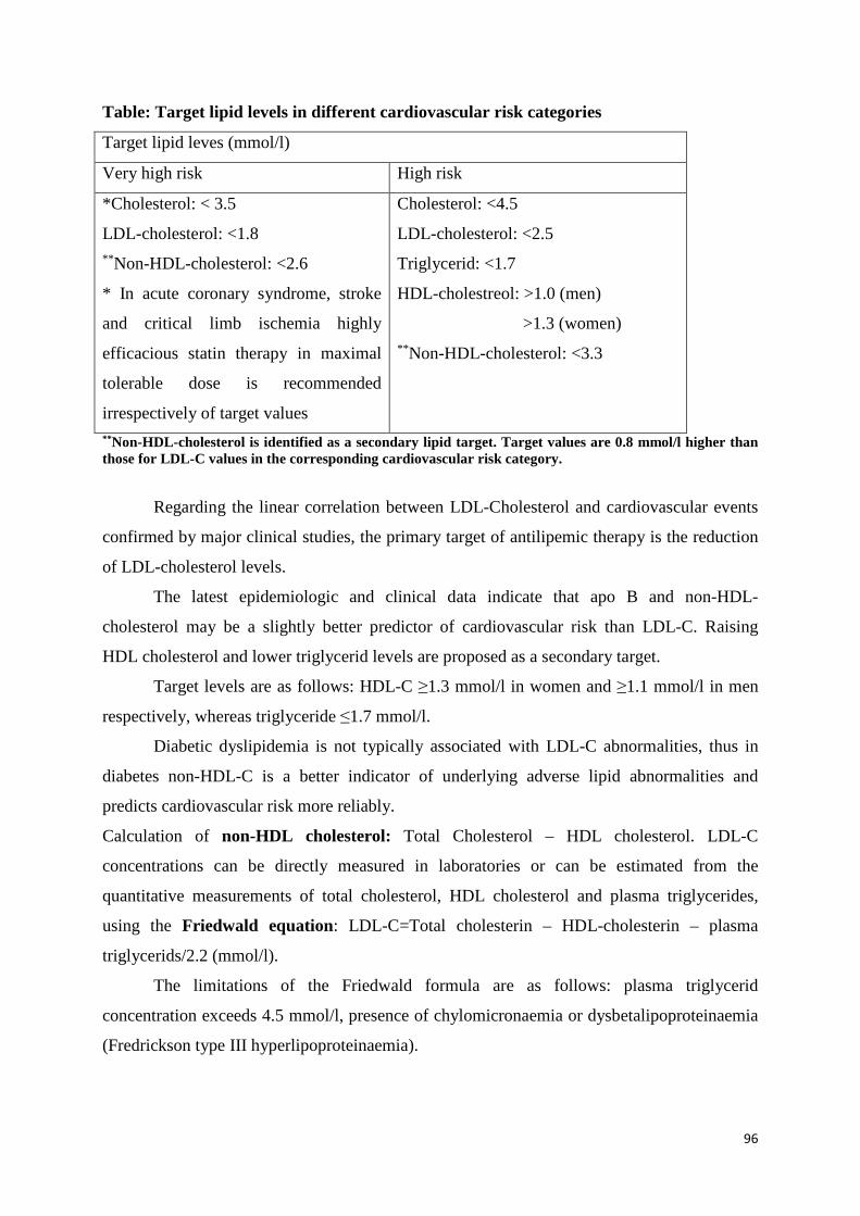
96
Table: Target lipid levels in different cardiovascular risk categories
Target lipid leves (mmol/l)
Very high risk High risk
*Cholesterol: < 3.5
LDL-cholesterol: <1.8 ** Non-HDL-cholesterol: <2.6
* In acute coronary syndrome, stroke
and critical limb ischemia highly
efficacious statin therapy in maximal
tolerable dose is recommended
irrespectively of target values
Cholesterol: <4.5
LDL-cholesterol: <2.5
Triglycerid: <1.7
HDL-cholestreol: >1.0 (men)
>1.3 (women) ** Non-HDL-cholesterol: <3.3
** Non-HDL-cholesterol is identified as a secondary lipid target. Target values are 0.8 mmol/l higher than those for LDL-C values in the corresponding cardiovascular risk category.
Regarding the linear correlation between LDL-Cholesterol and cardiovascular events
confirmed by major clinical studies, the primary target of antilipemic therapy is the reduction
of LDL-cholesterol levels.
The latest epidemiologic and clinical data indicate that apo B and non-HDL-
cholesterol may be a slightly better predictor of cardiovascular risk than LDL-C. Raising
HDL cholesterol and lower triglycerid levels are proposed as a secondary target.
Target levels are as follows: HDL-C ≥1.3 mmol/l in women and ≥1.1 mmol/l in men
respectively, whereas triglyceride ≤1.7 mmol/l.
Diabetic dyslipidemia is not typically associated with LDL-C abnormalities, thus in
diabetes non-HDL-C is a better indicator of underlying adverse lipid abnormalities and
predicts cardiovascular risk more reliably.
Calculation of non-HDL cholesterol: Total Cholesterol – HDL cholesterol. LDL-C
concentrations can be directly measured in laboratories or can be estimated from the
quantitative measurements of total cholesterol, HDL cholesterol and plasma triglycerides,
using the Friedwald equation: LDL-C=Total cholesterin – HDL-cholesterin – plasma
triglycerids/2.2 (mmol/l).
The limitations of the Friedwald formula are as follows: plasma triglycerid
concentration exceeds 4.5 mmol/l, presence of chylomicronaemia or dysbetalipoproteinaemia
(Fredrickson type III hyperlipoproteinaemia).

97
In atherogenic dyslipidaemia statin therapy combined with fibrates has resulted in
additional reduction in fatal and non-fatal cardiovascular events. This is called residual risk
reduction.
Lifestyle modification, including increased physical activity and appropriate diet is a
cornerstone in the treatment of lipid abnormalities.
The improvement of glycaemic control is an important first step in managing diabetic
dyslipdaemia.
Despite achieving an almost normoglycemic state, lipid abnormalities may remain and
so additional antilipemic therapy is usually required.
Because the majority of patients are overweight calorie restriction is commonly
recommended. For every 1-kg decrease in body weight, non-HDL cholesterol decreased by
0.06 mmol/l while increased physical activity resulted in a 4-5% decrease in non HDL
cholesterol. The cessation of smoking is essential.
Dietary therapy: The diet focuses on the reduction of consumption of cholesterol,
saturated and trans-fatty acids. The total dietary cholesterol intake should not exceed 200 g,
and limitation of saturated fat to less than 7 % of total calories is recommended.
Consumption of simple, rapidly absorbed carbohydrates and (even moderate) alcohol should
be avoided because they adversely influence glucose control and contribute to high
triglyceride levels.
A diet containing complex carbohydrates and rich in vegetables and omega-3 fatty
acids (e.g fish) which also limits the daily intake of saturated fat is recommended.
A daily intake of 1.5-6 g of fish oil as a nutrition supplement may be advocated
although its high energy content should be taken into consideration.
Pharmacologic therapy Lifestyle modification may be sufficient as long as initial
LDL-C is between 2.6-3.3 mmol/l and no cardiovascular disease is present. When LDL-C
exceeds 3.4 mmol/l or CVD is present, pharmacologic therapy accompanied with lifestyle
changes is recommended
HMG-CoA reductase inhibitors (statin) are the drug of first choice for lowering LDL
cholesterol. The use of statin results in a 30-50 % decrease in LDL cholesterol levels. It is
necessary to achieve treatment goals (e.g. a decrease of LDL cholesterol by at least 50 % from
baseline in high risk patients) in order to attain favourable cardiovascular outcomes. Statins
also moderately raise HDL cholesterol and lower triglyceride and exert many vascular
protective, pleiotropic effects.
98
For initiation therapy moderate intensity statins (simvastatin 40 mg, atorvastatin 20
mg, rosuvastatin 10 mg) are recommended due to their pleiotropic effect.
By doubling the statin dose there is an approximate 6% increase in LDL cholesterol-
lowering efficacy (the 6 percent rule).
If there is an inadequate response to statins, treatment may be complemented with
ezetimib, a selective cholesterol absorption inhibitor which provides an additional 20-25 %
reduction of LDL cholesterol. Ezetimib used as monotherapy is also beneficial in patients
who are unable to tolerate statins. Bile acid sequestrants might also be a further drug choice in
cases of poor therapeutic response.
When HDL cholesterol levels are proven to be low, lifestyle interventions with weight
loss, smoking cessation and increased physical activity along with PPAR-α agonist fibrates or
nicotinic acids are recommended.
When triglycerides are >2.3 mmol/l and LDL cholesterol is <2.5 mmol/l, fibrate
therapy might be initiated. Life style modification should be a first line treatment when
triglycerides are above target level, but under 4.5 mmol/l. When triglyedrides go above 4.5
mmol/l, pharmacological therapy associated with lifestyle changes is recommended. Fibrates
and nicotinic acid derivatives are the most effective agents in lowering triglyceride.
Fenofibrate also decreases the risk of microvascular complications.
In cases of severe hypertriglyceridaemia, when the triglycerid level is above 10
mmol/l, urgent treatment such as severe fat restriction (daily fat intake is reduced to less than
10 % of total energy intake) in addition to fibrate or nicotinic acid therapy is necessary to
reduce the risk of pancreatitis.
Combination therapy. Statin combined with ezetimib is considered a useful way to
reduce LDL cholesterol, whereas in atherogenic dyslipidaemia statin accompanied with
fibrates (or Ω-3 fatty acid) is regarded as being an efficacious combination. Combination
therapy is safe when used with caution and adherence to a summary of product characteristics,
although regular follow ups (control of liver function and creatinin kinase) are recommended.
Possible future treatment options include cholesterylester transfer protein (CETP) inhibitors
and proprotein convertase subtilisin /kexin type 9 (PCSK9) inhibitors.

99
Chapter 20. Rehabilitation in diabetes mellitus
Dr. Gábor Fülöp
During the process of medical rehabilitation the health care system provides help and
support for disabled people who, by developing their extant capabilities or using an assistive
technology device, become able to partially or completely regain independence and to
participate in the activities of a normal life. The first steps of rehabilitation include the
evaluation of current impairment level and the assessment of existent functions and capacity
for work, followed by improvements of conditions, development and maintenance in
individual functions.
Individuals who have diabetes are among those regarded as disabled people. Diabetes
leads to a substantial change in lifestyle: medical therapy, and particularly insulin treatment,
requires strict schedule. The risk of hypoglycaemia in insulin treated diabetics may restrict
access to some professions such as pilot, bus- and engine driver or work at heights or near a
high voltage power line. Night or shift work activities or jobs accompanied by extremely
varied physical activity might also not be recommended because fluctuant or high blood
glucose level and poor glucose control are expected. All these factors have to be considered in
the career choice process.
Chronic complications of diabetes further deteriorate the capacity to work and quality
of life (loss of vision, lower limb amputation, stroke, coronary heart disease, renal
replacement therapy).
People with diabetes often develop multiple, complex disabilities which require
comprehensive team work (e.g. care of a blind person, one with an amputated leg or on renal
replacement therapy).
Diabetes care includes rehabilitation. The foremost goals are the reduction of
cardiovascular risk and the optimal treatment of co-morbidities along with the maintenance of
adequate glycemic control in order to prevent late complications and slower progression.
If complications develop, strict control and timely interventions are required to avoid
more serious conditions and further health damage (e.g. laser therapy to prevent retinal
bleeding, retinal detachment and visual loss due to diabetic retinopathy).
Type 2 diabetes has been associated with increased risk of falls, fractures, depression,
and cognitive impairment.
100
In a diabetic group aged over 70 years and living in a community, difficulties in
performing daily activities were seen in 53%, urinary incontinence in 27%, fecal incontinence
in 11%, depression in 14%, and dementia in 15%. Only 36% of them were free from such
problems.
Risk factors include strokes, peripheral artery disease, depression, smoking, and rare
physical exercise, but the strongest independent risk factor was mobility limitation at baseline.
The process toward disability is complex and involves social-, economic- and
lifestyle-related factors (e.g. poor diet, obesity, smoking, sedentary habits), psychological
factors (personality type, coping strategies), psychiatric condition (mood disorders, changes in
cognition) and a range of disabling medical conditions (arthritis, cardiopulmonary disease,
cancer, stroke). Most factors contributing to disability are also significant in the development
of type 2 diabetes and its complications. A third of patients develop some degree of mobility
limitation after 5 years of diabetes, peripheral neuropathy, arthritis and history of stroke being
the most frequent contributing factors.
Mobility impairment is often an early step in the progression to future disability, thus
it may be a useful early marker for preventative and rehabilitation efforts in diabetes.
Rehabilitation is based on health care system underlying clinical evidence personalised
by a cooperative multidisciplinary team. After all, psychosocial support and involvement of
the community and society are inevitable. Comprehensive assessment of the patient and an
optimal treatment plan are also necessary for appropriate rehabilitation.
More careful diabetes care accompanied by frequent therapy modifications is usually
required. Treatment goals often have to be revised according to the condition and prognosis of
the patient. For example, an elderly patient with cognitive impairment and severe coronary
heart disease requires a more permissive glycemic control (HbA1c level could be kept in a
higher range), because strict glucose control is associated with a high risk of hypoglycaemia,
which is dangerous in this case.
Exercise is a cornerstone of treatment for type 2 diabetes. There is a variety of
literature data about the favourable effects of exercise on blood sugar levels and
cardiovascular risk; however, evidence is missing regarding how to treat or prevent disability
in diabetic patients.
A physiotherapist-supervised group training program has proved to be effective and
resulted in improved balance and reduced fear of falling in patients with neuropathy.
Although the rehabilitation of diabetic patients has aroused intense recent interest in
literature, so far there has been little clinical evidence in this field.
101
Chapter 21. Perioperative management of patients with diabetes mellitus
Dr. Botond Csiky
Diabetic patients have increased perioperative risk compared with their non-diabetic
counterparts. This risk is further increased in patients with inadequate glycemic ontrol. The
neurohumoral changes induced by surgery may lead to an imbalance of carbohydrate
metabolism, even in patients who have adequate glycemic control.
Diabetes should be properly under control prior to elective surgery. Surgery for
patients with diabetes should ideally be performed in the morning.
Biguanid therapy should be stopped 48 hours before the surgery and long-acting oral
hypoglycemic medications (sulphonylureas, basal insulin) should be witheld.
During the surgery and in the perioperative period blood glucose should be monitored
regularly.
We can distinguish minor or major surgical procedures from the diabetic point of
view.
Minor surgical procedure: this is short (general duration <30 min), it is performed
under local anesthesia and does not involve the opening of the pleural or abdominal cavity.
Major surgical procedure: this is longer, performed under general anesthesia and the
abdominal or pleural cavities have to be opened. Oral nutrition cannot be recommenced
immediately after the surgery.
Minor surgical procedure
In patients with properly controlled diabetes treated either solely through diet or
through a combination of diet and oral antidiabetic drugs minor surgical procedures can be
performed without any significant change in the therapy. Long-acting oral hypoglycemic
medication and metformin should be witheld as mentioned above. The patient should fast
prior to the operation, so the oral antidiabetic drugs should not be given to the patient before
the operation; they should be given before the first permitted meal.
Major surgical procedure
On the day of the operation, an insulin-glucose-potassium (GIK) infusion should be
used to maintain normoglycaemia (short-acting insulin in the infusion). The amount of

102
glucose, potassium and insulin administered to the patient should be set and modified
according to the results of frequent laboratory controls.
The infusion can be stopped as soon as the patient’s oral nutrition can be restarted and
the patient can resume the previous antidiabetic therapy. It is important to have an overlap of
at least 4 hours between the parenteral and oral nutrition. The final antidiabetic therapy can be
set following the stabilization of the patient.
If acute major surgical procedure is needed, the above mentioned insulin-glucose-
potassium infusion therapy can be used if the patient has satisfactory glycemic control; if not,
then the GIK infusion can be administered only after correction of the glucose control.
Top Related