


Languages
Pages
Legal


8/21/14
1
Teresa R. Kroeker, M.D. Thyroid, Parathyroid,
Head and Neck Surgery Austin, TX
Evaluation of a Neck Mass
Outline � Anatomy of the neck � Important questions to ask about history � How to examine the patient with a neck mass � Differential diagnosis based on location � Differential diagnosis based on etiology/
pathophysiology � Imaging/diagnostic tests � Evaluation of incidentally discovered thyroid
nodules
Goals � Understand the anatomy and zones of the neck � Know the “important stuff” to ask in the history � Understand how to examine the patient
presenting with a neck mass � Be able to formulate a differential diagnosis
based on location of the mass � Know what kind of imaging to obtain for further
work-up � Know what to do when you find a thyroid nodule
on physical exam or incidentally on another imaging modality

8/21/14
2
8/21/14
3
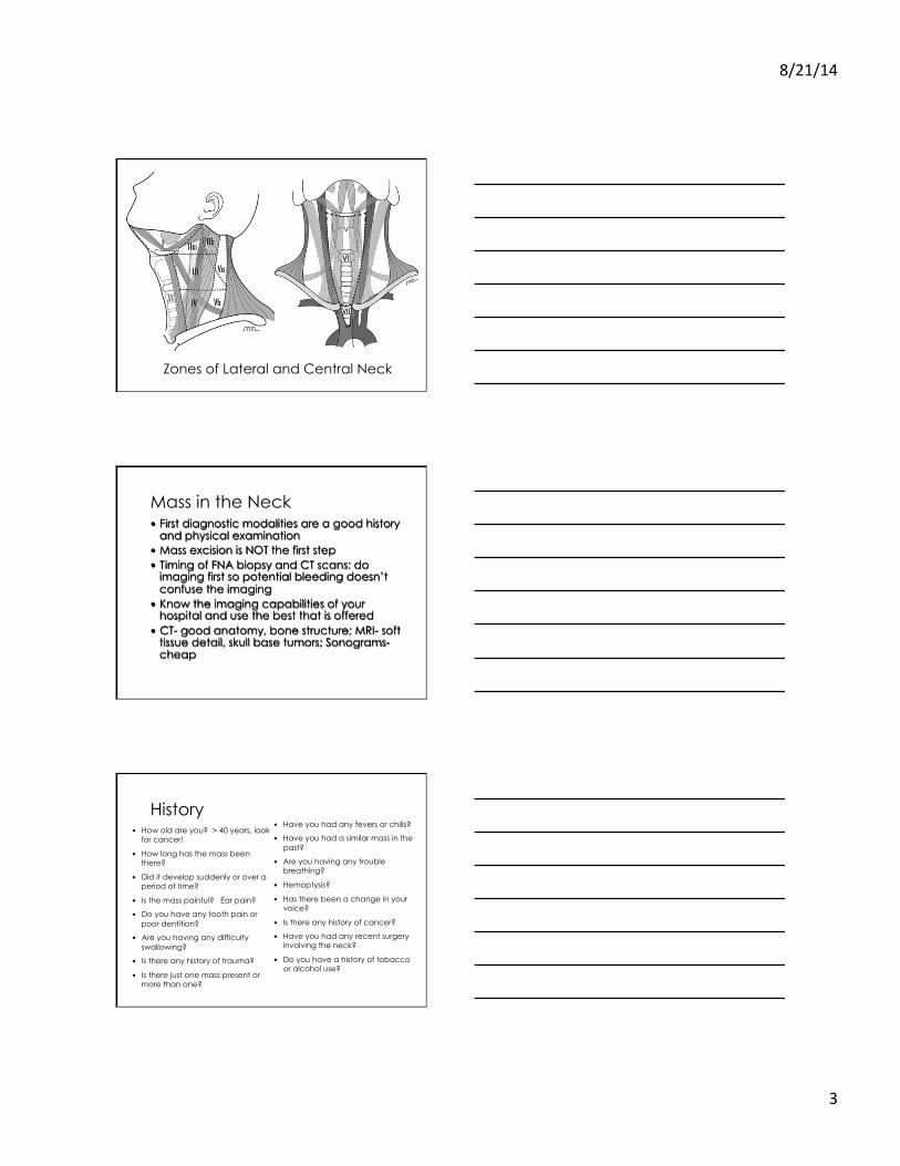
Zones of Lateral and Central Neck
Mass in the Neck � First diagnostic modalities are a good history
and physical examination � Mass excision is NOT the first step � Timing of FNA biopsy and CT scans: do
imaging first so potential bleeding doesn’t confuse the imaging
� Know the imaging capabilities of your hospital and use the best that is offered
� CT- good anatomy, bone structure; MRI- soft tissue detail, skull base tumors; Sonograms- cheap
History
� How old are you? > 40 years, look for cancer!
� How long has the mass been there?
� Did it develop suddenly or over a period of time?
� Is the mass painful? Ear pain?
� Do you have any tooth pain or poor dentition?
� Are you having any difficulty swallowing?
� Is there any history of trauma?
� Is there just one mass present or more than one?
� Have you had any fevers or chills?
� Have you had a similar mass in the past?
� Are you having any trouble breathing?
� Hemoptysis?
� Has there been a change in your voice?
� Is there any history of cancer?
� Have you had any recent surgery involving the neck?
� Do you have a history of tobacco or alcohol use?

8/21/14
4
Physical Exam � The mass should be palpated and its quality
assessed � Is it firm, matted, non-tender and fixed? � Or is it mobile and fleshy? � Is it tender, surrounding erythema/skin changes,
fluctuance, or the sensation of fluid? � Are there multiple lesions?
Cervical Lymph Nodes
Physical Exam � Oral examination (headlight needed) � Lips, gingivae, retromolar trigone, teeth, hard
palate, cheek mucosa, mobile tongue, floor of the mouth.

8/21/14
5
Diagnostic Pearls � 90% of pediatric masses are inflammatory � 90% of adult masses are metastatic � Rule of 80% (for non-thyroid neck masses in
adults) � 80% are neoplasms � 80% are malignant � 80% of parotid tumors are benign � 80% of parotid tumors are mixed tumors
Differential Dx: By Location � Midline
� Thyroid nodule/cancer � Thyroglossal duct cyst
� Lateral � Hyperplastic lymph node � Branchial cleft cyst � Metastatic thyroid
cancer � Vascular anomaly � Lipoma � Carotid body tumor � Metastases from
unknown primary
Differential Dx: By Location � Parotid
� Cystic hygroma, hemangioma, lymphadenitis, parotitis, lymphoma, neoplasm, Sjögren syndrome
� Post-auricular � Lymphadenitis, branchial cleft
cyst (1st), epidermoid cyst � Submental
� Lymphadenitis, cystic hygroma, thyroglossal duct cyst, dermoid, sialoadenitis
� Submandibular � Lymphadenitis, cystic hygroma,
neoplasm, sialoadenitis, sialolithiasis, sialectasis

8/21/14
6
Differential Dx: By Location � Sternocleidomastoid
� Lymphadenitis, branchial cleft cyst (2nd, 3rd)
� Supraclavicular � Cystic hygroma, lipoma,
lymphoma, metastasis, normal fat-pad, cervical rib, scoliosis, pneumatocele of upper lobe
� Suprasternal � thyroid, lipoma, dermoid,
thymus, mediastinal mass
Breakdown by Etiology
Congenital � Branchial cleft cysts: 20% of pediatric masses � Thyroglossal duct cysts: 40% diagnosed
during adulthood � Hemangioma � Laryngocele � Ranula � Teratoma cyst � Dermoid cyst � Thymic cyst

8/21/14
7
Branchial Cleft Cyst Tracks � 1st
� Type I: located near the external auditory canal, usually inferior and posterior to the tragus (base of the ear
� Type II: at the angle of the mandible and may involve the submandibular gland
� 2nd (95%): skin of the lateral neck, between the internal and external carotid arteries, and into the palatine tonsil; anterior border of SCM
� 3rd: skin of lateral neck, posterior to carotid arteries, pierces thyrohyoid membrane to enter the larynx, terminates on the lateral aspect of the pyriform sinus; deep to SCM
� 4th: skin of lateral neck, follows recurrent laryngeal nerve (around the aorta on the left and around the subclavian artery on the right), ends at pyriform sinus
Branchial Cleft Cyst � Most (95%) from 2nd branchial
cleft � Manifests as a sinus, fistula, or
cyst � Nontender, smooth, round
mass located along the anterior border of, or just deep to, the sternocleidomastoid muscle
� Kids: acute and painful enlargement of the cysts secondary to an upper respiratory infection
Thyroglossal Duct Cyst � 70% of all congenital neck
masses � Cystic remnant along the
course of the thyroglossal duct between the foramen cecum of the tongue base and the thyroid
� 50% of patients present before 20 years old
� Midline mass just below hyoid bone
� Asymptomatic, infection, 1-2% malignancy rate

8/21/14
8
Other Congenital Neck Masses � Hemangioma � Laryngocele: herniation of the
saccule of supraglottic larynx; external laryngocele can protrude through thyrohoid membrane and present as an anterior neck mass. Glassblower?
� Ranula: mucocele from obstruction of sublingual glands; submental mass extending from floor of mouth
� Teratoma � Dermoid � Thymic cyst
Inflammatory � Lymphadenopathy
� Viral: mono, EBV, HIV � Bacterial: staphylococcus, TB, cat scratch
(Bartonella) � Parasitic � Fungal: histoplasmosis, blastomycosis,
coccidiomycosis � Granulomatous: sarcoid, foreign body reaction
� Two week trial of antibiotics � Follow-up for further investigation
Neoplastic � Thyroid/parathyroid � Salivary glands � Carotid body tumor � Schwannoma � Lymphoma � Lipoma � Epidermoid inclusion cyst � Metastatic squamous cell carcinoma � Virchow’s node

8/21/14
9
Thyroid/Parathyroid � Thyroid nodules
� VERY common � Compressive symptoms?
� Dysphasia � Choking sensation when
supine � Hoarseness
� Family history of thyroid cancer?
� Radiation exposure? � More on this later…
� Parathyroid adenoma � Rare to palpate
Salivary Gland Neoplasms � 80% in parotid gland, 80%
benign � 10-15% in submandibular
gland, 50% benign � Rest in minor salivary glands,
< 40% benign � Benign:Malignant ratios
� Parotid 3:1 � Submandibular 1:1 � Minor and sublingual 1:9
Salivary Gland Neoplasms � Parotid gland
� Pleomorphic adenoma: 53%, benign BUT undergo malignant transformation after 10 years (carcinoma ex pleomorphic adenoma = BAD player)
� Warthin’s tumor: 28%, benign, older men, smokers, 10% bilateral, tail of parotid (more women recently)
� Mucoepidermoid carcinoma: 9% � Adenocarcinoma:1.5%
� Submandibular gland � Pleomorphic adenoma: 36% � Adenoid cystic carcinoma: 25% � Mucoepidermoid carcinoma:12%

8/21/14
10
Carotid Body Tumor � Develops within adventitia of medial
aspect of carotid bifurcation � Sporadic: 85% � Familial: 10-50% � Hyperplastic form common in chronic
hypoxia, COPD � 5% bilateral � 5-10% malignant (more so in familial) � Slow-growing, fixed mass � Usually asymptomatic unless a
functional CBT (rare): palpitations, paroxysmal HTN, diaphoresis
� May compresses carotid artery/nerves: pain, tongue paresis, hoarseness, Horner syndrome, dysphasia
� May hear bruit � Pulsatile mass
Schwannoma � Benign, slow-growing, encapsulated nerve
sheath tumor composed of Schwann cells � Most commonly involves the vagus nerve
and cervical sympathetic chain
Lymphoma � Most Non-Hodgkins
Lymphoma (NHL): 85% � Most NHLs are from B-cell:
85% � Median age of Dx: 50-60
years � Painless lymphadenopathy � “B symptoms” = extranodal
involvement: fevers, night sweats, unexplained weight loss
� Splenomegaly? � Hepatomegaly? � Abdominal mass? Burkitt
lymphoma

8/21/14
11
Subcutaneous Masses � Lipoma � Epidermoid inclusion cyst (keratinous cyst):
trapped keratin within pseudoepithelium � Sebaceous cysts
Metastatic SCC
Virchow’s Node � Firm, fixed supraclavicular lymph node (level IV)
� Metastatic breast cancer � Metastatic gastric cancer
8/21/14
12
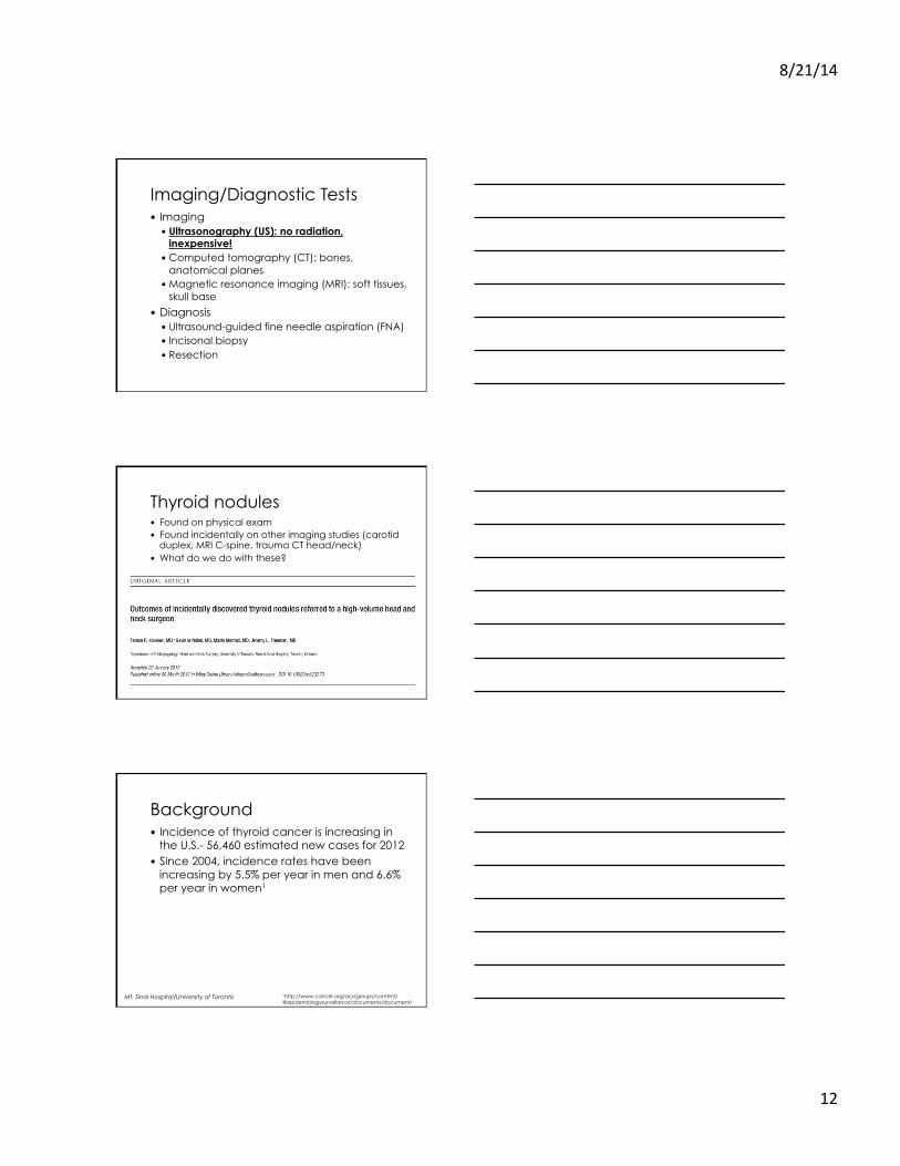
Imaging/Diagnostic Tests � Imaging
� Ultrasonography (US): no radiation, inexpensive!
� Computed tomography (CT): bones, anatomical planes
� Magnetic resonance imaging (MRI): soft tissues, skull base
� Diagnosis � Ultrasound-guided fine needle aspiration (FNA) � Incisonal biopsy � Resection
Thyroid nodules � Found on physical exam � Found incidentally on other imaging studies (carotid
duplex, MRI C-spine, trauma CT head/neck) � What do we do with these?
Background � Incidence of thyroid cancer is increasing in
the U.S.- 56,460 estimated new cases for 2012
� Since 2004, incidence rates have been increasing by 5.5% per year in men and 6.6% per year in women1
Mt. Sinai Hospital/University of Toronto 1http://www.cancer.org/acs/groups/content/@epidemiologysurveilance/documents/document/acspc-031941.pdf

8/21/14
13
Background � Incidence of incidental thyroid nodules
� US for parathyroid disease: 46%1
� Cross-sectional imaging (CT and MRI): 16%2-3 � US for carotid disease: 9-13%4-5 � PET scans: 2-3%6-8
Mt. Sinai Hospital/University of Toronto
1Horlocker . Frontiers in Thyroidology. 1985:1309–1312 2Shetty. AJR Am J Roentgenol 2006;187:1349 –56 3Youserm. AJNR Am J Neuroradiol 1997;18:1423– 8 4Steele. Arch Surg 2005;140:981–5 5Carroll. AJR Am J Roentgenol 1982; 138:499–501 6Cohen. Surgery 2001;130:941– 6 7Are. Ann Surg Oncol 2007;14:239–47 8Kim. Laryngoscope 2005;115:1074–8
Background � Prevalence of incidental thyroid nodules on
ultrasound in the general population : 42-67%1-2
� Mayo clinic: 821 autopsied patients with no history of thyroid disease- 50% found to have a thyroid nodule3
� Schlumberger : clinically occult thyroid nodules found in 30-60% of autopsy evaluations4
Mt. Sinai Hospital/University of Toronto
1Brander. Radiology 1991;181:683-7 2Ezzat. Ann Intern Med 1994;154:1838-40 3Mortensen. J Clin Endocrinol Metab 1955;15:1270-80 4Schlumberger. Thyroid Tumors 1999: 13–30
Methods � 729 new patients referred a single surgeon for
a thyroid nodule were reviewed between February 2009 and January 2011
� 133 patients referred for an incidental thyroid nodule/s per documented history
Mt. Sinai Hospital/University of Toronto

8/21/14
14
Study Group � 133 /729 patients (18.2%) were found to have
an incidental thyroid nodule � 133 patients: 29% male, 71% female � Mean age: 50 years
Mt. Sinai Hospital/University of Toronto
Referral Pattern Referring physician No. of patients % of patients
Primary Care Physician
69 52
Endocrinologist 33 25
Otolaryngologist 24 18
Other* 7 5
Mt. Sinai Hospital/University of Toronto
* obstetrician-gynecologist, neurologist, neurosurgeon, cardiologist, emergency physician
Radiologic Imaging Modality Imaging modality
No. of patients % of patients % malignant
Ultrasound 109 82 28
CT 13 9 38
MRI 8 6 13
Chest x-ray 4 3 0
Mt. Sinai Hospital/University of Toronto
CT, computed tomography; MRI, magnetic resonance imaging
8/21/14
15
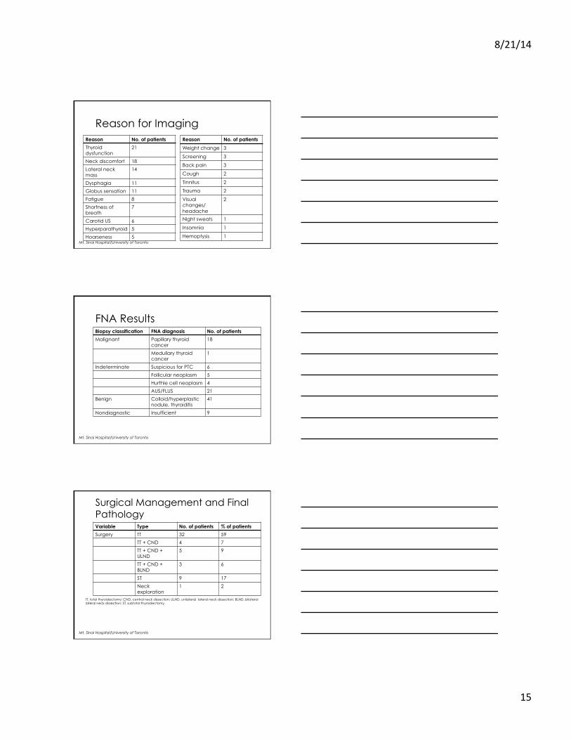
Reason for Imaging Reason No. of patients
Thyroid dysfunction
21
Neck discomfort 18
Lateral neck mass
14
Dysphagia 11
Globus sensation 11
Fatigue 8
Shortness of breath
7
Carotid US 6
Hyperparathyroid 5
Hoarseness 5 Mt. Sinai Hospital/University of Toronto
Reason No. of patients
Weight change 3
Screening 3
Back pain 3
Cough 2
Tinnitus 2
Trauma 2
Visual changes/ headache
2
Night sweats 1
Insomnia 1
Hemoptysis 1
FNA Results Biopsy classification FNA diagnosis No. of patients
Malignant Papillary thyroid cancer
18
Medullary thyroid cancer
1
Indeterminate Suspicious for PTC 6
Follicular neoplasm 5
Hurthle cell neoplasm 4
AUS/FLUS 21
Benign Colloid/hyperplastic nodule, thyroiditis
41
Nondiagnostic Insufficient 9
Mt. Sinai Hospital/University of Toronto
Surgical Management and Final Pathology Variable Type No. of patients % of patients
Surgery TT 32 59
TT + CND 4 7
TT + CND + ULND
5 9
TT + CND + BLND
3 6
ST 9 17
Neck exploration
1 2
Mt. Sinai Hospital/University of Toronto
TT, total thyroidectomy; CND, central neck dissection; ULND, unilateral lateral neck dissection; BLND, bilateral lateral neck dissection; ST, subtotal thyroidectomy.

8/21/14
16
Surgical Pathology Variable Type No. of patients % of patients
Pathology PTC 23 42
Papillary microcarcinoma
13 24
MTC 1 2
Dedifferentiated 1 2
Benign 16 30
Mt. Sinai Hospital/University of Toronto
PTC, papillary thyroid cancer; MTC, medullary thyroid carcinoma.
Results- Summary � 18% of all referrals for thyroid nodules were
picked up incidentally � Most referred from PCP � US by far most common imaging modality � Thyroid dysfunction and neck discomfort
most common reasons for imaging � 41% managed surgically with 29% found to
have thyroid cancer on final surgical pathology
Mt. Sinai Hospital/University of Toronto
Conclusion � Our malignancy rate (29%) is at the high end
of reported malignancy rates of incidentally-discovered thyroid nodules, ranging from 8% to 29%.1-2
� Incidental thyroid nodules should be evaluated in the same fashion as a palpable thyroid nodule, according to the ATA guidelines.
� Many can be observed and followed with serial ultrasounds.
1Papini. J Clin Endocrinol Metab 2002;87:1941– 6 2Kang. Thyroid 2004;14:29-33 Mt. Sinai Hospital/University of Toronto

8/21/14
17
American Thyroid Association Guidelines: Bottom Line � If you palpate a thyroid nodule or find one
incidentally on a CT, MRI, Carotid Duplex US � Thyroid US � TSH � Referral to head and neck surgeon or
endocrinologist for further work-up
� FNA usually indicated if a solid nodule > 1cm � FNA indicated if < 1cm with concerning US
features such as irregular borders or stippled calcifications
Questions/Comments?
Teresa R. Kroeker, MD Thyroid, Parathyroid,
Head and Neck Surgery
[email protected] Cell: (512) 799-1821
Top Related