


Languages
Pages
Legal


大韓ñHH흉醫學會註‘ 第 24 卷 第 2 號 pp. 278 - 281 , 1988 Journal of Korean Radiological Society, 24(2) 278-281 , 1988
CT Characteristics of Peripheral Organizing Pneumonia
Seoung Oh Yang, M.D. , Chul Soon Choi, M.D.치 Myung Joon Kim , M.D., Kyung SOO Lee, M.D.,
Hyung Sik Choi, M.D. , Young Hwan Jun, M.D. and Yong Koo Park, M.D.**
Department of Radiology, Capital Armed Forces Ceneral Hospital
〈국문초록〉 기질화 폐엽의 전산화단층촬영소견
-증례 보고-
국군수도병원 방사선과
앙승오 • 최칠순 김명준 • 이경수 • 최형식 • 전영환 • 박용구 ••
기질화 폐 엽이 원위 폐야에서 고링성으로 오래 지속되는 경우에 단순 흉부사진만으로는 악성 종양파
의 강벨이 어렵다. 최근에 고해상력의 천산화 단층촬영으로 폐실질의 명변의 진단에 많은 도웅을 얻고
있으나, 원위부의 기 질화 페 염 의 전산화 단충촬영 소견은 흔하지 않은 펀이 마. 저 자들은 수술을 하여 영
리학적」뜨로 확인펀 기질화 폐염 1예의 전산화 단층펠영소견을 보고하는 바이 다.
Diagnostic dilemma of persistent mass-forming parenchymal opacity in the lung periphe ry occurs occa
sionally in the realm of diagnostic radiology. Until recently, li terature on the ro le of computed tomography
in peripheral o rganizing pneumonia, which is difficult to differentiate from mali gnancy, has little been publish
ed. We expe ri enced one case of pathologica ll y proven o rganizing pneumonia diagnosed preoperatively by
chest ct When it comes to solitary peripheral mass density in the lung, we think that CT can be proved usef비
in the diagnosis of benign organizing pneumonia by showing regular and smoothly corrugate margin, peripheral
contrast enhancement with inner low density, and air-trapping by intervening normal lung parenchyma
Introduction
The application of computed tomography(CT)
in the evaluation of focal lung disorders has not
actively evolved at the same pace as other pul-
• 공군 항공의학연구원 * Department of Radiologι Aeromedical Research Center
• 국군수도영원 영리과 * * Department of Pathologκ Capital Armed Forces Ceneral
Hospital
이 논운은 1988년 2월 27일에 접 수하여 1988년 3월 19 일에 채택되었음.
monary diseases including mediastinum1,2). The
value and findings of chest CT in assessing mass
forming peripheral organizing pneumonia which is
often difficult to differentiate from a lveolar cell
carcinoma , solitary pulmonary infarct and so on
h ave not been f비ly clarified ye t. We h ave
observed some helpful CT findings of surgically
proven organizing pneumonia located in the lung
periphery , so we wish to present radiologic fin
dings with emphasis on the differential diagnosis
of solitary peripheral mass-fo rming density in the
lung.
- 278 -
- Seoung Oh Yang , et al.: CT Characterist ics 01 Peripheral Organizing Pneumonia
Case Report
A 33-year-이d man was referred to our hospital
because of flu-like symptoms for two weeks despite
empiric antibiotics coverage. There was a decre
aseè. breathing sound on right lower lung field
with inspiratory rale at admission . Laboratory data
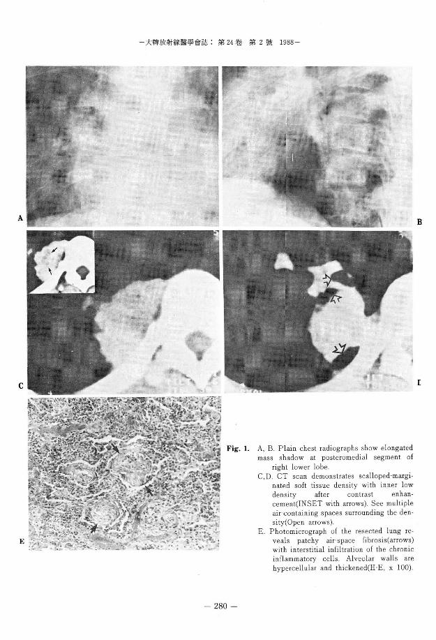
was not specific. Plain chest radiographs showed
elliptical opacity along right retrocardiac space
abutting thoracic spine with rather fluffy mar
gin(Fig. l-A , B). Owing to persistent mass-for
ming lesion , we performed chest CT which re
vealed craggy-surfaced ovoid soft tissue density
with contrast enhancement peripherally and inter
nal low density , and a few cr air-bronchograms
by intervening normal lung tissues(Fig. l-C , D).
On the 30th day following admission , right lower
lobectomy was performed to eliminate the mass
lesion which was shown 6 cm-sized relatively firm
soft tissue mass in the posteromedial segment of
right lower lobe adhered to the parietal pleura.
Pathologic cut surface showed yellowish tan necro
tic focus in the lung specimen and its final diag
nosis was orgamzmg penumonia with fibrosis(Fig .
l-E).
Discussion
To date , the role of CT in the evaluation of
patients with air-space disease has gone minimally
explored. This is primarily because of the ease
with which air-space disease is diagnosed from
plain chest radiographs 1) . According to Gener
eux2) , CT has little role to play during the acute
phase of the pneumonias apart from its valuable
assistance in identifying some of the compli
cations(cavity formation , bronchopleural fistula , empyema). Ovetall consideration of in f1ammatory
disorders is beyond the scope of this report , we
would like to focus our interest on the CT features
of peripheral mass-forming organizing pneumonia
which used to be found by us after its acute stage.
The term of organizing penumonia seems to be
less distinct from the standpoint of its underlying
pathologic morphology. Recently there has been
several articles reinvestigate bronchiolitis obliter
ans organizing pneumonia(BOOP) because it has a
favorable prognosis and good response to steriod
therapy3-S) McLoud et a l. said that localized le
sion with bronchilolitis obliterans is usually refer
red to as “ focal organizing pneumonia" and its
detailed description of CT feature is rare , but the
appearance is that of an irregular sublobar area of
air-space consolidation4) We think our case is not
quite dissimilar to that of localized BOOP ,
although the pathologic pattern of bronchial ob
struct lOn IS not promment
Differential diagnoses include unifocal alveolar
cell carcinoma , single pulmonary infarct , rounded
atelectasis and subpleural tuberculoma. Relatively
uncommon unifocal alveloar cell carcinoma is in
distinguishable on the basis of CT features ,
although frequent association with hilar or media
stinal lymphadenopathy will favor the malig
nancy6) We think smooth undulated surface and
surrounding contrast enhancement pattern and air
-<lensities by normal parenchyma are helpful
aspects of organizing pneumonic process on chest
cr. P비monary thromboembolism with pleural
based area of consolidation may mimic infla
mmation but often m비tiple. Pulmonary embolus
can be determined by radionuclide perfusion scan
when there is only one area of parenchymal den
sity on the plain radiograph2) . Rounded atelectasis
as a benign from of peripheral lung collapse may
be differentiated by the characteristic pleural de
formity with acute angle , curvilinear entrapment
of vessels and bronchi and some other CT signs7) .
Subpleural tuberculoma tends to be sharply cir
cumscribed and there may be small satellite lesions
in the vicinity of a somewhat large granuloma with
frequent parenchymal infiltration and calcifica
tion2) .
279 -
A
C
E
-大韓放射線훌훌學會誌 : 第 24 卷 第 2 號 1988-
280 -
A, B. Plain chest radiographs show elongated mass shadow at posteromedial segment of
right lower lobe C ,D. CT scan demonstrates scalloped-margi
nated soft tissue density with inner low density after contrast enhancement(INSET with arrows). See multiple air-containing spaces surrounding the density(Open arrows)
E. P hotomicrograph of the resected lung re veals patchy air-space fi bros is(arrows) with interstitial infiltration of the chronic inflammatory cells . Alveolar walls are hypercellular and thi ckened(H-E , x 100)
B
[

-Seoung Oh Yang , et al.: CT Characteristics of Peripheral Organizing Pneumonia-
In summary , CT features of rather smoothly
corrugate margin , peripheral contrast enhan
cement with inner low density , surrounding air
densities and absent lymphadenopathy in the case
of peripheral mass-forming opacity favor the diag
nosis of organizing pneumonia although not speci
fic . Appropriate cIinical setting and temporal
change are also helpful in the diagnosis .
REFERENCES
space(alveolar) disease. Seminars in R oentgenology
19:211 , 221 , 1984
3. Epler GR. Colby TV , McLoud TC et al: Bron.
chio/i tis ob/i terans organizing pneumonia. N Eng1
] Med 312.1 52'158, 1985
4. McLoud TC, Epler GR , Colby TV et al: Bronchi.
olitis ob/i terans. R adiology 159:1 , 8, 1986
5. Müller NL , Guerry'Force ML , Stap les CA et al
Differential diagn osis o[ bronchiolitis ob/i terans
with organizing pneumonia and usual interstitial
pneumonia: clinical, [unctional, and radiologic [in.
dings. Radiology 162:151 , 156, 1987
l. Naidich DP , Zerhouni EA , Siegelman SS: Compu. 6. Hill CA: Bron chioloalveolar carcinoma: a review
ted Tomography o[ the Thorax. 1st ed. pp.201'242, Radiology 150:15 , 20, 1984
Raven Press, New York , 1984 7. Doyle TC , Lawler GA: CT [eatures o[ ro unded
2. Genereux GP: CT o[ acute and chronic air' atelectasis o[ the lung. A]R 143:225 , 228, 1984
- 281-
Top Related