


Languages
Pages
Legal


Ângela Pascoal da Costa Crespo
Tese de Doutoramento em Biociências, especialização em Biologia Celular e Molecular, Orientada pelo Professor Doutor Jack Strominger (Universidade de Harvard, EUA) e
pelo Professor Doutor João Ramalho-Santos (Departamento de Ciências da Vida, FCTUC) e apresentada ao Departamento de Ciências da Vida,
Faculdade de Ciências e Tecnologia da Universidade de Coimbra
Characterization of KIR2DS1+ decidual Natural Killer cells in healthy and viral/bacterial – infected human pregnancy
Agosto de 2015

II
Ângela Pascoal da Costa Crespo
Characterization of KIR2DS1+ decidual
Natural Killer cell function in healthy and
viral/bacterial – infected human pregnancy
Tese de Doutoramento em Biociências, especialização em Biologia Celular e Molecular,
Orientada pelo Professor Doutor Jack Strominger (Universidade de Harvard, EUA) e
pelo Professor Doutor João Ramalho-Santos (Departamento de Ciências da Vida, FCTUC)
e apresentada ao Departamento de Ciências da Vida,
Faculdade de Ciências e Tecnologia da Universidade de Coimbra
Agosto de 2015

III
AO MEU AVÔ JOAQUIM
(1934-2010)
TO MY GRANDFATHER

IV
ACKNOWLEDGEMENTS
First and foremost, I would like to thank my supervisor at Harvard University, Professor Jack
Strominger. Jack is a living legend: at the age of 90, Jack still runs an active lab. I am very
grateful for the opportunity to develop my research in his lab and for all the advice and fruitful
discussions, in addition to providing me with the necessary funds to continue my research
after the term of my fellowship.
I wish I could officially assign Tamara Tilburgs as my co-supervisor. From the very
beginning of my project, Tamara has been the definition of what an amazing, responsible
and genuinely invested supervisor should be and it meant the world to me. I am extremely
thankful for the help in designing all the experiments, the availability to listen to my ideas and
make them a reality, the discussions that so much improved my interpretations, the complete
revision of this thesis, and all the instances where Tamara defended my interests. Tamara
was not only my rock in the lab, but also someone I could count on for support on personal
issues. I will hold all my memories with Tamara dearly in my heart.
Não posso deixar de agradecer ao meu orientador na Universidade de Coimbra, Prof. Dr.
João Ramalho-Santos, por todo o apoio desde o início do meu doutoramento. Estou-lhe
muito grata por ter aceite orientar-me e por toda a paciência e conselhos. O início não foi
fácil, mas o Prof. João acreditou em mim e ajudou-me a chegar até aqui com confiança.
To all the members of my lab, past and present (Anita van der Zwan, Ester Leno, Torsten
Meissner, Leonardo Ferreira, Lloyd Chen, Mei-Ling Wong, Basya Rybalov, Vernon Wu,
Zhenhong Guo, Leigh Guerin, Vita Dragos, Brandy Houser), I would like to extend my
sincerest gratitude for making the lab environment so welcoming and collaborative. I want to
thank Bill McCallum, our lab manager, for the help with all administrative issues and more –
Bill knows everything, everyone and everywhere. I have to especially acknowledge Henry
Evans, the former NK cell expert in the lab, for all the support. Finally, Sze-Ling Ng and
Anna Studwell were two of the most special friends I made while in my time in the
Strominger lab and I am deeply grateful for all the moments we shared.
My work wouldn’t have been possible without the contribution of many people in different
levels. First, I’d like to mention all the anonymous donors that contributed with tissue or
blood for my experiments. I hope I have honored them with this thesis. Also, I want to thank
all the members of the Flow Cores of the Northwest Building and Bauer, Harvard University,
for all the FACS sort work, in particular Patricia Rogers who worked several extra hours
every Tuesday. Professor Donald Coen (Harvard Medical School) kindly provided us with
the AD169-GFP strain of HCMV, and Wilfredo García-Beltran (Altfeld Lab) proved to be an
invaluable source of KIR knowledge that generated great discussion. I thank them all for the
support.

V
A little over a year ago, I had a crazy idea that ended up in a very important collaboration
with Professor Judy Lieberman’s lab (Harvard Medical School). I’d like to thank Prof.
Lieberman for welcoming me, and also Sachin Mulik and Farokh Dotiwala who worked
directly with me in the lab (Chapter 4). I am very happy to announce that the work will
continue as part of my postdoctoral project in the Lieberman lab.
I must also thank three very important institutions that made this work a reality. First of all,
the Fundação para a Ciência e Tecnologia, which awarded me my PhD fellowship
(SFRH/BD/33885/2009); the March of Dimes Foundation, which provided my stipend and
research funds for the past year; and the NIH, that funded the majority of the research.
O primeiro passo do meu doutoramento foi a aceitação no Programa Doutoral em
Biologia Experimental e Biomedicina (PDBEB) da Universidade de Coimbra. Quero
agradecer a todos os professores, oradores e organizadores pela oportunidade e
conhecimento transmitido. Em especial, agradeço aos meus colegas do BEB, companheiros
de aventura especialmente nos primeiros 6 meses, pelo espírito de entreajuda.
Boston tem uma comunidade portuguesa muito forte. Graças a todos os meus amigos
portugueses, senti sempre que a minha casa nunca esteve longe. A presença da
Portuguese American Post-Graduate Society (PAPS) foi uma bênção. Graças à
associação conheci portugueses e luso-descendentes que sabem como promover a nossa
cultura. Quero agradecer aos meus amigos mais especiais que nunca me deixaram sentir
sozinha: Inês Tenente e Madalena Barroso, e ao meu comité de boas vindas, Mariana
Fontes e Inês Freitas (and of course Luke Ryan, my favorite roommate ever).
My life in Boston was not only lab – I also found my other passion here. Salsa dancing kept
me sane and brought me a whole new group of friends. I have to especially acknowledge the
amazing work of Ana Haydeé Linares on my Introduction figures (6 and 7). And of course, I
have to thank my dear friend Delia Kong, my “sister”, for all the memories, laughs and tears
we shared in these years.
Os meus amigos e família estiveram sempre no meu coração. Agradeço muito aos meus
avós e tios, aos meus primos Cláudio Gonçalves (especialmente na formatação da tese) e
Adriano Gonçalves, ao meu irmão Luís Crespo e à minha irmã do coração Marta Barroso
por estarem sempre comigo quando as saudades apertam.
Salsa did not bring me only friends. It also brought me my other half. Words are not enough
to describe how much Franklin Condori’s support meant to me for the completion of my
PhD. From being with me through ups and downs and always believing in my worth, to
picking me up at lab on late nights, he has been my shelter. I love him with all my heart.
E finalmente, àqueles que mais merecem o meu agradecimento: os meus pais, Albertina
Crespo e Fernando Crespo. Sem eles nada faria sentido. Obrigada por tudo.

VI
ABSTRACT
Human pregnancy is a challenge for the maternal immune system, which must maintain
tolerance to a semi-foreign entity (the fetus) while keeping immunity against viral, bacterial
and parasite infections. While the mechanisms involved in placental immune tolerance have
been addressed for the last thirty years, very little attention has been given to the maternal
immune system role in the elimination of infections.
In preparation for implantation, the uterine endometrium undergoes modifications known as
decidualization. The placental cells (trophoblast) invade the decidua to facilitate nutrient and
gas exchanges. In particular, the extravillous trophoblast (EVT) of fetal origin detaches from
placental villi, migrates into the decidual tissue and contacts directly with maternal immune
cells such as decidual natural killer cells (dNK).
dNK are the most abundant leukocyte type in decidua and have a different phenotype from
peripheral NK cells (pNK). dNK have lower cytotoxicity in response to MHC-Class I –
negative targets and secrete more cytokines and growth factors than pNK. dNK express high
levels of killer cell immunoglobulin-like receptors (KIR) that recognize MHC Class I in
maternal tissue and EVT. EVT express HLA-E and HLA-G, in addition to polymorphic HLA-
C. Recently, women who lack the gene for the activating receptor KIR2DS1 and carry a fetus
expressing HLA-C2 (the ligand for KIR2DS1) were found to be at increased risk of recurrent
miscarriage, fetal growth restriction and preeclampsia. The molecular and cellular
mechanisms involved are unknown. The current hypothesis suggests that absence of
activating KIR2DS1 and expression of KIR2DL1 (inhibitory receptor for HLA-C2) results in
stronger inhibition of dNK upon interaction with fetal HLA-C2. This inhibition dampens dNK
secretion of growth factors involved in placentation and vessel remodeling, leading to
pregnancy complications. Particular combinations of KIR haplotypes and HLA allotypes are
also associated with improved clearance of viral infection. Therefore we hypothesized that
the protective effect of KIR2DS1 lies in a more efficient clearance of infections during early
pregnancy. This may prevent virus-induced placental pathology and subsequent fetal and
maternal complications. Both hypotheses are tested in the present work. In this thesis,
human dNK phenotype and function in healthy pregnancy and viral/bacterial infections are
characterized, focusing on the role of KIR2DS1.
Chapter 1 of this thesis provides a general introduction to contextualize the data presented
in the following chapters. Chapter 2 presents a characterization of KIR2DS1+ (S1+) dNK in
healthy pregnancy. dNK are shown to express receptors for MHC Class I in higher
frequencies than pNK, specifically KIR2DL1 and KIR2DS1, KIR2DL2/3 (inhibitory HLA-C1
receptors) and NKG2A/NKG2C (respectively, inhibitory and activating HLA-E receptors).
dNK express similar levels of the cytolytic molecules granzyme A and B as pNK, but lower

VII
levels of the pore forming protein perforin. Strikingly, dNK express much higher levels of the
antimicrobial peptide granulysin than pNK. In addition, the expression of granzyme B,
perforin and granulysin are increased in S1+ dNK. These observations point to a higher
cytolytic potential of S1+ dNK. In fact, cytotoxicity of dNK from KIR2DS1+ women was not as
efficiently inhibited by HLA-C2+ target cells as dNK from KIR2DS1- women and pNK from all
donors. S1+ dNK also displayed the highest levels of degranulation of all 4 subsets (L1+, S1+,
L1+S1+, L1-S1-) in response to HLA-C2+ target cells. Although dNK degranulated and
secreted cytokines in response to MHC Class I-negative or HLA-C2+ cell lines, co-culture
with EVT did not elicit secretion of IFN-γ, VEGF or GM-CSF.
The failure of KIR2DS1+ dNK to secrete cytokines in response to HLA-C2+ EVT led to the
hypothesis that the higher cytotoxic potential of KIR2DS1+ dNK contributes to an increased
clearance of placental viral infections, a possibility explored in chapter 3. Human
cytomegalovirus (HCMV) is the most common congenital viral infection and can lead to
miscarriages, fetal growth restriction and permanent hearing damage. In chapter 3, dNK
were shown to degranulate and secrete pro-inflammatory cytokines in response to HCMV-
infected decidual stromal cells (DSC). However, dNK did not respond to HCMV-infected
choriocarcinoma cell line (JEG3) and sample-matched primary EVT. This demonstrates the
high resistance to death by trophoblast and highlights the difficulties the maternal immune
system faces to clear infections in the tolerogenic compartment of the placenta.
Furthermore, KIR2DS1+ dNK were demonstrated to deliver an increased cytolytic response
to HCMV-infected DSC expressing HLA-C2. This observation may explain the protective
effect of KIR2DS1 in human pregnancy by limiting viral infection in the placenta. KIR2DS1+
dNK may control the spread of infection and reduce the risk for virus-induced pregnancy
complications.
Finally, in chapter 4, the role of dNK in the elimination of bacterial infections was
investigated. The high expression of antimicrobial peptide granulysin by dNK was the basis
for this study. dNK constitutively secreted high levels of granulysin. In addition, dNK cells
and dNK supernatants were able to kill both extracellular and intracellular bacteria in JEG3
without killing the host cell. This effect was independent of degranulation. This striking result
reveals a novel function for dNK and strengthens their role as immune effector cells.

VIII
RESUMO
A gravidez humana é um desafio para o sistema imunitário materno, o qual deve garantir
tolerância a um feto semi-incompatível enquanto mantém imunidade contra infeções. Os
mecanismos envolvidos na tolerância têm sido bastante explorados, mas o estudo da
imunidade a infeções congénitas tem sido negligenciado.
A implantação do embrião exige que o endométrio uterino sofra modificações profundas
(decidualização). As células fetais da placenta (trofoblastos) invadem a decidua para
permitir trocas eficientes de gases e nutrientes. Em especial, os trofoblastos extravilosos
(EVT) migram para o tecido decidual, contatando directamente com linfócitos maternos
como as células Natural Killer deciduais (dNK). As dNK são os leucócitos mais abundantes
na decidua, mas são menos citotóxicas em resposta a células alvo que não expressam
MHC Classe I que as NK do sangue (periféricas, pNK), e secretam mais citocinas e fatores
de crescimento. As dNK expressam níveis elevados de recetores KIR (killer cell
immunoglobulin-like receptors) que reconhecem proteínas MHC Classe I nos tecidos
maternos e EVT. Os EVT expressam HLA-E e HLA-G e também HLA-C, que é polimórfico.
Recentemente, foi encontrada uma associação prejudicial entre genes KIR maternos e
alelos HLA-C do feto. As grávidas que não possuem o gene para o recetor ativador
KIR2DS1 e cujo feto expressa HLA-C2 têm um risco elevado de aborto espontâneo,
restrição de crescimento fetal e pré-eclâmpsia. Os mecanismos moleculares e celulares
envolvidos não são conhecidos. A hipótese proposta atualmente sugere que a ausência de
KIR2DS1 ativador e expressão de KIR2DL1 (recetor inibidor de HLA-C2) leva a uma
inibição mais pronunciada das dNK aquando da interação com HLA-C2 fetal. Esta inibição
reduz a secreção pelas dNK de fatores de crescimento envolvidos na placentação e
remodelação de artérias uterinas, resultando em complicações na gravidez. São também
conhecidas combinações de haplótipos de KIR com alelos de HLA que estão associadas a
um melhor combate a infeções virais. Estes dados levaram à nossa hipótese de que o efeito
protetor de KIR2DS1 está relacionado com uma resposta imunitária mais eficiente a
infeções congénitas. Este fenómeno pode prevenir danos induzidos por vírus na placenta e
consequentes complicações na gravidez. Ambas as hipóteses foram testadas no trabalho
aqui apresentado. Nesta tese, é apresentada uma caraterização do fenótipo e função das
dNK humanas em gravidezes saudáveis e afetadas por infeções virais ou bacterianas, com
foco no papel de KIR2DS1.
O capítulo 1 desta tese fornece uma introdução geral para contextualizar os dados
apresentados nos capítulos seguintes. O capítulo 2 apresenta uma caraterização das dNK
que expressam KIR2DS1 (S1+) em gravidezes saudáveis. A frequência de expressão de
recetores para MHC Classe I nas dNK é mais elevada que nas pNK, em especial KIR2DL1

IX
e KIR2DS1, KIR2DL2/3 (recetores inibidores de HLA-C1) e NKG2A/NKG2C (recetor inibidor
e ativador, respetivamente, de HLA-E). As dNK contêm níveis semelhantes das proteínas
citolíticas granzima A e B aos das pNK, mas níveis mais reduzidos de perforina, que forma
poros nas membranas das células alvo. Em especial, as dNK contêm muito mais proteína
anti-microbiana granulisina que as pNK. Ainda, a expressão de granzima B, perforina e
granulisina é mais elevada em dNK S1+. Estas observações revelam um potencial citolítico
elevado das dNK S1+. De facto, a citotoxicidade das dNK isoladas de grávidas que possuem
o gene KIR2DS1 não foi inibida por células alvo HLA-C2+ com a mesma eficiência que dNK
isoladas de grávidas sem KIR2DS1 ou pNK de todos os indivíduos da amostra. As dNK S1+
também demonstraram possuir a mais elevada citotoxicidade dos 4 subgrupos (L1+, S1+,
L1+S1+ e L1-S1-) em resposta a células-alvo HLA-C2+. As dNK desgranularam (libertaram
moléculas citolíticas) e secretaram citocinas em resposta a células alvo MHC- ou HLA-C2+,
mas não secretaram IFN-γ, VEGF ou GM-CSF após cultura com EVT.
A falta de resposta das dNK à interação com EVT levou à hipótese de que o potencial
citotóxico das dNK S1+ contribui para um combate mais eficiente às infeções virais na
placenta. O vírus humano citomegalovírus (CMV) provoca a infeção congénita mais comum
e pode levar a aborto espontâneo, restrição do crescimento fetal e danos auditivos
permanentes. No capítulo 3, mostrou-se que as dNK desgranulam e secretam citocinas pró-
inflamatórias em resposta a células do estroma da decidua (DSC) infetadas com CMV. No
entanto, as dNK não responderam a células de coriocarcinoma (JEG3) ou EVT primários
infetados com CMV. Estas observações demonstram a resistência à morte dos trofoblastos
e sublinham a dificuldade que o sistema imunitário materno enfrenta para eliminar infeções
na placenta. Em especial, mostrou-se que as dNK S1+ são mais citolíticas em resposta a
DSC HLA-C2+ infetadas com CMV. Esta observação pode explicar o efeito protetor de
KIR2DS1 na gravidez humana ao limitar a infeção viral na placenta. As dNK S1+ podem
controlar a disseminação da infeção e reduzir o risco de complicações da gravidez
induzidas por vírus.
Finalmente, no capítulo 4, investigou-se o papel das dNK na eliminação de infeções
bacterianas. A expressão elevada do péptido anti-microbiano granulisina em dNK constituiu
a base para este estudo. As dNK secretam granulisina constitutivamente, e as células dNK
e sobrenadantes das suas culturas conseguiram eliminar bactérias extracelulares e
intracelulares em JEG3 sem matar as células hospedeiras. Este efeito foi independente de
desgranulação. Este importante resultado revela uma nova função das dNK e reforça o seu
papel como células imunitárias efetoras.
X
LIST OF ABBREVIATIONS
Ab Antibody
Ag Antigen
AML Acute myelogenous leukemia
APC Antigen Presenting Cell
BCR B cell receptor
CD14 Monocyte/macrophage marker
CD3 T cell marker
CD45 Lymphocyte lineage marker
CD56 NK cell marker
CD95L Fas Ligand
CFU Colony Forming Unit
CTL Cytotoxic T lymphocytes
DAP10 DNAX activation protein of 10kDa
DAP12 DNAX activation protein of 12kDa
DC Dendritic cells
dMϕs Decidual Macrophages
dNK Decidual NK cells
DSC Decidual Stromal Cells
EBI3 Epstein-Barr virus-induced gene 3 (interleukin-27 subunit β)
ER Endoplasmic reticulum
ETC Electron Transport Chain
EVT Extravillous trophoblast
GBS Group B Streptococcus
GM-CSF Granulocyte-monocyte colony stimulation factor
GNLY Granulysin
GvHD Graft vs Host Disease
Gzm Granzyme
HA Hemagglutinin
HCMV Human cytomegalovirus
HCV Hepatitis C virus
HIV Human Immunodeficiency virus
HLA Human Leukocyte Antigen
HSCT Hematopoietic Stem Cell Transplantation
HSV Herpes simplex virus
IFN Interferon
Ig Immunoglobulin
IL Interleukin
IS Immunological Synapse
ITAM Immunoreceptor tyrosine-based activation motif
ITIM Immunoreceptor tyrosine-based inhibitory motif
KIR Killer cell Immunoglobulin-like Receptor
LILR Leukocyte Immunoglobulin-like Receptor
LRC Leukocyte Receptor Region
MFI Mean Fluorescence Intensity
MHC Major Histocompatibility Complex

XI
MIC MHC Class I polypeptide-related sequence
MOI Multiplicity of infection
MTOC Microtubule Organization Center
NCR Natural Cytotoxicity Receptors
NK Natural Killer
PFN Perforin
pNK Peripheral NK cells
PRR Pattern Recognition Receptors
SAPLIP Saposin-like proteins
SHP Src homology region 2 domain-containing phosphatase
SYK Spleen Tyrosine Kinase
TAP Transporter associated with Antigen Processing
TCR T-cell receptor
TGF-β Transforming growth factor β
Th T helper cell
TNF Tumor Necrosis Factor
TRAIL TNF-related apoptosis-inducing ligand
Treg T regulatory cells
ULBP UL16 binding sequence
VEGF Vascular Endothelium Growth Factor
VT Villous trophoblast
ZAP70 Zeta-chain-associated protein kinase 70

XII
TABLE OF CONTENTS
ABSTRACT ................................................................................................................................. VI
RESUMO .................................................................................................................................. VIII
LIST OF ABBREVIATIONS .............................................................................................................. X
CHAPTER 1 – GENERAL INTRODUCTION ..................................................................................... 1
1. Self vs. Non-Self recognition ............................................................................................... 2
1.1 HLA recognition and the allogeneic response .............................................................. 6
1.1.1 Transplant allorecognition ..................................................................................... 6
2. Natural killer (NK) cells – between innate and adaptive immunity ................................... 8
2.1 NK cell phenotype ....................................................................................................... 9
2.2 NK cell immune synapse and degranulation ................................................................ 9
2.3 Other mechanisms of NK cell target killing ................................................................. 10
2.4 Activating and Inhibitory receptors in NK cells – a balance ........................................ 10
2.4.1 Natural Cytotoxicity Receptors (NCR) ................................................................. 11
2.4.2 C-type lectins ...................................................................................................... 11
2.4.3. Fc Receptors ...................................................................................................... 13
2.4.4 Leukocyte Immunoglobulin-like Receptors (LILR) ............................................... 13
2.4.5 Killer cell Immunoglobulin-like Receptors (KIR) ................................................... 14
2.5 NK cell education ....................................................................................................... 16
2.6 NK cell response to viruses, tumors and transplants ................................................. 16
2.6.1 Viral responses ................................................................................................... 17
2.6.2 Tumor responses ................................................................................................ 18
2.6.3 NK cells in haploidentical haemopoietic stem cell transplantation (HSCT) .......... 19
3. Pregnancy – an immunological paradox ...........................................................................19
3.1 MHC class I and II expression in human placenta ..................................................... 21
3.2 Decidual leukocytes and potential immune responses ............................................... 22
3.2.1 T helper cells, cytotoxic T cells and regulatory T cells ......................................... 22
3.2.2 Macrophages and dendritic cells ......................................................................... 23
3.2.3 Natural killer cells ................................................................................................ 23
3.3 HLA-KIR interactions in maternal and fetal health ...................................................... 25
3.4 Viral, bacterial and parasite infections in pregnancy .................................................. 26
3.4.1 Viral infections ..................................................................................................... 26
3.4.2 Bacterial infections .............................................................................................. 27
3.4.4 Parasite infections ............................................................................................... 27
AIMS .........................................................................................................................................28

XIII
CHAPTER 2 – CHARACTERIZATION OF THE PHENOTYPE, CYTOTOXICITY AND CYTOKINE
SECRETION CAPACITY OF KIR2DS1+ DECIDUAL NK CELLS ..........................................................29
Introduction ..................................................................................................................... 30
Materials and Methods .................................................................................................... 32
Results ............................................................................................................................ 36
Discussion ....................................................................................................................... 45
CHAPTER 3 – EXPRESSION OF KIR2DS1 BY DECIDUAL NK CELLS INCREASES THEIR ABILITY
TO CONTROL PLACENTAL HCMV INFECTION ...............................................................................48
Introduction ..................................................................................................................... 49
Materials and Methods .................................................................................................... 51
Results ............................................................................................................................ 54
Discussion ....................................................................................................................... 62
CHAPTER 4 – CONTROL OF BACTERIAL INFECTIONS BY DECIDUAL NATURAL KILLER CELL –
SECRETED GRANULYSIN .............................................................................................................65
Introduction ..................................................................................................................... 66
Materials and Methods .................................................................................................... 70
Results ............................................................................................................................ 73
Discussion ....................................................................................................................... 78
CHAPTER 5 – GENERAL DISCUSSION ........................................................................................81
REFERENCES .............................................................................................................................88
APPENDIX I. PRIMARY CELLS AND LINES USED IN THE PRESENT WORK ....................................... 104

1
CHAPTER 1
GENERAL INTRODUCTION

2
The human immune system is a complex set of cells and proteins that resulted from millions
of years of co-evolution of humans and their pathogens. To allow survival of the individual
and, more broadly, of human populations, each human being has a system ready to defend
it from bacteria, viruses, fungi and parasites (1).
The functioning of the immune system is complex and two main arms can be identified:
innate and adaptive immune system. The innate immune system constitutes the first line of
defense and delivers a quick response to any invading pathogen by recognizing common
patterns absent in the human body. The adaptive immune system, although slower, is more
efficient, because it can recognize specific markers in each pathogen and save that
information. This way the adaptive immune system can instantly eliminate subsequent
infections of the same pathogen and clear the infection before any significant damage
occurs. This property of the immune system to “remember” is named “immune memory” and
is the basis for the many vaccines available today (1).
How the immune system attacks only foreign (“non-self”) and dangerous pathogens, while
remaining controlled and not damaging (“tolerating”) our own (self) tissues will be discussed
in detail below. This introduction will cover three main areas: self vs non-self recognition in
the systemic immune system, the characteristics and role of Natural Killer cells and the
complexity of the local immune system regulation during pregnancy. In the first part, the
mechanisms of self and foreign tissue and pathogen recognition by immune leukocytes will
be discussed. In the second part, the function of Natural Killer cells, their subsets, receptors
and ligands will receive special attention. In the third part, the regulation and function of the
immune system during pregnancy will be described. This last part will emphasize the double-
edged challenge faced by the local immune system in the maternal-fetal interface:
maintaining immune tolerance to semi-foreign fetal tissues while remaining active against
potential viral, bacterial and parasite infections. The protective role of the NK cell receptor
KIR2DS1 (Killer cell Immunoglobulin-like Receptor, 2 extracellular Domains, Short
cytoplasmic tail 1) in healthy and complicated pregnancy will be addressed in detail.
1. Self vs. Non-Self recognition
The innate immune system recognizes common proteins and chemicals released by injured
cells. These molecules include chemicals that induce the secretion of cytokines, activate
innate immune leukocytes to secrete other chemicals or attack the pathogens, or
chemokines that recruit additional leukocytes to the site of infection. These leukocytes are,
for example, phagocytes (e.g. macrophages or dendritic cells) that express proteins at the
surface known as “pattern recognition receptors” (PRRs). PRRs recognize common proteins

3
from pathogens, leading the phagocytes to engulf the pathogens and lyse them
(phagocytosis) (2).
The adaptive immune system relies on Major Histocompatibility Complex (MHC) molecules
that present self and non-self peptides to T and B cells (3). The MHC locus contains the
most variable (polymorphic) functional genes described in vertebrates. In humans, these
proteins are known as human leukocyte antigens (HLA) and two classes can be recognized:
Class I and Class II. While Class I HLA are expressed by all nucleated cells, Class II are
constitutively expressed only by bone marrow-derived antigen1 presenting cells (APC), such
as macrophages, dendritic cells, B cells and thymic epithelial cells (1).
MHC Class I molecules are surface proteins which extracellular part is composed of 3 alpha
domains and a β2-microglobulin chain. The 1 and 2 domains form a groove where a
peptide can bind. The 3 domain contains a binding site for the co-receptor CD8 expressed
in CD8+ T cells, discussed below. During and following regular protein synthesis, a
proportion of the resulting proteins are rapidly degraded into peptides by the proteasome.
The resulting peptides are translocated into the lumen of the endoplasmic reticulum (ER) via
the transporter associated with antigen processing (TAP). Within the ER, the peptides are
loaded onto newly synthesized MHC I heavy chain/β2-microglobulin heterodimers (3). MHC
Class I constantly display short intracellular endogenous peptides (8-9 aminoacids long) as a
sign of the status of the cell to be surveyed by T cells. When a cell is infected by an
intracellular pathogen (e.g. virus), or
starts producing new and damaged
proteins due to tumor formation, the
complex formed by pathogen-derived
peptides and MHC Class I are recognized
by CD8+ T cells (cytolytic T lymphocytes)
and elicit an immune response (4).
In humans, six MHC Class I genes can
be identified: 3 classical highly
polymorphic genes (HLA-A, B and C) and
3 non-classical (HLA-E, G and F). All
nucleated cells express HLA-A, B and C,
which main function is antigen
presentation, and also HLA-E, which
although non-polymorphic, has a role in
Natural Killer cell inhibition. HLA-G is
1 Antigen – any molecule that elicits an immune response (1).
Table I – MHC Class I genes, proteins and number of
alleles. Source: HLA Nomenclature (http://hla.alleles.org/)
4
tissue restricted and is believed to be involved in immune modulation (e.g. induction of
regulatory T cells). HLA-F function is still unknown (5–7) (Table I).
MHC Class II molecules, unlike MHC Class I, are tissue restricted. Class II are only
expressed by professional antigen presenting cells (APC) like dendritic cells, macrophages
and B cells (antibody producing cells). MHC Class II proteins have 2 heavy chains, one with
two domains and one with two β domains. The groove formed by the 1 and β1 domains
can present a longer peptide than MHC Class I, which is usually derived from extracellular
pathogens (e.g. phagocytosed bacteria) which proteins were subsequently degraded. MHC
Class II contains a binding side for the co-receptor CD4 in CD4+ T cells (T helper cells). The
recognition of the MHC-peptide complex by CD4+ T cells will activate them and lead to the
immune response. In humans, 3 main MHC Class II proteins can be identified: HLA-DR,
HLA-DQ and HLA-DP, which are also highly polymorphic (8) (Table I).
T cells are the main immune lymphocytes involved in the recognition of MHC-peptide
complexes. T cells originate from hematopoietic precursors in the bone marrow and later
migrate to the thymus to finish their maturation. T lymphocytes come in two flavors, CD4+
and CD8+, and can be further divided into several subsets with different functions. All T cells
express a T cell receptor in the surface, the TCR, which is exclusive and different for each T
cell and recognizes a different MHC-peptide complex. The different TCRs are formed by
somatic rearrangement of the TCR genes, which leads to a different sequence and,
consequently, a different specificity of each T cell. The diversity of TCR in a given individual
is enormous, and since their formation is random, some TCRs recognize self-peptides that
can lead to auto-immune attack (9). To avoid this, T cells need to go through a mechanism
of positive and negative selection in the thymus during development. T cells that have too
low affinity to self MHC-peptide complex (negligent) or too high affinity to self MHC-peptide
complex (auto-reactive) are eliminated in the thymus (positive and negative selection,
respectively). This way, only T cells specific for self-MHC presenting foreign peptides and
can be activated by them are allowed to leave the thymus (10).
CD4+ T cells are usually identified as T helper cells (Th). When in the periphery, Th cells
interact with APCs expressing their specific MHC-Class II – peptide complex, leading to their
activation, proliferation and differentiation into different types of T helper cells (depending on
the cytokine environment) (11). CD4+ T cells can differentiate into Th1 cells, which produce
more inflammatory cytokines like interferon-γ (IFN- γ) and tumor necrosis factor (TNF-),
helping CD8+ T cell, NK cell and macrophage responses. CD4+ T cells can also differentiate
into Th2, which produce interleukins 4 and 5 (IL-4 and IL-5) that provide help to antibody-
producing B cells. Upon clearing of the infection, a subset of CD4+ T cells differentiates into

5
memory cells that will be able to respond faster to a second infection by the same pathogen
(12).
T regulatory cells are also CD4+, but have a very distinct phenotype (expressing high levels
of CD25 and the transcription factor FoxP3). These cells are involved in immune tolerance
and dampen the immune response, avoiding auto-immune reactions (13).
CD8+ T cells are the major immune cells involved in tumor and viral clearance. CD8+ T
lymphocytes directly recognize MHC-Class I-peptide complexes, which leads to their
activation, proliferation and differentiation into cytotoxic T lymphocytes (CTL) (11). CTLs
directly kill infected or tumor cells by several ways, the most important being the release of
cytotoxic molecules to the point of contact between the T cell and the target cell (immune
synapse - IS). Among the cytotoxic molecules are perforin, a pore forming protein that
creates an opening in the target cell membrane, allowing the entrance of granzymes, which
lead to apoptosis of the target cell by several pathways (caspase-dependent or independent)
(14, 15). Upon activation and clearance of infection, a subset of CD8 T cells differentiates
into memory T cells that can elicit a rapid response upon a subsequent infection with the
same pathogen (16).
The other adaptive immune lymphocyte which can play a role in the immune response are B
cells. B cells have a B cell receptor (BCR), also highly specific to foreign epitopes2 (since
each B cell undergoes somatic rearrangements in their BCR locus) (17). However, these
cells do not need to bind a MHC-peptide complex for activation. Instead, their BCR
recognizes free antigen, but the B cells need interaction with primed T cells of similar antigen
specificity to start their differentiation and proliferation (18, 19). Upon activation, B cells
produce antibodies (immunoglobulins, Ig), highly specific molecules. The antibodies
produced by a given B cell have the same specificity of the BCR, and their diversity is as
high as the TCR diversity (the result of somatic rearrangements in the immunoglobulin
genes) (17). Antibodies can be secreted and help in the elimination of pathogens by several
ways – opsonization (coating of a pathogen, flagging it for phagocytosis), aggregation of
pathogens or antibody-mediated cell death (by binding receptors in Natural Killer cells –
discussed in the section below - or macrophages) (20).
Natural Killer cells, which will be discussed in more detail on section 2, are lymphocytes
commonly categorized as innate immune cells, but recent studies have identified NK cell
functions that place NK in the border between immune and adaptive immunity. NK cells
share some characteristics with cytotoxic T lymphocytes, in particular the ability to form
immune synapses and deliver cytolytic granules for target cell killing (21). Unlike T cells, NK
do not recognize specific epitopes in target cells; instead, NK cells express activating
2 Epitope – specific region of an antigen recognized by TCR or antibodies (1).

6
receptors that identify stress-induced proteins upregulated by infected or cancerous cells,
and quickly eliminate them without prior stimulation (22). However, NK cells have recently
been shown to possess the ability to recognize specific viral proteins expressed in infected
cells, leading to their proliferation and stronger response upon second encounter with the
same antigen (23). These studies have been the subject of controversy and require further
development. In addition, NK cells express a wide range of inhibitory receptors that bind
groups of alleles of MHC Class I, as a way of protecting healthy self tissues from NK attack
(24).
1.1 HLA recognition and the allogeneic response
The MHC locus is the most variable (polymorphic) region in the human genome. These MHC
polymorphisms allow for a great variety of pathogenic peptides to be presented, resulting in
an increased ability to clear a large diversity of pathogens. However, these polymorphisms
also underlie the extreme difficulty in finding perfectly matched organs or unrelated bone
marrow donors that will not induce a strong anti-graft response (25). In the next paragraphs
the mechanisms of transplant allorecognition will be discussed to illustrate how the maternal
immune system could potentially respond to the allogeneic fetal tissues during pregnancy.
1.1.1 Transplant allorecognition
Transplantation is the artificial transfer of cells, tissues or organs from one individual to
another. When the graft is syngeneic (genetically identical to the host) or autologous (a
transplant from one individual into itself) there is perfect histocompatibility and no significant
immune response is elicited. When there is incompatibility, in general an immune response
is directed against the foreign antigens. Allorecognition is the term used to describe the
recognition of transplanted allogeneic tissues by the host, while alloresponse denotes the
effector mechanisms recruited in the reaction to the foreign tissue and the outcome of those
effects (25). Allorecognition can proceed via two mechanisms: direct allorecognition, where
T cells recognize epitopes on the intact donor MHC molecules displayed in the surface of
transplanted cells; or indirect allorecognition, in which donor MHC molecules are processed
and presented as peptides by self-MHC molecules (in a similar fashion to antigen
processing) (26).
1.1.1.1 T cell responses to transplants
Alloreactive T cells can identify foreign tissue by both direct allorecognition and
indirect allorecognition. While direct allorecognition can be carried out by both CD8+ and
CD4+ T cells (by recognizing donor MHC Class I and MHC Class II molecules, respectively),
indirect allorecognition is exclusive of CD4+ T cells, which identify the recipient APC
displaying MHC Class II - donor peptide complexes.

7
The vigorous nature of the direct alloresponse and its immediacy (acute rejection) in
comparison with the indirect pathway is the result of direct recognition of intact MHC by T
cells without the need for processing and presentation of self-MHC (25) (Figure 1A).
The indirect pathway differs from the direct pathway by the requirement for antigen
processing. Alloantigens shed from a graft are processed as exogenous antigens and
presented by APCs in association with self-MHC Class II. These responses are considerable
slower compared to the direct pathway (Figure 1B). The indirect response is likely to be
responsible for long term responses to engrafted tissues once donor APCs are exhausted.
The presence of donor APCs in transplanted tissues at the time of transplantation can lead
to a vigorous anti-donor alloresponse in the early period post-engraftment. The death and
removal of these APCs over time reduces this response. The indirect alloresponse, on the
contrary, requiring antigen capture and processing, is less rapid than the direct pathway but
continues for the life of the graft, as graft derived antigens are continuously acquired and
processed (chronic rejection). Acute rejection of transplanted tissues is more commonly
observed in the early post engraftment period, while tolerance to grafts develops at a later
time point. This correlates with demonstrations that regulatory T cells able to mediate
transplant tolerance have indirect rather than direct pathway alloreactivity (26).
Figure 1. Transplant allorecognition mechanisms by T and NK cells. A) Direct allorecognition of MHC Class
II in APC by CD4+ T cells (left) and of donor MHC Class I by CD8+ T cells (right). B) Indirect allorecognition of
recipient MHC/donor peptide complex in receptor APC by CD4+ T cells. Donor MHC fragments (antigens) are
taken up by APCs (black arrow) and presented via MHC Class II (arrowhead) to CD4+ T cells. C) NK recognition
of donor tissue. On the left, the donor shares MHC alleles with the recipient, inhibiting NK responses. On the
right, recipient NK does not recognize donor MHC (missing self), leading to activation.
8
1.1.1.2 B cell responses to transplants
B cells can contribute to transplant rejection via two major mechanisms – as antigen-
presenting cells (APC), contributing to indirect allorecognition, and as antibody producers. B
cells can be activated and secrete antibodies specific to donor MHC molecules. The
presence of donor-specific HLA antibodies in the circulation of transplant recipients has a
negative impact on transplantation outcome (18). These alloantibodies mainly induce
rejection by complement-dependent mechanisms. The complement system is a complex set
of proteins that, when triggered by antibody-antigen, lead to a cascade of reactions resulting
in cell lysis by membrane disruption, or chemo-attraction of macrophages and other
phagocytes able to clear the foreign cells (27).
1.1.1.3 Natural Killer cell responses to transplants
NK cells express inhibitory receptors that bind groups of alleles of self-MHC Class I
and are responsible for protection of self tissue from NK attack. In a MHC-mismatched
transplant, MHC class I alleles expressed in the donor tissue are different from the
recipient’s and unable to inhibit NK (missing-self). NK cell-mediated rejection of transplants
is included in the direct alloresponse, leading to acute rejection (Figure 1C) (28).
2. Natural Killer (NK) cells – between innate and adaptive immunity
Human Natural Killer cells (NK) are a subpopulation of lymphocytes, like B and T cells, that
originate from hematopoietic progenitors in the bone marrow. Unlike T cells, all their
development takes place in the bone marrow, from where they migrate into various tissues,
including secondary lymphoid organs and the blood (circulating or peripheral NK cells –
pNK) (22).
NK were initially defined by their ability to kill target cells without prior immunization. While T
cells require a first encounter with a pathogen to prime their activity, differentiation and
proliferation, leading to formation of memory, NK can deliver a rapid response upon first
encounter (21). One of the hallmarks of NK is their lack of need to recognize specific
epitopes or antigens to be activated. NK express non-rearranged somatic activating
receptors that identify common ligands on target cells – stress ligands. However, NK need
another layer of regulation to keep them from attacking healthy tissue. In fact, NK express
inhibitory receptors that recognize self MHC Class I molecules – different receptors
recognize different groups of alleles (28). Healthy cells always express MHC Class I
molecules, flagging them to be spared by NK. Infected or tumor cells upregulate stress-
induced surface proteins and, as a mechanism of immune evasion, also downregulate MHC
molecules to escape attack by T cells. The combination of stress signals and low MHC
expression works as a danger signal for NK, which take over the immune response (29).

9
2.1 NK cell phenotype
Two main populations of NK can be found in peripheral blood, defined by the expression of
two surface markers: CD56 and CD16. About 90% of NK in the blood are CD56lowCD16+, the
main cytotoxic subpopulation, containing cytolytic granules as CD8+ T cells. The remaining
10% are CD56highCD16- and are not cytotoxic. CD16- NK do not contain cytolytic molecules
and are defined by the secretion of cytokines (e.g. IFN-γ). CD16- NK are also the main
population of NK found in secondary lymphoid organs. CD56highCD16- and CD56lowCD16+
subsets are also sequential stages of maturation and CD56high can progress to CD56low cells
upon activation, as occurs in reactive lymph nodes or inflamed tissues (30).
NK can be further subdivided in an impressive array of subsets, based on their expression of
activating and inhibitory receptors (31). The receptors belong to different families of proteins
and recognize different ligands and will be addressed in more detail below. The most
interesting characteristic of NK cell receptors is their random expression in the population of
NK of a given individual. Each NK can express any number of receptors from zero to 30+ in
different combinations. The pattern of receptors of each NK is defined during maturation and
is maintained throughout the life of the cell and during division, generating clones. The
balance between activating and inhibitory receptors, combined with the expression of ligands
in the target cell, is crucial for the outcome of the NK cell response (32).
2.2 NK cell immune synapse and degranulation
Activation of NK triggers a complex and highly regulated response leading to cytolytic
granule release, resulting in the death of the target cell. Conversely, interaction of inhibitory
receptors with their ligands negatively regulates NK activity. Activation and inhibition take
place at the specialized contact sites between NK and target cells, known as the
immunological synapse (IS) (33). Upon encountering a target cell (i.e. a cell with
downregulated MHC Class I molecules and expressing stress signals at the surface), NK
form an activating synapse. This requires several steps: I. Contact and adhesion, led by
integrins such as LFA-1. II. Receptor ligation and segregation linked to initial signaling. III.
Actin cytoskeleton rearrangements (tight conjugation). IV. Further receptor clustering and
sustained signaling (signal amplification). V. Microtubule cytoskeleton rearrangements
(MTOC polarization). VI. Granule polarization and degranulation. VII. IS disassembly (34).
Similarly to T cells, NK contain cytolytic granules with molecules such as the serine
proteases granzymes and the pore-forming protein perforin. Upon synapse formation, these
granules exocytose their contents, releasing perforin which will polymerize in the target cell
surface, opening a pore which allows granzymes to enter. Granzymes are then responsible
for apoptosis by triggering caspase-dependent and independent pathways (Figure 2A) (14,
15).

10
2.3 Other mechanisms of NK cell target killing
Synapse formation is the main killing mechanism employed by NK, but other pathways can
be followed. For example, NK express death receptor ligands such as TRAIL or Fas-L, which
engage death receptors on the surface of target cells and initiate target cells apoptosis(35).
Furthermore, NK cells can also secrete cytokines such as IFN-γ and TNF- which can
increase inflammation and activate T cells and dendritic cells to ensure clearance of target
cells (Figure 2B and C) (36).
2.4 Activating and Inhibitory receptors in NK cells – a balance
NK express several receptors responsible for modulating NK function when facing a target
cell. The most important receptors can be subdivided in 5 families: Natural Cytotoxicity
Receptors (NCR), Fc receptors (FcR), C-type lectins, Leukocyte Immunoglobulin-like
receptors (LILR) and Killer cell Immunoglobulin-like receptors (KIR). While some families
include only activating (NCR and FcR) or inhibitory receptors (LILR), others include both
Figure 2. NK cell target killing mechanisms. A) Granule exocytosis. The recognition of activating ligands (and
lack of MHC Class I molecules) or antibodies bound to target cell antigens (antibody dependent cell cytotoxicity)
leads to immune synapse formation and degranulation of NK. The same stimuli lead to cytokine secretion (e.g.
interferon-gamma, IFN-γ) that increase inflammation and activate T cells, dendritic cells (DC) and macrophages
(Mϕ) (B). (C) Apoptosis induction. The engagement of death-inducing receptors (FasR, TRAIL receptor) by NK
ligands (FasL, TRAIL) and tumor necrosis factor (TNF)-α production by NK lead to apoptosis of target cells.
11
types (KIR and C-type lectins) (Figure 3 and Table II). NK cells express several other
receptors (e. g. adhesion molecules, integrins, etc.), that contribute to NK activation but, for
clarity, such proteins will not be addressed here.
2.4.1 Natural Cytotoxicity Receptors (NCR)
The major NK receptors able to induce NK-mediated killing are the natural cytotoxicity
receptors (NCRs) belonging to the Immunoglobulin-like superfamily — NKp46 (NCR1),
NKp44 (NCR2) and NKp30 (NCR3). The activating receptors are organized into multi-chain
complexes where the ligand-binding and signal-transducing sub-units are separate. The
ligands for these receptors are mostly unknown but believed to be viral proteins or stress
ligands expressed at the surface of infected cells. The signal transducing units are diverse,
and include CD3ζ, FcεRγ and DAP12 (DNAX activation protein of 12kDa). DAP12 contains
an intracellular immunoreceptor tyrosine-based activation motif (ITAM) that recruits protein
kinases involved in activating signaling such as Spleen tyrosine kinase (SYK) and Zeta-
chain-associated protein kinase 70 (ZAP70). These receptors also use different downstream
pathways leading to the NK activation (28).
2.4.2 C-type lectins
C-type lectins are a family of transmembrane proteins characterized by a type II membrane
orientation (extracellular C terminus) and the presence of a C-type lectin domain. This family
includes both activating and inhibitory receptors of NK.
The best-characterized activating C-type lectin is NKG2D. NKG2D recognizes “induced-self”
ligands – molecules not expressed or expressed at low levels on normal cells, but
upregulated on unhealthy cells due to the activation of pathways associated with cancer,
infection or stress. NKG2D ligands include MHC class I polypeptide-related sequence A and
B (MICA/MICB) and UL16-binding proteins (ULBP) 1-6 in humans. NKG2D associates with
the signal transducing proteins DAP10 or DAP12 (Figure 3). NKG2D recognition of target
cells is one of the most important pathways of killing of tumor cells (37).
In addition, two other C-type lectins have been well studied: NKG2A and NKG2C. Both of
these proteins form a heterodimeric receptor in association with another type II
transmembrane molecule: CD94 (Table II). Both CD94/NKG2C and CD94/NKG2A are
specific for the non-classical MHC class I (class Ib) molecules HLA-E (38). While NKG2C is
an activating receptor associated with DAP12 (Figure 3), NKG2A is an inhibitory receptor
that transduces negative signaling through immunoreceptor tyrosine-based inhibitory motif
(ITIM) sequences present in its cytoplasmic tails (Figure 3). Upon tyrosine phosphorylation,
ITIM interacts with Src homology region 2 domain-containing phosphatase (SHP)-1/2 that
disrupt the activating signaling pathways (39).

12
Figu
re 3
. NK
cel
l rec
epto
rs a
nd
liga
nd
s.

13
2.4.3. Fc Receptors
Fc receptors are commonly expressed in macrophages and bind the Fc common part of
antibodies bound to antigens on the surfaces of other cells. NK express the Fc receptor
FcγRIII (CD16), which triggers antibody-dependent cell cytotoxicity (ADCC). FcγRIII signals
through FcεRγ and CD3ζ transducers (Figure 3), leading to degranulation and cytokine
production (32).
2.4.4 Leukocyte Immunoglobulin-like Receptors (LILR)
The Leukocyte immunoglobulin-like receptor genes (LILRB1 and LILRB2) are mapped in
the chromosome 19 Leukocyte Receptor Complex (LRC) region. LILRB1/LIR1 and
LILRB2/LIR2 are inhibitory receptors that display four extracellular Ig-like domains and a
cytoplasmic portion characterized by ITIM motifs that mediate association with the SHP-1
phosphatase (Figure 3) (40). These receptors are expressed on monocytes and dendritic
Table II – NK cell receptors and ligands.

14
cells and LILRB1 is also expressed on B-cells, some T-cells and subsets of NK. They bind to
the non-classical class I molecules HLA-G, HLA-F and a similar broad spectrum of HLA-A, -
B and -C alleles (41).
2.4.5 Killer cell Immunoglobulin-like Receptors (KIR)
Killer cell Ig-like Receptors (KIRs) are the major MHC Class I NK receptors and have
specificity for discrete groups of HLA Class I alleles. Their genes map in the leukocyte
receptor complex (LRC) located on human chromosome 19q13.4 (42). Although most of the
functional KIRs are inhibitory, a few have activating function. The ~150 kb KIRs cluster
consists of 14 genes and 2 pseudogenes. However, due to rearrangements that lead to
deletions and duplications during evolution, each individual possesses a different number of
genes in this locus. Based on their gene content, two haplotypes have been defined:
haplotype A characterized by identical genes number and content (with mostly inhibitory
receptors and only one activating receptor gene), and haplotype B, highly variable in terms
of gene number (Figure 4). The KIRs cluster may have evolved almost 50 million years ago
from an ancestral gene (KIR3DL0 also known as KIR3DX1) (43).
The inhibitory KIR receptors have an extracellular portion with two (KIR2D) or three (KIR3D)
Ig-like C2-type domains, a transmembrane region characterized by non-polar amino acid
residues, and a long (L) cytoplasmic tail containing two ITIM sequences (Figure 3). Upon
recognition of sufficient levels of MHC class I molecules, KIRs generate src kinase-
dependent tyrosine phosphorylation of ITIMs. Phosphorylated ITIMs serve as docking motifs
for SHP1 or SHP2 through the formation of a ternary complex with -arrestin-2. This
complex finally blocks the signaling cascades induced by activating receptors (44). The
inhibition of NK-mediated cytotoxicity requires co-aggregation with triggering receptors and it
is focused in the immune-synapse area and not over the whole cell membrane. This way, an
NK cell, although inhibited in the limited area of contact with a particular target cell, may still
survey for transformed/infected tissues and maintain their killing potential (45).
Among the 14 KIR genes described so far in humans, five encode receptors associated with
activating rather than inhibitory potential (Table II) (43). These molecules have a shorter (S)
cytoplasmic tail not containing ITIM sequences, and a transmembrane portion displaying a
polar lysine residue. This residue forms a salt bridge with the opposite charged amino acid
present in the transmembrane region of the ITAM–containing adaptor DAP12 (Figure 3) (46).
15
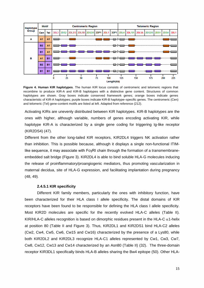
Activating KIRs are unevenly distributed between KIR haplotypes. KIR-B haplotypes are the
ones with higher, although variable, numbers of genes encoding activating KIR, while
haplotype KIR-A is characterized by a single gene coding for triggering Ig-like receptor
(KIR2DS4) (47).
Different from the other long-tailed KIR receptors, KIR2DL4 triggers NK activation rather
than inhibition. This is possible because, although it displays a single non-functional ITIM-
like sequence, it may associate with FcγRI chain through the formation of a transmembrane-
embedded salt bridge (Figure 3). KIR2DL4 is able to bind soluble HLA-G molecules inducing
the release of proinflammatory/proangiogenic mediators, thus promoting vascularization in
maternal decidua, site of HLA-G expression, and facilitating implantation during pregnancy
(48, 49).
2.4.5.1 KIR specificity
Different KIR family members, particularly the ones with inhibitory function, have
been characterized for their HLA class I allele specificity. The distal domains of KIR
receptors have been found to be responsible for defining the HLA class I allele specificity.
Most KIR2D molecules are specific for the recently evolved HLA-C alleles (Table II).
KIR/HLA-C alleles recognition is based on dimorphic residues present in the HLA-C 1-helix
at position 80 (Table II and Figure 3). Thus, KIR2DL1 and KIR2DS1 bind HLA-C2 alleles
(Cw2, Cw4, Cw5, Cw6, Cw15 and Cw16) characterized by the presence of a Lys80, while
both KIR2DL2 and KIR2DL3 recognize HLA-C1 alleles represented by Cw1, Cw3, Cw7,
Cw8, Cw12, Cw13 and Cw14 characterized by an Asn80 (Table II) (32). The three-domain
receptor KIR3DL1 specifically binds HLA-B alleles sharing the Bw4 epitope (50). Other HLA-
Figure 4. Human KIR haplotypes. The human KIR locus consists of centromeric and telomeric regions that
recombine to produce KIR-A and KIR-B haplotypes with a distinctive gene content. Structures of common
haplotypes are shown. Gray boxes indicate conserved framework genes; orange boxes indicate genes
characteristic of KIR-A haplotypes; purple boxes indicate KIR-B haplotype–specific genes. The centromeric (Cen)
and telomeric (Tel) gene-content motifs are listed at left. Adapted from reference (212).

16
A alleles (HLA-A3 and -A11) bind KIR3DL2 and apparently display some peptide specificity
(Table II) (51).
2.5 NK cell education
Until recently, it was thought all NK expressed at least one inhibitory receptor for self-MHC
class I molecules; this model provided a satisfactory explanation for self-tolerance and for
the involvement of NK in missing-self recognition (52). However, studies of humans and
mice that lacked MHC class I molecules had shown they were hyporesponsive, even though
these individuals had normal numbers of NK. These NK were not able to release their
cytotoxic granules to kill MHC Class I – negative target cells (53). Hyporesponsive NK were
thought to be unique to MHC class I-deficient individuals, but work from several laboratories
has now shown they also exist in normal humans and mice, and are characterized by a lack
of inhibitory receptors (53). The active engagement of inhibitory receptors on NK by self-
MHC class I molecules during maturation is the key event that determines whether an NK
will be functionally capable of mediating missing-self recognition (education), or whether it
will be of low cytotoxicity following stimulation (54). There are several models to explain how
this education/licensing occur. However, the rheostat model is the most widely accepted.
The rheostat model proposes NK have increased or decreased responsiveness depending
on the strength of the inhibitory signal received during maturation. For example, NK with no
or low expression of inhibitory KIR for self HLA molecules will establish weak interactions
with their ligands during maturation, leading to lower cytotoxicity upon later encounter of
target cells in the periphery. This suggests NK education operates in a quantitative manner
(cytotoxicity is increased or decreased) and states that NK cytotoxicity can either be ‘tuned
up’ (arming) or ‘tuned down’ (disarming) rather than in a binary manner (e.g. cytotoxicity is
completely shut down or turned on). The tuning of NK thus depends on the inhibitory signal
received by individual NK cells (55).
The licensing or education of NK by their receptors adds a level of sensitivity to self MHC
ligand expression. Productive licensing through inhibitory signaling provides a two-fold
benefit to NK function. It serves to simultaneously enhance effector responses (e.g. IFN-γ
secretion and cytotoxicity) and broaden the NK cell’s target specificity to include aberrant
cells that would not be detected by stimulatory receptors alone (56).
2.6 NK cell response to viruses, tumors and transplants
NK play a key role in the immune fight against viral infections and tumor cells and can both
have a beneficial or harmful role during transplantation. Due to the high polymorphism of
MHC molecules and the diversity of KIR haplotypes in the population, certain combinations
of MHC-KIR can lead to different outcomes (Figure 5).

17
2.6.1 Viral responses
Viruses are intracellular pathogens that take advantage of the host cell machinery to
replicate. Cells infected with viruses will enter a state of stress leading to the expression of
ligands for activating NK receptors (e. g. MICA/B, recognized by NKG2D). Certain viruses,
such as influenza and human cytomegalovirus (HCMV) express proteins that are displayed
at the surface of infected cells and act as ligands for NK receptors [e.g. influenza
hemagglutinin (HA) – NKp46 and HCMV pp65-NKp30]. To escape T cell immune responses,
most viruses interfere with the MHC Class I processing and surfacing, leading to a lower
expression of MHC by infected cells. This phenomena leads to “missing-self” recognition by
NK, tipping the balance towards activation instead of inhibition (Figure 5A) (57).
Interestingly, human studies provide evidence that individuals of matched KIR/HLA class I
may exhibit enhanced viral control. These correlations have been observed in immune
responses to HIV-1 (3DL1:HLA-Bw4), hepatitis C virus (HCV) (2DL3:HLA-C1), influenza
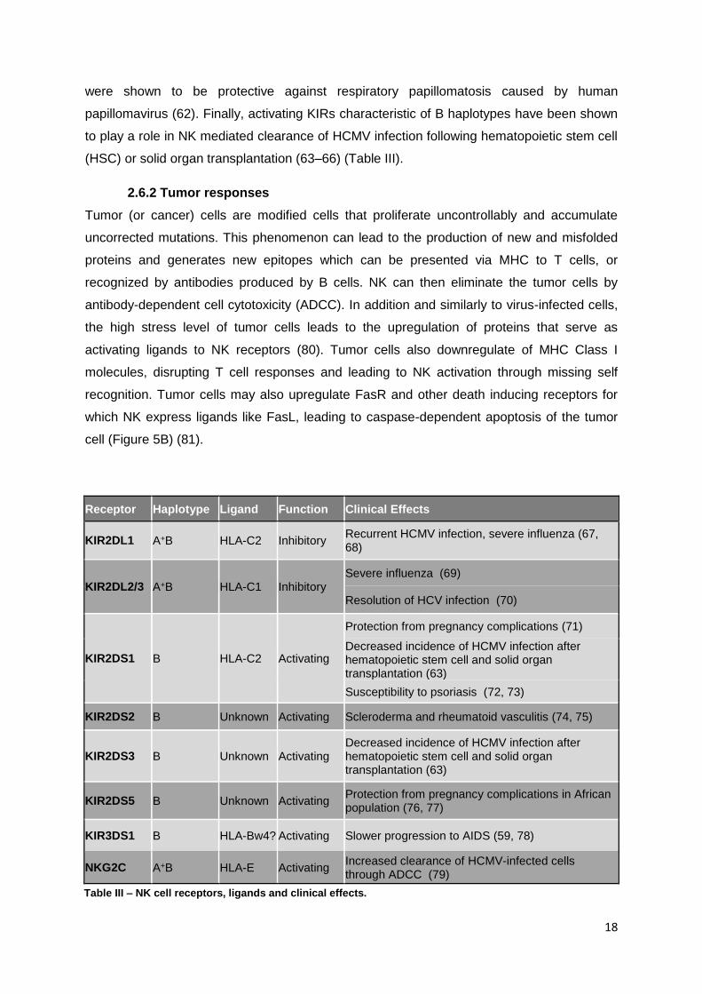
(3DL1:HLA-Bw4;2DL2/DL3:HLA-C1), and HCMV (58). The combined presence of KIR3DS1
and HLA-Bw4 alleles was associated with significantly slower progression to AIDS, lower
viral load and slower decline of CD4+ T cells (59–61). In addition, KIR3DS1 and KIR2DS1
Figure 5. NK cell response to viruses, tumors and transplants. A) Viral infected cells express viral and stress
proteins that serve as activating ligands for natural cytotoxicity receptors (NCR) in NK and downregulate MHC
Class I, leading to NK activation and degranulation. B) Tumor cells upregulate stress ligands (e.g. MICA/B),
death receptors (e.g. FasR) and misfolded proteins that can be recognized by antibodies. In addition, tumor cells
downregulate MHC Class I. NK cells are activated through NKG2D (MICA/B receptor) and FcR (antibody
receptor, triggering ADCC), leading to degranulation, and can engage death receptors in tumor cells through
TRAIL and FasL expression. C) MHC-matched transplant donor cells are protected from NK attack due to
inhibitory KIR engagement. Mismatched receptors (missing self) do not inhibit NK and are rejected. D) Missing
self recognition by donor NK, in addition to engagement of activating ligands expressed by recipient leukaemia
cells, lead to donor NK activation and elimination of leukaemia cells.
18
were shown to be protective against respiratory papillomatosis caused by human
papillomavirus (62). Finally, activating KIRs characteristic of B haplotypes have been shown
to play a role in NK mediated clearance of HCMV infection following hematopoietic stem cell
(HSC) or solid organ transplantation (63–66) (Table III).
2.6.2 Tumor responses
Tumor (or cancer) cells are modified cells that proliferate uncontrollably and accumulate
uncorrected mutations. This phenomenon can lead to the production of new and misfolded
proteins and generates new epitopes which can be presented via MHC to T cells, or
recognized by antibodies produced by B cells. NK can then eliminate the tumor cells by
antibody-dependent cell cytotoxicity (ADCC). In addition and similarly to virus-infected cells,
the high stress level of tumor cells leads to the upregulation of proteins that serve as
activating ligands to NK receptors (80). Tumor cells also downregulate of MHC Class I
molecules, disrupting T cell responses and leading to NK activation through missing self
recognition. Tumor cells may also upregulate FasR and other death inducing receptors for
which NK express ligands like FasL, leading to caspase-dependent apoptosis of the tumor
cell (Figure 5B) (81).
Table III 1
Table III 2
Receptor Haplotype Ligand Function Clinical Effects
KIR2DL1 A+B HLA-C2 Inhibitory Recurrent HCMV infection, severe influenza (67, 68)
KIR2DL2/3 A+B HLA-C1 Inhibitory Severe influenza (69)
Resolution of HCV infection (70)
KIR2DS1 B HLA-C2 Activating
Protection from pregnancy complications (71)
Decreased incidence of HCMV infection after hematopoietic stem cell and solid organ transplantation (63)
Susceptibility to psoriasis (72, 73)
KIR2DS2 B Unknown Activating Scleroderma and rheumatoid vasculitis (74, 75)
KIR2DS3 B Unknown Activating Decreased incidence of HCMV infection after hematopoietic stem cell and solid organ transplantation (63)
KIR2DS5 B Unknown Activating Protection from pregnancy complications in African population (76, 77)
KIR3DS1 B HLA-Bw4? Activating Slower progression to AIDS (59, 78)
NKG2C A+B HLA-E Activating Increased clearance of HCMV-infected cells through ADCC (79)
Table III – NK cell receptors, ligands and clinical effects.

19
2.6.3 NK cells in haploidentical haemopoietic stem cell transplantation (HSCT)
In some cases of hematopoietic stem cell transplantation (HSCT), there are mismatches
between the KIR-HLA combinations of donor and recipient. This situation is a classical
“missing-self” setting, which leads the recipient NK to attack the donor cells due to lack of
inhibition (Figure 5C). However, HSCT is a special transplant because it contains immune
cells from the donor, including NK cells. A fraction of NK from the donor may be able to kill
target cells from the recipient and lead to Graft-vs-Host Disease (GvHD) (82). Remarkably,
the contrary has been observed: the presence of alloreactive donor-derived NK have been
correlated with highly improved survival after HSCT for acute myelogenous leukaemia (AML)
and alloreactive NK promote engraftment and decrease leukaemic relapses (83). Leukaemic
cells express high levels of ligands for NCR that promote lysis by NK, and combined with
missing self, leads to their elimination by donor NK cells (Figure 5D). In particular, Moretta
and others showed a mismatch between homozygous HLA-C1+ and HLA-C2+ donor/receptor
pairs lead to the elimination of leukemia blasts (e.g. donor KIR2DL1+ NK kill leukemia blasts
from a HLA-C2- receptor) (84). (Figure 5D).
Recent experimental evidence has revealed some activating KIR are also involved in the
graft-vs-leukemia effect. In a recent study, Moretta and others provided direct evidence that
donor KIR2DS1+ NK play a crucial role in the lysis of receptor C2/C2 leukaemia blasts (85).
Cytolysis was inhibited by monoclonal antibody (mAb)-mediated blocking of KIR2DS1.
These data strongly support the clinical relevance of activating KIRs in the elimination of
leukaemic cells and the benefit of HLA-C mismatched HSCT.
3. Pregnancy - an immunological paradox
Human reproduction occurs by viviparity. This carries obvious advantages for the fetus,
which enjoys protection and efficient exchange of gas and nutrients with the maternal blood
(86). Human placentation is classified as haemochorial, the most invasive type of
placentation. In preparation for implantation, the human uterine mucosa is transformed into
decidua, a highly specialized tissue able to support placental invasion. Following
implantation, the blastocyst develops into an inner cell mass (the fetus precursor) and an
external layer of cells (trophoblast) which gives rise to the placenta and fetal membranes
(Figure 6). Over gestation, the trophoblast evolves into a fully formed placenta, constituted
by several layers of cells. Villous trophoblasts (VT) form tree-like structures that extend the
surface of the placenta in contact with maternal blood. VT include cytotrophoblast
(proliferating cells composing the inner cell layer of the villi) and syncytiotrophoblast
(composed by cells that fuse together to form a single layer over the villi). In addition, the
placenta contains extravillous trophoblast (EVT). EVT is the most invasive type, detaching
from villi and infiltrating the uterine tissue and the maternal vessels. EVT replaces the

20
endothelium of uterine spiral arteries, while the smooth muscle cell layer is degraded. These
processes open the maternal vessels and reduce resistance to allow for a low pressure
blood flow into the placenta and intervillous space. The maternal blood surrounds the
syncytiotrophoblast and facilitates the exchange of nutrients and gases with the fetus (86)
(Figure 7). Shallow placentation or inadequate maternal vessel transformation can lead to
serious conditions later in pregnancy that include intrauterine growth restriction, stillbirth and
preeclampsia, a hypertensive disorder of pregnancy (87–89).
The evolution of hemochorial placentation allowed for a better support of fetal development.
However, such an intimate contact between mother and fetus exposes the fetal trophoblast
(a semi-allogeneic entity) to the maternal immune system. In previous sections, the immune
responses that can be raised against mismatched transplants were discussed; however, in
human pregnancy, the mother tolerates a semi-allogeneic organ for 9 months. This
phenomenon raises a question that has fascinated immunologists for 60 years, starting with
Sir Peter Medawar. In his monolog “Some immunological and endocrinological problems
raised by the evolution of viviparity in vertebrates,”(90) he proposed three explanations for
maternal immunological tolerance: physical separation of maternal and fetal tissues, the
antigenic immaturity of the fetal tissues, and immunological inertness of the mother.
Although none of those explanations have held up, they had a profound influence on
research concerned with maternal-fetal tolerance and mechanisms of fetal immune evasion
(91).
Figure 6. Diagram of the gravid uterus showing the placenta, extraembryonic membranes and decidualized uterus wall. Image courtesy of Ana Haydeé Linares, adapted from reference (94).
21
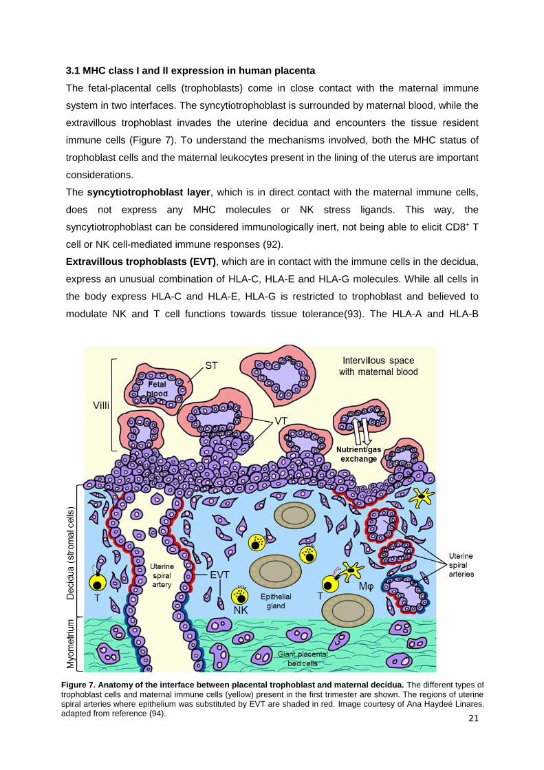
3.1 MHC class I and II expression in human placenta
The fetal-placental cells (trophoblasts) come in close contact with the maternal immune
system in two interfaces. The syncytiotrophoblast is surrounded by maternal blood, while the
extravillous trophoblast invades the uterine decidua and encounters the tissue resident
immune cells (Figure 7). To understand the mechanisms involved, both the MHC status of
trophoblast cells and the maternal leukocytes present in the lining of the uterus are important
considerations.
The syncytiotrophoblast layer, which is in direct contact with the maternal immune cells,
does not express any MHC molecules or NK stress ligands. This way, the
syncytiotrophoblast can be considered immunologically inert, not being able to elicit CD8+ T
cell or NK cell-mediated immune responses (92).
Extravillous trophoblasts (EVT), which are in contact with the immune cells in the decidua,
express an unusual combination of HLA-C, HLA-E and HLA-G molecules. While all cells in
the body express HLA-C and HLA-E, HLA-G is restricted to trophoblast and believed to
modulate NK and T cell functions towards tissue tolerance(93). The HLA-A and HLA-B
Figure 7. Anatomy of the interface between placental trophoblast and maternal decidua. The different types of
trophoblast cells and maternal immune cells (yellow) present in the first trimester are shown. The regions of uterine spiral arteries where epithelium was substituted by EVT are shaded in red. Image courtesy of Ana Haydeé Linares, adapted from reference (94).

22
molecules, which initiate allograft rejection, are not expressed, which leaves HLA-C as the
only polymorphic molecule in the trophoblast surface that could represent a mismatch
between mother and fetus. All 3 MHC Class I molecules interact with receptors — such as
KIRs, CD94/NKG2 and LILR — expressed by NK and subsets of T cells (94). Interestingly,
microarray and functional gene set enrichment analysis revealed a striking immune-
activating potential for EVT that is absent in villous trophoblast (95).
3.2 Decidual leukocytes and potential immune responses
During pregnancy, the decidua accumulates several types of immune cells. The relative
amounts and the phenotype of these cells are very different from the ones found in blood.
Decidual leukocytes are composed of 60-80% NK cells, followed by 15-20% macrophages
and 5-10% T cells in first trimester of pregnancy (Figure 7). Low percentages of dendritic
cells, γ T cells and NKT cells can also be found, but B cells are virtually absent. Below is a
short description of the most important immune cells found at the fetal-maternal interface
(Figure 7).
3.2.1 T helper cells, cytotoxic T cells and regulatory T cells
T cells are the main immune cell population found in blood (70%), which is in stark contrast
to decidua, where they represent only 5-10% in first trimester, but this percentage increases
to 60-80% at term. Their low frequency is considered a mechanism of immune evasion,
since T cells could recognize mismatched HLA-C alleles in EVT and elicit rejection. Both
CD4+ and CD8+ T cells in decidua display an activated phenotype; however, the percentage
of regulatory T cells found in decidua is much higher than in blood (96). Studies in abortion-
prone mice where Tregs were adoptively transferred to the site of pregnancy, avoiding fetal
resorption, showed the vital importance of these cells in pregnancy outcome (97). In
addition, Tilburgs et al demonstrated an HLA-C mismatch between mother and fetus leads to
an increase in the number and activity of activated T cells in the maternal-fetal interface. As
a compensatory mechanism, the function of Tregs in the mismatched pregnancies is also
enhanced. (98). Moreover, EVT have the ability to induce Treg differentiation in decidua
(95).
Some of the mechanisms employed by EVT to avoid T cell attack involved expression of Fas
ligand (which can induce apoptosis in Fas receptor-expressing cells like activated CD4+ and
CD8+ T cells) and expression of B7 family members (co-stimulatory molecules) that have an
inhibitory effect on T cell activation (B7-H1, B7-H2, B7-H3, B7-DC) (91). In addition,
trophoblast cells and APC produce the tryptophan catabolizing enzyme IDO, which inhibits T
cell proliferation by tryptophan depletion or production of toxic metabolites (99).
23
3.2.2 Macrophages and dendritic cells
Macrophages (Mϕ) comprise the second most abundant leukocyte in human decidua (10-
15%). The large abundance of CD14+ decidual macrophages (dMϕs) and the near absence
of CD14- dendritic cells (DC) suggest they are the most important professional APCs in the
decidua (100). Therefore, dMϕs, which express MHC class II, are likely to play a crucial role
in antigen presentation to decidual T cells and may contribute in the establishment of fetal–
maternal immune tolerance. In addition to their antigen presenting function, dMϕs have
been shown to express receptors important for phagocytosis of degraded extracellular matrix
products (101) as well as in apoptotic cell clearance and tissue remodeling (100, 102).
Furthermore, CD14+ dMϕs produce immunomodulatory proteins such as TGF-β, but also
proinflammatory IL-6 and IL-8 when stimulated by cell lines expressing the fetal-trophoblast
HLA-G homodimer (48). Recently, our lab identified two distinct populations of dMϕs in
decidua: CD11chigh and CD11clow. CD11chigh dMϕs express genes associated with lipid
metabolism and inflammation, whereas CD11clow dMϕs express genes associated with
extracellular matrix formation, muscle regulation, and tissue growth. The CD11chigh dMϕs
also differ from CD11clow dMϕs in their ability to process protein Ag and are likely to be the
major APCs in the decidua(103).
3.2.3 Natural killer cells
The infiltration of decidual NK (dNK) cells is part of the cyclical changes of the endometrium
and is clearly influenced by sex hormones, particularly progesterone. While in blood NK cells
represent about 10% of lymphocytes, in decidua they are particularly abundant, constituting
up to 70% of leukocytes. dNK start accumulating even before implantation, which may
implicate a role in placentation. During early pregnancy, dNK accumulate as a dense
infiltrate around the trophoblast and they progressively disappear from mid-gestation
onwards and at term represent only 10-20%. Therefore, their presence is coincident with the
period of trophoblast invasion (placentation is complete by about 20 weeks gestation) (94).
Our lab and others have shown dNK form a distinct NK cell population that has many
differences in gene expression profile, cytokine secretion profile and cytolytic capacity in
comparison with peripheral blood NK cells (pNK) (Table IV). While 90% of pNK are
CD56lowCD16+, all dNK are CD56highCD16-. dNK express KIRs and NKG2 receptors, and
contain high levels of the cytolytic molecules perforin, granzyme B, and granulysin (104,
105). These characteristics, however, do not lead to high cytotoxicity, as they are not able to
kill MHC Class I negative targets as efficiently as pNK (e.g. cell lines K652 or 721.221). The
low cytotoxicity of dNK is due to an intrinsic deficiency in polarizing cytolytic granules to the
immune synapse (106).

24
dNK have been shown to secrete high levels of Interleukin-6 (IL-6), IL-8 and vascular
endothelial growth factor (VEGF) in response to HLA-G transfected 721.221 cells (221.HLA-
G) and antibody stimulation (48, 107). The potential to secrete cytokines led to the
hypothesis that dNK facilitate implantation and placental growth by producing chemokines
for EVT invasion and growth factors involved in vascular remodeling of the spiral arteries.
However, our lab recently showed that contact with EVT alone does not induce secretion of
VEGF, IFN-γ or the granulocyte-monocyte colony-stimulating factor GM-CSF by dNK, which
indicates other stimuli are involved in the activation of dNK cytokine secretion (95). In mice,
uterine NK cells have in fact been shown to be crucial for proper vascularization of the
maternal-fetal interface(108).
dNK express many of the receptors that can interact with the MHC Class I expressed in the
EVT. HLA-G was thought to modulate NK responses, but more recent findings have added
controversy to that claim (109, 110). While HLA-E could potentially inhibit NK responses
against the trophoblast (111), mismatched paternal HLA-C alleles could lead to missing self
recognition by dNK (112).
Table IV – Differences between pNK and dNK.
25
3.3 HLA-KIR interactions in maternal and fetal health
Similarly to pNK, dNK express both activating and inhibitory receptors, especially MHC-
Class I receptors. Interestingly, they are found at higher frequencies and higher expression
levels in decidua (113). While KIR and MHC can influence viral clearance or HSCT outcome,
pregnancy outcome and complications can also be traced down to KIR-HLA interactions
between maternal dNK and trophoblast.
In 2004, Hiby et al reported that mothers homozygous for KIR-A haplotypes (genotype AA)
were at greater risk for preeclampsia than mothers having one (AB) or two (BB) KIR-B
haplotypes (114). The risk was further increased for the subset of KIR-AA mothers carrying
babies bearing the HLA-C2 epitope. KIR-A haplotypes are characterized for the presence of
the inhibitory receptor (KIR2DL1) and the absence of the activating receptor (KIR2DS1) for
HLA-C2 (Figure 4). More recently, by comparing the pregnancies of mothers whose KIR-B
haplotype genes are only centromerically or telomerically located (Figure 4), Hiby et al
focused the protection effect within the telomeric KIR-B haplotype genes: KIR3DS1,
KIR2DL5, KIR2DS3/5, and KIR2DS1 (71). Although genetic analysis cannot distinguish
between them, because of strong linkage disequilibrium, functional studies point to KIR2DS1
as the protective gene. KIR2DS1 is a well-defined HLA-C2 receptor and protection increased
when the fetus expressed HLA-C2. In addition, the protective effect was extended from only
pre-eclampsia to recurrent miscarriage and fetal growth restriction (Table III) (71).
Interestingly, the detrimental association of KIR-AA with HLA-C2 may have been a strong
evolutionary force for the distribution of KIR and MHC haplotypes in human populations.
Frequency of AA haplotype and HLA-C2 epitope is inversely correlated. For example, the
Japanese have high KIR AA and low C2 frequencies (115). In the US Caucasian population,
KIR AA has a frequency of 30%, while HLA-C2 epitope frequency is about 50%. The
KIR2DS1 gene frequency in the Eastern US population is of 33%(116).
The cellular mechanisms involved in this genetic association are mostly unknown, but the
current hypothesis is that dominance of the inhibitory HLA-C2 receptor (KIR2DL1) in women
with KIR-AA genotype reduces dNK stimulation of EVT and the depth of EVT invasion. This
hypothesis relies on the paternal HLA-C2 as responsible for the inhibition of dNK and was
tested in a mouse model of these associations. However, the mouse studies demonstrated
the parental origin of the inhibitory MHC is irrelevant (117). On the other hand, women with
telomeric KIR-B haplotypes are protected, because dNK are activated through
KIR2DS1/HLA-C2 interaction, counterbalancing inhibition and leading to the secretion of
cytokines facilitating trophoblast invasion and placental growth (118).
A recent study addressed the cytokine secretion capacity of KIR2DS1+ dNK and identified
GM-CSF as a growth factor secreted by KIR2DS1+, but not KIR2DS1- dNK. GM-CSF was
shown to promote the migration of trophoblast cell lines (117, 118). This increase in cytokine

26
production was observed when dNK were stimulated with KIR2DS1 antibodies and classical
MHC negative target cells, but our recent study demonstrated EVT do not elicit cytokine
responses by dNK even when KIR2DS1 and HLA-C2 are present (95). Therefore, these
genetic associations demand further investigation into the molecular mechanisms underlying
the protective effect of activating KIRs, and in particular KIR2DS1, from pregnancy
complications (described in chapters 2 and 3).
3.4 Viral, bacterial and parasite infections in pregnancy
The tolerant environment of the placenta allows the development of the fetus, but also
makes it an ideal place for pathogens to evade an immune attack. Viral [e. g. HCMV, HIV,
herpes simplex virus (HSV) and influenza virus], bacterial [e. g. Listeria monocytogenes,
Group B Streptococcus, Treponema (syphilis)] and parasitic infections (Toxoplasma,
malaria) can cause severe maternal and fetal disease and morbidity when they occur during
pregnancy (119–121). Infections have been reported to cause recurrent spontaneous
abortions at a rate of about 4%. However, difficulties in demonstrating the pathogenic role of
a wide variety of viral pathogens may result in under-diagnosis (122).
3.4.1 Viral infections
HCMV is the most common congenital viral infection and occurs in 0.5–2% of all live births.
HCMV causes placental thickening and insufficiency, leading to fetal growth restriction (123–
126). HCMV-infected individuals often have detectable virus specific T cell immunity, but a
process of immune tolerance combined with viral escape mechanisms overpower the
efficient eradication of the virus (127). Therefore, primary infection with HCMV leads to life-
long persistence of the virus in the infected host and 50 to 80% of all people in the United
States are infected with HCMV. HCMV infection is latent in the majority of the hosts, but
episode of reactivation are known to occur, particularly in immunocompromised patients
such as recipients of hematopoietic stem cell transplantation (82). HCMV significantly
inhibits the expression of HLA-A and HLA-B molecules on the cell surface of infected cells
(128), diminishing viral peptide presentation and subsequent immune recognition by CD8+
effector T cells. The low numbers of CD8+ T cells in decidual tissue and the HCMV-induced
down regulation of HLA-A and HLA-B molecules limit the effectiveness of CD8+ T cells to
respond to HCMV at the maternal-fetal interface. This leaves dNK cells as the predominant
immune cell able to control viral infections in decidua and placental tissue.
Interestingly, similar to HIV infection, HCMV specifically retains HLA-C molecules on the cell
surface (129). This allows for interaction with NK cells through KIR that bind to HLA-C2
(KIR2DL1, KIR2DS1) or HLA-C1 (KIR2DL2, KIR2DL3) group molecules. Infection of HLA-
C2+ placental cells may be more efficiently cleared by dNK expressing activating KIR2DS1
(present in women with KIR-B haplotypes) preventing pregnancy complications caused by

27
viral infection in later gestation. dNK from women with KIR-AA haplotypes (lacking the
KIR2DS1 gene) would be primarily inhibited by HLA-C2, preventing viral clearance. This
hypothesis lays the basis for the work presented in Chapter 3.
3.4.2 Bacterial infections
Bacterial infections are also common in pregnancy, and can lead to fetal loss, premature
labor, congenital anomalies and fetal distress. GBS colonize the vagina and can cause an
ascending infection in pregnant women, spontaneous abortion and neonatal meningitis and
sepsis (130). Listeria monocytogenes infection among pregnant women is 17-fold higher
than among the general population (12 per 100,000 vs. 0.7/ 100,000 respectively) (131). L.
monocytogenes is a Gram-positive rod that causes severe fetal sequelae with spontaneous
abortion in 10–20%, intrauterine fetal death in 11%, and preterm birth in 50% (132). The
mechanism of fetal death may result from impaired suppression of maternal T cells to fetal
antigens by maternal Foxp3+ T regulatory cells (133). Infection by L. monocytogenes during
pregnancy can result in miscarriage, stillbirth, preterm delivery, and maternal and neonatal
sepsis (134). Intracellular bacterial infections are primarily eliminated by killer cells (CTL and
NK), in a mechanism involving synapse formation with the infected target cells and the
release of perforin and granzymes, which lead to infected cell death (135). In addition, CTL
and NK cells release granulysin, a microbicidal protein that leads to bacterial cell death in
combination with granzymes (136, 137). Interestingly, dNK contain high levels of granulysin
(138), which indicates a possible role in placental bacterial infection and will be the focus of
Chapter 4 of this thesis.
3.4.4 Parasite infections
Parasites such as Toxoplasma gondii and the malaria-causing Plasmodium falciparium can
also affect pregnancy and cause fetal disorders. Toxoplasma gondii is a highly prevalent
infection throughout the world with human transmission generally occurring after ingestion of
raw meat or exposure to cat feces; fetal damage is characterized by brain and eye injury. T.
cruzi, also known as Chagas disease, is endemic in Mexico and Central and South America.
Congenital T. cruzi infection is associated with low birth weight, hepatosplenomegaly,
anasarca, and stillbirth (132). Plasmodium falciparum infection is especially prevalent in
regions of Africa, and leads to chronic anaemia and placental malaria infection, reducing the
birth weight and increasing the risk of neonatal death(139, 140).
The severity of these infections, both for mother and fetus, requires immediate action to
improve maternal and fetal health. To understand how and where to act, the achievement of
a better understanding of the balance between fetal tolerance and immune response to
infections is crucial.

28
AIMS
The aim of this thesis is to provide a detailed characterization of the decidual NK cell
responses to healthy and viral and bacterial-infected placental cells. A particular focus is
placed on the interaction between placental (fetal) HLA-C and maternal KIR receptors
expressed by dNK. Chapter 2 presents a study on the phenotype and function of dNK that
express the activating receptor KIR2DS1. Specifically, the influence of KIR2DS1 expression
in the cytolytic granule content, degranulation in response to HLA-C2+ targets and cytokine
secretion in response to primary trophoblast were investigated. In Chapter 3, the
KIR2DS1/HLA-C2 interactions were further studied in a model for placental viral-infection.
dNK and pNK response to HCMV-infected decidual stromal cells and fetal trophoblast, in
particular KIR2DS1+ dNK, were studied. Finally, Chapter 4 introduces a preliminary study on
dNK responses to bacterial-infected placental cells and the role of dNK-derived granulysin in
bacterial clearance. The data presented in this thesis contributes for a better understanding
of the dNK role in human pregnancy and aim to help establish dNK as true immune effector
cells, in addition to their function in the production of growth factors for facilitation of
trophoblast invasion.

29
CHAPTER 2
CHARACTERIZATION OF THE PHENOTYPE, CYTOTOXICITY
AND CYTOKINE SECRETION CAPACITY OF KIR2DS1+
DECIDUAL NK CELLS

30
INTRODUCTION
One of the most interesting observations in the last decade in reproductive immunology
research is the association between the presence of the killer cell immunoglobulin-like
receptor 2DS1 (KIR2DS1) in the mother’s genome and a lower risk of developing pregnancy
complications. In contrast, women who lack KIR2DS1 are at increased risk (71, 114). The
protective effect of maternal KIR2DS1 expression is most obvious when the fetus expresses
the ligand for KIR2DS1, the HLA-C2 group allotype of HLA-C (71). This association was also
found to increase birthweight (76). Recently, presence of KIR2DS5 was also identified as a
protection factor in pregnancies of African women, while the effect of KIR2DS1 seems to be
characteristic of European populations (77). The presence or absence of the KIR2DS1 gene
is due to the various rearrangements suffered by the KIR locus during human evolution
(141). These rearrangements resulted in two main KIR haplotypes, KIR-A and KIR-B. The
KIR-A haplotype lacks most activating KIR, including KIR2DS1, and is associated with
increased risk for pregnancy complications. In contrast, the KIR-B haplotypes include one or
more activating KIR genes and reduce the risk of developing complications. Protection from
pregnancy complications include severe maternal and fetal syndromes that occur in high
frequencies in the population such as pre-term birth (5-9%), fetal growth restriction (3-10%)
and preeclampsia (3-5%).
KIR2DS1 is an activating NK receptor expressed with increased frequency on decidual NK
cells (dNK) at the maternal-fetal interface compared to peripheral blood NK cells (pNK)
(113). The current hypothesis states that the protective effect of KIR2DS1 lies in the
activation of dNK through binding to its ligand, HLA-C2 group molecules, expressed by fetal
extravillous trophoblasts (EVT). The activation through HLA-C2/KIR2DS1 provides dNK with
the ability to secrete beneficial cytokines and growth factors, especially granulocyte-
monocyte colony stimulation factor (GM-CSF), to facilitate trophoblast invasion and placental
growth (71, 118). This increase in cytokine production was observed when dNK were
stimulated with KIR2DS1 antibodies and classical MHC negative target cells, but one recent
study demonstrated EVT do not elicit GM-CSF or even IFN-γ or VEGF secretion by dNK
(95).
dNK are by far the most abundant decidual leukocyte subset present at the maternal-fetal
interface in early pregnancy (60-80%). Our lab and others have shown dNK form a distinct
NK population which has many differences in gene expression profile, cytokine secretion
profile and cytolytic capacity in comparison with pNK (48, 105–107, 118). We recently
demonstrated that many dNK form immune synapses with one EVT in which perforin is not
localized to the synaptic region, typical for a non-lytic synapse. During the immune synapse
of dNK and EVT, dNK acquired HLA-G from the EVT through trogocytosis (Tilburgs et al,

31
provisionally accepted). Interestingly, KIR2DS1+ dNK incorporated significantly increased
levels of HLA-G into their membrane, suggesting a strong interaction between KIR2DS1+
dNK and EVT. Prolonged intracellular signaling and possibly distinct functional properties
may result.
The protective association between maternal KIR2DS1 and fetal HLA-C2 suggests the
activation of dNK through KIR2DS1 elicits dNK functions involved in the establishment and
maintenance of a successful pregnancy. However, activation may lead to higher cytotoxicity
which may be detrimental for placental cells if not controlled. To analyze the function of
KIR2DS1+ dNK in healthy pregnancy, the cytolytic capacity and cytokine secretion of dNK
from KIR2DS1+ versus KIR2DS1- women was studied. In particular, the expression of NK
receptors, cytolytic granule content, signaling molecules, degranulation in response to HLA-
C2+ cell lines, as well as cytokine production in response to primary EVT, were analyzed.
Our results reveal a higher cytolytic potential of KIR2DS1+ dNK which does not translate into
EVT-induced cytokine secretion.

32
MATERIALS AND METHODS
Isolation of NK and trophoblast
Discarded human placental and decidual material (gestational age 6-12 weeks) were
obtained from women undergoing elective pregnancy termination at a local reproductive
health clinic. Peripheral blood leukocytes were isolated from discarded Leukopacks® from
healthy volunteer blood donors at the Massachusetts General Hospital in Boston, MA. All of
the human tissue used for this research was de-identified, discarded clinical material. The
Committee on the Use of Human Subjects (the Harvard IRB) determined this use of
placental and decidual material is Not Human Subjects Research. The procedure to isolate
EVT, dNK and pNK has recently been described (95). Briefly: Decidual and villous tissues
were macroscopically identified and separated. Decidual tissue was washed with PBS,
minced and thereafter digested with 0.1% collagenase type IV and 0.01% DNase I (Sigma-
Aldrich) gently shaking in a water bath for 60-75 min at 37°C. Released lymphocytes were
washed with RPMI 10% FBS (8 min, 1800 rpm) and filtered through 100μm, 70μm and 40μm
sieves (BD, Labware; NJ). Lymphocytes were dissolved in 20 ml 0.830 g/ml Percoll (GE
Healthcare) and layered on a Percoll gradient (10 ml 1.085g/ml; 15 ml 1.054g/ml) for density
gradient centrifugation (30min/2000rpm). Lymphocytes were isolated from the 1.085 –
1.054g/ml interface, washed twice and directly stained for flow cytometric analysis or FACS
sort. Peripheral blood NK were isolated using a RosetteSep™ human NK enrichment
cocktail (StemCell Technologies) followed by Ficoll (GE Healthcare) density gradient
centrifugation (20 min, 2000 rpm).
Villous (VT) and extravillous (EVT) trophoblast were obtained as described previously (142).
Briefly, villous tissue was macroscopically identified and gently scraped from the basal
membrane. Thereafter the tissue was digested for 8 min at 37°C with a trypsin (0.2%) EDTA
(0.02%) solution. Trypsin was quenched with F12 medium containing 10% Newborn Calf
Serum (NCS) and 1% Pen/Strep (all from Gibco) and filtered over a gauze mesh. Filtrate
was washed once (7 min, 1800 rpm) and layered on Ficoll (GE Healthcare) for density
gradient centrifugation (20 min at 2000 rpm). Cells were collected, washed once (7 min,
1800 rpm) and incubated 20 min at 37°C in a 30 mm tissue culture dish for removal of
macrophages. Cells were collected and thereafter directly stained for flow cytometric
analysis or FACS sort. Decidual stromal cell lines were obtained as described previously
(143). HLA typing was performed by the American Red Cross tissue typing facility, Dedham,
Massachusetts.

33
Cell culture
pNK and dNK were cultured in X-VIVO 10TM media (Lonza) supplemented with gentamycin,
5% human AB serum (Corning) and 20 ng/ml recombinant IL-15 (Biolegend). 50.000 CD45-
sorted VT and EVT were plated in 48-well cell culture plates (Costar) pre-coated with
fibronectin (100μl of 20ng/ml for 45 min, BD), in DMEM/F12 medium (Gibco) supplemented
with 10% NCS, 1x Pen/Strep and 1x Glutamine, insulin, transferrin, selenium (Gibco), 5
ng/ml EGF (Peprotech) and 400 units human gonadotropic hormone (Sigma) as described
previously (95) . 721.221 (221) and K562 immortalized cell lines were cultured in RPMI 10%
FCS, while 721.221-HLA-Cw3 (221.Cw3), 721.221-HLA-Cw7 (221.Cw7), 721.221-HLA-Cw4
(221.Cw4), 721.221-HLA-Cw6 (221.Cw6) (144), K562-Cw1 and K562-Cw6 were cultured in
RPMI 10% FCS supplemented with 1.6 g/L G418.
Flow Cytometry
Antibodies used for flow cytometric analysis and FACS sort are listed in Supplemental Table
S1. For surface staining, cells were stained for 30 minutes on ice in the dark in PBS 1%FCS.
For KIR2DS1 staining, NK were stained with KIR2DL1 first (Clone 143211, 1.5μg/ml, 20
min) and thereafter with KIR2DL1/S1 (Clone EB6, 0.5μl, 15 min) as described previously
(145). Cells were analyzed immediately or fixed in 1% paraformaldehyde (PFA) for 10
minutes for next day analysis. For intracellular staining, cells were fixed and permeabilized
using the BD CytoFix/CytoPerm kit (BD). Analysis was performed on a FACSCalibur (BD) or
LSR II (BD) and FACS sort was done using a BD Aria III, MoFlo XDP or MoFlo Astrios
(Beckman Coulter).
Phosphorylation assessment
Freshly isolated pNK and dNK cells were incubated at 1 million/ml in X-VIVO 10 media
(Lonza) for 30 min at 37ºC with or without 100 μM pervanadate solution. Cells were
subsequently fixed for 10 minutes in equal volume of BD Cytofix buffer (BD) at 37ºC,
washed, and stained for surface markers for 30 min. The cells were then washed and
permeabilized with prechilled (-20ºC) BD Phosflow Perm Buffer III for 30 min on ice, followed
by intracellular staining with BD Phosflow antibodies for 20 minutes.
Degranulation assays
For degranulation assays, dNK and pNK were cultured overnight in X-VIVO 10TM
supplemented with 20ng/ml IL-15 (Biolegend). The dNK and pNK (effectors) were harvested,
counted and co-cultured with 721.221 (221), 221-HLA-C1 (221.C1) and 221.HLA-C2
(221.C2) in X-VIVO 10 TM in 96 well plates in a 1:3 (effector:target) ratio for 2 hours. For
NKG2A blocking experiments, dNK were incubated with anti-NKG2A antibody or isotype

34
control for 30 min at 37ºC prior to co-culture with targets. 250ng/ml CD107a PerCP-Cy5.5
antibody was added to all co-cultures. NK were collected and fixed for 10min in 1% PFA and
subsequently stained for all relevant surface markers for FACS analysis.
Cytokine secretion analysis
dNK were co-cultured for with 221, 221.C2 and EVT in a 1:1 mix of trophoblast media and X-
vivo media (supplemented with 20 ng/ml IL-15) for 18h. Supernatants were snap frozen at -
80 °C and all cytokines were analyzed using a multiplex cytokine assay (Magpix, Bio-Rad)
according to manufacturer’s protocols.
Statistical analyses
All data was analyzed using GraphPad Prism (version) software. To determine differences
between 2 unpaired groups, a non-parametric Mann-Whitney test was performed, whereas
between 2 paired groups a non-parametric Wilcoxon Signed Rank test was performed. To
determine differences among more than 2 unpaired groups, a non-parametric Kruskal-Wallis
test with Dunn’s multiple comparison post-test was performed. To determine differences
among more than 2 paired groups, a non-parametric Friedman’s test with Dunn’s multiple
comparison post-test was performed. For correlation analyses, Spearman correlation
analysis was used. All data are depicted with median and interquartile range. P-values <0.05
were considered to reflect significant differences.*-p<0.05;**-p<0.01; *** p<0.005
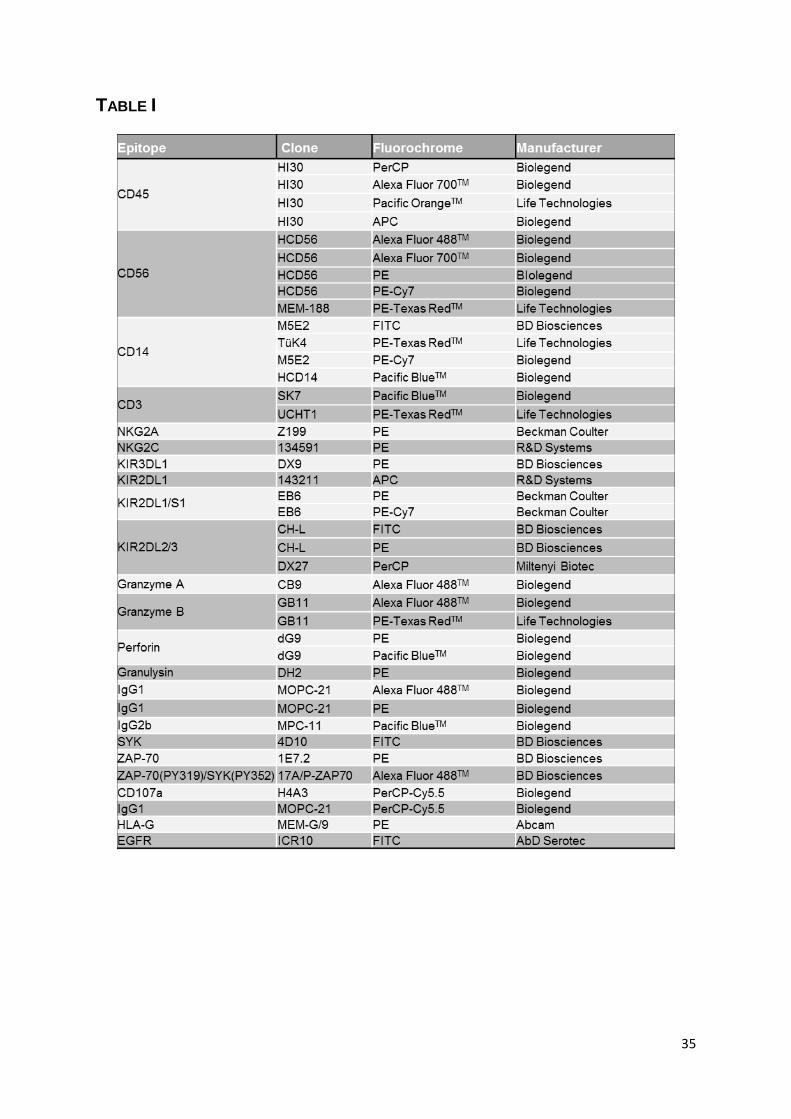
35
TABLE I

36
RESULTS
Frequency of KIR2DS1+ NK cells is significantly increased in decidua
The extracellular domains of KIR2DL1 and KIR2DS1 are almost identical, making the
development of specific antibodies a challenging task. No specific antibodies for KIR2DS1
are available, but the combination an antibody clone specific for KIR2DL1 (143211) with a
clone that binds both KIR2DL1 and KIR2DS1 (EB6) allows the identification of KIR2DL1 and
KIR2DS1 gene carriers (145). This strategy also discriminates four NK populations in
KIR2DL1+ and KIR2DS1+ gene carriers: KIR2DL1 single positive (L1+ S1-), KIR2DS1 single
positive (L1-S1+), KIR2DL1+ and KIR2DS1+ double positive (L1+S1+) and double negative
(L1-S1-). In KIR2DL1+ but KIR2DS1- gene carriers, two NK populations can be identified:
KIR2DL1 single positive (L1+) and KIR2DL1 negative (L1-) (Figure 1A). Among the KIR2DS1
Figure 1. Frequency of MHC Class I receptor-positive NK cells is significantly increased in decidua A) Representative FACS plots of KIR2DL1 and KIR2DS1 expression in dNK from KIR2DS1- (top left) and KIR2DS1+ (top right) women, and in pNK from KIR2DS1- (bottom left) and KIR2DS1+ (bottom right) donors. Cells
were gated in Live cells CD45+CD56+. B) Graphs depict the percentages of dNK and pNK subsets defined by KIR2DL1 (L1) and KIR2DS1 (S1) expression. Representative FACS plots of KIR2DL2/L3 (C), NKG2A (D) and NKG2C (E) expression in pNK and dNK. Graphs depict the percentages of dNK and pNK expressing KIR2DL2/L3 (L2/L3) (F), NKG2A (G) and NKG2C (H).

37
gene carriers, the frequency of both L1-S1+ and L1+S1+ NK was significantly higher in
decidua than blood (Figure 1B). In KIR2DS1- individuals, L1+ NK were more frequent in
decidua than in blood (Figure 1B). In addition, KIR2DL2/L3+, NKG2A+ and NKG2C+ NK
frequencies were also increased in decidua (Figure 1 C-H). These results confirm previous
reports demonstrating a skewing of dNK receptor expression towards recognition of MHC
Class I, especially HLA-C, and particularly, the potential of dNK to develop an activating
response to HLA-C2 through KIR2DS1 (118, 146).
KIR expression influences granule content in dNK
To determine whether the presence of KIR2DS1 influenced the cytolytic potential of NK
cells, freshly isolated dNK and pNK were stained for KIR2DS1 and KIR2DL1 and examined
for the intracellular expression of the cytolytic molecules granzyme A, granzyme B, perforin
and the 9 kDa active form of granulysin (147). pNK express significantly higher levels (as
demonstrated by mean fluorescence intensity, MFI) of perforin compared to dNK, whereas a
significantly higher percentage of dNK expressed granulysin compared to pNK. Granulysin
was also expressed at a significantly higher level in dNK. No difference was found between
expression of granzyme A and B by dNK and pNK (Figure 2 A-C).
pNK and dNK of KIR2DL1+/S1+ individuals were divided in the four NK subtypes and a
significantly higher percentage of S1+ dNK (L1-S1+ and L1+S1+) expressed granzyme B,
perforin and granulysin compared to the L1-S1- dNK subset (Figure 2 D and F). In addition,
L1-S1+ and L1+S1+ dNK also expressed significant higher levels (MFI) of granzyme B,
perforin and granulysin compared to the L1-S1- dNK subset (Figure 2D and H). The
increase of cytolytic molecules in S1+ cells was specific for dNK and was not observed in
S1+ pNK (Figure 2 D, E and G).
dNK have a higher potential for activation of signaling pathways downstream of
KIR2DS1
The binding of KIR2DS1 to its ligand HLA-C2 triggers DAP12 phosphorylation and
subsequent recruitment and phosphorylation of adaptor proteins SYK and ZAP-70 (148). To
determine the potential for activation of these signaling pathways, dNK and pNK from
KIR2DS1+ and KIR2DS1- donors were stimulated with pervanadate and the levels of
phosphorylated SYK and ZAP70 (p-SYK/p-ZAP70) were determined by Phosflow assay as
described in Methods. A significantly higher percentage of dNK had p-SYK/p-ZAP70
expression than pNK after pervanadate stimulation (Figure 3A-B). The expression levels
(MFI) of p-SYK/p-ZAP70 were also significantly increased in dNK compared to pNK (Figure
3B). No difference was found between total expression of SYK or ZAP70 between pNK and
dNK (data not shown). This data may suggest a previous activation of dNK in vivo or a
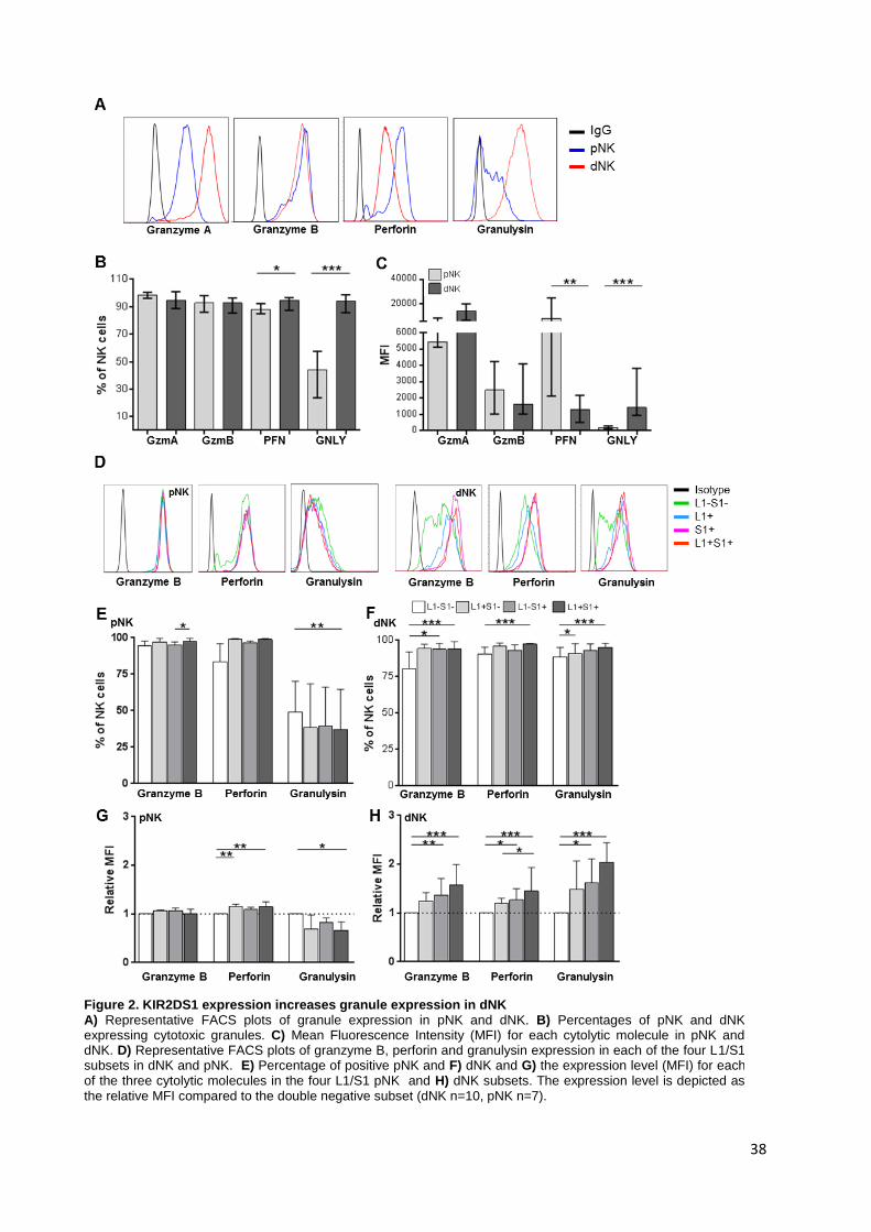
38
Figure 2. KIR2DS1 expression increases granule expression in dNK A) Representative FACS plots of granule expression in pNK and dNK. B) Percentages of pNK and dNK expressing cytotoxic granules. C) Mean Fluorescence Intensity (MFI) for each cytolytic molecule in pNK and dNK. D) Representative FACS plots of granzyme B, perforin and granulysin expression in each of the four L1/S1 subsets in dNK and pNK. E) Percentage of positive pNK and F) dNK and G) the expression level (MFI) for each of the three cytolytic molecules in the four L1/S1 pNK and H) dNK subsets. The expression level is depicted as the relative MFI compared to the double negative subset (dNK n=10, pNK n=7).
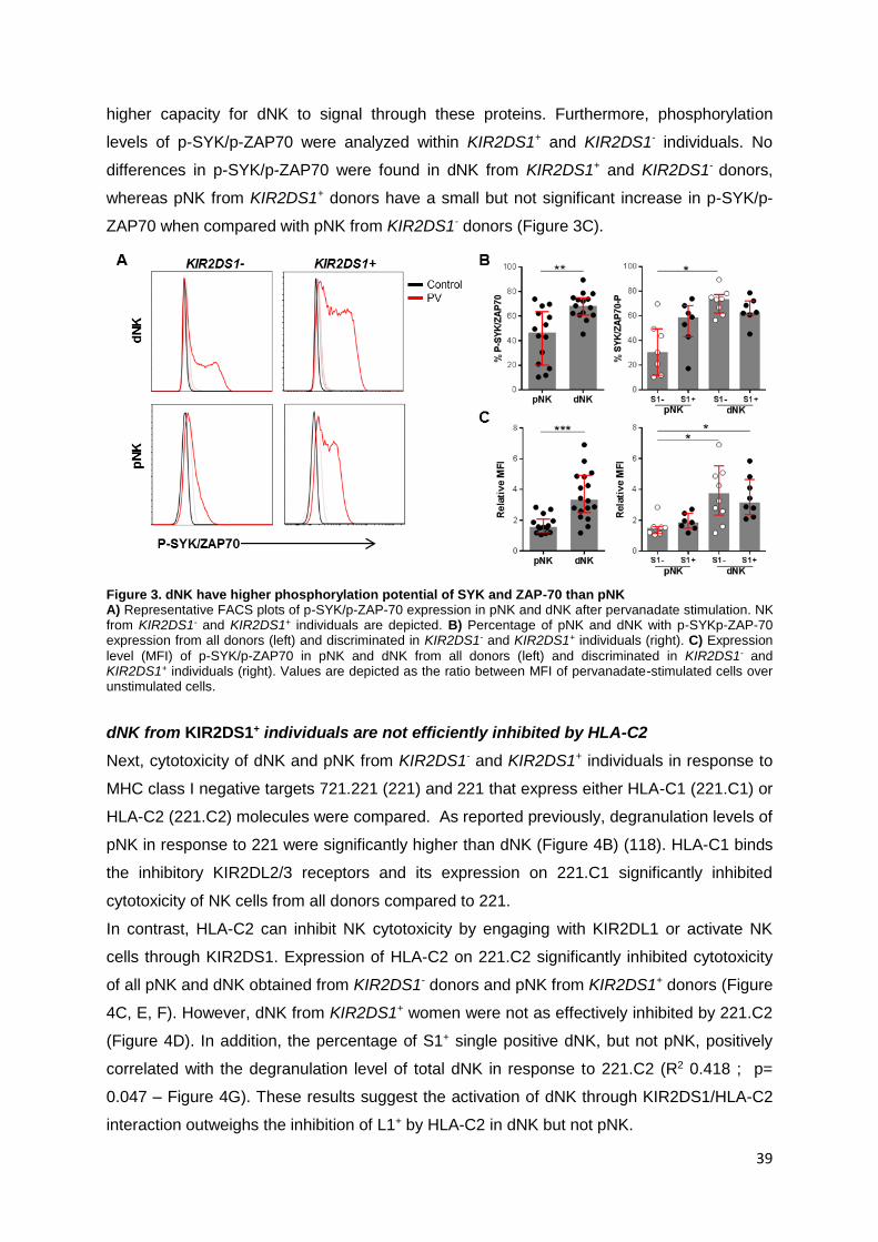
39
higher capacity for dNK to signal through these proteins. Furthermore, phosphorylation
levels of p-SYK/p-ZAP70 were analyzed within KIR2DS1+ and KIR2DS1- individuals. No
differences in p-SYK/p-ZAP70 were found in dNK from KIR2DS1+ and KIR2DS1- donors,
whereas pNK from KIR2DS1+ donors have a small but not significant increase in p-SYK/p-
ZAP70 when compared with pNK from KIR2DS1- donors (Figure 3C).
dNK from KIR2DS1+ individuals are not efficiently inhibited by HLA-C2
Next, cytotoxicity of dNK and pNK from KIR2DS1- and KIR2DS1+ individuals in response to
MHC class I negative targets 721.221 (221) and 221 that express either HLA-C1 (221.C1) or
HLA-C2 (221.C2) molecules were compared. As reported previously, degranulation levels of
pNK in response to 221 were significantly higher than dNK (Figure 4B) (118). HLA-C1 binds
the inhibitory KIR2DL2/3 receptors and its expression on 221.C1 significantly inhibited
cytotoxicity of NK cells from all donors compared to 221.
In contrast, HLA-C2 can inhibit NK cytotoxicity by engaging with KIR2DL1 or activate NK
cells through KIR2DS1. Expression of HLA-C2 on 221.C2 significantly inhibited cytotoxicity
of all pNK and dNK obtained from KIR2DS1- donors and pNK from KIR2DS1+ donors (Figure
4C, E, F). However, dNK from KIR2DS1+ women were not as effectively inhibited by 221.C2
(Figure 4D). In addition, the percentage of S1+ single positive dNK, but not pNK, positively
correlated with the degranulation level of total dNK in response to 221.C2 (R2 0.418 ; p=
0.047 – Figure 4G). These results suggest the activation of dNK through KIR2DS1/HLA-C2
interaction outweighs the inhibition of L1+ by HLA-C2 in dNK but not pNK.
Figure 3. dNK have higher phosphorylation potential of SYK and ZAP-70 than pNK A) Representative FACS plots of p-SYK/p-ZAP-70 expression in pNK and dNK after pervanadate stimulation. NK from KIR2DS1- and KIR2DS1+ individuals are depicted. B) Percentage of pNK and dNK with p-SYKp-ZAP-70 expression from all donors (left) and discriminated in KIR2DS1- and KIR2DS1+ individuals (right). C) Expression level (MFI) of p-SYK/p-ZAP70 in pNK and dNK from all donors (left) and discriminated in KIR2DS1- and KIR2DS1+ individuals (right). Values are depicted as the ratio between MFI of pervanadate-stimulated cells over unstimulated cells.
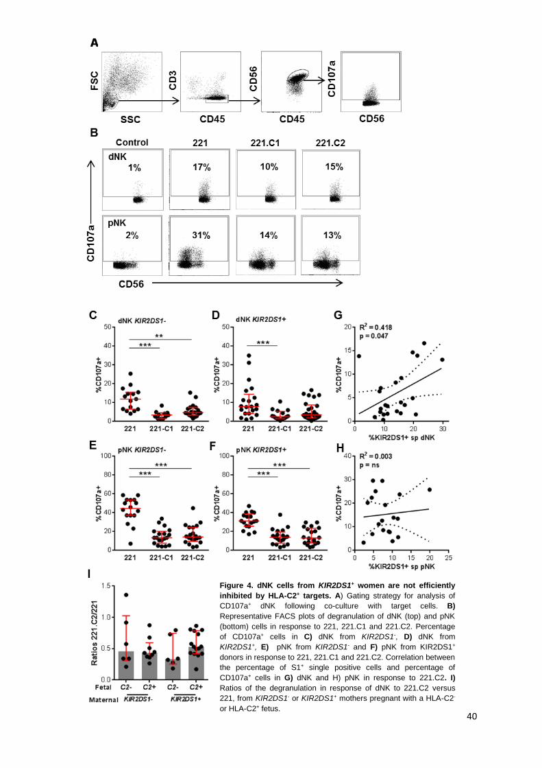
40
Figure 4. dNK cells from KIR2DS1+ women are not efficiently
inhibited by HLA-C2+ targets. A) Gating strategy for analysis of
CD107a+ dNK following co-culture with target cells. B)
Representative FACS plots of degranulation of dNK (top) and pNK
(bottom) cells in response to 221, 221.C1 and 221.C2. Percentage
of CD107a+ cells in C) dNK from KIR2DS1-, D) dNK from
KIR2DS1+, E) pNK from KIR2DS1- and F) pNK from KIR2DS1+
donors in response to 221, 221.C1 and 221.C2. Correlation between
the percentage of S1+ single positive cells and percentage of
CD107a+ cells in G) dNK and H) pNK in response to 221.C2. I)
Ratios of the degranulation in response of dNK to 221.C2 versus
221, from KIR2DS1- or KIR2DS1+ mothers pregnant with a HLA-C2-
or HLA-C2+ fetus.
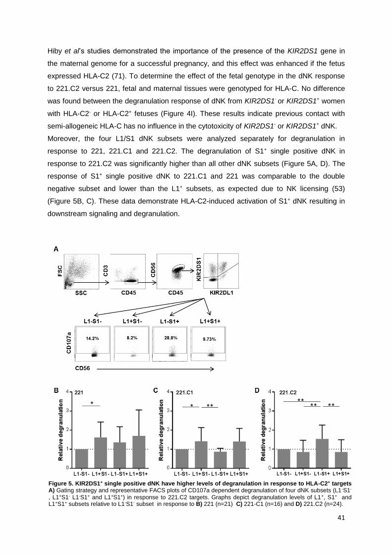
41
Hiby et al’s studies demonstrated the importance of the presence of the KIR2DS1 gene in
the maternal genome for a successful pregnancy, and this effect was enhanced if the fetus
expressed HLA-C2 (71). To determine the effect of the fetal genotype in the dNK response
to 221.C2 versus 221, fetal and maternal tissues were genotyped for HLA-C. No difference
was found between the degranulation response of dNK from KIR2DS1- or KIR2DS1+ women
with HLA-C2- or HLA-C2+ fetuses (Figure 4I). These results indicate previous contact with
semi-allogeneic HLA-C has no influence in the cytotoxicity of KIR2DS1- or KIR2DS1+ dNK.
Moreover, the four L1/S1 dNK subsets were analyzed separately for degranulation in
response to 221, 221.C1 and 221.C2. The degranulation of S1+ single positive dNK in
response to 221.C2 was significantly higher than all other dNK subsets (Figure 5A, D). The
response of S1+ single positive dNK to 221.C1 and 221 was comparable to the double
negative subset and lower than the L1+ subsets, as expected due to NK licensing (53)
(Figure 5B, C). These data demonstrate HLA-C2-induced activation of S1+ dNK resulting in
downstream signaling and degranulation.
Figure 5. KIR2DS1+ single positive dNK have higher levels of degranulation in response to HLA-C2+ targets A) Gating strategy and representative FACS plots of CD107a dependent degranulation of four dNK subsets (L1-S1-
, L1+S1- L1-S1+ and L1+S1+) in response to 221.C2 targets. Graphs depict degranulation levels of L1+, S1+ and L1+S1+ subsets relative to L1-S1- subset in response to B) 221 (n=21) C) 221-C1 (n=16) and D) 221.C2 (n=24).

42
HLA-E expression in 221.C2 has no influence in dNK and pNK degranulation
The 721.221 cell line has genomic deletions in the HLA-A, B and C locus, but not in HLA-E
(149). HLA-E presents peptides derived from the leader sequence of other MHC Class I
molecules. 221 do not express MHC class I and this prevents the surfacing of HLA-E.
However, the transfection of 221 with HLA-C1 and HLA-C2 provide such leader peptides
and increases the expression of HLA-E on the cell surface (150). dNK and pNK express
inhibitory receptors for HLA-E (NKG2A); while clear NKG2A- and NKG2A+ populations can
be identified among pNK, virtually all dNK express NKG2A (Figure 1D, G). To examine if
HLA-E had an effect in the degranulation results observed in response to 221.C1 and
221.C2, NKG2A- and NKG2A+ pNK responses were compared. No difference in the
percentage of CD107a+ NK cells in response to 221.C1 and 221.C2 was found between
NKG2A- and NKG2A+ pNK (Figure 6A). A clear NKG2A- population cannot be identified in
dNK and thus blocking of NKG2A was necessary. No difference in the percentage of
CD107a+ cells in response to 221.C2 was found between dNK previously incubated with
anti-NKG2A blocking antibody or isotype control (Figure 6B).These results suggest the effect
observed is due to HLA-C expression only.
K562 is not a suitable cell line for the study of MHC influence in dNK responses
To overcome some of the disadvantages of the use of 221 (MHC Class II and HLA-E
expression – Appendix I), the MHC Class I and II-negative cell line K562 (151) (and K562
transfected with HLA-C1 (K562.C1) and HLA-C2 (K562.C2)) were also used as targets for
Figure 6: HLA-E expression does not influence
dNK responses to 221.C2. A) ) Comparison of the
degranulation levels of NKG2A- with NKG2A+ pNK in
response to 221.C1 and 221.C2 B) Percentage of
CD107a+ dNK after co-culture with 221.C2 in the
presence or absence of anti-NKG2A antibody (or
isotype control). C) Representative FACS plots of a
comparison between use of 221 or K562 as target cells
for degranulation assay, using the same dNK sample
as effectors. D) Degranulation of pNK (left) and dNK
(right) in response to K562.C1 or K562.C2 relative to
K562, compared to degranulation in response to
221.C1 or C2 relative to 221.

43
degranulation assay. K562, K562.C1 and K562.C2 were co-cultured with dNK and pNK in
parallel with 221, 221.C1 and 221.C2. The degranulation of pNK and dNK in response to
K652 was similar to 221. However, while MHC Class I transfected 221 were able to inhibit
degranulation, MHC Class I transfected K562 were not (Figure 6C-D). These results showed
221 are a more reliable NK target for the study of the effect of MHC Class I molecules in NK
responses.
KIR2DS1 does not influence cytokine secretion of dNK in response to EVT
dNK have been shown to secrete both pro- and anti-inflammatory cytokines, as well as
growth factors and chemokines involved in trophoblast invasion and vascular remodeling
(48, 107, 118). However, all of these studies used cell lines, antibodies or mitogens to
stimulate dNK. Recently, it was demonstrated that co-culture of dNK with primary EVT did
not lead to secretion of the
cytokines identified in these
studies (IFN-γ, VEGF) (95). To
determine if KIR2DS1
expression has an influence in
the cytokine response to HLA-
C2+ EVT, dNK were co-cultured
alone or with 221, 221.C2 and
matched primary EVT. dNK
constitutively secreted high
levels of IL-8 (neutrophil
chemotactic factor), and the
secretion was amplified by co-
culture 221 and 221.C2, but
only slightly with EVT (Figure
7F). In fact, EVT alone also
constitutively secreted IL-8
(data not shown). Co-culture of
dNK with 221 induced secretion
of IFN-γ, TNF-, GM-CSF,
VEGF and IL-6 confirming
previous studies (48, 107)
(Figure 7A-E). Interestingly,
expression of HLA-C2 in 221
Figure 7. . KIR2DS1 does not influence cytokine secretion of dNK in response to EVT.Cell culture supernatants of dNK incubated alone or with 221, 221.C2 and EVT analyzed for A) IFNγ, B) TNFα, C) VEGF, D) GM-CSF, E) IL-6 and F) IL-8.

44
partially inhibited the secretion of IFN-γ, TNF-α and IL-6, but not GM-CSF and VEGF (Figure
7A-E), confirming MHC class I may play a direct role in the suppression of pro-inflammatory
cytokines. Co-culture of dNK with EVT stimulated dNK secretion of IL-6 (Figure 7E), but not
to the same levels of 221, and did not elicit secretion of IFN-γ, TNF-α, GM-CSF or VEGF
(Figure 7A-D).
No significant differences were found in cytokine secretion of dNK from KIR2DS1- vs
KIR2DS1+ individuals when co-cultured with 221.C2 and HLA-C2+ EVT (Figure 7A-F),
contrarily to previous published studies regarding GM-CSF secretion by KIR2DS1+ dNK
(118).
Together, these data indicate HLA-C expression by EVT has no influence on cytokine
secretion by dNK, suggesting other stimuli at the maternal-fetal interface may activate dNK
to secrete cytokines and growth factors.

45
DISCUSSION
This chapter demonstrates dNK from KIR2DS1+ individuals acquire higher cytotoxic function
than dNK from KIR2DS1- individuals when exposed to HLA-C2-expressing targets, an effect
not observed in pNK. Furthermore, the cytotoxic function was correlated with the frequency
of KIR2DS1+ dNK. KIR2DS1+ dNK had an increased capacity to degranulate in response to
221 cells that expressed HLA-C2 than KIR2DS1- dNK. This effect was independent of the
HLA-C genotype of the fetus with which the dNK had had contact in vivo. In addition, HLA-
C2+ EVT did not elicit GM-CSF, IFN-γ or VEGF secretion by dNK from either KIR2DS1- or
KIR2DS1+ individuals.
HLA-C expressed by fetal EVT has a unique role in pregnancy, as the only polymorphic
molecule that can present peptides to maternal T cells and as the main molecule to which
immune tolerance needs to be established (127). The importance of HLA-C was further
demonstrated by the observation that the combination of the HLA-C allotype (HLA-C2,
expressed by fetal trophoblasts) and the expression of its receptor KIR2DS1 in maternal NK
cells reduce the risk for pregnancy complications and regulate fetal birth weight (71, 76,
114).
The previous observation that dNK express higher levels of KIR2DL1 and KIR2DS1 than
pNK was confirmed. In addition, it was shown expression of KIR2DS1 (both L1-S1+ and
L1+S1+ subsets) increased the expression of key cytolytic molecules (perforin, granzyme B
and granulysin) stored intracellularly in dNK but not pNK. Furthermore, dNK from KIR2DS1+
individuals were less efficiently inhibited by HLA-C2 expression on target cells. HLA-
E/NKG2A interaction did not influence the HLA-C2 effect. Overall, these data indicate a
skewing of dNK receptor expression towards recognition of HLA-C, and particularly, the
potential of dNK to develop an activating response through KIR2DS1 and its interaction with
HLA-C2.
Interestingly, the cell line K652 proved not to be a suitable target to study the effect of MHC
Class I on NK cell cytotoxicity. K562 is an erythroleukemia cell line that does not express
MHC Class II or MHC Class I (151, 152). Because of this lack of MHC Class I molecules,
K562 are widely used as classical targets for T cells and NK cells in a variety of cytotoxicity
assays (153). However, expression of MHC Class I molecules on transfected K562 lines,
even at very high levels, was not enough to inhibit dNK and pNK degranulation against
K562. K562 is a tumor cell line and is known to express very high levels of molecules that
serve as activating ligands for NK receptors, (e. g. MICA and MICB ligands for NKG2D)
(154). The expression of MHC Class I was not sufficient to tip the balance towards inhibition
of NK cells.

46
Although dNK have a higher potential to be activated through the p-SYK/ZAP70 signaling
pathway, this does not result increased cytolytic activity. On the contrary, dNK have reduced
ability to degranulate and kill target cells including classical MHC-negative 221.
Requirements for activation by pro-inflammatory signals may be different between pNK and
dNK, possibly due to different maturation conditions. The origin of uterine NK cells is not
completely clear. Several lines of evidence point to an origin in mature peripheral NK cells
that migrate to the uterus, where their phenotype is modified due to the low inflammatory
environment of the placenta. Some studies have developed protocols involving the addition
of cytokines abundant in the maternal fetal interface (such as TGF-β) and low oxygen
pressure that are able to convert pNK into dNK-like cells (155–157). However, a recent study
in mice demonstrated mature NK cells do not migrate to the uterus. Instead, immature NK
precursors migrate from the bone marrow to the endometrium where maturation is
completed (158). Independently of the origin, NK cells have high plasticity and ability to
adapt to the surrounding environment to prevent tissue damage due to excess of
inflammation (53, 159). The threshold for dNK activation is thought to be higher than pNK to
avoid detrimental effects to pregnancy. However, the expression of high levels of cytolytic
molecules in dNK shows a cytotoxic potential that may be employed in situations such as
viral or bacterial infections.
The increased activation state of S1+ dNK was previously linked to increased ability to
secrete cytokines and growth factors that facilitate trophoblast invasion and placental growth
(6). However co-culture of dNK with primary EVT did not enhance cytokine secretion in dNK
and no differences were found in cytokine responses between dNK or pNK from KIR2DS1+
and KIR2DS1− individuals toward any of the target cells, including 221, 221.C2 and EVT (7).
This suggests other stimuli present in the maternal-fetal interface may be involved in dNK
function. Interaction of dNK with other immune cells (e.g. macrophages) or decidual stromal
cells (DSC), in combination with the cytokine environment of the placenta, may lead to dNK
secretion of growth factors. EVT interaction alone was not sufficient to elicit these functions,
but may modulate the dNK phenotype. Future focus will be placed on the analysis of the
cytokine secretion of dNK stimulated with mitogens (e.g. PMA – Ionomycin) after or during
co-culture with EVT or decidual stromal cells. Furthermore, the analysis of cytokine content
in the dNK cell culture supernatant only shows the secretion of the total sample of dNK. To
analyze the contribution of the KIR2DS1+ dNK subset, intracellular staining of cytokines
following stimulation will be included.
The protective effect of maternal KIR2DS1 and fetal HLA-C2 was previously thought to be
related to activation of dNK to secrete growth factors. In this chapter, KIR2DS1+ dNK
demonstrate a high activation and cytolytic potential that does not translate into cytokine

47
secretion following stimulation with EVT. In the next chapter, the role of KIR2DS1+ dNK in
the clearance of placental HCMV infection will be discussed.

48
CHAPTER 3
EXPRESSION OF KIR2DS1 BY DECIDUAL NK CELLS INCREASES
THEIR ABILITY TO CONTROL PLACENTAL HCMV INFECTION

49
INTRODUCTION
The establishment of a healthy human pregnancy requires the maternal immune system to
tolerate fetal alloantigens. HLA-C is the only polymorphic MHC molecule expressed by
extravillous trophoblast (EVT) that can be directly recognized by maternal NK and T cells
and elicit an allogeneic response in case of a mismatch between mother and fetus.
Tolerance is achieved via several mechanisms, such as reduced numbers of effector T cells
in the maternal decidua, high activity of regulatory T cells, lower cytotoxicity of decidual NK
and CD8+ T cells and a primarily anti-inflammatory microenvironment with high levels of
TGF-β and IL-10 (92, 160). However, the immune system must still be able to fight viral and
bacterial infections that arise during gestation. The balance between fetal tolerance and
infection clearance is one of the greatest challenges the maternal immune system faces
during pregnancy.
Viral infections [e.g. human cytomegalovirus (HCMV), HIV, herpes simplex virus (HSV) and
influenza virus], can cause severe maternal and fetal disease and morbidity (119–121).
However, most infections are under-diagnosed due to the high variety of pathogens and the
difficulty in demonstrating the pathogenic role of the viral pathogens in pregnancy
complications (122). The most common congenital infection HCMV, occurring in 0.5–2% of
all live births. HCMV infects the placenta and can inhibit trophoblast differentiation and
invasion, which can lead to placental insufficiency and fetal growth restriction (123–126). If
the infection reaches the fetus, it can cause long-term health problems and disabilities such
as hearing and vision loss, mental disability and seizures (121). CD8+ effector T cells are the
most important cell type to respond and clear viral infections in the periphery, recognizing
MHC-viral peptide complexes in infected cells (161). However, in early pregnancy decidual
tissue, the frequency of CD8+ T cells is very low (2-7% of total CD45+ lymphocytes). In
addition, decidual effector-memory T cells contain reduced levels of the cytolytic molecules
perforin and granzyme B, which may limit their cytotoxicity (162). This leaves dNK cells as
the main potential actors in the clearance of placental viral infections. Decidual NK cells
represent 60-80% of lymphocytes in decidua. In spite of their low cytotoxicity , dNK express
very high levels of cytolytic granules (granzymes, perforin, granulysin) and can become
cytotoxic when activated by pro-inflammatory cytokines (e.g. IL-15, IL-2 IL-12 and IL-18)
(105, 106). A recent study demonstrated dNK are capable of killing HCMV-infected
placental fibroblasts (163). Nevertheless, whether or not dNK or pNK can respond and kill
HCMV-infected HLA-G+ EVT has not been addressed.
The presence of activating KIRs, especially KIR2DS1, in the maternal genome has been
associated with protection from pregnancy complications such as miscarriage, fetal growth
restriction and preeclampsia (71, 114). In addition, individuals who carry the KIR-B

50
haplotype (with more activating KIR) have a significantly improved outcome after viral
infections such as HIV and HCMV (64, 164). The combined presence of KIR3DS1 and its
putative ligand HLA-Bw4 was associated with significantly slower progression to AIDS, lower
viral load and slower decline of CD4+ T cells (59–61, 164). In addition, KIR3DS1 and
KIR2DS1 were shown to be protective against respiratory papillomatosis caused by human
papillomavirus (62). Activating KIRs have also been shown to play a role in NK mediated
clearance of HCMV infection following hematopoietic stem cell (HSC) or solid organ
transplantation (63–66). We therefore postulated the activation of KIR2DS1+ dNK may lead
to an earlier and more efficient clearing of virus-infected placental cells. This avoids virus
induced pathology and prevents development of complications in later gestation and would
provide an explanation to the protective effect of KIR2DS1 in human pregnancy.
In this chapter, KIR2DS1+ dNK are shown to acquire higher cytotoxicity than KIR2DS1- dNK
when exposed to HCMV-infected decidual stromal cells (DSC), particularly when DSC
expresses HLA-C2. Furthermore, dNK were unable to degranulate or secrete cytokines in
response to HCMV-infected JEG3 and primary EVT. This emphasizes the immunological
challenge to clear placental viral infections within the immune privileged compartment of the
placenta. Additional activation of dNK through KIR2DS1/HLA-C2 interaction may contribute
to a more efficient control of pregnancy-associated HCMV infection and prevent the spread
of the infection through the placenta and fetal tissues.

51
MATERIALS AND METHODS
Isolation of NK, decidua and trophoblast
Discarded human placental and decidual material (gestational age 6-12 weeks) were
obtained from women undergoing elective pregnancy termination at a local reproductive
health clinic. Peripheral blood leukocytes were isolated from discarded Leukopacks® from
healthy volunteer blood donors at the Massachusetts General Hospital in Boston, MA. All of
the human tissue used for this research was de-identified, discarded clinical material. The
Committee on the Use of Human Subjects (the Harvard IRB) determined this use of
placental and decidual material is Not Human Subjects Research. The procedure to isolate
dNK, pNK and trophoblast was described in Chapter 2. Decidual stromal cell lines were
obtained as described previously (143). HLA typing was performed by the American Red
Cross tissue typing facility, Dedham, Massachusetts.
Cell culture
pNK and dNK were cultured in X-VIVO 10TM media (Lonza) supplemented with gentamycin,
5% human AB serum (Corning) and 2.5 ng/ml rIL-15 (Biolegend). Decidual stromal cells
were cultured in OPTI-MEM media (GIBCO) supplemented with 3% FCS and 25μg/ml
gentamycin and passaged once to twice a week. 50.000 CD45- sorted EVT were plated in
48 well cell culture plates or 8-chamber slides (Costar) pre-coated with fibronectin (100μl of
20ng/ml for 45 min, BD), in supplemented DMEM/F12 medium (Gibco) as described
previously (95). The choriocarcinoma cell line JEG-3 was cultured in RPMI 10% FCS.
Flow Cytometry
Antibodies used for flow cytometric analysis and FACS sort are listed in Supplemental Table
S1. For surface staining, cells were stained for 30 minutes on ice in the dark in PBS 1%FCS.
For KIR2DS1 staining, NK were stained with KIR2DL1 first (Clone 143211, 1.5μg/ml, 20
min) and thereafter with KIR2DL1/S1 (Clone EB6, 0.5μl, 15 min) as described previously
(145). Cells were analyzed immediately or fixed in 1% PFA for 10 minutes for next day
analysis. Analysis was performed on a FACSCalibur (BD) or LSR II (BD) and FACS sort was
done using a BD Aria III, MoFlo XDP or MoFlo Astrios (Beckman Coulter).
Degranulation assays
For degranulation assays, freshly isolated dNK and pNK were co-cultured with uninfected
and HCMV-infected DSC and JEG-3. For co-culture with EVT, dNK and pNK were cultured
in X-vivo media (Lonza) supplemented with 5% human serum (Corning) and 2.5ng/ml IL-15
(Biolegend) for 24h to allow optimal HCMV infection of dNK-matched primary EVT. 150.000

52
dNK or pNK were added to confluent DSC, JEG3 or 50.000 EVT plated in 48 well plates in
X-VIVO 10TM supplemented with 2.5 ng/ml IL-15 for 10h. 250ng/ml CD107a PerCP-Cy5.5
antibody was added to all co-cultures. NK were collected and fixed for 10min in 1% PFA and
subsequently stained for all relevant surface markers for FACS analysis.
Cytokine secretion analysis
Supernatants from the same co-cultures used for degranulation assays were snap frozen at
-80 °C and all cytokines were analyzed using a multiplex cytokine assay (Magpix, Bio-Rad)
according to manufacturer’s protocols.
Infection of DSC, JEG3 cells and EVT
High titer virus stocks of HCMV AD169 (IE-1-GFP) (a gift from Prof. Donald Coen at Harvard
Medical School) were obtained by infecting Human Foreskin Fibroblasts (HFF) (ATCC) and
collecting supernatants. Supernatants were aliquoted and snap frozen in liquid nitrogen until
use (46). DSC at 80% confluence in 48 well plates were infected at a MOI 0.5 for 1 hour at
37ºC with frequent shaking, followed by normal culture for 48h before use in assays. JEG-3
and primary EVT were infected at a MOI 3-4 for 2 or 3 periods of 12 hours (including 1h of
centrifugation at 2800rpm) before assays.
Fluorescence microscopy
HCMV-AD169-GFP - infected DSC, JEG3, and EVT were fixed with 4% PFA for 10 minutes
followed by a 10 min permeabilization with PBS/0.01% Triton-X. Purified primary anti-pp65
antibody (Abcam) or mouse IgG2a (Biolegend) were added in a 1:200 concentration for 1h,
followed by washing and staining with goat anti-mouse – AlexaFluor 546 secondary antibody
(1:500) for 1h. Cells were imaged in a Nikon Eclipse Ti fluorescence microscope.
Statistical analyses
All data was analyzed using GraphPad Prism (version 6.0) software. To determine
differences between 2 unpaired groups, a non-parametric Mann-Whitney test was
performed, whereas between 2 paired groups a non-parametric Wilcoxon Signed Rank test
was performed. To determine differences among more than 2 unpaired groups, a non-
parametric Kruskal-Wallis test with Dunn’s multiple comparison post-test was performed. To
determine differences among more than 2 paired groups, a non-parametric Friedman’s test
with Dunn’s multiple comparison post-test was performed. For correlation analyses,
Spearman correlation analysis was used. All data are depicted with median and interquartile
range, except when otherwise indicated. p-values <0.05 were considered to reflect
significant differences.*-p<0.05;**-p<0.01; *** p<0.005

53
TABLE I.

54
RESULTS
dNK degranulate in response to HCMV-infected DSC but not to infected JEG3 or EVT
dNK have recently been shown to be able to eliminate HCMV-infected placental fibroblasts
(163). The target cells used in the referenced study were a mix of maternal and fetal
fibroblasts. To discriminate the NK activity in response to HCMV-infected maternal or fetal
tissue, maternal decidual stromal cells (DSC), the HLA-G+ choriocarcinoma cell line JEG3
and primary fetal HLA-G+ EVT were infected with HCMV-AD169-GFP. Both dNK and pNK
showed significantly higher levels of degranulation in response to HCMV-infected DSC
compared to healthy DSC (Figure 1B and C).
Interestingly, infection of JEG3 and primary fetal EVT with HCMV significantly increased
degranulation by pNK compared to uninfected cells. However, dNK did not have an
increased response to HCMV-infected JEG3 or EVT compared to uninfected cells. (Figure
1D-E). In addition, both dNK and pNK secreted significantly more IFN-γ, TNF-α and GM-
CSF in response to HCMV-infected DSC compared to healthy DSC, whereas pNK but not
dNK secreted more of these cytokines in response to HCMV-infected JEG3 and EVT (Figure
2A-C). In addition, HCMV infection of EVT induced higher IL-6 secretion by pNK but not dNK
(Figure 2D). These data emphasize the difficulties of dNK in clearing placental viral
infections, possibly due to the immune privileged status of the placenta.

55
Figure 1. dNK degranulate in response
to HCMV infected DSC but not to
HCMV infected JEG3 and EVT
A( Gating strategy for analysis of
CD107a+ dNK and pNK following co-
culture with uninfected or HCMV-infected
target cells. One representative dNK
sample co-cultured with HCMV-infected
DSC is shown. B) Representative FACS
plots of degranulation of dNK (top) and
pNK (bottom) in response to uninfected
or infected DSC, JEG3 and EVT. Gates
for CD107a+ are based on dNK and pNK
cultured alone. Percentage of CD107a+
pNK (left) and dNK (middle) after co-
culture with healthy and HCMV-infected
D) DSC, E) JEG3 and F) EVT for 10
hours in the presence of 2.5 ng/ml IL-15.
Relative degranulation of pNK and dNK
in response to HCMV-infected relative to
healthy cells are depicted (right panels).
Co-culture with DSC and JEG3 was
established immediately after pNK/dNK
isolation. To allow for an optimal HCMV
infection of EVT, dNK/pNK were cultured
with 2.5ng/ml IL-15 for 24h prior to co-
incubation with dNK matched EVT.

56
HLA-C type of DSC has no influence in total dNK response to HCMV infection
To evaluate the importance of HLA-C allotype in the response of dNK and pNK from
KIR2DS1- and KIR2DS1+ donors, two DSC lines were used: one homozygous for HLA-C1
(DSC.C1) and one expressing both HLA-C1 and C2 (DSC.C2). No difference was found in
the ratio of degranulation in response to HCMV-infected cells versus uninfected cells
between dNK from KIR2DS1- or KIR2DS1+ donors, or in dNK response to HLA-C2+ vs HLA-
C2- DSC (Figure 3A). In contrast, the ratio of degranulation in response to HCMV-infected
cells vs uninfected cells of pNK was higher in response to HLA-C2+ DSC, particularly in
KIR2DS1- donors (Figure 3B). These results indicate the presence of activating KIR
(KIR2DS1) does not enhance the responses of total dNK population to HCMV infection.
Increased degranulation of KIR2DS1+ dNK in response to HCMV-infected HLA-C2+
DSC
To determine the degranulation capacity of each of the four L1/S1 NK subtypes,
degranulation assays for the response to DSC.C1 and DSC.C2 were performed as
described previously (Chapter 2). dNK that express KIR2DS1 (L1-S1+ and L1+S1+)
Figure 2. dNK produce cytokines in response to HCMV- infected DSC but not to HCMV-infected JEG3 and EVT. Cell culture supernatants of dNK and pNK incubated with healthy and HCMV infected DSC, JEG3 and EVT were analyzed for A) IFNγ B) TNFα, C) GM-CSF and D) IL-6. Cytokine production is depicted as the ratio of
concentrations in HCMV infected cultures relative to uninfected cultures. (e. g. all ratios >1 indicate a significant increased response to HCMV infected versus uninfected cells).

57
demonstrated a significantly increased response to HCMV-infected DSC.C2 compared to L1-
S1- dNK. A similar increase was also showed in response to infected DSC.C1 but it was not
significant (Figure 4A-B). Moreover, the percentage of total S1+ dNK (both L1-S1+ and
L1+S1+) positively correlated with the relative degranulation in response to infected DSC.C2
(Figure 4C). In pNK, the L1+S1+ double positive subset also showed an increased response
to HCMV-infected DSC but this was independent of HLA-C2 expression on DSC (Figure 4
D-E). Furthermore S1+ pNK did not correlate with the degranulation level in response to
infected DSC.C2 (Figure 4F). These data demonstrate the combination of KIR2DS1
expression on dNK and the presence of HLA-C2 on DSC increased the ability of dNK to
clear decidual HCMV infection.
Early HCMV infection reduced HLA-C on DSC, JEG3 and EVT while late infection
restored HLA-C expression
HCMV has many molecular strategies to circumvent immune recognition and avoid lysis of
infected cells (128). Immune evasion strategies by HCMV include the blockade of peptide
presentation and downregulation of cell surface expression of MHC class I molecules (165).
Figure 3. dNK KIR2DS1 genotype and DSC HLA-C genotype have no influence in the degranulation response to HCMV infected targets (Left) Percentage of CD107a+ dNK (A) and pNK (B) after co-culture with
uninfected or HCMV-infected DSC. The response to HLA-C2- and HLA-C2+ DSC is compared. Relative degranulation of pNK and dNK in response to HCMV-infected relative to healthy HLA-C2- or HLA-C2+ DSC are depicted (right panels), comparing KIR2DS1- with KIR2DS1+ donors.

58
This prevents cytolysis by CD8+ effector T cells that require MHC/peptide-TCR binding.
However downregulation of MHC class I molecules increases the susceptibility of HCMV-
infected cells to lysis by NK cells through missing self-recognition (165). Downregulation of
MHC, including HLA-C, may also prevent activation of NK cells through KIR2DS1.
DSC, JEG3 and EVT were infected with HCMV-AD169-GFP. This HCMV strain has GFP
tagged to an HCMV-Immediate Early (IE) gene. Infected cells were analyzed 48h post
infection for expression of IE-GFP and stained for pp65 (a late expression gene) for
fluorescence microscopy (Figure 5). HCMV infection of DSC reached nearly 100% with
~70% IE-GFP+ and ~30 % pp65+GFP- cells. HCMV infection levels for JEG3 reached closer
to 40%, with 10% IE-GFP+ cells and ~30% pp65+GFP- cells (Figure 5 and 6A). At 48h, 100%
of EVT expressed IE-GFP and also pp65, although different levels of expression could be
observed for the 2 proteins. HCMV-infected and uninfected DSC, JEG3 and EVT were
analyzed for expression MHC class I molecules. HLA-A2, HLA-E and especially HLA-C
expression in HCMV-infected DSC was significantly reduced on all infected DSC that
expressed the IE-GFP protein (Figure 6A, C, D, F). However, when HCMV-infected DSC lost
Figure 4. S1+ and L1+S1+ dNK respond strongly to HCMV infected HLA-C2+ DSC. Specific degranulation of
the four L1/S1 NK subsets in response to HCMV infected DSC depicted as the ratio of %CD107a+ NK to HCMV- infected versus uninfected DSC. A) dNK HLA-C2+ DSC (n=24) B) dNK to DSC.C1 (n=13) C) pNK to DSC.C2 (n=13) and D) pNK to DSC.C1.C2 (n=10) E) Correlation plot between percentage of S1 positive dNK and relative degranulation of total dNK population in response to HCMV-infected or healthy DSC.C2. F) Correlation plot
between percentage of S1 positive pNK and relative degranulation of total pNK population in response to HCMV-infected or healthy DSC.C2.
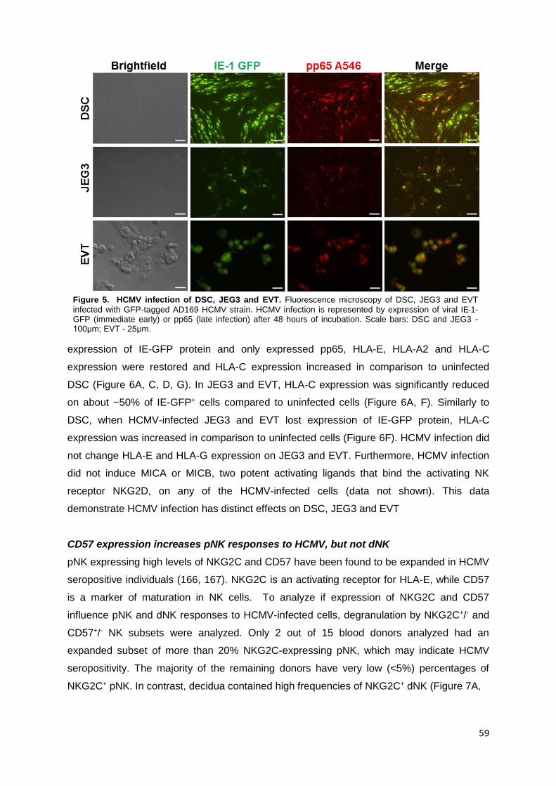
59
expression of IE-GFP protein and only expressed pp65, HLA-E, HLA-A2 and HLA-C
expression were restored and HLA-C expression increased in comparison to uninfected
DSC (Figure 6A, C, D, G). In JEG3 and EVT, HLA-C expression was significantly reduced
on about ~50% of IE-GFP+ cells compared to uninfected cells (Figure 6A, F). Similarly to
DSC, when HCMV-infected JEG3 and EVT lost expression of IE-GFP protein, HLA-C
expression was increased in comparison to uninfected cells (Figure 6F). HCMV infection did
not change HLA-E and HLA-G expression on JEG3 and EVT. Furthermore, HCMV infection
did not induce MICA or MICB, two potent activating ligands that bind the activating NK
receptor NKG2D, on any of the HCMV-infected cells (data not shown). This data
demonstrate HCMV infection has distinct effects on DSC, JEG3 and EVT
CD57 expression increases pNK responses to HCMV, but not dNK
pNK expressing high levels of NKG2C and CD57 have been found to be expanded in HCMV
seropositive individuals (166, 167). NKG2C is an activating receptor for HLA-E, while CD57
is a marker of maturation in NK cells. To analyze if expression of NKG2C and CD57
influence pNK and dNK responses to HCMV-infected cells, degranulation by NKG2C+/- and
CD57+/- NK subsets were analyzed. Only 2 out of 15 blood donors analyzed had an
expanded subset of more than 20% NKG2C-expressing pNK, which may indicate HCMV
seropositivity. The majority of the remaining donors have very low (<5%) percentages of
NKG2C+ pNK. In contrast, decidua contained high frequencies of NKG2C+ dNK (Figure 7A,
Figure 5. HCMV infection of DSC, JEG3 and EVT. Fluorescence microscopy of DSC, JEG3 and EVT
infected with GFP-tagged AD169 HCMV strain. HCMV infection is represented by expression of viral IE-1-GFP (immediate early) or pp65 (late infection) after 48 hours of incubation. Scale bars: DSC and JEG3 - 100μm; EVT - 25μm.

60
Figure 6. HCMV early infection reduces expression of HLA-C in DSC, JEG3 and EVT. A) Representative FACS plots of percentage of infection and expression of total MHC Class I (W6/32), HLA-C, HLA-
E, HLA-A2 and HLA-G in healthy or GFP-/GFP+ HCMV-infected DSC, JEG3 and EVT. Graphs depict the ratio between the expression levels (MFI) of the MHC Class I molecules (B), HLA-E (C), HLA-A2 (D) and HLA-G (E) in GFP-/GFP+ HCMV-infected and healthy DSC (n=10), JEG3 (n=3) and EVT (n=5).. F) Percentage of HLA-C+ DSC (n=10), JEG3 (n=3) and EVT (n=3) after infection with HCMV. G) Ratio between expression levels (MFI) of HLA-C in
GFP- HCMV-infected and healthy DSC and JEG3. For HCMV-infected EVT, no GFP- cells were detected. The grey bar refers to the ratio of the MFI of HLA-C+ population in GFP+ HCMV-infected EVT and MFI of HLA-C expression in
healthy EVT. Bars indicate mean SE.
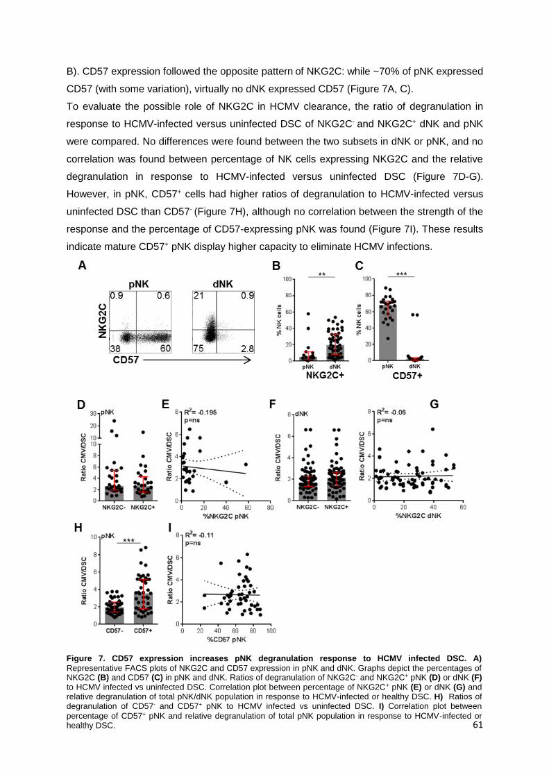
61
Figure 7. CD57 expression increases pNK degranulation response to HCMV infected DSC. A)
Representative FACS plots of NKG2C and CD57 expression in pNK and dNK. Graphs depict the percentages of NKG2C (B) and CD57 (C) in pNK and dNK. Ratios of degranulation of NKG2C- and NKG2C+ pNK (D) or dNK (F) to HCMV infected vs uninfected DSC. Correlation plot between percentage of NKG2C+ pNK (E) or dNK (G) and relative degranulation of total pNK/dNK population in response to HCMV-infected or healthy DSC. H) Ratios of degranulation of CD57- and CD57+ pNK to HCMV infected vs uninfected DSC. I) Correlation plot between
percentage of CD57+ pNK and relative degranulation of total pNK population in response to HCMV-infected or healthy DSC.
B). CD57 expression followed the opposite pattern of NKG2C: while ~70% of pNK expressed
CD57 (with some variation), virtually no dNK expressed CD57 (Figure 7A, C).
To evaluate the possible role of NKG2C in HCMV clearance, the ratio of degranulation in
response to HCMV-infected versus uninfected DSC of NKG2C- and NKG2C+ dNK and pNK
were compared. No differences were found between the two subsets in dNK or pNK, and no
correlation was found between percentage of NK cells expressing NKG2C and the relative
degranulation in response to HCMV-infected versus uninfected DSC (Figure 7D-G).
However, in pNK, CD57+ cells had higher ratios of degranulation to HCMV-infected versus
uninfected DSC than CD57- (Figure 7H), although no correlation between the strength of the
response and the percentage of CD57-expressing pNK was found (Figure 7I). These results
indicate mature CD57+ pNK display higher capacity to eliminate HCMV infections.

62
DISCUSSION
HLA-C expressed by fetal EVT has a unique role in pregnancy, as the only polymorphic
molecule that can express viral peptides to maternal T cells and as the main molecule to
which immune tolerance needs to be established (161). The importance of HLA-C was
further demonstrated by the observation that the combination of the HLA-C allotype HLA-C2
expressed by fetal trophoblasts and the presence of its receptor KIR2DS1 expressed by
maternal NK cells reduces the risk for pregnancy complications and are related to fetal birth
weight (71, 76, 118). Here we present an exciting and novel mechanism to explain how
expression of KIR2DS1 by dNK increases their ability to control placental HCMV infection
and thus may limit subsequent virus-induced placental inflammation and pathology.
dNK and in particular KIR2DS1+ dNK degranulated in response to HCMV-infected decidual
stromal cells, but failed to respond (degranulate and produce cytokines) to HCMV-infected
JEG3 and EVT. Thus EVT are tolerant for dNK lysis even when infected. Many molecules, in
particular HLA-G, but also others (e. g. surface expression of PDL1, CTRAM and secretion
of anti-inflammatory cytokines such as TGFβ and EBI3) have been shown to be expressed
on EVT and may explain how EVT are protected (95).
Interestingly pNK degranulated and produced pro-inflammatory cytokines (IFN-γ, TNF-α and
GM-CSF) in response to HCMV-infected JEG3 and EVT. pNK are not in direct contact with
EVT in vivo and whether influx of pNK to HCMV-infected decidua contributes to clearance of
HCMV-infected EVT requires further investigation. Furthermore, degranulation and cytokine
production of pNK in response to HCMV-infected EVT demonstrates EVT are not completely
resistant to killing by immune cells. Additional activation of dNK during virus-induced
inflammation may be required for dNK to clear infected EVT. Further investigation is needed
to determine if there are conditions under which dNK are able to kill infected EVT. In the
present study, dNK were activated with a low dose of IL-15 (2.5ng/ml). However during a
placental viral infection other decidual immune cells, most importantly macrophages can
secrete pro-inflammatory cytokines (TNF-α, IL-1β, IL-6, IL-12) that can enhance dNK
activation.
dNK and in particular KIR2DS1+ dNK may limit the spread of HCMV infection by lysis of
DSC. DSC require a MOI of 0.5 to be infected and infection reaches 100% after 48 hours. In
contrast EVT and especially JEG3 require an MOI of 3-4 for only 30-50% of the cells to
become infected. Previously, miRNAs produced and secreted by EVT exosomes were
shown to limit HCMV infection of trophoblast cells (168), but HCMV-infected EVT can be
found in placental tissue (169). Other pathogens such as Listeria monocytogenes have
previously shown to preferentially infect EVT (170).

63
HLA-C expression was downregulated in HCMV-infected IE-GFP+ DSC, JEG3 and EVT, and
upregulated during late infection. Other MHC molecules such as HLA-A2 and HLA-E were
downregulated during early infection of DSC. Interestingly, HLA-G expression in EVT and
JEG3 was not affected, contrarily to what was claimed in an earlier study (171). Park et al
demonstrated HCMV infection of HLA-G-transfected human foreskin fibroblasts (HFF)
downregulated HLA-G expression and identified the HCMV protein US10 as the main factor
involved in HLA-G degradation. In addition, transfection of JEG3 with US10 also led to
decrease of HLA-G expression (171). In our study, JEG3 were directly infected with HCMV
and the efficiency of infection is much lower than HFF. HCMV infection of HFF and US10
transfection of JEG3 potentially yield high levels of US10 that are not observed in direct
JEG3 infection. Thus, HCMV infection of JEG3 does not produce sufficient levels of US10 to
downregulate HLA-G. This hypothesis is reinforced by the observation that HCMV early
infection of DSC reduces HLA-C expression in ~100% of IE-1-GFP+ cells, compared to
~50% of JEG3 and EVT. The modulation of HLA-C expression by HCMV potentially leads to
different responses by dNK in early and late infection. While early infected cells may
become susceptible to missing self-recognition through inhibitory KIR (KIR2DL1/2/3), late
infection and the increase in HLA-C expression levels may provide more potent activation
through activating KIR such as KIR2DS1. The presence of KIR2DS1+ cells among the total
sample of dNK does not provide an advantage on viral clearance over dNK from KIR2DS1-
individuals (Figure 3). However, expression of KIR2DS1 in individual dNK increases their
cytotoxicity to HCMV-infected cells and may be important in clearing local foci of infections
(Figure 4).
The increased activation state of S1+ dNK was previously linked to increased ability to
secrete cytokines and growth factors that facilitate trophoblast invasion and placental growth
(118). However co-culture of dNK with primary EVT did not enhance cytokine secretion in
dNK and no differences were found in cytokine responses between dNK or pNK from
KIR2DS1+ and KIR2DS1− individuals toward any of the target cells, including 221, 221.C2,
DSC, JEG3 and EVT (Chapter 2) (95). Our results suggest KIR2DS1 provides protection
from pregnancy complications due to early elimination of local infections in decidua by
KIR2DS1+ dNK. This may avoid the spread of the infection in the placenta, which is known to
inhibit trophoblast invasion that can lead to fetal growth restriction and miscarriage (123).
Improvement of the diagnosis of congenital infections is crucial to study associations of
KIR/HLA with susceptibility to pregnancy related HCMV infection or reactivation.
HCMV reactivation leads to long-term expansion of NKG2C+ pNK, an effect particularly
observed in recipients of hematopoietic stem cell transplantation. The expanded NKG2C+
population was long lived and also expressed the maturation marker CD57. NKG2C+CD57+
pNK population expressed several other markers of activation and displayed potent cytolytic
64
function in response to classical NK targets and NKG2C antibody stimulation (172). In this
study, high percentages of NKG2C+ dNK were present in the decidua, whereas only two
blood donors had an expanded population of NKG2C+ pNK. Furthermore no CD57+ dNK
were found in decidua, while the majority of pNK were CD57+. NKG2C expression was not
associated with increased degranulation to HCMV-infected DSC in pNK or dNK, but CD57
expression in pNK increased the response to HCMV. This indicates the maturation status of
pNK is an important determinant of their anti-viral activity.
NKG2C expression was associated with lack of FcRγ in NK cells from HCMV-seropositive
individuals. NKG2C+FcRγ- cells were found primarily in individuals infected with HCMV and
showed an increased response to HCMV-infected cells in the presence of anti-HCMV
antibodies or serum from seropositive individuals. Thus, the potent response of NKG2C+
pNK is ADCC-dependent (173, 174). NK cells are not able to rearrange somatic receptor
genes as T or B cells and their ability to generate an immunological memory to antigens is
controversial. However, several studies of murine models demonstrated that subsets of NK
cells can proliferate in response to a pathogen and mount a stronger response upon a
second infection with the same pathogen. An example is mouse cytomegalovirus (MCMV)
which upon infection produces a MHC-like protein (m157) that is expressed in the surface of
infected cells and functions as a ligand for the activating NK receptor Ly49H. Ly49H+ mouse
NK cells proliferate and mount stronger responses upon second MCMV infection (23). In
humans, more research is required to identify memory NK cells, but NKG2C may serve as a
marker for memory NK cells formed during virus infections. The existence of NKG2C+FcRγ-
dNK has not been studied. It will be important to identify and characterize this subset and to
find associations with the previous HCMV history of the individual.
Many pregnancy complications are associated with insufficient trophoblast invasion,
inadequate spiral artery remodeling and underdeveloped placentas. The interaction of
maternal immune cells with fetal extravillous trophoblast at the maternal-fetal interface
regulates these processes. Here we demonstrate that maternal immune cells, especially
dNK, are able to fight infections in the maternal fetal interface, reducing the risk of infection-
related pregnancy disorders. Moreover, the expression of KIR2DS1 in dNK, in association
with HLA-C2 expression in decidual stromal cells, increases the response of dNK to HCMV
infections and contributes to explain the protective effect of KIR2DS1 in pregnancy. These
results open new pathways for the development of early diagnosis and preventative
therapies to fight congenital infections, by identifying high-risk pregnancies and enhancing
dNK immune responses.
65
CHAPTER 4
CONTROL OF BACTERIAL INFECTIONS BY DECIDUAL NATURAL
KILLER CELL – SECRETED GRANULYSIN
66
INTRODUCTION
Decidual NK cells (dNK) are the most abundant leukocyte type in the maternal fetal
interface, but their cytotoxic response to MHC Class I – negative cells and virus-infected
cells is lower compared to peripheral NK (pNK) (106). dNK contain cytolytic molecules
(granzymes and perforin) in similar amounts to pNK, and in particular, express increased
levels of the active form of the anti-bacterial granulysin (Chapter 2). These observations
suggest dNK may require distinct signals to display their full cytotoxicity and, particularly,
granulysin may be important in bacterial infection clearance in the placenta.
Granulysin, an unusual lytic peptide
Cytotoxic T cells (CTL) and NK cells have multiple ways to eliminate target cells. The most
extensively studied is the directional release of cytolytic granules into the targets during the
immune synapse. Cytolytic granules contain several types of molecules involved in target
cell death. Two families of these molecules have been extensively studied: Granzymes and
Perforin. The granzyme family of serine proteases is involved in the cell death (apoptosis):
Granzyme B has a role in caspase-dependent apoptosis, while Granzyme A leads to cell
death independently of caspases. Cell membranes are not permeable to granzymes and for
these proteins to reach the cytoplasm they require perforin. Perforin polymerizes in the
plasma membrane of the target cells and forms a pore through which granzymes can enter
the cytoplasm (14).
A third cytolytic molecule, granulysin, was identified by Alan Krensky’s lab. The expression
pattern of granulysin falls in the category of “late” after T cell activation and is similar to that
of perforin and granzymes (175). Granulysin was found to be in cytoplasmic granules and to
be exocytosed following stimulation of cytotoxic T lymphocytes (CTL) through the TCR and
was also identified in NK cells (176). Granulysin is synthesized as a propeptide of 15kDa
that can be proteolytically cleaved to generate a 9kDa peptide, which in high concentrations
has lytic activity to tumor cell lines. The carboxyl-terminal region of granulysin shares
structural homology with a family of proteins named saposin-like proteins (SAPLIP), which
are generally small peptides that interact with lipids. The most similar member of the SAPLIP
family is NK-lysin, a porcine NK and T cell granule protein, known to associate with the
membrane of target cells and for having cytolytic and antibacterial activity (177). The
proteins of the SAPLIP family share a common structure based primarily on the conserved
positioning of cysteines and a series of hydrophobic residues. The predicted structure of
SAPLIP family members, including granulysin, suggests the presence of four relatively
amphipathic helices held together as a bundle by disulfide bonds (Figure 1) (178).

67
Granulysin has antimicrobial activity
Although 9 kDa granulysin shows lytic activity to mammalian cells, the activity is dose
dependent and only very high concentrations of recombinant granulysin are able to yield
significant lysis (10-100 μM) (177). 9kDa granulysin was subsequently found to be able to
lyse mycobacteria and parasites when used in much lower concentrations (1-5 μM) (136),
showing antimicrobial activity similar to NK-lysin. Bacterial death is due to granulysin-
induced lesions in the membrane, an activity similar to that of perforin in mammalian cell
membranes – creation of pores in bacterial membranes that alter permeability (179). The
higher affinity of granulysin to bacterial membranes versus mammalian cell membranes is
due to the cholesterol content – granulysin preferentially lyses negatively charged
cholesterol-poor membranes(180).
Low micromolar concentrations of 9kDa granulysin are enough to lyse extracellular
mycobacteria, but not to kill intracellular mycobacteria in macrophages. For lysis of
intracellular bacteria, the addition of recombinant perforin is necessary. Thus CTL must
respond to bacteria-infected cells and release both perforin (which creates a pore in the
mammalian cell through which granulysin can enter) and granulysin. In general, formation of
an immune synapse and granule exocytosis also involves the transfer of granzymes, which
will then cause the infected cell to die by apoptosis (136).
Concerted action of low-dose granulysin and granzymes lyses bacteria
Granulysin has lytic activity to mammalian cells, but only at irrelevantly high doses. The
Lieberman lab recently described that at nanomolar concentrations (~25 fold less than is
needed by granulysin on its own to kill bacteria), granulysin delivers granzymes into bacteria.
The granzymes then rapidly kill the bacteria by cleaving components of bacterial electron
Figure 1. Sequence of the granulysin protein. After cleavage of the predicted hydrophobic leader sequence, a
protein of 15 kDa is produced. The carboxyl-terminal region of the molecule contains a SAPLIP domain that is retained in the 9 kDa granulysin protein. Five cysteines important for protein folding in SAPLIP are shown in bold, underlined and numbered . The first tyrosine in granulysin-9 kDa (Tyr 1), which normally corresponds to Cys 1 in SAPLIP, is also numbered. Disulfide bonding patterns in NK-lysin and PSP-B are: Cys 1-Cys 6, Cys 2-Cys 5, and Cys 3-Cys 4. Adapted from reference (178).
68
transport chain (ETC) complex-I to disrupt electron transport and generate superoxide
anions. At the same time, granzymes cleave and disrupt key oxidative stress defense
enzymes that detoxify superoxide anion, rendering the bacteria defenseless to superoxide
anion-induced death. Killer cell lysis of intracellular bacteria relies on the concerted action of
all three cytotoxic effector molecules – granzymes, perforin and granulysin, in which perforin
delivers granzymes and granulysin into the infected mammalian cells and then granulysin
binds to bacterial membranes to deliver granzymes into bacteria to kill them (135).
Furthermore these cytolytic molecules activate a similar death pathway in intracellular
protozoan pathogens (T. cruzi, L. major, T. gondii) (F. Dotiwala et al, Nature Med,
provisionally accepted).
Decidual NK cells express higher levels of 9kDa granulysin than peripheral NK cells
dNK at the maternal fetal interface have been shown by various labs, including ours, to
express cytolytic molecules, even though their cytotoxicity is lower than that of pNK (104,
105). In 2008, granulysin expression was shown to be increased in spontaneous abortion
tissue compared to healthy placental tissue, and transfection with granulysin gene was able
to induce apoptosis in the trophoblast cell lines HTR8/SV40neo and JEG3 (181). In 2011,
the Rukavina lab reported that dNK contained more granulysin than pNK. dNK also
constitutively secreted granulysin independently of in vitro activation, while pNK did not
(138). The secretion of granulysin may be due to previous activation in situ (at the maternal
fetal interface) to which pNKs are not subjected.
Although the only functional observation of granulysin in the maternal-fetal interface was
related to tissue apoptosis during spontaneous abortion, its striking abundance raises the
question of its function in healthy pregnancies. Given its antimicrobial properties, the
analysis of granulysin role in the clearance of placental bacterial infections is imperative.
In utero infection with viruses (diverse herpesviruses, rubella, B19 parvovirus), bacteria (L.
monocytogenes, GBS, syphilis), and parasites (T. gondii) can lead to fetal loss, premature
labor, congenital anomalies and fetal distress. For example, Listeria monocytogenes
infection among pregnant women is 17-fold higher than among the general population (12
vs. 0.7 per 100,000) (131). L. monocytogenes is a Gram-positive rod associated with
ingestion of raw meats, unwashed raw vegetables, and soft unpasteurized cheeses; fetal
sequelae are severe with spontaneous abortion in 10–20%, intrauterine fetal death in 11%,
and preterm birth in 50% (132). L. monocytogenes may spread to the uterus via either an
ascending infection or a hematogenous route. EVT may control the spread of L.
monocytogenes at the maternal–fetal interface by confining the bacteria within vacuolar
compartments destined for lysosome degradation (170). Infection by L. monocytogenes
during pregnancy can result in miscarriage, stillbirth, preterm delivery, and maternal and

69
neonatal sepsis (134). The mechanism of fetal death may result from impaired suppression
of maternal T cells to fetal antigens by maternal Foxp3+ T regulatory cells (133).
Understanding the immune response to infection in the pregnant uterus is critical to
protecting the fetus and the mother from infection to improve pregnancy outcome. In this
study, the main aim is to characterize the antimicrobial effect of dNK and pNK on a
trophoblast-like cell line, JEG3, and the role of granulysin in this process. The abundance of
dNK in the maternal fetal interface and their potential role in bacterial infection clearance are
key points that can be targeted for prenatal therapy.

70
MATERIALS AND METHODS
Isolation of dNK and pNK
Discarded human placental and decidual material (gestational age 6-12 weeks) were
obtained from women undergoing elective pregnancy termination at a local reproductive
health clinic. Peripheral blood leukocytes were isolated from discarded Leukopacks® from
healthy volunteer blood donors at the Massachusetts General Hospital in Boston, MA. All of
the human tissue used for this research was de-identified, discarded clinical material. The
Committee on the Use of Human Subjects (the Harvard IRB) determined this use of
placental and decidual material is Not Human Subjects Research. The procedure to isolate
dNK and pNK was described in Chapter 2.
Cell culture
pNK and dNK were cultured in X-VIVO 10TM media (Lonza) supplemented with 5% human
AB serum (Corning) and 2.5 ng/ml recombinant IL-15 (Biolegend). The choriocarcinoma cell
line JEG-3 was cultured in RPMI 10% FCS supplemented with 1% Pen/Strep (Gibco).
721.221 (221) immortalized cell line was cultured in RPMI 10% FCS supplemented with
1%Pen/Strep (Gibco). Whenever target cells (721.221 or JEG3) were plated for bacterial
infection, cells were washed three times in antibiotic-free media and seeded for overnight
culture in antibiotic-free media.
Flow Cytometry
Antibodies used for flow cytometric analysis and FACS sort were as follows: CD45 Pacific
Orange (HI30), CD14 PE-Texas Red (TüK4), CD3 PE-Texas RedTM (S4.1) (Invitrogen),
CD14 FITC (ME52), CD56 PE (HCD56), CD56 AlexaFluor 700TM (HCD56), Granulysin
AlexaFluor 647TM (DH2), Granulysin PE (DH2), CD107a PerCP-Cy5.5 (H4A3) (Biolegend),
Granulysin AlexaFluor 488TM (RB1) (BD Pharmingen). For surface staining, cells were
stained for 30 minutes on ice in the dark in PBS 1% FCS. Cells were analyzed immediately
or fixed in 1% paraformaldehyde (PFA) for 10 minutes for next day analysis. For intracellular
staining, cells were fixed and permeabilized using the BD CytoFix/CytoPerm kit (BD
Biosciences). Analysis was performed on a FACSCalibur (BD) or LSR II (BD) and FACS sort
was done using a BD Aria III, MoFlo XDP or MoFlo Astrios (Beckman Coulter).
Cytolytic granule secretion
dNK and pNK were cultured overnight in antibiotic-free X-vivo 10TM (Lonza) at a
concentration of 2x10⁶ cells/ml with 2.5ng/ml IL-15 (Biolegend). Supernatants from these
cultures were collected and snap frozen at -80ºC till analysis. Granulysin concentration was

71
measured by ELISA (LEGENDMAX, Biolegend). Granzyme A, Granzyme B and Perforin
concentration were measured by Milliplex MAX (EMD Millipore) in a Luminex instrument.
Colony forming unit assays
dNK and pNK were cultured overnight in antibiotic-free X-vivo 10TM (Lonza) at a
concentration of 2x10⁶ cells/ml with 2.5ng/ml IL-15 (Biolegend). Supernatants were collected
and cells were harvested. JEG3 were seeded overnight in 96 well plates (Corning) at a
concentration of 10.000 cells/well, and then infected with L. monocytogenes at MOI=5 for 15
minutes. JEG3 were washed 3 times with RPMI 10% FCS (Gibco) and cultured with 100.000
dNK/pNK cells or 100 μl of dNK/pNK supernatant in duplicate for 3 hours. 10.000 free L.
monocytogenes were also cultured in 100 μl of supernatant for 3 hours in duplicate. At
endpoint, media was removed from cells and replaced with 200μl of deionized water for
hypotonic lysis. Released bacteria were plated in LB plates at 10-1, 10-2 and 10-3
concentrations. For free bacteria cultured with dNK/pNK supernatant, aliquots of the cultures
were plated in LB plates in the same 3 concentrations. LB plates were incubated overnight
and colonies were counted.
51Cr release assays
10.000 JEG3 plated on 96 well plates were incubated with 100 μCi of 51Cr for 1h. Half of the
wells were infected with L. monocytogenes at MOI=5 for the last 15 minutes. Uninfected and
infected target cells were incubated with 100.000 dNK/pNK or 100 μl of dNK/pNK
supernatant from the overnight cultures for 4h. Controls for spontaneous release and total
release (lysed with 0.1%SDS) were also prepared. 50ul of supernatant was transferred to a
Luma Plate, dried overnight and read in a scintillation counter. Percentage of specific lysis
was calculated as follows: (𝑆𝑎𝑚𝑝𝑙𝑒 𝑟𝑒𝑙𝑒𝑎𝑠𝑒 𝑐𝑜𝑢𝑛𝑡𝑠)−(𝑆𝑝𝑜𝑛𝑡𝑎𝑛𝑒𝑜𝑢𝑠 𝑟𝑒𝑙𝑒𝑎𝑠𝑒 𝑐𝑜𝑢𝑛𝑡𝑠)
(𝑇𝑜𝑡𝑎𝑙 𝑟𝑒𝑙𝑒𝑎𝑠𝑒 𝑐𝑜𝑢𝑛𝑡𝑠)−(𝑆𝑝𝑜𝑛𝑡𝑎𝑛𝑒𝑜𝑢𝑠 𝑟𝑒𝑙𝑒𝑎𝑠𝑒 𝑐𝑜𝑢𝑛𝑡𝑠) × 100
Degranulation assays
For degranulation assays, dNK and pNK were cultured overnight in antibiotic-free X-VIVO
10TM (Lonza) supplemented with 2.5ng/ml IL-15 (Biolegend). The dNK and pNK (effectors)
were harvested, counted and co-cultured with 721.221 (221) in a 1:3 E:T ratio and with
uninfected or L. monocytogenes-infected confluent JEG3 in antibiotic free RPMI 10% FCS
(Gibco) for 4 hours. 250ng/ml CD107a PerCP-Cy5.5 antibody (Biolegend) was added to all
co-cultures. NK cells were collected and fixed for 10min in 1% PFA and subsequently
stained for all relevant surface markers for FACS analysis.
72
Confocal microscopy
Fresh dNK and pNK were plated in 10mm 1.5 coverslips coated with 0.01% poly-L-lysine
(Sigma) and allowed to adhere for 1 hour. Cells were fixed with 4% paraformaldehyde for 10
minutes and then permeabilized with PBS/0.01% Triton-X for 10 minutes. Primary
conjugated antibodies (Perforin AlexaFluorTM 488 (dG9), Perforin AlexaFluor 647TM (dG9),
Granulysin AlexaFluor 647TM (DH2) (Biolegend) Granulysin AlexaFluor 488 (RB1) (BD) were
added for 1h. Cells were washed and stained with Hoechst (Immunochemistry
Technologies) for 10 minutes, and coverslips were mounted in slides for confocal
microscopy using ProLong Gold Antifade mounting media (Life Technologies). Cells were
imaged in a Zeiss LSM510 microscope.
Statistical analysis
All data was analyzed using GraphPad Prism (version 6.0) software. To determine
differences between 2 unpaired groups, a non-parametric Mann-Whitney test was
performed. To determine differences among more than 2 unpaired groups, a non-parametric
Kruskal-Wallis test with Dunn’s multiple comparison post-test was performed. To determine
differences among more than 2 paired groups, a non-parametric Friedman’s test with Dunn’s
multiple comparison post-test was performed. P-values <0.05 were considered to reflect
significant differences.*-p<0.05;**-p<0.01; *** p<0.005

73
RESULTS
dNK express higher levels of 9kDa and 15kDa granulysin than pNK
To characterize granulysin expression, dNK and pNK were intracellularly stained for total
granulysin (15kDa and 9kDa) and the 9kDa isoform separately and analyzed by flow
cytometry. dNK express significantly higher levels of total granulysin and the 9kDa isoform
than pNK. pNK do not express the 9kDa isoform and thus the low expression levels as found
with the total granulysin stain must identify the 15kDa isoform (Figure 2 A and B).
9kDa granulysin has been described to be confined to the same granular vesicles and
perforin and granzymes in pNK, while 15kDa was localized in different vesicles (182). To
determine the localization of granulysin in dNK, freshly isolated dNK and pNK were stained
intracellularly for total granulysin, 9kDa granulysin and perforin and analyzed by confocal
microscopy. Granulysin was expressed in granules both with and without perforin, while
some of the granulysin was diffusely distributed in the cytoplasm of dNK (Figure 2C, upper
panels). Staining with the antibody specific for 9kDa granulysin combined with the total
granulysin antibody revealed 9kDa granulysin is confined in granules, which identifies the
diffuse cytoplasmic granulysin as the 15kDa form (Figure 2C, middle panels). Furthermore,
pNK did not stain for 9 kDa granulysin (confirming the flow cytometry data), while 15 kDa
granulysin was found confined to granules that were either perforin-positive or perforin-
negative (Figure 2C, lower panels).

74
dNK spontaneously release high levels of granulysin
dNK have been shown to spontaneously secrete granulysin, while pNK do not have that
capacity (138). dNK secreted granulysin in the absence of stimulation (e. g. cytokine
stimulation IL-15 or IL-2, or stimulation with target cells). To confirm this observation, freshly
isolated dNK and pNK were cultured for 12 hours in the presence or absence of IL-15 and
the culture supernatants were analyzed for the presence of cytolytic molecules by ELISA
(granulysin) and Bioplex assay (granzyme A, B and perforin). The levels of granzyme A,
granzyme B and perforin found in the supernatant were negligible (<0.5 nM). In contrast,
granulysin levels in dNK supernatants were variable, (mean 1.42 ± SD 2.27 nM), while pNK
supernatants contained virtually no granulysin (0.067 ± 0.064 nM, p<0.001) (Figure 3A). The
concentrations of all cytolytic molecules found in the supernatants of dNK and pNK cultured
Figure 2. dNK express higher levels of granulysin than pNK and granulysin has distinct localization in intracellular granules. A) Representative FACS plots of 9kDa and total granulysin in pNK and dNK. B) Graph depicts expression level (MFI) of 9kDa isoform and total granulysin in pNK and dNK. C) Confocal microscopy of
dNK and pNK showing the localization of perforin, total granulysin and 9kDa granulysin. Granules containing 1. perforin and granulysin; 2. perforin only; 3. granulysin only.

75
in the absence of IL-15 were lower, but only granulysin secretion was significantly decreased
by lack of IL-15 (Figure 3B). Thus, granulysin release by dNK was independent from perforin
and granzymes and may point to the existence of special granulysin-exclusive vesicles.
dNK and pNK cells and dNK supernatants eliminate intracellular bacteria without
lysing the host cell
Given the high levels of granulysin in dNK and the antimicrobial functions of granulysin, the
next step was to determine if dNK have a role in eliminating bacterial infections in placenta.
JEG3 cells (a choriocarcinoma cell line with the same MHC pattern of EVT) were infected
with Listeria monocytogenes and co-cultured with dNK or pNK at a ratio of 10:1
(effector:target) for 3 hours. Bacterial and target cell death were analyzed by colony forming
unit (CFU) assay and 51Cr release assay, respectively. Both dNK and pNK were able to kill a
significant fraction of the intracellular bacteria compared to control. However, none were able
to kill infected or uninfected JEG3 (Fig 4 A and C).
pNK and dNK cell culture supernatants obtained were added to free L. monocytogenes or to
Figure 3. dNK secrete granulysin and granzyme A in higher levels than pNK. A) pNK and dNK were
cultured for 12h in the presence of 2.5 ng/ml IL-15. Granulysin levels analyzed by ELISA. Granzyme A, Granzyme B and Perforin levels in the culture supernatant were analyzed by Luminex B) Cytolytic granule
concentrations in the supernatant of dNK cultured in the presence or absence of IL-15. Bars represent meanSE.
Figure 4. dNK and their supernatants kill intracellular L. monocytogenes but spare infected cells. A) Relative colony forming units (CFU) of intracellular L. monocytogenes (MOI 5) in JEG3 co-cultured with dNK or pNK (E:T 10:1) for 3h. B) Relative CFU of intracellular L. monocytogenes (MOI 5) in JEG3 cultured with dNK or pNK supernatant for 3h C) Killing of L. monocytogenes -infected JEG3 by dNK or pNK (E:T 10:1, 4h) D) Killing of
L. monocytogenes-infected JEG3 4h by supernatants from dNK or pNK. Bars represent meanSE.
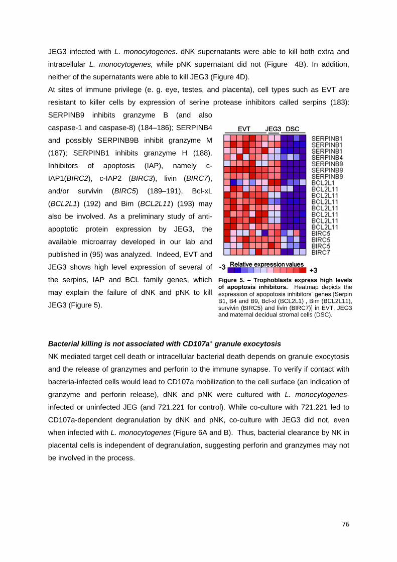
76
JEG3 infected with L. monocytogenes. dNK supernatants were able to kill both extra and
intracellular L. monocytogenes, while pNK supernatant did not (Figure 4B). In addition,
neither of the supernatants were able to kill JEG3 (Figure 4D).
At sites of immune privilege (e. g. eye, testes, and placenta), cell types such as EVT are
resistant to killer cells by expression of serine protease inhibitors called serpins (183):
SERPINB9 inhibits granzyme B (and also
caspase-1 and caspase-8) (184–186); SERPINB4
and possibly SERPINB9B inhibit granzyme M
(187); SERPINB1 inhibits granzyme H (188).
Inhibitors of apoptosis (IAP), namely c-
IAP1(BIRC2), c-IAP2 (BIRC3), livin (BIRC7),
and/or survivin (BIRC5) (189–191), Bcl-xL
(BCL2L1) (192) and Bim (BCL2L11) (193) may
also be involved. As a preliminary study of anti-
apoptotic protein expression by JEG3, the
available microarray developed in our lab and
published in (95) was analyzed. Indeed, EVT and
JEG3 shows high level expression of several of
the serpins, IAP and BCL family genes, which
may explain the failure of dNK and pNK to kill
JEG3 (Figure 5).
Bacterial killing is not associated with CD107a+ granule exocytosis
NK mediated target cell death or intracellular bacterial death depends on granule exocytosis
and the release of granzymes and perforin to the immune synapse. To verify if contact with
bacteria-infected cells would lead to CD107a mobilization to the cell surface (an indication of
granzyme and perforin release), dNK and pNK were cultured with L. monocytogenes-
infected or uninfected JEG (and 721.221 for control). While co-culture with 721.221 led to
CD107a-dependent degranulation by dNK and pNK, co-culture with JEG3 did not, even
when infected with L. monocytogenes (Figure 6A and B). Thus, bacterial clearance by NK in
placental cells is independent of degranulation, suggesting perforin and granzymes may not
be involved in the process.
Figure 5. – Trophoblasts express high levels of apoptosis inhibitors. Heatmap depicts the
expression of apopotosis inhibitors’ genes [Serpin B1, B4 and B9, Bcl-xl (BCL2L1) , Bim (BCL2L11), survivin (BIRC5) and livin (BIRC7)] in EVT, JEG3 and maternal decidual stromal cells (DSC).
Figure 5. – Trophoblasts express high levels of apoptosis inhibitors. Heatmap depicts the
expression of apopotosis inhibitors’ genes [Serpin B1, B4 and B9, Bcl-xl (BCL2L1) , Bim (BCL2L11), survivin (BIRC5) and livin (BIRC7)] in EVT, JEG3 and maternal decidual stromal cells (DSC).

77
Figure 6. L. monocytogenes infection of JEG3 does not elicit degranulation of dNK and pNK. A)
Representative FACS plots of degranulation of dNK (top) and pNK (bottom) in response to 221 and uninfected or L. monocytogenes -infected JEG3. B) Percentage of CD107a+ pNK and dNK after co-culture with 221, healthy and L. monocytogenes -infected JEG3.

78
DISCUSSION
The present work describes a new and exciting phenomenon – NK cells are able to eliminate
microbes within infected cells without killing the host cell. The killing of intracellular microbes
does not rely on CD107a-dependent degranulation and is possibly due to granulysin that is
constitutively secreted by dNK. In dNK, granulysin was found to be diffusely distributed in the
cytoplasm as well as contained in vesicles. Although some of the granulysin-positive
vesicles also contained perforin, granulysin-exclusive vesicles were identified. In addition,
the granulysin secretion was independent of the release of granzymes and perforin. Thus
granulysin exclusive vesicles may be responsible for the constitutive secretion of granulysin.
The ability to secrete granulysin constitutively may be due to a high activation state of dNK
(e.g. constitutive expression of CD69, high levels of cytotoxic granules) (105).
The concentration of granulysin in the dNK supernatants do not fall within the range
considered to be microbicidal by itself (1-3μM) (136). In addition, which isoform of granulysin
is found in dNK supernatants is not known. Previous studies of pNK demonstrated the
15kDa isoform of granulysin is preferentially secreted. The 15kDa form has not been studied
extensively, and until recently it was not clear whether it serves only as a precursor of the
9kDa isoform or if it has a separate function by itself (177). In activated CTLs and pNK,
9kDa granulysin co-localizes with granzymes and perforin in cytolytic granules, while 15kDa
is localized in its own granules that can exocytose upon activation without target cell contact
and synapse formation(182). Also, the 15kDa form has no cytolytic activity against
mammalian cells or extracellular microbes. However, it can stimulate monocytes to
differentiate into dendritic cells, and work as a chemoattractant for leukocytes (182). An
interesting hypothesis concerns the possibility of the 15kDa form being taken up by bacteria-
infected JEG3 and then proteolytically cleaved within the cells to release 9kDa form, leading
to intracellular bacteria death. These questions are currently being addressed by performing
detailed analysis of the contents of the dNK cell culture supernatant.
The levels of granulysin in dNK supernatants may contribute to the extracellular bacterial
killing, in combination with other putative antimicrobial substances present in the
supernatant. In fact, dNK constitutively secrete other molecules, such as galectin-1, which
may have a role in bacteria elimination (105). Galectins are β-galactoside-binding lectins that
accumulate in the cytosol before being secreted via a leader-peptide-independent pathway.
Extracellular galectins bind glycans on cell membranes and modulate cellular behavior.
However, they can also occur in the cytosol and can act as danger and pattern-recognition
receptors (194). Galectin 3, for example, binds bacterial lipopolysaccharide (LPS) and acts
as a negative regulator of LPS functions (195), and Galectin 8 targets damaged vesicles for
autophagy to defend cells against bacterial invasion (194). pNK supernatants have negligible

79
levels of granulysin and were not able to kill extracellular bacteria, and have also been
shown to not secrete Galectin-1 (105).
The ability of dNK supernatants to kill intracellular bacteria may lie in the capacity of
granulysin to enter JEG3 by binding lipid rafts and subsequently being transported to the
intracellular space. This mechanism is independent of perforin. Recombinant granulysin
was shown to bind lipid rafts of Listeria innocua-infected DCs. Subsequently the
granulysin/lipid rafts were endocytosed and transported to bacteria-containing vesicles,
leading to microbe killing (196).
Although pNK supernatants do not kill bacteria, pNK and dNK cells can kill intracellular L.
monocytogenes. In this case, an immune synapse may be formed where all three granules
are transferred to the infected cells and act together to eliminate bacteria. dNK have been
shown to form immature synapses with EVT, where perforin was not polarized to the
synapse (Tilburgs et al, provisionally accepted). In our study, no degranulation was observed
during dNK and pNK co-culture with infected JEG3, which indicates perforin and granzymes
were not released. Thus, bacterial death may be due to granulysin in combination with other
antibacterial peptides. dNK contact with infected cells may increase secretion or dNK may
form a new kind of synapse with JEG3 where only granulysin-containing vesicles are
polarized, resulting in exclusive transfer of granulysin between cells. This mechanism may
apply to pNK, which are able to kill intracellular bacteria when in contact with infected cells,
but do not secrete granulysin without activating stimuli.
Uninfected or infected JEG3 were not killed during interaction with NK, at least during the
time of assay (4h), which was sufficient to inhibit bacterial growth. A longer exposure of
infected JEG3 to NK may result in cell death; indeed, a previous study on HCMV-infected
primary decidual stromal cells (DSC) demonstrated efficient lysis of infected DSC only
occurs after 18h of contact (163). In addition, dNK do not degranulate against HCMV-
infected JEG3 or EVT even after 10-12 hours of contact, while pNK are able to degranulate
in response to infected JEG3 and EVT (chapter 3). pNK do not contact EVT in vivo, but
whether there is influx of pNK to the decidua during infection has not been explored. dNK
may form synapses with infected JEG3 that lead to bacterial death. JEG3 may be resistant
to NK killing due to expression of anti-apoptotic proteins such as serpins ((95) and figure 5).
The findings reported in this chapter point to an important role of dNK in clearance of
bacterial infection through the action of granulysin. These observations reveal a novel
function of dNK as true immune effector cells besides their role in secretion of cytokines and
growth factors that facilitates trophoblast invasion. Further experiments are necessary to
study the mechanism of action of dNK-secreted granulysin in the elimination of placental
infections. Current studies in include the detailed analysis of dNK supernatants and potential
binding of granulysin to lipid rafts for internalization in infected JEG3. Furthermore, the

80
cytolytic potential of NK in response to bacteria infected-JEG3 and JEG3 resistance to death
will be studied, in addition to immune synapse formation between dNK and JEG3. The
influence of cytokines in the expression and secretion of granulysin by dNK and pNK will
also be analyzed.
In summary, these data unveil a new target for pre-natal therapies. Bacterial infections in
early gestations can be treated by increasing the dNK potential to secrete granulysin, or by
administration of granulysin in microbicidal doses that do not affect the placental cells. The
treatment of placental infections in an early stage may avoid complications later in
pregnancy such as miscarriage, congenital malformations or fetal growth restriction.

81
CHAPTER 5
GENERAL DISCUSSION

82
The discovery of the presence of high numbers of large granular lymphocytes (LGL) in
human decidua, later identified as decidual Natural Killer (dNK) cells (197), led to the
hypothesis that fetal placental cells actively inhibit maternal dNK to avoid immunologic
rejection (198). The subsequent characterization of dNK as poor cytotoxic lymphocytes and
major cytokine and growth factor producers distanced dNK from true immune effector cells.
The main role for dNK was established as agents that facilitate implantation, trophoblast
invasion and vascular remodeling. These processes are of key importance for placental
development and enhance the exchange of gases and nutrients from mother to fetus (94,
107, 199). Until recently the role of dNK in clearance of infections, a key function of pNK,
was largely ignored. In 2013, Siewiera et al demonstrated the ability of dNK to clear HCMV-
infected cells (163). The results presented in this thesis build upon this observation and
highlight the dual role of dNK, capable to mount immune effector responses during viral and
bacterial infections as well as provide immune tolerance to the fetus and facilitate placental
growth. The expression of the activating MHC Class I receptor KIR2DS1 by dNK was
associated with improved clearance of HCMV-infected decidual stromal cells (DSC). In
addition, dNK eliminated placental bacterial infections without affecting the host cells,
possibly though secretion of the anti-microbial peptide granulysin. These results emphasize
the importance of dNK to provide both immunity to infections and tolerance to fetal cells.
KIR2DS1, HLA-C2 and HCMV
The MHC Class I molecule HLA-C expressed by fetal EVT is the main factor that can cause
allogeneic recognition and possible immune rejection by maternal immune cells. This
immune response can be driven by T cells through direct allo-recognition (e.g. fetal EVT
express a foreign HLA-C molecule that directly activates maternal T cells) or by NK cells
due to missing-self (e.g. EVT lack a MHC molecule and fail to inhibit NK cytotoxicity through
inhibitory receptors) (91, 94). dNK express high levels of receptors for MHC class I [of which
the killer cell immunoglobulin-like receptors (KIR) are most important for HLA-C]. Particular
combinations of KIR and MHC haplotypes influence reproductive success, clearance of viral
infections and development of auto-immune diseases. In particular, women with KIR-AA
haplotypes (lacking most activating KIR) are in high risk of developing complications such as
pre-eclampsia, recurrent miscarriage or fetal growth restriction. The effect is enhanced when
the fetus expresses group 2 HLA-C alleles (71, 112, 114). This observation led to the
hypothesis that the lack of activating KIR (KIR2DS1) allows for a strong inhibition of dNK
through KIR2DL1 (inhibitory receptor), reducing dNK secretion of cytokines that enhance
trophoblast invasion and placental development (117, 118). In this thesis, KIR2DS1+ dNK did
not respond directly to EVT by degranulation or secretion of growth factors such as GM-CSF
or VEGF. In addition, degranulation of KIR2DS1+ dNK in response to HLA-C2+ target cells

83
did not correlate with the HLA-C epitope of the fetus, indicating de novo education fetal HLA-
C does not occur (Chapter 2). Instead, KIR2DS1+ dNK were shown to have increased
cytolytic granule content (granzymes, perforin and granulysin) and to have higher levels of
degranulation in response to 221.C2 (Chapter 2). Most importantly, KIR2DS1+ dNK
displayed higher capacity to clear human cytomegalovirus (HCMV)-infected decidual stromal
cells (DSC), in particular when HLA-C2 molecules were present (Chapter 3). These
observations were specific for dNK and not observed in pNK.
HCMV employs many strategies for immune evasion and prevent the lysis of infected cells.
These strategies include interference with MHC Class I processing and surface expression
(128). As presented in chapter 3, HCMV infection downregulated HLA-C in DSC in early
stages, but upregulated HLA-C expression in late infection. Consequently, cells in early
infection stages are not able to inhibit dNK through KIR2DL1, while cells in late infection
stages can increase activation of single-positive KIR2DS1+ dNK. Thus the presence of
KIR2DS1+ dNK can enhance elimination of decidual HCMV-infection and limit virus-induced
placental pathology. These observations may lead to novel pathways for development of
immune therapies that prevent congenital infections and virus-induced pregnancy
complications.
Interaction of EVT did not induce secretion of growth factors by dNK. Whether or not
interaction with other cell types in decidua (e.g. decidual macrophages, T cells and stromal
cells) and modulation by the decidual environment induce cytokine secretion by dNK
remains to be investigated. Several lines of evidence indicate DSC as conditioners of dNK
phenotype and function, via direct interaction or production of cytokines such as IL-33 and
TGF-β (156, 200). DSC decrease pNK cytotoxicity and upregulate the expression of
inhibitory receptors in dNK (200, 201). Other sources of anti-inflammatory cytokines that can
influence dNK behavior are decidual macrophages (103). dMϕs have been shown to secrete
TGF-β and to inhibit dNK cytotoxicity in vitro (202). Crosstalk between dNK and dMϕs or
dendritic cells in the maternal fetal interface is also involved in decidualization in mice (203).
The detrimental association of the maternal KIR-AA haplotype with fetal HLA-C2 may have
been a strong evolutionary force for the distribution of KIR and MHC haplotypes in human
populations. Frequency of AA haplotype and HLA-C2 epitope is inversely correlated and this
reduces the frequency of potentially complicated pregnancies: women who are homozygous
for the group A KIR haplotype paired with men who carry the HLA-C2 epitope (204).
Infections are also known to be strong forces of evolution and natural selection.
Combinations of KIR and HLA are associated with increased clearance of viral infections
(59, 64, 164). For example, KIR2DL2/3 and HLA-C1 are associated with control of HCV
infection, due to the low affinity interaction of KIR2DL2/3 with HLA-C1 (compared with the
high affinity interaction of KIR2DL1 and HLA-C2). This leads to a less effective inhibition of

84
NK and consequently higher activation (70). Although the evidence is correlative, it raises
the hypothesis that KIR-A haplotypes are mainly selected for their role in immune defense,
whereas KIR-B haplotypes are selected for their role in placental reproduction. An episode of
viral infection would select for KIR-A haplotypes, which will be enriched in the survivors, but
in subsequent expansion of the surviving population there will be selection for KIR-B
haplotypes and against KIR-A. Thus, long-term survival of human populations have been
selected for retain both KIR-A and KIR-B haplotypes (205, 206). In this thesis, the KIR-B
haplotype correlates with improved clearance of viral infection in pregnancy, and not KIR-A.
However, elimination of viral infection leads to reproductive success and contributes to the
maintenance of KIR B haplotypes in the population. These results suggest a more complex
model for KIR haplotype influence in evolution and demand further investigation.
dNK, infections and pregnancy complications
Viral infections in pregnancy, in particular HCMV, are frequent and lead to pregnancy
complications that include miscarriage and pre-term birth (122, 132). HCMV persists as a
latent infection and, in the absence of high levels of protective antibodies, HCMV can
reactivate in the placenta. Placental HCMV infection interferes with trophoblast invasion and
placental development and can give rise to placental pathology and congenital syndromes
(123, 126) Due to the high variety of pathogens involved and subclinical nature of some
infections, occurrence of infections during pregnancy is underdiagnosed. CD8+ T cells
represent only 2-7% of CD45+ lymphocytes in decidua and thus dNK are the prime
candidates to respond to viral infections in decidua (162). Siewiera et al provided the first
evidence that dNK are able to clear HCMV infected DSC (163). In this thesis, we expanded
these studies and showed while dNK can clear HCMV-infection of maternal DSC, HCMV-
infected trophoblast cannot be cleared (Chapter 3). While dNK degranulated and secreted
pro-inflammatory cytokines in response to HCMV-infected DSC, no response was observed
to HCMV-infected JEG3 or primary trophoblast (Chapter 3). The clearance of infected DSC
limits the viral spread to trophoblast, which are naturally more resistant (168). The failure of
dNK to kill EVT, even when infected with HCMV, may be due to the immunomodulatory
properties of EVT, to avoid detrimental consequences to the fetus caused by excessive
inflammation. Virus, bacteria and parasites may take advantage of the immune privileged
status of the placenta and EVT to avoid immune attack. However, pNK are able to kill
infected trophoblasts in vitro, although pNK and EVT do not interact in vivo. These
observations underline the importance of early diagnosis of infections during pregnancy to
avoid the spread of pathogens to the placenta and fetus. Understanding the mechanisms
that regulate the switch in dNK immune tolerance and immunity contribute to discover key

85
factors involved in the immunopathology of placental HCMV infection and lead to novel
strategies to limit congenital infection.
dNK, granulysin and the control of bacterial infections
Similarly to viral infections, bacterial (e.g. Listeria monocytogenes, Group B streptococcus)
and parasite infections (e.g. Toxoplasma gondii, Plasmodium spp) can also cause major
complications during pregnancy and this includes miscarriage, preterm delivery and
maternal and fetal sepsis (139, 140). In this thesis, the importance of the high level of
antimicrobial peptide granulysin in dNK was demonstrated in the clearance of Listeria
monocytogenes. dNK were shown for the first time to clear intracellular Listeria
monocytogenes in the trophoblast cell line JEG3 without killing the JEG3 cells (Chapter 4).
The failure of dNK to lyse infected JEG3 may be due to the expression of anti-apoptotic
proteins that neutralize the effect of granzymes by JEG3 (95). This reveals a novel function
of dNK as immune effectors able to clear bacterial infections while maintaining tolerance to
the trophoblast.
Killing of intracellular bacteria by NK in most cell types is dependent on the concerted action
of granzyme, perforin and granulysin (135). However, the elimination of L. monocytogenes
by dNK in trophoblast lines was independent of degranulation (Chapter 4). This suggests the
existence of other pathways such as specialized immune synapses that may transfer
granulysin and other antimicrobial substances to target cells. Furthermore dNK may target
bacteria through constitutive secretion of granulysin or other antimicrobial peptides that enter
the infected host cells without killing them (138).
The stimuli that induce the high granulysin expression and constitutive secretion of
granulysin by dNK are unknown and may be related to the contact of dNK with the placenta
bacterial flora. Until recently, the placenta was thought to be sterile, but a multi-center study
reported the existence of a unique microbiome (207). pNK may require activating signals
(e.g. of placental cells or the placental microbiome) to increase the expression and secretion
of granulysin.
The data presented in Chapter 4 is preliminary and raises several questions related to the
regulation of granulysin expression and secretion by dNK, the mechanisms involved in the
bacterial elimination and trophoblast protection, as well as other microbial compounds that
may be involved in bacterial elimination. Nevertheless, these novel findings provide
promising new possibilities for congenital therapies and for the elimination of bacterial and
parasite infections. This includes a focus on the enhancement of dNK function and
granulysin activity.

86
Mice are not humans
Although mouse placentation is not as invasive as human placentation, both mouse and
human placentation are haemochorial. These arguments have been used to support the
claim that mouse are both a good and not a good model to study human reproduction. Other
differences between mouse and humans are gestational length (3 versus 40 weeks),
exposure to pathogens (mouse are kept in pathogen free environments) and MHC
differences (mouse are mainly inbred strains with limited polymorphisms) (208). Most
importantly, the NK receptor repertoire for MHC Class I of mice and humans have evolved
separately (mouse have Ly49 genes instead of KIR genes) (204). Despite these differences,
mice are useful to study placental immunology and development of pregnancy complications
as long as these differences are taken in consideration.
In particular, Anne Croy and others have shown VEGF and IFN-γ produced by uterine NK
(uNK) is crucial for an effective vascularization of mouse decidua (108, 209, 210). Mice that
lack both T and NK cells (Rag2-/-Il2rg-/-) or only NK cells (IL-15 KO), although still bearing
successful semi-allogeneic pregnancies, have delayed vascularization compared to WT and
the litter sizes are smaller (211). Although both KO mice were capable to successful semi-
allogeneic pregnancies, the fact that these mice were kept in a pathogen-free environment
should be considered. This does not address the crucial role of uNK cells in the resolution of
infection. Interestingly, no interactions occurred between uNK cells and trophoblasts in the
live tissues studied at early gestation. This is in stark contrast with the claims that interaction
of dNK with EVT in humans is the main event that triggers cytokine and growth factor
secretion by dNK (107, 117, 118). In fact, our lab demonstrated, both in early publications
and in this thesis, that direct co-culture of dNK with EVT does not elicit cytokine or growth
factor secretion such as IFN-γ, VEGF or GM-CSF (Chapter 2) (95).
The maternal KIR AA/fetal HLA-C2 detrimental association has been hypothesized to be due
to strong inhibition of KIR2DS1- dNK cytokine secretion by HLA-C2+ EVT. This theory was
explored with the use of a mouse pregnancy model in which the fetus expressed an extra
MHC Class I molecule to increase the inhibition of maternal NK cells. The litter sizes in this
model were smaller and the secretion of IFN-γ by uNK cells was significantly lower than in
pregnancies without the extra MHC. However, the effects were independent of the paternal
or maternal origin of the extra MHC and the results did not account for the presence of NK
activating receptors (117). These results leave open the possibility that maternal HLA-C2+
DSC, and not only fetal EVT, can be involved in the inhibitory interactions in KIR2DS1-
women and dNK activation in KIR2DS1+ women. Chapter 3 demonstrated maternal HCMV-
infected HLA-C2+ DSC did increase the cytotoxicity of S1+ dNK – showing the influence of
maternal HLA-C.
87
Other notes and final conclusion
All the work presented in this thesis was performed using primary NK cells and trophoblasts,
isolated from human tissue. The use of primary cells yields results relevant for human
clinical applications However, primary cells have several limitations, such as short culture
life, lack of proliferation, limited cell numbers, and a great variability in phenotype and activity
that is inherent to human populations. In addition, it was not possible to obtain peripheral
blood cells from the same women that donated decidual tissue. All the pNK were obtained
from unrelated blood donors, and the results may be different from pNK from pregnant
women.
The data presented in this thesis on dNK and EVT in healthy and infected pregnancies
opens exciting new possibilities for therapies that can improve maternal and fetal health, with
special focus in enhancing dNK function in viral and bacterial infections. Identifying high risk
pregnancies (e.g. women with KIR-AA genotypes) at early stages for monitoring viral and
bacterial infections will be crucial to avoid disorders that affect both mother and fetus. The
preliminary data on the possible role of dNK-derived granulysin will need further
development, but it presents a promising strategy to eliminate bacteria without disturbing the
delicate placental tissue. Finally, this work represents a step further on the understanding of
Peter Medawar’s proposed problem – the balance between immunity and tolerance in the
maternal-fetal interface.

88
REFERENCES
1. Goldsby, R. A., T. J. Kindt, B. A. Osborne, and J. Kuby. 2003. Immunology, 5th ed. (J. Noe, M. Ryan, J. Ciprioni, K. Mergen, G. Hadler, A. Kudlacik, and N. Villamora, eds). W.H. Freeman and Company, New York.
2. Medzhitov, R., and C. a Janeway. 2002. Decoding the patterns of self and nonself by the innate immune system. Science 296: 298–300.
3. York, I. a, and K. L. Rock. 1996. Antigen processing and presentation by the class I major histocompatibility complex. Annu. Rev. Immunol. 14: 369–396.
4. Chervin, A. S., J. D. Stone, P. D. Holler, A. Bai, J. Chen, H. N. Eisen, and D. M. Kranz. 2009. The impact of TCR-binding properties and antigen presentation format on T cell responsiveness. J. Immunol. 183: 1166–78.
5. Bogunia-Kubik, K., and M. Iwaszko. 2011. Clinical Significance of the HLA-E and CD94 / NKG2 Interaction. Arch. Immunol. Ther. Exp. 59: 353–367.
6. Amiot, L., N. Vu, and M. Samson. 2014. Immunomodulatory Properties of HLA-G in Infectious Diseases. J. Immunol. Res. 2014: 298569.
7. Petroff, M. G. 2005. Immune interactions at the maternal – fetal interface. J. Reprod. Immunol. 68: 1–13.
8. Roche, P. a., and K. Furuta. 2015. The ins and outs of MHC class II-mediated antigen processing and presentation. Nat. Rev. Immunol. 15: 203–216.
9. Abbey, J. L., and H. C. O’Neill. 2008. Expression of T-cell receptor genes during early T-cell development. Immunol. Cell Biol. 86: 166–174.
10. Starr, T. K., S. C. Jameson, and K. a Hogquist. 2003. Positive and negative selection of T cells. Annu. Rev. Immunol. 21: 139–176.
11. Shah, D. K., and J. C. Zúñiga-Pflücker. 2014. An overview of the intrathymic intricacies of T cell development. J. Immunol. 192: 4017–23.
12. O’Garra, A. 1998. Cytokines induce the development of functionally heterogeneous T helper cell subsets. Immunity 8: 275–283.
13. Toda, A., and C. a Piccirillo. 2006. Development and function of naturally occurring CD4+CD25+ regulatory T cells. J. Leukoc. Biol. 80: 458–70.
14. Chowdhury, D., and J. Lieberman. 2008. Death by a thousand cuts: granzyme pathways of programmed cell death. Annu. Rev. Immunol. 26: 389–420.
15. Thiery, J., D. Keefe, S. Boulant, E. Boucrot, M. Walch, D. Martinvalet, I. S. Goping, R. C. Bleackley, T. Kirchhausen, and J. Lieberman. 2011. Perforin pores in the endosomal membrane trigger the release of endocytosed granzyme B into the cytosol of target cells. Nat. Immunol. 12: 770–7.
16. Van Lier, R. A. W., I. J. M. ten Berge, and L. E. Gamadia. 2003. HUMAN CD8+ T-CELL DIFFERENTIATION IN RESPONSE TO VIRUSES. Nat. Rev. Immunol. 3: 913–918.

89
17. Ehlich, A., and R. Küppers. 1995. Analysis of immunoglobulin gene rearrangements in single B cells. Curr. Opin. Immunol. 7: 281–284.
18. Hodgkin, P. D., and A. Basten. 1995. B cell activation, tolerance and antigen-presenting function. Curr. Opin. Immunol. 7: 121–129.
19. Armitage, R. J., and M. R. Alderson. 1995. B-cell stimulation. Curr. Opin. Immunol. 7: 243–247.
20. Fridman, W. H. 1991. Fc receptors and immunoglobulin binding factors. FASEB J. 5: 2684–2690.
21. Leavy, O. 2012. Maturation and function of NK cells. Nat. Rev. Immunol. 12: 150.
22. Bellora, F., R. Castriconi, A. Dondero, P. Carrega, A. Mantovani, G. Ferlazzo, A. Moretta, and C. Bottino. 2014. Human NK cells and NK receptors. Immunol. Lett. 161: 168–173.
23. Sun, J. C., J. N. Beilke, and L. L. Lanier. 2009. Adaptive immune features of natural killer cells. Nature 457: 557–561.
24. Biassoni, R., E. Ugolotti, and A. De Maria. 2009. NK cell receptors and their interactions with MHC. Curr. Pharm. Des. 15: 3301–10.
25. Afzali, B., R. I. Lechler, and M. P. Hernandez-Fuentes. 2007. Allorecognition and the alloresponse: Clinical implications. Tissue Antigens 69: 545–556.
26. Afzali, B., G. Lombard, and R. I. Lechler. 2008. Pathways of major histocompatibility complex allorecognition. Curr. Opin. Organ Transplant. 13: 438–444.
27. Charpentier, B., P. Lang, C. Hiesse, and D. Fries. 1986. Effector mechanisms of graft rejection. Nephrologie 7: 15–18.
28. Pegram, H. J., D. M. Andrews, M. J. Smyth, P. K. Darcy, and M. H. Kershaw. 2010. Activating and inhibitory receptors of natural killer cells. Immunol. Cell Biol. 89: 216–224.
29. Moretta, L., E. Montaldo, P. Vacca, G. Del Zotto, F. Moretta, P. Merli, F. Locatelli, and M. C. Mingari. 2014. Human Natural Killer Cells: Origin, Receptors, Function, and Clinical Applications. Int. Arch. Allergy Immunol. 164: 253–264.
30. Vivier, E. 2006. What is natural in natural killer cells? Immunol. Lett. 107: 1–7.
31. Horowitz, A., D. M. Strauss-albee, M. Leipold, J. Kubo, O. C. Dogan, C. L. Dekker, S. Mackey, H. Maecker, G. E. Swan, M. M. Davis, P. J. Norman, L. a Guethlein, M. Desai, P. Parham, and C. a Blish. 2013. Genetic and Environmental determinants of human NK cell Diversity Revealed By Mass Cytometry. Sci. Transl. Med. 5: 208ra145.
32. Biassoni, R. 2009. Human natural killer receptors, co-receptors, and their ligands. Curr. Protoc. Immunol. Chapter 14: 14.10.1–40.
33. Krzewski, K., and J. L. Strominger. 2008. The killer’s kiss: the many functions of NK cell immunological synapses. Curr. Opin. Cell Biol. 20: 597–605.

90
34. Rak, G. D., E. M. Mace, P. P. Banerjee, T. Svitkina, and J. S. Orange. 2011. Natural killer cell lytic granule secretion occurs through a pervasive actin network at the immune synapse. PLoS Biol. 9: e1001151.
35. Shifrin, N., D. H. Raulet, and M. Ardolino. 2014. NK cell self tolerance, responsiveness and missing self recognition. Semin. Immunol. 26: 138–144.
36. Brady, J., S. Carotta, R. P. L. Thong, C. J. Chan, Y. Hayakawa, M. J. Smyth, and S. L. Nutt. 2010. The interactions of multiple cytokines control NK cell maturation. J. Immunol. 185: 6679–88.
37. Upshaw, J. L., and P. J. Leibson. 2006. NKG2D-mediated activation of cytotoxic lymphocytes: unique signaling pathways and distinct functional outcomes. Semin. Immunol. 18: 167–75.
38. Arlettaz, L., J. Villard, C. De Rham, S. Degermann, B. Chapuis, B. Huard, and E. Roosnek. 2004. Activating CD94 : NKG2C and inhibitory CD94 : NKG2A receptors are expressed by distinct subsets of committed CD8 + TCR ab lymphocytes. Eur. J. Cell Biol. 34: 3456–3464.
39. Borrego, F., M. Masilamani, A. I. Marusina, X. Tang, and J. E. Coligan. 2006. The CD94 / NKG2 Family of Receptors. Immunol. Res. 35: 263–277.
40. Mori, Y., S. Tsuji, M. Inui, Y. Sakamoto, S. Endo, Y. Ito, S. Fujimura, T. Koga, A. Nakamura, H. Takayanagi, E. Itoi, and T. Takai. 2008. Inhibitory immunoglobulin-like receptors LILRB and PIR-B negatively regulate osteoclast development. J. Immunol. 181: 4742–51.
41. Davidson, C. L., N. L. Li, and D. N. Burshtyn. 2010. LILRB1 polymorphism and surface phenotypes of natural killer cells. Hum. Immunol. 71: 942–9.
42. Middleton, D., and F. Gonzelez. 2010. The extensive polymorphism of KIR genes. Immunology 129: 8–19.
43. Parham, P. 2008. The genetic and evolutionary balances in human NK cell receptor diversity. Semin. Immunol. 20: 311–6.
44. Alari-pahissa, E., C. Grandclément, B. Jeevan-raj, and W. Held. 2014. Inhibitory Receptor-Mediated Regulation of Natural Killer Cells. Crit. Rev. Immunol. 34: 455–465.
45. Culley, F. J., M. Johnson, J. H. Evans, S. Kumar, R. Crilly, J. Casasbuenas, T. Schnyder, M. Mehrabi, M. P. Deonarain, D. S. Ushakov, V. Braud, G. Roth, R. Brock, K. Köhler, and D. M. Davis. 2009. Natural killer cell signal integration balances synapse symmetry and migration. PLoS Biol. 7: e1000159.
46. Lanier, L. L. 2010. DAP10- and DAP12-associated receptors in innate immunity. Immunol. Rev. 227: 150–160.
47. Moretta, a, D. Pende, F. Locatelli, and L. Moretta. 2009. Activating and inhibitory killer immunoglobulin-like receptors (KIR) in haploidentical haemopoietic stem cell transplantation to cure high-risk leukaemias. Clin. Exp. Immunol. 157: 325–31.
91
48. Li, C., B. L. Houser, M. L. Nicotra, and J. L. Strominger. 2009. HLA-G homodimer-induced cytokine secretion through HLA-G receptors on human decidual macrophages and natural killer cells. Proc. Natl. Acad. Sci. U. S. A. 106: 5767–72.
49. Rajagopalan, S., Y. T. Bryceson, S. P. Kuppusamy, D. E. Geraghty, A. Van Der Meer, I. Joosten, and E. O. Long. 2006. Activation of NK Cells by an Endocytosed Receptor for Soluble HLA-G. PLoS Biol. 4: e9.
50. Parham, P., P. J. Norman, L. Abi-Rached, and L. a Guethlein. 2011. Variable NK cell receptors exemplified by human KIR3DL1/S1. J. Immunol. 187: 11–9.
51. Colantonio, A. D., B. N. Bimber, W. J. Neidermyer, R. K. Reeves, G. Alter, M. Altfeld, R. P. Johnson, M. Carrington, D. H. O’Connor, and D. T. Evans. 2011. KIR polymorphisms modulate peptide-dependent binding to an MHC class I ligand with a Bw6 motif. PLoS Pathog. 7: e1001316.
52. Elliott, J. M., and W. M. Yokoyama. 2011. Unifying concepts of MHC-dependent natural killer cell education. Trends Immunol. 32: 364–72.
53. Anfossi, N., P. André, S. Guia, C. S. Falk, S. Roetynck, C. A. Stewart, V. Breso, C. Frassati, D. Reviron, D. Middleton, F. Romagné, S. Ugolini, and E. Vivier. 2006. Human NK cell education by inhibitory receptors for MHC class I. Immunity 25: 331–42.
54. Brodin, P., T. Lakshmikanth, S. Johansson, K. Kärre, and P. Höglund. 2009. The strength of inhibitory input during education quantitatively tunes the functional responsiveness of individual natural killer cells. Blood 113: 2434–41.
55. Höglund, P., and P. Brodin. 2010. Current perspectives of natural killer cell education by MHC class I molecules. Nat. Rev. Immunol. 10: 724–34.
56. Brodin, P., and P. Höglund. 2008. Beyond licensing and disarming: a quantitative view on NK-cell education. Eur. J. Immunol. 38: 2934–7.
57. Jost, S., and M. Altfeld. 2013. Control of human viral infections by natural killer cells. Annu. Rev. Immunol. 31: 163–94.
58. Nash, W. T., J. Teoh, H. Wei, A. Gamache, and M. G. Brown. 2014. Know thyself: NK-cell inhibitory receptors prompt self-tolerance, education, and viral control. Front. Immunol. 5: 1–12.
59. Martin, M. P., Y. Qi, X. Gao, E. Yamada, J. N. Martin, F. Pereyra, S. Colombo, E. E. Brown, W. L. Shupert, J. Phair, J. J. Goedert, S. Buchbinder, G. D. Kirk, A. Telenti, M. Connors, S. J. O’Brien, B. D. Walker, P. Parham, S. G. Deeks, D. W. McVicar, and M. Carrington. 2007. Innate partnership of HLA-B and KIR3DL1 subtypes against HIV-1. Nat. Genet. 39: 733–740.
60. Zipeto, D., and A. Beretta. 2012. HLA-C and HIV-1: friends or foes? Retrovirology 9: 39.
61. Körner, C., and M. Altfeld. 2012. Role of KIR3DS1 in human diseases. Front. Immunol. 3: 1–11.

92
62. Bonagura, V. R., L. J. Hatam, D. W. Rosenthal, J. a. De Voti, F. Lam, B. M. Steinberg, and A. L. Abramson. 2010. Recurrent respiratory papillomatosis: A complex defect in immune responsiveness to human papillomavirus-6 and -11. Apmis 118: 455–470.
63. Stern, M., K. Hadaya, G. Hönger, P.-Y. Martin, J. Steiger, C. Hess, and J. Villard. 2011. Telomeric rather than centromeric activating KIR genes protect from cytomegalovirus infection after kidney transplantation. Am. J. Transplant 11: 1302–7.
64. Stern, M., H. Elsässer, G. Hönger, J. Steiger, S. Schaub, and C. Hess. 2008. The number of activating KIR genes inversely correlates with the rate of CMV infection/reactivation in kidney transplant recipients. Am. J. Transplant. 8: 1312–1317.
65. Cook, M., D. Briggs, C. Craddock, P. Mahendra, D. Milligan, C. Fegan, P. Darbyshire, S. Lawson, E. Boxall, and P. Moss. 2006. Donor KIR genotype has a major influence on the rate of cytomegalovirus reactivation following T-cell replete stem cell transplantation. Blood 107: 1230–2.
66. Béziat, V., L. L. Liu, J.-A. Malmberg, M. a Ivarsson, E. Sohlberg, A. T. Björklund, C. Retière, E. Sverremark-Ekström, J. Traherne, P. Ljungman, M. Schaffer, D. a Price, J. Trowsdale, J. Michaëlsson, H.-G. Ljunggren, and K.-J. Malmberg. 2013. NK cell responses to cytomegalovirus infection lead to stable imprints in the human KIR repertoire and involve activating KIRs. Blood 121: 2678–88.
67. Gazit, R., B.-Z. Garty, Y. Monselise, V. Hoffer, Y. Finkelstein, G. Markel, G. Katz, J. Hanna, H. Achdout, R. Gruda, T. Gonen-Gross, and O. Mandelboim. 2004. To the editor: Expression of KIR2DL1 on the entire NK cell population: a possible novel immunodeficiency syndrome. Blood 103: 1965–1967.
68. Aranda-Romo, S., C. a. Garcia-Sepulveda, A. Comas-García, F. Lovato-Salas, M. Salgado-Bustamante, A. Gómez-Gómez, and D. E. Noyola. 2012. Killer-cell immunoglobulin-like receptors (KIR) in severe A (H1N1) 2009 influenza infections. Immunogenetics 64: 653–662.
69. La, D., C. Czarnecki, H. El-Gabalawy, A. Kumar, A. F. a Meyers, N. Bastien, J. N. Simonsen, F. a. Plummer, and M. Luo. 2011. Enrichment of variations in KIR3DL1/s1 and KIR2DL2/L3 among H1N1/09 ICU patients: An exploratory study. PLoS One 6: e29200.
70. Khakoo, S. I., C. L. Thio, M. P. Martin, C. R. Brooks, X. Gao, J. Astemborski, J. Cheng, J. J. Goedert, D. Vlahov, M. Hilgartner, S. Cox, A.-M. Little, G. J. Alexander, M. E. Cramp, S. J. O’Brien, W. M. C. Rosenberg, D. L. Thomas, and M. Carrington. 2004. HLA and NK cell inhibitory receptor genes in resolving hepatitis C virus infection. Science (80-. ). 305: 872–874.
71. Hiby, S. E., R. Apps, A. M. Sharkey, L. E. Farrell, L. Gardner, A. Mulder, F. H. Claas, J. J. Walker, C. C. Redman, L. Morgan, C. Tower, L. Regan, G. E. Moore, M. Carrington, and A. Moffett. 2010. Maternal activating KIRs protect against human reproductive failure mediated by fetal HLA-C2. J. Clin. Invest. 120: 4102–4110.
72. Łuszczek, W., M. Mańczak, M. Cisło, P. Nockowski, A. Wiśniewski, M. Jasek, and P. Kuśnierczyk. 2004. Gene for the activating natural killer cell receptor, KIR2DS1, is associated with susceptibility to psoriasis vulgaris. Hum. Immunol. 65: 758–766.

93
73. Kuśnierczyk, P. 2013. Killer cell immunoglobulin-like receptor gene associations with autoimmune and allergic diseases, recurrent spontaneous abortion, and neoplasms. Front. Immunol. 4: 8.
74. Momot, T., S. Koch, N. Hunzelmann, T. Krieg, K. Ulbricht, R. E. Schmidt, and T. Witte. 2004. Association of Killer Cell Immunoglobulin-Like Receptors with Scleroderma. Arthritis Rheum. 50: 1561–1565.
75. Yen, J. H., B. E. Moore, T. Nakajima, D. Scholl, D. J. Schaid, C. M. Weyand, and J. J. Goronzy. 2001. Major histocompatibility complex class I-recognizing receptors are disease risk genes in rheumatoid arthritis. J. Exp. Med. 193: 1159–1167.
76. Hiby, S. E., R. Apps, O. Chazara, L. E. Farrell, P. Magnus, L. Trogstad, H. K. Gjessing, M. Carrington, and A. Moffett. 2014. Maternal KIR in combination with paternal HLA-C2 regulate human birth weight. J. Immunol. 192: 5069–73.
77. Nakimuli, A., O. Chazara, S. E. Hiby, L. Farrell, S. Tukwasibwe, J. Jayaraman, J. a Traherne, J. Trowsdale, F. Colucci, E. Lougee, R. W. Vaughan, A. M. Elliott, J. Byamugisha, P. Kaleebu, F. Mirembe, N. Nemat-Gorgani, P. Parham, P. J. Norman, and A. Moffett. 2015. A KIR B centromeric region present in Africans but not Europeans protects pregnant women from pre-eclampsia. Proc. Natl. Acad. Sci. U. S. A. 112: 845–50.
78. Martin, M. P., X. Gao, J.-H. Lee, G. W. Nelson, R. Detels, J. J. Goedert, S. Buchbinder, K. Hoots, D. Vlahov, J. Trowsdale, M. Wilson, S. J. O’Brien, and M. Carrington. 2002. Epistatic interaction between KIR3DS1 and HLA-B delays the progression to AIDS. Nat. Genet. 31: 429–34.
79. Costa-Garcia, M., A. Vera, M. Moraru, C. Vilches, M. Lopez-Botet, and A. Muntasell. 2015. Antibody-Mediated Response of NKG2Cbright NK Cells against Human Cytomegalovirus. J. Immunol. 194: 2715–2724.
80. Wu, L., and L. Van Kaer. 2011. Natural killer cells: in health and disease. Front. Biosci. (Schol. Ed). 3: 236–251.
81. Sungur, C. M., and W. J. Murphy. 2014. Positive and Negative Regulation by NK Cells in Cancer. Crit. Rev. Oncog. 19: 57–66.
82. Palmer, J. M., K. Rajasekaran, M. S. Thakar, and S. Malarkannan. 2013. Clinical relevance of natural killer cells following hematopoietic stem cell transplantation. J. Cancer 4: 25–35.
83. Venstrom, J. M., G. Pittari, T. a Gooley, J. H. Chewning, S. Spellman, M. Haagenson, M. M. Gallagher, M. Malkki, E. Petersdorf, B. Dupont, and K. C. Hsu. 2012. HLA-C-dependent prevention of leukemia relapse by donor activating KIR2DS1. N. Engl. J. Med. 367: 805–16.
84. Pende, D., S. Marcenaro, M. Falco, S. Martini, M. E. Bernardo, D. Montagna, E. Romeo, C. Cognet, M. Martinetti, R. Maccario, M. C. Mingari, E. Vivier, L. Moretta, F. Locatelli, and A. Moretta. 2009. Anti-leukemia activity of alloreactive NK cells in KIR ligand-mismatched haploidentical HSCT for pediatric patients: evaluation of the functional role of activating KIR and redefinition of inhibitory KIR specificity. Blood 113: 3119–29.

94
85. Marcenaro, E., S. Carlomagno, S. Pesce, M. Della Chiesa, A. Moretta, and S. Sivori. 2011. Role of alloreactive KIR2DS1(+) NK cells in haploidentical hematopoietic stem cell transplantation. J. Leukoc. Biol. 90: 661–7.
86. Moffett, A., and C. Loke. 2006. Immunology of placentation in eutherian mammals. Nat. Rev. Immunol. 6: 584–94.
87. Zhou, Y., K. Bianco, L. Huang, J. K. Nien, M. McMaster, R. Romero, and S. J. Fisher. 2007. Comparative analysis of maternal-fetal interface in preeclampsia and preterm labor. Cell Tissue Res. 329: 559–69.
88. Yeh, C.-C., K.-C. Chao, and S. J. Huang. 2013. Innate immunity, decidual cells, and preeclampsia. Reprod. Sci. 20: 339–53.
89. Straszewski-Chavez, S. L., V. M. Abrahams, and G. Mor. 2005. The role of apoptosis in the regulation of trophoblast survival and differentiation during pregnancy. Endocr. Rev. 26: 877–97.
90. Billington, W. D. 2003. The immunological problem of pregnancy : 50 years with the hope of progress . A tribute to Peter Medawar. J. Reprod. Immunol. 60: 1–11.
91. Trowsdale, J., and A. Betz. 2006. Mother’s Little Helpers: Mechanisms of maternal-fetal tolerance. Nat. Immunol. 7: 241–246.
92. Guleria, I., and M. H. Sayegh. 2007. Maternal acceptance of the fetus: true human tolerance. J. Immunol. 178: 3345–51.
93. Apps, R., L. Gardner, and A. Moffett. 2008. A critical look at HLA-G. Trends Immunol. 29: 313–321.
94. Moffett-King, A. 2002. Natural killer cells and pregnancy. Nat. Rev. Immunol. 2: 656–63.
95. Tilburgs, T., Â. C. Crespo, A. van der Zwan, B. Rybalov, T. Raj, B. Stranger, L. Gardner, A. Moffett, and J. L. Strominger. 2015. Human HLA-G+ extravillous trophoblasts: Immune-activating cells that interact with decidual leukocytes. Proc. Natl. Acad. Sci. U. S. A. 112: 7219–7224.
96. Guerin, L. R., J. R. Prins, and S. a Robertson. 2009. Regulatory T-cells and immune tolerance in pregnancy: a new target for infertility treatment? Hum. Reprod. Update 15: 517–35.
97. Zenclussen, A. C., K. Gerlof, M. L. Zenclussen, S. Ritschel, A. Zambon Bertoja, S. Fest, S. Hontsu, S. Ueha, K. Matsushima, J. Leber, and H.-D. Volk. 2006. Regulatory T cells induce a privileged tolerant microenvironment at the fetal-maternal interface. Eur. J. Immunol. 36: 82–94.
98. Tilburgs, T., S. a Scherjon, B. J. van der Mast, G. W. Haasnoot, M. Versteeg-V D Voort-Maarschalk, D. L. Roelen, J. J. van Rood, and F. H. J. Claas. 2009. Fetal-maternal HLA-C mismatch is associated with decidual T cell activation and induction of functional T regulatory cells. J. Reprod. Immunol. 82: 148–57.

95
99. Munn, D. H., M. Zhou, J. T. Attwood, I. Bondarev, S. J. Conway, B. Marshall, C. Brown, and a L. Mellor. 1998. Prevention of allogeneic fetal rejection by tryptophan catabolism. Science 281: 1191–3.
100. Mor, G., and V. M. Abrahams. 2003. Potential role of macrophages as immunoregulators of pregnancy. Reprod. Biol. Endocrinol. 1: 119.
101. Repnik, U., T. Tilburgs, D. L. Roelen, B. J. van der Mast, H. H. H. Kanhai, S. Scherjon, and F. H. J. Claas. 2008. Comparison of Macrophage Phenotype Between Decidua Basalis and Decidua Parietalis by Flow Cytometry. Placenta 29: 405–412.
102. Abrahams, V. M., Y. M. Kim, S. L. Straszewski, R. Romero, and G. Mor. 2004. Macrophages and apoptotic cell clearance during pregnancy. Am. J. Reprod. Immunol. 51: 275–282.
103. Houser, B. L., T. Tilburgs, J. Hill, M. L. Nicotra, and J. L. Strominger. 2011. Two unique human decidual macrophage populations. J. Immunol. 186: 2633–42.
104. King, A., P. Wooding, and L. Gardner. 1993. Expression of perforin , granzyme A and TIA-1 by human uterine CD56 + NK cells implies they are activated and capable of effector functions. Hum. Reprod. 8: 2061–2067.
105. Koopman, L. a, H. D. Kopcow, B. Rybalov, J. E. Boyson, J. S. Orange, F. Schatz, R. Masch, C. J. Lockwood, A. D. Schachter, P. J. Park, and J. L. Strominger. 2003. Human decidual natural killer cells are a unique NK cell subset with immunomodulatory potential. J. Exp. Med. 198: 1201–12.
106. Kopcow, H. D., D. S. J. Allan, X. Chen, B. Rybalov, M. M. Andzelm, B. Ge, and J. L. Strominger. 2005. Human decidual NK cells form immature activating synapses and are not cytotoxic. Proc. Natl. Acad. Sci. U. S. A. 102: 15563–8.
107. Hanna, J., D. Goldman-Wohl, Y. Hamani, I. Avraham, C. Greenfield, S. Natanson-Yaron, D. Prus, L. Cohen-Daniel, T. I. Arnon, I. Manaster, R. Gazit, V. Yutkin, D. Benharroch, A. Porgador, E. Keshet, S. Yagel, and O. Mandelboim. 2006. Decidual NK cells regulate key developmental processes at the human fetal-maternal interface. Nat. Med. 12: 1065–74.
108. Zhang, J., Z. Chen, G. N. Smith, and A. Croy. 2011. Natural Killer Cell Triggered Vascular Transformation: Maternal Care Before Birth? Cell. Mol. Immunol. 8: 1–11.
109. Hunt, J. S., D. K. Langat, R. H. McIntire, and P. J. Morales. 2006. The role of HLA-G in human pregnancy. Reprod. Biol. Endocrinol. 4 (Suppl 1: S10.
110. Rizzo, R., M. Vercammen, H. van de Velde, P. a Horn, and V. Rebmann. 2011. The importance of HLA-G expression in embryos, trophoblast cells, and embryonic stem cells. Cell. Mol. Life Sci. 68: 341–52.
111. King, a, D. S. Allan, M. Bowen, S. J. Powis, S. Joseph, S. Verma, S. E. Hiby, a J. McMichael, Y. W. Loke, and V. M. Braud. 2000. HLA-E is expressed on trophoblast and interacts with CD94/NKG2 receptors on decidual NK cells. Eur. J. Immunol. 30: 1623–31.
96
112. Hiby, S. E., L. Regan, W. Lo, L. Farrell, M. Carrington, and a Moffett. 2008. Association of maternal killer-cell immunoglobulin-like receptors and parental HLA-C genotypes with recurrent miscarriage. Hum. Reprod. 23: 972–6.
113. Male, V., A. Sharkey, L. Masters, P. R. Kennedy, L. E. Farrell, and A. Moffett. 2011. The effect of pregnancy on the uterine NK cell KIR repertoire. Eur. J. Immunol. 41: 3017–27.
114. Hiby, S. E., J. J. Walker, K. M. O’shaughnessy, C. W. G. Redman, M. Carrington, J. Trowsdale, and A. Moffett. 2004. Combinations of maternal KIR and fetal HLA-C genes influence the risk of preeclampsia and reproductive success. J. Exp. Med. 200: 957–965.
115. Parham, P., P. J. Norman, L. Abi-rached, H. G. Hilton, and L. A. Guethlein. 2012. Review : Immunogenetics of human placentation. Placenta 33: S71–S80.
116. Gonzalez-Galarza, F., L. Takeshita, E. Santos, F. Kempson, M. Maia, A. Silva, A. Silva, G. Ghattaoraya, A. Alfirevic, A. Jones, and D. Middleton. 2015. Allele frequency net 2015 update: new features for HLA epitopes, KIR and disease and HLA adverse drug reaction associations. Nucleic Acid Res. 28: D784–8.
117. Kieckbusch, J., L. M. Gaynor, A. Moffett, and F. Colucci. 2014. MHC-dependent inhibition of uterine NK cells impedes fetal growth and decidual vascular remodelling. Nat. Commun. 5: 3359.
118. Xiong, S., A. M. Sharkey, P. R. Kennedy, L. Gardner, L. E. Farrell, O. Chazara, J. Bauer, S. E. Hiby, F. Colucci, and A. Moffett. 2013. Maternal uterine NK cell – activating receptor KIR2DS1 enhances placentation. J. Clin. Invest. 123: 4265–4272.
119. Pass, R. F., C. Zhang, A. Evans, T. Simpson, W. Andrews, M.-L. Huang, L. Corey, J. Hill, E. Davis, C. Flanigan, and G. Cloud. 2009. Vaccine prevention of maternal cytomegalovirus infection. N. Engl. J. Med. 360: 1191–1199.
120. Rasmussen, S. a., D. J. Jamieson, and T. M. Uyeki. 2012. Effects of influenza on pregnant women and infants. Am. J. Obstet. Gynecol. 207: S3–S8.
121. Kenneson, A., and M. J. Cannon. 2007. Review and meta-analysis of the epidemiology of congenital cytomegalovirus (CMV) infection. Rev. Med. Virol. 17: 253–276.
122. Nigro, G., M. Mazzocco, E. Mattia, G. C. Di Renzo, G. Carta, and M. M. Anceschi. 2011. Role of the infections in recurrent spontaneous abortion. J. Matern. Fetal. Neonatal Med. 24: 983–989.
123. Pereira, L., M. Petitt, a Fong, M. Tsuge, T. Tabata, J. Fang-Hoover, E. Maidji, M. Zydek, Y. Zhou, N. Inoue, S. Loghavi, S. Pepkowitz, L. M. Kauvar, and D. Ogunyemi. 2014. Intrauterine growth restriction caused by underlying congenital cytomegalovirus infection. J Infect Dis 209: 1573–1584.
124. Revello, M. G., D. Lilleri, M. Zavattoni, M. Furione, E. Genini, G. Comolli, and G. Gerna. 2006. Lymphoproliferative response in primary human cytomegalovirus (HCMV) infection is delayed in HCMV transmitter mothers. J. Infect. Dis. 193: 269–276.
125. Warner, J. a, K. J. Zwezdaryk, B. Day, D. E. Sullivan, G. Pridjian, and C. a Morris. 2012. Human cytomegalovirus infection inhibits CXCL12- mediated migration and invasion of human extravillous cytotrophoblasts. Virol. J. 9: 255.

97
126. LaMarca, H. L., a. B. Nelson, a. B. Scandurro, G. S. J. Whitley, and C. a. Morris. 2006. Human cytomegalovirus-induced inhibition of cytotrophoblast invasion in a first trimester extravillous cytotrophoblast cell line. Placenta 27: 137–147.
127. Tilburgs, T., and J. L. Strominger. 2013. CD8+ effector T cells at the fetal-maternal interface, balancing fetal tolerance and antiviral immunity. Am. J. Reprod. Immunol. 69: 395–407.
128. Jackson, S. E., G. M. Mason, and M. R. Wills. 2011. Human cytomegalovirus immunity and immune evasion. Virus Res. 157: 151–160.
129. Cohen, G. B., R. T. Gandhi, D. M. Davis, O. Mandelboim, B. K. Chen, J. L. Strominger, and D. Baltimore. 1999. The selective downregulation of class I major histocompatibility complex proteins by HIV-1 protects HIV- infected cells from NK cells. Immunity 10: 661–671.
130. Chen, V. L., F. Y. Avci, and D. L. Kasper. 2013. A maternal vaccine against group B Streptococcus: Past, present, and future. Vaccine 31: D13–D19.
131. Benshushan, A. 2002. Listeria Infection during Pregnancy : A 10 Year Experience. Isr. Med. Assoc. J. 4: 776–780.
132. Adams Waldorf, K. M., and R. M. McAdams. 2013. Influence of infection during pregnancy on fetal development. Reproduction 146.
133. Rowe, J. H., J. M. Ertelt, L. Xin, and S. S. Way. 2012. Listeria monocytogenes Cytoplasmic Entry Induces Fetal Wastage by Disrupting Maternal Foxp3+ Regulatory T Cell-Sustained Fetal Tolerance. PLoS Pathog. 8.
134. Mateus, T., J. Silva, R. L. Maia, and P. Teixeira. 2013. Listeriosis during Pregnancy : A Public Health Concern. ISRN Obstet. Gynecol. 2013.
135. Walch, M., F. Dotiwala, S. Mulik, J. Thiery, T. Kirchhausen, C. Clayberger, A. M. Krensky, D. Martinvalet, and J. Lieberman. 2014. Cytotoxic cells kill intracellular bacteria through granulysin-mediated delivery of granzymes. Cell 157: 1309–23.
136. Stenger, S., D. a Hanson, R. Teitelbaum, P. Dewan, K. R. Niazi, C. J. Froelich, T. Ganz, S. Thoma-Uszynski, a Melián, C. Bogdan, S. a Porcelli, B. R. Bloom, a M. Krensky, and R. L. Modlin. 1998. An antimicrobial activity of cytolytic T cells mediated by granulysin. Science 282: 121–125.
137. Dieli, F., M. Troye-blomberg, J. Ivanyi, A. M. Krensky, M. Bonneville, M. A. Peyrat, N. Caccamo, G. Sireci, and A. Salerno. 2001. Granulysin-Dependent Killing of Intracellular and Extracellular Mycobacterium tuberculosis by V g 9 / V d 2 T Lymphocytes. J. Infect. Dis. 184: 1082–1085.
138. Vujaklija, D. V., T. Gulic, S. Sucic, K. Nagata, K. Ogawa, G. Laskarin, S. Saito, H. Haller, and D. Rukavina. 2011. First trimester pregnancy decidual natural killer cells contain and spontaneously release high quantities of granulysin. Am. J. Reprod. Immunol. 66: 363–72.
139. Kourtis, A. P., J. S. Read, and D. J. Jamieson. 2014. Pregnancy and infection. N. Engl. J. Med. 370: 2211–8.
98
140. Sappenfield, E., D. J. Jamieson, and A. P. Kourtis. 2013. Pregnancy and susceptibility to infectious diseases. Infect. Dis. Obstet. Gynecol. 2013.
141. Uhrberg, M., N. M. Valiante, B. P. Shum, H. G. Shilling, K. Lienert-Weidenbach, B. Corliss, D. Tyan, L. L. Lanier, and P. Parham. 1997. Human diversity in killer cell inhibitory receptor genes. Immunity 7: 753–63.
142. Trundley, A., L. Gardner, J. Northfield, C. Chang, and A. Moffett. 2006. Methods for Isolation of Cells from the Human Fetal – Maternal Interface. In Placenta and Trophoblast: Methods and Protocols, Volume 2, 1st ed. vol. 122. M. J. Soares, and J. S. Hunt, eds. Humana Press, New Jersey. 109–122.
143. Blanco, O., I. Tirado, R. Muñoz-Fernández, A. C. Abadía-Molina, J. M. García-Pacheco, J. Peña, and E. G. Olivares. 2008. Human decidual stromal cells express HLA-G: Effects of cytokines and decidualization. Hum. Reprod. 23: 144–52.
144. Pazmany, L., O. Mandelboim, M. Valés-Gómez, D. M. Davis, H. T. Reyburn, and J. L. Strominger. 1996. Protection from natural killer cell-mediated lysis by HLA-G expression on target cells. Science 274: 792–5.
145. Fauriat, C., M. a Ivarsson, H.-G. Ljunggren, K.-J. Malmberg, and J. Michaëlsson. 2010. Education of human natural killer cells by activating killer cell immunoglobulin-like receptors. Blood 115: 1166–74.
146. Sharkey, A. M., L. Gardner, S. Hiby, L. Farrell, R. Apps, L. Masters, J. Goodridge, L. Lathbury, C. A. Stewart, S. Verma, and A. Moffett. 2008. Killer Ig-like receptor expression in uterine NK cells is biased toward recognition of HLA-C and alters with gestational age. J. Immunol. 181: 39–46.
147. Hanson, D. A., A. A. Kaspar, F. R. Poulain, and A. M. Krensky. 1999. Biosynthesis of granulysin , a novel cytolytic molecule. Mol. Immunol. 36: 413–422.
148. Lanier, L. L. 2009. DAP10- and DAP12-associated receptors in innate immunity. Immunol. Rev. 227: 150–60.
149. Kavathas, P., F. H. Bach, and R. DeMars. 1980. Gamma ray-induced loss of expression of HLA and glyoxalase I alleles in lymphoblastoid cells. Proc. Natl. Acad. Sci. U. S. A. 77: 4251–4255.
150. O’Callaghan, C. a. 2000. Molecular basis of human natural killer cell recognition of HLA-E (human leucocyte antigen-E) and its relevance to clearance of pathogen-infected and tumour cells. Clin. Sci. (Lond). 99: 9–17.
151. Benz, E. J., M. J. Murnane, B. L. Tonkonow, B. W. Berman, E. M. Mazur, C. Cavallesco, T. Jenko, E. L. Snyder, B. G. Forget, and R. Hoffman. 1980. Embryonic-fetal erythroid characteristics of a human leukemic cell line. Proc. Natl. Acad. Sci. U. S. A. 77: 3509–13.
152. Day, N. E., H. Ugai, K. K. Yokoyama, and A. T. Ichiki. 2003. K-562 cells lack MHC class II expression due to an alternatively spliced CIITA transcript with a truncated coding region. Leuk. Res. 27: 1027–1038.

99
153. Duske, H., A. Sputtek, T. Binder, N. Kr, S. Schrepfer, and T. Eiermann. 2011. Assessment of physiologic natural killer cell cytotoxicity in vitro. Hum. Immunol. 72: 1007–1012.
154. Bae, D. S., Y. K. Hwang, and J. K. Lee. 2012. Importance of NKG2D-NKG2D ligands interaction for cytolytic activity of natural killer cell. Cell. Immunol. 276: 122–127.
155. Allan, D. S. J., B. Rybalov, G. Awong, J. C. Zúñiga-Pflücker, H. D. Kopcow, J. R. Carlyle, and J. L. Strominger. 2010. TGF-β affects development and differentiation of human natural killer cell subsets. Eur. J. Immunol. 40: 2289–95.
156. Keskin, D. B., D. S. J. Allan, B. Rybalov, M. M. Andzelm, J. N. H. Stern, H. D. Kopcow, L. a Koopman, and J. L. Strominger. 2007. TGFbeta promotes conversion of CD16+ peripheral blood NK cells into CD16- NK cells with similarities to decidual NK cells. Proc. Natl. Acad. Sci. U. S. A. 104: 3378–83.
157. Cerdeira, A. S., A. Rajakumar, C. M. Royle, A. Lo, Z. Husain, R. I. Thadhani, V. P. Sukhatme, S. A. Karumanchi, and H. D. Kopcow. 2013. Conversion of peripheral blood NK cells to a decidual NK-like phenotype by a cocktail of defined factors. J. Immunol. 190: 3939–48.
158. Chiossone, L., P. Vacca, P. Orecchia, D. Croxatto, P. Damonte, S. Astigiano, O. Barbieri, C. Bottino, L. Moretta, and M. C. Mingari. 2014. In vivo generation of decidual natural killer cells from resident hematopoietic progenitors. Haematologica 99: 448–57.
159. Juelke, K., M. Killig, A. Thiel, J. Dong, and C. Romagnani. 2009. Education of hyporesponsive NK cells by cytokines. Eur. J. Cell Biol. 39: 2548–2555.
160. Von Rango, U. 2008. Fetal tolerance in human pregnancy--a crucial balance between acceptance and limitation of trophoblast invasion. Immunol. Lett. 115: 21–32.
161. Tilburgs, T., and J. L. Strominger. 2013. CD8+ Effector T Cells at the Fetal-Maternal Interface, Balancing Fetal Tolerance and Antiviral Immunity. Am. J. Reprod. Immunol. 69: 395–407.
162. Tilburgs, T., D. Schonkeren, M. Eikmans, N. M. Nagtzaam, G. Datema, M. Godelieve, F. Prins, J. M. Van Lith, B. J. Van Der, D. L. Roelen, S. A. Scherjon, and H. Frans. 2010. Human Decidual Tissue Contains Differentiated CD8 + Effector-Memory T Cells with Unique Properties. J. Immunol. 185: 4470–4477.
163. Siewiera, J., H. El Costa, J. Tabiasco, A. Berrebi, G. Cartron, P. Le Bouteiller, P. Bouteiller, and N. Jabrane-Ferrat. 2013. Human cytomegalovirus infection elicits new decidual natural killer cell effector functions. PLoS Pathog. 9: e1003257.
164. Qi, Y., M. P. Martin, X. Gao, L. Jacobson, J. J. Goedert, S. Buchbinder, G. D. Kirk, S. J. O’Brien, J. Trowsdale, and M. Carrington. 2006. KIR/HLA pleiotropism: Protection against both HIV and opportunistic infections. PLoS Pathog. 2: 0741–0745.
165. Halenius, A., S. Hauka, L. Dölken, J. Stindt, H. Reinhard, C. Wiek, H. Hanenberg, U. H. Koszinowski, F. Momburg, and H. Hengel. 2011. Human cytomegalovirus disrupts the major histocompatibility complex class I peptide-loading complex and inhibits tapasin gene transcription. J. Virol. 85: 3473–85.

100
166. Björkström, N. K., T. Lindgren, M. Stoltz, C. Fauriat, M. Braun, M. Evander, J. Michaëlsson, K.-J. Malmberg, J. Klingström, C. Ahlm, and H.-G. Ljunggren. 2011. Rapid expansion and long-term persistence of elevated NK cell numbers in humans infected with hantavirus. J. Exp. Med. 208: 13–21.
167. Foley, B., S. Cooley, M. R. Verneris, M. Pitt, J. Curtsinger, X. Luo, S. Lopez-Vergès, L. L. Lanier, D. Weisdorf, and J. S. Miller. 2012. Cytomegalovirus reactivation after allogeneic transplantation promotes a lasting increase in educated NKG2C + natural killer cells with potent function. Blood 119: 2665–2674.
168. Delorme-Axford, E., R. B. Donker, J.-F. Mouillet, T. Chu, A. Bayer, Y. Ouyang, T. Wang, D. B. Stolz, S. N. Sarkar, A. E. Morelli, Y. Sadovsky, and C. B. Coyne. 2013. Human placental trophoblasts confer viral resistance to recipient cells. Proc. Natl. Acad. Sci. U. S. A. 110: 12048–53.
169. McDonagh, S., E. Maidji, H.-T. Chang, and L. Pereira. 2006. Patterns of human cytomegalovirus infection in term placentas: a preliminary analysis. J. Clin. Virol. 35: 210–215.
170. Cao, B., and I. U. Mysorekar. 2014. Intracellular bacteria in placental basal plate localize to extravillous trophoblasts. Placenta 35: 139–42.
171. Park, B., E. Spooner, B. L. Houser, J. L. Strominger, and H. L. Ploegh. 2010. The HCMV membrane glycoprotein US10 selectively targets HLA-G for degradation. J. Exp. Med. 207: 2033–2041.
172. Lopez-Vergès, S., J. M. Milush, B. S. Schwartz, M. J. Pando, J. Jarjoura, V. a York, J. P. Houchins, S. Miller, S.-M. Kang, P. J. Norris, D. F. Nixon, and L. L. Lanier. 2011. Expansion of a unique CD57+NKG2Chi natural killer cell subset during acute human cytomegalovirus infection. Proc. Natl. Acad. Sci. U. S. A. 108: 14725–14732.
173. Zhang, T., J. M. Scott, I. Hwang, and S. Kim. 2013. Cutting edge: antibody-dependent memory-like NK cells distinguished by FcRγ deficiency. J. Immunol. 190: 1402–6.
174. Hwang, I., T. Zhang, J. M. Scott, a R. Kim, T. Lee, T. Kakarla, a Kim, J. B. Sunwoo, and S. Kim. 2012. Identification of human NK cells that are deficient for signaling adaptor FcRgamma and specialized for antibody-dependent immune functions. Int Immunol 24: 793–802.
175. Jongstra, B. Y. J. A. N., T. J. Schall, B. J. Dyer, C. Clayberger, J. Jorgensen, and M. M. Davis. 1987. The isolation and sequence of a novel gene from a human functional T cell line. J. Exp. Med. 165: 601–614.
176. Houchins, J., T. Yabe, C. McSherry, N. Miyokawa, and F. Bach. 1990. Isolation and characterization of NK cell or NK/T cell-specific cDNA clones. J. Mol. Cell Biol. 4: 295–304.
177. Peña, S. V, D. a Hanson, B. a Carr, T. J. Goralski, and a M. Krensky. 1997. Processing, subcellular localization, and function of 519 (granulysin), a human late T cell activation molecule with homology to small, lytic, granule proteins. J. Immunol. 158: 2680–2688.
178. Peña, S. V, and a M. Krensky. 1997. Granulysin, a new human cytolytic granule-associated protein with possible involvement in cell-mediated cytotoxicity. Semin. Immunol. 9: 117–125.

101
179. Ernst, W. a, S. Thoma-Uszynski, R. Teitelbaum, C. Ko, D. a Hanson, C. Clayberger, a M. Krensky, M. Leippe, B. R. Bloom, T. Ganz, and R. L. Modlin. 2000. Granulysin, a T cell product, kills bacteria by altering membrane permeability. J. Immunol. 165: 7102–7108.
180. Barman, H., M. Walch, S. Latinovic-Golic, C. Dumrese, M. Dolder, P. Groscurth, and U. Ziegler. 2006. Cholesterol in negatively charged lipid bilayers modulates the effect of the antimicrobial protein granulysin. J. Membr. Biol. 212: 29–39.
181. Nakashima, A., and A. Shiozaki. 2008. Granulysin Produced by Uterine Natural Killer Cells Induces Apoptosis of Extravillous Trophoblasts in Spontaneous Abortion. Am. J. Pathol. 173: 653–664.
182. Clayberger, C., M. W. Finn, T. Wang, R. Saini, C. Wilson, V. a Barr, M. Sabatino, L. Castiello, D. Stroncek, and A. M. Krensky. 2012. 15 kDa granulysin causes differentiation of monocytes to dendritic cells but lacks cytotoxic activity. J. Immunol. 188: 6119–26.
183. Kaiserman, D., and P. I. Bird. 2010. Control of granzymes by serpins. Cell Death Differ. 17: 586–595.
184. Bird, C. H., V. R. Sutton, J. Sun, C. E. Hirst, a Novak, S. Kumar, J. a Trapani, and P. I. Bird. 1998. Selective regulation of apoptosis: the cytotoxic lymphocyte serpin proteinase inhibitor 9 protects against granzyme B-mediated apoptosis without perturbing the Fas cell death pathway. Mol. Cell. Biol. 18: 6387–6398.
185. Bladergroen, B. a, M. C. Strik, N. Bovenschen, O. van Berkum, G. L. Scheffer, C. J. Meijer, C. E. Hack, and J. a Kummer. 2001. The granzyme B inhibitor, protease inhibitor 9, is mainly expressed by dendritic cells and at immune-privileged sites. J. Immunol. 166: 3218–3225.
186. Buzza, M. S., P. Hosking, and P. I. Bird. 2006. The granzyme B inhibitor, PI-9, is differentially expressed during placental development and up-regulated in hydatidiform moles. Placenta 27: 62–69.
187. De Koning, P. J. a, J. A. Kummer, S. a H. de Poot, R. Quadir, R. Broekhuizen, A. F. McGettrick, W. J. Higgins, B. Devreese, D. M. Worrall, and N. Bovenschen. 2011. Intracellular serine protease inhibitor SERPINB4 inhibits granzyme M-induced cell death. PLoS One 6: 1–9.
188. Wang, L., Q. Li, L. Wu, S. Liu, Y. Zhang, X. Yang, P. Zhu, H. Zhang, K. Zhang, J. Lou, P. Liu, L. Tong, F. Sun, and Z. Fan. 2013. Identification of SERPINB1 as a physiological inhibitor of human granzyme H. J. Immunol. 190: 1319–30.
189. Heazell, A. E. P., H. R. Buttle, P. N. Baker, and I. P. Crocker. 2008. Altered expression of regulators of caspase activity within trophoblast of normal pregnancies and pregnancies complicated by preeclampsia. Reprod. Sci. 15: 1034–1043.
190. Ka, H., and J. S. Hunt. 2003. Temporal and spatial patterns of expression of inhibitors of apoptosis in human placentas. Am. J. Pathol. 163: 413–422.
191. Kasof, G. M., and B. C. Gomes. 2001. Livin, a Novel Inhibitor of Apoptosis Protein Family Member. J. Biol. Chem. 276: 3238–3246.

102
192. Boise, L. H., M. Gonzblez-garcia, C. E. Postema, L. Ding, T. Lindsten, L. A. Turka, X. Mao, G. Nuaez, and C. Thompson. 1993. bcl-x , a bcl-2-Related Gene That Functions as a Dominant Regulator of Apoptotic Cell Death. Cell 74: 597–606.
193. Connor, L. O., A. Strasser, L. A. O. Reilly, G. Hausmann, J. M. Adams, S. Cory, and D. C. S. Huang. 1998. Bim : a novel member of the Bcl-2 family that promotes apoptosis. EMB 17: 384–395.
194. Thurston, T. L. M., M. P. Wandel, N. von Muhlinen, Á. Foeglein, and F. Randow. 2012. Galectin 8 targets damaged vesicles for autophagy to defend cells against bacterial invasion. Nature 482: 414–418.
195. Rabinovich, G. a, and M. a Toscano. 2009. Turning “sweet” on immunity: galectin-glycan interactions in immune tolerance and inflammation. Nat. Rev. Immunol. 9: 338–352.
196. Walch, M., E. Eppler, C. Dumrese, H. Barman, P. Groscurth, and U. Ziegler. 2005. Uptake of Granulysin via Lipid Rafts Leads to Lysis of Intracellular Listeria innocua. J. Immunol. 174: 4220–4227.
197. King, a., V. Wellings, L. Gardner, and Y. W. Loke. 1989. Immunocytochemical characterization of the unusual large granular lymphocytes in human endometrium throughout the menstrual cycle. Hum. Immunol. 24: 195–205.
198. King, a, P. Kalra, and Y. W. Loke. 1990. Human trophoblast cell resistance to decidual NK lysis is due to lack of NK target structure. Cell. Immunol. 127: 230–237.
199. Hanna, J., and O. Mandelboim. 2007. When killers become helpers. Trends Immunol. 28: 201–6.
200. Hu, W.-T., L.-L. Huang, M.-Q. Li, L.-P. Jin, D.-J. Li, and X.-Y. Zhu. 2015. Decidual stromal cell-derived IL-33 contributes to Th2 bias and inhibits decidual NK cell cytotoxicity through NF-κB signaling in human early pregnancy. J. Reprod. Immunol. 109: 52–65.
201. Croxatto, D., P. Vacca, F. Canegallo, R. Conte, P. L. Venturini, L. Moretta, and M. C. Mingari. 2014. Stromal cells from human decidua exert a strong inhibitory effect on NK cell function and dendritic cell differentiation. PLoS One 9: 1–11.
202. Co, E. C., M. Gormley, M. Kapidzic, D. B. Rosen, M. a. Scott, H. a. R. Stolp, M. McMaster, L. L. Lanier, a. Barcena, and S. J. Fisher. 2013. Maternal Decidual Macrophages Inhibit NK Cell Killing of Invasive Cytotrophoblasts During Human Pregnancy. Biol. Reprod. 88: 155–155.
203. González, I. T., G. Barrientos, N. Freitag, T. Otto, V. L. J. L. Thijssen, P. Moschansky, P. von Kwiatkowski, B. F. Klapp, E. Winterhager, S. Bauersachs, and S. M. Blois. 2012. Uterine NK Cells Are Critical in Shaping DC Immunogenic Functions Compatible with Pregnancy Progression. PLoS One 7: 1–10.
204. Parham, P. 2005. MHC class I molecules and KIRs in human history, health and survival. Nat. Rev. Immunol. 5: 201–214.
205. Gendzekhadze, K., P. J. Norman, L. Abi-Rached, T. Graef, A. K. Moesta, Z. Layrisse, and P. Parham. 2009. Co-evolution of KIR2DL3 with HLA-C in a human population retaining

103
minimal essential diversity of KIR and HLA class I ligands. Proc. Natl. Acad. Sci. U. S. A. 106: 18692–18697.
206. Gendzekhadze, K., P. J. Norman, L. Abi-Rached, Z. Layrisse, and P. Parham. 2006. High KIR diversity in Amerindians is maintained using few gene-content haplotypes. Immunogenetics 58: 474–480.
207. Aagaard, K., J. Ma, K. M. Antony, R. Ganu, J. Petrosino, and J. Versalovic. 2014. The placenta harbors a unique microbiome. Sci. Transl. Med. 6: 237ra65.
208. Ramathal, C. T., I. C. Bagchi, R. N. Taylor, and M. K. Bagchi. 2010. Endometrial Decidualization: of mice and men. Semin. Reprod. Med. 28: 17–26.
209. Ratsep, M. T., a. M. Felker, V. R. Kay, L. Tolusso, a. P. Hofmann, and B. a. Croy. 2014. Uterine natural killer cells: supervisors of vasculature construction in early decidua basalis. Reproduction 149: R91–R102.
210. Murphy, S. P., C. Tayade, A. a Ashkar, K. Hatta, J. Zhang, and B. A. Croy. 2009. Interferon gamma in successful pregnancies. Biol. Reprod. 80: 848–59.
211. Hofmann, a. P., S. a. Gerber, and B. a. Croy. 2014. Uterine natural killer cells pace early development of mouse decidua basalis. Mol. Hum. Reprod. 20: 66–76.
212. Parham, P., and L. A. Guethlein. 2010. Pregnancy immunogenetics : NK cell education in the womb ? J. Clin. Invest. 136: 4–7.

104
APPENDIX I. PRIMARY CELLS AND LINES USED IN THE PRESENT WORK
Primary cells Description
pNK
Peripheral NK cells Defined as CD45+CD14-CD3-CD56lowCD16+ Isolated from Leukopacks® processed from healthy blood donations Proliferation in culture; growth in suspension
dNK
Decidual NK cells Defined as CD45+CD14-CD3-CD56highCD16- Isolated from discarded decidual tissue from elective pregnancy terminations No proliferation in culture; cultured in suspension (1-2 days survival)
VT
Villous trophoblast Defined as CD45-EGFR+HLA-G- Isolated from discarded placental tissue from elective pregnancy terminations MHC Class I-negative No proliferation in culture, growth in suspension
EVT
Extravillous trophoblast Defined as CD45-EGFRlowHLA-G+ Isolated from discarded placental tissue from elective pregnancy terminations. Express HLA-C, HLA-E and HLA-G No proliferation in culture. Cultured in attachment in fibronectin-coated surfaces (3-4 days of survival)
DSC
Decidual Stromal Cells Defined as CD45-CD10+CD29+ Isolated from discarded decidual tissue from elective pregnancy terminations Express HLA-A, HLA-B, HLA-C, HLA-E Proliferation in culture; growth in attachment; lines survive up to 15 passages
Cell Line Description
221
721.221 cell line EBV-transformed B cell line HLA-A, B and C negative MHC Class II positive Immortalized cell line; growth in suspension
221.C1 721.221 cell line constitutively expressing HLA-C1 group alleles Retrovirally transfected with HLA-Cw3 or HLA-Cw7
221.C2 721.221 cell line constitutively expressing HLA-C2 group alleles Retrovirally transfected with HLA-Cw6 or HLA-Cw4
K562
Erythroleukemia cell line Immortalized myelogenous leukemia line MHC Class I negative Growth in suspension
K562.C1 K562 cell line constitutively expressing HLA-C1 group alleles Retrovirally transfected with HLA-Cw1
K562.C2 K562 cell line constitutively expressing HLA-C2 group alleles Retrovirally transfected with HLA-Cw6
JEG3
Choriocarcinoma cell line Immortalized cell line isolated from placenta affected with choriocarcinoma Expresses HLA-C, HLA-E, HLA-G (same pattern as EVT) Growth in attachment
Top Related