


Languages
Pages
Legal

© 2004 Wadsworth – Thomson Learning
Chapter 3Chapter 3Methods of Studying Methods of Studying
MicroorganismsMicroorganisms
© 2004 Wadsworth – Thomson Learning
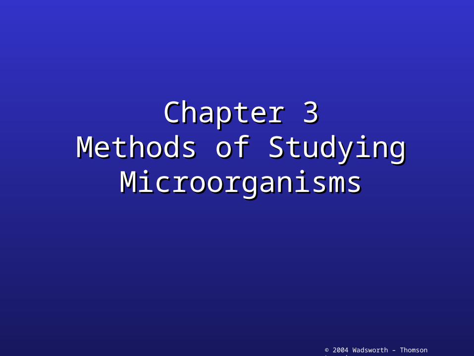
Properties of Light• Electromagnetic waves
– Visible light• 400-700 nm
– Longer wavelengths• Infrared rays, microwaves,
radio waves
– Shorter wavelengths• Ultraviolet rays, x-rays,
gamma rays
Figure 3.1
© 2004 Wadsworth – Thomson Learning
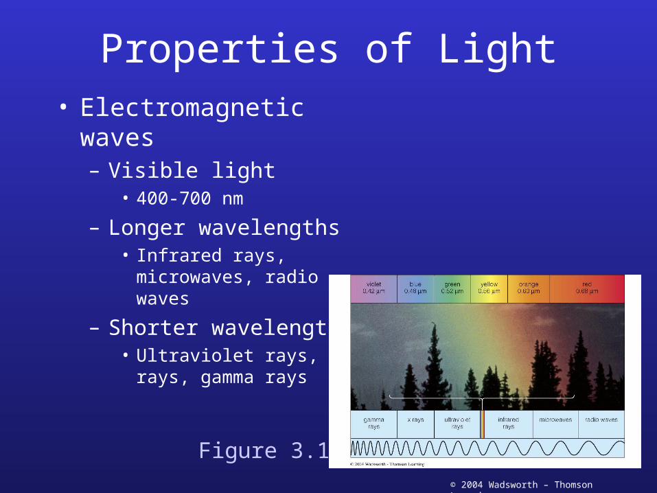
Properties of Light• Reflection
– Light hits an opaque object
• Rays bounce off the object
• Transmission– Rays pass through the
object– Must be clear
transparent• Glass
• Water
• Absorption– Some light does not pass
through• Certain wavelengths
can be absorbed
• Different colors result
Figure 3.2
© 2004 Wadsworth – Thomson Learning
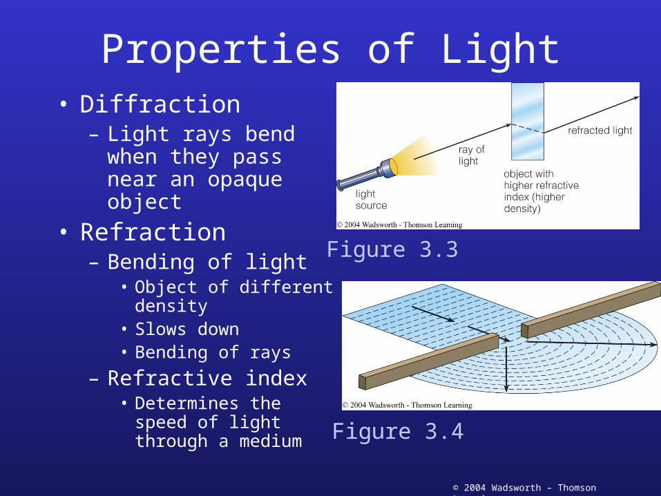
Properties of Light• Diffraction
– Light rays bend when they pass near an opaque object
• Refraction– Bending of light
• Object of different density
• Slows down• Bending of rays
– Refractive index• Determines the speed
of light through a medium
Figure 3.3
Figure 3.4
© 2004 Wadsworth – Thomson Learning
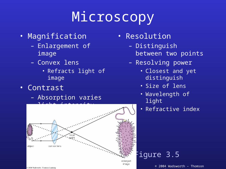
Microscopy• Magnification
– Enlargement of image– Convex lens
• Refracts light of image
• Contrast– Absorption varies light
intensity– Specimen absorbs light
• Resolution– Distinguish between two
points– Resolving power
• Closest and yet distinguish
• Size of lens
• Wavelength of light
• Refractive index
Figure 3.5
© 2004 Wadsworth – Thomson Learning
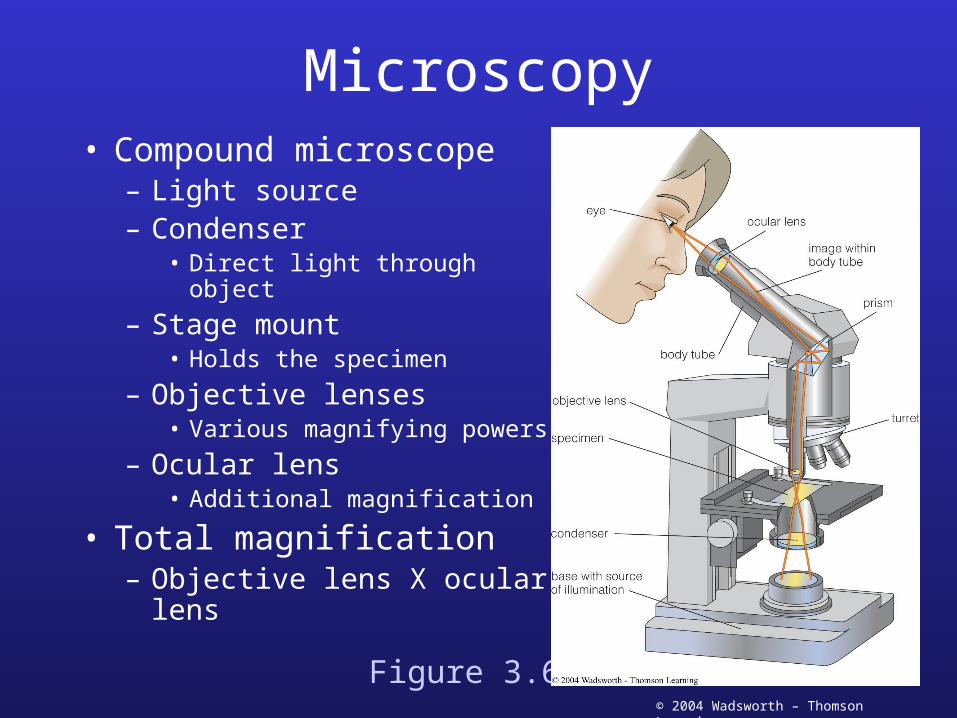
Microscopy• Compound microscope
– Light source– Condenser
• Direct light through object
– Stage mount• Holds the specimen
– Objective lenses• Various magnifying powers
– Ocular lens• Additional magnification
• Total magnification– Objective lens X ocular
lensFigure 3.6
© 2004 Wadsworth – Thomson Learning
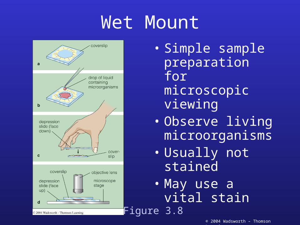
Wet Mount• Simple sample
preparation for microscopic viewing
• Observe living microorganisms
• Usually not stained
• May use a vital stain
Figure 3.8
© 2004 Wadsworth – Thomson Learning
Stains• Increase contrast• Require fixation of sample
– Heat fixation• Coagulates proteins and causes to stick to slide
– Chemical fixation
• Types of dyes– Acidic: safranin, acid fuchsin, crystal violet, methylene blue– Basic: eosin, basic fuchsin, congo red
• Simple stains– Make cells visible with one dye
• Differential stains– Distinguish between types of microorganisms
© 2004 Wadsworth – Thomson Learning
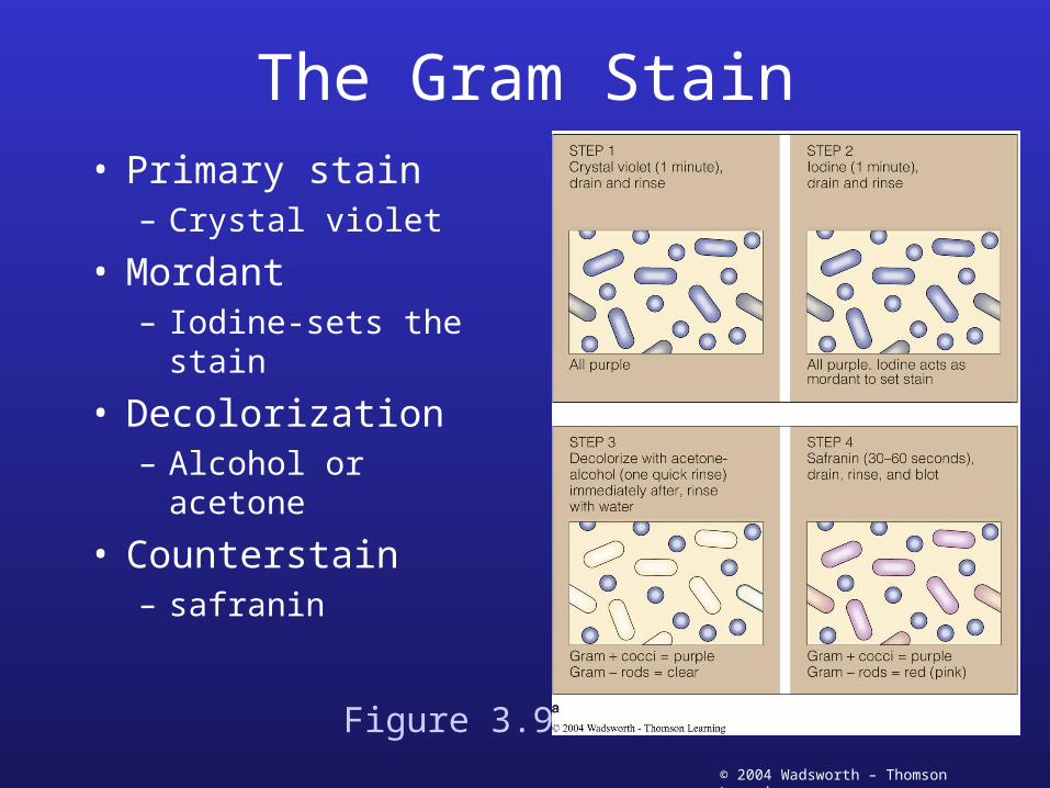
The Gram Stain• Primary stain
– Crystal violet
• Mordant– Iodine-sets the stain
• Decolorization– Alcohol or acetone
• Counterstain– safranin
Figure 3.9
© 2004 Wadsworth – Thomson Learning
Other Light Microscopes• Phase contrast microscopy
– Rings in objective and condenser• Increase contrast of certain parts of specimen
– Cellular movement and internal structure
• Darkfield microscopy– Light is scattered off of object
• Only light entering objectives is from specimen
– Viewing surface structures
• Nomarsky microscopy– Prisms in objective and condenser– Living organisms in animal tissues
• Fluorescence microscopy– Fluorescent material illuminated by UV light
© 2004 Wadsworth – Thomson Learning
Scanning microscopes
• Concentrating on small field of view
• Confocal microscopy– Same object viewed simultaneously from
opposite sides• Illuminating microscope
– Focused on very small area
• Receiving microscope with photodetector– Connected to computer– Computer generates image
– Three-dimensional• Multiple scans and different depths
© 2004 Wadsworth – Thomson Learning
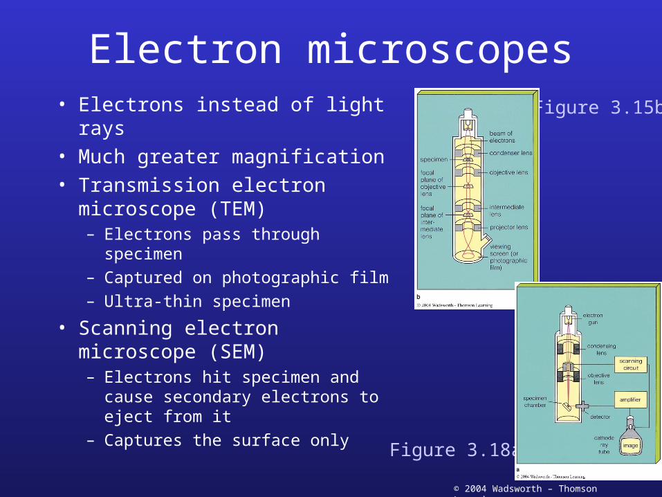
Electron microscopes• Electrons instead of light rays• Much greater magnification• Transmission electron
microscope (TEM)– Electrons pass through specimen– Captured on photographic film– Ultra-thin specimen
• Scanning electron microscope (SEM)– Electrons hit specimen and cause
secondary electrons to eject from it– Captures the surface only
Figure 3.15b
Figure 3.18a
© 2004 Wadsworth – Thomson Learning
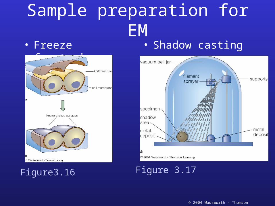
Sample preparation for EM• Freeze fracturing • Shadow casting
Figure3.16 Figure 3.17

© 2004 Wadsworth – Thomson Learning
Viewing atoms and molecules
• Scanned-proximity probe microscopes– Scanning tunneling microscope
• View surfaces that conduct electricity– Metals– Semi-conducting materials
– Atomic force microscope• Biologically important molecules• Attractive and repulsive forces• Diamond probe detects forces• Laser beam detects bending of beam• Resolution of 10 pm: 1/100th of nm
© 2004 Wadsworth – Thomson Learning
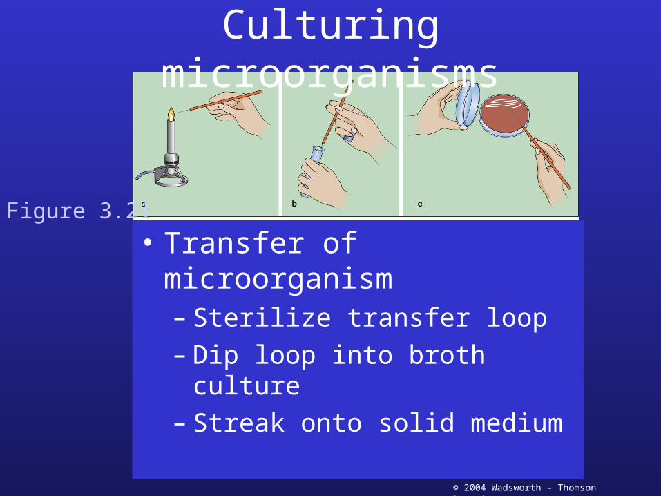
Culturing microorganisms
• Transfer of microorganism– Sterilize transfer loop– Dip loop into broth culture– Streak onto solid medium
Figure 3.21
© 2004 Wadsworth – Thomson Learning
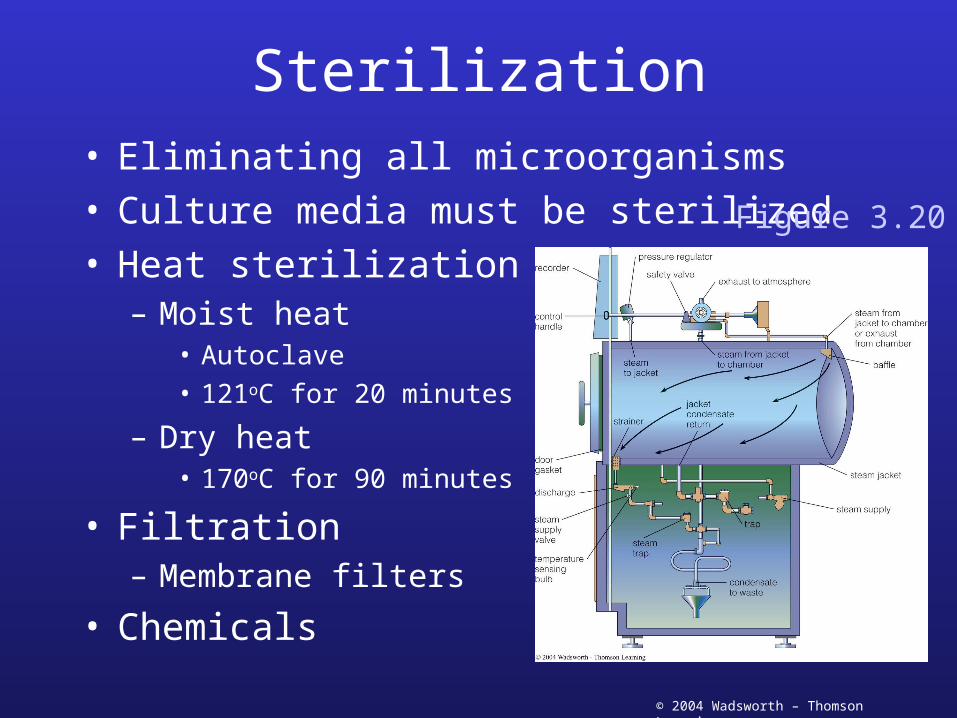
Sterilization• Eliminating all microorganisms• Culture media must be sterilized• Heat sterilization
– Moist heat• Autoclave• 121oC for 20 minutes
– Dry heat• 170oC for 90 minutes
• Filtration– Membrane filters
• Chemicals
Figure 3.20
© 2004 Wadsworth – Thomson Learning
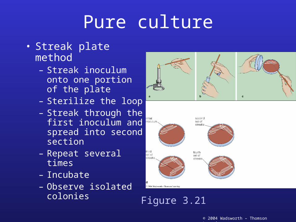
Pure culture• Streak plate method
– Streak inoculum onto one portion of the plate
– Sterilize the loop– Streak through the
first inoculum and spread into second section
– Repeat several times– Incubate– Observe isolated
coloniesFigure 3.21
© 2004 Wadsworth – Thomson Learning
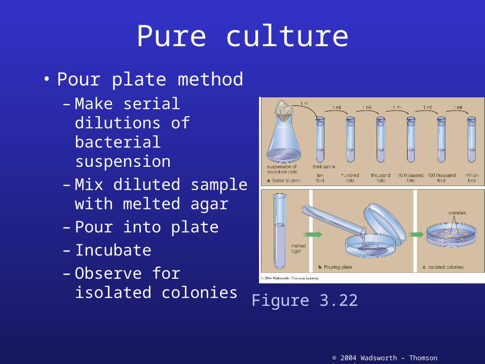
Pure culture
• Pour plate method– Make serial
dilutions of bacterial suspension
– Mix diluted sample with melted agar
– Pour into plate– Incubate– Observe for isolated
coloniesFigure 3.22
© 2004 Wadsworth – Thomson Learning
Culture media• Defined media
– Produced from pure chemicals
• Complex media– Extracts of natural sources
• Beef, blood, milk, protein, yeast, soybeans• Precise composition not known
• Selective media– Contents select for specific
microorganism
• Differential media– Identification of microorganisms
© 2004 Wadsworth – Thomson Learning
Growth conditions
• Temperature– Incubators– Water baths
• pH– Growth medium at optimal pH– Buffers maintain pH over period of growth
© 2004 Wadsworth – Thomson Learning
Growth conditions• Oxygen
– Strict aerobes• Require oxygen
– Strict anaerobes• Oxygen is toxic
– Facultative anaerobes• Use oxygen when available• Can grow without oxygen
– Aerotolerant anaerobes• Can’t use oxygen but not toxic
– Microaerophilic• Need low concentrations of oxygen
© 2004 Wadsworth – Thomson Learning
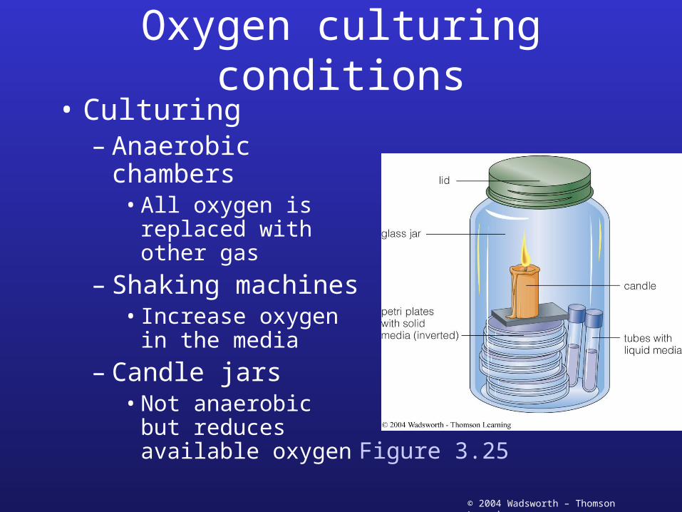
Oxygen culturing conditions• Culturing
– Anaerobic chambers• All oxygen is
replaced with other gas
– Shaking machines• Increase oxygen in
the media
– Candle jars• Not anaerobic but
reduces available oxygen
Figure 3.25

© 2004 Wadsworth – Thomson Learning
Preserving cultures
• Cold storage– Short-term: refrigeration slows growth
• Must continually transfer
– Long-term: freezing• Add substance to reduce freeze-killing
– Glycerol, skim milk, dimethyl sulfoxide (DMSO)
– Lyophilization• Long term—freeze drying• Frozen and dried under vacuum
Top Related