Transgenically-expressed secretoglobin 3A2 accelerates ... · RESEARCH ARTICLE Open Access...
13
RESEARCH ARTICLE Open Access Transgenically-expressed secretoglobin 3A2 accelerates resolution of bleomycin-induced pulmonary fibrosis in mice Yan Cai 1,5 , Mitsuhiro Yoneda 1 , Takeshi Tomita 1,6 , Reiko Kurotani 1,2 , Minoru Okamoto 1,7 , Taketomo Kido 1,8 , Hiroyuki Abe 2 , Wayne Mitzner 3 , Arjun Guha 4 and Shioko Kimura 1* Abstract Background: Secretoglobin (SCGB) 3A2, a cytokine-like secretory protein of small molecular weight, is predominantly expressed in airway epithelial cells. While SCGB3A2 is known to have anti-inflammatory, growth factor, and anti-fibrotic activities, whether SCGB3A2 has any other roles, particularly in lung homeostasis and disease has not been demonstrated in vivo. The aim of this study was to address these questions in mice. Methods: A transgenic mouse line that expresses SCGB3A2 in the lung using the human surfactant protein-C promoter was established. Detailed histological, immunohistochemical, physiological, and molecular characterization of the Scgb3a2-transgenic mouse lungs were carried out. Scgb3a2-transgenic and wild-type mice were subjected to bleomycin-induced pulmonary fibrosis model, and their lungs and bronchoalveolar lavage fluids were collected at various time points during 9 weeks post-bleomycin treatment for further analysis. Results: Adult Scgb3a2-transgenic mouse lungs expressed approximately five-fold higher levels of SCGB3A2 protein in comparison to wild-type mice as determined by western blotting of lung tissues. Immunohistochemistry showed that expression was localized to alveolar type II cells in addition to airway epithelial cells, thus accurately reflecting the site of surfactant protein-C expression. Scgb3a2-transgenic mice showed normal lung development and histology, and no overt gross phenotypes. However, when subjected to a bleomycin-induced pulmonary fibrosis model, they initially exhibited exacerbated fibrosis at 3 weeks post-bleomycin administration that was more rapidly resolved by 6 weeks as compared with wild-type mice, as determined by lung histology, Masson Trichrome staining and hydroxyproline content, inflammatory cell numbers, expression of collagen genes, and proinflammatory cytokine levels. The decrease of fibrosis coincided with the increased expression of SCGB3A2 in Scgb3a2-transgenic lungs. Conclusions: These results demonstrate that SCGB3A2 is an anti-fibrotic agent, and suggest a possible therapeutic use of recombinant SCGB3A2 in the treatment of pulmonary fibrosis. Keywords: Secretoglobin, SCGB, SCGB3A2, Bleomycin-induced pulmonary fibrosis model, Spontaneous resolution of bleomycin-induced pulmonary fibrosis, Transgenic mouse * Correspondence: [email protected] 1 Laboratory of Metabolism, National Cancer Institute, National Institutes of Health, Bethesda, MD 20892, USA Full list of author information is available at the end of the article © 2015 Cai et al. This is an Open Access article distributed under the terms of the Creative Commons Attribution License (http://creativecommons.org/licenses/by/4.0), which permits unrestricted use, distribution, and reproduction in any medium, provided the original work is properly credited. The Creative Commons Public Domain Dedication waiver (http:// creativecommons.org/publicdomain/zero/1.0/) applies to the data made available in this article, unless otherwise stated. Cai et al. BMC Pulmonary Medicine (2015) 15:72 DOI 10.1186/s12890-015-0065-4
Transcript of Transgenically-expressed secretoglobin 3A2 accelerates ... · RESEARCH ARTICLE Open Access...
RESEARCH ARTICLE Open Access
Transgenically-expressed secretoglobin 3A2accelerates resolution of bleomycin-inducedpulmonary fibrosis in miceYan Cai1,5, Mitsuhiro Yoneda1, Takeshi Tomita1,6, Reiko Kurotani1,2, Minoru Okamoto1,7, Taketomo Kido1,8,Hiroyuki Abe2, Wayne Mitzner3, Arjun Guha4 and Shioko Kimura1*
Abstract
Background: Secretoglobin (SCGB) 3A2, a cytokine-like secretory protein of small molecular weight, is predominantlyexpressed in airway epithelial cells. While SCGB3A2 is known to have anti-inflammatory, growth factor, and anti-fibroticactivities, whether SCGB3A2 has any other roles, particularly in lung homeostasis and disease has not beendemonstrated in vivo. The aim of this study was to address these questions in mice.
Methods: A transgenic mouse line that expresses SCGB3A2 in the lung using the human surfactant protein-Cpromoter was established. Detailed histological, immunohistochemical, physiological, and molecular characterizationof the Scgb3a2-transgenic mouse lungs were carried out. Scgb3a2-transgenic and wild-type mice were subjected tobleomycin-induced pulmonary fibrosis model, and their lungs and bronchoalveolar lavage fluids were collectedat various time points during 9 weeks post-bleomycin treatment for further analysis.
Results: Adult Scgb3a2-transgenic mouse lungs expressed approximately five-fold higher levels of SCGB3A2 protein incomparison to wild-type mice as determined by western blotting of lung tissues. Immunohistochemistry showed thatexpression was localized to alveolar type II cells in addition to airway epithelial cells, thus accurately reflecting the siteof surfactant protein-C expression. Scgb3a2-transgenic mice showed normal lung development and histology, and noovert gross phenotypes. However, when subjected to a bleomycin-induced pulmonary fibrosis model, they initiallyexhibited exacerbated fibrosis at 3 weeks post-bleomycin administration that was more rapidly resolved by 6 weeks ascompared with wild-type mice, as determined by lung histology, Masson Trichrome staining and hydroxyprolinecontent, inflammatory cell numbers, expression of collagen genes, and proinflammatory cytokine levels. The decreaseof fibrosis coincided with the increased expression of SCGB3A2 in Scgb3a2-transgenic lungs.
Conclusions: These results demonstrate that SCGB3A2 is an anti-fibrotic agent, and suggest a possible therapeutic useof recombinant SCGB3A2 in the treatment of pulmonary fibrosis.
Keywords: Secretoglobin, SCGB, SCGB3A2, Bleomycin-induced pulmonary fibrosis model, Spontaneous resolution ofbleomycin-induced pulmonary fibrosis, Transgenic mouse
* Correspondence: [email protected] of Metabolism, National Cancer Institute, National Institutes ofHealth, Bethesda, MD 20892, USAFull list of author information is available at the end of the article
© 2015 Cai et al. This is an Open Access article distributed under the terms of the Creative Commons Attribution License(http://creativecommons.org/licenses/by/4.0), which permits unrestricted use, distribution, and reproduction in any medium,provided the original work is properly credited. The Creative Commons Public Domain Dedication waiver (http://creativecommons.org/publicdomain/zero/1.0/) applies to the data made available in this article, unless otherwise stated.
Cai et al. BMC Pulmonary Medicine (2015) 15:72 DOI 10.1186/s12890-015-0065-4

BackgroundSecretoglobin (SCGB) 3A2, also called UGRP1 (uteroglobin-related protein 1), is a member of the SCGB gene superfam-ily, consisting of cytokine-like secretory proteins of smallmolecular weight (~10 kDa) [1–3]. SCGB3A2 is highlyexpressed in airway Club cells, while low expression isobserved at the growing tips of bronchi around embryonicday (E) 11.5 of mouse gestation [4]. SCGB3A2 plays a rolein lung development as demonstrated using ex vivo embry-onic lung organ cultures in the presence of SCGB3A2, andin vivo by administration of SCGB3A2 to pregnant femalemice, followed by examination of pre-term pups [4].Recently, SCGB3A2 was shown to be an early marker forClub cells (formerly called Clara cells) in conjunction withNotch signaling. Expression of SCGB3A2 appears earlierthan SCGB1A1, the founding member of the SCGB genesuperfamily, also called Club cell secretory protein (CCSP) orClub cell 10 kDa protein (CC10). SCGB1A1 was previouslythought to be the only definitive marker for Club cells [5].SCGB1A1 is the most well-characterized SCGB
protein, exhibiting anti-inflammatory, anti-fibrotic, andimmunomodulatory functions [6–9]. SCGB1A1 possessesphospholipase A2 (PLA2) inhibitory activity, which wasthought to be at least partially responsible for the anti-inflammatory and immunomodulatory activity of SCGB1A1[10, 11]. SCGB1A1 also exhibits tumor suppressor activityas demonstrated by decreased invasiveness of human lungadenocarcinoma-derived A549 cells in vitro [12], and theincreased incidence of tumors in chemical carcinogenesisbioassay and the increased lung metastasis of B16F10melanoma cells using Scgb1a1-null mice in vivo [13, 14].SCGB3A2 also exhibits anti-inflammatory and anti-
fibrotic activities [15, 16]; the anti-inflammatory functionwas originally suggested by the fact that Scgb3a2 mRNAlevels were reduced in the lungs of fungal-induced aller-gic inflammation model mice, which was almost restoredby dexamethasone treatment [3]. Further, in the ovalbu-min (OVA)-induced airway inflammation model mice,reduced levels of lung Scgb3a2 mRNA were inverselycorrelated with the increased levels of proinflammatorycytokines, IL-5 and IL-9 in bronchoalveolar lavage fluid(BALF) [17, 18]. When OVA-induced airway inflamma-tion model mice were intranasally administered recom-binant adenovirus expressing SCGB3A2 before OVAchallenge, OVA-induced airway inflammation was sup-pressed [15]. Lastly, Scgb3a2-null mice when subjected toOVA-inflammation model, showed exacerbated airwayinflammation [19]. On the other hand, anti-fibrotic activityof SCGB3A2 was demonstrated by using a bleomycin(BLM)-induced mouse pulmonary fibrosis model [16]. Thisactivity was through SCGB3A2-induced STAT1 phosphor-ylation and increased expression of inhibitory SMAD7,which inhibited the TGFβ signaling, resulting in reducedexpression of various collagen genes and development of
fibrosis [16] SCGB3A2 can also be used as a marker forpulmonary carcinomas in mice and humans [20, 21]. Takentogether, SCGB3A2 has multiple biological activities, play-ing a role in lung homeostasis and function, and influencingvarious lung diseases. Whether SCGB3A2 possesses anyother activities and the mechanisms for these activities haveyet to be determined.To understand the role of SCGB3A2 in lung homeostasis
and diseases, an Scgb3a2-transgenic mouse was establishedthat over-expresses SCGB3A2 in a lung-specific fashionunder the control of the human surfactant protein C(SP-C) gene promoter. Detailed characterization dem-onstrated that the lungs of Scgb3a2-transgenic micewere histologically and functionally normal as comparedto wild-type. When subjected to the BLM-induced pul-monary fibrosis model, however, they exhibited increasedfibrosis at 3 weeks post-BLM administration, which wasmore quickly resolved by 6 weeks as compared to wild-type mice. These results demonstrate that SCGB3A2has anti-fibrotic activity and suggest a potential use ofSCGB3A2 as a therapeutic agent in treating lung fibrosis.
MethodsTransgenic constructAn expression plasmid with the human SP-C gene promoter(3.7 kb) cloned into pUC18 vector with SV40 small T intronand poly A (0.4 kb) (SPC3.7-SV40-pUC18), was providedfrom Dr. Jeffrey Whitsett (University of Cincinnati, OH) [22].The mouse Scgb3a2 cDNA that covers the entire proteincoding sequence (50–427) was inserted into the SPC3.7-SV40-pUC18 plasmid. The resultant SPC3.7-SCGB3A2-SV40-pUC18 was double-digested with restrictionenzymes, Nde I and Not I. The linearized SPC3.7-SCGB3A2-SV40 fragment was purified before microinjection intopronuclei of C57BL/6NCr mouse eggs. Production ofScgb3a2-transgenic mouse lines was confirmed by Southernblotting of genomic DNAs isolated from clipped mouse-tails.
Northern blottingTotal RNA (3 μg) isolated from adult lungs of wild typeand Scgb3a2-transgenic mice was electrophoresed on 1 %agarose gel containing 0.22 M formaldehyde and trans-ferred onto nitrocellulose membrane (Immobilon-Ny+,Millipore, Billerica, MA). Filters were hybridized withSCGB3A2 probe obtained from Eco RI digestion of theSCGB3A2/pCR2.1 construct. Hybridization was performedin Perfect Hybridization solution (GE Healthcare LifeSciences, Piscataway, NJ) at 68 °C overnight. The mem-brane was washed twice with 2 x SSC containing 0.1 %SDS at 68 °C for 30 min, followed by exposure to aphosphoimager screen (Storm 840, GE Healthcare LifeSciences, Piscataway, NJ). Data processing was carriedout using ImageQuant TL 2005 software (GE HealthcareLife Sciences).
Cai et al. BMC Pulmonary Medicine (2015) 15:72 Page 2 of 13

Western blottingLung from wild type and transgenic mice were frozen andcrushed in 50 mM Tris-HCl, pH 8.0, 5 mM EDTA, 1 mMDTT, 1 mM phenylmethylsulfonyl fluoride (PMSF) withprotein inhibitor cocktail (Roche Applied Science, Branford,CT). Protein concentrations were determined by Brad-ford assay (Bio-Rad Laboratories, Hercules, CA) withbovine serum albumin (BSA) as standard, and sampleswere mixed with equal volume of 2 x SDS sample buffer(125 mM Tris-HCl, pH 6.8, 4 % SDS, 20 % glycerol, 0.1 %mercaptoethanol). Ten microgram of sample was appliedin each well of 20 % polyacrylamide gel and was run withrunning buffer of 50 mM Tris, 384 mM glycine, 2 % SDS.After electrophoresis, protein was transferred to Polyviny-lidene fluoride (PVDF) membrane using a tank transfersystem (Mini Trans-Blot Cell, Bio-Rad) with blotting buf-fer (50 mM Tris, 40 mM glycine, 20 % methanol) andelectric field of 30 V for 6 h. To visualize SCGB3A2band, PVDF membrane was treated as follows; 1 hblocking with PBST (phosphate buffered saline + 0.05 %Tween 20) +5 % BSA, 3 h incubation with 0.2 μg/mlpolyclonal rabbit anti-SCGB3A2 IgG in PBST + 5 % BSA,PBST wash 3 times, incubation with 0.1 μl/ml horseradishperoxidase (HRP)-linked anti-Rabbit IgG F(ab’) fragment(GE Healthcare, NA9340) in PBST + 5 % BSA, and PBSTwash 3 times before ECL plus (PerkinElmer, Waltham,MA) reaction. Polyclonal rabbit anti-SCGB3A2 antibodywas produced as previously described [3]. The anti-SCGB3A2 IgG was purified using the Montage antibodypurification kit (EMD Millipore, Billerica, MA) and usedfor all experiments. Labeled proteins were visualized usinga SuperSignal West Pico Substrate (Thermo Scientific,Rockford, IL), and signals were detected using FluoChemHD2 System (ProteinSimple, San Jose, CA).
Animal studiesAll animal studies were carried out after approval by theNational Cancer Institute Animal Care and Use Commit-tee. Mouse embryonic lungs were collected from wild-typeand Scgb3a2-transgenic pregnant females at various embry-onic days (E). Noon of the day on which a vaginal plug wasfound was considered as E 0.5. Branching degree of ex vivocultured embryonic lungs were counted after 3 days of cul-ture as previously described [4]. Breathing score assessmentwas performed as previously described [4] according to thecriteria described by Ozdemir et al. [23]; 0, no breathing; 1,gasping; 2, gasping/labored breathing; 3, labored breathing;4, labored breathing/unlabored breathing; 5, unlaboredbreathing. For the BLM-induced pulmonary fibrosis model,mice of approximately 8 weeks old (at least 5 mice pergroup) were intratracheally intubated and dosed with BLM(1.2 U/kg) at day 0. Mice were killed on 3, 6, and 9 weeksafter BLM intubation, and bronchoalveolar lavage (BAL)fluids obtained by lavaging lungs with 1 mL PBS [16]. The
collected BAL fluids were used for counting and differenti-ating inflammatory cell numbers with Cytospin 4 (ThermoScientific). PBS-treated mice killed at 3 weeks were used asnormal control. Experiments were repeated more than 2times, and the combined data points were used for analysis.
ELISASCGB3A2 protein levels were determined by ELISA aspreviously described [24]. Briefly, samples diluted with acoating solution (500 mM bicarbonate buffer, pH 9.6),were applied onto each well of 96-well plates and theplates were incubated at 4 °C overnight. Calibration curveswere constructed with twelve points by serially diluting asolution of recombinant mouse SCGB3A2 (1 μg/ml). Theplates were washed four times with washing solution (PBS,pH 7.4 containing 0.5 % of Tween), followed by addition ofblocking buffer (PBS, pH 7.4 containing 1 % of BSA) toeach well. After incubation for 2 h at 37 °C, the plates werewashed four times with the washing solution. Purified anti-mouse SCGB3A2 IgG [3] was applied to each well and theplates were incubated for 4 h at 37 or 4 °C overnight.The plates were washed seven times with the washingbuffer. One hundred μl of ECL anti-rabbit IgG HRP-linked F(ab’) fragment (from donkey) was added to eachwell and the plates were placed at 37 °C for 2 h. After fur-ther washing, the amount of SCGB3A2 was determinedby addition of 3,3′,5,5′-tetramethylbenzidine (TMB,Sigma) and was read at 450 nm after stopping the reactionby adding 1 N HCl.
Lung histological analysisLungs were fixed in 4 % paraformaldehyde for one dayat 4 °C, dehydrated, and embedded in paraffin. Lung tissueswere sectioned at 4 μm and stained with hematoxyline andeosin (H&E). For immunohistochemistry, sections were atfirst rinsed with 0.05 % Triton-X 100 in PBS, and non-specific binding sites were blocked using 10 % normal goatserum in PBS containing 0.05 % Tween 20. Epitope re-trieval was carried out using autoclave (5 min in citratebuffer, pH 6.0 or 1X TE, pH 9.0). After cooling to roomtemperature, the sections were incubated overnight at4 °C with rabbit polyclonal anti-mouse SCGB3A2 [3] oranti-pro-surfactant protein-C (Seven Hills Bioreagents,Cincinnati, OH) primary antibodies. The sections wererinsed in distilled water, followed by treating with HRP-conjugated goat anti-rabbit IgG using the ABC methodwith a commercially available kit (Vector Laboratories,Burlingame, CA) according to the manufacturer’s instruc-tion. Immunovisualization was carried out with 3, 3′-diaminobenzidine as substrate (Sigma, St Louis, MO), andcounterstained with hematoxylin.For double immunofluorescence labeling of adult lungs
using two primary antibodies from the same species, thesections were first incubated with anti-SCGB3A2 antibody
Cai et al. BMC Pulmonary Medicine (2015) 15:72 Page 3 of 13

(1:1000) at 4 °C overnight, followed by labeled goatanti-rabbit IgG (Alexa Fluor 594 or 488, 1:200, LifeTechnology) as the secondary antibody for 1 h at roomtemperature. The sections were then incubated with 5 %rabbit serum for 1 h at room temperature, followed by in-cubation with unconjugated Fab Fragment goat anti-rabbitIgG for 1 h at room temperature. The sections were finallyincubated with the secondary primary antibody, rabbitanti-pro-SP-C antibody (1:500), followed by labeled goatanti-rabbit IgG (Alexa Fluor 488 or 594, 1:200, LifeTechnology). Cell nuclei were identified by counterstainingwith 4,6-diamino-2-phenylindolyl-dihydrochloride (DAPI,Life Technology). Fluorescence images were obtained andprocessed using a Zeiss 780 laser-scanning confocal micro-scope. Matching confocal planes were analyzed in allco-localization studies.Severity of fibrosis was quantified from H&E stained
entire lungs using the Ashcroft scoring system [25]. Thedegree of fibrosis was graded from 0 (normal lung) to 8(severe distortion of structure, large fibrous areas, andhoneycomb lesions). The mean score from all fields(magnification X200, average 30 fields/animal) was takenas the fibrosis score.
Quantitation of hydroxyproline content in lungHydroxyproline content was measured using hydroxy-proline assay kit from Biovision (Milpitas, CA) accordingto the manufacture’s instruction with slight modification.In brief, whole lungs were homogenized in dH2O, using100 μl H2O for every 10 mg of tissue. To 100 μl of tissuehomogenate, 200 μl concentrated HCl (6 N) was addedin a pressure-tight, teflon capped vial, and the mixturewas hydrolyzed at 120 °C for 3 h, followed by filtrationthrough a 45 μm syringe filter (Millipore, Bedford, MA).Ten μl of hydrolyzed sample was transferred to a 96-wellplate and was evaporated to dryness under vacuum, towhich 100 μl Chloramine T reagent was added perwell. After incubation at room temperature for 5 min,100 μl p-dimethylaminobenzaldehyde reagent was added toeach well and further incubated for 90 min at 60 °C. Ab-sorbance was measured at 560 nm in a microplate reader(SpectroMax Plus384, Molecular Devices, Sunnyvale, CA).
Microarray analysisLung total cellular RNAs were individually isolated fromfour mice each of no-treatment wild-type and Scgb3a2transgenic mice (approximately 8-weeks old) using RNeasyMini Kit (Qiagen Science, Maryland, USA). The sampleRNAs were labeled with Cy5, while pooled RNAs con-taining the same amount of RNA from each samplewere labeled with Cy3 and used as a reference. Labelingwas carried out using CyDye™ Post-Labeling ReactiveDye Pack (GE Healthcare) according to the manufacturer’sinstructions. The purified Dye-coupled RNA samples were
hybridized to an Agilent Whole Mouse Genome 4X44Koligo microarray kit (Agilent Techonologies, G4122F, SantaClub, CA), and were incubated for 17 h at 65 °C. The slideswere washed, dried, and scanned using Agilent G26000microarray scanner. The data were processed and analyzedby Genespring GX 11.0.2 software package (AgilentTechnologies). All effective genes of microarray ana-lysis were submitted to the Gene Expression Omnibus(GEO: ID # GSE47931).
Quantitative RT-PCR analysisTotal RNAs isolated using TRIzol (Lifetechnologies) anddigested with DNase I were reverse-transcribed by Super-script II reverse transcriptase (Life Technologies). Quantita-tive RT-PCR (qRT-PCR) was performed with ABI Prism7900 Sequence Detection System (Applied Biosystems,Foster City, CA) using SYBR Green master mixture.The ΔΔ Ct method was used using β-actin or 18S asnormalization control. PCR condition used was 50 °C,2 min and 95 °C, 10 min followed by 95 °C, 15 s and60 °C, 40 s for 40 cycles with the following primers:Sftpa (forward) 5′- ACT CCC ATT GTT TGC AGAATC -3′, (reverse) 5′- AAG GGA GAG CCT GGA GAAAG -3′; Sftpb (forward) 5′- ACA GCC AGC ACA CCCTTG -3′, (reverse) 5′- TTC TCT GAG CAA CAG CTCCC -3′; Sftpc (forward) 5′- ATG AGA AGG CGT TTGAGG TG -3′, (reverse) 5′- AGC AAA GAG GTC CTGATG GA -3′; Sftpd (forward) 5′- GAG AGC CCC ATAGGT CCT G -3′, (reverse) 5′- GTA GCC CAA CAGAGA ATG GC -3′; Aqp1 (forward) 5′- TGC AGA GTGCCA ATG ATC TC -3′, (reverse) 5′- GGC ATC ACCTCC TCC CTA GT −3; Col1a1 (forward) 5′-TAG GCCATT GTG TAT GCA GC-3′, (reverse) 5′- ACA TGTTCA GCT TTG TGG ACC-3′; Col3a1 (forward) 5′-TAGGAC TGA CCA AGG TGG CT-3′, (reverse) 5′- GGAACC TGG TTT CTT CTC ACC-3′; Col4a1 (forward)5′-CAC ATT TTC CAC AGC CAG AG-3′, (reverse)5′- GTC TGG CTT CTG CTG CTC TT-3′; Col5a2(forward) 5′-CAT GGA GAA GGT TTC CAA ATG-3′,(reverse) 5′- AAA GCC CAG GAA CAA GAG AA-3′;Col12a1 (forward) 5′-TGA GGT CTG GGT AAA GGCAA-3′, (reverse) 5′- GTA TGA GGT CAC CGT CCAGG-3′; Acta2 (forward) 5′-GTT CAG TGG TGC CTCTGT CA-3′, (reverse) 5′-ACT GGG ACG ACA TGGAAA AG-3′; Ctgf (forward) 5′-GCT TGG CGA TTTTAG GTG TC-3′, (reverse) 5′-CAG ACT GGA GAAGCA GAG CC-3′; Mmp2 (forward) 5′-GGG GTC CATTTT CTT CTT CA-3′, (reverse) 5′-CCA GCA AGTAGA TGC TGC CT-3′; Mmp12 (forward) 5′-TTT GGATTA TTG GAA TGC TGC-3′, (reverse) 5′-ATG AGGCAG AAA CGT GGA CT-3′; β-actin (forward) 5′-ATGGAG GGG AAT ACA GCC C-3′, (reverse) 5′-TTC TTTGCA GCT CCT TCG TT-3′; and 18S (forward) 5′- CGC
Cai et al. BMC Pulmonary Medicine (2015) 15:72 Page 4 of 13
GGT TCT ATT TTG TTG GT-3′, (reverse) 5′- AGTCGG CAT CGT TTATGG TC-3′.
Statistical analysisAll animal studies were carried out at least twice, eachusing at least 5–6 mice per group. Statistical analysiswas carried out between wild-type and transgenic miceof various ages using a student’s t-test. P < 0.05 wasconsidered significant.
ResultsProduction of Scgb3a2-transgenic mouse overexpressingSCGB3A2 in lung-specific fashionLung-specific over-expression of SCGB3A2 was achievedby producing a transgenic mouse line which harbors atransgenic construct containing a mouse Scgb3a2 cDNAcoding sequence, under control of the human SP-C genepromoter at the 5′ end, and the SV40 small T intronand poly A addition site at the 3′ end [22] (Fig. 1a). Onefounder mouse had high lung-specific expression ofScgb3a2 as determined by Northern blotting (Fig. 1b).The highest mobility band corresponded to endogen-ously expressed Scgb3a2, while the transgene producedtwo bands with slower mobility.Western blotting using a whole lung of 3-month-old
mice demonstrated that the expression of SCGB3A2protein was approximately five-fold higher in transgenicmouse lungs compared to wild-type control lungs (Fig. 1c).The mRNA level of Scgb3a2 was measured by qRT-PCRusing lung tissues and the protein level by bronchoalveolarlavage (BAL) fluids at various ages from embryonic day(E) 14.5 to 12 or 24-weeks old (Fig. 1d, e). The Scgb3a2mRNA levels drastically increased towards the end ofgestation [26], and the levels in E18.5 transgenic embryolungs were approximately two-fold higher than that ofwild-type control lungs, which remained more or less atsimilar levels thereafter through 12 weeks (Fig. 1d).Transgenic mouse BAL fluids had about 6-fold higherlevels of SCGB3A2 as compared to wild-type mice whenmeasured at 2-weeks old (~6 μg/ml vs. ~1 μg/ml, respect-ively), which gradually decreased as the mice aged (Fig. 1e).Wild-type mice had ~1 μg/ml of SCGB3A2 for the entireperiod of 2 to 24 weeks. Immunohistochemistry andimmunofluorescence analysis demonstrated that thetransgenic mice expressed SCGB3A2 in alveolar type IIcells, in addition to bronchiolar epithelial cells, the normalexpression site for SCGB3A2 [3, 26, 27] (Fig. 1f, g),consistent with the expected expression of the alveolartype II cell-specific human SP-C [28]. Even though withthis higher ectopic expression of SCGB3A2, both lungsof wild-type and transgenic mice looked histologicallynormal at 2 to 24-weeks old age. Further, scanning elec-tron microscopy, using adult wild-type and Scgb3a2-nullmouse lungs, demonstrated that ciliated cells appeared to
be similar in morphology, size and number between wild-type and Scgb3a2-null airways (Additional file 1: Figure S1).
Characterization of Scgb3a2-transgenic mouse lungsPrevious results showed that SCGB3A2 promoted lungdevelopment as demonstrated by branching morphogenesisin ex vivo organ culture studies as well as by in vivo admin-istration of SCGB3A2 to pregnant females, followed byexamination of preterm pups [4]. When E14.5, 16.5, and18.5 wild-type and transgenic embryos were analyzed forbody weight, body lengths, lung weights, and breathingscores of premature pups (Additional file 1: Figure S2A-D),no statistically significant differences were obtained be-tween the two genotypes for all parameters examined.The development of embryonic lungs was also examinedhistologically, by immunohistochemistry and immuno-fluorescence. Like adults, E14.5 and 18.5 wild-type andtransgenic embryo lungs presented similar histology(Additional file 1: Figure S2E). Immunohistochemicalstaining for SCGB3A2 confirmed that in E18.5 transgeniclungs, SCGB3A2 was strongly expressed in bronchiolarepithelial cells, the normal expression site for SCGB3A2and the precursor to type II cells, the site of SP-C expres-sion (Additional file 1: Figure S2E, bottom panel) [3, 26,27]. Immunofluorescence studies using SCGB1A1 (CCSP)as a marker for Club cells, β-tubulin as a marker for ciliatedcells, PGP9.5 as a maker for neuroendocrine cells, T1α as amarker for alveolar type I cells, and SP-C as a marker foralveolar type II cells showed no differences in the spatialand temporal expression patterns of these proteins betweenwild-type and transgenic embryo lungs at E14.5, 16.5 and18.5 (Additional file 1: Figure S2F, and data from E14.5 and16.5 not shown). These results thus suggested that bothwild-type and transgenic embryo lungs developed similarlyduring lung morphogenesis.In order to examine whether Scgb3a2-transgenic mice
have normal lung function, lung chord lengths and lungvolumes were measured to calculate surface areas usingthe point and intersection counting method [29]. Thesurface areas were not different between wild-type andtransgenic mice of either 2-, 3-, 4-, or 12-weeks-old(Additional file 1: Figure S3A). Further, when mice weresubjected to MouseOx to monitor O2 saturation, heartrate, pulse distention, and breath rate, none of these pa-rameters had significant differences between wild-typeand transgenic mice of either ages (Additional file 1:Figure S3B-E). These results suggested that Scgb3a2-transgenic mice had normal lung function at two weeksand up to 12 weeks old.
Accelerated resolution of BLM-induced pulmonary fibrosisin Scgb3a2-transgenic lungsIn order to examine whether Scgb3a2-transgenic miceexhibit any phenotypes upon challenge, both wild-type
Cai et al. BMC Pulmonary Medicine (2015) 15:72 Page 5 of 13

and Scgb3a2-transgenic mice were subjected to theBLM-induced pulmonary fibrosis model. After BLM in-tubation, both wild-type and transgenic mice lost weightat similar rates for the first five days, from which trans-genic mice lost significantly more weight as compared towild-type (Fig. 2a). On day 21 (3 weeks post-BLM), wild-type and transgenic mice weighed 85 and 75 % of theiroriginal weight, respectively. However, after day 21, the
weight of transgenic mice gradually increased and reachedthe wild-type weight by day 49. There was no difference inthe survival curves between BLM-treated wild-type andScgb3a2-transgenic mice (Additional file 1: Figure S4).Histological examination revealed that both BLM-treatedwild-type and Scgb3a2-transgenic mice developed exten-sive pulmonary fibrosis by 3 weeks based on a whole lungH&E stained images (Fig. 2b) and Masson Trichrome
Fig. 1 Generation of the SCGB3A2 transgenic mouse line. a Illustration of the construct showing the human SP-C gene promoter cloned into thepUC18 vector with SV40 small T intron and poly A. b Northern Blot analysis. The high level of Scgb3a2 is observed only in lung tissue. The bandsindicated by an arrow represent exogenously expressed Scgb3a2 mRNAs. The highest mobility band shown by an arrowhead is derived fromendogenous Scgb3a2. c Representative Western blot result showing increased level of SCGB3A2 protein in Scgb3a2-transgenic mouse lungs ascompared with wild-type mice. GAPDH was used as a loading control. Lower panel shows the result of quantitation, normalized to GAPDH. N = 3. dqRT-PCR analysis of relative expression levels of Scgb3a2 in lungs of various gestational days and ages of wild-type (WT) and transgenic (TG)mice. e SCGB3A2 levels in BALF of different ages of WT and TG mice. For D, E: N = 5 in each group. The results are shown as the mean ± SD.**P < 0.01, ***P < 0.001 by student t-test in comparison between WT and TG. f Immunohistochemistry for SP-C and SCGB3A2 in 4-month-oldWT and TG mouse lungs. Arrows indicate representative positive signals in brown. TG lungs express SCGB3A2 in alveolar type II cells (middlepanel) in addition to airway epithelial cells (right panel). Scales are as indicated. g Co-immunofluorescence analysis of TG mouse lungs forSP-C and SCGB3A2. Airway cells express only SCGB3A2 (red, shown by an asterisk) while alveolar type II cells express both SP-C and SCGB3A2(yellow, shown by arrows). Scales are as indicated
Cai et al. BMC Pulmonary Medicine (2015) 15:72 Page 6 of 13

staining (Fig. 2c). When the extent of fibrous damage wascalculated using these histological sections with theAshcroft score system, BLM-treated Scgb3a2-transgenicmice had slightly increased damaged areas by 3 weeks ascompared to the BLM-treated wild-type group with statis-tical significance (Fig. 2d). Ashcroft scores were thendecreased by 6 weeks in both wild-type and transgenicmice, which continued until 9 weeks post-BLM treat-ment. BLM-induced pulmonary fibrosis with single doseadministration is known to partially resolve after weeks ofBLM treatment due to unknown reasons [30–32]. Atboth 6 and 9 weeks post-BLM administration, the Ashcroftscore was significantly lower in transgenic lungs than theirwild-type counterparts, suggesting that BLM-induced pul-monary fibrosis may have dissolved more quickly in trans-genic lungs than wild-type lungs. This result was supportedby Masson Trichrome staining that detects collagen fibers(Fig. 2C), and hydroxyproline contents (Fig. 2e), bothof which were increased in both transgenic and wild-type lungs at 3 weeks. These levels gradually decreasedpost-BLM over the course of 9 weeks. The transgenic
lungs demonstrated a much more rapid decrease ofhydroxyproline content; by 9 weeks post-BLM, the levelin the transgenic lungs was less than a half of its peaklevel, and was only 2 fold higher than the control level.
Gene expression analysis of BLM-induced pulmonaryfibrosis in Scgb3a2-transgenic lungsTotal inflammatory cell numbers in BALF and levels ofmRNAs encoding collagen 1a1, 3a1, 4a1, and 5a2 weredrastically increased at 3 weeks post-BLM treatment andthe levels were higher in Scgb3a2-transgenic than wild-type lungs (Fig. 3a left panel, b). Collagen 12a1 mRNAlevels did not change after BLM treatment in both mouselines (data not shown). The increased inflammatory cellswere due to increased numbers of monocytes/lymphocytesand neutrophils (Fig. 3a right panel). The number of mac-rophages was not different between Scgb3a2-transgenicand wild-type lungs. Inflammatory cell numbers and allcollagen mRNA levels rapidly decreased to almost controllevels in both BLM-treated wild-type as well as Scgb3a2-transgenic mouse lungs by 6 weeks post-BLM treatment.
Fig. 2 BLM-induced pulmonary fibrosis in wild-type (WT) and Scgb3a2-transgenic (TG) mice. a Body weight curves of mice intubated and treatedwith BLM on day 0, followed by necropsy on day 63. Body weights are shown as the percentage of Day 0 weight set as 100 %. *P < 0.05, WT-BLMgroup vs. TG-BLM, **P < 0.01, BLM-treated groups vs. PBS-treated groups. b Whole lung H&E images of WT and TG lungs at 3 weeks (BLM 3 W), 6 weeks(BLM 6 W), and 9 weeks (BLM 9 W) post-BLM treatment, and their corresponding PBS-treated lungs collected at 3 W (PBS) as control. Magnification 40Ximages were stitched. c Masson Trichrome staining of lung sections of WT and TG mice at 3, 6, and 9 weeks post-BLM treatment. Magnification, 100X.d Ashcroft scores of BLM-induced damaged areas at 3, 6, and 9 weeks post-BLM treatment. PBS control levels are those from 3 W. e Hydroxyprolinecontent at 3, 6, and 9 weeks post-BLM treatment. PBS control levels are those from 3 W. N > 6 in each group. The results are shown as the mean ± SD.*P < 0.05, NS, not significant. Statistical analysis was carried out by using student’s t-test
Cai et al. BMC Pulmonary Medicine (2015) 15:72 Page 7 of 13

The expression of mRNAs encoded by other genesinvolved in bleomycin-induced fibrosis such as Acta2,Ctgf, Mmp2, and Mmp12 were also determined (Fig. 3c).The expression of all these mRNAs markedly increased at3 weeks post-BLM with the levels in transgenic lungsbeing higher than those of wild-type lungs. However, theincreased expression again quickly decreased by 6 weekspost-BLM; in particularly expression in the transgeniclungs returned to control levels.The levels of Scgb3a2 mRNA and protein in BALF
were determined by qRT-PCR and ELISA using PBS andBLM-treated lungs and BALF of wild-type and transgenic
mice, respectively (Fig. 4a). The differences in Scgb3a2mRNA and protein levels initially observed betweenScgb3a2-transgenic and wild-type mice were no longerfound in 3 week post-BLM-treated lungs, and their levelswere similar to those of PBS treated wild-type lungs.Scgb3a2 mRNA and protein levels of transgenic micegradually returned to the PBS control levels by 6 weeks;the levels of transgenic mice again became higher thanthose of wild-type. Immunohistochemistry did not showclear positive staining for SCGB3A2 in 3 weeks-post-BLMlungs of both genotypes, however very weak signals weresometimes noted in wild-type bronchiolar epithelial cells
Fig. 3 Characterization of BLM-induced pulmonary fibrosis-harboring lungs of wild-type (WT) and Scgb3a2-transgenic (TG) mice. a Number ofinflammatory cells in BAL fluids at 3, 6, and 9 weeks post-BLM treatment and PBS control groups collected at 3 weeks. (right panel) Number ofmonocytes/lymphocytes, neutrophils, and macrophages from 3 weeks post-BLM treatment mice were separately counted. *P < 0.05 by student’st-test. b qRT-PCR analysis for various mRNA levels. The results are shown as the mean ± SD from N = 7-10. *P < 0.05, **P < 0.01, NS, not significantby student’s t-test
Cai et al. BMC Pulmonary Medicine (2015) 15:72 Page 8 of 13
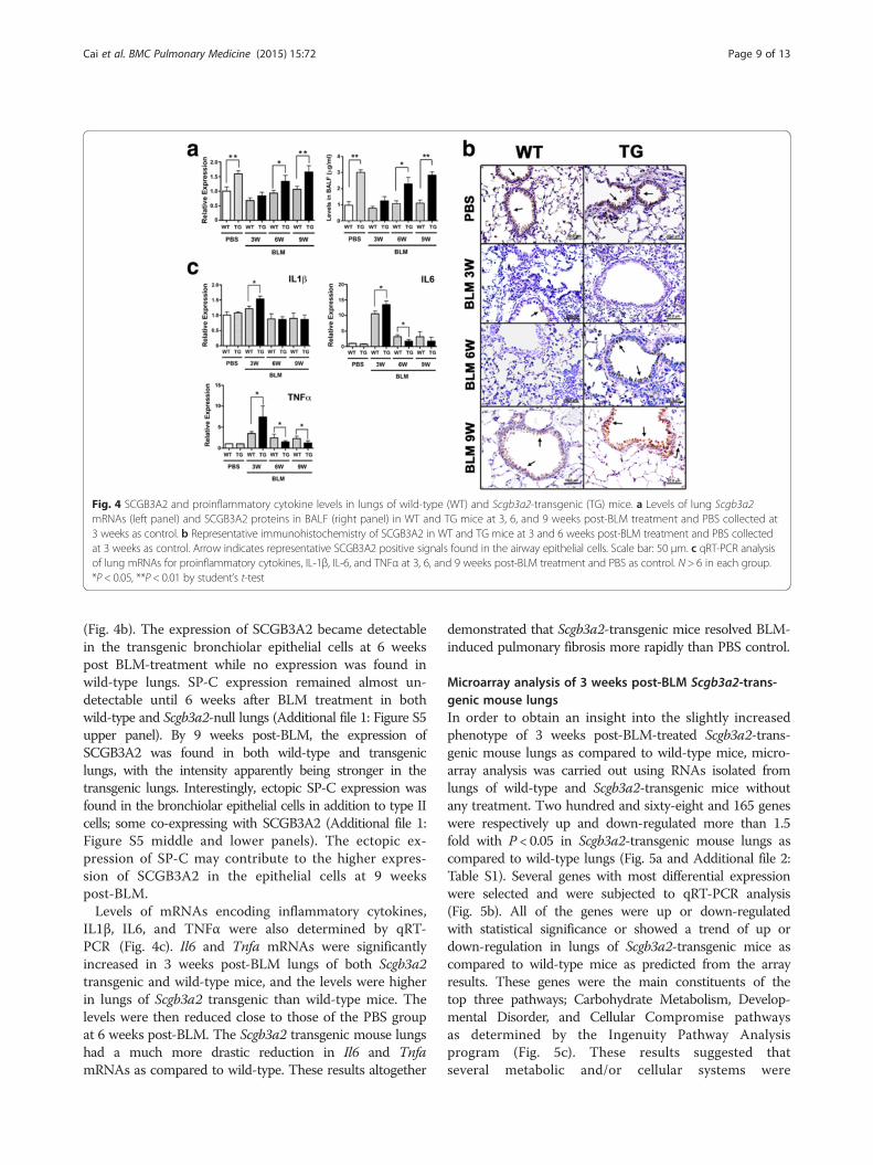
(Fig. 4b). The expression of SCGB3A2 became detectablein the transgenic bronchiolar epithelial cells at 6 weekspost BLM-treatment while no expression was found inwild-type lungs. SP-C expression remained almost un-detectable until 6 weeks after BLM treatment in bothwild-type and Scgb3a2-null lungs (Additional file 1: Figure S5upper panel). By 9 weeks post-BLM, the expression ofSCGB3A2 was found in both wild-type and transgeniclungs, with the intensity apparently being stronger in thetransgenic lungs. Interestingly, ectopic SP-C expression wasfound in the bronchiolar epithelial cells in addition to type IIcells; some co-expressing with SCGB3A2 (Additional file 1:Figure S5 middle and lower panels). The ectopic ex-pression of SP-C may contribute to the higher expres-sion of SCGB3A2 in the epithelial cells at 9 weekspost-BLM.Levels of mRNAs encoding inflammatory cytokines,
IL1β, IL6, and TNFα were also determined by qRT-PCR (Fig. 4c). Il6 and Tnfa mRNAs were significantlyincreased in 3 weeks post-BLM lungs of both Scgb3a2transgenic and wild-type mice, and the levels were higherin lungs of Scgb3a2 transgenic than wild-type mice. Thelevels were then reduced close to those of the PBS groupat 6 weeks post-BLM. The Scgb3a2 transgenic mouse lungshad a much more drastic reduction in Il6 and TnfamRNAs as compared to wild-type. These results altogether
demonstrated that Scgb3a2-transgenic mice resolved BLM-induced pulmonary fibrosis more rapidly than PBS control.
Microarray analysis of 3 weeks post-BLM Scgb3a2-trans-genic mouse lungsIn order to obtain an insight into the slightly increasedphenotype of 3 weeks post-BLM-treated Scgb3a2-trans-genic mouse lungs as compared to wild-type mice, micro-array analysis was carried out using RNAs isolated fromlungs of wild-type and Scgb3a2-transgenic mice withoutany treatment. Two hundred and sixty-eight and 165 geneswere respectively up and down-regulated more than 1.5fold with P < 0.05 in Scgb3a2-transgenic mouse lungs ascompared to wild-type lungs (Fig. 5a and Additional file 2:Table S1). Several genes with most differential expressionwere selected and were subjected to qRT-PCR analysis(Fig. 5b). All of the genes were up or down-regulatedwith statistical significance or showed a trend of up ordown-regulation in lungs of Scgb3a2-transgenic mice ascompared to wild-type mice as predicted from the arrayresults. These genes were the main constituents of thetop three pathways; Carbohydrate Metabolism, Develop-mental Disorder, and Cellular Compromise pathwaysas determined by the Ingenuity Pathway Analysisprogram (Fig. 5c). These results suggested thatseveral metabolic and/or cellular systems were
Fig. 4 SCGB3A2 and proinflammatory cytokine levels in lungs of wild-type (WT) and Scgb3a2-transgenic (TG) mice. a Levels of lung Scgb3a2mRNAs (left panel) and SCGB3A2 proteins in BALF (right panel) in WT and TG mice at 3, 6, and 9 weeks post-BLM treatment and PBS collected at3 weeks as control. b Representative immunohistochemistry of SCGB3A2 in WT and TG mice at 3 and 6 weeks post-BLM treatment and PBS collectedat 3 weeks as control. Arrow indicates representative SCGB3A2 positive signals found in the airway epithelial cells. Scale bar: 50 μm. c qRT-PCR analysisof lung mRNAs for proinflammatory cytokines, IL-1β, IL-6, and TNFα at 3, 6, and 9 weeks post-BLM treatment and PBS as control. N > 6 in each group.*P < 0.05, **P < 0.01 by student’s t-test
Cai et al. BMC Pulmonary Medicine (2015) 15:72 Page 9 of 13
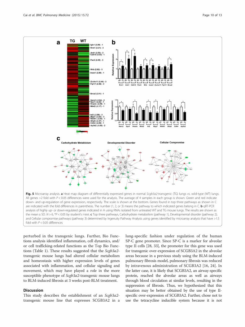
perturbed in the transgenic lungs. Further, Bio Func-tions analysis identified inflammation, cell dynamics, and/or cell trafficking-related functions as the Top Bio Func-tions (Table 1). These results suggested that the Scgb3a2-transgenic mouse lungs had altered cellular metabolismand homeostasis with higher expression levels of genesassociated with inflammation, and cellular signaling andmovement, which may have played a role in the moresusceptible phenotype of Scgb3a2-transgenic mouse lungsto BLM-induced fibrosis at 3 weeks post-BLM treatment.
DiscussionThis study describes the establishment of an Scgb3a2-transgenic mouse line that expresses SCGB3A2 in a
lung-specific fashion under regulation of the humanSP-C gene promoter. Since SP-C is a marker for alveolartype II cells [28, 33], the promoter for this gene was usedfor transgenic over-expression of SCGB3A2 in the alveolarareas because in a previous study using the BLM-inducedpulmonary fibrosis model, pulmonary fibrosis was reducedby intravenous administration of SCGB3A2 [16, 24]. Inthe latter case, it is likely that SCGB3A2, an airway-specificprotein, reached the alveolar areas as well as airwaysthrough blood circulation at similar levels, resulting in thesuppression of fibrosis. Thus, we hypothesized that thissituation may be better obtained by the use of type II-specific over-expression of SCGB3A2. Further, chose not touse the tetracycline inducible system because it is not
Fig. 5 Microarray analysis. a Heat map diagram of differentially expressed genes in normal Scgb3a2-transgenic (TG) lungs vs. wild-type (WT) lungs.All genes >2 fold with P < 0.05 differences were used for the analysis. The average of 4 samples in each group is shown. Green and red indicatedown- and up-regulation of gene expression, respectively. The scale is shown at the bottom. Genes found in top three pathways as shown in Care indicated with the fold differences in parenthesis. The number (1, 2, or 3) means the pathway to which indicated genes belong in C. b qRT-PCRanalysis of highly up- or down-regulated genes indicated in A using RNAs isolated from untreated WT and TG mouse lungs. The results are shown asthe mean ± SD. N > 6, *P < 0.05 by student’s t-test. c Top three pathways, Carbohydrate metabolism (pathway 1), Developmental disorder (pathway 2),and Cellular compromise pathways (pathway 3) determined by Ingenuity Pathway Analysis using genes identified by microarray analysis that have >1.5fold with P < 0.05 differences
Cai et al. BMC Pulmonary Medicine (2015) 15:72 Page 10 of 13

known whether and/or how doxycycline affects formationand/or resolution of BLM-induced pulmonary fibrosis, inan otherwise well-established BLM-induced pulmonaryfibrosis model.SCGB3A2 expression faithfully mimicked the expression
pattern of the lung specific SP-C gene. Over-expression ofSCGB3A2 in transgenic mouse lungs was confirmed byqRT-PCR for mRNA and Western blotting for proteinusing lung tissues, and levels in BALF by ELISA. The dif-ference in mRNA expression levels between wild-type andtransgenic mouse lungs was observed as early as E14.5.The SCGB3A2 mRNA and protein levels in lung werehighest at around two weeks of age. Immunohistochemis-try further demonstrated expression of SCGB3A2 inairway epithelial cells, the endogenous expression site forSCGB3A2 as well as type II cells, the specific site for SP-Cexpression [28, 33]. Since alveolar type II cells constitute~15 % of the peripheral lung cells [34], the expression ofSCGB3A2 in type II cells is likely to be a main contributorto over-expression of SCGB3A2 in the transgenic lung.Two different sizes of Scgb3a2 mRNAs were observed byNorthern blotting for Scgb3a2-transgenic mice that werelarger than that of endogenously expressed Scgb3a2. Wedo not know the exact reason for this phenomenon. It
could be due to longer polyA tails present in the trans-gene-derived mRNA. Alternatively, since the transgeneconstruct contains a Scgb3a2 cDNA that does not have anintron, the mRNA processing may have been affected,resulting in a larger mRNA. The way the transgene wasinserted to a chromosome might also have affected thesize of Scgb3a2 mRNA.While Scgb3a2-transgenic lungs exhibited no phenotypes
with normal lung function, they showed exacerbated fi-brosis at 3 weeks after subjected to the BLM-inducedpulmonary fibrosis model as determined by lung histology,hydroxyproline content, inflammatory cell numbers, colla-gen gene expression, and inflammatory cytokine levels.Microarray analysis revealed that this was due to alteredexpression of genes caused by ectopic overexpression ofSCGB3A2 that are involved in inflammation, cell dynam-ics, and/or cell trafficking-related functions as comparedto wild-type. These changes in gene expression patternsmay have affected metabolism and homeostasis of thetransgenic lungs, which rendered the transgenic lungsmore susceptible to BLM challenge. However the changesmay be too subtle to affect lung homeostasis withoutchallenge since we did not detect any gross metabolicor abnormal inflammatory phenotypes in the transgenicmice. Of most interest is that after 3 weeks, the fibrosisof Scgb3a2-transgenic lungs quickly resolved as comparedwith wild-type lungs. BLM-induced pulmonary fibrosis isknown to partially resolve after weeks of BLM treatmentdue to unknown reasons [30–32]. Previously we dem-onstrated that SCGB3A2 exhibits anti-fibrotic activity[16, 24]. The anti-fibrotic activity of SCGB3A2 was dueto suppression of the TGFβ-induced differentiation offibroblasts to myofibroblasts, a hallmark of the fibrogenicprocess through increased phosphorylation of STAT1 andexpression of SMAD7, and decreased phosphorylationof SMAD2 and SMAD3 [16]. The BLM-induced injuryincludes damage to and involvement of alveolar andbronchial epithelium, and decreased SCGB1A1 expres-sion in the airway epithelial cells after BLM treatmentwas described following BLM [35]. In the current study,SCGB3A2 overexpression predisposed mice to a moresevere phenotype 3 weeks post-BLM, at which timeSCGB3A2 expression was almost at the level of wild-typemice, suggesting that changes in gene expression patternsin transgenic mice predominate in determination of thephenotype. However by 9 weeks post-BLM, the expressionof SCGB3A2 in transgenic lungs recovered to levels foundin pre-BLM lungs, which was significantly higher than thatof wild-type lungs. Ectopic expression of SP-C was alsoobserved in the bronchial epithelial cells, which may havecontributed to the more rapid increase in SCGB3A2 ex-pression in the transgenic lungs. Previously it was shownthat after BLM administration, SP-C is co-expressed inbronchial epithelial cells with SCGB1A1, the marker for
Table 1 Top Bio Functions identified in lungs of normalScgb3a2-transgenic vs. wild-type mouse
Name of functions p-value # ofmolecules
Diseases and disorders
Inflammatory response 6.76E-04–2.75E-02 9
Organismal injury and abnormalities 1.12E-03–4.06E-02 16
Neurological response 1.83E-03–4.10E-02 20
Nutritional disease 1.83E-03–1.83E-03 3
Cancer 2.76E-03–4.10E-02 15
Molecular and cellular functions
Cellular development 8.79E-05–3.99E-02 25
Cell growth and proliferation 8.79E-05–3.52E-02 21
Drug metabolism 1.91E-04–2.75E-02 7
Small molecule biochemistry 5.68E-04–4.10E-02 20
Cellular movement 6.76E-04–3.68E-02 17
Physiological system developmentand function
Cardiovascular system developmentand function
8.79E-05–2.80E-02 10
Connective tissue development andfunction
8.79E-05–4.10E-02 10
Hematological system developmentand function
6.76E-04–4.10E-02 14
Immune cell trafficking 6.76E-04–4.10E-02 8
Nervous system development andfunction
1.12E-03–4.10E-02 24
Cai et al. BMC Pulmonary Medicine (2015) 15:72 Page 11 of 13
Club cells that is the site for SCGB3A2 expression[36, 37]. The temporary SP-C-positive, SCGB1A1-positivecells were suggested to eventually differentiate into type IIcells during the repair after severe pulmonary injury[36, 37]. In the current study, the resolution of fibrosiscoincided with the rapid increase of SCGB3A2 expres-sion in lungs of Scgb3a2-transgenic mice, suggestingthe anti-fibrotic activity of SCGB3A2. How SCGB3A2promotes natural resolution of BLM-induced pulmon-ary fibrosis requires further studies.In the single dose BLM model, pulmonary fibrosis
develops through sequential events after BLM administra-tion; first in inflammation phase (≤7 days), followed byfibrosis phase (≥7 days) [30]. Many compounds werereported as anti-fibrotic agents, however most of them(over 220 compounds) were given ≤7 days of BLM ad-ministration and thus considered as preventive agents.Only a handful compounds were administered in the fi-brosis phase as therapeutic agents. The present studiesdemonstrated that expression of SCGB3A2 markedlyincreased in transgenic mice after the severity of fibrosisreached peak levels, and thus the situation may resemblethat of SCGB3A2 being administered in the therapeuticphase, which likely resulted in the rapid decrease offibrosis. These results are in good agreement with theprevious reports using BLM-induced pulmonary fibro-sis model mice with intravenously administered SCGB3A2that SCGB3A2 possesses anti-fibrotic activity and maybe used as a therapeutic agent in treatment of pulmonaryfibrosis [16, 24].
ConclusionsA transgenic mouse over-expressing SCGB3A2 in lung-specific fashion under the promoter of human SP-Cgene was established. The lungs of the Scgb3a2-trans-genic mice were histologically and functionally normal.When these mice were subjected to the BLM-inducedpulmonary fibrosis model, a slightly exaggerated fibrosiswas initially noted at 3 weeks post-BLM, however thefibrosis more rapidly resolved in Scgb3a2-transgenic miceas compared to wild-type by 6 weeks post-BLM. Theseresults demonstrate the possible therapeutic use ofSCGB3A2 in treatment of lung fibrosis.
Additional files
Additional file 1: Figure S1. Scanning electron microscopy of wild-type(WT) and Scgb3a2-transgenic mouse (TG) airways. Figure S2. Characterization ofScgb3a2-transgenic embryo lungs. Figure S3. Lung morphometric analysis.Figure S4. Survival curve of wild-type (WT) and Scgb3a2-transgenic mice (TG)after BLM treatment. Figure S5. SP-C expression in wild-type (WT) and Scgb3a2-transgenic mouse (TG) lungs at 6 and 9 weeks post-BLM administration.
Additional file 2: Table S1. Up and down-regulated genes in lungs ofScgb3a2-transgenic vs. wild-type mice.
AbbreviationsSCGB: Secretoglobin; BLM: Bleomycin.
Competing interestsThe authors declare that they have no competing interests.
Authors’ contributionsYC conceived of the study, designed the experiments, characterizedScgb3a2-transgenic mouse lungs, carried out BLM study, and wrote a draftof the manuscript. TT established and characterized the Scgb3a2-transgenicmouse line. MY, RK, TK participated in characterization of the transgenicmouse lungs. MY, RK, MO carried out immunohistochemistry/immunofluorescence studies, and analyzed the data. HA carried outelectron microscopy analysis. WM intellectually and technically contributedto morphometric analysis of mouse lungs, AG carried out characterization ofmouse embryonic lungs, SK designed and integrated this study, wrote andrevised the manuscript. All authors read and approved the final manuscript.
AcknowledgementsWe thank Jeffrey Whitsett (Cincinnati, OH) for the human SP-C gene promoterplasmid and Poonam Mannan, Langston Lim, and Susan H. Garfield (CCRConfocal Microscopy Core Facility, Laboratory of Cancer Biology and Genetics,NCI) for their help in confocal microscopy. This study was funded by theIntramural Research Program of the National Cancer Institute.
Author details1Laboratory of Metabolism, National Cancer Institute, National Institutes ofHealth, Bethesda, MD 20892, USA. 2Biochemical Engineering, GraduateSchool of Science and Engineering, Yamagata University, Yonezawa,Yamagata 992-8510, Japan. 3Bloomberg School of Public Health, JohnsHopkins University, Baltimore, MD 21205, USA. 4Department of Medicine,Pulmonary Center, Boston University School of Medicine, Boston, MA 02118,USA. 5Laboratory of Liver Diseases, National Institute on Alcohol Abuse andAlcoholism, National Institutes of Health, Bethesda, MD 20892, USA.6Department of Pharmacology, Tokyo Women’s Medical University, Tokyo162-8666, Japan. 7Department of Veterinary Immunopathology, School ofVeterinary Medicine, Rakuno Gakuen University, Ebetsu, Hokkaido 069-8501,Japan. 8Laboratory of Cell Growth and Differentiation, Institute of Molecularand Cellular Biosciences, The University of Tokyo, Tokyo 113-0032, Japan.
Received: 20 November 2014 Accepted: 28 June 2015
References1. Jackson BC, Thompson DC, Wright MW, McAndrews M, Bernard A, Nebert DW,
et al. Update of the human secretoglobin (SCGB) gene superfamily and anexample of ‘evolutionary bloom’ of androgen-binding protein genes withinthe mouse Scgb gene superfamily. Hum Genomics. 2011;5(6):691–702.
2. Klug J, Beier HM, Bernard A, Chilton BS, Fleming TP, Lehrer RI, et al.Uteroglobin/Clara cell 10-kDa family of proteins: nomenclature committeereport. Ann N Y Acad Sci. 2000;923:348–54.
3. Niimi T, Keck-Waggoner CL, Popescu NC, Zhou Y, Levitt RC, Kimura S.UGRP1, a uteroglobin/Clara cell secretory protein-related protein, is a novellung-enriched downstream target gene for the T/EBP/NKX2.1 homeodomaintranscription factor. Mol Endocrinol. 2001;15(11):2021–36.
4. Kurotani R, Tomita T, Yang Q, Carlson BA, Chen C, Kimura S. Role ofsecretoglobin 3A2 in lung development. Am J Respir Crit Care Med.2008;178(4):389–98.
5. Guha A, Vasconcelos M, Cai Y, Yoneda M, Hinds A, Qian J, et al. Neuroepithelialbody microenvironment is a niche for a distinct subset of Clara-like precursorsin the developing airways. Proc Natl Acad Sci U S A. 2012;109(31):12592–7.
6. Harrod KS, Mounday AD, Stripp BR, Whitsett JA. Clara cell secretory proteindecreases lung inflammation after acute virus infection. Am J Physiol.1998;275(5 Pt 1):L924–930.
7. Lee YC, Zhang Z, Mukherjee AB. Mice lacking uteroglobin are highlysusceptible to developing pulmonary fibrosis. FEBS Lett. 2006;580(18):4515–20.
8. Mandal AK, Zhang Z, Ray R, Choi MS, Chowdhury B, Pattabiraman N, et al.Uteroglobin represses allergen-induced inflammatory response by blockingPGD2 receptor-mediated functions. J Exp Med. 2004;199(10):1317–30.
Cai et al. BMC Pulmonary Medicine (2015) 15:72 Page 12 of 13

9. Mukherjee AB, Zhang Z, Chilton BS. Uteroglobin: a steroid-inducibleimmunomodulatory protein that founded the Secretoglobin superfamily.Endocr Rev. 2007;28(7):707–25.
10. Facchiano A, Cordella-Miele E, Miele L, Mukherjee AB. Inhibition of pancreaticphospholipase A2 activity by uteroglobin and antiflammin peptides: possiblemechanism of action. Life Sci. 1991;48(5):453–64.
11. Miele L, Cordella-Miele E, Facchiano A, Mukherjee AB. Inhibition of phospholipaseA2 by uteroglobin and antiflammin peptides. Adv Exp Med Biol. 1990;279:137–60.
12. Linnoila RI, Szabo E, DeMayo F, Witschi H, Sabourin C, Malkinson A. The roleof CC10 in pulmonary carcinogenesis: from a marker to tumor suppression.Ann N Y Acad Sci. 2000;923:249–67.
13. Saha A, Lee YC, Zhang Z, Chandra G, Su SB, Mukherjee AB. Lack of anendogenous anti-inflammatory protein in mice enhances colonization ofB16F10 melanoma cells in the lungs. J Biol Chem. 2010;285(14):10822–31.
14. Yang Y, Zhang Z, Mukherjee AB, Linnoila RI. Increased susceptibility of micelacking Clara cell 10-kDa protein to lung tumorigenesis by 4-(methylnitrosamino)-1-(3-pyridyl)-1-butanone, a potent carcinogen in cigarette smoke. J Biol Chem.2004;279(28):29336–40.
15. Chiba Y, Kurotani R, Kusakabe T, Miura T, Link BW, Misawa M, et al.Uteroglobin-related protein 1 expression suppresses allergic airway inflammationin mice. Am J Respir Crit Care Med. 2006;173(9):958–64.
16. Kurotani R, Okumura S, Matsubara T, Yokoyama U, Buckley JR, Tomita T,et al. Secretoglobin 3A2 suppresses bleomycin-induced pulmonary fibrosisby transforming growth factor beta signaling down-regulation. J Biol Chem.2011;286(22):19682–92.
17. Chiba Y, Kusakabe T, Kimura S. Decreased expression of uteroglobin-relatedprotein 1 in inflamed mouse airways is mediated by IL-9. Am J Physiol LungCell Mol Physiol. 2004;287(6):L1193–1198.
18. Chiba Y, Srisodsai A, Supavilai P, Kimura S. Interleukin-5 reduces the expressionof uteroglobin-related protein (UGRP) 1 gene in allergic airway inflammation.Immunol Lett. 2005;97(1):123–9.
19. Kido T, Yoneda M, Cai Y, Matsubara T, Ward JM, Kimura S. Secretoglobinsuperfamily protein SCGB3A2 deficiency potentiates ovalbumin-inducedallergic pulmonary inflammation. Mediators Inflamm. 2014;2014:216465.
20. Tachihara-Yoshikawa M, Ishida T, Watanabe K, Sugawara A, Kanazawa K,Kanno R, et al. Expression of secretoglobin3A2 (SCGB3A2) in primarypulmonary carcinomas. Fukushima J Med Sci. 2008;54(2):61–72.
21. Kurotani R, Kumaki N, Naizhen X, Ward JM, Linnoila RI, Kimura S. Secretoglobin3A2/uteroglobin-related protein 1 is a novel marker for pulmonary carcinomain mice and humans. Lung Cancer. 2011;71(1):42–8.
22. Wikenheiser KA, Clark JC, Linnoila RI, Stahlman MT, Whitsett JA. Simian virus40 large T antigen directed by transcriptional elements of the humansurfactant protein C gene produces pulmonary adenocarcinomas intransgenic mice. Cancer Res. 1992;52(19):5342–52.
23. Ozdemir H, Guvenal T, Cetin M, Kaya T, Cetin A. A placebo-controlled comparisonof effects of repetitive doses of betamethasone and dexamethasone on lungmaturation and lung, liver, and body weights of mouse pups. Pediatr Res.2003;53(1):98–103.
24. Cai Y, Winn ME, Zehmer JK, Gillette WK, Lubkowski JT, Pilon AL, et al. Preclinicalevaluation of human secretoglobin 3A2 in mouse models of lung developmentand fibrosis. Am J Physiol Lung Cell Mol Physiol. 2014;306(1):L10–22.
25. Ashcroft T, Simpson JM, Timbrell V. Simple method of estimating severity ofpulmonary fibrosis on a numerical scale. J Clin Pathol. 1988;41(4):467–70.
26. Tomita T, Kido T, Kurotani R, Iemura S, Sterneck E, Natsume T, et al.CAATT/enhancer-binding proteins alpha and delta interact with NKX2-1 tosynergistically activate mouse secretoglobin 3A2 gene expression.J Biol Chem. 2008;283(37):25617–27.
27. Reynolds SD, Reynolds PR, Pryhuber GS, Finder JD, Stripp BR. SecretoglobinsSCGB3A1 and SCGB3A2 define secretory cell subsets in mouse and humanairways. Am J Respir Crit Care Med. 2002;166(11):1498–509.
28. Kalina M, Mason RJ, Shannon JM. Surfactant protein C is expressed inalveolar type II cells but not in Clara cells of rat lung. Am J Respir Cell MolBiol. 1992;6(6):594–600.
29. Knudsen L, Weibel ER, Gundersen HJ, Weinstein FV, Ochs M. Assessment ofair space size characteristics by intercept (chord) measurement: an accurateand efficient stereological approach. J Appl Physiol. 2010;108(2):412–21.
30. Moeller A, Ask K, Warburton D, Gauldie J, Kolb M. The bleomycin animalmodel: a useful tool to investigate treatment options for idiopathicpulmonary fibrosis? Int J Biochem Cell Biol. 2008;40(3):362–82.
31. Mouratis MA, Aidinis V. Modeling pulmonary fibrosis with bleomycin.Curr Opin Pulm Med. 2011;17(5):355–61.
32. Scotton CJ, Chambers RC. Bleomycin revisited: towards a morerepresentative model of IPF? Am J Physiol Lung Cell Mol Physiol.2010;299(4):L439–441.
33. Wert SE, Glasser SW, Korfhagen TR, Whitsett JA. Transcriptional elementsfrom the human SP-C gene direct expression in the primordial respiratoryepithelium of transgenic mice. Dev Biol. 1993;156(2):426–43.
34. Wang D, Haviland DL, Burns AR, Zsigmond E, Wetsel RA. A pure populationof lung alveolar epithelial type II cells derived from human embryonic stemcells. Proc Natl Acad Sci U S A. 2007;104(11):4449–54.
35. Daly HE, Baecher-Allan CM, Barth RK, D’Angio CT, Finkelstein JN. Bleomycininduces strain-dependent alterations in the pattern of epithelial cell-specificmarker expression in mouse lung. Toxicol Appl Pharmacol. 1997;142(2):303–10.
36. Zheng D, Limmon GV, Yin L, Leung NH, Yu H, Chow VT, et al. Regenerationof alveolar type I and II cells from Scgb1a1-expressing cells following severepulmonary damage induced by bleomycin and influenza. PLoS One.2012;7(10), e48451.
37. Zheng D, Limmon GV, Yin L, Leung NH, Yu H, Chow VT, et al. A cellularpathway involved in Clara cell to alveolar type II cell differentiation aftersevere lung injury. PLoS One. 2013;8(8), e71028.
Submit your next manuscript to BioMed Centraland take full advantage of:
• Convenient online submission
• Thorough peer review
• No space constraints or color figure charges
• Immediate publication on acceptance
• Inclusion in PubMed, CAS, Scopus and Google Scholar
• Research which is freely available for redistribution
Submit your manuscript at www.biomedcentral.com/submit
Cai et al. BMC Pulmonary Medicine (2015) 15:72 Page 13 of 13