Tosoh BILS 2016
18
TOSOH BIOSCIENCE GmbH Fcɣ Receptor immobilized Resin for Chromatogaphy Judith Vajda, Toru Tanaka, Egbert Müller Tosoh Bioscience BILS conference 2016, Berlin
-
Upload
gbx-summits -
Category
Presentations & Public Speaking
-
view
153 -
download
0
Transcript of Tosoh BILS 2016

TOSOH BIOSCIENCE GmbH
Fcɣ Receptor immobilized Resin for Chromatogaphy
Judith Vajda, Toru Tanaka, Egbert Müller Tosoh Bioscience
BILS conference 2016, Berlin
TOSOH BIOSCIENCE GmbH
Outline
• R&D of innovative chromatography resins
• Fcγ Receptors
• Fcγ-RIII immobilized on TSKgel resin for an analytical column
• Application examples
TOSOH BIOSCIENCE GmbH
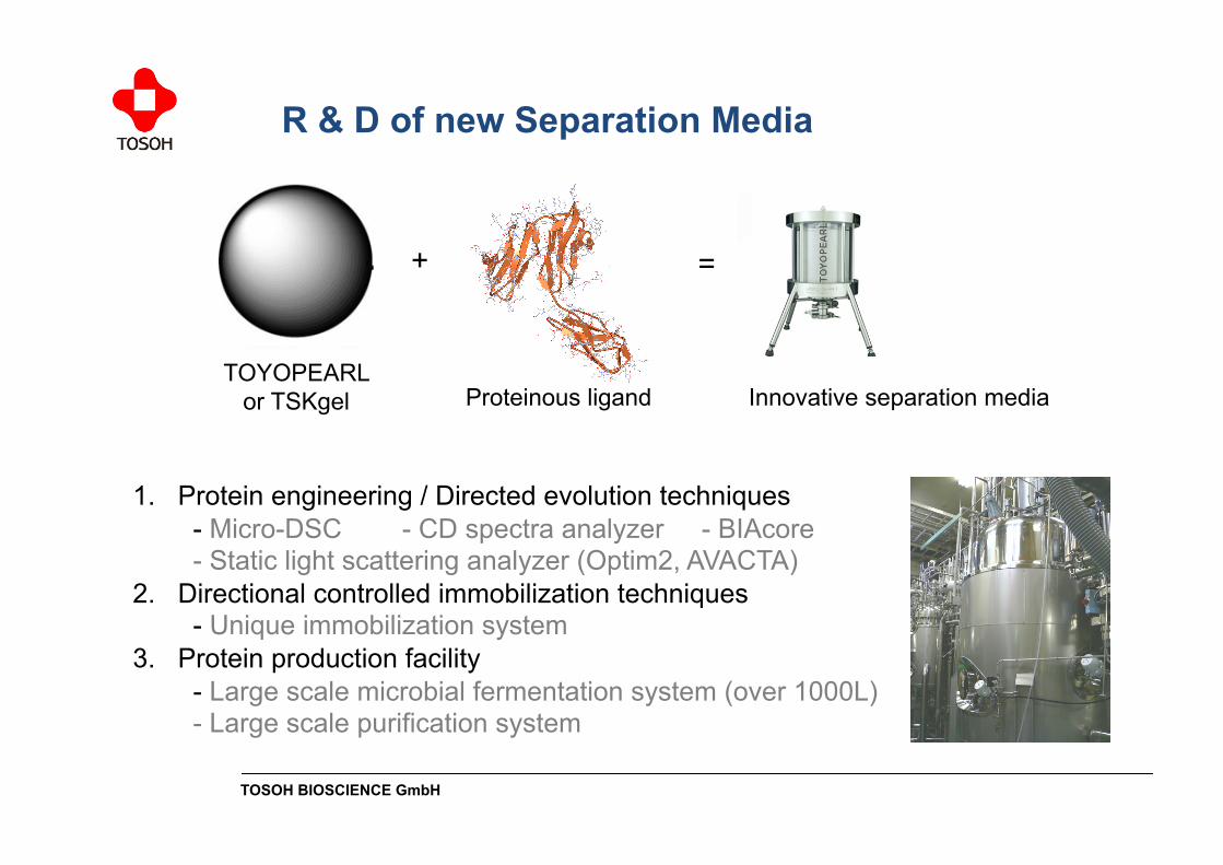
1. Protein engineering / Directed evolution techniques - Micro-DSC - CD spectra analyzer - BIAcore - Static light scattering analyzer (Optim2, AVACTA) 2. Directional controlled immobilization techniques - Unique immobilization system 3. Protein production facility - Large scale microbial fermentation system (over 1000L) - Large scale purification system
R & D of new Separation Media
TOYOPEARL or TSKgel
+
Proteinous ligand
=
Innovative separation media
TOSOH BIOSCIENCE GmbH
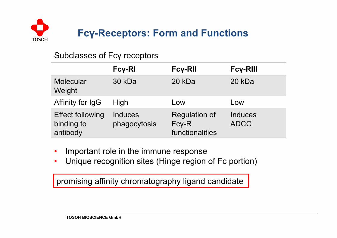
Fcγ-RI Fcγ-RII Fcγ-RIII Molecular Weight
30 kDa 20 kDa 20 kDa
Affinity for IgG High Low Low
Effect following binding to antibody
Induces phagocytosis
Regulation of Fcγ-R functionalities
Induces ADCC
Subclasses of Fcγ receptors
• Important role in the immune response • Unique recognition sites (Hinge region of Fc portion)
Fcγ-Receptors: Form and Functions
promising affinity chromatography ligand candidate

TOSOH BIOSCIENCE GmbH
• Quality control of therapeutic antibodies is still very difficult.
Background and Target Application of Fcγ-R Resins
Lot to lot variation of Rituxan®/MabThera®
Source: Martin Schiestl et al. (Sandoz Biopharmaceuticals), Nature Biotechnology 29, 310-312 (2011)
TOSOH BIOSCIENCE GmbH
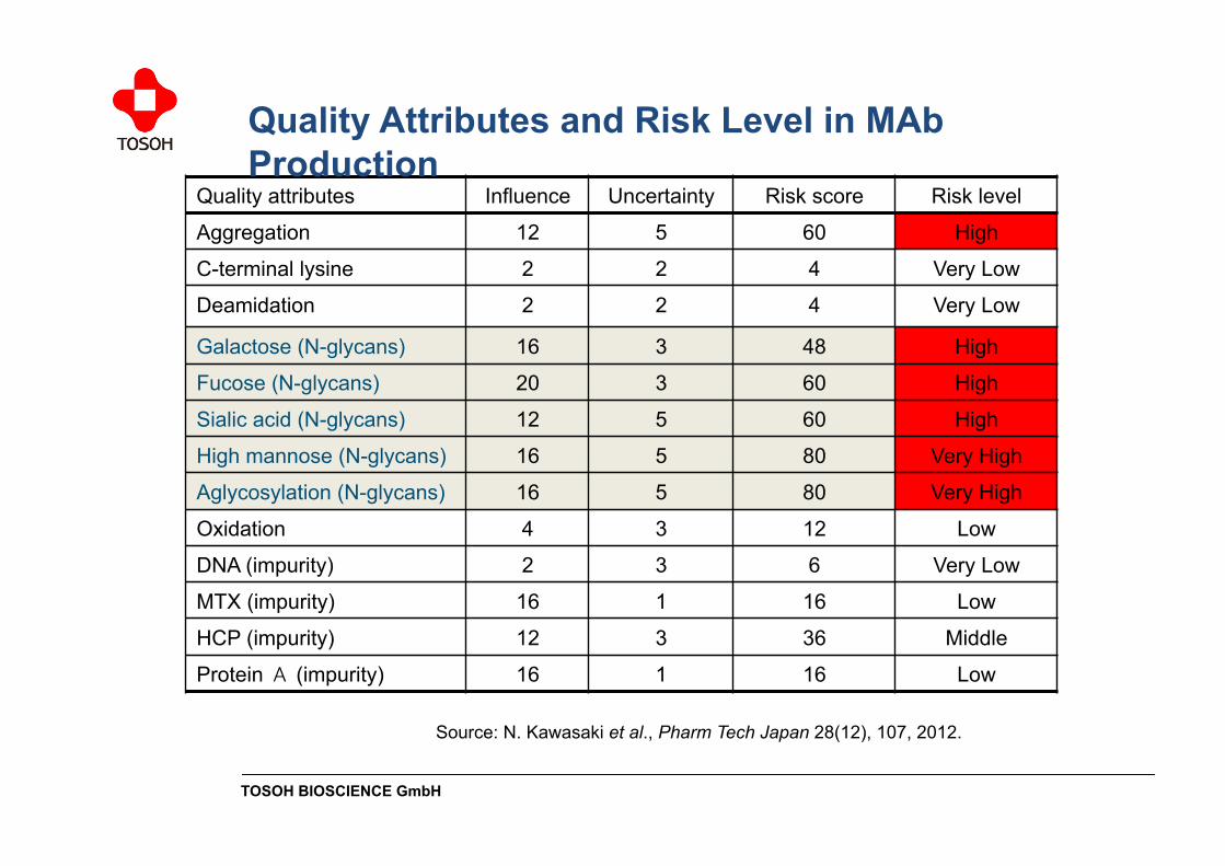
Quality attributes Influence Uncertainty Risk score Risk level
Aggregation 12 5 60 High
C-terminal lysine 2 2 4 Very Low
Deamidation 2 2 4 Very Low
Galactose (N-glycans) 16 3 48 High
Fucose (N-glycans) 20 3 60 High
Sialic acid (N-glycans) 12 5 60 High
High mannose (N-glycans) 16 5 80 Very High
Aglycosylation (N-glycans) 16 5 80 Very High
Oxidation 4 3 12 Low
DNA (impurity) 2 3 6 Very Low
MTX (impurity) 16 1 16 Low
HCP (impurity) 12 3 36 Middle
Protein A (impurity) 16 1 16 Low
Source: N. Kawasaki et al., Pharm Tech Japan 28(12), 107, 2012.
Quality Attributes and Risk Level in MAb Production

TOSOH BIOSCIENCE GmbH
The binding affinity between Fcγ-RIII and a mAb is very important to destroy a cancer cell. The N-glycan structure of an antibody affects the affinity to the Fc receptor.
Mechanism of therapeutic Ab ADCC activity

TOSOH BIOSCIENCE GmbH
0
20
40
60
80
100
120
0 50 100 150 200
Sta
bilit
y (R
em
ainin
g ac
tivi
ty, %)
Incubation time (hours)
• Wild-type Fcγ-RIII is unstable under acidic conditions
! Modification of Fcγ-RIII by directed evolution techniques
Ligand and resin development
• FcR-immobilized resin/column using TSKgel resin packed into stainless steel hardware
Stability of wild-type (black) and modified FcR (red) at pH 3.0, 25 °C
TOSOH BIOSCIENCE GmbH
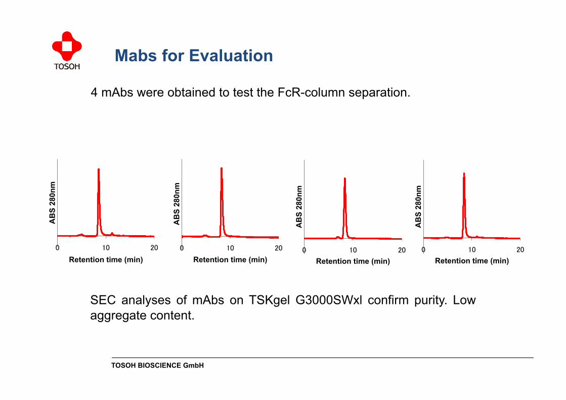
4 mAbs were obtained to test the FcR-column separation.
0 10 20
AB
S 28
0nm
Retention time (min) 0 10 20
AB
S 28
0nm
Retention time (min) 0 10 20
AB
S 28
0nm
Retention time (min) 0 10 20
AB
S 28
0nm
Retention time (min)
SEC analyses of mAbs on TSKgel G3000SWxl confirm purity. Low aggregate content.
Mabs for Evaluation

TOSOH BIOSCIENCE GmbH
0 20 40
AB
S 28
0nm
Retention time (min) 0 20 40
AB
S 28
0nm
Retention time (min)
0 20 40
AB
S 28
0nm
Retention time (min) 0 20 40
AB
S 28
0nm
Retention time (min)
FcR-column analyses of mAbs on FcR-column (proto-type)
• 4 mAbs were separated into three or four peaks • All antibodies have unique separation profiles
Buffer A: 20 mM Na Acetate pH 5.0, 50 mM NaCl Buffer B: 10 mM Gly-HCl pH 3.0 Gradient: B 0% (0-2 min), B 0-100% (2-40 min), B 100% (40-50 min)
Fcγ-RIII Column Evaluation

TOSOH BIOSCIENCE GmbH
0 20 40
AB
S 28
0nm
Retention time (min) 0 20 40
AB
S 28
0nm
Retention time (min) 0 20 40
AB
S 28
0nm
Time (min) 0 20 40
AB
S 28
0nm
Time (min)
Rituxan® mAb A mAb B mAb C
FcR-column analyses of mAbs on FcR-column (proto-type)
Buffer A: 20 mM Na Acetate pH 5.0, 200 mM NaCl Buffer B: 10 mM Gly-HCl pH 3.0 Gradient: B 0% (0-2 min), B 0-100% (2-40 min), B 100% (40-50 min)
Fcγ-RIII Column Evaluation at optimized Conditions
• Resolution improved by method optimization

TOSOH BIOSCIENCE GmbH
Buffer system Buffer A, 20 mM Sodium Acetate buffer pH 5.0, 50 mM NaCl Buffer B, 10 mM Gly-HCl buffer pH 3.0
Reproducibility
• Separation profiles indicate well stability of Fcγ-RIII column
TOSOH BIOSCIENCE GmbH
-50
0
50
100
150
200
250
300
350
400
0 2 4 6 8 10 12 14 16 18 20 22 24 26 28 30 32 34 36 38 40
AB
S280
nm
Time (min)
Fr 1 Fr 2 Fr 3
20 25 30 35 40 45
AB
S280
nm
Time (min)
Fr 1
Fr 2 Fr 3
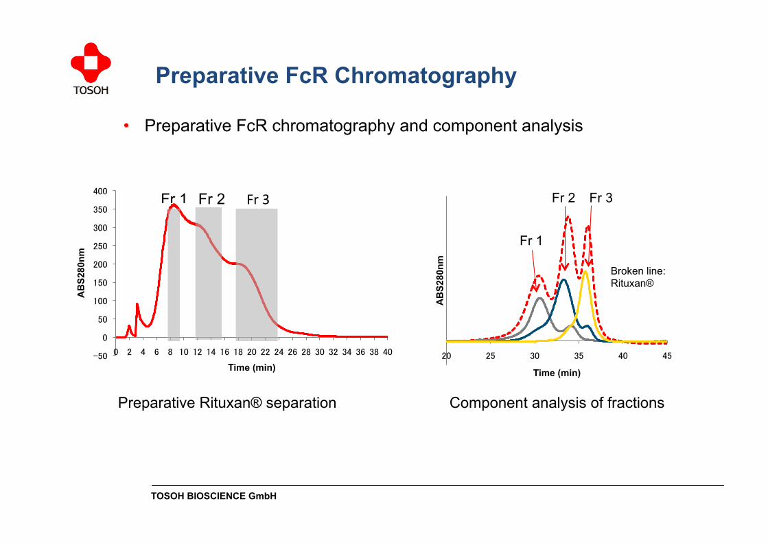
Broken line: Rituxan®
Preparative Rituxan® separation Component analysis of fractions
Preparative FcR Chromatography
• Preparative FcR chromatography and component analysis
TOSOH BIOSCIENCE GmbH
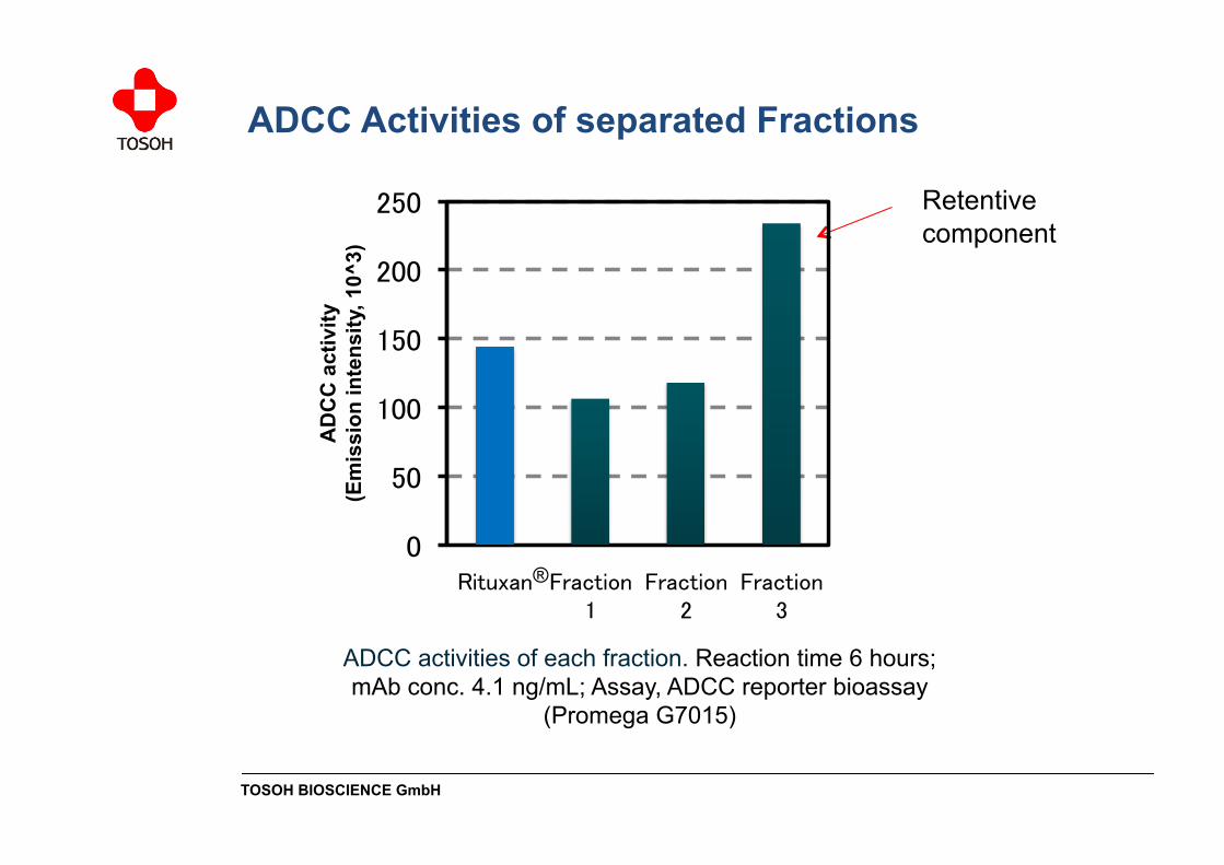
ADCC activities of each fraction. Reaction time 6 hours; mAb conc. 4.1 ng/mL; Assay, ADCC reporter bioassay
(Promega G7015)
Retentive component
ADCC Activities of separated Fractions
0
50
100
150
200
250
Rituxan Fraction 1
Fraction 2
Fraction 3
AD
CC
act
ivity
(E
mis
sion
inte
nsity
, 10^
3)
®

TOSOH BIOSCIENCE GmbH
G0F
G1F
G1F
G2F
Rituxan® First Last Fraction
unknown
(%)
N-glycan Structure Analysis
TOSOH BIOSCIENCE GmbH
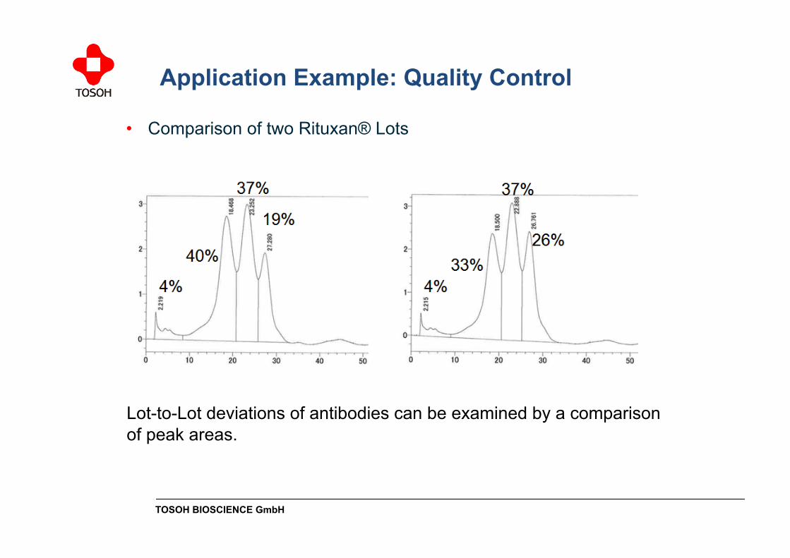
Lot-to-Lot deviations of antibodies can be examined by a comparison of peak areas.
• Comparison of two Rituxan® Lots
Application Example: Quality Control
TOSOH BIOSCIENCE GmbH
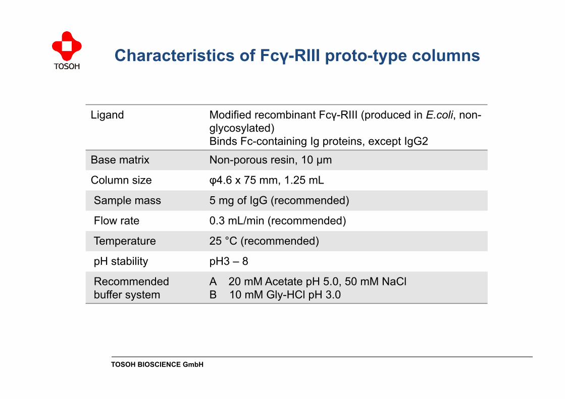
Ligand Modified recombinant Fcγ-RIII (produced in E.coli, non-glycosylated) Binds Fc-containing Ig proteins, except IgG2
Base matrix Non-porous resin, 10 µm
Column size φ4.6 x 75 mm, 1.25 mL
Sample mass 5 mg of IgG (recommended)
Flow rate 0.3 mL/min (recommended)
Temperature 25 °C (recommended)
pH stability pH3 – 8
Recommended buffer system
A 20 mM Acetate pH 5.0, 50 mM NaCl B 10 mM Gly-HCl pH 3.0
Characteristics of Fcγ-RIII proto-type columns
TOSOH BIOSCIENCE GmbH
Conclusions
• The Fcγ-RIII proto-type column can separate mAbs according to their glycan structure and ADCC activity
• Non-glycosylated ligand produced in E.coli
• Ligand stability at acidic conditions was achieved by directed evolution techniques


![EL.LE [online] ISSN 2280-6792 › media › pdf › journals › ... · 2018-09-07 · Progettare la valutazione scolastica degli studenti con BiLS ... complex variables, some of](https://static.fdocument.pub/doc/165x107/5ed687e9ff0e593c0b6404b7/elle-online-issn-2280-6792-a-media-a-pdf-a-journals-a-2018-09-07.jpg)