Toshiba Medical Celebrates 10 Years of Area … · Toshiba Medical Celebrates 10 Years of Area...
44
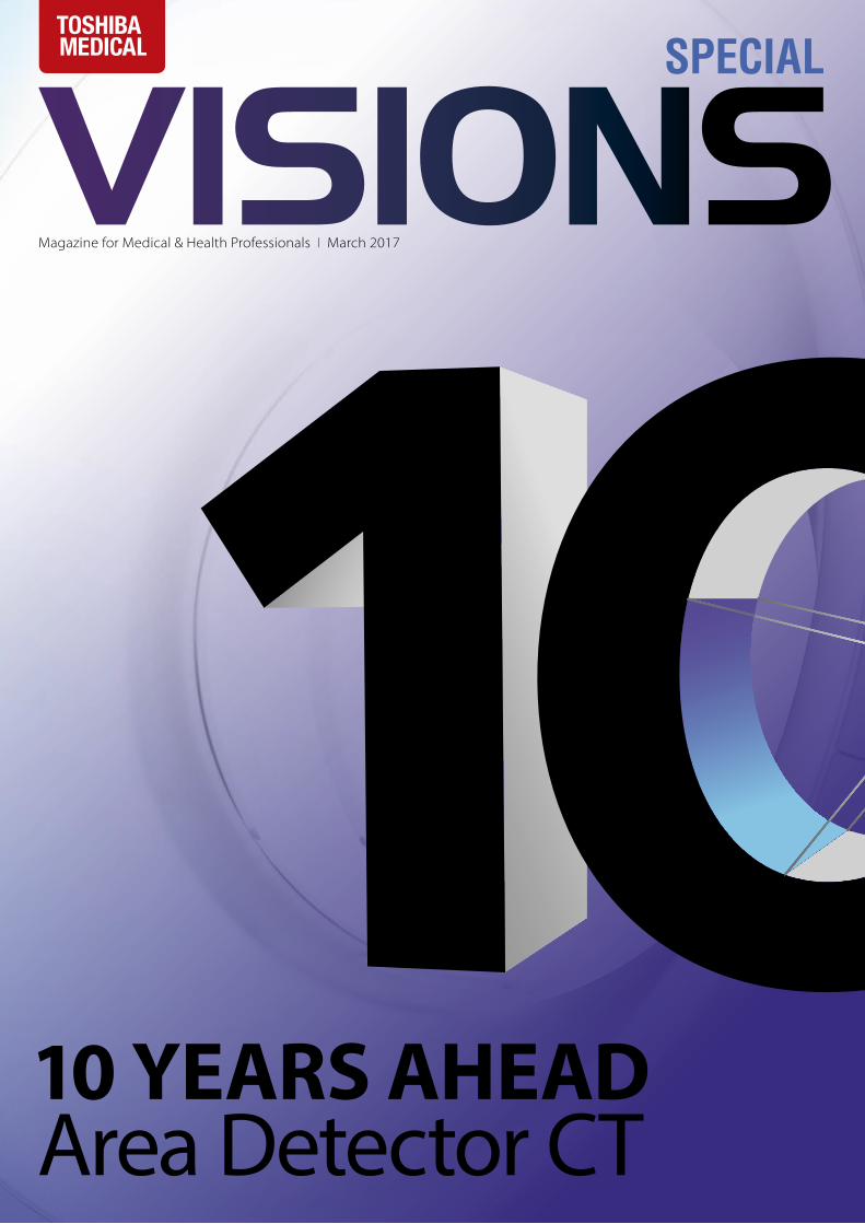
VISIONS Magazine for Medical & Health Professionals I March 2017 SPECIAL 10 YEARS AHEAD Area Detector CT
Transcript of Toshiba Medical Celebrates 10 Years of Area … · Toshiba Medical Celebrates 10 Years of Area...
VISIONSMagazine for Medical & Health Professionals I March 2017
SPECIAL
10 YEARS AHEADArea Detector CT
Toshiba Medical Celebrates10 Years of Area Detector CTDescribed as ‘a breakthrough CT system’ back in 2007, Toshiba Medical introduced the world’s very first dynamic volume CT scanner, The Aquilion ONE™. Driven by the clinical need for a volume scanner, the Aquilion ONE’s unique Area Detector opened doors to new ways of medical imaging. Ten years later, Toshiba Medical is celebrating this memorable introduction!
This VISIONS Special highlights personal experiences of Key Opinion Leaders. It outlines Toshiba Medical’s journey in Area Detector CT and shows our continuing spirit to explore the unknown.
To download the digital version of this CT Special, visit: www.toshiba-medical.eu/eu/ct-campaign-10-years-ahead
To stay up to date with our latest innovations in medical imaging, visit: www.toshiba-medical.eu
This CT Special edition of VISIONS magazine is a publication of Toshiba Medical Europe and is offered free of charge to medical and health professionals. To download the digital edition of this CT Special, please visit the campaign web site: www.toshiba-medical.eu/eu/ct-campaign-10-years-ahead. No part of this publication may be reproduced in whole or in part, stored in an automated storage and retrieval system or transmitted in any manner whatsoever without written permission of the publisher. The opinions expressed in this publication are solely those of the authors and not necessarily those of Toshiba Medical. Toshiba Medical does not guarantee the accuracy or reliability of the information provided herein.
PublisherToshiba Medical Systems Europe B.V.Zilverstraat 1NL-2718 RP ZoetermeerTel.: +31 79 368 92 22Fax: +31 79 368 94 44Web: www.toshiba-medical.euEmail: [email protected]
Campaign web pages: www.toshiba-medical.eu/eu/ct-campaign-10-years-ahead
Editor-in-chiefJack Hoogendoorn ([email protected])
EditorJacqueline de Graaf ([email protected])
Coordinator & ReviewerRoy Verlaan (International Market Development Manager CT)
Design & LayoutBoerma Reclame (www.boermareclame.com)
PhotographyCojan van Toor (www.cojanvantoor.nl)
PrintmanagementHet Staat Gedrukt (www.hetstaatgedrukt.nl)
Text contributions and editingThe Creative Practice (www.thecreativepractice.com)
© 2017 by TOSHIBA Medical EuropeAll rights reserved
ISSN 1617-2876
10 YEARS AHEADArea Detector CT
VISIONS | CT SPECIAL
3
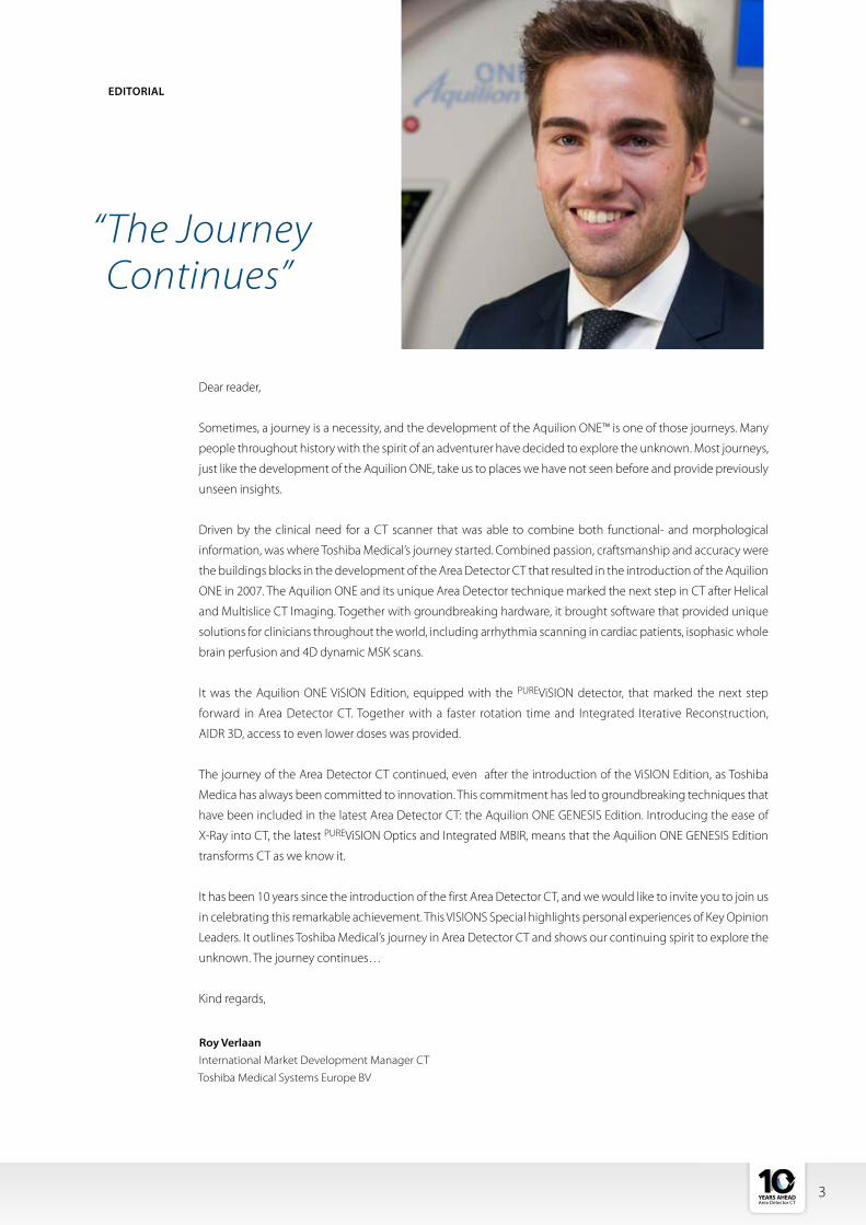
Dear reader,
Sometimes, a journey is a necessity, and the development of the Aquilion ONE™ is one of those journeys. Many
people throughout history with the spirit of an adventurer have decided to explore the unknown. Most journeys,
just like the development of the Aquilion ONE, take us to places we have not seen before and provide previously
unseen insights.
Driven by the clinical need for a CT scanner that was able to combine both functional- and morphological
information, was where Toshiba Medical’s journey started. Combined passion, craftsmanship and accuracy were
the buildings blocks in the development of the Area Detector CT that resulted in the introduction of the Aquilion
ONE in 2007. The Aquilion ONE and its unique Area Detector technique marked the next step in CT after Helical
and Multislice CT Imaging. Together with groundbreaking hardware, it brought software that provided unique
solutions for clinicians throughout the world, including arrhythmia scanning in cardiac patients, isophasic whole
brain perfusion and 4D dynamic MSK scans.
It was the Aquilion ONE ViSION Edition, equipped with the PUREViSION detector, that marked the next step
forward in Area Detector CT. Together with a faster rotation time and Integrated Iterative Reconstruction,
AIDR 3D, access to even lower doses was provided.
The journey of the Area Detector CT continued, even after the introduction of the ViSION Edition, as Toshiba
Medica has always been committed to innovation. This commitment has led to groundbreaking techniques that
have been included in the latest Area Detector CT: the Aquilion ONE GENESIS Edition. Introducing the ease of
X-Ray into CT, the latest PUREViSION Optics and Integrated MBIR, means that the Aquilion ONE GENESIS Edition
transforms CT as we know it.
It has been 10 years since the introduction of the first Area Detector CT, and we would like to invite you to join us
in celebrating this remarkable achievement. This VISIONS Special highlights personal experiences of Key Opinion
Leaders. It outlines Toshiba Medical’s journey in Area Detector CT and shows our continuing spirit to explore the
unknown. The journey continues…
Kind regards,
EDITORIAL
Roy VerlaanInternational Market Development Manager CT
Toshiba Medical Systems Europe BV
“ The Journey Continues”
4 ©2017 TOSHIBA MEDICALVISIONSCT SPECIAL
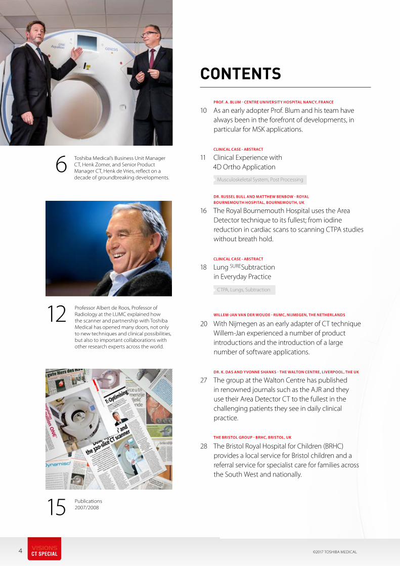
10 As an early adopter Prof. Blum and his team have always been in the forefront of developments, in particular for MSK applications.
11 Clinical Experience with 4D Ortho Application
16 The Royal Bournemouth Hospital uses the Area Detector technique to its fullest; from iodine reduction in cardiac scans to scanning CTPA studies without breath hold.
18 Lung SURESubtraction in Everyday Practice
20 With Nijmegen as an early adapter of CT technique Willem-Jan experienced a number of product introductions and the introduction of a large number of software applications.
27 The group at the Walton Centre has published in renowned journals such as the AJR and they use their Area Detector CT to the fullest in the challenging patients they see in daily clinical practice.
28 The Bristol Royal Hospital for Children (BRHC) provides a local service for Bristol children and a referral service for specialist care for families across the South West and nationally.
CONTENTS
DR. K. DAS AND YVONNE SHANKS - THE WALTON CENTRE, LIVERPOOL, THE UK
THE BRISTOL GROUP - BRHC, BRISTOL, UK
PROF. A. BLUM - CENTRE UNIVERSITY HOSPITAL NANCY, FRANCE
DR. RUSSEL BULL AND MATTHEW BENBOW - ROYAL BOURNEMOUTH HOSPITAL, BOURNEMOUTH, UK
WILLEM-JAN VAN DER WOUDE - RUMC, NIJMEGEN, THE NETHERLANDS
CLINICAL CASE - ABSTRACT
CLINICAL CASE - ABSTRACT
Musculoskeletal System, Post Processing
CTPA, Lungs, Subtraction
12
15
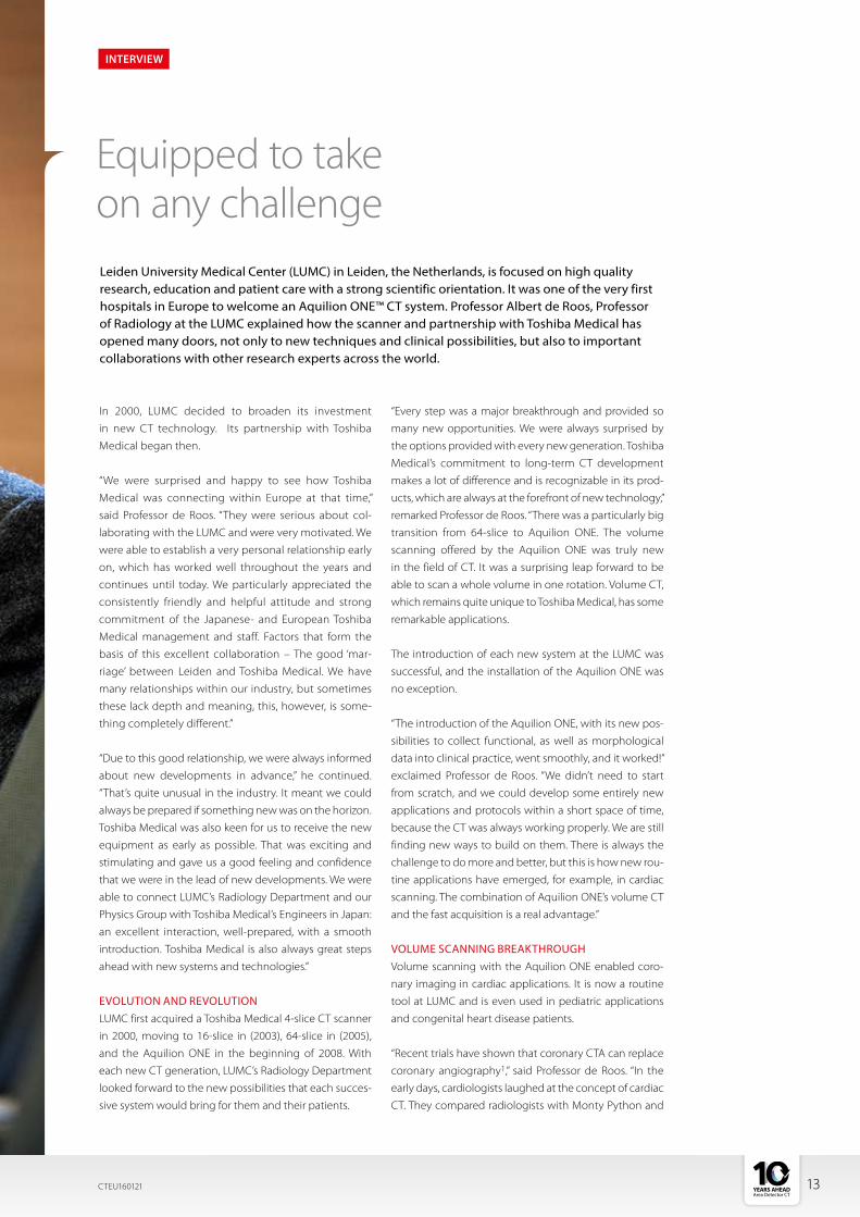
Professor Albert de Roos, Professor of Radiology at the LUMC explained how the scanner and partnership with Toshiba Medical has opened many doors, not only to new techniques and clinical possibilities, but also to important collaborations with other research experts across the world.
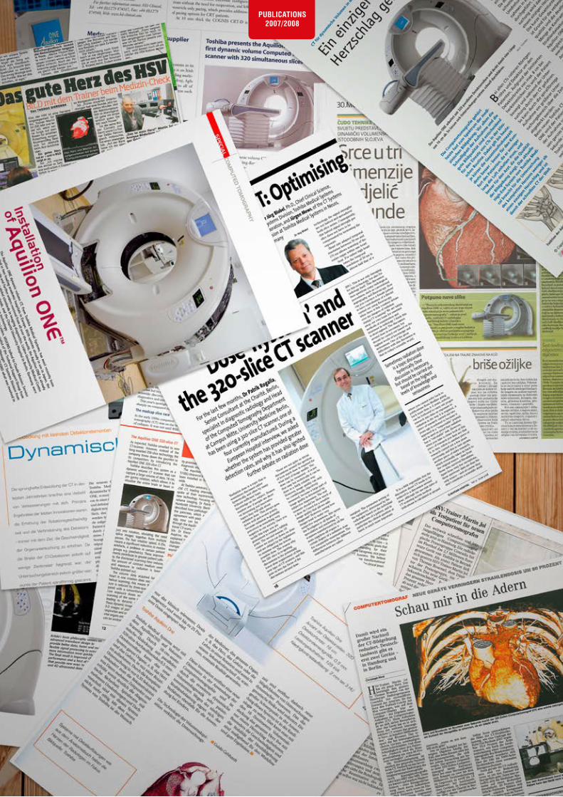
Publications 2007/2008
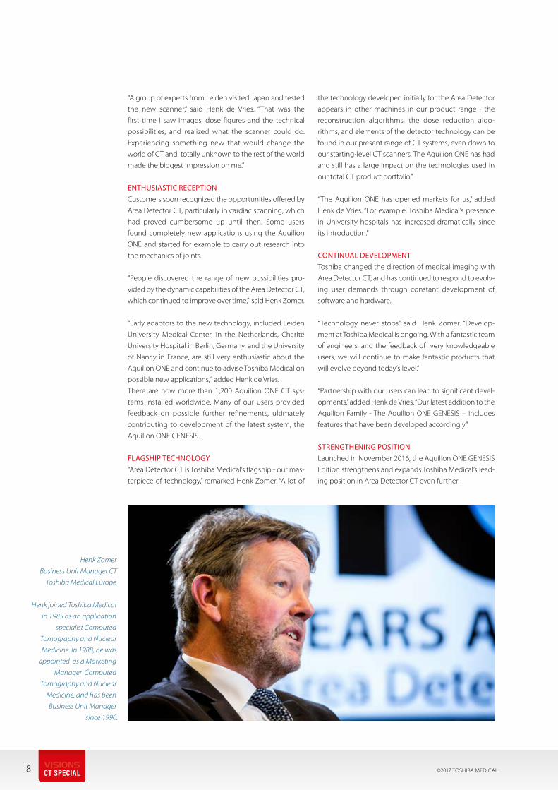
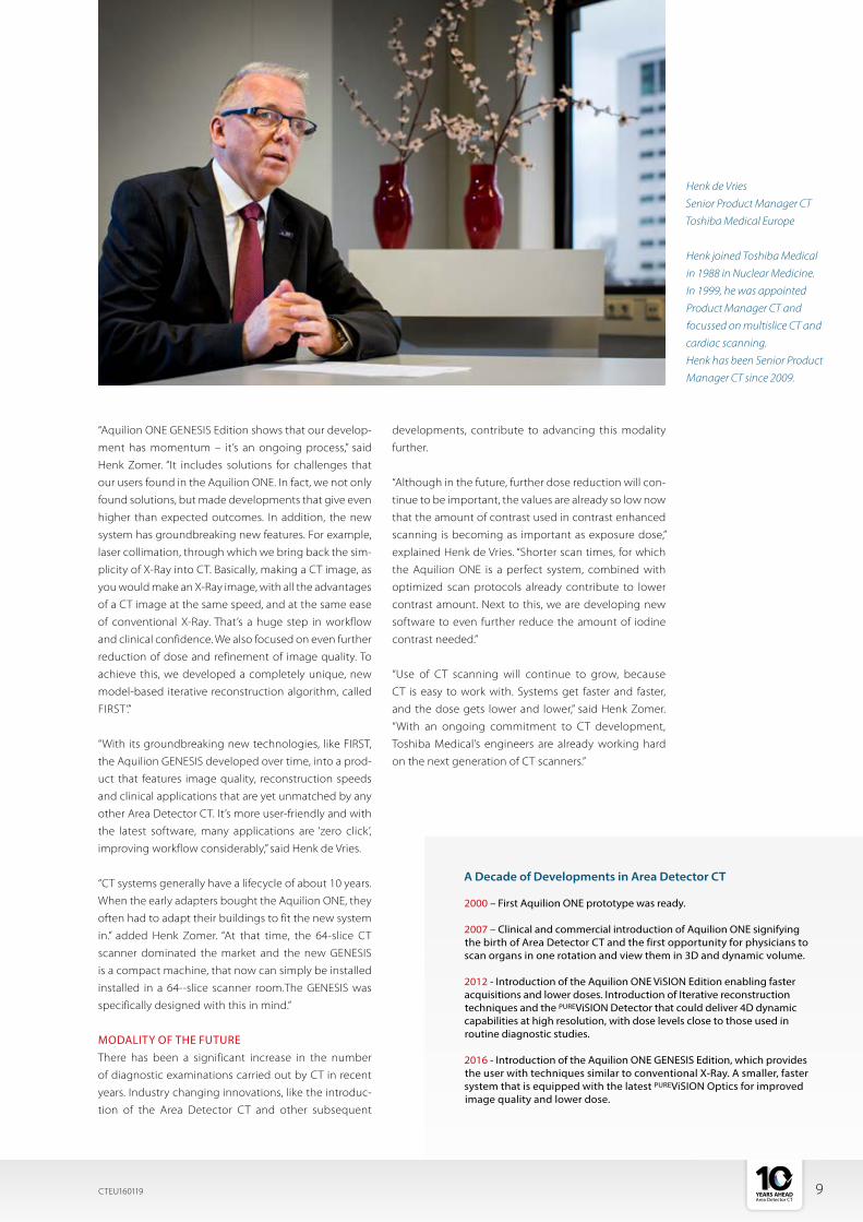
6 Toshiba Medical’s Business Unit Manager CT, Henk Zomer, and Senior Product Manager CT, Henk de Vries, reflect on a decade of groundbreaking developments.
5
Dose, Metal artefact reduction
03 Editorial
19 CT Workshops 2017
23 10 Years Advertising
32 VISIONS articles 2007-2017
42 Made For Life Campaign
30 German Armed Forces and Patients Benefit from New Options for Low-Dose Volume CT
31 As a lead radiographer Joost Roelofs has extensive, long-term experience in using Toshiba CT’s including the Area Detector CT’s.
38 Prof. C. Roy and her team in Strasbourg have worked in many different areas using Toshiba Medical’s Area Detector technique.
39 First Clinical Results of Coronary CT Subtraction
32 Dr. Viladés Medel, Dr. Leta Petracca and Dr. Alomar have been very active in Coronary CTA imaging. As leading experts in Coronary CTA they have published extensively using Toshiba Medical’s Area Detector CT.
CLINICAL CASE - ABSTRACT
Coronary Subtraction, CT Angiography, Coronary Artery Disease
PROF. C. ROY - UNIVERSITY HOSPITAL STRASBOURG, STRASBOURG, FRANCE
DR. VILADÉS MEDEL, DR. LETA PETRACCA AND DR. ALOMAR - CLINICA CREU BLANCA, BARCELONA, SPAIN.
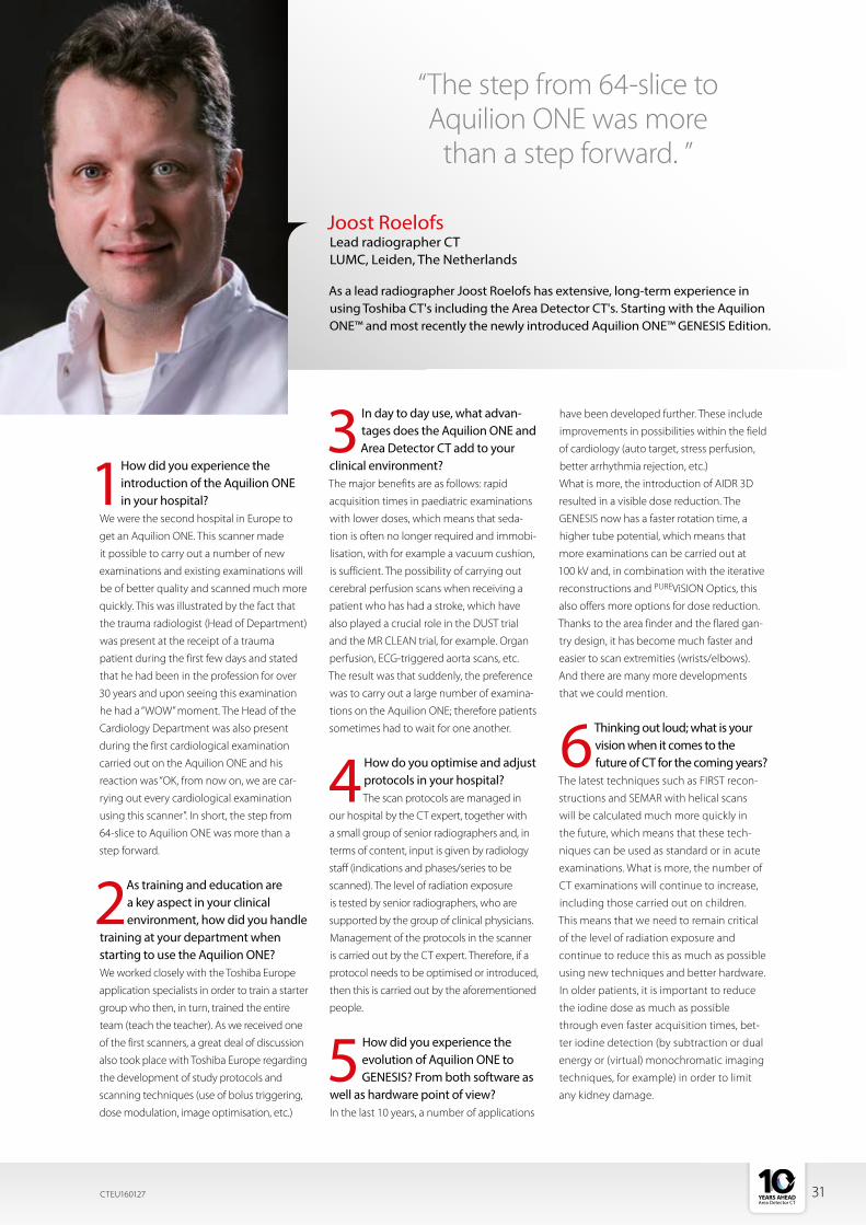
JOOST ROELOFS - LUMC, LEIDEN, THE NETHERLANDS
CLINICAL CASE - ABSTRACT
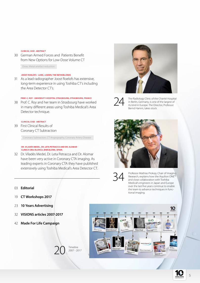
The Radiology Clinic of the Charité Hospital in Berlin, Germany, is one of the largest of its kind in Europe. The Director, Professor Bernd Hamm, takes stock.
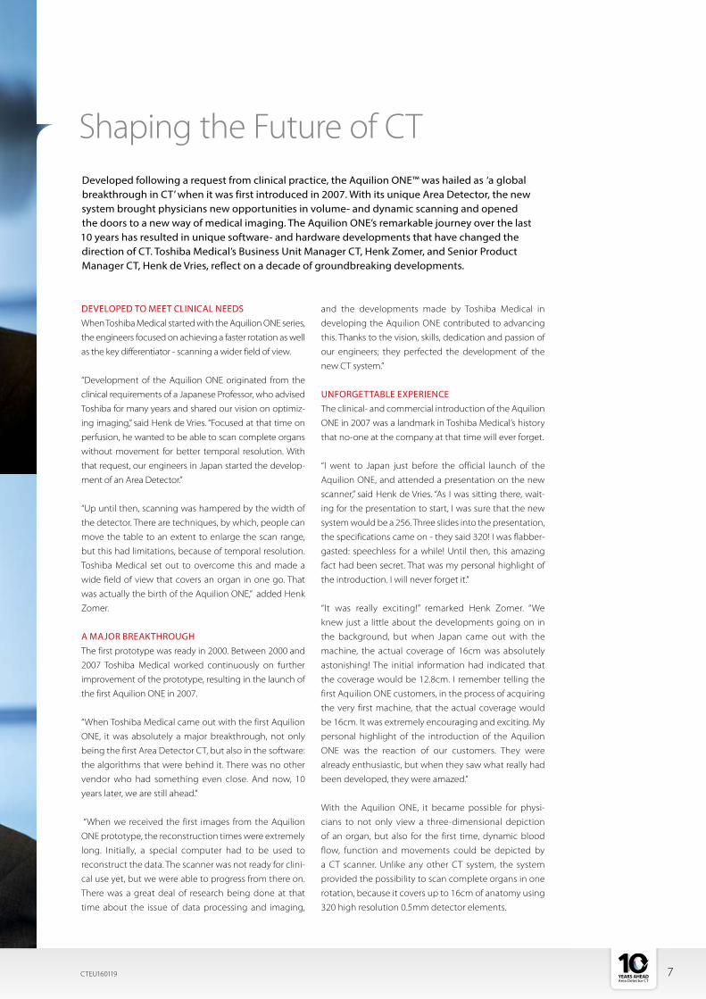
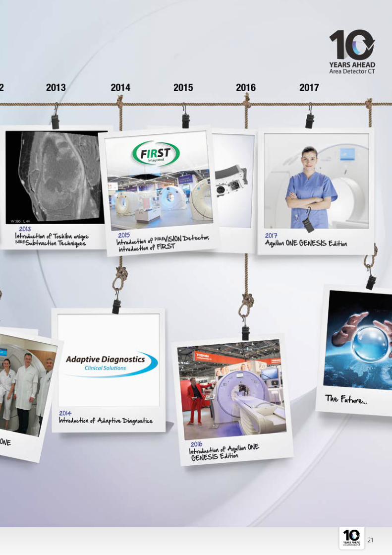
Timeline 2007 - 2017
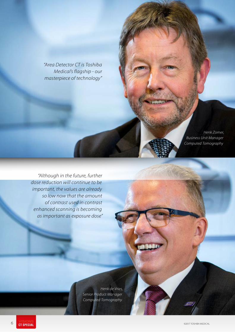
Professor Mathias Prokop, Chair of Imaging Research, explains how the Aquilion ONE™ and close collaboration with Toshiba Medical’s engineers in Japan and Europe over the last five years continue to enable the team to advance techniques in func-tional imaging.
34
24
20
6 ©2017 TOSHIBA MEDICALVISIONSCT SPECIAL
Henk de Vries, Senior Product Manager Computed Tomography
Henk Zomer, Business Unit Manager
Computed Tomography
“Area Detector CT is Toshiba Medical’s flagship - our
mas terpiece of technology”
“Although in the future, further dose reduction will con tinue to be important, the values are already
so low now that the amount of contrast used in contrast
enhanced scanning is becoming as important as exposure dose”
DEVELOPED TO MEET CLINICAL NEEDSWhen Toshiba Medical started with the Aquilion ONE series,
the engineers focused on achieving a faster rotation as well
as the key differentiator - scanning a wider field of view.
“Development of the Aquilion ONE originated from the
clinical requirements of a Japanese Professor, who advised
Toshiba for many years and shared our vision on optimiz-
ing imaging,” said Henk de Vries. “Focused at that time on
perfusion, he wanted to be able to scan complete organs
without movement for better temporal resolution. With
that request, our engineers in Japan started the develop-
ment of an Area Detector.”
“Up until then, scanning was hampered by the width of
the detector. There are techniques, by which, people can
move the table to an extent to enlarge the scan range,
but this had limitations, because of temporal resolution.
Toshiba Medical set out to overcome this and made a
wide field of view that covers an organ in one go. That
was actually the birth of the Aquilion ONE,” added Henk
Zomer.
A MAJOR BREAKTHROUGHThe first prototype was ready in 2000. Between 2000 and
2007 Toshiba Medical worked continuously on further
improvement of the prototype, resulting in the launch of
the first Aquilion ONE in 2007.
“When Toshiba Medical came out with the first Aquilion
ONE, it was absolutely a major breakthrough, not only
being the first Area Detector CT, but also in the software:
the algorithms that were behind it. There was no other
vendor who had something even close. And now, 10
years later, we are still ahead.”
“When we received the first images from the Aquilion
ONE prototype, the reconstruction times were extremely
long. Initially, a special computer had to be used to
reconstruct the data. The scanner was not ready for clini-
cal use yet, but we were able to progress from there on.
There was a great deal of research being done at that
time about the issue of data processing and imaging,
and the developments made by Toshiba Medical in
developing the Aquilion ONE contributed to advancing
this. Thanks to the vision, skills, dedication and passion of
our engineers; they perfected the development of the
new CT system.”
UNFORGETTABLE EXPERIENCEThe clinical- and commercial introduction of the Aquilion
ONE in 2007 was a landmark in Toshiba Medical’s history
that no-one at the company at that time will ever forget.
“I went to Japan just before the official launch of the
Aquilion ONE, and attended a presentation on the new
scanner,” said Henk de Vries. “As I was sitting there, wait-
ing for the presentation to start, I was sure that the new
system would be a 256. Three slides into the presentation,
the specifications came on - they said 320! I was flabber-
gasted: speechless for a while! Until then, this amazing
fact had been secret. That was my personal highlight of
the introduction. I will never forget it.”
“It was really exciting!” remarked Henk Zomer. “We
knew just a little about the developments going on in
the background, but when Japan came out with the
machine, the actual coverage of 16cm was absolutely
astonishing! The initial information had indicated that
the coverage would be 12.8cm. I remember telling the
first Aquilion ONE customers, in the process of acquiring
the very first machine, that the actual coverage would
be 16cm. It was extremely encouraging and exciting. My
personal highlight of the introduction of the Aquilion
ONE was the reaction of our customers. They were
already enthusiastic, but when they saw what really had
been developed, they were amazed.”
With the Aquilion ONE, it became possible for physi-
cians to not only view a three-dimensional depiction
of an organ, but also for the first time, dynamic blood
flow, function and movements could be depicted by
a CT scanner. Unlike any other CT system, the system
provided the possibility to scan complete organs in one
rotation, because it covers up to 16cm of anatomy using
320 high resolution 0.5mm detector elements.
Shaping the Future of CT
INTERVIEW
Developed following a request from clinical practice, the Aquilion ONE™ was hailed as ‘a global breakthrough in CT’ when it was first introduced in 2007. With its unique Area Detector, the new system brought physicians new opportunities in volume- and dynamic scanning and opened the doors to a new way of medical imaging. The Aquilion ONE’s remarkable journey over the last 10 years has resulted in unique software- and hardware developments that have changed the direction of CT. Toshiba Medical’s Business Unit Manager CT, Henk Zomer, and Senior Product Manager CT, Henk de Vries, reflect on a decade of groundbreaking developments.
7CTEU160119
8 ©2017 TOSHIBA MEDICALVISIONSCT SPECIAL
“A group of experts from Leiden visited Japan and tested
the new scanner,” said Henk de Vries. “That was the
first time I saw images, dose figures and the technical
possibilities, and realized what the scanner could do.
Experiencing something new that would change the
world of CT and totally unknown to the rest of the world
made the biggest impression on me.”
ENTHUSIASTIC RECEPTIONCustomers soon recognized the opportunities offered by
Area Detector CT, particularly in cardiac scanning, which
had proved cumbersome up until then. Some users
found completely new applications using the Aquilion
ONE and started for example to carry out research into
the mechanics of joints.
“People discovered the range of new possibilities pro-
vided by the dynamic capabilities of the Area Detector CT,
which continued to improve over time,” said Henk Zomer.
“Early adaptors to the new technology, included Leiden
University Medical Center, in the Netherlands, Charité
University Hospital in Berlin, Germany, and the University
of Nancy in France, are still very enthusiastic about the
Aquilion ONE and continue to advise Toshiba Medical on
possible new applications,” added Henk de Vries.
There are now more than 1,200 Aquilion ONE CT sys-
tems installed worldwide. Many of our users provided
feedback on possible further refinements, ultimately
contributing to development of the latest system, the
Aquilion ONE GENESIS.
FLAGSHIP TECHNOLOGY“Area Detector CT is Toshiba Medical’s flagship - our mas-
terpiece of technology,” remarked Henk Zomer. “A lot of
the technology developed initially for the Area Detector
appears in other machines in our product range - the
reconstruction algorithms, the dose reduction algo-
rithms, and elements of the detector technology can be
found in our present range of CT systems, even down to
our starting-level CT scanners. The Aquilion ONE has had
and still has a large impact on the technologies used in
our total CT product portfolio.”
“The Aquilion ONE has opened markets for us,” added
Henk de Vries. “For example, Toshiba Medical’s presence
in University hospitals has increased dramatically since
its introduction.”
CONTINUAL DEVELOPMENTToshiba changed the direction of medical imaging with
Area Detector CT, and has continued to respond to evolv-
ing user demands through constant development of
software and hardware.
“Technology never stops,” said Henk Zomer. “Develop-
ment at Toshiba Medical is ongoing. With a fantastic team
of engineers, and the feedback of very knowledgeable
users, we will continue to make fantastic products that
will evolve beyond today’s level.”
“Partnership with our users can lead to significant devel-
opments,” added Henk de Vries. “Our latest addition to the
Aquilion Family - The Aquilion ONE GENESIS – includes
features that have been developed accordingly.”
STRENGTHENING POSITIONLaunched in November 2016, the Aquilion ONE GENESIS
Edition strengthens and expands Toshiba Medical’s lead-
ing position in Area Detector CT even further.
Henk Zomer
Business Unit Manager CT
Toshiba Medical Europe
Henk joined Toshiba Medical
in 1985 as an application
specialist Computed
Tomography and Nuclear
Medicine. In 1988, he was
appointed as a Marketing
Manager Computed
Tomography and Nuclear
Medicine, and has been
Business Unit Manager
since 1990.
9
“Aquilion ONE GENESIS Edition shows that our develop-
ment has momentum – it’s an ongoing process,” said
Henk Zomer. “It includes solutions for challenges that
our users found in the Aquilion ONE. In fact, we not only
found solutions, but made developments that give even
higher than expected outcomes. In addition, the new
system has groundbreaking new features. For example,
laser collimation, through which we bring back the sim-
plicity of X-Ray into CT. Basically, making a CT image, as
you would make an X-Ray image, with all the advantages
of a CT image at the same speed, and at the same ease
of conventional X-Ray. That’s a huge step in workflow
and clinical confidence. We also focused on even further
reduction of dose and refinement of image quality. To
achieve this, we developed a completely unique, new
model-based iterative reconstruction algorithm, called
FIRST’.”
“With its groundbreaking new technologies, like FIRST,
the Aquilion GENESIS developed over time, into a prod-
uct that features image quality, reconstruction speeds
and clinical applications that are yet unmatched by any
other Area Detector CT. It’s more user-friendly and with
the latest software, many applications are ‘zero click’,
improving workflow considerably,” said Henk de Vries.
“CT systems generally have a lifecycle of about 10 years.
When the early adapters bought the Aquilion ONE, they
often had to adapt their buildings to fit the new system
in.” added Henk Zomer. “At that time, the 64-slice CT
scanner dominated the market and the new GENESIS
is a compact machine, that now can simply be installed
installed in a 64--slice scanner room.The GENESIS was
specifically designed with this in mind.”
MODALITY OF THE FUTUREThere has been a significant increase in the number
of diagnostic examinations carried out by CT in recent
years. Industry changing innovations, like the introduc-
tion of the Area Detector CT and other subsequent
developments, contribute to advancing this modality
further.
“Although in the future, further dose reduction will con-
tinue to be important, the values are already so low now
that the amount of contrast used in contrast enhanced
scanning is becoming as important as exposure dose,”
explained Henk de Vries. “Shorter scan times, for which
the Aquilion ONE is a perfect system, combined with
optimized scan protocols already contribute to lower
contrast amount. Next to this, we are developing new
software to even further reduce the amount of iodine
contrast needed.”
“Use of CT scanning will continue to grow, because
CT is easy to work with. Systems get faster and faster,
and the dose gets lower and lower,” said Henk Zomer.
“With an ongoing commitment to CT development,
Toshiba Medical’s engineers are already working hard
on the next generation of CT scanners.”
A Decade of Developments in Area Detector CT
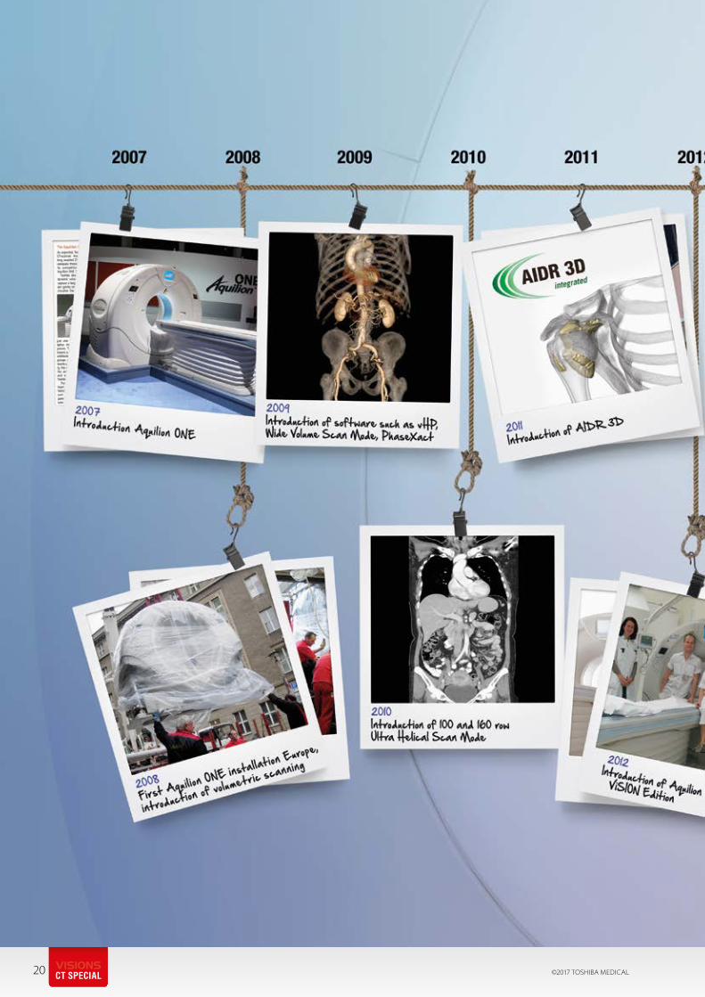
2000 – First Aquilion ONE prototype was ready.
2007 – Clinical and commercial introduction of Aquilion ONE signifying the birth of Area Detector CT and the first opportunity for physicians to scan organs in one rotation and view them in 3D and dynamic volume. 2012 - Introduction of the Aquilion ONE ViSION Edition enabling faster acquisitions and lower doses. Introduction of Iterative reconstruction techniques and the PUREViSION Detector that could deliver 4D dynamic capabilities at high resolution, with dose levels close to those used in routine diagnostic studies.
2016 - Introduction of the Aquilion ONE GENESIS Edition, which provides the user with techniques similar to conventional X-Ray. A smaller, faster system that is equipped with the latest PUREViSION Optics for improved image quality and lower dose.
Henk de Vries
Senior Product Manager CT
Toshiba Medical Europe
Henk joined Toshiba Medical
in 1988 in Nuclear Medicine.
In 1999, he was appointed
Product Manager CT and
focussed on multislice CT and
cardiac scanning.
Henk has been Senior Product
Manager CT since 2009.
CTEU160119
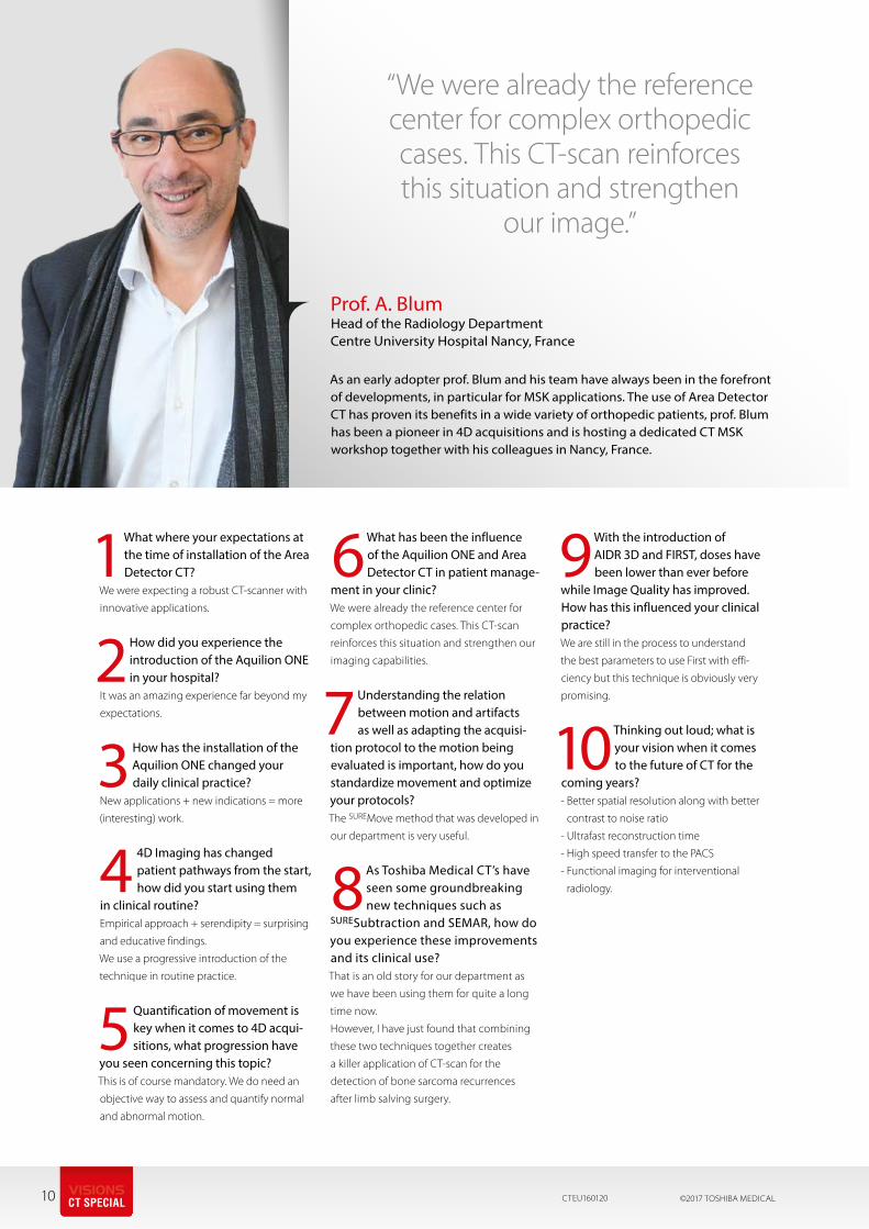
Prof. A. BlumHead of the Radiology DepartmentCentre University Hospital Nancy, France
As an early adopter prof. Blum and his team have always been in the forefront of developments, in particular for MSK applications. The use of Area Detector CT has proven its benefits in a wide variety of orthopedic patients, prof. Blum has been a pioneer in 4D acquisitions and is hosting a dedicated CT MSK workshop together with his colleagues in Nancy, France.
“We were already the reference center for complex orthopedic cases. This CT-scan reinforces this situation and strengthen
our image.”
10 ©2017 TOSHIBA MEDICAL
1 What where your expectations at the time of installation of the Area Detector CT?
We were expecting a robust CT-scanner with
innovative applications.
2 How did you experience the introduction of the Aquilion ONE in your hospital?
It was an amazing experience far beyond my
expectations.
3 How has the installation of the Aquilion ONE changed your daily clinical practice?
New applications + new indications = more
(interesting) work.
4 4D Imaging has changed patient pathways from the start, how did you start using them
in clinical routine? Empirical approach + serendipity = surprising
and educative findings.
We use a progressive introduction of the
technique in routine practice.
5 Quantification of movement is key when it comes to 4D acqui-sitions, what progression have
you seen concerning this topic? This is of course mandatory. We do need an
objective way to assess and quantify normal
and abnormal motion.
6 What has been the influence of the Aquilion ONE and Area Detector CT in patient manage-
ment in your clinic?We were already the reference center for
complex orthopedic cases. This CT-scan
reinforces this situation and strengthen our
imaging capabilities.
7 Understanding the relation between motion and artifacts as well as adapting the acquisi-
tion protocol to the motion being evaluated is important, how do you standardize movement and optimize your protocols?The SUREMove method that was developed in
our department is very useful.
8 As Toshiba Medical CT’s have seen some groundbreaking new techniques such as
SURESubtraction and SEMAR, how do you experience these improvements and its clinical use?That is an old story for our department as
we have been using them for quite a long
time now.
However, I have just found that combining
these two techniques together creates
a killer application of CT-scan for the
detection of bone sarcoma recurrences
after limb salving surgery.
9 With the introduction of AIDR 3D and FIRST, doses have been lower than ever before
while Image Quality has improved. How has this influenced your clinical practice?We are still in the process to understand
the best parameters to use First with effi-
ciency but this technique is obviously very
promising.
10 Thinking out loud; what is your vision when it comes to the future of CT for the
coming years?- Better spatial resolution along with better
contrast to noise ratio
- Ultrafast reconstruction time
- High speed transfer to the PACS
- Functional imaging for interventional
radiology.
CTEU160120VISIONSCT SPECIAL
11
Motion is frequently involved in the pathogenesis of musculoskeletal diseases. With static imaging
methods, the diagnosis of dynamic pathology (e.g. friction and impingement syndromes) is based on
secondary findings only1. This fact and the frequency of these conditions underscore the importance
of dynamic imaging modalities in the evaluation of musculoskeletal diseases. Wide area-detector CT is
suited to dynamic study of joints, allowing volumetric study of bone and intra-articular ligaments during
physiologic motion or under stress maneuvers. Dynamic CT is complementary to other dynamic methods,
helping overcome some of their limitations, such as evaluation of bony and intra-articular structures with
ultrasound or superimposition of structures on fluoroscopy2. Dynamic CT is most frequently used for the
evaluation of the wrist, but can be used on various joints (shoulder, hip, elbow, knee, and ankle)3-5.
Teixeira Gondim, Pedro Augusto MD, PhD. Blum, Alain MD, PhD
CLINICAL CASE ABSTRACT
Musculoskeletal System, Post Processing
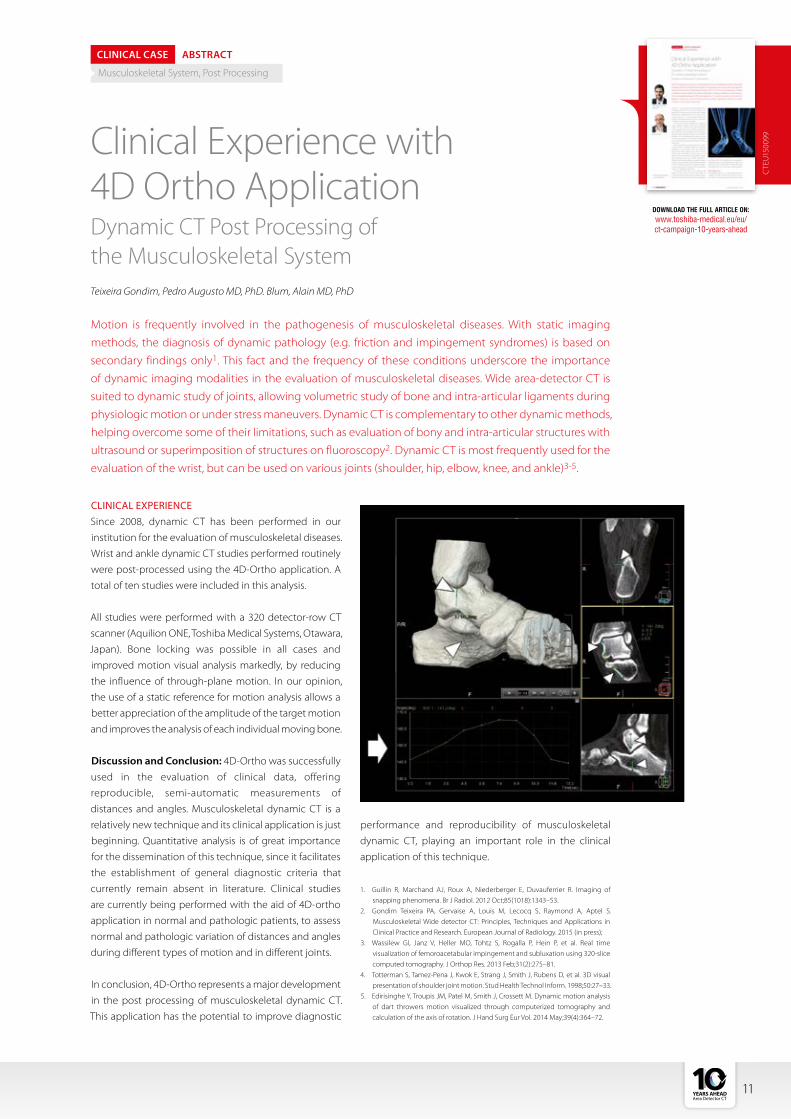
CLINICAL EXPERIENCESince 2008, dynamic CT has been performed in our
institution for the evaluation of musculoskeletal diseases.
Wrist and ankle dynamic CT studies performed routinely
were post-processed using the 4D-Ortho application. A
total of ten studies were included in this analysis.
All studies were performed with a 320 detector-row CT
scanner (Aquilion ONE, Toshiba Medical Systems, Otawara,
Japan). Bone locking was possible in all cases and
improved motion visual analysis markedly, by reducing
the influence of through-plane motion. In our opinion,
the use of a static reference for motion analysis allows a
better appreciation of the amplitude of the target motion
and improves the analysis of each individual moving bone.
Discussion and Conclusion: 4D-Ortho was successfully
used in the evaluation of clinical data, offering
reproducible, semi-automatic measurements of
distances and angles. Musculoskeletal dynamic CT is a
relatively new technique and its clinical application is just
beginning. Quantitative analysis is of great importance
for the dissemination of this technique, since it facilitates
the establishment of general diagnostic criteria that
currently remain absent in literature. Clinical studies
are currently being performed with the aid of 4D-ortho
application in normal and pathologic patients, to assess
normal and pathologic variation of distances and angles
during different types of motion and in different joints.
In conclusion, 4D-Ortho represents a major development
in the post processing of musculoskeletal dynamic CT.
This application has the potential to improve diagnostic
1. Guillin R, Marchand AJ, Roux A, Niederberger E, Duvauferrier R. Imaging of snapping phenomena. Br J Radiol. 2012 Oct;85(1018):1343–53.
2. Gondim Teixeira PA, Gervaise A, Louis M, Lecocq S, Raymond A, Aptel S. Musculoskeletal Wide detector CT: Principles, Techniques and Applications in Clinical Practice and Research. European Journal of Radiology. 2015 (in press);
3. Wassilew GI, Janz V, Heller MO, Tohtz S, Rogalla P, Hein P, et al. Real time visualization of femoroacetabular impingement and subluxation using 320-slice computed tomography. J Orthop Res. 2013 Feb;31(2):275–81.
4. Totterman S, Tamez-Pena J, Kwok E, Strang J, Smith J, Rubens D, et al. 3D visual presentation of shoulder joint motion. Stud Health Technol Inform. 1998;50:27–33.
5. Edirisinghe Y, Troupis JM, Patel M, Smith J, Crossett M. Dynamic motion analysis of dart throwers motion visualized through computerized tomography and calculation of the axis of rotation. J Hand Surg Eur Vol. 2014 May;39(4):364–72.
DOWNLOAD THE FULL ARTICLE ON:
www.toshiba-medical.eu/eu/ ct-campaign-10-years-ahead
CTE
U15
0099Clinical Experience with
4D Ortho ApplicationDynamic CT Post Processing of the Musculoskeletal System
performance and reproducibility of musculoskeletal
dynamic CT, playing an important role in the clinical
application of this technique.
12 ©2017 TOSHIBA MEDICAL
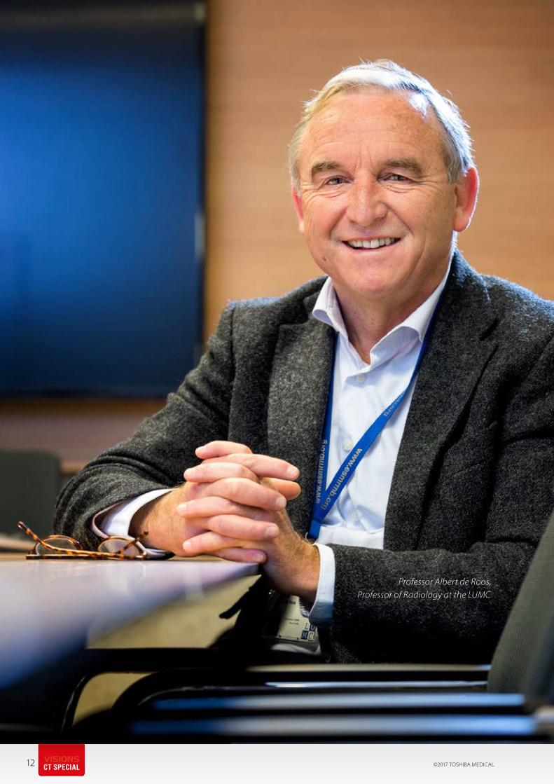
Professor Albert de Roos, Professor of Radiology at the LUMC
VISIONSCT SPECIAL
13
In 2000, LUMC decided to broaden its investment
in new CT technology. Its partnership with Toshiba
Medical began then.
“We were surprised and happy to see how Toshiba
Medical was connecting within Europe at that time,”
said Professor de Roos. “They were serious about col-
laborating with the LUMC and were very motivated. We
were able to establish a very personal relationship early
on, which has worked well throughout the years and
continues until today. We particularly appreciated the
consistently friendly and helpful attitude and strong
commitment of the Japanese- and European Toshiba
Medical management and staff. Factors that form the
basis of this excellent collaboration – The good ‘mar-
riage’ between Leiden and Toshiba Medical. We have
many relationships within our industry, but sometimes
these lack depth and meaning, this, however, is some-
thing completely different.”
“Due to this good relationship, we were always informed
about new developments in advance,” he continued.
“That’s quite unusual in the industry. It meant we could
always be prepared if something new was on the horizon.
Toshiba Medical was also keen for us to receive the new
equipment as early as possible. That was exciting and
stimulating and gave us a good feeling and confidence
that we were in the lead of new developments. We were
able to connect LUMC’s Radiology Department and our
Physics Group with Toshiba Medical’s Engineers in Japan:
an excellent interaction, well-prepared, with a smooth
introduction. Toshiba Medical is also always great steps
ahead with new systems and technologies.”
EVOLUTION AND REVOLUTIONLUMC first acquired a Toshiba Medical 4-slice CT scanner
in 2000, moving to 16-slice in (2003), 64-slice in (2005),
and the Aquilion ONE in the beginning of 2008. With
each new CT generation, LUMC’s Radiology Department
looked forward to the new possibilities that each succes-
sive system would bring for them and their patients.
“Every step was a major breakthrough and provided so
many new opportunities. We were always surprised by
the options provided with every new generation. Toshiba
Medical’s commitment to long-term CT development
makes a lot of difference and is recognizable in its prod-
ucts, which are always at the forefront of new technology,”
remarked Professor de Roos. “There was a particularly big
transition from 64-slice to Aquilion ONE. The volume
scanning offered by the Aquilion ONE was truly new
in the field of CT. It was a surprising leap forward to be
able to scan a whole volume in one rotation. Volume CT,
which remains quite unique to Toshiba Medical, has some
remarkable applications.
The introduction of each new system at the LUMC was
successful, and the installation of the Aquilion ONE was
no exception.
“The introduction of the Aquilion ONE, with its new pos-
sibilities to collect functional, as well as morphological
data into clinical practice, went smoothly, and it worked!”
exclaimed Professor de Roos. “We didn’t need to start
from scratch, and we could develop some entirely new
applications and protocols within a short space of time,
because the CT was always working properly. We are still
finding new ways to build on them. There is always the
challenge to do more and better, but this is how new rou-
tine applications have emerged, for example, in cardiac
scanning. The combination of Aquilion ONE’s volume CT
and the fast acquisition is a real advantage.”
VOLUME SCANNING BREAKTHROUGHVolume scanning with the Aquilion ONE enabled coro-
nary imaging in cardiac applications. It is now a routine
tool at LUMC and is even used in pediatric applications
and congenital heart disease patients.
“Recent trials have shown that coronary CTA can replace
coronary angiography1,” said Professor de Roos. “In the
early days, cardiologists laughed at the concept of cardiac
CT. They compared radiologists with Monty Python and
Equipped to takeon any challenge
INTERVIEW
Leiden University Medical Center (LUMC) in Leiden, the Netherlands, is focused on high quality research, education and patient care with a strong scientific orientation. It was one of the very first hospitals in Europe to welcome an Aquilion ONE™ CT system. Professor Albert de Roos, Professor of Radiology at the LUMC explained how the scanner and partnership with Toshiba Medical has opened many doors, not only to new techniques and clinical possibilities, but also to important collaborations with other research experts across the world.
CTEU160121
Henk de Vries, Senior Product Manager Computed Tomography
14 ©2017 TOSHIBA MEDICAL
the Holy Grail! They envisaged us wandering through the
desert, searching for a tool to image the coronary arteries,
and they laughed and said: “These radiologists are crazy:
it will never happen, every option has already been tried
over many years!”…. Nowadays, it’s standard. That shows
how these things evolve. There’s always the same pattern
that happens in development. Many leading professors
are skeptical of new technology at first. You need to have
great vision and accept the challenge to develop these
techniques to a higher level. It’s then a pattern of accept-
ance by early adapters and then the wider audience.”
OPENING DOORS TO MULTICENTER STUDIESWith the Aquilion ONE, LUMC were able to join multi-
center trials – international research collaborations
between institutes located across the world.
“Toshiba Medical showed great foresight in starting
scientific research initiatives alongside to commercial
activities to evaluate and promote new CT technologies,”
he said. LUMC was able to work on multicenter trials
between Europe, Japan and the US, and that started a
significant collaboration: the so-called ‘CORE-64 Project2’;
and later the ‘CORE-320 Project3’. The projects were very
successful and the results were published widely. Toshiba
Medical unusually and successfully combines European,
Japanese and American standards. The projects also
created a sort of ‘Toshiba Family’ between the Aquilion
ONE users in different global locations.”
SETTING NEW STANDARDS IN DOSE REDUCTIONAt the time of introduction of the Aquilion ONE, a major
focus was on dose reduction in the CT industry. Dose
reduction is very important for general health issues,
for imaging wisely, particularly for example in using low
doses for imaging children.
“Toshiba Medical has been in the forefront of dose reduc-
tion techniques, but I think the issue is now becoming
less and less relevant, because low-dose has become
standard,” added Professor de Roos. “With doses becom-
ing so low, I believe that standard radiography appli-
cations will be taken by CT in the future. For example,
routine chest films and other applications will maybe
increasingly be performed as low-dose CT applications.
I think that CT technologies also provide new opportuni-
ties for new ways of image analysis. So that a computer
with a self-learning algorithm will actually go through the
data sets by CT and pick out the abnormalities. Maybe
we can achieve this with automated techniques more
efficiently than with the eye of a physician? I think this
might fit with the CT modality especially well. We will see
how these things develop.
NEXT STEPS IN CARDIAC CTAProfessor de Roos shares Toshiba Medical’s commitment
to continual progress.
“It never stops. At every step, people always think that:
“This is the top of the hill!”…but after every hill, there is
a new hill,” he concluded. “There will certainly be other
new applications and possibly new detectors introduced,
such as Photon Counting detectors. Perhaps, people will
integrate CT technology with other modalities, because
CT currently has some limitations. Maybe we will see CT
and MR or some other modality combined to leverage
the strengths of each. People always want to have one
modality that fits all, but mostly that carries some limita-
tions. Will have to wait and see how these things might
improve…So onwards and upwards to the next hill!
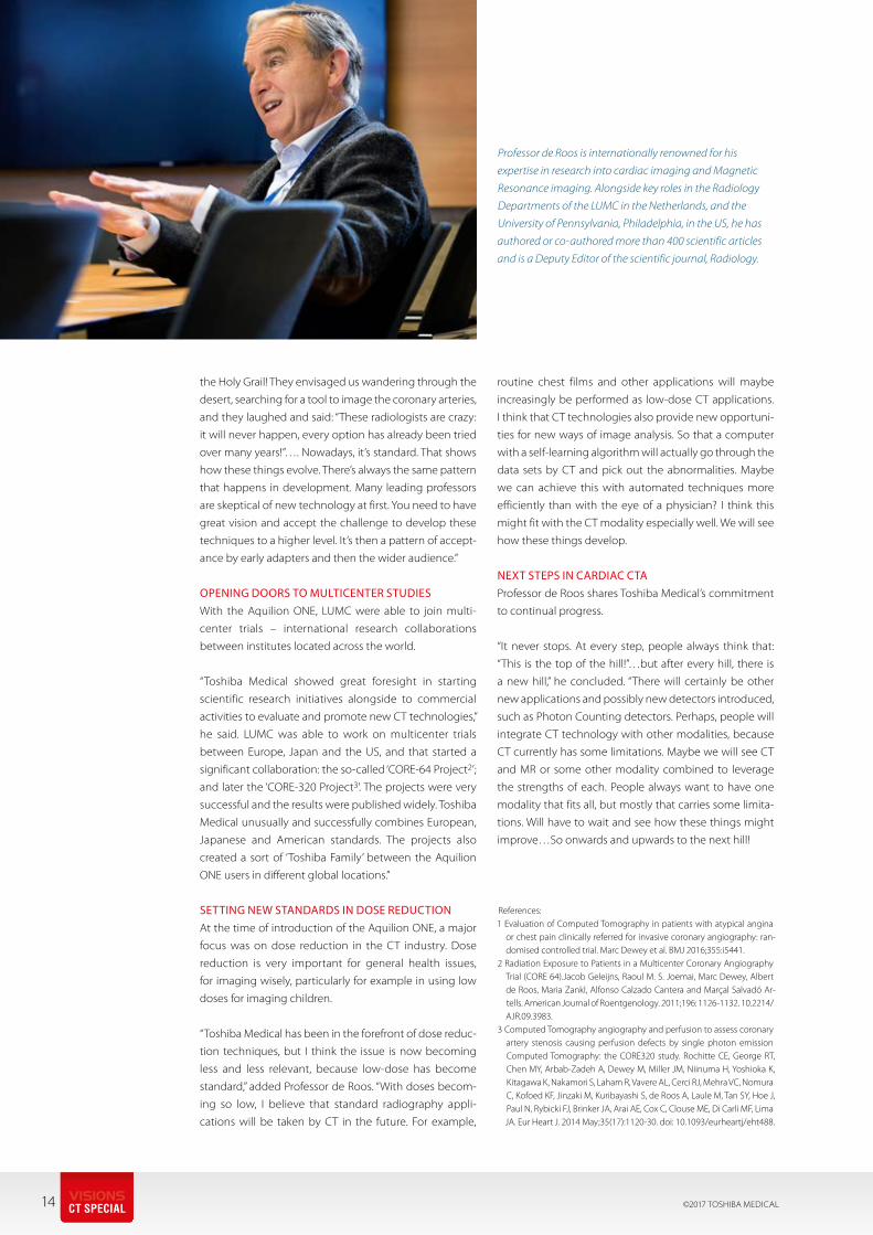
Professor de Roos is internationally renowned for his
expertise in research into cardiac imaging and Magnetic
Resonance imaging. Alongside key roles in the Radiology
Departments of the LUMC in the Netherlands, and the
University of Pennsylvania, Philadelphia, in the US, he has
authored or co-authored more than 400 scientific articles
and is a Deputy Editor of the scientific journal, Radiology.
References:1 Evaluation of Computed Tomography in patients with atypical angina
or chest pain clinically referred for invasive coronary angiography: ran-domised controlled trial. Marc Dewey et al. BMJ 2016;355:i5441.
2 Radiation Exposure to Patients in a Multicenter Coronary Angiography Trial (CORE 64).Jacob Geleijns, Raoul M. S. Joemai, Marc Dewey, Albert de Roos, Maria Zankl, Alfonso Calzado Cantera and Marçal Salvadó Ar-tells. American Journal of Roentgenology. 2011;196: 1126-1132. 10.2214/AJR.09.3983.
3 Computed Tomography angiography and perfusion to assess coronary artery stenosis causing perfusion defects by single photon emission Computed Tomography: the CORE320 study. Rochitte CE, George RT, Chen MY, Arbab-Zadeh A, Dewey M, Miller JM, Niinuma H, Yoshioka K, Kitagawa K, Nakamori S, Laham R, Vavere AL, Cerci RJ, Mehra VC, Nomura C, Kofoed KF, Jinzaki M, Kuribayashi S, de Roos A, Laule M, Tan SY, Hoe J, Paul N, Rybicki FJ, Brinker JA, Arai AE, Cox C, Clouse ME, Di Carli MF, Lima JA. Eur Heart J. 2014 May;35(17):1120-30. doi: 10.1093/eurheartj/eht488.
VISIONSCT SPECIAL
PUBLICATIONS2007/2008
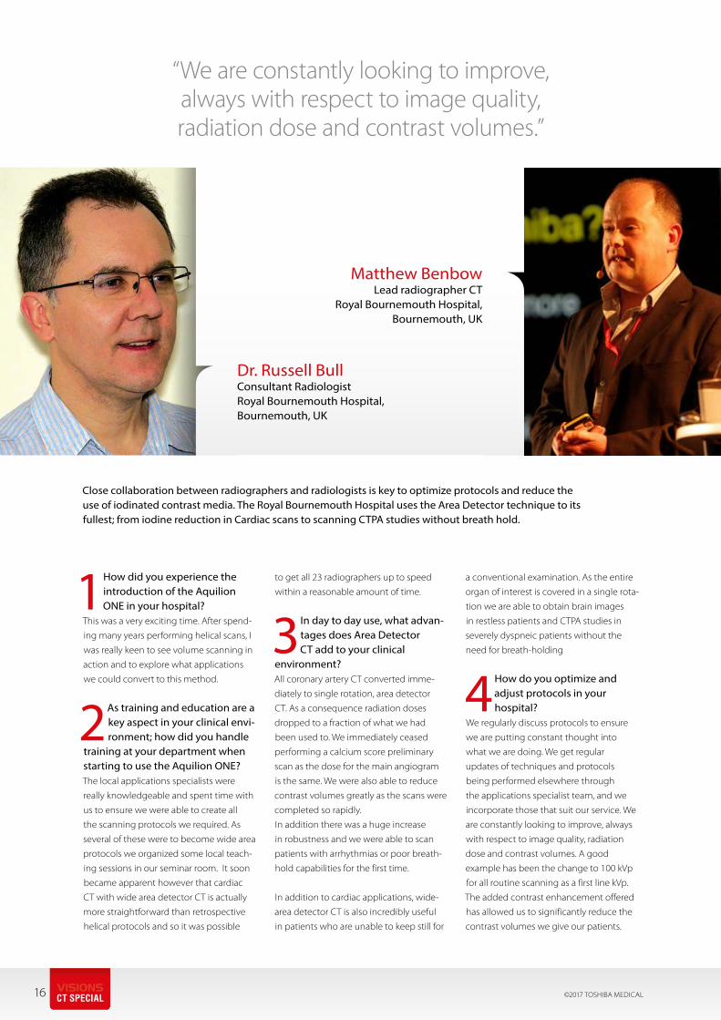
“We are constantly looking to improve, always with respect to image quality, radiation dose and contrast volumes.”
16 ©2017 TOSHIBA MEDICAL
a conventional examination. As the entire
organ of interest is covered in a single rota-
tion we are able to obtain brain images
in restless patients and CTPA studies in
severely dyspneic patients without the
need for breath-holding
4 How do you optimize and adjust protocols in your hospital?
We regularly discuss protocols to ensure
we are putting constant thought into
what we are doing. We get regular
updates of techniques and protocols
being performed elsewhere through
the applications specialist team, and we
incorporate those that suit our service. We
are constantly looking to improve, always
with respect to image quality, radiation
dose and contrast volumes. A good
example has been the change to 100 kVp
for all routine scanning as a first line kVp.
The added contrast enhancement offered
has allowed us to significantly reduce the
contrast volumes we give our patients.
1How did you experience the introduction of the Aquilion ONE in your hospital?
This was a very exciting time. After spend-
ing many years performing helical scans, I
was really keen to see volume scanning in
action and to explore what applications
we could convert to this method.
2 As training and education are a key aspect in your clinical envi-ronment; how did you handle
training at your department when starting to use the Aquilion ONE?The local applications specialists were
really knowledgeable and spent time with
us to ensure we were able to create all
the scanning protocols we required. As
several of these were to become wide area
protocols we organized some local teach-
ing sessions in our seminar room. It soon
became apparent however that cardiac
CT with wide area detector CT is actually
more straightforward than retrospective
helical protocols and so it was possible
to get all 23 radiographers up to speed
within a reasonable amount of time.
3 In day to day use, what advan-tages does Area Detector CT add to your clinical
environment?All coronary artery CT converted imme-
diately to single rotation, area detector
CT. As a consequence radiation doses
dropped to a fraction of what we had
been used to. We immediately ceased
performing a calcium score preliminary
scan as the dose for the main angiogram
is the same. We were also able to reduce
contrast volumes greatly as the scans were
completed so rapidly.
In addition there was a huge increase
in robustness and we were able to scan
patients with arrhythmias or poor breath-
hold capabilities for the first time.
In addition to cardiac applications, wide-
area detector CT is also incredibly useful
in patients who are unable to keep still for
Dr. Russell BullConsultant RadiologistRoyal Bournemouth Hospital, Bournemouth, UK
Matthew BenbowLead radiographer CT
Royal Bournemouth Hospital, Bournemouth, UK
Close collaboration between radiographers and radiologists is key to optimize protocols and reduce the use of iodinated contrast media. The Royal Bournemouth Hospital uses the Area Detector technique to its fullest; from iodine reduction in Cardiac scans to scanning CTPA studies without breath hold.
VISIONSCT SPECIAL
17CTEU160122
5 How did you experience the evolution of Aquilion ONE in the last 10 years? From both
software as well as hardware point of view?Reliability of our Aquilion scanners has
always been superb, and this has persisted
as changes have been made. Tube
outputs were improved with the Aquilion
ONE™ ViSION Edition, such that high mA
scanning has been possible routinely.
This, in conjuction with greatly improved
detector efficiency has enabled lower
kVps to be used, improving the effect of
IV contrast and allowing us to reduce the
dose. Rotation times have shortened and
as such this improves the temporal resolu-
tion achievable for all studies, but par-
ticularly for coronary artery CT. Radiation
doses for coronary CT are now so low that
we are able to use this technology even in
very young patients. Other options such as
lateral table movement, ultrahelical scan
mode and variable helical pitch have all
allowed more challenging examinations
to be achieved with good image quality.
Iterative reconstruction (AIDR 3D) has
reduced doses whilst maintaining image
quality and metal artifact reduction
(SEMAR) has become available in both
volume and helical modes which not only
massively improves orthopedic studies,
but the soft tissues around implants, such
as enabling good visualization of the blad-
der when hip prosteheses are present.
6 Thinking out loud; what is your vision when it comes to the future of CT for the com-
ing years?Firstly, wide area CT has to be the single
biggest benefit that all scanners should
aspire to. Whilst much of our general
work is performed in helical mode , and
always will be, for specialist examinations
such as cardiac CT, the benefits of a wide,
ultra-efficient detector in terms of image
quality and patient dose are considerable
and our vision would be that all large
volume cardiac centres will use this
type of technology in the future. Further
improvements in detector technology,
reconstruction systems and high output
tubes will reduce radiation doses still
further, allowing lower kVps to become
the norm. With this comes the benefit of
really being able to utilise extremely low
volumes of contrast media and as such I
believe that syringe injectors will become
inappropriate for use, and continuous
delivery systems will become exclusively
purchased.
I would predict that within the next 10
years there will be major advances in the
spatial resolution of CT due to a combina-
tion of ultra-high-resolution detectors
and advanced model based iterative
reconstruction. This will allow CT to equal
or even surpass the resolution of catheter
angiography. This, in combination with
functional assessment of lesion severity
based on advanced fluid dynamic model-
ling will allow CT to become the standard
diagnostic test for all cases of suspected
coronary artery disease thus allowing car-
diac cath labs to concentrate exclusively
on interventional procedures.
For non-coronary applications, CT doses
will be down to such low levels that there
will be very little justification for perform-
ing conventional plain films for many
indications. My prediction is that in 10
years time an average mid-sized general
hospital will have perhaps 2 plain film
rooms and 8 ultra-low dose CT scanners.
The challenge for the imaging depart-
ments of the future will be to report all
these studies in a timely manner and we
are likely to need help from advanced
visualisation and processing techniques
in conjunction with computer-aided
diagnosis.
18 ©2017 TOSHIBA MEDICAL
Lung SURESubtraction in Everyday Practice
Post contrast iodine maps were introduced as part of dual-energy imaging over 10 years ago but
these have never become part of routine practice in most centers for the investigation of pulmonary
thromboembolic disease. As dual energy imaging utilizes the post contrast difference in attenuation of
iodine between 2 separate kVp images, the amount of signal generated is rather small meaning that noise
levels have to be low in order to generate images with sufficient signal to noise ratio.
Dr R. Bull, Royal Bournemouth Hospital, UK
CLINICAL CASE ABSTRACT
CTPA, Lungs, Subtraction
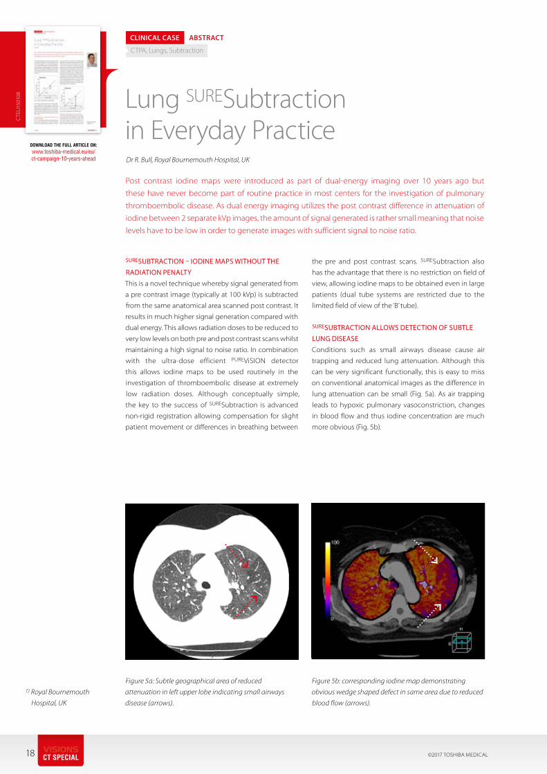
SURESUBTRACTION – IODINE MAPS WITHOUT THE RADIATION PENALTYThis is a novel technique whereby signal generated from
a pre contrast image (typically at 100 kVp) is subtracted
from the same anatomical area scanned post contrast. It
results in much higher signal generation compared with
dual energy. This allows radiation doses to be reduced to
very low levels on both pre and post contrast scans whilst
maintaining a high signal to noise ratio. In combination
with the ultra-dose efficient PUREViSION detector
this allows iodine maps to be used routinely in the
investigation of thromboembolic disease at extremely
low radiation doses. Although conceptually simple,
the key to the success of SURESubtraction is advanced
non-rigid registration allowing compensation for slight
patient movement or differences in breathing between
the pre and post contrast scans. SURESubtraction also
has the advantage that there is no restriction on field of
view, allowing iodine maps to be obtained even in large
patients (dual tube systems are restricted due to the
limited field of view of the ‘B’ tube).
SURESUBTRACTION ALLOWS DETECTION OF SUBTLE LUNG DISEASEConditions such as small airways disease cause air
trapping and reduced lung attenuation. Although this
can be very significant functionally, this is easy to miss
on conventional anatomical images as the difference in
lung attenuation can be small (Fig. 5a). As air trapping
leads to hypoxic pulmonary vasoconstriction, changes
in blood flow and thus iodine concentration are much
more obvious (Fig. 5b).
DOWNLOAD THE FULL ARTICLE ON:
www.toshiba-medical.eu/eu/ ct-campaign-10-years-ahead
CTE
U15
0108
Figure 5a: Subtle geographical area of reduced
attenuation in left upper lobe indicating small airways
disease (arrows).
Figure 5b: corresponding iodine map demonstrating
obvious wedge shaped defect in same area due to reduced
blood flow (arrows).
1) Royal Bournemouth
Hospital, UK
VISIONSCT SPECIAL
CT WORKSHOPS2017
Please visit: www.toshiba-medical.eu/eu/education
for more information.
20 ©2017 TOSHIBA MEDICALVISIONSCT SPECIAL
21
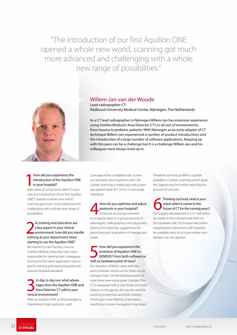
“The introduction of our first Aquilion ONE opened a whole new world, scanning got much
more advanced and challenging with a whole new range of possibilities.”
22 ©2017 TOSHIBA MEDICALVISIONSCT SPECIAL
Therefore scanning at 80kV is quicker
available in Cardiac scanning which gives
the opportunity for further reducing the
amount of contrast.
6 Thinking out loud; what is your vision when it comes to the future of CT for the coming years?
The biggest developments in CT will rather
be made on the software side then on
the hardware side. Techniques like iodine
mapping and subtraction will hopefully
be available soon for al scans where mul-
tiphase scan are applied.
coverage of the complete brain, is now
our standard care in patients with CVA.
Cardiac scanning is made easy with a aver-
age patient dose of 1-2mSv in one single
heartbeat.
4 How do you optimise and adjust protocols in your hospital?Protocols are being reviewed
on a regular base in a group existing of
radiologists, radiographers and physicians.
During this meetings suggestions for
optimizing and evaluation of changes are
made.
5 How did you experience the evolution of Aquilion ONE to GENESIS? From both software as
well as hardware point of view?Our Aquilion GENESIS came with the
same software version as the Vision so no
changes there. On the hardware point of
view there were some great changes. The
CT is equipped with a view finder and start
button on the gantry, this can be used for
scanning extremities and little children.
There’s also more filtering of the beam,
resulting in a more homogene X-ray beam.
1 How did you experience the introduction of the Aquilion ONE in your hospital?
After years of using some older CT scan-
ners the introduction of our first Aquilion
ONE™ opened a whole new world,
scanning got much more advanced and
challenging with a whole new range of
possibilities.
2 As training and education are a key aspect in your clinical environment, how did you handle
training at your department when starting to use the Aquilion ONE?We started to train five key-users at
Toshiba Medical, these key-users were
responsible for training their colleagues.
During the first week application was on
site for training and improving protocols
towards hospital standards.
3 In day to day use, what advan-tages does the Aquilion ONE and Area Detector CT add to your
clinical environment?With an Aquilion ONE on the Emergency
Department brain perfusion, with
As a CT lead radiographer in Nijmegen Willem-Jan has extensive experience using Toshiba Medical's Area Detector CT's in all sort of environments; from trauma to pediatric patients. With Nijmegen as an early adapter of CT technique Willem-Jan experienced a number of product introductions and the introduction of a large number of software applications. Keeping up with this pace can be a challenge but it is a challenge Willem-Jan and his colleagues have always lived up to.
Willem-Jan van der WoudeLead radiographer CTRadboud University Medical Center, Nijmegen, The Netherlands
CTEU160123

GENESIS Edition – Transforming CTBuilding on over 10 years of clinical experience in Area Detector Technology, Aquilion ONE GENESIS sets a new standard in delivering higher quality CT examinations for superior diagnostic confidence in a patient-centric and cost-conscious design.
www.toshiba-medical.eu
MAD
CT0
001E
UC
10 YEARS ADVERTISING
2008
2008
2010
2012
2007
2009
2017
24 ©2017 TOSHIBA MEDICAL
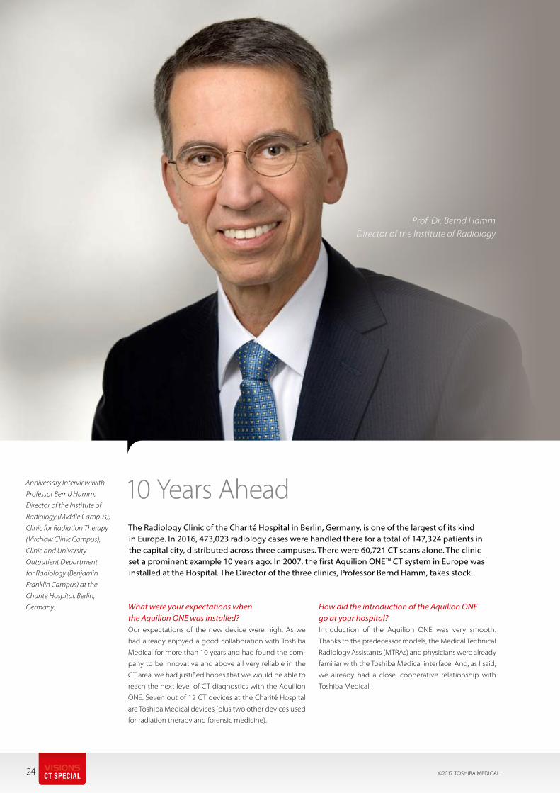
Prof. Dr. Bernd HammDirector of the Institute of Radiology
VISIONSCT SPECIAL
10 Years Ahead The Radiology Clinic of the Charité Hospital in Berlin, Germany, is one of the largest of its kind in Europe. In 2016, 473,023 radiology cases were handled there for a total of 147,324 patients in the capital city, distributed across three campuses. There were 60,721 CT scans alone. The clinic set a prominent example 10 years ago: In 2007, the first Aquilion ONE™ CT system in Europe was installed at the Hospital. The Director of the three clinics, Professor Bernd Hamm, takes stock.
Anniversary Interview with
Professor Bernd Hamm,
Director of the Institute of
Radiology (Middle Campus),
Clinic for Radiation Therapy
(Virchow Clinic Campus),
Clinic and University
Outpatient Department
for Radiology (Benjamin
Franklin Campus) at the
Charité Hospital, Berlin,
Germany. What were your expectations when the Aquilion ONE was installed? Our expectations of the new device were high. As we
had already enjoyed a good collaboration with Toshiba
Medical for more than 10 years and had found the com-
pany to be innovative and above all very reliable in the
CT area, we had justified hopes that we would be able to
reach the next level of CT diagnostics with the Aquilion
ONE. Seven out of 12 CT devices at the Charité Hospital
are Toshiba Medical devices (plus two other devices used
for radiation therapy and forensic medicine).
How did the introduction of the Aquilion ONE go at your hospital?Introduction of the Aquilion ONE was very smooth.
Thanks to the predecessor models, the Medical Technical
Radiology Assistants (MTRAs) and physicians were already
familiar with the Toshiba Medical interface. And, as I said,
we already had a close, cooperative relationship with
Toshiba Medical.

25CTEU160124
Why was the Aquilion ONE selected? Other than the fact that the CT was advertised through-
out Europe, there was a functional and emotional reason
for this. At the functional level, the convincing aspect was
that the Aquilion ONE allows you to collect a combina-
tion of morphological and functional data, which was not
possible before. These data include those for cerebral per-
fusion, myocardial function, and myocardial perfusion,
as well as cardiac valve function, to some extent. In the
meantime, we've introduced pancreatic perfusion in low-
dose technology. Today, the Aquilion ONE is being used
in all areas, including for full organ- and tumor perfusion.
What was the emotional reason?The emotional aspect was equally important. We had
amassed very good experiences with Toshiba Medical
over the years. We were ready to take the next step
together with the use and evaluation of the Aquilion
ONE. There was no reason to change manufacturers.
How did installing the Aquilion ONE change clinical practice?Installation of the device clearly expanded the range of
indications. Or to put it in other words: with the Aquilion
ONE there are fewer contraindications. The CT enables
diagnostics to be performed on a much larger patient
population. For example, in the past, patients with
arrhythmias were hardly ever considered for CT diagnos-
tics. Due to the arrhythmias, the diagnostic informative
value of the images was greatly reduced. Today, such
examinations are stable and provide much better diag-
nostic information, thanks to 'Single Beat Cardio Imaging'.
We even achieve good results with the Aquilion ONE
for obese patients. We can increase the tube current –
thereby, accepting a higher dose. The diagnostic informa-
tion is persuasive and trend-setting for future therapy.
In what areas did the Aquilion ONE bring about new developments?Dual-energy technology in diagnosing gout is one new
indication, for example. With this technology, two images
are taken of the joint with two different X-Ray radiation
energies. Comparing these images allows you to see if a
deposit is made of calcium- or urate crystals. Dual-energy
technology makes it easier to demarcate inflammatory-
or degenerative changes of gout-specific deposits. There
are also major advances in stroke diagnostics. Volume
CT makes it possible to take 3D perfusion images of
the entire brain. The decision as to whether the patient
is eligible for intravascular therapy or not can be made
much faster.
When the Aquilion ONE was introduced, there was a large 'dose discussion' going on in the CT arena. How did you tackle the issue of optimizing protocols in your clinical routine? Actually, there was an intense dose competition going
on at that time. The Aquilion ONE had the advantage of
allowing flexible optimization of protocols. For example,
certain problems can be assessed with a low dose and
image noise. The examiner minimizes the dose with a
dedicated design of the examination protocol. The algo-
rithm of the adaptive iterative dose reduction also results
in a significant dose reduction. The Aquilion ONE is an
excellent tool for reducing the radiation dose.
What impact have the Aquilion ONE and flat-panel detector CT (with a 16 cm detector) had on patient management?The frequency with which patients are examined has
not increased significantly due to the introduction of the
Aquilion ONE. That's because we already had Multislice
CTs and a high throughput before. Furthermore, the
actual CT exam is not the limiting factor at a maximum-
care facility. Instead, the limiting factor is much more the
placement and positioning of bed-ridden and intensive-
care patients. The Aquilion ONE makes it possible to
achieve good diagnostic results with stable quality for a
larger patient population.
Which hardware and/or software advances have you witnessed since 2007? It may sound like a small thing, but the rotation time
reduction from 350 milliseconds to 275 milliseconds
really pays off. We now have significantly improved image
quality in cardiac diagnostics in particular. The AIDR 3D
software has also proven itself in terms of iterative image
reconstruction. The variable tube output is also an advan-
tage, because we can increase it for obese patients. And
vice versa, we can better use the contrast effect in the
low kilowatt range – and we can decrease the amount of
contrast medium for patients with renal failure.
How would you describe the relationship between Charité and Toshiba Medical?Allow me to give you a brief retrospective on that: I admit
that I was skeptical when the decision was first made to
install a Toshiba Medical device. We didn't know the com-
pany very well. We weren't sure whether a development
department in ‘far-away’ Japan would be to our advan-
tage. However, over the years, the reliability of Toshiba
Medical’s employees has been exemplary. I personally
value the fact that a promise is an absolute promise in
INTERVIEW
26 ©2017 TOSHIBA MEDICALVISIONSCT SPECIAL
Japanese culture. Scheduled updates, for example,
have always been installed by the agreed-upon date at
the latest. And Toshiba Medical's employees continue
to impress me. They are technically oriented and less
interested in marketing. They are Medical Engineers, not
Sales people.
Which recent improvements are important to you?The development of increasingly customized imaging is
an advantage. This includes saving on contrast medium,
thanks to low tube output, depending upon the individ-
ual situation. Or reducing the radiation exposure thanks
to the iterative reconstruction and customized exam pro-
tocol design. A reduced dose and a decreased contrast
medium amount have become a reality. Measuring the
intra-arterial blood flow and the myocardial perfusion are
future technical advances that are currently still in the
clinical investigation stage.
Which trends are you currently seeing in Radiology?The current trends are Big Data and Artificial Intelligence.
It's very exciting. Of course, innovations also have the
power to frighten people, but there's no reason for this.
As even though Artificial Intelligence will certainly make
its way into the Radiology field, it will remain an aid, as
opposed to becoming competition. You can draw a com-
parison with civil aviation: the autopilot feature is highly
valued. But no one would take a flight if the plane were
being piloted only by the autopilot without an actual
cockpit crew. Artificial Intelligence will enrich radiology
but won't replace the competence of Radiologists.
What does the future of CT looklike in your opinion?CT could develop in parallel with the self-driving car in
the future. The CT 'accelerates, brakes and parks', just like
a self-driving car. Depending on the medical question
and the patient data, CT will optimize the examination
protocol in all relevant aspects.
Once the device has calculated the best possible result
from all the data, it will give the Radiologist a recommen-
dation for an optimized individual protocol. There will be
fewer standardized examination protocols the way these
are usual and justified today. Software programs that flex-
ibly align the examination protocol with the individual
case in the future would further increase the quality of
the radiological findings.
27
1 What where your expectations at the time of installation of Aquilion ONE?
Our expectation were high with the hope
of doing better and more complex imaging.
We knew upgrading from a 4 slice to a 320
slice scanner would be a learning curve.
2 How did you experience the introduction of the Aquilion ONE in your hospital?
The installation was very well managed
with minimal disruption to clinical services.
3How has the installation of the Aquilion ONE changed your daily clinical practice?
CT has become a more versatile tool and
as a result usage has increased.
4 What has been the influence of the Aquilion ONE and Area Detector CT in patient
management?The main influence has been in better
neurovascular imaging.
5In a recent publication The Walton Centre presented two cases where an intranidal
aneurysm was demonstrated on four-dimensional CT angiography (4D-CTA). Where do you see 4D-CTA in the detection and management of AVM’s?4D-CTA is likely to have a significant
impact on charcterisation of AVM with the
aim of using 4D-CTA to replace diagnostic
catheter angiography in relevant cases. We
have also worked to significantly reduce
doses in 4D-CTA as published recently in
American Journal of Neuroradiology.
6 In the American Journal of Roentgenology The Walton Centre has published an article
describing a method for Cerebral CTV scanning using a low volume of con-trast agent, how has the Area Detector Technique supported the use of this method and the reduction of contrast agent used?We have utilized the faster scanning tech-
niques to develop new CT Venography
technique. The 16 cm detector allowing
whole head imaging in one rotation has
allowed us to develop better venous
phase imaging for CTV with added
advantage of lower contrast dose. We
also published a similar technique for CT
Angiography in Clinical Radiology Journal
– again with better arterial phase with
reduced contrast dose.
7 Combined CTA/CTP data can provide visualization of dynamic flow and perfusion. Area
Detector CT allows for whole-brain perfusion, what role do you see for whole brain perfusion other than in stroke imaging?We have used whole brain perfusion in
determining cerebrovascular reserve in
patients with haemodynamic transient
ischaemic attacks to help determine selec-
tion for arterial bypass. We have also used
it to assess vasospasm in post- aneurysm
coiling patients.
8 How has the introduction of Single Energy Metal Artifact Reduction (SEMAR) impacted
diagnostic image quality to your opinion?It has had provided much quality imaging
in both patients with aneusym clips and
spinal instrumentation.
9 Thinking out loud; what is your vision when it comes to the future of CT for the
coming years?CT provides specific contrast detail not
achievable by other imaging modality and
I expect its use will continue to increase
and diversify. With improvements in dose
reduction, it may replace many of the
of plain radiography techniques such as
chest X-rays.
CTEU160125
Dr. K. Das Consultant Neuroradiologist
Yvonne ShanksRadiology Manager
“The 16 cm detector allowing whole head imaging in one rotation has allowed us to develop better
venous phase imaging for CTV with added advantage of lower contrast dose.”
The Walton Centre is the only NHS Trust in the UK dedicated to Neuroscience. The group at the Walton Centre has published in renowned journals such as the AJR and they use their Area Detector CT to the fullest in the challenging patients they see in daily clinical practice. Please find below their view on the role of Area Detector CT in Neuroradiology.
The Bristol groupBristol Royal Hospital for Children, Bristol, UK
“We are able to scan many patients we wouldn’t have attempted
previously with out anaesthesia.”
The Bristol Royal Hospital for Children (BRHC) provides a local service for Bristol children and a referral service for specialist care for families across the South West and nationally. Currently they have 11 full time and 2 part time Paediatric Radiographers and 5 consultant Paediatric Radiologists including one Professor.
28 ©2017 TOSHIBA MEDICAL
1What where your expectations at the time of installation of Area Detector CT’s?
Faster acquisition times, less table
movement therefore less sedation and
decreased risk of failed scans
2How did you experience the introduction of the Aquilion ONE / ViSION Edition in your hospital?
Very smooth. The Applications training and
support thereafter was second to none.
3How has the installation of the Aquilion ONE / ViSION Edition changed your daily clinical
practice?We are able to scan many patients we
wouldn’t have attempted previously with-
out anaesthesia.
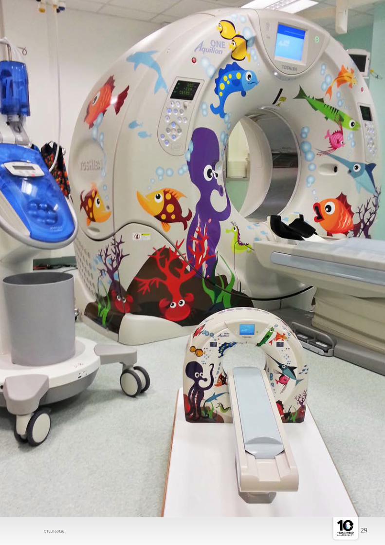
4How do you prepare your pedi-atric patients for a CT scan?
- Play Specialist Team
- Model scanner
- Breath hold practice using the iStation
console
- Demonstration- ride on the table, press
buttons to move table etc.
5To what extent can CT replace or add information in the visu-alization of airway disease in
pediatric patients?4D Dynamic Airway CT- allows dynamic
visualization of airway collapsibility
without having to inject contrast into the
bronchus. In addition the volumetric scan-
ning precludes mis-registration of the loca-
tion of any trachea-bronchomalacia and
allows full cine viewing of dynamic airway
collapse. By adding IV contrast we can see
vascular information and its relationship to
the airway.
6What has been the influence of the Area Detector CT in patient management?
We have found reduced requirements
for anaesthesia and less motion artifacts.
Furthermore we have improved image
quality in general at lower doses and
improved cardiac imaging at lower
doses. Due to the 4D Dynamic Airway
CT we undertake less bronchograms
examinations.
7Limiting the amount of Iodinated contrast used in a scan is key to reducing the risks of a CT scan.
An example is the split bolus tech-nique you are using in Bristol. How do you use this technique in your clinic?With faster acquisition times at 80-100kV
we have reduced our body contrast dosing
from 2mls/kg to 1.5mls/kg. For trauma
patients we use a biphasic contrast proto-
col that means the patient is only scanned
once for arterial and venous phases in one
scan. Our modified camp bastion protocol
uses a split bolus technique with a pause in
between boluses. This has resulted in better
biphasic contrast scans with less contrast
than the camp bastion wheel method.
8How do you handle the opti-mization of protocols and the optimization of dose in your
department?We have a very good working relationship
with our applications specialist who has
been instrumental in helping us tailor pro-
tocols to the requirements of our patients.
We started out by setting a baseline of
achieving doses within DRL and what we
were achieving previously. Currently we
are working towards optimizing paediatric
chest protocols. Using sure exposure 3D
we have been able to reduce dose incre-
mentally by small increases in standard
deviation without noticeable reduction in
image quality.
9 How has the introduction of the Aquilion ONE opened doors to new ways of imaging in your
department?It is ideal in our current status as a Level 1
paediatric major trauma centre.
We offer a new solution for imaging
tracheo-bronchomalacia for neonatal ICU
patients and for children with complex
cardiovascular abnormalities.
In our capacity as a tertiary referral centre
for orthopaedic limb lengthening and cor-
rection surgery as well as spinal scoliosis
surgery the SEMAR technology has greatly
improved our imaging of patients with
metallic hardware.
10 Thinking out loud; what is your vision when it comes to the future of CT for the
coming years?Considering the existing disfavour of CT for
paediatric practice in the United Kingdom
because of concerns regarding radiation
doses, the Aquilion One™ is serving to dem-
onstrate significant advantages of CT with
regards to speed (reducing the requirement
for anaesthesia), dynamic airway imaging
and artefact reduction when imaging
metallic hardware, with doses well within
acceptable levels for paediatric imaging.
VISIONSCT SPECIAL
CTEU160126 29
30 ©2017 TOSHIBA MEDICAL
DOWNLOAD THE FULL ARTICLE ON:
www.toshiba-medical.eu/eu/ ct-campaign-10-years-ahead
CTE
U16
0114 German Armed Forces and
Patients Benefit from New Options for Low-Dose Volume CT
The Radiologists at the Bundeswehr Central Hospital in Koblenz (BWZK), Germany, used the high-end
volume CT - Toshiba’s Aquilion ONE™ / ViSION Edition - for several months, along with the Vitrea Advanced
web-based image-processing software. The new, low-dose volume CT expanded clinical diagnostics and
provided added value for trauma- and routine CTs, as well as special examinations.
Dr. S. Waldeck1
CLINICAL CASE ABSTRACT
Dose, Metal artefact reduction
The Diagnostic and Interventional Radiology department comprises of a team of 19 doctors and 24 technicians who work under the direction of Head Doctor, Dr. Stephan Waldeck. The new Volume CT scanner is predominantly employed in the following areas: low dose scans of all body regions and organs (e.g. for diagnosis of accident victims and patients with multiple injuries); for complete diagnosis of heart and brain disease; and in angiography. It is also used for examinations of the face, paranasal sinuses, upper and lower jaw, temporal bones and dental CT. it is used in planning CT guided interventions, eg stenting of thoracic, abdominal and carotid arteries and neurological interventions; minimally invasive treatment of tumors as well as targeted pain therapies in the spine.
Dr. Waldeck and his Team are excited both by the new technology and the clinically advanced applications options. Examinations can now be performed with the Aquilion ONE / ViSION Edition, in which the balance between low dose and excellent image quality is standardized for all patients. The following examples show the added value that volume CT offers routine applications.
Case 1: Subarachnoid hemorrhage with hemorrhagic bifurcation aneurysm and acom aneurysm A comatose patient was brought to the emergency room
by emergency services. In a third-party anamnesis, the
husband reported sudden extremely intense headaches
and increasing disorientation.
The initial Brain CT in the emergency room detected a
massive subarachnoid hemorrhage with ventricular
rupture. The subsequently performed CTA revealed a large
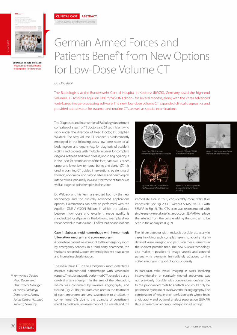
cerebral artery aneurysm in the area of the bifurcation,
which was confirmed by invasive angiography and
treated (Fig. 2). The platinum coils used in the treatment
of such aneurysms are very susceptible to artefacts in
conventional CTs due to the quantity of constituent
metal. In particular, an assessment of the vessels and the
immediate area, is thus, considerably more difficult or
impossible (see Fig. 2: CCT without SEMAR vs. CCT with
SEMAR in Fig. 2). The CTA scan was reconstructed with
single energy metal artefact reduction (SEMAR) to reduce
the artefact from the coils, enabling the contrast to be
seen in the aneurysm (Fig. 2).
The 16-cm detector width makes it possible, especially in
cases involving such complex issues, to acquire highly-
detailed vessel imaging and perfusion measurements in
the shortest possible time. The new SEMAR technology
also makes it possible to image vessels and cerebral
parenchyma elements immediately adjacent to the
coiled aneurysm in good diagnostic quality.
In particular, valid vessel imaging in cases involving
interventionally- or surgically treated aneurysms was
not previously possible with conventional devices due
to the pronounced metallic artefacts and could only be
performed by means of invasive catheter angiography. The
combination of whole-brain perfusion with whole-brain
angiography and optional artefact suppression (SEMAR),
thus, represents an enormous diagnostic advantage.
1) Army Head Doctor,
Head Doctor and
Department Manager
of the Viii Radiology
Department, Armed
Forces Central Hospital,
Koblenz, Germany
Figure 2a: A CTA of the brain demonstrating severe metallic artefact
Figure 2b: CTA with SEMAR shows reduced artefact
Figure 2d: 3D of the CTA demonstrat-ing the aneurysms foliwong coiling
Figure 2e: Catheter angiogram showing the aneurysm prior to coiling
Figure 2c: Coil placement during the interventional procedure
VISIONSCT SPECIAL
1 How did you experience the introduction of the Aquilion ONE in your hospital?
We were the second hospital in Europe to
get an Aquilion ONE. This scanner made
it possible to carry out a number of new
examinations and existing examinations will
be of better quality and scanned much more
quickly. This was illustrated by the fact that
the trauma radiologist (Head of Department)
was present at the receipt of a trauma
patient during the first few days and stated
that he had been in the profession for over
30 years and upon seeing this examination
he had a “WOW” moment. The Head of the
Cardiology Department was also present
during the first cardiological examination
carried out on the Aquilion ONE and his
reaction was “OK, from now on, we are car-
rying out every cardiological examination
using this scanner”. In short, the step from
64-slice to Aquilion ONE was more than a
step forward.
2 As training and education are a key aspect in your clinical environment, how did you handle
training at your department when starting to use the Aquilion ONE?We worked closely with the Toshiba Europe
application specialists in order to train a starter
group who then, in turn, trained the entire
team (teach the teacher). As we received one
of the first scanners, a great deal of discussion
also took place with Toshiba Europe regarding
the development of study protocols and
scanning techniques (use of bolus triggering,
dose modulation, image optimisation, etc.)
3 In day to day use, what advan-tages does the Aquilion ONE and Area Detector CT add to your
clinical environment?The major benefits are as follows: rapid
acquisition times in paediatric examinations
with lower doses, which means that seda-
tion is often no longer required and immobi-
lisation, with for example a vacuum cushion,
is sufficient. The possibility of carrying out
cerebral perfusion scans when receiving a
patient who has had a stroke, which have
also played a crucial role in the DUST trial
and the MR CLEAN trial, for example. Organ
perfusion, ECG-triggered aorta scans, etc.
The result was that suddenly, the preference
was to carry out a large number of examina-
tions on the Aquilion ONE; therefore patients
sometimes had to wait for one another.
4 How do you optimise and adjust protocols in your hospital?The scan protocols are managed in
our hospital by the CT expert, together with
a small group of senior radiographers and, in
terms of content, input is given by radiology
staff (indications and phases/series to be
scanned). The level of radiation exposure
is tested by senior radiographers, who are
supported by the group of clinical physicians.
Management of the protocols in the scanner
is carried out by the CT expert. Therefore, if a
protocol needs to be optimised or introduced,
then this is carried out by the aforementioned
people.
5 How did you experience the evolution of Aquilion ONE to GENESIS? From both software as
well as hardware point of view?In the last 10 years, a number of applications
have been developed further. These include
improvements in possibilities within the field
of cardiology (auto target, stress perfusion,
better arrhythmia rejection, etc.)
What is more, the introduction of AIDR 3D
resulted in a visible dose reduction. The
GENESIS now has a faster rotation time, a
higher tube potential, which means that
more examinations can be carried out at
100 kV and, in combination with the iterative
reconstructions and PUREViSION Optics, this
also offers more options for dose reduction.
Thanks to the area finder and the flared gan-
try design, it has become much faster and
easier to scan extremities (wrists/elbows).
And there are many more developments
that we could mention.
6 Thinking out loud; what is your vision when it comes to the future of CT for the coming years?
The latest techniques such as FIRST recon-
structions and SEMAR with helical scans
will be calculated much more quickly in
the future, which means that these tech-
niques can be used as standard or in acute
examinations. What is more, the number of
CT examinations will continue to increase,
including those carried out on children.
This means that we need to remain critical
of the level of radiation exposure and
continue to reduce this as much as possible
using new techniques and better hardware.
In older patients, it is important to reduce
the iodine dose as much as possible
through even faster acquisition times, bet-
ter iodine detection (by subtraction or dual
energy or (virtual) monochromatic imaging
techniques, for example) in order to limit
any kidney damage.
Joost RoelofsLead radiographer CTLUMC, Leiden, The Netherlands
“The step from 64-slice to Aquilion ONE was more
than a step forward. ”
As a lead radiographer Joost Roelofs has extensive, long-term experience in using Toshiba CT's including the Area Detector CT's. Starting with the Aquilion ONE™ and most recently the newly introduced Aquilion ONE™ GENESIS Edition.
CTEU160127 31
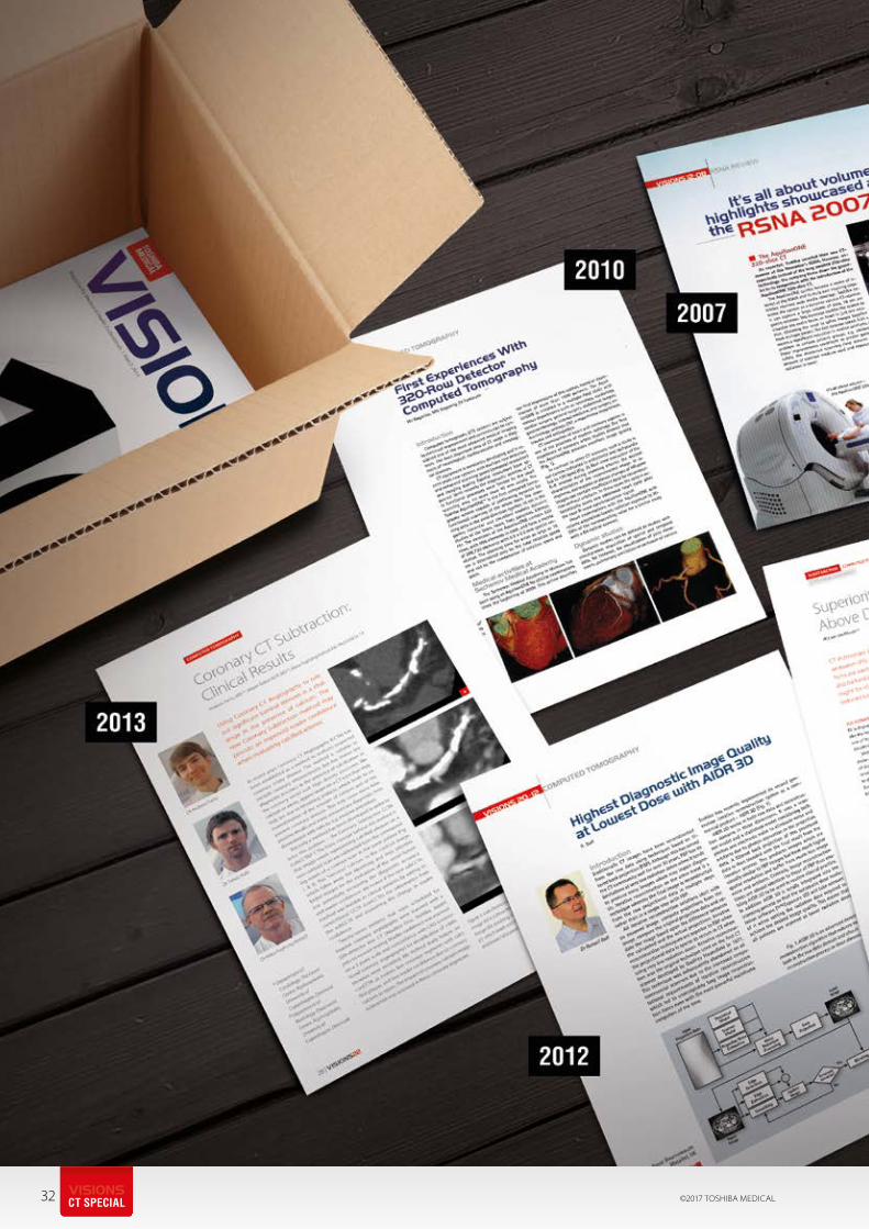
32 ©2017 TOSHIBA MEDICAL
Professor Albert de Roos, Professor of Radiology at the LUMC
VISIONSCT SPECIAL

33
Visit our CT Campaign webpagewww.toshiba-medical.eu/eu/ct-campaign-10-years-ahead and download these digital CT articles from VISIONS 2007 – 2017!
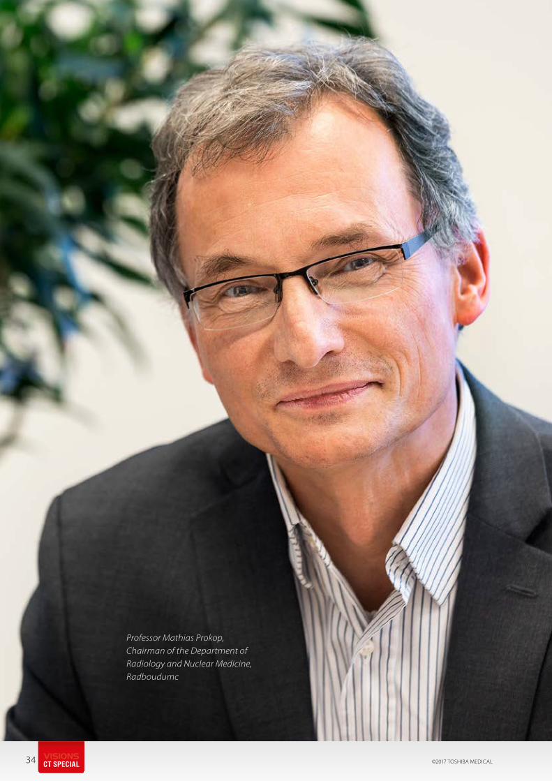
34 ©2017 TOSHIBA MEDICAL
Professor Mathias Prokop, Chairman of the Department of Radiology and Nuclear Medicine, Radboudumc
VISIONSCT SPECIAL
35
Radboudumc purchased an Aquilion ONE in 2011 to
advance research in functional imaging, recognizing the
importance of this field for the future.
SYNERGY“The market options available at the time meant that we
could choose for speed (Dual Source CT scanners) or large
coverage per rotation (Aquilion ONE). Functional imaging
was in its infancy, so we expected to be able to impact
the further development. We were looking for a partner
and had a good click with Toshiba Medical,” said Professor
Prokop. “Before making a final decision, we visited the
company’s engineers in Japan, because we wanted to
know how much they listened to users. I find that very
important, because the field only moves forward if you
get a good interaction between users and engineers.
What we found out was impressive, and convinced us
that working with Toshiba Medical was the way forward.”
Since then, the collaboration has steadily grown.
Radboudumc Imaging Research share their thoughts
about the future with the engineers in Japan and co-
develop in some areas. Regular meetings in Japan and
Nijmegen ensure the continuity of discussions about
ongoing projects and fine-tuning of the imaging systems
currently in use at the hospital.
RAPID INTRODUCTIONBefore installation of their new Aquilion ONE,
Radboudumc planned the positioning of the new sys-
tem strategically to ensure easy access and optimal flex-
ibility for use by the Emergency Rooms and poli-clinics
alike. Installation of the scanner went fast.
“We could use the system within a short period of time,”
said Professor Prokop. “Training went smoothly, although
the interface was substantially different to those of the
scanners that we had used before. Due to this, we first
trained small groups of radiographers and expanded it to
the rest, so that everyone felt comfortable.”
EXPLORING NEW POSSIBILITIESWith the introduction of the Aquilion ONE, it became
possible for the Radboudumc Imaging Research to col-
lect functional together with morphological data. While
employed for routine clinical practice, the Aquilion ONE
allowed to continually explore new possibilities.
“The development of any new techniques starts with a
vision that we initially evaluate. We see what is currently
missing, and then approach Toshiba Medical to discuss
what is needed to make things a reality. So far, this has
gone very well,” explained Professor Prokop. “Toshiba
Medical also implements our feedback on optimization
into new software. I have been amazed by the speed at
which the feedback is implemented."
The brain is a classic region for testing a lot of new
techniques in radiology. Brain perfusion imaging (CTP)
using the Aquilion ONE was introduced early on and is
used in addition to a pre-contrast CT and CTA for the
evaluation of stroke. “With full brain coverage provided
by Aquilion ONE, we can investigate stroke outside the
classical field around the basal ganglia,” said Professor
Prokop. “Perfusion with the Aquilion ONE gives us a
higher detection rate of vascular occlusions, even in
peripheral branches. Malperfused regions indicate
areas that we need to return to in order to look for
occlusions. CTP is easy to set up, works quite rapidly
and doesn’t delay our clinical process.”
"Our ultimate goal for CT imaging of stroke, however,
is to eliminate the CTA and pre-contrast scan and rely
on CTP only. A prerequisite is that the information from
CTA and pre-contrast CT can be extracted from a single
CTP sequence. In the past years we have developed
and optimized such techniques. As a result, we now
can reduce overall examination time, the amount of
iodinated contrast and the overall radiation dose."
From Molecule to Man to Population
INTERVIEW
The Radboud university medical centre (Radboudumc) in Nijmegen, the Netherlands, aims to have a significant impact on healthcare with research spanning ‘From Molecule to Man to Population’. With a team of more than 150 people , the Imaging Research group within the Department of Radiology and Nuclear Medicine develops imaging applications that bridge the gap between research and practice and help shape the future of healthcare. Professor Mathias Prokop, Chairman of the department, explains how the Aquilion ONE™ and close collaboration with Toshiba Medical’s engineers in Japan and Europe over the last five years continue to enable the team to advance techniques in functional imaging.
CTEU160127
Henk de Vries, Senior Product Manager Computed Tomography
36 ©2017 TOSHIBA MEDICAL
DEVELOPING NEW TECHNIQUESIn their quest for optimum use of CTP, Radboudumc
Imaging Research have been developing an interesting
technique for a subset of particularly vulnerable patients.
The One-Step-Stroke protocol, a single examination
sequence, in which the neck CTA is interleaved with CTP.
One-Step-Stroke imaging has the potential to replace a
separate CTA, which saves radiation dose and contrast
agent dose. It can reduce the amount of contrast required
by almost half (1, 2).
“The nice thing about this technique is that it doesn’t
affect the qualitative perfusion values,” said Professor
Prokop.“In addition, it acts as bolus triggering for CT per-
fusion: in a patient with delayed contrast arrival, a 60-sec-
ond acquisition can stop too early before the contrast
is optimally distributed. The One-Step-Stroke protocol
interrupts the standard CTP sequence for a wide volume
CTA of the neck and then returns to the brain, where a
fixed amount of scans (e.g. 20) is performed after the
contrast has arrived, which ensures constant CTP quality
and avoids truncation artifacts.”
“We are currently exploring combining the wide volume
CTA of the neck with subtraction; performing an initial
pre-contrast scan, followed by the One-Step-Stroke
technique, and finally subtracting in the neck region to
gain vessel-only CTA. This substantially reduces scatter
artifacts in the shoulder region,” he added.
OPPORTUNITIES TO ADVANCE EXISTING APPLICATIONS The Aquilion ONE as well as the Aquilion ONE ViSION
Edition and Aquilion ONE GENESIS Edition offer full flex-
ibility in planning scan protocols, which makes them
ideal for perfusion studies.
“You can chose continuous volumetric scanning for high
temporal resolution, which results in 4D data that over-
laps in the time domain,” said Professor Prokop. “We do
that in patients with arteriovenous (AV) malformations
to find the nidus, for example. You can also perform a set
of rapid volumetric scans in the early phase of enhance-
ment, followed by longer time delays between scans in
later phases, and you can also change the amount of
dose that you give for each of these phases.”
The flexibility of Toshiba’s area detector CTs makes it
possible to develop unique protocols that combine the
advantages of regular abdominal examinations with
those of CT perfusion. “A diagnostic scan interleaved with
a perfusion scan gives the best of two worlds - That’s pos-
sible on this scanner,” he added.
Transferring new techniques like these to clinicians
so that they understand the full implications of
opportunities can be challenging. For the best results,
Radboudumc pairs pre-clinical researchers with radiolo-
gist, who explore this together with their clinical partners.
Professor Prokop’s dream for the future is to use CT perfu-
sion for many of the indications that currently require a
multiphasic CT. However, he admits that many prerequi-
sites must be fulfilled before that will be possible.
“We have recently joined forces with Toshiba Medical’s
engineers to develop a very potent noise reduction algo-
rithm that enables high resolution CT perfusion imaging,
at a much higher resolution than we can achieve at the
moment. It also allows us to do excellent 4D analysis of
the vasculature in the region of interest. It is an essential
step forward,” he remarked. “We hope to see our noise
reduction algorithm introduced in mid-2017. It’s a very
powerful tool that we think will become a ‘game changer’.
When transferring this to other areas of the body, such
as the lungs or abdomen; motion correction becomes a
key factor. The Aquilion ONE scanners already have great
motion correction in the abdomen and lung, which is
good for subtraction imaging, but we are working to
improve it further to create a totally new image quality.”
SUBTRACTION “We are really happy with the subtraction technique for
the Aquilion ONE scanners. It’s very versatile. We have
used it routinely for pulmonary angiographs for more than
two years. In the lung, it helps us find perfusion defects,
which we then examine further; the enhancement of
nodules; and areas of infection or active inflammation,”
he said.“One of the big advantages SURESubtraction is that
you get a much better signal at a lower noise - around
40-50% less noise - than with Dual Energy CT scanning
techniques. We believe that subtraction will be very
important in the future.”
“I would, however, like to see even better image registra-
tion for the lung that enables not only the lung paren-
chyma, but also the vessels, to be registered perfectly or
almost perfectly,” he continued. “We can then use that
information for many different things, like being able to
reduce the amount of contrast administered by virtu-
ally increasing contrast enhancement - similar to virtual
monoenergetic images from Dual Energy CT. We can cre-
ate these images by adding the enhancement from the
subtraction image onto the original contrast-enhanced
images and combining this with noise reduction. Toshiba
Medical has already implemented the first step of this in
the abdomen and we hope to see it in other areas of the
body in the future.”
PIVOTAL IN THE FUTURE Over the next decades, Professor Prokop anticipates that
radiology, and medicine in general, will become far more
VISIONSCT SPECIAL
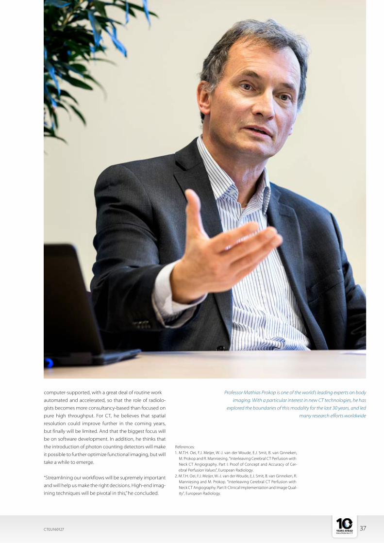
37
Professor Mathias Prokop is one of the world’s leading experts on body
imaging. With a particular interest in new CT technologies, he has
explored the boundaries of this modality for the last 30 years, and led
many research efforts worldwide
References:1. M.T.H. Oei, F.J. Meijer, W.-J. van der Woude, E.J. Smit, B. van Ginneken,
M. Prokop and R. Manniesing. "Interleaving Cerebral CT Perfusion with Neck CT Angiography. Part I: Proof of Concept and Accuracy of Cer- ebral Perfusion Values", European Radiology.
2. M.T.H. Oei, F.J. Meijer, W.-J. van der Woude, E.J. Smit, B. van Ginneken, R. Manniesing and M. Prokop. "Interleaving Cerebral CT Perfusion with Neck CT Angiography. Part II: Clinical Implementation and Image Qual- ity", European Radiology.
computer-supported, with a great deal of routine work
automated and accelerated, so that the role of radiolo-
gists becomes more consultancy-based than focused on
pure high throughput. For CT, he believes that spatial
resolution could improve further in the coming years,
but finally will be limited. And that the biggest focus will
be on software development. In addition, he thinks that
the introduction of photon counting detectors will make
it possible to further optimize functional imaging, but will
take a while to emerge.
“Streamlining our workflows will be supremely important
and will help us make the right decisions. High-end imag-
ining techniques will be pivotal in this,” he concluded.
CTEU160127
38 ©2017 TOSHIBA MEDICAL SYSTEMS
“We are now able to perform cardiacexaminations very easily, they are not time
consuming anymore.”
VISIONSCT SPECIAL
Prof. C. Roy and her team in Strasbourg have worked in many
different areas using Toshiba Medical's Area Detector tech-
nique. Ranging from the use of Wide Volume scanning for dose reduction, optimization of TAVI
scans and kidney perfusion.
Prof. C. RoyProfessor of Radiology
University Hospital Strasbourg, Strasbourg,
France
1What where your expectations at the time of installation of the Area Detector CT?
We expected to have a reliable partner for
clinical routine. Furthermore we expected
to have robust and convenient material to
work with.
2 How did you experience the introduction of the Aquilion ONE in your hospital?
We experienced the introduction of the
Aquilion ONE™ quite well. Only thing was
that we experienced some technical prob-
lems with the detector immediately after
the installation but these were resolved
easily
3 How has the installation of the Aquilion ONE changed your daily clinical practice?
We are now able to perform cardiac
examinations very easily, they are not time
consuming anymore. Other than that we
have found that vascular diseases can be
well evaluated except for the lower limbs
where we see that the distal part become
difficult to visualize.
4 What has been the influence of the Aquilion ONE and Area Detector CT in patient
management?We have found that all the Cardiac applica-
tions are now being used in our clinical
routine.
5 Have you been able to accept a wider variety of patients after the introduction of the
Area Detector CT?We did not encounter a wider variety of
patients being scanned but we did notice
that the scanner is always overbooked. It is
therefor always open, 24 hours per day, 7
days a week.
6 How do you use the 16 cm Z-axis coverage in daily clini-cal routine? And how did this
affect radiation dose and contrast dose used in your clinic?Used in routine for cardiac, TAVI, renal
perfusion and to obtain a dose reduction
in abdominal exploration for oncologic
patients (especially young patients)
7 How has the visualization of Kidney pathology changed with the availability of the Area
Detector CT?The use of the Area Detector CT provides
us with better functional information due
to the perfusion studies we perform.
8 You have also been very active regarding Kidney Perfusion in CT, what will be
the next step in CT Kidney Perfusion to your opinion?To evaluate the treatment in oncologic
patients and perhaps to evaluate the
renal functional (instead of scintigraphic
exploration). The radiation dose addition-
ally should come down to even more
acceptable levels.
9 What role do you see for Perfusion CT in general for the future?
Please find the answer above in the previ-
ous question.
10 Thinking out loud; what is your vision when it comes to the future of CT for the
coming years?Reduction in the use of iodinated contrast
thanks to Auto kV and further reduction
of radiation dose with improved iterative
reconstruction techniques.
Additionally an improvement for the speed
of the reconstruction and presentations of
the clinical images.
Finally we see the development of more
convenient software to compare data
between different examinations.
CTEU160128
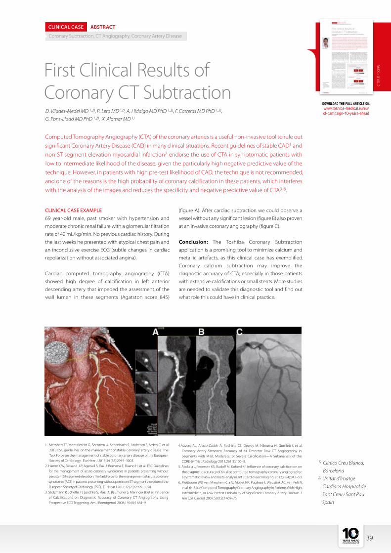
DOWNLOAD THE FULL ARTICLE ON:
www.toshiba-medical.eu/eu/ ct-campaign-10-years-ahead
CTE
U14
0095First Clinical Results of
Coronary CT Subtraction
Computed Tomography Angiography (CTA) of the coronary arteries is a useful non-invasive tool to rule out
significant Coronary Artery Disease (CAD) in many clinical situations. Recent guidelines of stable CAD1 and
non-ST segment elevation myocardial infarction2 endorse the use of CTA in symptomatic patients with
low to intermediate likelihood of the disease, given the particularly high negative predictive value of the
technique. However, in patients with high pre-test likelihood of CAD, the technique is not recommended,
and one of the reasons is the high probability of coronary calcification in these patients, which interferes
with the analysis of the images and reduces the specificity and negative predictive value of CTA3-6.
D. Viladés-Medel MD 1,2), R. Leta MD1,2), A. Hidalgo MD PhD 1,2), F. Carreras MD PhD 1,2),
G. Pons-Lladó MD PhD 1,2), X. Alomar MD 1)
CLINICAL CASE ABSTRACT
Coronary Subtraction, CT Angiography, Coronary Artery Disease
CLINICAL CASE EXAMPLE69 year-old male, past smoker with hypertension and
moderate chronic renal failure with a glomerular filtration
rate of 40 mL/kg/min. No previous cardiac history. During
the last weeks he presented with atypical chest pain and
an inconclusive exercise ECG (subtle changes in cardiac
repolarization without associated angina).
Cardiac computed tomography angiography (CTA)
showed high degree of calcification in left anterior
descending artery that impeded the assessment of the
wall lumen in these segments (Agatston score 845)
1) Clinica Creu Blanca,
Barcelona2) Unitat d’Imatge
Cardíaca Hospital de
Sant Creu i Sant Pau
Spain
(figure A). After cardiac subtraction we could observe a
vessel without any significant lesion (figure B) also proven
at an invasive coronary angiography (figure C).
Conclusion: The Toshiba Coronary Subtraction
application is a promising tool to minimize calcium and
metallic artefacts, as this clinical case has exemplified.
Coronary calcium subtraction may improve the
diagnostic accuracy of CTA, especially in those patients
with extensive calcifications or small stents. More studies
are needed to validate this diagnostic tool and find out
what role this could have in clinical practice.
1. Members TF, Montalescot G, Sechtem U, Achenbach S, Andreotti F, Arden C, et al. 2013 ESC guidelines on the management of stable coronary artery disease: The Task Force on the management of stable coronary artery disease of the European Society of Cardiology . Eur Hear J 2013;34 (38):2949–3003.
2. Hamm CW, Bassand J-P, Agewall S, Bax J, Boersma E, Bueno H, et al. ESC Guidelines for the management of acute coronary syndromes in patients presenting without persistent ST-segment elevation: The Task Force for the management of acute coronary syndromes (ACS) in patients presenting without persistent ST-segment elevation of the European Society of Cardiology (ESC) . Eur Hear J 2011;32 (23):2999–3054.
3. Stolzmann P, Scheffel H, Leschka S, Plass A, Baumüller S, Marincek B, et al. Influence of Calcifications on Diagnostic Accuracy of Coronary CT Angiography Using Prospective ECG Triggering. Am J Roentgenol. 2008;191(6):1684–9.
4. Vavere AL, Arbab-Zadeh A, Rochitte CE, Dewey M, Niinuma H, Gottlieb I, et al. Coronary Artery Stenoses: Accuracy of 64–Detector Row CT Angiography in Segments with Mild, Moderate, or Severe Calcification—A Subanalysis of the CORE-64 Trial. Radiology 2011;261(1):100–8.
5. Abdulla J, Pedersen KS, Budoff M, Kofoed KF. Influence of coronary calcification on the diagnostic accuracy of 64-slice computed tomography coronary angiography: a systematic review and meta-analysis. Int J Cardiovasc Imaging. 2012;28(4):943–53.
6. Meijboom WB, van Mieghem C a G, Mollet NR, Pugliese F, Weustink AC, van Pelt N, et al. 64-Slice Computed Tomography Coronary Angiography in Patients With High, Intermediate, or Low Pretest Probability of Significant Coronary Artery Disease. J Am Coll Cardiol. 2007;50(15):1469–75.
39
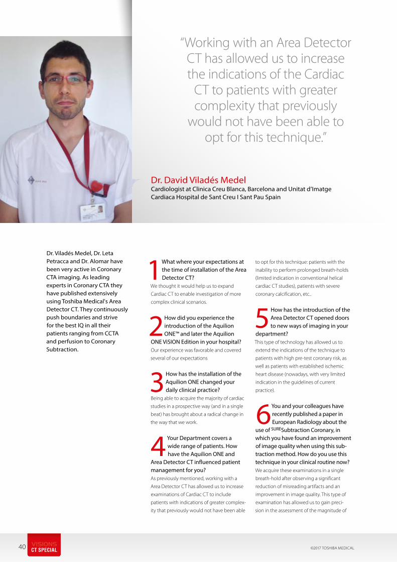
Dr. David Viladés MedelCardiologist at Clinica Creu Blanca, Barcelona and Unitat d’Imatge Cardiaca Hospital de Sant Creu I Sant Pau Spain
“Working with an Area Detector CT has allowed us to increase the indications of the Cardiac
CT to patients with greater complexity that previously
would not have been able to opt for this technique.”
40 ©2017 TOSHIBA MEDICAL
1 What where your expectations at the time of installation of the Area Detector CT?
We thought it would help us to expand
Cardiac CT to enable investigation of more
complex clinical scenarios.
2 How did you experience the introduction of the Aquilion ONE™ and later the Aquilion
ONE ViSION Edition in your hospital?Our experience was favorable and covered
several of our expectations
3 How has the installation of the Aquilion ONE changed your daily clinical practice?
Being able to acquire the majority of cardiac
studies in a prospective way (and in a single
beat) has brought about a radical change in
the way that we work.
4 Your Department covers a wide range of patients. How have the Aquilion ONE and
Area Detector CT influenced patient management for you? As previously mentioned, working with a
Area Detector CT has allowed us to increase
examinations of Cardiac CT to include
patients with indications of greater complex-
ity that previously would not have been able
to opt for this technique: patients with the
inability to perform prolonged breath-holds
(limited indication in conventional helical
cardiac CT studies), patients with severe
coronary calcification, etc..
5 How has the introduction of the Area Detector CT opened doors to new ways of imaging in your
department?This type of technology has allowed us to
extend the indications of the technique to
patients with high pre-test coronary risk, as
well as patients with established ischemic
heart disease (nowadays, with very limited
indication in the guidelines of current
practice).
6 You and your colleagues have recently published a paper in European Radiology about the
use of SURESubtraction Coronary, in which you have found an improvement of image quality when using this sub-traction method. How do you use this technique in your clinical routine now? We acquire these examinations in a single
breath-hold after observing a significant
reduction of misreading artifacts and an
improvement in image quality. This type of
examination has allowed us to gain preci-
sion in the assessment of the magnitude of
Dr. Viladés Medel, Dr. Leta Petracca and Dr. Alomar have been very active in Coronary CTA imaging. As leading experts in Coronary CTA they have published extensively using Toshiba Medical's Area Detector CT. They continuously push boundaries and strive for the best IQ in all their patients ranging from CCTA and perfusion to Coronary Subtraction.
VISIONSCT SPECIAL
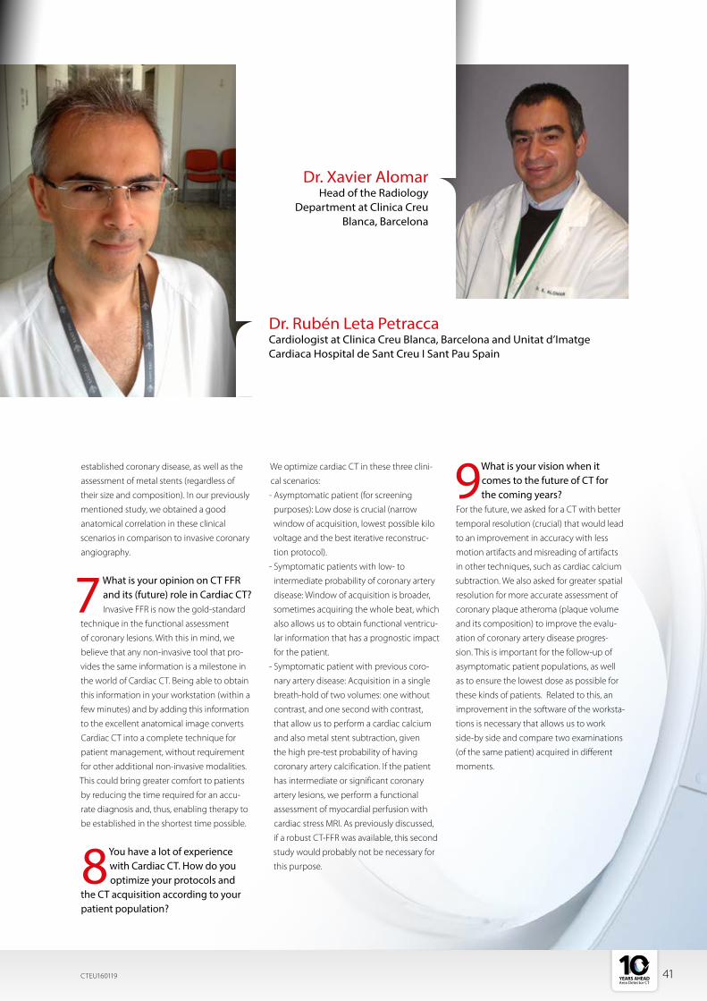
41
Dr. Rubén Leta PetraccaCardiologist at Clinica Creu Blanca, Barcelona and Unitat d’Imatge Cardiaca Hospital de Sant Creu I Sant Pau Spain
Dr. Xavier AlomarHead of the Radiology
Department at Clinica Creu Blanca, Barcelona
CTEU160119
We optimize cardiac CT in these three clini-
cal scenarios:
- Asymptomatic patient (for screening
purposes): Low dose is crucial (narrow
window of acquisition, lowest possible kilo
voltage and the best iterative reconstruc-
tion protocol).
- Symptomatic patients with low- to
intermediate probability of coronary artery
disease: Window of acquisition is broader,
sometimes acquiring the whole beat, which
also allows us to obtain functional ventricu-
lar information that has a prognostic impact
for the patient.
- Symptomatic patient with previous coro-
nary artery disease: Acquisition in a single
breath-hold of two volumes: one without
contrast, and one second with contrast,
that allow us to perform a cardiac calcium
and also metal stent subtraction, given
the high pre-test probability of having
coronary artery calcification. If the patient
has intermediate or significant coronary
artery lesions, we perform a functional
assessment of myocardial perfusion with
cardiac stress MRI. As previously discussed,
if a robust CT-FFR was available, this second
study would probably not be necessary for
this purpose.
9What is your vision when it comes to the future of CT for the coming years?
For the future, we asked for a CT with better
temporal resolution (crucial) that would lead
to an improvement in accuracy with less
motion artifacts and misreading of artifacts
in other techniques, such as cardiac calcium
subtraction. We also asked for greater spatial
resolution for more accurate assessment of
coronary plaque atheroma (plaque volume
and its composition) to improve the evalu-
ation of coronary artery disease progres-
sion. This is important for the follow-up of
asymptomatic patient populations, as well
as to ensure the lowest dose as possible for
these kinds of patients. Related to this, an
improvement in the software of the worksta-
tions is necessary that allows us to work
side-by side and compare two examinations
(of the same patient) acquired in different
moments.
established coronary disease, as well as the
assessment of metal stents (regardless of
their size and composition). In our previously
mentioned study, we obtained a good
anatomical correlation in these clinical
scenarios in comparison to invasive coronary
angiography.
7 What is your opinion on CT FFR and its (future) role in Cardiac CT? Invasive FFR is now the gold-standard
technique in the functional assessment
of coronary lesions. With this in mind, we
believe that any non-invasive tool that pro-
vides the same information is a milestone in
the world of Cardiac CT. Being able to obtain
this information in your workstation (within a
few minutes) and by adding this information
to the excellent anatomical image converts
Cardiac CT into a complete technique for
patient management, without requirement
for other additional non-invasive modalities.
This could bring greater comfort to patients
by reducing the time required for an accu-
rate diagnosis and, thus, enabling therapy to
be established in the shortest time possible.
8 You have a lot of experience with Cardiac CT. How do you optimize your protocols and
the CT acquisition according to your patient population?
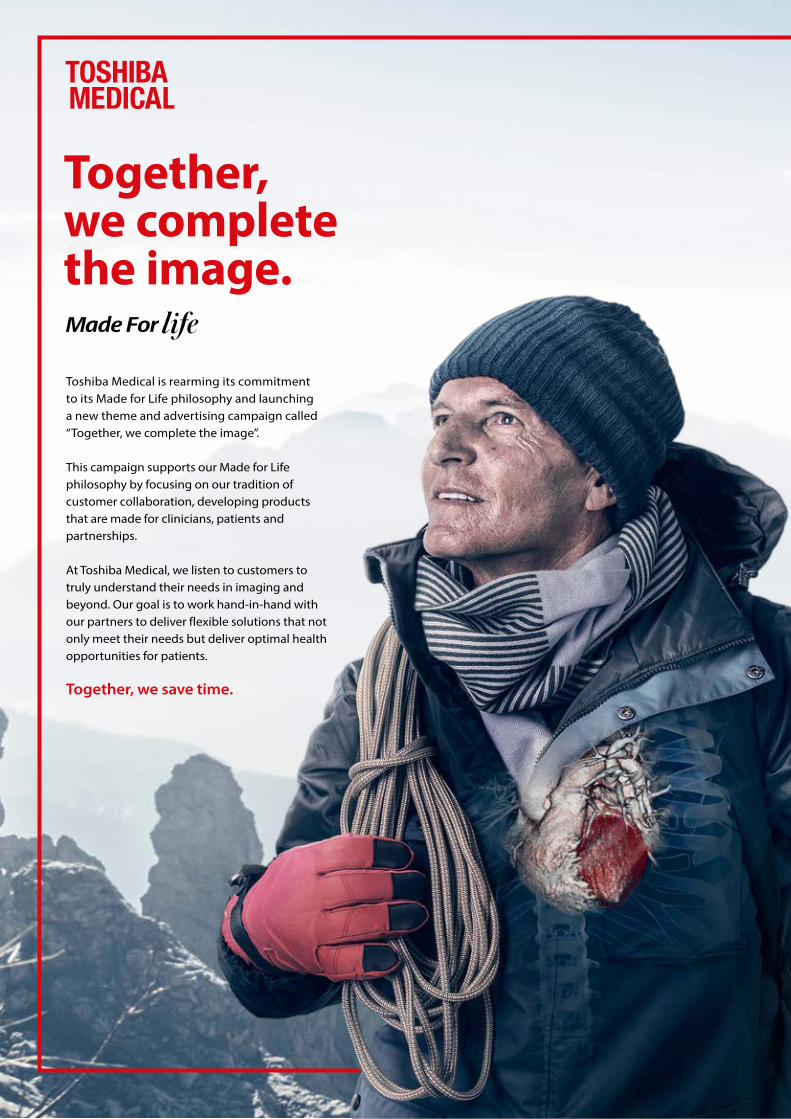
Together,we complete the image.
Toshiba Medical is rearming its commitment to its Made for Life philosophy and launching a new theme and advertising campaign called “Together, we complete the image”.
This campaign supports our Made for Life philosophy by focusing on our tradition of customer collaboration, developing products that are made for clinicians, patients and partnerships.
At Toshiba Medical, we listen to customers to truly understand their needs in imaging and beyond. Our goal is to work hand-in-hand with our partners to deliver flexible solutions that not only meet their needs but deliver optimal health opportunities for patients.
Together, we save time.
This CT Special edition of VISIONS magazine is a publication of Toshiba Medical Europe and is offered free of charge to medical and health professionals. To download the digital edition of this CT Special, please visit the campaign web site: www.toshiba-medical.eu/eu/ct-campaign-10-years-ahead. No part of this publication may be reproduced in whole or in part, stored in an automated storage and retrieval system or transmitted in any manner whatsoever without written permission of the publisher. The opinions expressed in this publication are solely those of the authors and not necessarily those of Toshiba Medical. Toshiba Medical does not guarantee the accuracy or reliability of the information provided herein.
PublisherToshiba Medical Systems Europe B.V.Zilverstraat 1NL-2718 RP ZoetermeerTel.: +31 79 368 92 22Fax: +31 79 368 94 44Web: www.toshiba-medical.euEmail: [email protected]
Campaign web pages: www.toshiba-medical.eu/eu/ct-campaign-10-years-ahead
Editor-in-chiefJack Hoogendoorn ([email protected])
EditorJacqueline de Graaf ([email protected])
Coordinator & ReviewerRoy Verlaan (International Market Development Manager CT)
Design & LayoutBoerma Reclame (www.boermareclame.com)
PhotographyCojan van Toor (www.cojanvantoor.nl)
PrintmanagementHet Staat Gedrukt (www.hetstaatgedrukt.nl)
Text contributions and editingThe Creative Practice (www.thecreativepractice.com)
© 2017 by TOSHIBA Medical EuropeAll rights reserved
ISSN 1617-2876
10 YEARS AHEADArea Detector CT
VISIONS | CT SPECIAL
VISIONSMagazine for Medical & Health Professionals I March 2017
SPECIAL
10 YEARS AHEADArea Detector CT
Toshiba Medical Celebrates10 Years of Area Detector CTDescribed as ‘a breakthrough CT system’ back in 2007, Toshiba Medical introduced the world’s very first dynamic volume CT scanner, The Aquilion ONE™. Driven by the clinical need for a volume scanner, the Aquilion ONE’s unique Area Detector opened doors to new ways of medical imaging. Ten years later, Toshiba Medical is celebrating this memorable introduction! This VISIONS Special highlights personal experiences of Key Opinion Leaders. It outlines Toshiba Medical’s journey in Area Detector CT and shows our continuing spirit to explore the unknown.
To download the digital version of this CT Special, visit: www.toshiba-medical.eu/eu/ct-campaign-10-years-ahead
To stay up to date with our latest innovations in medical imaging, visit: www.toshiba-medical.eu