Title EXPERIMENTAL STUDY ON CROSSED … · nerve and also between the median or ulnar and the...
23
Title EXPERIMENTAL STUDY ON CROSSED ANASTOMOSIS BETWEEN ANTAGONISTIC PERIPHERAL NERVES Author(s) Watanabe, Kousaku Citation 日本外科宝函 (1955), 24(2): 132-153 Issue Date 1955-03-01 URL http://hdl.handle.net/2433/206173 Right Type Departmental Bulletin Paper Textversion publisher Kyoto University
Transcript of Title EXPERIMENTAL STUDY ON CROSSED … · nerve and also between the median or ulnar and the...
Title EXPERIMENTAL STUDY ON CROSSED ANASTOMOSISBETWEEN ANTAGONISTIC PERIPHERAL NERVES
Author(s) Watanabe, Kousaku
Citation 日本外科宝函 (1955), 24(2): 132-153
Issue Date 1955-03-01
URL http://hdl.handle.net/2433/206173
Right
Type Departmental Bulletin Paper
Textversion publisher
Kyoto University
132 !J寸、:/Hl宝曲第24耗第2号
EXPERL¥IE.NTAL STUDY (l)J CROSSED ANASTOMOSIS
BET¥YEEN ANTAGONISTIC PERIPHERAL NERVES
by
KOUSAKU WATANABE
From the !st Surgical Division, Kyoto University Medical School. (Director: Prof. Dr. CersATO ARAKI) 、
〔Receivedfor Publication : Jan. 10, 1955.〕
INTRODUCTION
As early as in 1873, LETIEVANT attempted, for the purpose of restoration of the
peripheral nerve function lost by injuries, anastomosis between that nerve and another
spinal nerve with di任erentinnervation. Later, RAWA (1885), GUNN (1886), SICK
and SANGER (1897), MENASSE (1900), KENNEDY (1910), OSBORNE and KILVINGTON
(1911), SHIBUYA (1934), NORIOKA (1934), BARRON (1934), SPERRY (1940,’ 41 and
’47) and ARIZAWA (1952) made anastomosis, both in patient and in animals,
between h巴teronymousor heterogenous nerves.
But there were diversさ opinionsas to the possibility of reestablishment of physi・
ologic function of the a'tastomosed nerve; some were in the affirmative, and others
of the opposite.
The literature concerned m乱y coveniently b巴 classified, from the viewpoint of
fur.ctional recovery, into the following three categories:
A) th巴orywhich admits the recovery occurring rather early in the postopera-
tive period,
B) theory of r. on-recovery, and
C) theory iてl which the recovery can tak巴 placeby exercise after th巴 nerve
regeneration.
MENAおE(1900) reportd that in a patient anastomosis of the peripheral portion
of the paralyzed facial nerve with the accessory nerve resulted in successful recovery
of movements of the face after the operation. Since then, CUSHING (1903), BALLANCE
(1904) and DAVIDSON (1907) did similar facial anastomosis with the accessory or
a spinal nerve and recognized that in all the cases improvement of tonus of facial
muscles apparently occurred. However, they stated that complete recovery of delicate
movement of the face, especially the facial expression, could not be attained.
GUNN (1886) and SICK and SANGER (1897) reported that improvement of paralysis
was obtained by the implantation of the peripheral cut end of the ulnar nerve
paralyzed or by that of the radial nerve paralyzed, respectively, irてto the r. ormal median r.erve.
Anastomosis betwee.1 heteronymous nerves had been accomplished also exp巴rime・
ntally by various authors. Some of them believed the reestablishment of fur.ction,
but others came to the opposite conclusion.
Recovery to the normal function of the nerves shortly after the operation was
reported by RAWA (1885) when the tibial and五bularnerves were crosswise anast・
omosed in rabbits, dogs, cats and pigs. This was true of KENNEDγs experiment
CROSSED ANAST0'¥10SIS OF PERIPHERAL l¥'ERVES 133
(1911) in dogs, in which crossed anastomosis between the tibial and the五bular
nerve and also between the median or ulnar and the radial nerve was accomplished.
OSBORNE and KILVINGTON (1911) confirmed that normal function was regained
ten months after crossed anastomosis between the middle cords of the brachial
plexus on toth sides, when they observed that both fore legs reacted to unilate al
electrical stimulation of the cortical motor areas.
NORIOKA (1934) observed functional recovery after crossed anastomosis between
the ph enicus and the vagus, and SHIBUYA (1934) between the tibial and the五bularnerve.
BARRON (1934) performed in rats anastomosis tetween the follつwingnerves :
peripheral end of the n. femoralis with central end of the n. medianus orロ.ulnaris;
peripheral end of the n. ulnaris with central end of th号 n.tibialis; peripheral end of
the n. tibialis with central ends of the n. medianus and n. ulnaris. And of the 34
rats, a great majority showed recovery to normal functioning of the legs, but in 4
unusual movements were observed.
The experimental results obtained by these authors may conform to the theory
(A) described above.
In 1940, SPERRY carried out transposition bξtween the自exor muscle ( m. gastro-
cnemius) and the extensor muscles (m. tibialis anterior et m. extensor digitorum
longus) in rats. He found that normal function of the leg did never recover post-
operatively despite all possible exercises. However, he could confirm recovery to
normal function when he made crossed anastomosis of the 五bularnerve with the
tibial in addition. Further, he observed in 1941 that crossed anastomosis between
the五bularand the tibial nerve iロratsgave rise to reversed movement of the leg
after the operation. This reversed movement persisted despite every kind of train-
ing, and was recovered only by crossed transposition of the muscles concerned.
SPERRY’s experimental results may justify the theory (B) abovestated.
In 1947, SPERRY made anastomosis between the nerves innervating the muscles
of the elbow-joint of the spider and macaques monkeys and observed, that reversed
movements of the elbow-joint appeared, three years thereafter, when the nerve
regeneration seemed to have been completed. With training with a special apparatus,
however, the monkeys regained the ability to perform purposive movements volun-
tarily. He, therefore, laid claim to the case of monkey to the theory ( C) as
described.
ARIZAWA (1952) in our lavoratory accomplished crossed anastomosis between the
tibial and the五bularnerve i口 33rabbits and 3 dogs, all of which showed recovery
of the function to the normal. He consequently assumed that the functional reco-
very of the peripheral nerves which had undergone crossed anastomosis might be
due to the compensation either in the brain or in the spinal cord.
Subsequently, ISHII (1954) in our laboratory anastomosed a cervical spinal nerve
,, with the vagal nerve crosswise at the level of the neck in cats; it was then confirmed
t’that SU伍cientnerve regeneration could take place even in heterogenous nerve ana-
13』 日木外科室副号1~24巻第 2号
stomosis, although the function reestablished was not physiological in such a case.
Whether or not does physiological function once lost of the peripheral nerve
recover after anastomosis betw田 n the two nerves with di任er巴ntfunctions, and, if
the recovery does occur, in what part of the brain or the spinal cord does functional
compensation actually take place? For studying these problems, I made crossed
anastomosis at the level of the upper leg in dogs, between the tibial nerve, which
innervates fl.exor muscles of the hind leg, and the五bularnerve innervating the
extensors.
Further, the peripheral nerves were crosswise anastomosed after previous d巴corti-
cation of the motor areas or medullary pyramidotomy.
In the control animals, partially crossed anastomosis of the peripheral nerves was
done and the animals were observed for the peripheral functions for 3 to 12 months
postoperatiYely, during and after which period electrical stimulation, and macro-
and microscopic examinations of the anastomosed nerves were carried out. The
present report concerns with the results thus obtained.
MATERIALS AND METHOD OF EXPERIMENT
I) Experimental animals. Healthy adults dogs, weighing around 10 kg., were
used, as they readily survive su伍cientlylong postoperatively until the nerve regen-
eration is completed and they haYe the well developed pyramidal tract. In 30
dogs, various operations described in the following were done successfully.
II) Experimental method.
Group I : Completely crossed anastomosis of the peripheral nerves (C. C. A)・
20 dogs.
Group 2 : Completely crossed peripheral nerve anastomosis after recovery from
the hemiplegia as the result of the preceding decortication of the
contralateral motor areas ( D. + C. C. A.). 3 dogs.
Group 3 : Completely crossed peripheral nerve anastomosis after recovery from
the hemiplegia resulting from previous contralateral pyramidotomy
(P.十C.C. A.). 2 dogs.
Group 4 : Partially cross巴danastomosis of the peripheral nerves (P. C. A.)・
5 dogs.
Postoperative impairments of th巴 functionwere compared with each other among
these four groups from variable points of view as were described later.
III) Operative procedures.
え) Anaesthesia ・ General narcosis with injection into the free peritoneal cavity of 10.%
Is~my凶 solution, 0.3c.c. per kg. body weight after pr巴Ii口iinarybasis narcosis with subcutaneous
miection of 4,% N arcopon scopolamine I c.c.
B) C~mpl悦ly. c:os~e_d anastomosis of the periphe凶 nen-es With the right hind leg
in extenswn, a skm rnc1s1on, ca 5 cm. long, was made so that m. hiceps femoris and m. semi・
tendineus could easily he retracted when the tibial and fibular nerves were seen to bifur・
cate from the main trunk of the sciatic n巴rve.
i入ftermobili出 ion~nd cutti暗 ofth巴senerves, AR山 WAmade E吋ーto-e吋 anastomosisafter
the routine method ot nerve anastomosis and tubulation with autogenous fascia. At autopsy,
CROSSED ANASTO"¥rGSIS OF PERIPHERAL NERVES 135
it was found in his experiments that the sites of the anastomosis adhered to each other,
showing conglomerated swelling, which histologically revealed comglomeration of nerve fibers,
some of them passing into the homonymous peripheral nerve stump. For a successful crossed
anastomosis, such comglomeration of nerve fibers must be avoided. Th巴refore,I devised a new
technique, a combination of the autoarterial tubulation method of WEISS with TAKETOMO’s
non-suture method using a tube of auto-fascia or vein. In my technique an arterial tube
五xd iロ 70;'~ alcohol was used for tubulation. This method was previously published in
detail. It will be summarized in the following : each of the two nerves is doubly ligat巴d,
and then cut. At the middle of the arterial tube preserved in 70;'-0 alcohol, a small hole
is made, through which the ctit end of the one nerve inserted from one end of the tube is pulled out with a fine thread attached to that cut end. In this position, two or three sutures
with fine silk thread were made between the arterial tube end on this side and the epineurium
of the nerve without injuring the nerve parenchyma. Then the cut end of another nerv巴 is
likewise pulled out of the hole and the end of the arterial tube on this side is sutured with
the epineurium. The two nerv巴 endsappearing outside the hole of the tube are refreshed
and then reposed within the tube so that the refreshed ends are exactly adjusted without
direct suturing. In this way, the central end of the tibial nerve and the peripheral end of the fibular
nerve, or the central end of the fibular nerve and the peripheral end of the tibial nぞrYeare
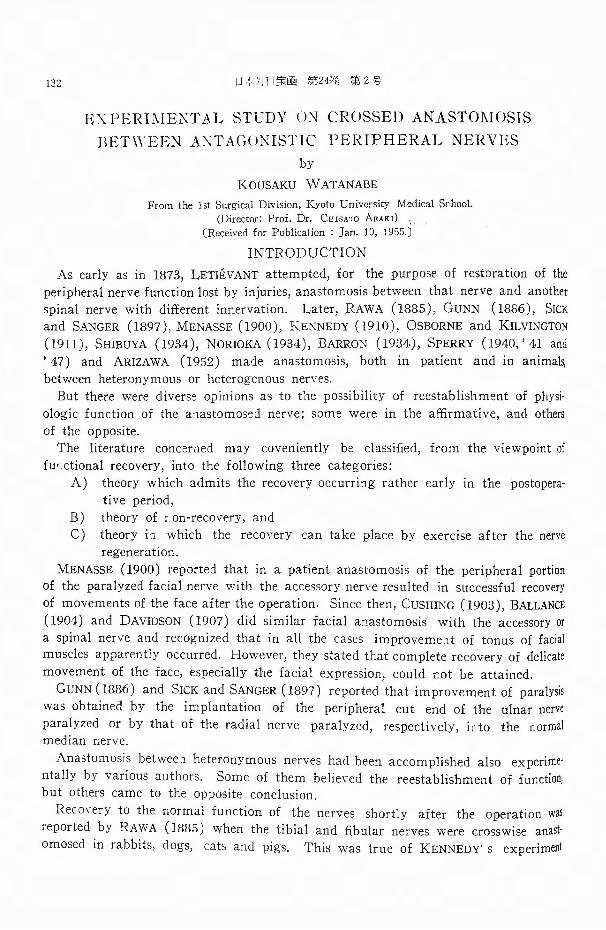
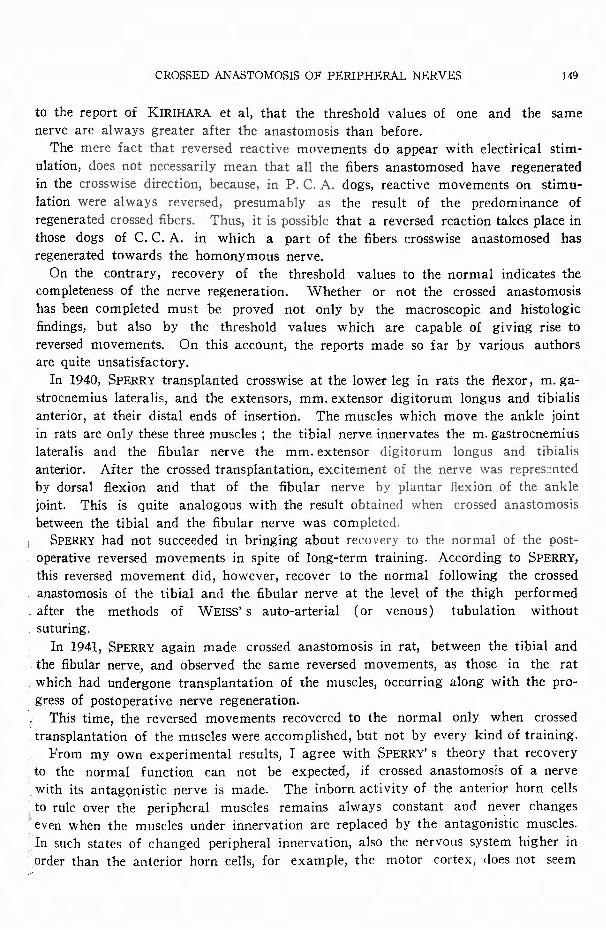
crosswise anastomosed (Fig. 1, a).
a
f.
'T. 子.
b C) Partially crossed anastomosis of the per-
ipheral nerves : After exposing the two nerves,
F. their epineuriums are incised lo昭 itudinallyand each nerve is divided into one larger and one
smaller bundle (6: 1 to 8: 1 in proportion). The smaller one is left unsevered while the larger one
is severed and each end is crosswise anastomosed
with that of another nene in the same way as
described in (B) (Fig. 1, b).
' 'r. f
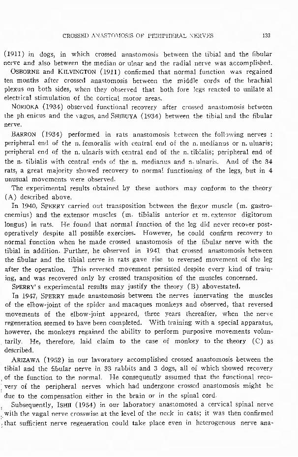
D) Unilateral decortication of the moter
areas: Although exact location and extension of
the cortical motor areas in dogs are not definitely
established, gyri sigmoideus anterior et posterior
are generally considered to be these areas. A
part of gyri proreus and coronalis are also
included in the motor areas : this fact was taken
into consideration in my operation.
Fig-. I. Schematical illustration of crossed nerve anastoロiosis.
Nomenclature of the gyri and sulci of the
dog's brain is given in Fig. 2. a : Completely crossed anastomosis b : Partially crossed anastomosis Tib. : N.tibialis Fib. : N.五bularis
The head of a dog in prone position is fixed,
a median incision from the glabella to the occi-
pital protuberance is made, the fascia of the
left temporal muscle is cut at its attachment,
and the temporal muscles are turned laterocaudally, resulting in su伍cientexposure of the
temporal and frontal bones. After the periosteum is detached at the upper margin of the
temporal bone, is made a burr hole, which is enlarged, so as to e文posethe frontal sinus
frontally and extend to the midline at the vertex. Then sulcus cruciatus and gyrus sigmoideus
are visualized through the transparent dura mater. The dura is then incised at the part
136 日!列、科宝’画第21巻第2号r
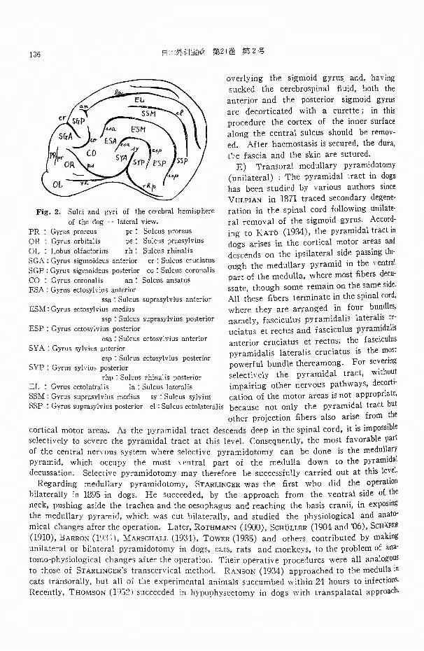
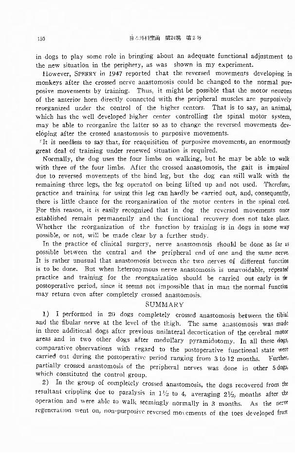
Fig・. 2. Suki and gyri of the cerebral hemisphere of the dog --lateral view.
PR : Gyrus pror巴口s pr : Snlcus proreus OR '. Gyrus orbitalis ps : Sulcns praesylvins OL : Lobus o!factorins rh : Sulcus rhinalis
overlying the sigmoid gyrus and, having
sucked the cerebrospinal fluid, both the
anterior and the posterior sigmoid gyrus
are decorticated with a curette ; in this procedure the cortex of the inner surface
along the central sulcus should be remov・
ed. After haemostasis is s巴cured,th巴 dura,
t~' e fascia and the skin are sutured. E) Transoral medullary pyramidotomy
(unilateral) : The pyramidal tract in dogs has been studied by various authors since VuLPIAN in 1871 traced secondary degene-
ration in the spinal cord following unilate・
ral removal of the sigmoid gyrus. Accord-
ing to KATδ (1934), the pyramidal tract i~ dogs arises in the cortical motor areas and
descends on the ipsilateral side passing thr・ SGA: Gyrus叩 oideusanterior り ulcuscrnc ia~1~s 岬 1the medullary pyramid in the 、巴削
~~p::包;::ヱ:::s postご~r S~~c~::~ニ:r山 15 part of the r帥仇 wheremost伽 rsdei~::・ESA: Gyrus ectosyl¥"ius anterior ssate, though some remain on the same Slde.
ssa: Su!cus suprasylvius anterior All these fibers terminate in the spinal cord, ESM: Gyrus ectosylvius medius where they are arranged in four bundles,
ssp: S口lcussuprasylvius posterior namely, fasciculus pyramidalis lateralis er・ ESP ・ Gyrus ectosy[vit1s post巴rior uciatus et rectus and fasciculus pyramidalis
esa : Sulcns ectosylvius anterior SYA ・ Gyms sylvius anterior
anterior cruciatus et rectus; the fasciculus
pyramidalis lateralis cruciatus is the叩stesp : Sulcus ectosylvius posterior powerful bundle thereamong. For seven【
町 p:Gy川 5山 5~~~e:i~:lcus rhinalis P口sterior sel
I g
EL : Gvrus ectolatralis la : Sulcus Jateralis impairing other nervous pathways, decorti-SSM: Gvrus suprasvlvius medius sy ・ Sulcus sylvius cation of the motor areas is not appropriate, SSP : Gvrus叫 1叫 Ivinsposterior el : Sulcns ectolat叫 isbecause not only the pyramidal t附 tbut
other projection fibers also arise from the
cortical motor areas. As the pyramidal tract descends deep in the spinal cord, it is impossible selectively to severe the pyramidal tract at this level. Consequently, the most favorable part of the central nervous system where selective pyramidotomy can be done is the medullarY pyramid, which occupy the most ventral part of the medulla down to the pyramidal decus凶 ion. Selective pyramidotomy may t1町 efore be successfully carried out at this le~el. Regarding medullary pyramidotomy, STARLINGER was the first who did the operation
bilaterally in 1895 in dogs. He succeeded, by the approach from the ventral side of. the neck, pushing aside the trachea and the oesophagus and reaching the basis cranii, in exposing the medullary pyramid, which was cut bilaterally, and studied the physiological and anato-mica! changes after the operation. Later, RoTHMANN (1900), SCHULLER (1904 and ’06), ScH~FER (1910), BARRON (1リ:lei),MARSCHALL (1934), TOWER (1935) and others contributed by making unilateral or bilateral pyramidotomy in dogs, cats, rats and monkeys, to the problem of ana・ tomo-physiological changes after the operation. Their operative procedures were all analogous to those of ST ARLINGER’s transcervical method. RANSON (1934) approached to the medulla in cats transorally, but all of the experimental animals succumbed within 24 hours to infections. Recently, THOMSON (l:).")2) succeeded in hypophysectomy in dogs with transpalatal approach.
CROSSED ANASTOMOSIS OF PERIPHERAL NERVES 137
Kuoδet al. (1952) was able to expose the ventral surface of the midbrain by a new transoral
approach with previous luxation or fracture of the mandibule. In modifying and improving
the methods of THOMSON and Kuo5, I devised a transoral method, in which the ventral
surface of the medulla was exposed pretty widely so that the pyramid could be cut exactly
unilaterally.
After the narcosis, the dog is laid in supine position and, prior to the operation, a canula
is introduced through an incision into the trachea.
The maxilla is五xed,the mouth is opened maximally by a retractor and the tongue is
pulled out and sutured to the skin over the mandibule. The lips and the oral caYity are
desinfected with 0.5,% marsonin alcohol, draped, only exposing the soft palate and the
pharynx, and the drape is sutured to the mucous membrane in order to avoid slipping. The
soft palate is incised in the midline and the posterior nasal cavity and the posterior phary-
ngeal wall are exposed which are immediately desinfected.
The soft palate divided is turned aside and pull巴dlaterally and the edges are sutured to
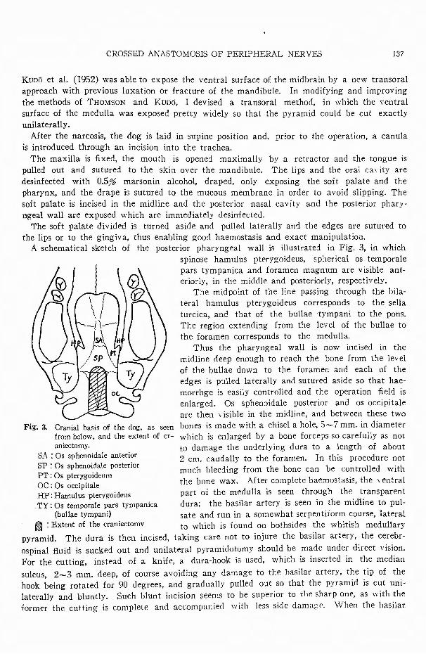
the lips or to the gingiva, thus enabling good haemostasis and exact manipulation. A schematical sketch of the posterior pharyngeal wall is illustrated in Fig. 3, in which
spinose hamulus pterygoideus, spherical os temporale
pars tympanica and foramen magnum are visible ant-eriorly, in the middle and posteriorly, respectively.
The midpoint of the line passing through the bila-
teral hamulus pterygoideus corresponds to the sella
turcica, and that of the bullae tympani to the pons.
The region extending from the level of the bullae to
the foramen corresponds to the medulla.
Thus the pharyngeal wall is now incised in the
midline deep enough to reach the bone from the level
of the bullae down to the foramen and each of the
edges is pulled laterally and sutured aside so that hae-
morrhge is easily controlled and the operation五eldis
enlarged. Os sphenoidale posterior and os occipitale
are then visible in the midline, and between these two
Fig・. 3. Cranial basis of the dog, as seen bones is made with a chisel a hole,ヨ~7mm. in diameter from below, and the extent of er-which is enlarged by a bone forceps so carefully as not aniectomy. to damage the underlying dura to a length of about
SA : Os sphenoidale anterior 2 cm. caudally to the foramen. In this procedure not SP : Os sphenoidale posterior much bleeding from the bone can be controlled with PT : Os pterygoideum
the bone wax. After complete haemostasis, the ventral OC : Os occipitale HP:Hami TY: Os temporale pars tympanica dura; the basilar artery is seen in the midline to pul
- (bullae tympani) sate and run in a somewhat serpentiform course, lateral
~ : Extent of the craniectomy to which is found on bothsides the whitish medullary
pyramid. The dura is then incised, taking care not to injure the basilar artery, the cerebr-
ospinal fluid is sucked out and unilateral pyramidotomy should be made under direct vision.
For the cutting, instead of a knife, a dura司hookis used, which is inserted in the median
sulcus, 2~3 mm. deep, of course avoiding any damage to the basilar artery, the tip of the
hook being rotated for 90 degrees, and gradually pulled out so that the pyramid is cut uni-
laterally and bluntly. Such blunt incision seems to be superior to the sharp one, as with the
former the cutting is cornplete and accompanied with less side damage. When the basilar
138 日本外科宝画第24巻 第2号
artery is once injured, the resultant haemorrhage is uncontrollable owing to the narrow and deeply situated operation五eld. Furthermore, even a seemingly trivial haemorrhage is liable to form a clot to which the animal readily succumbs. When haemostasis is completed, place a small piece of gelatine sponge soaked with penicillin solution in the bony defect, leaving the dura unsutured, and then the pericsteum and the mucosa are sutured together. Antibiotics combined with haemostatica is systemically administered after the operation and the tracheal canula is removed on the 2nd or 3rd postoperative day.
IV) Postoperative examinations.
The following items are examined postop巴ratively.
A) Functional examination( clinical signs).
a) Unilateral decortication or medullary pyramidotomy :
Paresis of the head and the four limbs were recognized by the changes in behavior
at the time of movements, such as walking, running and standing with the hind
legs. Paresis of the hind leg, in particular, was carefully analyzed by the state
of both extension and flexion of the toes of that leg when the dog stood with the
hind legs. In addition, abnormalities of tendon reflexes and of musclar tonus and
the course of the recovery from the paresis were concurrently observed.
b) Completely or partially crossed anastomosis of the peripheral nerves:
i) Lameness (or crippling) appeared most frequently. State of crippling,
particularly fl.exions of the toes, position and form of the ankle joint, state of
standing with the hind legs, and coordination or non-coordination with the healthy
legs at movements, were carefi.;lly examined.
ii) Trophic disturbanc巴s. Loss of hair, deformation of nail, blush, bleeding,
ulceration and necrosis of the leg were observed.
iii) Resistance at passive flexion of the ankle joint. Immediately after the
operation all the cases show巴d flaccidity, but along with the progress of nerve
regen巴rationthe resistance gradually increased.
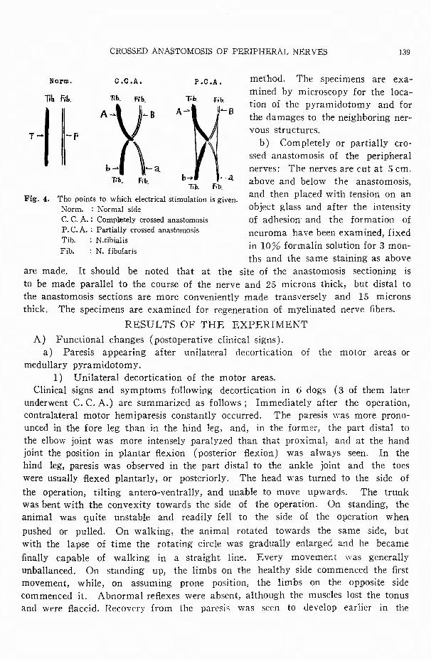
B) Electrical stimulation by induced current.
Prior t'J killing the animal, both the tibiai and fibular nerves on the healthy side
and the tibio-fibular and fibulo-tibial nerves on the paralyzed side are exposed
and, with Du BOJS-REYMOND’s apparatus (2 volt), electrical stimulation is given
to the points shown in Fig. 4; i. e. points T and F on the healthy side and A
and B (central), and a and b (periph巴ral) on the side operated on. The motor
reactions elicited and the threshold values oロ bothsides are compared with each other (Fig. 4).
C) Anatomical examination.
a) Decortication of the motor areas or medullary pyram idotomy : When
the animal is sacrificed, the brain and the spinal cord are removed and the medulla
is fixed for 3 months in 107" formalin solution after the presence of adhesions
in the previous op巴ration五巴Idand extent of the decortication or of the pyrami-
dotomy have been determined, stained with 羽TEIGERTSCHNELL BEIZE for 2 weeks,
dehydrated with alcohol, embedded in celloidin. ;-・,erial sections of 2.'i microns
in thickness are made and stained wi・(h WEIGERT-PAL' s myelin sheath staining
CROSSED ANASTOMOSIS OF PERIPHERAL NERVES 139
Norm. C.C.A. P.C.A.
li~ F;b. lib. fib. T・"b_ 干ib_
』 8 A-"l jιB
l』 F、j
T;b_ fib.
Fig. 4. The points to which electrical stimulation is given. Norm. : Normal side C. C. A. : Compk!tely crossed anastomosis P. C. A. : Partially crossed anastomosis Tib. : N.tibialis Fib. : N. fibularis
method. The specimens are exa-
mined by microscopy for the loca-
tion of the pyramidotomy and for
the damages to the neighboring ner-
vous structures.
b) Completely or partially cro-
ssed anastomosis of the peripheral
nerves: The nerves are cut at 5 cm.
above and below the anastomosis,
and then placed with tension on an
object glass and after the intensity
of adhesion and the formation of
neuroma have been examined, fixed
in 10% formalin solution for 3 mon-
ths and the same staining as above
are made. It should be noted that at the site of the anastomosis sectioning is
to be made parallel to the course of the nerve and 25 microns thick, but distal to
the anastomosis sections are more conveniently made transversely and 15 microns
thick. The specimens are examined for regeneration of myelinated nerve五bers.
RESULTS OF THE EXPERIMENT
A) Functional changes (postoperative clinical signsj.
a) Paresis appearing after unilateral decortication of the motor areas or
medullary pyramidotomy.
I) Unilateral decortication of the motor areas.
Clinical signs and symptoms following decortication in 6 dogs (3 of them later
underwent C. C. A.) are summarized as follows ; Immediately after the operation,
contralateral motor hemiparesis constantly occurred. The paresis was more prono-
unced in the fore leg than in the hind leg, and, in the former, the part distal to
the elbow joint was more intensely paralyzed than that proximal, and at the hand
joint the position in plantar flexion (posterior fl.exion) was always seen. In the
hind leg, paresis was observed in the part distal to the ankle joint and the toes
were usually乱exedplantarly, or posteriorly. The head was turned to the side of
the operation, tilting antero-ventrally, and unable to move upwards. The trunk
was bent with the convexity towards the side of the operation. On standing, the
animal was quite unstable and readily fell to the side of the operation when
pushed or pulled. On walking, the animal rotated towards the same side, but
with the lapse of time the rotating circle was gradually enlarged and he became
五nally capable of walking in a straight line. Every movement was generally
unballanced. On standing up, the limbs on the healthy side commenced the五rst
movement, while, on assuming prone position, the limbs on the opposite side
commenced it. Abnormal reflexes were absent, although the muscles lost the tonus
and were flaccid. Recovery from the paresis was seen to develop earlier in the
140 日本外科宝曲第24巻第2号
hind leg than in the fore leg. The last to recover was the part distal to the
hand joint. 2 to 3 weeks at most after the operation, the animals recovered from
the paresis nearly to the normal, so that by usual examination the abnormality in
the movements, if any remained, could not be found.
2) Unilateral medullary pyramidotomy.
Clinical signs demonstrated in 3 dogs (2 of them later underwent C. C. A.) are
summarized as follows ; motor hemiparesis below the neck on the side contralateral
to the operation constantly developed in more or less degree. The paresis was
more severe in degree in the fore leg than in the hind leg, particularly in the part
distal to the hand joint. Paresis of the hind leg was only slight. The movement
of the head was not influenced by the operation・Thetrunk showed a slight arched
bending towards the side contralateral to the operation. Standing up movements
were unsteady, at which the contralateral hand joint was自exedplantarly. But
once the animal stood up, th巴 handjoint returned to the normal position and the
animal could stand straight. The fore legs were usually in crossed positions on
standing and, when pulled or pushed, the animal easily fell to the contralateral
side. On Wdlking, the animal at五rstrotat巴dslightly toward the side of the ope-
ration, but soon was able to go straight, though liable to fall to the contralateral
side. The hind leg recovered from the paresis earlier than the fore leg, of which
the part distal to the hand joint was the last to recover. In 7~IO days postopera“
tively, recovery to nearly the normal tool王 placewithout any permanent disturbance.
Abnormal reflexes and increased tonicity of the muscles were entirely absent both
immediately after the operation and after the recovぞryof movements. Hemiparesis
was of nearly the same nature in the case of pyramidotomy as in the case of
decortication, but it was less in severity and recovered prompter in the former than in the latter.
b) Changes after completely or partially crossed peripheral nerve anastomosis. I) Completely cross巴danastomosis.
i) Crippling : This was constantly ~een after the operation. The part
distal to the ankle joint on the side of the operation was maximally flaccid and
lost activ巴 movements. During the period from the 3 rd to the 14th postoperative
day, the paralyz巴dhind leg was lifted up and the animal walked or ran with the
remaining three limbs. This may be attributed to the pain arising from the ope-
rative wound. Thereafter, the toes became plantarly flexed and the animal walked
with the dorsal surface of the toes dragging the ground. On running, however, the
paralyzed leg was lifted up and the other three limbs were used. Such lameness
was observed for Iろな~4months, averagingころと months postoperatively, which I
should like to term 1st stadium of paralysis Thereafter, the animal gradually
recovered from this crippling state and became capable of walking and running
normally at a glance. However, the ankle joint took a position of extreme dorsal
fl.exion, although the toes were in the normal position. This stadium suggestive of
I町 owryto normal function lasted for .J. ~'> months, 5 ~~ months in average, which
is termed 2日dstadium of compensatio日・ During the period of compensation, the

CROSSED ANASTOJ¥IOSIS OF PERIPHERAL NERVES 141
position of the ankle joint returned from hyper丑exion to the normal and the toes
began to move actively. About the end of this stadium, peculiar movements, just
opposite to the movements on the healthy side developed at meals, anger and
astonishment. Such reversed movements became gradually more pronounced, espe-
cially in the ankle joint, and at the same time the toes and frequently the ankle
also were again丑exedplantarly. Consequently, due to such reversed movements,
walking became unskillful and running was carried out with the other three limbs.
In 7 to IO months the reversed movements became the most manifest, and this
stage is termed 3 rd stadium of reversed movements. The position and form of
the leg in the three different stadiums are shown in Fig. 5. Crippling by reversed
movements never recovered later. As is shown in Fig. 6, postoperative crippling
Norm. 1st stad. 2nd stad. 3rd stad.
ソ
1 s七 S.I ♀nd s. 』, 3rds. • ~ y_
Op. 1 2 3 4 5 6 7 8 ~ 10 ll M
Fig・. 5. Postoperative state of the hind leg on walking (C. C. A.)
improved by the 4th month in nearly
all dogs, when it again began to appear
and fully developed in the 9 th postope-
rative month, after which period crippling
persisted for ever.
Of the 25 (20 C. C. A., 3 D.十 C.C.A.,
and 2 P.十C.C. A.) dogs, 7 (28%) went
%
F F
〆J,r
/
/
〆/ 〆
司、
a
k
q
且
TIE--+11品BILE
n
u
n
u
A
U
《
U
岬
N
A
M
O
O
F
0
・Rw・内d
op. 1 2 3 4 !; ι7 g 9 10 n M. into the second stadium soon after the
Fig. 6. Frequency of crippling after the operation. initial stage of walking with three legs. Percentage of the number of the dogs In 2 of the 25 ( 8)ト) the crippling due showing crippling. to reversed movements (the 3rd stadium)
developed in immediate succession to the五rststadium without demonstrating com-
pensatory normal walking (the 2nd stadium). Crippling both in the自rstand the third stadium was more outstanding in D.十
C. C. A. dogs than in C C. A. dogs, although the period of walking with three legs was shorter in the former.τherefore D.十C.C. A. animals were seen more frequently to fall laterally. The reason is that the reduction in tonus and 日trengthof muscles, which is still remaining latently after decortication although undetectable by usual

142 日本外科宝曲第24巻第2号
examination, is added to the paralysis due to C. C. A・, so that D. + C. C. A. animals tend to cripple more severely and to fall laterally on walking. There was not
much difference in the postoperative state between the P. + C. C. A. and the C. C. A. dogs.
ii) Trophic disturbances: Trophic disturbances were seen in the following
number of cases, all appearing in the first stadium.
No disterbance 4 dogs
Loss of hair and blush only 2 dogs
Bleeding and ulcer formation 14 dogs
Formation of deep ulcer reaching the tendon or bone 3 dogs
Necrosis and falling o任 ofthe distal end 2 dogs
Formation of ulcer took place suddenly one week to one month after the operation
and the ulcers were one to three or four in number, 0.5 to 1.0 cm. in diameter,
round in shape and located mostly in the dorsum of the foot or on the tip of a
toe. The ulcers were gradually covered with granulation tissue and healed in the
4th postoperative month (Fig. 7).
% 100
80
i;o
40
20
In the third stadium, when crippling reap-
peared, loss of hair, deformation of nails
and bleeding were occasionally seen, but
there was no ulcer formation. Of the 25
dogs, 4 (16 % ) did not show a slightest
dystrophy and 2 (8%) manifested loss of
hair and blush only. The dogs, in which
the crippling in the first stadium was not
remarkable, were usually devoid of trophic op. 1 2 3 4門 disturbances.
In all the D.十C.C. A. dogs, ulcer form-
ation was severe and in some of them the
phalangeal bones were exposed for a certain
period. The ulcers, however, healed in the 4th postoperative month. There was
no differ巴ncein ulceration between the P. + C. C. A. and the C. C. A. dogs.
Fig. 7. Postoperative trophic disturbances. Percentage of the number of the dogs showing ulcer formation.
In 2 (Nos. 7 and IO, C. C. A.) dogs, there developed 4 and 45 days, respectively,
after the operation at the distal end of the foot an ulcer, which was quickly widened
and deepened and in the course of 15 days the distal half of the phalanx became
necrotic and fell o任・ The cause of this ulcer was not evident, but it seemed that
a neurotrophic factor might play a role, although infection by some specific micr-oorganisms could not entirely be excluded.
iii) Resistance to passive flexion of the ankle joint: The normal resistance
disappeared after the operation in all the cases. The hind limb distal to the ankle
joint was completely flaccid and no longer actively movable. However. this resis-
tance reappeared at the end of the I st stadium, i.e. in 2 to 3 months post~peratively, improved gradually and at the end of the 2nd stadium. i.e. in 4 to 6 months after the operation, returned to the normal. ・

CROSSED ANASTOMOSIS OF PERIPHERAL NERVES 143
2) Partially crossed anastomosis of the peripheral nerves.
In all the 5 dogs no neurological changes appeared after the operation and they
were capable of walking and running without crippling, despite that the unsevered
part of both the tibial and the五bular nerve was only ~8 to弘 inthickness of
the whole nerve. On standing with the hind legs, the dogs sometimes stood with
the dorsal surface of the toes touching the ground, but they soon turned actively
the toes to the normal position. Postoperative trophic disturbances were also slight
in these dogs. In only 2 dogs were found small subcutaneous haemorrhages or ulcers
on the plantar surface of the foot, which healed in 3 to 4 months postoperatively.
These dogs, however, began to cripple 6 to 7 months after the operation in the
same way as in C. C. A. dogs, showing reversed movements of increasing severity,
from which they never recovered.
B) Reactive movements to electrical stimulation by induction current.
The experiment was carried out 6 to 12 months after the operation. Both in C. C.
A. and in P. C. A. dogs, th円 stimulationgiven to the portions of nerves central to
the anastomoses (point A and B) gave rise to the reactions contrary to those on
the healthy side : at the time of stimulation of the tibio-fibular nerve (point A),
a weak current gave rise to spreading of the toes and a strong current dorsal
自exion of the ankle besides. On stimulation of the的 ulo-tibialnerve (point B),
a weak current gaYe rise to plantar flexion of the toes only and a strong
current to that of the ankle besides. In P. C. A. dogs, some of the toes showed,
at times, normal reaction on weak stimulation, but, on somewhat stronger
stimulation, all the toes demonstrated reversed reactive movements. Threshold
values, when measured at point A and B (central to the anastomosis), and more
than 7 to 8 months after the operation, returned in most cases to the normal and _
did rarely show a rising (the coil distance being less )-5ノl at maximum, if compared
with those on the healthy side (Fig. 8). In a few cases (No・1,1 year; No. 15, IO
a) C. C. A.
T F A B a b
'.fax. 29.0 29.0 26.6 28.0 25.8 25.0
Min. 22.3 23.0 21.9 22.3 18.0 17.3
A、・erage. 25.3 25.8 24.0 25.2 19.8 19.5
b) P. C. A.
T F 人 B a b
:11司x. 27.0 26目8 28.5 28.0 25.0 24.7
:¥fin. 25.3 25.0 23.0 23.0 19.5 19.3
Average. 26.3 26;1 26.1 25.7 23.2 23.0
Fig・. 8. Threshold values of el巴ctricalstimulation by induced current (cm.) using Du Bor~- R"·orn:-;1> s apparatus. Voltage: 2 volt.
months; No. 20, 11 months; and No. 41, 1 year after the operation) even a decrease

144 日本外科宝曲第24巻第2号
(the coil distance being greater) in the values was noted.
On stimulation at points a and b (distal to the anastomosis), on the other hand,
a greater majority showed a slight increase (less than 20拓) in the threshold values,
although some showed nearly the same values as those obtained by the stimulation
of the central portions. This may be accounted for that the stimulation given to
the distal portion is weakened by the massive connective tissue proliferated in
between the nerve fibers. In one case (No. 14, 5 1h months after the operation),
the reactive movements were analogous to those described above, but the threshold
values were markely increased (the coil distance being less) even when th巴 central
portion was stimulated. Return of the threshold values to nearly the normal 7 to 8
months after the operation suggested that the nerve regeneration was almost
completed after crossed anastomosis.
C) Anatomical Findings.
a) Decortication of the motor areas or m巴dullarypyramidotomy.
i) Extent of the 白cortication(Fig. 9).
Fig・. 9. Extent and location of the d巴cortication.@ーー一一一- '.¥o. 41 dog @一一一一 '¥o. 42 dog ⑬・・・・υ ・一一 No. 46 dog
In No. 41 dog (1 year after the operation). えnteriorand posterior sigmoid gyri (total)
and also proreus and coronary gyri (partial)
Fig・. 10. Extent and location of the decortication iロト\o.41 dog. (1 year after the opera-tion). lu巾巾randp口町riorsigmoid gyri( totally) and gnri proreus and coronal is (partially) were removed, all on the left side.
were decorticated on the left side : complete decortication of the so-called motor
areas (Fig・10)・ InNo. 42 dog (3弘 monthsafter the operation), whole anterior
sigmoid gyrus, most of posterior sigmoid gyrus and a part of coroロarygyrus, all
on the left side, were decorticated : in other words, decortication of nearly all the
motor areas. In No. 46 dog (11 months after the operation), left anterior and
posterior sigmoid gyri were removed : almost complete deco山~tion.In the dogs with a rather long postoperative period of survival (Nos. 41 and 46),
the 町 dul
size than oロthehealthy sid巴.

ii) Location of the medullary pyramidotomy and
boring nervous pathways.
In No. 59 dog 16 months postoperatively!, the pyramidotomized part of the medulla was seen to be slightly adhered to the dura mater, although the adhesion could be
separated bluntly. In this case, the left medullary pyramid was cut across at the
level 5 mm. below the pons and the pyramidal bundles both above and below the
cut line reduced its size grossly to弘 ofthe normal. Histologically, the pyramid
was found to be completely cut through at the level of the inferior olivary nucleus.
The longitudinal fasciculus dorsal to the pyramid was seen severed and the myelin
sheaths were faintly stained (Fig. 11 and 12 ).
In No. 60 dog (8 months postoperative-ly ), the medullary pyramid on the left
side was cut through at the level of the
transition between the pons and the me-
dulla. The pyramid distal to the cut
line was拾 in size of the pyramid on
the healthy side. Histological examinat-
ion disclosed that the pyramid was cut
across at the level of the五laredicularia
of the vagal nerve and the cut reached
in the depth of stratum interolive.
b) Completely crossed anastomo-
sis or partially crossed anastomosis.
1) Mac、roscopicalfindings :
In the case of C.C.A., the arterial piece used for tubulation remained and appe-
ared as a white membrane even I year
after the operation. At the site of the
anastomosis, adhesion to the surrounding
tissue was found in few cases only. The
two arterial pieces were seen embedded
together in the connective tissue, althou-
gh they could be readily and bluntly
shelled out. The two nerves appeared
white and glistening. The periphery of
the fibular n巴rve anastomosed to the
central end of the tibial nerve doubled
its preoperative size so that it became
as large as the latter nerve, which rem-
ained unchanged. Irisu伍ciency of the
anastomosis, dense adhesion and forma-
tion of neuroma were not observed. In short, crossed nerve anastomosis healed
145
damages to the neigh-
CROSSED ANASTOMOSIS OF PERIPHERAL NERVES
Fig・. 11. Me<lullary pyrami<lotomy. No. 59 dog (6 months after the opεration). The left pyramid has b民 ncut across at the point marked with an arrow (〆).The pyr-amid, both above and below the cut line, is seen to have reduced the size to 11~ of that of the right pyramid.
特例代/
嘉一・一一誠2正i7J:i.{c';'.tq.
~ -~ - ι ー 、;狩iプτ
産設Fig・. 12. Photomicrograph of the medullary pyra-
midotomy. :-¥o. 59 dog. Th巴 leftpyramid is entirely absent. The longitudinal fascicu!us on that side shows an obvious demyelinization (com-pare with the contralateral side). W EWERT-
PAL' s staining method for myelin sheath. ( x 4)
146 白木外科宝曲第24巻第2号
completely (Fig. 13).
千Tーナプ?っFig・. 13. Gross appearance of the nerves、、hich
have undergone C. C. A. No.13 dog, 10 months after the operation (enlarge-ment×2).
The arterial tubes used for tubulation remain and appear as white membranes. The t¥¥'o nerves (n. 五bulo-tibialisis seen abov巴 andn. tibio・五bnlarisbelow) appear white and glistening. They are seen embedded together in the ocnnective tissue, although they can be readily and bluntly shelled out. Crossed nen・c anastomosis is complete. Pro-ximo-distal direction from left to right.
In P. C. A. dogs, the n巴rveswhich had
undergone crossed anastomosis were seen
to arrange in 2 parallel rows, white and
glistening, on which were seen X-form
nerve fiber bundles which had not been
severed and anastomosed at the operation.
In general, adhesion around and between
the two nerves was more prominent in
these cases than in the C. C A. cases.
Yet, th巴 partially crossed anastomosis
seemed to have healed successfully.
2) Histological自ndings:
In more than 6 months after the opera-
tion, the nerve fibers anastomosed showed
SU伍cientregeneration. The regenerated
fib巴rs of the tibialis proceeded with a
well arranged course towards the peripheral portion of the fibularis inside the arterial
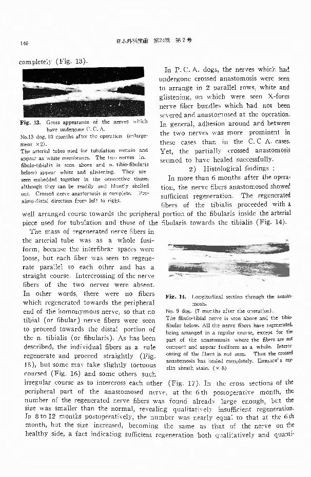
piece used for tubulation and those of the五bularistowards the tibialis (Fig. 14).
The mass of regenerated nerve fibers iロ
the arterial tube was as a whole fusi-
form, because the inter五brarspaces were
loose, but each fiber was seen to regene-
rate parallel to each other and has a
straight course. Intercrossing of the nerYe
fibers of the two nerves were absent.
In other words, there were no fibers
which regenerated towards the peripheral
end of the homonymous nerve, so that no
tibial (or五bular)nerve fibers were seen
to proceed towards the distal portion of
the n. tibialis (or fibularis). As has been
described, the individual fibers as a rule
regenerate and proceed straightly (Fig.
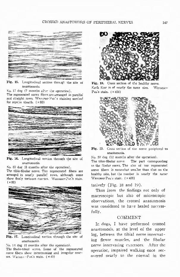
l S ), but some may take slightly tortuous
coursed (Fig. 16) and some others such
Fig-. 14. Longitudinal section through the anasto-官lOS!S.
No. 9 dog. (7 months after the operation). The fibuloーtibial口en・eis seen above and the tibio・ 五bularbelow. All the n巴rve五bershave regenerated, being arranged in a regular course, except for the part of the anastomosis where the五bersare not compact and appεar fusiform as a whole. Intercr-ossing of the五bersis not seen. Thns the crossed anastomosis has healed completely. EHRLICH' s nw-elin sheath stain. ( x 8)
irregular course as to intercross each other (Fig. 17 ). In the cross sections of the
peripheral part of the anastomosed nぞれ-e, at the 6th postoperative month, the
number of the regenerated nerve fibers was found already large enough, but the
size was smaller than the normal, revealing qualitatively insu伍cient regeneration.
In 8 to 12 months postoperatively, the number was nearly equal to that at the 6th
month, but the size increased, becoming th巴 same as that of th巴 nerve on the
healthy side, a fact indicating su伍cientregeneration both q¥1alitatively and quanti-
CROSSED ANASTOMOSIS OF PERIPHERAL NERVES 147
Fig・. 15. LongituιJina! section through the site of
anastomosis.
No. 17 dog (7 months after the operation). The regenerated nerve :fibers are arranged in parallel and straight rows. WEr<JERT-PAL’s staining method for myelin sheath. ( x 80)
Fig・. 16. Longitudinal sεction through t.he site of
anastomosis.
No. 60 dog (8 months after the operation). The tibio-五bularnerve. The regenerated :fibers are arranged in nearly parallel rows, although some show五nelytortuous comses. WEWERT-PAL’s stain. (×80)
孟主主子事事
anastomosis.
No. 14 dog (6 months after th巴 operation).The五blllo-tibial nεrve. Some of the regenerated nerve五bersshow intercrossing and irregular cour-ses. ¥V EIGE'《IPAr/s stain. (x40)
Fig・. 18. Cross section of the healthy nerve.
Each五heris of nearly the same size. \九TEI臼ERT」
PAL stain. ( x 400)
anastomosis. ]¥:o. 20 dog (12 months after the operation).
The tibio-fibular口erve. The part corresponding
to the fibular nerve. The size of tne regenerated
nerve五hersis somewhat smaller than that on the
healthy side, but the number is nearly the same-
もIVEWERT-PAL's stain. (×400)
tatively (Fig. 18 and 19 ).
Thus from the findings not only of
macroscopic but also of microscopic
observations, the crossed anastomosis
was considered to have healed success-
fully.
CO:i¥ffMENT
In dogs, I have performed crossed
anastomosis, at the level of the upper
leg, between the tibial nerve innervat-
ing fl.exor muscles, and the 五bular
nerve innervating extensors. After the
operation, impaired walking once rec-
overed nearly to the normal in the
148 日オ外科宝曲第24巻第2号
period of 2弘 to5拍 postoperativemonths. After this period, or subsequent to
complete nerve regeneration, impaired gait gradually reappeared, demonstrating such contra-purposive reversed movements as to flex the toes plantarly when dorsal
-flexion should occur, and vice versa. From the impaired gait of this kind, the
animals were seen never to recover. In the case of P. C. A., the uncrossed, or unsevered nerve fibers, 1Ai to % of
the original thickness of the nerve, seemed at first to be enough to rule normal walking. ¥Vith the lapse of time, however, the nerve fibers which had undergone crossed anastomosis, regenerated and finally they became predominant to the uncrossed 五bersin the e妊ectof innervation, when revers巴dmovements of the toes de\・ぞlopedin the same way as in the case of C. C. A・, thus resulting in crippling on walking.
After the C. C. A. crippling due to immediate paralysis naturally appeared, from which the animal recovered in 1 ~j to 4, averaging 2日 months, when the nerve regeneration was probably still going on and seemed to have advanced only so far as to allow the muscles under innervation to maintain the tonus and to hold the normal position of the leg. In such a stadium of incomplete nerve regeneration, one is apt to assume that complete recovery to normal function has already taken place. But such an assumption is not correct, because, in P. C. A. cases, only ~i:~ 1/k of the original nerve fibers was enough to control nearly normal walking. As the regeneration went on and was finally completed, the disturbance in
walking reappeared, due to the occurrence of the contra-purposive reversed move-ments. This seems to be the result of the establishment of the complete innervation
of the nerves which had been crosswise anastomosed. The e旺巴rentimpulse of plantar a巴xionfrom the anterior horn celis of the spinal
cord was conveyed through th巴 crosswise anastomosed tibio-fibular nerve to the extensor muscles, and vice versa, resulting in contra-purposive reversed movements of the limb and, consequently, in walking disturbances.
Regarding the theory which admits the recovery occurring rather early in the postoperative period, I presume that th巴 nerve regeneration might have occurred only incompletely owing to some failure in the technique of the問 rveanastomosis, i. e. the process of the nene regeneration advanced no further than the 2 nd stadium of compensation. Also it is possible that eveロ in C. C. A. , if performed with insu伍cienttechnique, some per cent of the regenerated n巴rvefibers should proceed towards the homonymous nerve. In such imperfect operations, formation of a neuroma and adhesion are constantly present, hindering the regeneration of the nerve fibers, so that reversed movements hardly巴nsue. A ris~ in the threshold values (lo~2九%), or reduction in nervous excitability, as reported by many authors, would likewise justify this view. More than 7 to 8 months after the anastomosis, the threshold values measured at the central portion of the nervぞ :inastomoseddid actually di任ernot much in my exp巴rimentfrom those measured o九 thehealthy side (the di妊erencebeing 5% at the maximum). In a great majority of the. cases, the threshold values w巴reequal on both sides, but in some cases, they were巴venlower on the side of the anastomosis than on the healthy side. This fact is contrary
:~
CROSSED ANASTOMOSIS OF PERIPHERAL NERVES 149
to the report of KIRIHARA et al, that the threshold values of one and the same
nerve are always greater after the anastomosis than before.
The mere fact that reversed reactive movements do appear with electirical stim-
ulation, does not necessarily mean that all the五hersanastomosed have regenerated
in the crosswise direction, because, in P. C. A. dogs, reactive movements on stimu-
lation were always reversed, presumably as the result of the predominance of
regener且tedcrossed日bers. Thus, it is possible that a reversed reaction takes place in
those dogs of C. C. A. in which a part of the fibers crosswise anastomosed has regenerated towards the homonymous nerve.
On the contrary, recovery of the threshold values to the normal indicates the
completeness of the nerve regeneration. Whether or not the crossed anastomosis
has been completed must be proved not only by the macroscopic and histologic
findings, but also by the threshold values which are capable of giving rise to
reversed movements. On this account, the reports made so far by various authors
are quite unsatisfactory.
In 1940, SPERRY transplanted crosswise at the lower leg in rats the fl.exor, m. ga-
strocnemius lateralis, and the extensors, mm. extensor digitorum longus and tibialis
anterior, at their distal ends of insertion. The muscles which move the ankle joint
in rats are only these three muscles ; the tibial nerve innervates the m. gastrocnemius
lateralis and the fibular nerve the mm. extensor digitorum longus and tibialis
anterior. After the crossed transplantation, excitement of the n巴rvewas repres三nted
by dorsal fl.exion and that of the fibular nerve by plantar flexion of the ankle
joint. This is quite analogous with the result obtained wh巴n cross巴danastomosis
between the tibial and the五bularnerve was completed. SPERRY had not succeeded in bringing about recovery to th巴 normalof the post-
operative reversed movements in spite of long-term training. According to SPERRY,
this reversed movement did, however, recover to the normal following the crossed
anastomosis of the tibial and the五bularnerve at the level of the thigh performed
after the methods of WEISS’s auto-arterial (or venous) tubulation without
suturing.
In 1941, SPERRY again made crossed anastomosis in rat, between the tibial and
the fibular nerve, and observed the same reversed movements, as those in the rat
which had undergone transplantation of the muscles, occurring along with the pro-
gress of postoperative nerve regeneration.
This time, the reversed movements recovered to the normal only when crossed
transplantation of the muscles were accomplished, but not by every kind of training.
From my own experimental results, I agree with SPERRY’s theory that recovery
to the normal function can not be expected, if crossed anastomosis of a nerve
with its antagc;mistic nerve is made. The inborn activity of the anterior horn cells
to rule over the peripheral muscles remains always constant and never changes
even when the muscles under innervation are replaced by the antagonistic muscles.
In such states of changed peripheral innervation, also the nervous system higher in
order than the anterior horn cells, for example, the motor cortex, does not seem
150 日,j:外科宝曲第24巻第2号
in .dogs to play some role in bringing about an adequate functional adjustment to
the new situation in the periphery, as was shown in my experiment.
However, SPERRY in 1947 reported that the reversed movements developing in
monkeys after the crossed nerve anastomosis could be changed to the normal pur-
posive movements by training. Thus, it might be possible that the motor neurons
of the anterior horn directly connected with the peripheral muscles are purposively
reorganized under the control of the higher centers. That is to say, an animal,
which has the well developed higher center controlling the spinal motor system,
may be able to reorganize the latter so as to change the reversed movements dev-
eloping after the crossed anastomosis to purposive movements.
<It is needless to say that, for reaquisition of purposive movements, an enormou・sly
great deal of training under renewed situation is required.
Normally, the dog uses the four limbs on walking, but he may be able to walk
with three of the four limbs. After the crossed anastomosis, the gait is impaired
due to reversed movements of the hind leg, but the dog can still walk with the
remaining three legs, the leg operated on being lifted up and not used. Therefore,
practice and training for using this leg can hardly be carried out, and, consequently,
there is little chance for the reorganization of the motor centers in the spinal cord.
For this reason, it is easily recognized that in dog the reversed movements once
established remain permanently and the functional recovery does not take place.
Whether the reorganization of the function by training is in dogs in some way
possible, or not, will be made clear by a further study.
In the practice of clinical surgery, nerve anastomosis should be done as far as
possible between the central and the peripheral end of one and the same nerve.
It is rather unu包ualthat anastomosis between the two nerves of di妊erentfunction
is to be done. But when heteronymous nerve anastomosis is unavoidable, repeated
practice and training for the reorganization should be carried out early in the
postoperative period, since it seems not impossible that in man the normal function
may return even after completely crossed anastomosis.
SUMMARY
1) I performed in 20 dogs completely crossed anastomosis between the tibial
and the五bularn巴rveat the level of the thigh. The same anastomosis was made
in thre巴 additionaldogs after previous unilateral decortication of the cerebral motor
areas and in two other dogs after medullary pyramidotomy. In all these dogs,
comparative observations with regard to the postoperative functional state were
carried out during the postoperative period ranging from 3 to 12 months. Further,
partially crossed anastomosis of the peripheral nerves was done in other 5 dogs, which constituted the control group.
2) In the group of completely crossed anastomosis, the dogs recover巴dfrom the
resultant crippling due to paralysis in I見 to4, averaging 2弘, months after the
operation and were able to walk seemingly normally in 3 months. As the ne問
regeneration went on, non-purposive reversed movements of the toes developed from
CROSSED ANASTOMOSIS OF PERIPHERAL NERVES 151
the 4th to 9th, averaging 5 1ii th, postoperative month so that the animals again showed pronounced crippling on walking, which never improved even after a fairly long time.
3) In the case of partially crossed anastomosis, the animals were seen capable of normal walking, if some引ito % in thickness of the original neryes were left unsevered. However, when the nerve fibers, which had undergone crossed anastom-osis, fully regenerated, the reversed movements of the toes, as seen in the case of completely crossed anastomosis, appeared, resulting in the walking disturbances which did hardly improve later.
4) Electrical stimulation applied to the point central to the anastomosis gave rise to reactive movements reverse to those on the healthy side in the cases of both C. C. A .. and P. C. A .. But the threshold values were not much different from those on the healthy side; a fact which indicated that the nerve regeneration in the crossed direction was completed.
5) Both the macroscopic and the histologic examinations of the anastomosed ' sites of the nerves con五rmedthat the crossed anastomosis healed successfully. In other words, the nerve fibers were found to have regenerated well and to have
1 proceeded into the periphery of the heteronymous nerve. 6) Decortication of the cerebral motor areas or medullary pyramidotomy did
not essentially influence on the functional changes after the crossed anastomosis of the peripheral nerves.
7) However, it is presumed from the literature that the animals which have . the well developed brain may have an ability of reorganization of the peripheral motor system so as to change the reversed movements to the normal purposive '.movements if adequately trained at the time when the nerve regeneration has been . completed.
8) It is my belief that, in the cases, where the recovery of function took place ~ rather early after the crossed anastomosis of the peripheral nerves, the nerve regeneration was not complete and remained in the 2nd stadium of compensation probably due to some failure in the technique of the nerve anastomosis. 9) In the present experiment a new method of anastomosis has been adopted
・・in which an arterial tube五xedand preserved in 70% alcohol is used for tubulation ’and the nerve ends within it are not sutured with each other. This method is {simple and enables an ideal regeneration of the nerve 印)ers. It should be widely 'useful in clinical practice.
REFERENCES
f; 1) Araki, C. : Surgical exp~rience with inj-uries of the peripheral nerv巴s.Saishin Igaku, 2 ; 9, 1947.
2) Arizawa, G. : Experimental studies on the crossed anastomosis of antagonistic peripheral
~ nerves. ]. Jap. Surg. Soc. , 53 ; 91, 195:2. ¢ 3) Barron, D. H. : The results of periphe凶
anastomosis between the fore and hind limb nerves of albino rats. J. Comp. Nenrol.,59 ; 301, 1934. 4) Barron, D.日.: The retults of unilateral
pyramidal section in the rat. J. Comp. Neurol., 60 ; 45, 1934. 5) Ballance, C.: A case of facial palsy treated
by facio-hypoglossal anastomosis in which an
152 日本外科宝前第24者第2号
anastomosis was made between the spinal acce-
ssory and the distal segment of the divided
hypogloss泡1nerve. Lancet, 1; 1675, 1909.
6) Davidson, A.: Ueber die Nervenpfropfung im
Gebiete des Nervus facialis. Beitr. z. ldin. Chir.,
55 j 427, 1907.
7) Ishii, S.: Experimental study on the possibility
of the functional restoration after anastomosis
between a spinal nerve and the vagal nerve.
Folia Psychiat. Neurol. Jap., 8; 69, 1954.
8) Ishii, S. : Histological studies of the anasto-
mosis between a spinal and the vagal nerve. Folia
Psychiat. Neural. Jap., 8; 87, 1954. 9) Kata, K. : Experimental comparative-anatomi-
cal study on the pyramidal system. 日okuetsu
Igalm Zasshi, 49; 851, 1934.
10) Kirihara, S. and Y. Kobayashi: Experimental study on transplantation of the peripheral ne-
rves. Brain and Nerve, 2; 347, 1949.
11) Kizawa, K. : Histological review on the
regeneration of the peripheral nerves. Nisshin
Igaku, 29; 31 and 183, 1940.
12) Kudo, T., S. Nawa and S. Sato: Decerebrate
rigidity by cross section of the midbrain. Brain and Nerve, 4; 139, 1952.
13) Marshall, C. : Experimental lesion of the
pyramidal tract. Arch. Neurol. Psychiat., 32; 778,
1934.
14) Norioka, E.: Ueber die Nervennaht zwischen
dem N. vagus und dem N. phrenicus. Kyoto Daigaku Daisan Kaibogaku Rombunsh丸 I;4, 14, 1934.
15) Osborne, W. A.叩 dB. Kilvington : Central
nervous response to peripheral nervous distortion. Brain, 33; 288, 1911.
16) Ranson, S. W.: Rigidity caused by pyramidal
lesions in the cat. J. Comp. Neurol., 55; 91, 1932.
17) Rothmann, :¥I. : Die Zerstorung der Pyrami-
denbahnen in der Kreuzung. Neurol. Centralbl., 19; 1055, 1900.
18) Schuller, A. : Experimentelle Pyramiden
Durchschneidロngbeim Hunde und Affen. Wien. klin. Wochenschr., 19; 57, 1906.
19) Shibt1ya, K. : Eine Funktionumstimmung
nach der totalen Kreuznaht zwischen dem N.
peroneus und N. tibialis. Kyoto Daigaku Daisan
Kaibogaku Rombunshu, I; 4,65, 1934.
20) Sick, C. und A. Sanger : Heilung einer in
Folge traumatischen Defekts be<lingten L泊mung
des Ra<lialis <lurch Vern益hung des peripheren
Endes dieses Nerven mit dem :¥Iedianus. Langen・beck's Arch., 54; 271, 1897.
21) Sperry, R. W.: The functional results of
muscle transposition in the hind limb of the
rat. J. Comp. Neueol., 73; 379, 1940.
22) Sperry, R. W.: The e妊ectof crossing nerves
to antagonistic muscles in the hind limb of the
rat. J. Comp. Neural., 75; l, 1941.
23) Sperry, R. W. : E妊ectof crossing nerves to
antagonistic limb muscles in the monkey. Arch.
Neural. Psychiat., 58; 452, 1947.
24) Starlinger, J. : Die Durchschneidung beider
Pyramiden beim Hun de. Neural. Central bl., 14;
390, 1895. Jahrb. Psychiat. Neural., 15; 1, 1897.
25) Taketomo, T.: A new method of nerve suture
and of repair of the gap of the nerve. Kyoto
Igakkai Zasshi, 2 ; 628, 1952.
26) Thomson, A.: A transpalatal approach to the
hypothalamic area in the ferret. J. Endocrinol.,
8 j 234, 1952.
27) Tower, S. S. : ・The dissociation of cortical
excitation from cortical inhibition l:y pyramid
section, and the symdrome of that lesion in the
cat. Brain, 58; 238, 1935.
28) Watanabe, K.: A new method of peripl世間!
nerve anastomosis: Reunion of a severed nerve by
tubulation with an arterial tube fixed and pre-
served in 709占 alcohol. Arch. Jap. Chir., 23; 458, 1954.
29) Weiss, P.: Reunion of stumps of small nerves
by tubulation instead of suture. Science, 93 ; 67,
1941.
30) Weiss,P.: The technology of nerve regenera・
tion: A review, sutureless tubulation and related
method of nerve repair. J. Neurost時., 1; 400. 1944. 1
CROSSED ANASTOMOSIS OF PERIPHERAL NERVES 153
和文抄録
異名末梢神経交叉縫合に関する実験的研究
京都大学医学部外科学教室第 1講座(指導荒木千里教授)
大学院学生 渡辺浩策
1) 私は犬(20頭)の鹿骨,肱骨両神経を大腿部に が,刺戟騎値は健側に比して差異のない襟度に快復
て完全交叉経合を行い,更にー側大脳運動領皮質切除 し,神経再生が完全に営なまれた事を示している.
( 3頭)及び延髄ー側錐体東切断( 2頭)の後に対側 5) 縫合部の肉眼的,組織学的所見により,交叉縫
の同様な末梢神経完全交叉縫合を行って,術後の機能 合ぷ成功している事を確かめ,再生神経線維は異名神
状況を 3ヶ月~1ヶ年に豆り,比較観察すると共に, 経聞にでも,略与完全に再生延長する事を実証した.
対照群として,部分的交叉縫合( 5頭)を行った. 6) か Lる末梢機能の変化に対して,大脳運動領皮
2)完全交叉縫合では,術後 1ヶ月半~4ヶ月,平 質切除,延髄錐体切断等は認むべき影響を及ぼさな
均2ヶ月半で当初の麻簿性披行より脱し,約 3ヶ月間 い.
略L正常歩行を営むが,神経再生の完成と共に,術後 7) 大脳発育の良好なるAtiJ物にては,逆運動発現後,
4~9ヶ月,平均5ヶ月半頃より足E止の反目的々逆運 即ち神経再生が完成された時に訓練練習により合目的
動を塁して再び著明に敏行し,其後長時日の経過の後 運動へと再編成,湾統一が可能で・あるかも知れない.
も再び後能の快復は認めない. 8) 諸家の報告の内,術後早期に機能欧復を認める
3) 部分的交叉縫合によれ神経の%~%程度が残 論説に対しては,神経縫合法の不備により,私のいう
されであれば,それによって歩行後能は略L完全に営 第2期,代償期の状態にて神経再生が留ったものと信
む事が出来るが,残余の交叉されたより沢山の神経線 ずる.
維が再生を完成して来れば,完全交叉縫合と同様に, 9) 神経縫合に際し70%アルコール固定動脈管を用
足世の逆運動を来して,機能は障碍され,これはその い,断端に全く糸を通じない縫合術式を考案し採用し
後に至っても恢復しない. Tこが,本術式は簡便であり,神経再生も完全に営なま
4) 電気刺戟により,縫合中継側の刺戟では,完全 れる事を実証しTこから,臨床的応用も広いものと考え
交叉,部分的交叉共に健側とは逆の反応運動を呈する る.
Pancreatiti日.
R. M. Zollinger
New Engl. J. Med., 251; 13, 497, 1954.
勝磁炎の診断は腹痛に際し疑診を置き,史に血中アミラーゼ試験を行うことにより,以前よりも透かに高率に
於て確診を下し得るに至った.
又治療法に就ては急性豚臓炎の場合は相当標準的な治療法があるが,慢性目撃滋炎では今日倫その治療法に就て
意見のー数をみていない現状である.著者の経験によれば急性勝抜炎の診断に当つては,本症の特徴である腹痛
に関連した発痛試験h邸貢,血中誌に腹水のアミラーゼ値の測定値,更にレ線所見を念頭に置いて診断を下すべき
である.又その治療法はあくまで豚放機能の休息にふれ鎮痛,勝分泌抑制,合血量の復元に努め,抗生物質の
投与を行うべきでp 急性症状のある時期の手術は危険であるから,敢て手1'1~を施行するならば急性症状の去った
後に行うべきであるとしている.
慢性豚臓炎に就ては再燃性結石性のものを特に採り上げ,手術以外にその適切な治療法はなく,その術式とし
て, Cholecystectomy,Transduodenal Sphincterotomy, Gastrectomy, Vagotomy, Biliaryshunt等を上げてL、る.
而して患者の病因,病理,条件等を良く熟考の上,これら術式の何れかを決定すべきとしている.
(藤原憲和抄訳)


![zz.fjtcm.edu.cn · Web viewLI Beibei, XU Yiming, LI Lixing,et al. Effect of Electromyographic Biofeedback Therapy on Ulnar Nerve Injury in Different Degrees[J].Rehabilitation Medicine,2020,30(3):197-201.](https://static.fdocument.pub/doc/165x107/6111aa954c26fb23aa15f6b8/zzfjtcmeducn-web-view-li-beibei-xu-yiming-li-lixinget-al-effect-of-electromyographic.jpg)


![The Elbo · Ulnar nerve dislocation & injury 3]. proximal ulna fracture 4]. fracture of ulnar component 5]. impingement of the radial head 6]. hardware failure 7]. Loosening 8]. Wound](https://static.fdocument.pub/doc/165x107/601baef6c039f322a241fc86/the-ulnar-nerve-dislocation-injury-3-proximal-ulna-fracture-4-fracture.jpg)