Therapeutic monoclonal antibody targeting of neuronal ...
14
RESEARCH Open Access Therapeutic monoclonal antibody targeting of neuronal pentraxin receptor to control metastasis in gastric cancer Mitsuro Kanda 1* , Dai Shimizu 1 , Koichi Sawaki 1 , Shunsuke Nakamura 1 , Shinichi Umeda 1 , Takashi Miwa 1 , Haruyoshi Tanaka 1 , Chie Tanaka 1 , Masamichi Hayashi 1 , Yohei Iguchi 2 , Suguru Yamada 1 , Masahisa Katsuno 2 and Yasuhiro Kodera 1 Abstract Background: Controlling metastasis is essential for improving the prognosis of patients with gastric cancer (GC). Here, we aimed to identify a molecule required for GC metastasis and to investigate its potential utility as a target for the development of therapeutic antibodies (Abs). Methods: Transcriptome and bioinformatics analyses of human GC cell lines identified the neuronal pentraxin receptor (NPTXR) as a candidate molecule. NPTXR function was probed by modulating its expression in GC cells and assessing the effects on intracellular signaling and malignant behaviors in vitro and in mouse xenograft models. We also generated anti-NPTXR Abs and Nptxr -/- mice, and assessed the clinical significance of NPTXR expression in GC specimens. Results: NPTXR mRNA expression in clinical specimens was associated with disease progression and was significantly higher in tissues from GC patients with distant metastasis compared with those without. NPTXR regulated expression of genes involved in metastatic behaviors as well as activation of the PI3K–AKT–mTOR, FAK– JNK, and YAP signaling pathways. NPTXR silencing promoted caspase-mediated apoptosis and attenuated GC cell proliferation, cell cycle progression, migration, invasion, adhesion, stem cell-like properties, and resistance to 5- fluorouracil in vitro, and also inhibited the tumorigenicity of GC cells in vivo. Anti-NPTXR Abs inhibited GC peritoneal metastasis in mice. Nptxr -/- mice showed no abnormalities in reproduction, development, metabolism, or motor function. Conclusions: NPTXR plays an essential role in controlling the malignant behavior of GC cells in vitro and in vivo. NPTXR-targeting Abs may thus have utility as novel diagnostic tools and/or treatment modalities for GC. Keywords: Gastric cancer, Neuronal pentraxin receptor, Antibody, Knockout mouse © The Author(s). 2020 Open Access This article is licensed under a Creative Commons Attribution 4.0 International License, which permits use, sharing, adaptation, distribution and reproduction in any medium or format, as long as you give appropriate credit to the original author(s) and the source, provide a link to the Creative Commons licence, and indicate if changes were made. The images or other third party material in this article are included in the article's Creative Commons licence, unless indicated otherwise in a credit line to the material. If material is not included in the article's Creative Commons licence and your intended use is not permitted by statutory regulation or exceeds the permitted use, you will need to obtain permission directly from the copyright holder. To view a copy of this licence, visit http://creativecommons.org/licenses/by/4.0/. The Creative Commons Public Domain Dedication waiver (http://creativecommons.org/publicdomain/zero/1.0/) applies to the data made available in this article, unless otherwise stated in a credit line to the data. * Correspondence: [email protected] 1 Department of Gastroenterological Surgery (Surgery II), Nagoya University Graduate School of Medicine, 65 Tsurumai-cho, Showa-ku, Nagoya 466-8550, Japan Full list of author information is available at the end of the article Kanda et al. Molecular Cancer (2020) 19:131 https://doi.org/10.1186/s12943-020-01251-0
Transcript of Therapeutic monoclonal antibody targeting of neuronal ...

RESEARCH Open Access
Therapeutic monoclonal antibody targetingof neuronal pentraxin receptor to controlmetastasis in gastric cancerMitsuro Kanda1* , Dai Shimizu1, Koichi Sawaki1, Shunsuke Nakamura1, Shinichi Umeda1, Takashi Miwa1,Haruyoshi Tanaka1, Chie Tanaka1, Masamichi Hayashi1, Yohei Iguchi2, Suguru Yamada1, Masahisa Katsuno2 andYasuhiro Kodera1
Abstract
Background: Controlling metastasis is essential for improving the prognosis of patients with gastric cancer (GC).Here, we aimed to identify a molecule required for GC metastasis and to investigate its potential utility as a targetfor the development of therapeutic antibodies (Abs).
Methods: Transcriptome and bioinformatics analyses of human GC cell lines identified the neuronal pentraxinreceptor (NPTXR) as a candidate molecule. NPTXR function was probed by modulating its expression in GC cells andassessing the effects on intracellular signaling and malignant behaviors in vitro and in mouse xenograft models. Wealso generated anti-NPTXR Abs and Nptxr−/− mice, and assessed the clinical significance of NPTXR expression in GCspecimens.
Results: NPTXR mRNA expression in clinical specimens was associated with disease progression and wassignificantly higher in tissues from GC patients with distant metastasis compared with those without. NPTXRregulated expression of genes involved in metastatic behaviors as well as activation of the PI3K–AKT–mTOR, FAK–JNK, and YAP signaling pathways. NPTXR silencing promoted caspase-mediated apoptosis and attenuated GC cellproliferation, cell cycle progression, migration, invasion, adhesion, stem cell-like properties, and resistance to 5-fluorouracil in vitro, and also inhibited the tumorigenicity of GC cells in vivo. Anti-NPTXR Abs inhibited GC peritonealmetastasis in mice. Nptxr−/− mice showed no abnormalities in reproduction, development, metabolism, or motorfunction.
Conclusions: NPTXR plays an essential role in controlling the malignant behavior of GC cells in vitro and in vivo.NPTXR-targeting Abs may thus have utility as novel diagnostic tools and/or treatment modalities for GC.
Keywords: Gastric cancer, Neuronal pentraxin receptor, Antibody, Knockout mouse
© The Author(s). 2020 Open Access This article is licensed under a Creative Commons Attribution 4.0 International License,which permits use, sharing, adaptation, distribution and reproduction in any medium or format, as long as you giveappropriate credit to the original author(s) and the source, provide a link to the Creative Commons licence, and indicate ifchanges were made. The images or other third party material in this article are included in the article's Creative Commonslicence, unless indicated otherwise in a credit line to the material. If material is not included in the article's Creative Commonslicence and your intended use is not permitted by statutory regulation or exceeds the permitted use, you will need to obtainpermission directly from the copyright holder. To view a copy of this licence, visit http://creativecommons.org/licenses/by/4.0/.The Creative Commons Public Domain Dedication waiver (http://creativecommons.org/publicdomain/zero/1.0/) applies to thedata made available in this article, unless otherwise stated in a credit line to the data.
* Correspondence: [email protected] of Gastroenterological Surgery (Surgery II), Nagoya UniversityGraduate School of Medicine, 65 Tsurumai-cho, Showa-ku, Nagoya 466-8550,JapanFull list of author information is available at the end of the article
Kanda et al. Molecular Cancer (2020) 19:131 https://doi.org/10.1186/s12943-020-01251-0
IntroductionAlthough the incidence of gastric cancer (GC) has de-creased over the past few decades, it is still one of thethree most common malignancies worldwide and re-mains a global health burden [1]. GC can be treatedwhen diagnosed sufficiently early, and patients undergo-ing resection can expect an excellent prognosis. How-ever, patients diagnosed with advanced cancer face adire prognosis, mainly because of the propensity for GCto metastasize [2].The only therapeutic options currently available for
patients with unresectable or metastatic GC are combi-nations of cytotoxic anti-cancer agents and targetedtherapies such as monoclonal antibodies (mAbs) [3].Trastuzumab (anti-Her2), ramucirumab (anti-vascularendothelial growth factor receptor 2), nivolumab (anti-PD-1), and pembrolizumab (anti-PD-L1) are mAbs thattarget growth factor receptors or immune checkpointsand have demonstrated efficacy for GC in large-scaleclinical trials [4–6]. However, the efficacy and prognosisof these treatments are unpredictable owing to the clin-ical heterogeneity and molecular complexity of GC [7,8]. Moreover, some mAbs are not well tolerated, leavingthe patient with few treatment options [9]. There is thusan urgent need to identify candidate therapeutic targetsfor the development of agents that can control cancermetastasis through novel mechanisms.To this end, we performed transcriptome and bioinfor-
matics analysis of GC tissues from patients with or with-out metastasis to identify novel candidate targetsinvolved in metastasis. We identified neuronal pentraxinreceptor (NPTXR) as being specifically overexpressed inGC tissues with metastatic potential. NPTXR is a type IItransmembrane protein that functions as a trans-synaptic organizer and anchors neuronal pentraxin com-plexes to plasma membranes [10, 11]. However, little isknown about its possible roles in cancer [12]. We inves-tigated the expression and function of NPTXR byin vitro and in vivo analysis of human GC cell lines,tumor xenograft mouse models, and Nptxr-deficient(Nptxr−/−) mice. We also developed polyclonal Abs(pAbs) and mAbs against NPTXR and evaluated theirpotential utility as diagnostic tools and/or therapeuticagents for GC.
Materials and methodsMore details were provided in Additional file 1.
Cell lines and clinical samplesThe GC cell lines GCIY, IM95, MKN1, MKN7, MKN45,MKN74, NUGC2, NUGC3, NUGC4, OCUM-1, and SC-6-JCK were obtained from the Japanese Collection ofResearch Bioresources Cell Bank (JCRB, Osaka, Japan).AGS, KATOIII, and N87 GC cell lines and a
nontumorigenic epithelial cell line (FHs74) were ac-quired from the American Type Culture Collection (Ma-nassas, VA, USA). Three hundred pairs of surgicallyresected GC and adjacent noncancerous tissues were ob-tained from patients who underwent gastrectomy. Afreely available integrated dataset (n = 1065 GC patients)was accessed at http://kmplot.com/analysis/ [13].
Transcriptome analysisThe HiSeq System (Illumina, San Diego, CA, USA) wasused to perform global expression profiling of 57,749genes, including splice variants, in clinical specimens(n = 4 each) from patients with no metastasis for > 5years, or patients with peritoneal recurrence, liver recur-rence, or distant node metastasis within 2 years aftersurgery.
Quantitative reverse-transcription PCR (qRT-PCR) analysisof NPTXR and 84 cancer-related genesTotal RNA was extracted from clinical specimens or celllines using an RNeasy Mini Kit (Qiagen, Hilden,Germany). Specific primers are listed in Additional file 2(Table S1). Genes expressed in association with NPTXRin GC cell lines were analyzed using the Human Epithe-lial to Mesenchymal Transition RT2 Profiler PCR Array(Qiagen) [14].
NPTXR knockdown (KD), knockout (KO), andoverexpression in GC cell linesTo modulate NPTXR expression, we generated GC celllines with small interfering RNA (siRNA)-mediated KD,short hairpin RNA (shRNA)-mediated KD, CRISPR-Cas9-mediated stable KO, and forced overexpression(see Additional file 2: Table S1 1 for sequence details).Genome editing using the CRISPR-Cas9 system wasused to generate stable NPTXR-KO GC cell lines fromMKN1 cells stably expressing luciferase because itexpressed one of the highest levels of NPTXR mRNA,had high abilities in cell migration and invasion in ourprevious studies and is engrafted in nude mice for sub-cutaneous and peritoneal xenograft models [15, 16].
Proliferation, apoptosis, and cell cycle assaysCell proliferation was analyzed using the Cell CountingKit-8 (CCK-8) assay (Dojindo Molecular Technologies,Inc., Kumamoto, Japan). Apoptosis was measured usingan annexin V-Alexa Fluor 568 conjugate (A13202,Thermo Fisher Scientific). Total caspase activity wasmeasured using a Muse Multi-caspase kit (MCH100109,Merck Millipore, Billerica, MA, USA). A Caspase Colori-metric Assay Kit (BioVision, Milpitas, CA, USA) wasused to measure the activity of caspase-3, 8, 9, and − 12individually. The Muse MitoPotential kit (Merck Milli-pore) was used to assess the mitochondrial membrane
Kanda et al. Molecular Cancer (2020) 19:131 Page 2 of 14

potential. Cell cycle progression was analyzed using aMuse Cell Cycle Kit (Merck Millipore). We also ana-lyzed the cell cycle using a Cell Cycle Assay Cell-ClockKit (Biocolor, Carrickfergus, UK).
Cell migration, invasion, adhesion and aldehydedehydrogenase (ALDH) assaysCell migration was assessed using wound-healing assays.Cell invasion was measured using BioCoat Matrigel inva-sion chambers (BD Biosciences, Bedford, MA, USA).The CytoSelect 48-Well Cell Adhesion Assay (Cell Bio-labs, San Diego, CA, USA) was used to determine celladhesion to the extracellular matrix proteins. ALDH wasmeasured using the ALDEFLUOR fluorescent reagentsystem (Stem Cell Technologies, Vancouver, Canada).
Mouse subcutaneous xenograft modelThe Animal Research Committee of Nagoya Universityapproved all animal experiments (approval number31370). Cells (106/injection) were resuspended in 100 μLof a 1:1 mixture of PBS and Matrigel (BD Biosciences)and injected subcutaneously (s.c.) into both flanks of 9-week-old male BALB/c nu/nu mice (n = 3/group; JapanSLC, Inc. Hamamatsu, Japan).
Generation of rabbit anti-NPTXR pAbsTwo anti-NPTXR pAbs (pAb-1 and pAb-2; antiserum)were generated by immunizing rabbits with syntheticpeptides containing NPTXR epitopes predicted to be im-munogenic by in silico analysis. Peptides were synthe-sized by solid-phase peptide synthesis with the fluorenyl-methoxy-carbonyl method (CESGLPRGLQGAGPRRDTfor pAb-1 and KERVALSHSSRRQRQEVE for pAb-2).
Immunofluorescence microscopy andimmunohistochemistry (IHC)Cells were seeded on coverslips and fixed, and then in-cubated with anti-NPTXR pAb-1 overnight. The cellswere washed and then incubated with Alexa Fluor 488-conjugated anti-rabbit IgG (H + L) secondary Ab (CellSignaling Technology, Danvers, MA, USA).NPTXR protein expression was analyzed in 80
formalin-fixed and paraffin-embedded sections of well-preserved tissues from patients with GC using anti-NPTXR pAb-1 diluted 1:100 in Ab diluent (Dako,Glostrup, Denmark).
Generation of mAbs against NPTXRThree 6-week-old female BALB/c mice were immunizeds.c. twice at 3-week intervals with 40 μg of peptide CESGLPRGLQGAGPRRDT and then once more 3 weeks laterwith 40 μg peptide delivered by intraperitoneal (i.p.) in-jection. Splenocytes were prepared, fused with P3U1myeloma cells. The best three clones based on Ab titer
and inhibitory activity were selected for further evalu-ation and designated anti-NPTXR mAb-1, − 2, and − 3.NPTXR mAb-1 was further characterized by epitopemapping and Ab-dependent cell-mediated cytotoxicity(ADCC) assay using an ADCC Reporter Bioassay Kit(Promega, Madison, WI, USA).
Mouse xenograft model of GC peritoneal metastasisMKN1 cells stably expressing luciferase (106 cells/mouse) were injected into peritoneal cavity (i.p.) of10-week-old male BALB/c nu/nu mice (n = 4) to es-tablish a model of GC metastasis to the peritoneum.The mice were then injected i.p. twice a week for 6weeks with 6 μg of normal mouse IgG (control; 140–09511, FUJIFILM Wako Pure Chemical Corporation,Tokyo, Japan), or anti-NPTXR pAb-1, pAb-2, mAb-1,mAb-2, or mAb-3.
Profiling of intracellular signalingPhosphorylation of 1006 unique sites in 409 signalingproteins using the PTMScan® Direct Multi-Pathway Kit(Cell Signaling Technology). Pull-down assay was per-formed to assess activation of small GTPases using anArf6 Activation Assay Kit and Pan-Ras Activation AssayKit (STA-407-6 and STA-400, respectively; Cell Biolabs,San Diego, CA, USA).
Generation of Nptxr−/− miceNptxr−/− mice were generated using the CRISPR/Cas9system [17]. Mutations in the Nptxr allele were con-firmed by direct sequencing (Eurofins Genomics CoLtd., Tokyo, Japan). Appearance and body weight weremonitored for 8 weeks, and the development of majororgans (macroscopic appearance and histology) andblood tests (blood counts and biochemistry) were evalu-ated at 8 weeks after birth in groups of Nptxr+/+,Nptxr+/−, and Nptxr−/−mice (n = 8 each). We alsoassessed general motor coordination and motor abilityusing the rotarod test (Economex Rotarod; ColumbusInstruments, Columbus, OH, USA) when the mice were6–8 weeks of age [18]. Three tests were performed (n =10 mice/group) to measure the time spent on the rod,and the longest time was recorded. An arbitrary timelimit of 300 s was set for the experiment.
ResultsElevated expression of NPTXR in GC tissues of patientswith peritoneal, hematogenous, and nodal metastasisWe analyzed the transcriptomes of GC tissue specimensfrom four patients each with no metastasis for > 5 yearsor with liver, peritoneal, or distant nodal metastasiswithin 2 years after surgery. An extraction diagram ofcandidate genes identified by global expression analysiswas shown in Additional file 3 (Fig. S1). A total of 14
Kanda et al. Molecular Cancer (2020) 19:131 Page 3 of 14

genes were expressed at significantly higher levels in the12 patients with metastasis compared to those without(Additional file 4: Table S2). Of these, we selected NPTXRfor further analysis for three reasons: (i) NPTXR encodes asingle-pass transmembrane protein that may serve as atarget of therapeutic antibodies, (ii) the roles of NPTXR inGC have not yet been investigated, and (iii) the nucleotidesequence of NPTXR is available (NM_014293.4).
Expression of NPTXR and co-expression of cancer-relatedgenes in GC cell linesWe performed qRT-PCR analysis of 14 human GC celllines and found that NPTXR mRNA was differentiallyexpressed compared with the control normal epithelialcell line FHs74 (Fig. 1a). Levels of microtubule associ-ated protein 1B (MAP 1B) and matrix metallopeptidase9 (MMP9) mRNAs correlated positively with those ofNPTXR mRNA, whereas there was an inverse correlationbetween the levels of nudixtype motif 13 (NUDT13) andNPTXR mRNAs (Fig. 1a).
Effect of NPTXR KD and overexpression on the metastaticbehavior of GC cells in vitroTo investigate the involvement of NPTXR in GC cellfunction, we examined the effects of modulating NPTXR
levels. MKN1 (differentiated type), N87 (differentiatedtype), and NUGC3 (undifferentiated type) GC cells wereselected for siRNA-mediated and shRNA-mediatedNPTXR KD based on their relatively high expressionlevels. We confirmed that siNPTXR transfection specific-ally reduced NPTXR mRNA levels in MKN1, N87, andNUGC3 cells compared with the correspondingsiControl-transfected cells (Additional file 5: Fig. S2a). Inaddition, NPTXR KD inhibited the proliferation of allthree cells lines compared with the siControl anduntransfected cells (Fig. 1b). To verify these results,we generated GC cell lines stably expressing NPTXRshRNA or a control shRNA. After confirmation ofdownregulation of NPTXR mRNA in shNPTXR-trans-fected cells compared with the control cells (Add-itional file 5: Fig. S2b), we examined cell proliferation(Fig. 1c), migration (Fig. 1d), and invasion (Fig. 1e)in vitro, and we found that NPTXR KD significantlyattenuated all three functions. To confirm that theseresults translated to the growth of GC cells in vivo,we injected shControl- or shNPTXR-expressingMKN1 cells s.c. into BALB/c nu/nu mice and moni-tored the tumor growth. Indeed, the growth ofNPTXR KD tumors was reduced compared with thatof the control tumors (Fig. 1f).
Fig. 1 Expression of NPTXR in GC cell lines and effect of NPTXR knockdown, knockout, or overexpression on cell phenotypes. a qRT-PCR analysisof NPTXR, MAP 1B, MMP9, and NUDT13 mRNA in the indicated human GC cell lines. b CCK-8 proliferation assay of parental and NPTXR-knockdown(KD, siRNA-mediated) MKN1, N87, and NUGC3 cells. c-f CCK-8 proliferation assay c, wound healing migration assay d, Matrigel invasion assay, eand in vivo tumorigenicity f of parental and NPTXR-KD (shRNA-mediated) MKN1 cells. g qRT-PCR analysis and CCK-8 proliferation assay of controland NPTXR-overexpressing NUGC3 cells. *P < 0.05. Mean ± standard deviation
Kanda et al. Molecular Cancer (2020) 19:131 Page 4 of 14

To determine whether NPTXR overexpression had theopposite effects on GC cell behavior, we transfectedNUGC4 cells, which expressed low levels of endogenousNPTXR, with a control (empty) or NPTXR expressionplasmid (Additional file 5: Fig. S2c) and examined cellproliferation. Compared with cells expressing the emptyplasmid, NPTXR-overexpressing NUGC4 cells exhibitedenhanced proliferation (Fig. 1g). Thus, NPTXR plays apivotal role in controlling the proliferation and othermetastatic behaviors of GC cells.
CRISPR/Cas9-mediated NPTXR deletion in GC cellsWe selected MKN1 cells for NPTXR KO because itexpressed high NPTXR expression levels and was origin-ally derived from a liver metastasis lesion from a GC pa-tient. After CRISPR/Cas9 editing, two clones with stableNPTXR KO were generated (KO-1 and KO-2), and con-firmed by direct sequence analysis to harbor the expecteddeletion (Fig. 2a). NPTXR protein was also undetectablein both clones by western blot analysis (Fig. 2b).
Effect of stable NPTXR KO on the survival phenotype ofGC cell linesTo further investigate the oncological functions of NPTXR,we examined the proliferation, cell cycle progression, and
apoptosis of KO-1, KO-2, and parental MKN1 cells. As ex-pected from the results with NPTXR KD cells, the prolifera-tion of KO-1 and KO-2 cells was attenuated compared withthe control cells (Fig. 2c). In addition, apoptosis was signifi-cantly more prevalent among the KO cell lines than the par-ental line, as determined by annexin V staining (Fig. 2d). Todetermine whether apoptosis in NPTXR KO cells occurredvia the mitochondrial pathway, we also evaluated differencesin mitochondrial membrane potential using a fluorescenceassay. Notably, a higher proportion of KO-1 and KO-2 cellsthan control cells exhibited disruption of the mitochondrialmembrane potential (Fig. 2e). Moreover, total caspase activ-ity was increased in KO-1 and KO-2 cells compared withcontrol MKN1 cells (Fig. 2f), with preferential increases de-tected in caspase-3 and -9 activities (Fig. 2f). To evaluate theeffects of NPTXR KO on cell cycle progression, we examinedthe proportion of cells in each phase of the cycle. NPTXRKO resulted in an accumulation of cells in G2/M phasecompared with control MKN1 cells, as measured using bothflow cytometric and colorimetric cell cycle assays (Fig. 2g).
Effects of stable NPTXR KO on the metastatic potentialphenotype of GC cellsNext, we evaluated several characteristic oncological be-haviors in NPTXR KO and control MKN1 cells. Both
Fig. 2 Effects of NPTXR knockout on survival phenotypes of GC cell lines. a Direct sequence analysis confirming successful editing of NPTXR exon1. Insertion of a T around the target site of the gRNA was observed. Red arrow indicates the frame-shift site. b Western blot analysis confirmingNPTXR knockout (KO) in MKN1 cells. c-g CCK-8 proliferation assay c, annexin V flow cytometric apoptosis assay d, flow cytometric mitochondrialmembrane potential assay e, colorimetric activated caspase-3 and -9 assay f, and flow cytometric cell cycle analysis assay g of parental and NPTXRKO MKN1 cells. *P < 0.05. Mean ± standard deviation
Kanda et al. Molecular Cancer (2020) 19:131 Page 5 of 14

migration, as measured using a wound-healing assay(Fig. 3a), and invasion, as measured using Matrigelassays (Fig. 3b) were attenuated by NPTXR KO inboth cell lines compared with control MKN1 cells.In addition, the KO cells exhibited reduced adhesionto five ECM proteins compared with control MKN1cells (Fig. 3c). To determine whether NPTXR KO af-fects the presence of GC subpopulations with cancerstem cell-like properties, we examined ALDH levelsand found that ALDH was reduced in the KO-1 andKO-2 cells compared with the control MKN1 cells(Fig. 3d). Consistent with the essential role ofNPTXR in GC cell biology detected here, we alsofound that NPTXR KO reduced the sensitivity ofMKN1 cells to 5-FU, as measured by cell prolifera-tion (Fig. 3e). Finally, we confirmed the influence ofstable NPTXR KO on tumor growth in vivo usingthe BALB/c nu/nu mouse xenograft model, in whichcontrol or NPTXR-KO-1 MKN1 cells were injecteds.c. While tumors formed by the control cells grewprogressively over eight weeks, NPTXR KO greatlyreduced cell growth (Fig. 3f), as indicated by the sig-nificantly smaller tumor volumes formed by NPTXR-KO-1 cells compared with the control MKN1 cells(Fig. 3f).
Production of pAbs against NPTXR and efficacy in vivoTo assess the potential anti-cancer therapeutic value ofAb-mediated blockade of NPTXR in GC and to deter-mine the optimal antigenic site, we next generated andtested pAbs. Rabbits were immunized with two NPTXRpeptides predicted to be immunogenic (Fig. 4a). Antiserawere collected and two preparations of pAbs were puri-fied and designated pAb-1 and pAb-2. The pAbs werethen used to examine the subcellular localization ofNPTXR by immunofluorescence microscopy. In bothMKN1 and N87 cells, NPTXR was detected at theplasma membrane, as expected from its known structure(Fig. 4b). We also determined whether pAb-mediatedblockade of NPTXR could attenuate GC cell prolifera-tion, and found that both pAb-1 and pAb-2 significantlyinhibited the proliferation of MKN1 cells compared withIgG control treatment (Additional file 6: Fig. S3a).We next evaluated the potential therapeutic effects of
the NPTXR pAbs in vivo. Whole-body imaging of themice revealed an intense luciferin fluorescence signal inmice injected with control IgG but a much reduced sig-nal in animals in the pAb-1 and pAb-2 treatment groupsthree weeks after cell injection (Fig. 4c). The macro-scopic appearance of peritoneal metastases 6 weeks aftercell injection is shown in Additional file 6 (Fig. S3b).
Fig. 3 Effects of NPTXR knockout on the carcinogenic and drug resistance phenotypes of GC cell lines. a-c Wound-healing migration assay a,Matrigel invasion assay b, and extracellular matrix protein adhesion assay c of parental, NPTXR-KO-1, and NPTXR-KO-2 MKN1 cells. d Flowcytometric assay of ALDH-positive control (left panels) and NPTXR-KO (test, right panels) MKN1 cells. e CCK-8 proliferation assay of 5-fluorouracil(FU) sensitivity of parental and NPTXR-KO MKN1 cells after 3 days incubation. f Growth of parental and NPTXR-KO MKN1 xenografts in BALB/c nu/nu mice. *P < 0.05. Mean ± standard deviation
Kanda et al. Molecular Cancer (2020) 19:131 Page 6 of 14

Few peritoneal nodules were observed in the micetreated with anti-NPTXR pAbs, whereas many largetumor nodules were present in the omentum and mes-enteric tissue in mice treated with control IgG (Add-itional file 6: Fig. S3b). Consistent with this, the totalvolume of peritoneal nodules was significantly smaller inmice treated with pAbs compared with control IgG(Additional file 6: Fig. S3b). Importantly, over the 6-week experiment, anti-NPTXR pAbs had no significanteffect on gross clinical signs, food intake, or body weight.These results that Ab-mediated blockade of NPTXRmay be a feasible approach to the treatment of GC.
Production and characterization of mAbs against NPTXRHaving established that anti-NPTXR pAbs suppresstumor growth in vitro and in vivo, we next generatedmouse mAbs against the same NPTXR sequence (161–178 CESGLPRGLQGAGPRRDT) as that used to raisepAb-1. After fusion of splenocytes and screening andcloning of hybridomas, we selected three mAb-producingclones for expansion and purification of mAbs (designatedmAb-1, − 2, and − 3; Fig. 4d and Fig. 4e). The therapeuticpotential of the three mAbs was assessed using the mousexenograft model. BALB/c nu/nu mice were injected i.p.
with parental MKN1 cells and then injected i.p. with 6 μgof control IgG or mAb-1, − 2, or − 3 twice weekly for 4weeks. The macroscopic appearance of peritoneal metas-tases 4 weeks after cell injection is shown in Fig. 4f. Tumornodules were observed in the omentum and mesenterictissue of all mice treated with control IgG, whereas nonewere found in the mAb-treated mice. As expected fromthis observation, the total volume of peritoneal noduleswas smaller for the groups treated with mAb-1, mAb-2,and mAb-3 groups compared with the control IgG group(Additional file 6: Fig. S3c). No overt signs of mAb toxicitywere observed in any of the mice over the 4-week treat-ment period.Based on these findings, we selected NPTXR-mAb-1
for further characterization. mAb-1 inhibited the prolif-eration of a range of GC cell lines of differential histo-logic types and with different NPTXR expression levels(Additional file 7: Fig. S4a), including N87 (differenti-ated) and MKN45 (undifferentiated) cells. To determinewhether mAb-1 treatment affects drug resistance,MKN1 cells were incubated with mAb-1 in combinationwith various concentrations of 5-FU or cisplatin. Not-ably, mAb-1 synergistically enhanced the antiprolifera-tive effect of both 5-FU (Fig. 4g) and cisplatin
Fig. 4 Characterization of anti-NPTXR pAbs and mAbs. a Bioinformatics analysis of predicted immunogenic epitopes in NPTXR. bImmunofluorescence microscopy of MKN1 and N87 cells labeled with NPTXR-pAb-1 and Alexa Fluor 488 (green). Nuclei were stained with DAPI(blue). c IVIS analysis of representative BALB/c nu/nu mice injected with luciferase-expressing parental or NPTXR-KO MKN1 cells. Mice wereinjected with D-luciferin for 15 min and imaged 3 weeks after cell injection. d ELISA assay of sera from mice 3 weeks after immunization andboosting with NPTXR peptide. e CCK-8 proliferation assay of MKN1 cells incubated with NPTXR mAb-1, − 2 and − 3. f Therapeutic effects ofintraperitoneal administration of anti-NPTXR monoclonal antibodies. g CCK-8 proliferation assay of MKN1 cells treated with 5-fluorouracil (5-FU)and NPTXR-mAb-1. *P < 0.05. Mean ± standard deviation
Kanda et al. Molecular Cancer (2020) 19:131 Page 7 of 14
(Additional file 7: Fig. S4b), consistent with the findingthat NPTXR KO sensitized MKN1 cells to 5-FU.To determine the precise epitope in NPTXR recognized
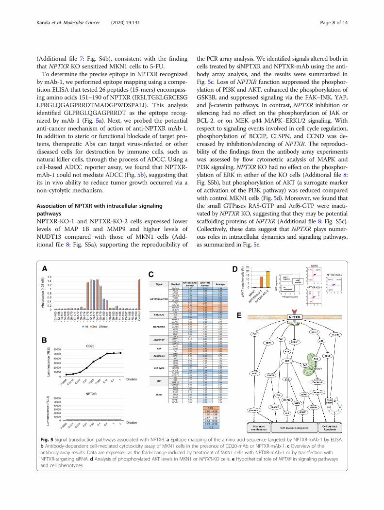
by mAb-1, we performed epitope mapping using a compe-tition ELISA that tested 26 peptides (15-mers) encompass-ing amino acids 151–190 of NPTXR (IRELTGKLGRCESGLPRGLQGAGPRRDTMADGPWDSPALI). This analysisidentified GLPRGLQGAGPRRDT as the epitope recog-nized by mAb-1 (Fig. 5a). Next, we probed the potentialanti-cancer mechanism of action of anti-NPTXR mAb-1.In addition to steric or functional blockade of target pro-teins, therapeutic Abs can target virus-infected or otherdiseased cells for destruction by immune cells, such asnatural killer cells, through the process of ADCC. Using acell-based ADCC reporter assay, we found that NPTXR-mAb-1 could not mediate ADCC (Fig. 5b), suggesting thatits in vivo ability to reduce tumor growth occurred via anon-cytolytic mechanism.
Association of NPTXR with intracellular signalingpathwaysNPTXR-KO-1 and NPTXR-KO-2 cells expressed lowerlevels of MAP 1B and MMP9 and higher levels ofNUDT13 compared with those of MKN1 cells (Add-itional file 8: Fig. S5a), supporting the reproducibility of
the PCR array analysis. We identified signals altered both incells treated by siNPTXR and NPTXR-mAb using the anti-body array analysis, and the results were summarized inFig. 5c. Loss of NPTXR function suppressed the phosphor-ylation of PI3K and AKT, enhanced the phosphorylation ofGSK3B, and suppressed signaling via the FAK–JNK, YAP,and β-catenin pathways. In contrast, NPTXR inhibition orsilencing had no effect on the phosphorylation of JAK orBCL-2, or on MEK–p44 MAPK–ERK1/2 signaling. Withrespect to signaling events involved in cell cycle regulation,phosphorylation of BCCIP, CLSPN, and CCND was de-creased by inhibition/silencing of NPTXR. The reproduci-bility of the findings from the antibody array experimentswas assessed by flow cytometric analysis of MAPK andPI3K signaling. NPTXR KO had no effect on the phosphor-ylation of ERK in either of the KO cells (Additional file 8:Fig. S5b), but phosphorylation of AKT (a surrogate markerof activation of the PI3K pathway) was reduced comparedwith control MKN1 cells (Fig. 5d). Moreover, we found thatthe small GTPases RAS-GTP and Arf6-GTP were inacti-vated by NPTXR KO, suggesting that they may be potentialscaffolding proteins of NPTXR (Additional file 8: Fig. S5c).Collectively, these data suggest that NPTXR plays numer-ous roles in intracellular dynamics and signaling pathways,as summarized in Fig. 5e.
Fig. 5 Signal transduction pathways associated with NPTXR. a Epitope mapping of the amino acid sequence targeted by NPTXR-mAb-1 by ELISA.b Antibody-dependent cell-mediated cytotoxicity assay of MKN1 cells in the presence of CD20-mAb or NPTXR-mAb-1. c Overview of theantibody array results. Data are expressed as the fold-change induced by treatment of MKN1 cells with NPTXR-mAb-1 or by transfection withNPTXR-targeting siRNA. d Analysis of phosphorylated AKT levels in MKN1 or NPTXR-KO cells. e Hypothetical role of NPTXR in signaling pathwaysand cell phenotypes
Kanda et al. Molecular Cancer (2020) 19:131 Page 8 of 14
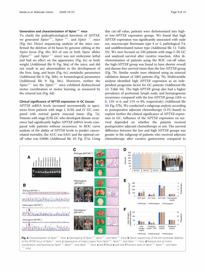
Generation and characterization of Nptxr−/− miceTo clarify the pathophysiological functions of NPTXR,we generated Nptxr+/+, Nptxr +/−
, and Nptxr −/− mice(Fig. 6a). Direct sequencing analysis of the mice con-firmed the deletion of 64 bases by genome editing at theNptxr locus (Fig. 6b). KO of one or both Nptxr alleles(Nptxr+/− and Nptxr−/− mice) was not embryonic lethaland had no effect on the appearance (Fig. 6c) or bodyweight (Additional file 9: Fig. S6a) of the mice, and didnot result in any abnormalities in the development ofthe liver, lung, and brain (Fig. 6c), metabolic parameters(Additional file 9: Fig. S6b), or hematological parameters(Additional file 9: Fig. S6c). Moreover, neither theNptxr+/− nor the Nptxr−/− mice exhibited dysfunctionalmotor coordination or motor learning, as measured bythe rotarod test (Fig. 6d).
Clinical significance of NPTXR expression in GC tissuesNPTXR mRNA levels increased incrementally in speci-mens from patients with stage I, II/III, and IV GC com-pared with normal gastric mucosal tissue (Fig. 7a).Patients with stage II/III GC who developed disease recur-rence had significantly higher NPTXR mRNA levels com-pared with patients without recurrence. In ROC curveanalysis of the ability of NPTXR levels to predict cancer-related mortality, the AUC was 0.651 and the optimal cut-off value was 0.0086 (Additional file 10: Fig. S7a). Using
this cut-off value, patients were dichotomized into high-or low-NPTXR expression groups. We found that highNPTXR expression was significantly associated with malesex, macroscopic Borrmann type 4 or 5, pathological T4,and undifferentiated tumor type (Additional file 11: TableS3). We next focused on 230 patients with stage I–III GCand analyzed survival after curative resection. After di-chotomization of patients using the ROC cut-off value,the high-NPTXR group was found to have shorter overalland disease-free survival times than the low-NPTXR group(Fig. 7b). Similar results were obtained using an externalvalidation dataset of 1065 patients (Fig. 7b). Multivariableanalysis identified high NPTXR expression as an inde-pendent prognostic factor for GC patients (Additional file12: Table S4). The high-NPTXR group also had a higherprevalence of peritoneal, lymph node, and hematogenousrecurrence compared with the low-NPTXR group (16% vs6, 13% vs 6, and 11% vs 8%, respectively) (Additional file10: Fig. S7b). We conducted a subgroup analysis accordingto postoperative adjuvant chemotherapy (5-FU-based) toexplore further the clinical significance of NPTXR expres-sion in GC. Influence of the NPTXR expression on sur-vival depended on whether the patients receivedpostoperative adjuvant chemotherapy or not. The survivaldifference between the low and high NPTXR groups wasgreater in the subgroup of patients who received adjuvantchemotherapy after curative gastrectomy compared to
Fig. 6 Characterization of Nptxr−/− mice. a Genotyping of Nptxr+/+, Nptxr+/−, and Nptxr−/− mice. b Direct sequencing of the 64-nucleotide deletionat the NPTXR locus of Nptxr−/− mice. c Appearance of major organs from Nptxr+/+, Nptxr+/−, and Nptxr −/− mice. d Rotarod test of motorcoordination and learning by Nptxr+/+, Nptxr+/−, and Nptxr −/− mice. e and f Renal e and liver f function tests of Nptxr+/+, Nptxr+/−, and Nptxr−/− mice
Kanda et al. Molecular Cancer (2020) 19:131 Page 9 of 14

Fig. 7 Clinical significance of NPTXR expression in gastric tissues from patients with GC. a qRT-PCR analysis of NPTXR mRNA levels in 80 pairs ofGC and matched adjacent normal gastric tissues according to disease stage and the presence or absence of disease recurrence (Rec). b Kaplan–Meier curves of overall and disease-free survival of patients in the institutional and validation cohorts with stage I–III GC according to NPTXRexpression. c Representative images of IHC staining of NPTXR protein expression in gastric tissue sections. Left panel, section with undetectableNPTXR expression; right panels, sections with positive NPTXR expression
Kanda et al. Molecular Cancer (2020) 19:131 Page 10 of 14

that of patients who underwent surgery alone (Add-itional file 10: Fig. S7c). These results suggest thatNPTXR expression reflects resistance to adjuvantchemotherapy.
IHC analysis of NPTXR expression in gastric tissuesFinally, we explored the utility of the anti-NPTXRpAb-1 as a potential diagnostic tool by performingIHC staining of GC tissues. Images of representativesections with negative or positive NPTXR immunore-activity are shown in Fig. 7c. Among the 80 patientsassessed, 32 were positive for NPTXR in the primarytumor, and the incidence of disease recurrence washigher in patients with NPTXR-positive tumors com-pared with those with NPTXR-negative tumors (31and 8%, respectively, P = 0.009; Fig. 7c).
DiscussionMetastasis is a major cause of death in patients with GC[1, 19]. A better understanding of the molecular mecha-nisms underlying metastasis is therefore essential for de-veloping novel and innovative therapeutic strategies andrisk assessment biomarkers [20, 21]. As a first step to-wards this, we conducted transcriptome analysis of GCtissues from patients with and without metastasis andidentified NPTXR as one of 14 genes with significantoverexpression in GC tissues metastasized to diverse or-gans. We generated GC cell lines with NPTXR KD, KO,and overexpression, developed anti-NPTXR pAbs andmAbs, investigated the involvement of NPTXR in GCcell functions in vitro and in vivo, and generated a newstrain of Nptxr−/− mice. Our results indicate that NPTXRregulates several cell behaviors associated with the ma-lignant phenotype and metastatic potential of GC cellsvia multiple intracellular signaling pathways, includingthe PI3K–AKT–mTOR and FAK–JNK pathways. Wealso showed that anti-NPTXR pAb and mAbs had thera-peutic effects in a mouse model of peritoneal metastasis.It is a significant advance in further understanding ofphysiological roles of NPTXR that we first found no ad-verse effects on reproduction, development, or metabolicor motor functions by genetic deletion of NPTXR inmice. Collectively, these results not only suggest thatNPTXR may be an excellent therapeutic target for meta-static GC but also indicate that NPTXR-targeting mAbscan effectively suppress GC growth in vivo in the ab-sence of ADCC.NPTXR is a receptor for synapse-associated neuronal
NPTX, and it is expressed at higher levels in the braincompared with other organs [11, 22]. Among the NPTXfamily proteins, NPTXR is unique in possessing a trans-membrane domain and in its ability to anchor NPTXcomplexes to plasma membranes [23, 24]. NPTXR is alsocleaved by the extracellular protease tumor necrosis
factor-α converting enzyme to release a soluble form ofNPTXR [10, 25]. Members of the NPTX family functionduring synapse formation and remodeling by facilitatingthe uptake of synaptic materials [24, 25]. NPTXR andNPTX2 are overexpressed in human Schwannian stroma-poor neuroblastoma [12, 22, 26], and NPTX2 expressionserves as a marker of poor prognosis of neuroblastoma pa-tients [12]. Bartolini et al. investigated expression andfunctions of NPTX1, NPTX2 and NPTXR in glioblastoma[12]. They found that blockade of ligand-receptor connec-tion between NPTX2 and NPTXR reduces tumor burdenin orthotopic mouse models of human neuroblastoma[12]. However, the oncological roles of NPTXR in gastro-enterological malignancies are unknown.We focused on determining the involvement of
NPTXR in the malignant phenotype of GC cells by gene-specific ablation or overexpression and Ab-mediated in-hibition of function. NPTXR KD, KO, or Ab-mediatedblockade significantly inhibited the proliferation of mul-tiple GC cell lines, possibly via facilitation of apoptosisand/or induction of G2/M arrest [27]. Apoptosis is pre-dominantly mediated via two pathways; the intrinsicmitochondria pathway, which involves activation of cas-pases and the disruption of mitochondrial membranepotential, and the extrinsic death receptor pathway,which requires engagement of cell surface death recep-tors [28, 29]. Notably, NPTXR KO increased caspase ac-tivity, particularly caspase-9, and induced a strikingdecrease in mitochondrial membrane potential. Caspase-9 is a cysteine-aspartic protease associated with cytokinesignaling and apoptosis [30, 31]. Apoptotic signals causethe release of cytochrome c from mitochondria and acti-vation of apoptosis protease-activating factor 1, whichthen cleaves procaspase-9 into the active homodimer[30, 31]. Our results thus suggest that NPTXR regulatesapoptosis through mitochondrial-dependent activationof caspase-9.The involvement of NPTXR in oncogenesis is also sup-
ported by our findings that NPTXR KO attenuated manybiological properties (migration, invasion, and stemness)required for GC metastasis. High ALDH activity is a hall-mark of cancer stem cells [32], which are able to evade theeffects of chemotherapy and radiotherapy through theirability to self-renew and differentiate into tumor cells aftertreatment is discontinued, leading to recurrence and/ormetastasis [33]. In the present study, NPTXR KO de-creased the frequency of ALDH-positive GC cells, indicat-ing that NPTXR influences the stemness of GC cells. Wespeculate that the decreased stemness of NPTXR-KO cellsmay be associated with their increased sensitivity to 5-FU.NPTXR KO also resulted in a reduction of tumor growthin the mouse xenograft model of s.c. GC.Consistent with the paucity of information on the roles
of NPTXR in oncogenesis, little is known about the
Kanda et al. Molecular Cancer (2020) 19:131 Page 11 of 14
interactions between NPTXR and cancer-related path-ways. We explored NPTXR-associated gene expressionusing two approaches. First, we performed PCR arrayanalysis, which showed that NPTXR expression may in-fluence or be influenced by MAP 1B, MMP9, andNUDT13, which contribute to the EMT, a crucialprocess in carcinogenesis. MAP 1B is upregulated incells during an EMT-like process that operates duringwound healing [34, 35]. MMP9 is a matrix metallopro-teinase involved in the degradation of the ECM inphysiological processes such as tissue remodeling andembryonic development, as well as in pathological pro-cesses such as the progression of arthritis and cancermetastasis [36, 37]. NUDT13 is a mitochondrial enzymereported to be downregulated during the EMT [38]. Asa second method to explore cancer-related pathways, weperformed antibody array analysis to identify alterationsin the phosphorylation of intracellular signaling proteinsGC cells with loss of NPTXR expression or function.NPTXR KO or mAb treatment resulted in inactivationof the PI3K–AKT–GSK3B, FAK–JNK, and YAP signal-ing pathways, but not of MAPK and JAK–STAT signal-ing. PI3K–AKT–mTOR is a key signaling pathwayinvolved in numerous cellular processes [39]; YAP1modulates cancer stem cell properties, such as sphereformation, self-renewal, invasiveness, and drug resist-ance, via the Hippo and Wnt/β-catenin pathways [40];and FAK regulates cell growth, survival, migration, andinvasion through its dual functions as a kinase and ascaffold protein [41]. FAK autophosphorylation leads tothe recruitment of Src and the phosphorylation of bind-ing sites for downstream effector pathways, includingJNK and the transcriptional regulation of pro-invasivegenes like MMP9 [41, 42]. Previous studies indicate thatthe PI3K–AKT–GSK3B, FAK–JNK, and YAP signalingpathways interact with each other during cancer devel-opment and progression. Kinase function of FAK hasbeen shown to activate the PI3K-AKT pathway [43].FAK binds p85 subunit of PI3K, and phospholipid pro-duction stimulated by FAK association and activation ofPI3K can activate AKT kinase, leading to inhibition ofapoptosis by regulating various cell death machineryproteins [43, 44]. Moreover, it has been reported thatconcomitant activation of YAP1 and PI3K promotes de-velopment of liver tumors through activation of AKT/mTOR signaling [40]. Our findings suggest that NPTXRmodulates the network of AKT, FAK and YAP pathwaysand contributes to GC progression.We investigated the potential therapeutic effects of anti-
NPTXR Abs using a mouse xenograft model of peritonealmetastasis. Both pAbs and mAbs significantly inhibitedtumor formation in this model and the anti-NPTXRmAbs also enhanced the cytotoxicity of 5-FU and cisplatinin vitro, supporting the possibility that NPTXR may be a
promising therapeutic target for controlling GC progres-sion and/or for overcoming chemotherapy resistance. Wealso showed that the anti-cancer effects of the mAbsin vivo resulted from NPTXR antagonism rather than asmediators of ADCC [45]. Finally, we identified the preciseepitope recognized by our NPTXR mAbs, which is a crit-ical step in developing and engineering therapeutic mAbs.Further work, including humanization of the mAbs andadditional preclinical efficacy and toxicity testing, will berequired before these promising findings can be translatedto the clinic. Along these lines, another important findingin the present study is that Nptxr−/− mice exhibit no ab-normalities in reproduction, development, metabolism, ormotor functions, and that treatment of mice for up to 6weeks with anti-NPTXR pAbs and mAbs elicited no overtclinical signs or adverse effects. These are important ob-servations for understanding the potential effects of clin-ical NPTXR targeting on physiological homeostasis.Our findings have clinical relevance, as demonstrated
by the association between NPTXR expression in patientspecimens and outcomes. We found that NPTXR mRNAlevel in clinical samples is an independent prognosticfactor in GC, and these findings were confirmed usingan independent external validation dataset. Our IHCanalysis also indicated that NPTXR protein expressionmay serve as a novel biomarker of disease recurrence,which can easily be determined through analysis of sur-gical specimens or endoscopic biopsy samples. Further,like HER2 amplification NPTXR expression may be uti-lized, for example, as a companion diagnostic to identifypatients potentially sensitive to anti-NPTXR therapy.Several limitations of our study should be acknowl-
edged. It remains unclear whether NPTX1 and NPTX2act as ligands of NPTXR in GC cells or not. We evaluatedthe effects of anti-NPTXR pAbs and mAbs only in amouse model, and the frequency, administration route,optimal dose, and optimal duration of treatment and sur-vival benefits were not determined. Patient-derived xeno-graft models may be helpful for understanding theoncological involvement of NPTXR. The development ofassays to detect NPTXR expression levels in serum willlikely facilitate the translation of our findings to the clinic.
ConclusionsWe identified NPTXR as a cell surface receptor thatmodulates multiple oncological processes in GC cells,including behaviors associated with metastasis. NPTXR-targeting Abs may have utility in GC, not only as in-novative therapeutic agents but also as companion diag-nostic tools.
Supplementary informationSupplementary information accompanies this paper at https://doi.org/10.1186/s12943-020-01251-0.
Kanda et al. Molecular Cancer (2020) 19:131 Page 12 of 14
Additional file 1. Supplementary Materials and Methods.
Additional file 2: Table S1. Nucleotide sequences.
Additional file 3: Figure S1. Extraction diagram of candidate genesidentified by global expression analysis. Sixteen patients were categorizedinto four groups (4 patients for each) according to clinical courses asfollows: group 1, no recurrences for > 5 years; group 2, hepatic-confinedmetastasis within 2 years after surgery; group 3, peritoneal metastasiswithin 2 years after surgery; and group 4, nodal metastasis within 2 yearsafter surgery. Global expression profiling was conducted to compare theexpression levels of 57,749 genes between each cluster. Among them, 14we considered 14 genes as candidates shown in Supplemental Table 2.
Additional file 4: Table S2. List of candidate genes upregulated ingastric cancer tissues of patients with metachronous metastasis.
Additional file 5: Figure S2. Verification of NPTXR knockdown oroverexpression in GC cells. a-c qRT-PCR analysis of NPTXR mRNA expres-sion in MKN1, N87, and/or NUGC3 cells with siRNA-mediated knockdown(a), shRNA-mediated knockdown (b), or overexpression (c) of NPTXR. *P <0.05. Mean ± standard deviation.
Additional file 6: Figure S3. Characterization of polyclonal anti-NPTXRAbs effects in vitro and in vivo. a CCK-8 proliferation assay of MKN1 cellsincubated with pAbs in vitro. b Therapeutic effects of intraperitoneal ad-ministration of anti-NPTXR polyclonal antibodies. Macroscopic appearanceof peritoneal nodules 6 weeks after injection of MKN1 cells and treatmentwith control IgG or NPTXR-pAbs. c Total volumes of peritoneal nodulescollected from BALB/c nu/nu mice 6 weeks after injection of MKN1 cellsand treatment with control IgG or NPTXR-mAb-1, − 2, or − 3. *P < 0.05.Mean ± standard deviation.
Additional file 7: Figure S4. Effects of NPTXR-mAb-1 on gastric cancercells. CCK-8 proliferation assay of MKN45 (a) and N87 (b) cells incubatedwith NPTXR-mAb-1 in dose-dependent manner.
Additional file 8: Figure S5. a qRT-PCR analysis of NUDT13, MAP 1B,MMP9 mRNA levels in parental MKN1, NPTXR-KO-1 and -KO-2 cells. b Flowcytometric analysis of the frequency parental and NPTXR-KO MKN1 cellswith phosphorylated ERK1/2. c Pull-down of small GTPases (Ras and Arf6)in parental and NPTXR-KO MKN1 cells.
Additional file 9: Figure S6. a Body weights of Nptxr+/+, Nptxr+/−, andNptxr−/− mice at 4 and 8 weeks after birth. b and c Metabolic (b) andhematological (c) tests in Nptxr+/+, Nptxr+/−, and Nptxr−/− mice.
Additional file 10: Figure S7. a ROC curve analysis of the ability ofNPTXR expression level in tissue specimens to predict peritonealmetastasis in GC patients. b Frequency of the site of initial recurrence inGC patients according to NPTXR expression level. c Disease-free survivalrates in subgroups according to administration of adjuvantchemotherapy.
Additional file 11: Table S3. Patients’ clinical characteristics associatedwith NPTXR expression.
Additional file 12: Table S4. Prognostic factors of patients withresectable gastric cancer.
AbbreviationsALDH: Aldehyde dehydrogenase; AUC: Area under the curve;IHC: Immunohistochemistry; NPTXR: Neuronal pentraxin receptor; qRT-PCR: Quantitative reversetranscription polymerase chain reaction;ROC: Receiver operating characteristic; siRNA: Small interfering RNA
AcknowledgementsWe thank Edanz Group (www.edanzediting.com/ac) for editing a draft of thismanuscript.
Authors’ contributionsMK (M. Kanda) designed the study, acquired materials and wrote themanuscript. DS and CT performed expression analyses including qPCR assayand immunohistochemical staining of the human tissues. HT and TMperformed and analyzed the in vitro experiments. SU and SN performed thein vivo experiments. YI and KS analyzed knockout mice. SY and MH criticallyreviewed the manuscript. YK and MK (M. Katsuno) supervised the study andrevised the manuscript. All authors read and approved the final manuscript.
FundingThis work was supported by the Japan Agency for Medical Research andDevelopment (17lm0203005j0001, 2017; 19cm0106163h0001, 2019–2020),the Center for Advanced Medicine and Clinical Research at NagoyaUniversity 2018, grant 2018 from the Kobayashi Foundation for CancerResearch, and the Mochida Memorial Foundation for Medical andPharmaceutical Research.
Availability of data and materialsAll the data obtained and/or analyzed during the current study wereavailable from the corresponding authors on reasonable request.
Ethics approval and consent to participateThis study was approved by the institutional review board/ethics committeeof the Nagoya University Hospital (2014–0043).
Consent for publicationAll authors give consent for the publication of manuscript in MolecularCancer.
Competing interestsThe authors declare that there is no potential competing interest.
Author details1Department of Gastroenterological Surgery (Surgery II), Nagoya UniversityGraduate School of Medicine, 65 Tsurumai-cho, Showa-ku, Nagoya 466-8550,Japan. 2Department of Neurology, Nagoya University Graduate School ofMedicine, Nagoya, Japan.
Received: 23 June 2020 Accepted: 17 August 2020
References1. Van Cutsem E, Sagaert X, Topal B, Haustermans K, Prenen H. Gastric cancer.
Lancet. 2016;388:2654–64.2. Thrift AP, El-Serag HB. Burden of gastric Cancer. Clin Gastroenterol Hepatol.
2020;18:534–42.3. Wilke H, Muro K, Van Cutsem E, Oh SC, Bodoky G, Shimada Y, Hironaka S,
Sugimoto N, Lipatov O, Kim TY, et al. Ramucirumab plus paclitaxel versusplacebo plus paclitaxel in patients with previously treated advanced gastricor gastro-oesophageal junction adenocarcinoma (RAINBOW): a double-blind, randomised phase 3 trial. Lancet Oncol. 2014;15:1224–35.
4. Muro K, Chung HC, Shankaran V, Geva R, Catenacci D, Gupta S, Eder JP,Golan T, Le DT, Burtness B, et al. Pembrolizumab for patients with PD-L1-positive advanced gastric cancer (KEYNOTE-012): a multicentre, open-label,phase 1b trial. Lancet Oncol. 2016;17:717–26.
5. Shitara K, Ozguroglu M, Bang YJ, Di Bartolomeo M, Mandala M, Ryu MH,Fornaro L, Olesinski T, Caglevic C, Chung HC, et al. Pembrolizumab versuspaclitaxel for previously treated, advanced gastric or gastro-oesophagealjunction cancer (KEYNOTE-061): a randomised, open-label, controlled, phase3 trial. Lancet. 2018;392:123–33.
6. Fuchs CS, Tomasek J, Yong CJ, Dumitru F, Passalacqua R, Goswami C, SafranH, dos Santos LV, Aprile G, Ferry DR, et al. Ramucirumab monotherapy forpreviously treated advanced gastric or gastro-oesophageal junctionadenocarcinoma (REGARD): an international, randomised, multicentre,placebo-controlled, phase 3 trial. Lancet. 2014;383:31–9.
7. Kanda M, Suh YS, Park DJ, Tanaka C, Ahn SH, Kong SH, Lee HJ, Kobayashi D,Fujiwara M, Shimada H, et al. Serum levels of ANOS1 serve as a diagnosticbiomarker of gastric cancer: a prospective multicenter observational study.Gastric Cancer. 2020;23:203–11.
8. Lei Z, Tan IB, Das K, Deng N, Zouridis H, Pattison S, Chua C, Feng Z, GuanYK, Ooi CH, et al. Identification of molecular subtypes of gastric cancer withdifferent responses to PI3-kinase inhibitors and 5-fluorouracil.Gastroenterology. 2013;145:554–65.
9. Tan IB, Ivanova T, Lim KH, Ong CW, Deng N, Lee J, Tan SH, Wu J, Lee MH,Ooi CH, et al: Intrinsic subtypes of gastric cancer, based on gene expressionpattern, predict survival and respond differently to chemotherapy.Gastroenterology 2011, 141:476–485, 85 e1–11.
10. Lee SJ, Wei M, Zhang C, Maxeiner S, Pak C, Calado Botelho S, Trotter J,Sterky FH, Südhof TC. Presynaptic Neuronal Pentraxin Receptor OrganizesExcitatory and Inhibitory Synapses. J Neurosci. 2017;37:1062–80.
Kanda et al. Molecular Cancer (2020) 19:131 Page 13 of 14
11. Dodds DC, Omeis IA, Cushman SJ, Helms JA, Perin MS. Neuronal pentraxinreceptor, a novel putative integral membrane pentraxin that interacts withneuronal pentraxin 1 and 2 and taipoxin-associated calcium-binding protein49. J Biol Chem. 1997;272:21488–94.
12. Bartolini A, Di Paolo D, Noghero A, Murgia D, Sementa AR, Cilli M, PasqualiniR, Arap W, Bussolino F, Ponzoni M, et al. The neuronal Pentraxin-2 pathwayis an unrecognized target in human neuroblastoma, which also offersprognostic value in patients. Cancer Res. 2015;75:4265–71.
13. Szasz AM, Lanczky A, Nagy A, Forster S, Hark K, Green JE, Boussioutas A,Busuttil R, Szabo A, Gyorffy B. Cross-validation of survival associatedbiomarkers in gastric cancer using transcriptomic data of 1,065 patients.Oncotarget. 2016;7:49322–33.
14. Kanda M, Shimizu D, Tanaka H, Tanaka C, Kobayashi D, Hayashi M, Iwata N,Niwa Y, Yamada S, Fujii T, et al. Significance of SYT8 for the detection,prediction, and treatment of peritoneal metastasis from gastric Cancer. AnnSurg. 2018;267:495–503.
15. Miwa T, Kanda M, Umeda S, Tanaka H, Shimizu D, Tanaka C, Kobayashi D,Hayashi M, Yamada S, Nakayama G, et al. Establishment of peritoneal andhepatic metastasis mouse Xenograft models using gastric Cancer cell lines.Vivo. 2019;33:1785–92.
16. Kanda M, Tanaka H, Shimizu D, Miwa T, Umeda S, Tanaka C, Kobayashi D,Hattori N, Suenaga M, Hayashi M, et al. SYT7 acts as a driver of hepaticmetastasis formation of gastric cancer cells. Oncogene. 2018;37:5355–66.
17. Wang H, Yang H, Shivalila CS, Dawlaty MM, Cheng AW, Zhang F, Jaenisch R.One-step generation of mice carrying mutations in multiple genes by CRISPR/Cas-mediated genome engineering. Cell. 2013;153:910–8.
18. Katsuno M, Adachi H, Kume A, Li M, Nakagomi Y, Niwa H, Sang C,Kobayashi Y, Doyu M, Sobue G. Testosterone reduction prevents phenotypicexpression in a transgenic mouse model of spinal and bulbar muscularatrophy. Neuron. 2002;35:843–54.
19. Wang SM, Tie J, Wang WL, Hu SJ, Yin JP, Yi XF, Tian ZH, Zhang XY, Li MB, LiZS, et al. POU2F2-oriented network promotes human gastric cancermetastasis. Gut. 2016;65:1427–38.
20. Razzak M. Genetics: new molecular classification of gastric adenocarcinomaproposed by the Cancer genome atlas. Nat Rev Clin Oncol. 2014;11:499.
21. Liu X, Zhang X, Zhang Z, Chang J, Wang Z, Wu Z, Wang C, Sun Z, Ge X,Geng R, et al. Plasma microRNA-based signatures to predict 3-yearpostoperative recurrence risk for stage II and III gastric cancer. Int J Cancer.2017;141:2093–102.
22. Yin GN, Lee HW, Cho JY, Suk K. Neuronal pentraxin receptor incerebrospinal fluid as a potential biomarker for neurodegenerative diseases.Brain Res. 2009;1265:158–70.
23. Andreiuolo F, Puget S, Peyre M, Dantas-Barbosa C, Boddaert N, Philippe C,Mauguen A, Grill J, Varlet P. Neuronal differentiation distinguishessupratentorial and infratentorial childhood ependymomas. Neuro-Oncology.2010;12:1126–34.
24. Pelkey KA, Barksdale E, Craig MT, Yuan X, Sukumaran M, Vargish GA, MitchellRM, Wyeth MS, Petralia RS, Chittajallu R, et al. Pentraxins coordinateexcitatory synapse maturation and circuit integration of parvalbumininterneurons. Neuron. 2015;85:1257–72.
25. Kirkpatrick LL, Matzuk MM, Dodds DC, Perin MS. Biochemical interactions ofthe neuronal pentraxins. Neuronal pentraxin (NP) receptor binds to taipoxinand taipoxin-associated calcium-binding protein 49 via NP1 and NP2. J BiolChem. 2000;275:17786-92.
26. von Roemeling CA, Radisky DC, Marlow LA, Cooper SJ, Grebe SK,Anastasiadis PZ, Tun HW, Copland JA. Neuronal pentraxin 2 supports clearcell renal cell carcinoma by activating the AMPA-selective glutamatereceptor-4. Cancer Res. 2014;74:4796–810.
27. Acikgoz E, Guven U, Duzagac F, Uslu R, Kara M, Soner BC, Oktem G.Enhanced G2/M arrest, Caspase Related Apoptosis and Reduced E-CadherinDependent Intercellular Adhesion by Trabectedin in Prostate Cancer StemCells. PLoS One. 2015;10:e0141090.
28. Liu XD, Fan RF, Zhang Y, Yang HZ, Fang ZG, Guan WB, Lin DJ, Xiao RZ,Huang RW, Huang HQ, et al. Down-regulation of telomerase activity andactivation of caspase-3 are responsible for Tanshinone I-induced apoptosisin monocyte leukemia cells in vitro. Int J Mol Sci. 2010;11:2267–80.
29. Samykutty A, Shetty AV, Dakshinamoorthy G, Kalyanasundaram R, Zheng G,Chen A, Bosland MC, Kajdacsy-Balla A, Gnanasekar M. Vitamin k2, a naturallyoccurring menaquinone, exerts therapeutic effects on both hormone-dependent and hormone-independent prostate cancer cells. Evid BasedComplement Alternat Med. 2013;2013:287358.
30. Cheng CH, Cheng YP, Chang IL, Chen HY, Wu CC, Hsieh CP. Dodecyl gallateinduces apoptosis by upregulating the caspase-dependent apoptoticpathway and inhibiting the expression of anti-apoptotic Bcl-2 familyproteins in human osteosarcoma cells. Mol Med Rep. 2016;13:1495–500.
31. Poli G, Guasti D, Rapizzi E, Fucci R, Canu L, Bandini A, Cini N, Bani D,Mannelli M, Luconi M. Morphofunctional effects of mitotane onmitochondria in human adrenocortical cancer cells. Endocr Relat Cancer.2013;20:537–50.
32. Mele L, Liccardo D, Tirino V. Evaluation and isolation of Cancer stem cellsusing ALDH activity assay. Methods Mol Biol. 1692;2018:43–8.
33. Zhou Y, Xia L, Wang H, Oyang L, Su M, Liu Q, Lin J, Tan S, Tian Y, Liao Q,Cao D. Cancer stem cells in progression of colorectal cancer. Oncotarget.2018;9:33403–15.
34. Tessema M, Yingling CM, Picchi MA, Wu G, Liu Y, Weissfeld JL, Siegfried JM,Tesfaigzi Y, Belinsky SA. Epigenetic repression of CCDC37 and MAP 1B linkschronic obstructive pulmonary disease to lung Cancer. J Thorac Oncol.2015;10:1181–8.
35. Yasuda T, Iizuka K, Fukuda M, Takeda J, Natsume T, Horikawa Y, Gadoth A,Kryzer TJ, Fryer J, McKeon A, et al. Microtubule-associated protein 1B: novelparaneoplastic biomarker. Sci Rep. 2017;81:266–77.
36. Qin Z, Wang Y, Tang J, Zhang L, Li R, Xue J, Han P, Wang W, Qin C, Xing Q,et al. High LINC01605 expression predicts poor prognosis and promotestumor progression via up-regulation of MMP9 in bladder cancer. Biosci Rep.2018;38.
37. Wang X, Wang B, Xie J, Hou D, Zhang H, Huang H. Melatonin inhibitsepithelialtomesenchymal transition in gastric cancer cells via attenuation ofIL1beta/NFkappaB/MMP2/MMP9 signaling. Int J Mol Med. 2018;42:2221–8.
38. McLennan AG. The Nudix hydrolase superfamily. Cell Mol Life Sci. 2006;63:123–43.
39. Singh SS, Yap WN, Arfuso F, Kar S, Wang C, Cai W, Dharmarajan AM, Sethi G,Kumar AP. Targeting the PI3K/Akt signaling pathway in gastric carcinoma: areality for personalized medicine? World J Gastroenterol. 2015;21:12261–73.
40. Shibata M, Ham K, Hoque MO. A time for YAP1: tumorigenesis,immunosuppression and targeted therapy. Int J Cancer. 2018;143:2133–44.
41. Murphy JM, Jeong K, Rodriguez YAR, Kim JH, Ahn EE, Lim SS. FAK and Pyk2activity promote TNF-α and IL-1β-mediated pro-inflammatory geneexpression and vascular inflammation. Sci Rep. 2019;9:7617.
42. Zhang Y, Peng X, Zheng Q. Ropivacaine inhibits the migration ofesophageal cancer cells via sodium-channel-independent but prenylation-dependent inhibition of Rac1/JNK/paxillin/FAK. Biochem Biophys ResCommun. 2018;501:1074–9.
43. Sulzmaier FJ, Jean C, Schlaepfer DD. FAK in cancer: mechanistic findingsand clinical applications. Nat Rev Cancer. 2014;14:598–610.
44. Kurenova E, Xu LH, Yang X, Baldwin AS Jr, Craven RJ, Hanks SK, Liu ZG,Cance WG. Focal adhesion kinase suppresses apoptosis by binding to thedeath domain of receptor-interacting protein. Mol Cell Biol. 2004;24:4361–71.
45. Martin TC, Ilieva KM, Visconti A, Beaumont M, Kiddle SJ, Dobson RJB,Mangino M, Lim EM, Pezer M, Steves CJ, et al. Dysregulated antibody,natural killer cell and immune mediator profiles in autoimmune thyroiddiseases. Cells. 2020;9.
Publisher’s NoteSpringer Nature remains neutral with regard to jurisdictional claims inpublished maps and institutional affiliations.
Kanda et al. Molecular Cancer (2020) 19:131 Page 14 of 14