The regulation of phosphorylation of τ in SY5Y neuroblastoma cells: the role of protein...
7
The regulation of phosphorylation of d in SY5Y neuroblastoma cells : the role of protein phosphatases Toshihisa Tanaka b , Jun Zhong a , Khalid Iqbal a , Ekkhart Trenkner a , Inge Grundke-Iqbal a; * a New York State Institute for Basic Research in Developmental Disabilities, 1050 Forest Hill Road, Staten Island, NY 10314, USA b Osaka Medical School, Department of Psychiatry, Osaka, Japan Received 23 February 1998; revised version received 16 March 1998 Abstract In Alzheimer disease brain the microtubule associated protein (MAP) d is abnormally hyperphosphorylated. The role of protein phosphatases (PP) in the regulation of phosphorylation of d was studied in undifferentiated SY5Y cells. In cells treated with 10 nM okadaic acid (OA), a PP-2A/PP-1 inhibitor, the PP- 1 and -2A activities decreased by 60% and 100% respectively and the activities of MAPKs, cdc2 kinase and cdk5, but not of GSK- 3, increased. OA increased the phosphorylation of d at Thr-231/ Ser-235 and Ser-396/404, but not at Ser-262/356 or Ser-199/202. An increase in tyrosinated/detyrosinated tubulin ratio, a decrease in the microtubule binding activities of d, MAP1b and MAP2, and cell death were observed. Treatment with 1 Wm taxol partially inhibited the cell death. These data suggest (1) that OA induced hyperphosphorylation of d is probably the result of activated MAPK and cdks in addition to decreased PP-2A and PP-1 activities and (2) that in SY5Y cells the OA induced cell death is associated with a decrease in stable microtubules. z 1998 Federation of European Biochemical Societies. Key words: Microtubule associated protein ; Microtubule ; Protein phosphatase 2A; Proline dependent protein kinase; Alzheimer disease ; SY5Y cell 1. Introduction Accumulation of paired helical ¢laments (PHFs) in selected neurons is one of the histopathological hallmarks of the Alz- heimer disease (AD) brain, and the microtubule associated protein (MAP) d is the major protein subunit of PHF [1,2]. In the AD brain, d protein, the biological activity of which is regulated by its degree of phosphorylation [3], is abnormally hyperphosphorylated [4]. The AD hyperphosphorylated d (ADP-d) contains 5^9 mol phosphate per protein molecule as compared with 2^3 mol phosphate per mole normal d [5]. To date, 21 phosphorylated Ser/Thr sites have been identi¢ed in PHF-d [6]. The ADP-d does not promote assembly of tu- bulin into microtubules [7]; it sequesters normal d and high molecular weight MAPs, MAP1 and MAP2, and causes dis- assembly of microtubules in vitro [8]. The disruption of micro- tubules and the consequent compromised axoplasmic trans- port are believed to be responsible for the retrograde degeneration in neurons with neuro¢brillary pathology in AD brain [7,8]. Therefore, understanding of the processes leading to hyperphosphorylation of d is important. Several kinases and phosphatases are known to be involved in the regulation of phosphorylation of d. The proline directed kinases, i.e. mitogen activated protein kinases (MAPKs) [9,10], cyclin dependent kinases (cdks) [10,11], and glycogen synthase kinase-3 (GSK-3) [12], are thought to be active par- ticipants in d phosphorylation in the brain since these kinases can phosphorylate d in vitro at many of the sites characteristic for AD. To date, phosphorylation of only Ser and Thr and not of Tyr has been observed in AD d. Among the Ser/Thr protein phosphatases, PP-1, -2A and -2B have been shown to catalyze dephosphorylation of the ADP-d [13,14]. Furthermore, the activities of PP-1 and PP-2A towards phosphorylase kinase (Phk) and the phosphatase activity towards the ADP-d are compromised in AD brain [15,16]. Previously we reported that SH-SY5Y human neuroblasto- ma cells, if grown in 5% fetal calf serum, contained d hyper- phosphorylated at several of the same sites as in AD and it had less ability to bind to microtubules [17]. Furthermore, d hyperphosphorylation by treatment with okadaic acid (OA), a PP-2A and PP-1 inhibitor, and its decrease in biological ac- tivity had been reported in di¡erent cell lines [18]. However, the protein kinase and protein phosphatase activities involved in the phosphorylation of d in these cells were not known. In the present study, phosphatase inhibitors were employed to elucidate the role of protein phosphatases in the phosphory- lation of d and the regulation of the microtubule system. We found that in SY5Y cells OA inhibits the activities of PP-2A and PP-1 and increases in an acute fashion the activities of MAPK, cdc2 kinase and cdk5. These changes in phosphatase and kinase activities alter the levels of phosphorylation of d. Furthermore, OA and other PP-1 and -2A inhibitors produce degeneration of SY5Y cells that is accompanied by a decrease of detyrosinated tubulin (stable microtubules), and this degen- eration is inhibited by taxol, a microtubule stabilizer. 2. Materials and methods 2.1. Reagents OA, calyculin A, and tautomycin were purchased from Calbiochem (San Diego, CA), deltamethrin from Alomone Labs (Jerusalem, Isra- el) and taxol (paclitaxel) from Sigma (St. Louis, MO.). Cyclosporin A was a gift from Sandoz (Tokyo, Japan). 2.2. Antibodies and recombinant d The following site speci¢c phosphorylation dependent antibodies to d, d-1 (d not phosphorylated at Ser-195, Ser-198, Ser-199, or Ser-202 [19]), PHF-1 (d phosphorylated at Ser-396, Ser-404 [20]), M4 (d phos- phorylated at Thr-231, Ser-235 [21]), 12E8 (d phosphorylated at Ser- 262, Ser-356 [22]) and R145d (d phosphorylated at Ser-422) were employed. Rabbit antiserum R145d was raised against the synthetic peptide corresponding to residues 417^426 of d 410 , with Ser-422 phos- phorylated. The peptide was conjugated to keyhole limpet hemocya- nin and injected into two rabbits (400 Wg peptide at the initial immu- nization and 200 Wg peptide for the booster injections in 3 weekly intervals). Serum from one of the rabbits reacted strongly with neuro- ¢brillary tangles in AD hippocampus and on Western blots with ADP-d but not with recombinant or normal brain d. Other primary 0014-5793/98/$19.00 ß 1998 Federation of European Biochemical Societies. All rights reserved. PII S0014-5793(98)00346-9 *Corresponding author. Fax: (1) (718) 494-1080. E-mail: [email protected] FEBS 20113 FEBS Letters 426 (1998) 248^254
-
Upload
toshihisa-tanaka -
Category
Documents
-
view
212 -
download
0
Transcript of The regulation of phosphorylation of τ in SY5Y neuroblastoma cells: the role of protein...
The regulation of phosphorylation of d in SY5Y neuroblastoma cells :the role of protein phosphatases
Toshihisa Tanakab, Jun Zhonga, Khalid Iqbala, Ekkhart Trenknera, Inge Grundke-Iqbala;*aNew York State Institute for Basic Research in Developmental Disabilities, 1050 Forest Hill Road, Staten Island, NY 10314, USA
bOsaka Medical School, Department of Psychiatry, Osaka, Japan
Received 23 February 1998; revised version received 16 March 1998
Abstract In Alzheimer disease brain the microtubule associatedprotein (MAP) dd is abnormally hyperphosphorylated. The role ofprotein phosphatases (PP) in the regulation of phosphorylationof dd was studied in undifferentiated SY5Y cells. In cells treatedwith 10 nM okadaic acid (OA), a PP-2A/PP-1 inhibitor, the PP-1 and -2A activities decreased by 60% and 100% respectively andthe activities of MAPKs, cdc2 kinase and cdk5, but not of GSK-3, increased. OA increased the phosphorylation of dd at Thr-231/Ser-235 and Ser-396/404, but not at Ser-262/356 or Ser-199/202.An increase in tyrosinated/detyrosinated tubulin ratio, a decreasein the microtubule binding activities of dd, MAP1b and MAP2,and cell death were observed. Treatment with 1 WWm taxolpartially inhibited the cell death. These data suggest (1) that OAinduced hyperphosphorylation of dd is probably the result ofactivated MAPK and cdks in addition to decreased PP-2A andPP-1 activities and (2) that in SY5Y cells the OA induced celldeath is associated with a decrease in stable microtubules.z 1998 Federation of European Biochemical Societies.
Key words: Microtubule associated protein; Microtubule;Protein phosphatase 2A; Proline dependent protein kinase;Alzheimer disease; SY5Y cell
1. Introduction
Accumulation of paired helical ¢laments (PHFs) in selectedneurons is one of the histopathological hallmarks of the Alz-heimer disease (AD) brain, and the microtubule associatedprotein (MAP) d is the major protein subunit of PHF [1,2].In the AD brain, d protein, the biological activity of which isregulated by its degree of phosphorylation [3], is abnormallyhyperphosphorylated [4]. The AD hyperphosphorylated d(ADP-d) contains 5^9 mol phosphate per protein moleculeas compared with 2^3 mol phosphate per mole normal d [5].To date, 21 phosphorylated Ser/Thr sites have been identi¢edin PHF-d [6]. The ADP-d does not promote assembly of tu-bulin into microtubules [7]; it sequesters normal d and highmolecular weight MAPs, MAP1 and MAP2, and causes dis-assembly of microtubules in vitro [8]. The disruption of micro-tubules and the consequent compromised axoplasmic trans-port are believed to be responsible for the retrogradedegeneration in neurons with neuro¢brillary pathology inAD brain [7,8]. Therefore, understanding of the processesleading to hyperphosphorylation of d is important.
Several kinases and phosphatases are known to be involvedin the regulation of phosphorylation of d. The proline directedkinases, i.e. mitogen activated protein kinases (MAPKs)[9,10], cyclin dependent kinases (cdks) [10,11], and glycogen
synthase kinase-3 (GSK-3) [12], are thought to be active par-ticipants in d phosphorylation in the brain since these kinasescan phosphorylate d in vitro at many of the sites characteristicfor AD.
To date, phosphorylation of only Ser and Thr and not ofTyr has been observed in AD d. Among the Ser/Thr proteinphosphatases, PP-1, -2A and -2B have been shown to catalyzedephosphorylation of the ADP-d [13,14]. Furthermore, theactivities of PP-1 and PP-2A towards phosphorylase kinase(Phk) and the phosphatase activity towards the ADP-d arecompromised in AD brain [15,16].
Previously we reported that SH-SY5Y human neuroblasto-ma cells, if grown in 5% fetal calf serum, contained d hyper-phosphorylated at several of the same sites as in AD and ithad less ability to bind to microtubules [17]. Furthermore, dhyperphosphorylation by treatment with okadaic acid (OA), aPP-2A and PP-1 inhibitor, and its decrease in biological ac-tivity had been reported in di¡erent cell lines [18]. However,the protein kinase and protein phosphatase activities involvedin the phosphorylation of d in these cells were not known. Inthe present study, phosphatase inhibitors were employed toelucidate the role of protein phosphatases in the phosphory-lation of d and the regulation of the microtubule system. Wefound that in SY5Y cells OA inhibits the activities of PP-2Aand PP-1 and increases in an acute fashion the activities ofMAPK, cdc2 kinase and cdk5. These changes in phosphataseand kinase activities alter the levels of phosphorylation of d.Furthermore, OA and other PP-1 and -2A inhibitors producedegeneration of SY5Y cells that is accompanied by a decreaseof detyrosinated tubulin (stable microtubules), and this degen-eration is inhibited by taxol, a microtubule stabilizer.
2. Materials and methods
2.1. ReagentsOA, calyculin A, and tautomycin were purchased from Calbiochem
(San Diego, CA), deltamethrin from Alomone Labs (Jerusalem, Isra-el) and taxol (paclitaxel) from Sigma (St. Louis, MO.). Cyclosporin Awas a gift from Sandoz (Tokyo, Japan).
2.2. Antibodies and recombinant dThe following site speci¢c phosphorylation dependent antibodies to
d, d-1 (d not phosphorylated at Ser-195, Ser-198, Ser-199, or Ser-202[19]), PHF-1 (d phosphorylated at Ser-396, Ser-404 [20]), M4 (d phos-phorylated at Thr-231, Ser-235 [21]), 12E8 (d phosphorylated at Ser-262, Ser-356 [22]) and R145d (d phosphorylated at Ser-422) wereemployed. Rabbit antiserum R145d was raised against the syntheticpeptide corresponding to residues 417^426 of d410, with Ser-422 phos-phorylated. The peptide was conjugated to keyhole limpet hemocya-nin and injected into two rabbits (400 Wg peptide at the initial immu-nization and 200 Wg peptide for the booster injections in 3 weeklyintervals). Serum from one of the rabbits reacted strongly with neuro-¢brillary tangles in AD hippocampus and on Western blots withADP-d but not with recombinant or normal brain d. Other primary
FEBS 20113 17-4-98
0014-5793/98/$19.00 ß 1998 Federation of European Biochemical Societies. All rights reserved.PII S 0 0 1 4 - 5 7 9 3 ( 9 8 ) 0 0 3 4 6 - 9
*Corresponding author. Fax: (1) (718) 494-1080.E-mail: [email protected]
FEBS 20113FEBS Letters 426 (1998) 248^254

antibodies employed were monoclonal antibodies YL 1/2 (anti-Tyrtubulin, Sera Lab, Westbury NY), anti-Glu tubulin [23], 3G5 (anti-MAP1B), HM2 (anti-MAP2A-C, Sigma), anti-MAP kinase antibodyand agarose conjugated anti-MAP kinase antibody (clone Z033,Zymed, South San Francisco, CA), anti-cdc2 kinase antibody (sc-53,Santa Cruz Biotechnology, Santa Cruz, CA), anti-cdk5 antibody (sc-173, Santa Cruz). Antibodies sc-53 and sc-173 are highly speci¢c anddo not crossreact (information obtained from the supplier). The fol-lowing rabbit antisera to synthetic peptides of PP-1, -2A and -2B [24]and glycogen synthase kinase-3 [25] were employed: R129d (anti-PP-1), R123d (anti-PP-2A), R126d (anti-PP-2B), R133d (anti-GSK-3).Secondary antibodies employed were alkaline phosphatase conjugatedanti-mouse and anti-rabbit IgG antibodies (Sigma), 125I-conjugatedanti-mouse and anti-rabbit IgG antibodies (0.1 Wg/ml, Amersham,Arlington Heights, IL), and £uorescein isothiocyanate (FITC) conju-gated anti-mouse IgG antibodies (Cappel, Durham, NC). d410 (3-re-peat d with two N-terminal inserts) was prepared as previously de-scribed [26].
2.3. Cells, protein and viability assaysSH-SY5Y human neuroblastoma cells were obtained from Dr. June
L. Biedler (Sloan Kettering Institute, NY). Cells were cultured up toabout 70^80% con£uence in 100 mm or 60 mm diameter dishes at 5%CO2 and 37³C, employing D-MEM/F-12 medium (Gibco BRL, Gai-thersburg, MD) supplemented with 5% fetal calf serum (FCS), 100IU/ml of penicillin, and 100 Wg/ml of streptomycin. The medium waschanged every 3 days. For harvesting, the cells were ¢rst washed withwarm (37³C) PBS to remove the loosely £oating dead or dying cellsand then collected by trituration. Cells were lysed on ice for 20 min inlysis bu¡er (100 mM PIPES, pH 6.8, 0.1% Triton X-100, 2 mMMgCl2, 1 mM EGTA, 0.1 mM EDTA, 25 mM NaF, 1 mM PMSF,5 Wg/ml leupeptin, 5 Wg/ml aprotinin, 2 Wg/ml pepstatin A, 25 Wg/mlphosphoramidon), and centrifuged at 200 000Ug for 30 min. Thesupernatants, called `cell extracts' in the following, were used forWestern blots, radioimmuno-dot-blot and microtubule binding assays.The protein concentrations were assayed by the modi¢ed Lowrymethod [27]. Live/Dead Eukolight Viability/Cytotoxicity kit (Molec-ular Probes Inc., Eugene, OR) was employed to assay cytotoxicity.
2.4. Assay of site speci¢c d phosphorylationLevels of phosphorylation of d at d-1 and PHF-1 sites were assayed
by radioimmuno-dot-blot as described previously [17,28]. Triplicatesamples (from three individual dishes) of cell extracts containing 1,2, and 3 Wg of protein were applied to nitrocellulose membranes(Schleicher and Schuell, Keene, NH) and dried at 37³C. As a standardfor d-1 antibody 0.5, 1 and 2 ng of d410 was applied to the membranes.d-1, with or without prior dephosphorylation of the protein on themembrane with alkaline phosphatase (258 U/ml, 7^10 h, 37³C, Boehr-inger-Mannheim, Indianapolis, IL), and PHF-1 were employed asprimary antibodies and 125I-conjugated anti-mouse IgG as a secon-dary antibody. The phosphorylation of d at M4, R145d and 12E8 siteswere assayed by Western blotting using 125I-conjugated anti-mouseIgG as the secondary antibody.
All radioblots were scanned with a Fuji BAS 1500 Bio Image ana-lyzer (Raytest USA Inc., Wilmington, DE). Images were processedusing the Tina software system and expressed as PSL (photo stimu-latable luminescence units). From the di¡erent sample concentrationsfor the dot blots used, curves were constructed and the slopes (PSL/Wgprotein) determined. Values for phosphorylation of the d-1 epitopewere determined by subtraction of the values of the untreated blotfrom those of the alkaline phosphatase treated blot (total d). Changesof phosphorylation at the PHF-1, M4 and 12E8 sites were calculatedfrom the di¡erences between the immunoreactivity levels of the un-treated (100%) and the OA treated cells.
2.5. Protein phosphatase assaysCells were lysed in lysis bu¡er containing no NaF and centrifuged
as above (see Section 2.3). For the PP-1 and -2A assays, 32P-labeledphosphorylase was prepared as a substrate by phosphorylating phos-phorylase b with Phk [15]. PP-1 and -2A activities were assayed in thereaction mixture containing 20 mM Tris, pH 7.0, 0.1 mM EGTA,15 mM L-mercaptoethanol, 5 WM ca¡eine, 0.2 mg/ml 32P-phosphoryl-ase and 0.03 Wg/ml of cell extract protein in the absence or presence of0.2 mg/ml of PP-1 inhibitor-I, respectively.
For PP-2B assay, Phk 32P-labeled catalytic subunit of cAMP de-
pendent kinase (Sigma) [15] was employed as a substrate. PP-2B ac-tivity was assayed in the reaction mixture containing 50 mM Tris, pH7.0, 10 mM L-mercaptoethanol, 4.0 WM 32P-Phk, and 0.025 Wg/ml ofcell extract protein in the presence or absence of 1 mM CaCl2 and 1WM calmodulin. PP-2B activity was calculated by subtraction of phos-phatase activity in the absence of CaCl2/calmodulin from the activityin the presence of CaCl2/calmodulin.
2.6. Protein kinase assaysActivities of cdc2 kinase, cdk5 and MAPK were determined as
described previously [26] but in immunoprecipitates of these enzymesisolated from the cell extracts with appropriate speci¢c antibodies. 2 Wgof antibodies to cdc2 kinase or cdk5 together with immobilized pro-tein G (Pierce, Rockford, IL) or 20 Wl of agarose conjugated antibodyto MAPK were mixed with 50 Wg of cell extract protein in 50 mMTris, pH 7.4, 150 mM NaCl, 10 mM NaF, 1 mM Na3VO4, 2 mMEGTA, 1 mM PMSF, 5 Wg/ml leupeptin, 5 Wg/ml aprotinin, 2 Wg/mlpepstatin and 25 Wg/ml phosphoramidon. After incubation at 4³Covernight the mixtures were centrifuged and precipitates were washedthree times. The precipitates were then dissolved in 50 mM Tris, pH7.4, containing 10 mM MgCl2. Cdc2 protein kinase peptide substrate(Promega, Madison, WI) was employed for the cdc2 kinase and cdk5assays, and MAPK substrate peptide (UBI, Lake Placid, NY) wasemployed for the MAPK assay. Precipitates were suspended in 20Wl of bu¡er and incubated at 30³C for 30 min in the reaction mixturecontaining 30 mM Tris, pH 7.4, 10 mM MgCl2, 10 mM NaF, 1 mMNa3VO4, 2 mM EGTA, 10 mM L-mercaptoethanol, and 200 WM[Q-32P]ATP with each substrate.
For the GSK-3 assay, 20 Wl of cell extracts (diluted to 1 mg/ml) wasincubated at 30³C for 30 min in the same reaction bu¡er as aboveexcept that phosphoglycogen synthase peptide 2 (UBI, Lake Placid,NY) was used as a substrate. GSK-3 activity was also determined byimmunoprecipitating the enzyme from 50 Wg of cell extracts with 2 Wlof R133 antibody which recognizes both GSK-3 K and L [25]. Non-speci¢c phosphorylation was subtracted from each total kinase activ-ity.
2.7. Microtubule binding assayMAPs binding to taxol stabilized microtubules was assayed as de-
scribed previously [17]. Brie£y, 900 Wg of cell extracts was mixed with100 Wg of taxol polymerized microtubules (from MAPs free tubulin),in lysis bu¡er without Triton X-100 but containing 20 WM taxol, andincubated at 37³C for 30 min. The microtubule bound proteins (pellet)were separated from proteins not bound to microtubules (superna-tant) by centrifugation at 70 000Ug for 30 min. The pellets weresuspended in the same volume as that of the supernatants and 1/10of the volume analyzed on Western blots. d, MAP1b and MAP2 weredetected with antibodies d-1 (after dephosphorylation of the blot),3G5 and HM2. Levels of bound and unbound MAPs were determinedwith 125I-labeled secondary antibody. The radioactivities were scannedand quanti¢ed and amounts of both microtubule bound and unboundMAPs determined. The percentages of the bound MAPs were calcu-lated from the amounts of total, i.e. bound and unbound, MAPs.
3. Results
3.1. E¡ect of protein phosphatase inhibitors on cell viabilityOA is a speci¢c inhibitor towards PP-2A and PP-1. This
compound, though tumorigenic in most organs, causes degen-eration of cells of neuronal origin [29]. To establish conditionsof phosphatase inhibition least cytotoxic to SY5Y cells, vari-ous concentrations of the PP-1 and PP-2A inhibitors, OA(Fig. 1a), calyculin A (Fig. 1b) and tautomycin (data notshown) were tested for their cytotoxicity towards SY5Y cells.The three inhibitors, though chemically di¡erent from oneanother, were all cytotoxic. However, the toxicity was ob-served at about 10^100 times the concentration of the in vitroIC50 of each inhibitor. In contrast, the PP-2B inhibitors del-tamethrin (Fig. 1c) and cyclosporin A (data not shown) didnot show any cytotoxic e¡ect even though the concentrationsemployed were 100 times more than their IC50. Since 10 nM
FEBS 20113 17-4-98
T. Tanaka et al./FEBS Letters 426 (1998) 248^254 249
OA was the least cytotoxic of the three PP-1 and -2A inhib-itors (even at 48 h 50% of the cells were viable), for furtherstudies this PP-2A/PP-1 inhibitor was used.
3.2. E¡ects of OA and cyclosporin A on the activities of PP-1,PP-2A and PP-2B
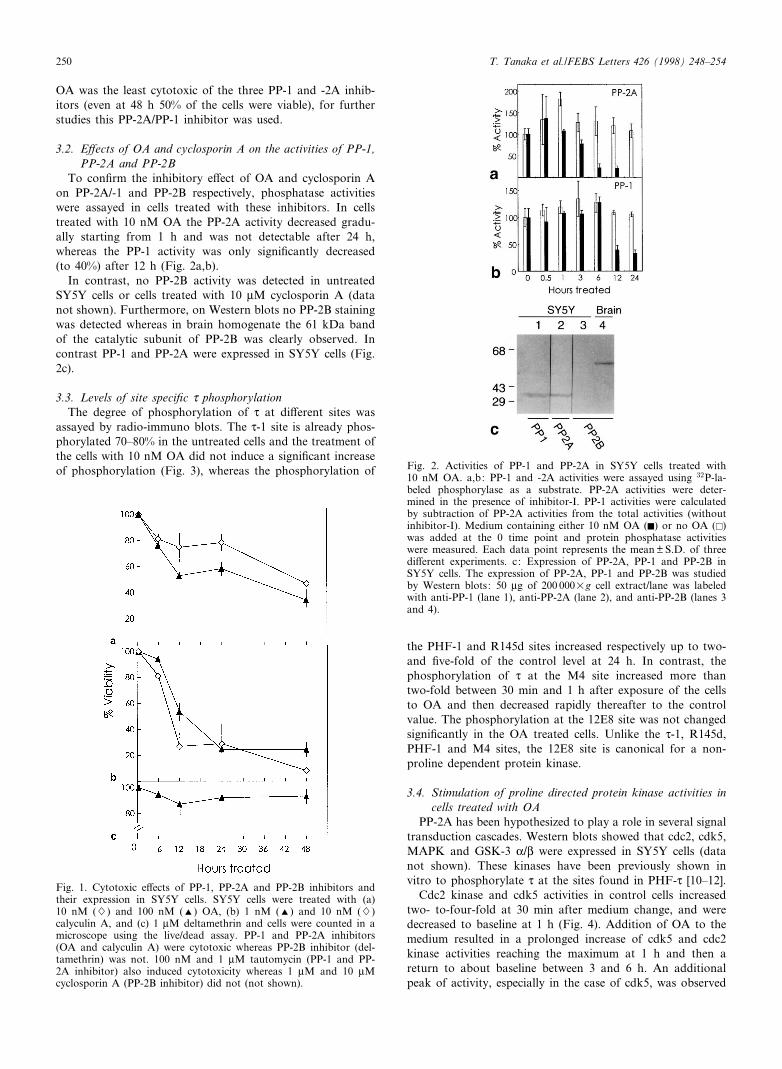
To con¢rm the inhibitory e¡ect of OA and cyclosporin Aon PP-2A/-1 and PP-2B respectively, phosphatase activitieswere assayed in cells treated with these inhibitors. In cellstreated with 10 nM OA the PP-2A activity decreased gradu-ally starting from 1 h and was not detectable after 24 h,whereas the PP-1 activity was only signi¢cantly decreased(to 40%) after 12 h (Fig. 2a,b).
In contrast, no PP-2B activity was detected in untreatedSY5Y cells or cells treated with 10 WM cyclosporin A (datanot shown). Furthermore, on Western blots no PP-2B stainingwas detected whereas in brain homogenate the 61 kDa bandof the catalytic subunit of PP-2B was clearly observed. Incontrast PP-1 and PP-2A were expressed in SY5Y cells (Fig.2c).
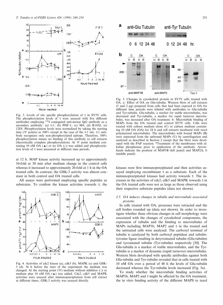
3.3. Levels of site speci¢c d phosphorylationThe degree of phosphorylation of d at di¡erent sites was
assayed by radio-immuno blots. The d-1 site is already phos-phorylated 70^80% in the untreated cells and the treatment ofthe cells with 10 nM OA did not induce a signi¢cant increaseof phosphorylation (Fig. 3), whereas the phosphorylation of
the PHF-1 and R145d sites increased respectively up to two-and ¢ve-fold of the control level at 24 h. In contrast, thephosphorylation of d at the M4 site increased more thantwo-fold between 30 min and 1 h after exposure of the cellsto OA and then decreased rapidly thereafter to the controlvalue. The phosphorylation at the 12E8 site was not changedsigni¢cantly in the OA treated cells. Unlike the d-1, R145d,PHF-1 and M4 sites, the 12E8 site is canonical for a non-proline dependent protein kinase.
3.4. Stimulation of proline directed protein kinase activities incells treated with OA
PP-2A has been hypothesized to play a role in several signaltransduction cascades. Western blots showed that cdc2, cdk5,MAPK and GSK-3 K/L were expressed in SY5Y cells (datanot shown). These kinases have been previously shown invitro to phosphorylate d at the sites found in PHF-d [10^12].
Cdc2 kinase and cdk5 activities in control cells increasedtwo- to-four-fold at 30 min after medium change, and weredecreased to baseline at 1 h (Fig. 4). Addition of OA to themedium resulted in a prolonged increase of cdk5 and cdc2kinase activities reaching the maximum at 1 h and then areturn to about baseline between 3 and 6 h. An additionalpeak of activity, especially in the case of cdk5, was observed
FEBS 20113 17-4-98
Fig. 2. Activities of PP-1 and PP-2A in SY5Y cells treated with10 nM OA. a,b: PP-1 and -2A activities were assayed using 32P-la-beled phosphorylase as a substrate. PP-2A activities were deter-mined in the presence of inhibitor-I. PP-1 activities were calculatedby subtraction of PP-2A activities from the total activities (withoutinhibitor-I). Medium containing either 10 nM OA (F) or no OA (E)was added at the 0 time point and protein phosphatase activitieswere measured. Each data point represents the mean þ S.D. of threedi¡erent experiments. c: Expression of PP-2A, PP-1 and PP-2B inSY5Y cells. The expression of PP-2A, PP-1 and PP-2B was studiedby Western blots: 50 Wg of 200 000Ug cell extract/lane was labeledwith anti-PP-1 (lane 1), anti-PP-2A (lane 2), and anti-PP-2B (lanes 3and 4).
Fig. 1. Cytotoxic e¡ects of PP-1, PP-2A and PP-2B inhibitors andtheir expression in SY5Y cells. SY5Y cells were treated with (a)10 nM (W) and 100 nM (R) OA, (b) 1 nM (R) and 10 nM (W)calyculin A, and (c) 1 WM deltamethrin and cells were counted in amicroscope using the live/dead assay. PP-1 and PP-2A inhibitors(OA and calyculin A) were cytotoxic whereas PP-2B inhibitor (del-tamethrin) was not. 100 nM and 1 WM tautomycin (PP-1 and PP-2A inhibitor) also induced cytotoxicity whereas 1 WM and 10 WMcyclosporin A (PP-2B inhibitor) did not (not shown).
T. Tanaka et al./FEBS Letters 426 (1998) 248^254250
at 12 h. MAP kinase activity increased up to approximately10-fold at 30 min after medium change in the control cellswhereas it increased to approximately 20-fold at 1 h in the OAtreated cells. In contrast, the GSK-3 activity was almost con-stant in both control and OA treated cells.
All assays were performed employing speci¢c peptides assubstrates. To con¢rm the kinase activities towards d, the
kinases were ¢rst immunoprecipitated and then activities as-sayed employing recombinant d as a substrate. Each of theimmunoprecipitated kinases had activity towards d. The in-creases in the activities of cdc2, cdk5 and MAPK towards d inthe OA treated cells were not as large as those observed usingtheir respective substrate peptides (data not shown).
3.5. OA induces changes in tubulin and microtubule associatedproteins
In cells treated with OA, processes were retracted and thecell bodies rounded up (data not shown). In order to inves-tigate whether these obvious changes in cell morphology wereassociated with the changes of cytoskeletal components, theexpression of tubulin and the binding to microtubules ofMAPs including MAP1b, MAP2 and d in the treated andthe untreated cells were analyzed. The carboxyl terminal oftubulin is catalyzed by both carboxyl peptidase and tubulin-tyrosine ligase resulting in detyrosinated tubulin (Glu-tubulin)and tyrosinated tubulin (Tyr-tubulin) respectively [30]. TheGlu-tubulin is a marker of stable microtubules, and the Tyr-tubulin is a marker of dynamic (rapid turnover) microtubules.Western blots developed with speci¢c antibodies against bothGlu-tubulin and Tyr-tubulin revealed that in cells treated with10 nM OA over a period of 48 h the levels of Glu-tubulindecreased whereas the Tyr-tubulin levels increased (Fig. 5a).
To study whether the microtubule binding activities ofMAP1b, MAP2 and d might be a¡ected by the OA treatment,the in vitro binding activity of the di¡erent MAPS to taxol
FEBS 20113 17-4-98
Fig. 5. Changes in cytoskeletal protein in SY5Y cells treated withOA. a: E¡ect of OA on Glu-tubulin. Western blots of cell extracts(5 and 2 Wg) prepared from cells that had been exposed to OA fordi¡erent time periods were labeled with antibodies to Glu-tubulinand Tyr-tubulin. Glu-tubulin, a marker for stable microtubules, wasdecreased and Tyr-tubulin, a marker for rapid turnover microtu-bules, was increased after OA treatment. b: Microtubule binding ofMAPs from the OA treated and control SY5Y cells. Cells weretreated with culture medium alone (C) or culture medium contain-ing 10 nM OA (OA) for 24 h and cell extracts incubated with taxolpolymerized microtubules. The microtubules with bound MAPs (B)were separated from the unbound MAPs (U) by centrifugation andanalyzed as described in Section 2 except that the blots were devel-oped with the PAP reaction. *Treatment of the membranes with al-kaline phosphatase prior to application of the antibody. Arrow-heads indicate the position of MAP1B (left panel) and MAP2A, b(middle panel).
Fig. 4. Activities of cdc2 kinase (a), cdk5 (b), MAPK (c) and GSK-3 (d). 36 h before the start of the experiment the medium waschanged. At the starting point (`0') medium without inhibitor (E) ormedium plus 10 nM OA (R) was added. Cdc2, cdk5 and MAPKactivities were assayed after immunoprecipitation from cell extractat di¡erent times; GSK-3 activity was assayed directly.
Fig. 3. Levels of site speci¢c phosphorylation of d in SY5Y cells.The phosphorylation levels of d were assayed with ¢ve di¡erentantibodies employing 125I conjugated anti-mouse IgG antibody as asecondary antibody: (a) d-1; (b) PHF-1; (c) M4; (d) R145d; (e)12E8. Phosphorylation levels were normalized by taking the startingtime (`0' points) as 100% except in the case of the d-1 site. d-1 anti-body recognizes only non-phosphorylated epitope. Therefore, 100%phosphorylation means no binding of this antibody to cell extracts(theoretically complete phosphorylation). At `0' point medium con-taining 10 nM OA (R) or no OA (a) was added and phosphoryla-tion levels of d were measured at di¡erent time periods.
T. Tanaka et al./FEBS Letters 426 (1998) 248^254 251
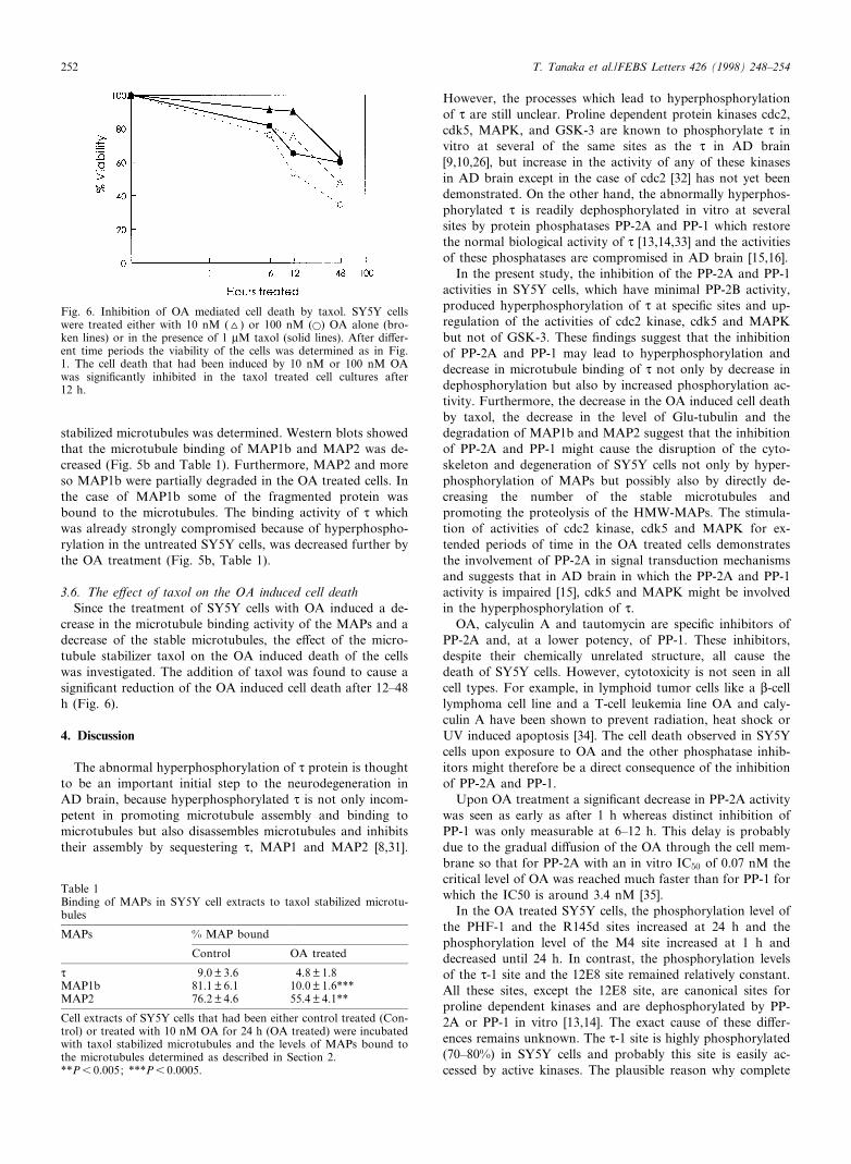
stabilized microtubules was determined. Western blots showedthat the microtubule binding of MAP1b and MAP2 was de-creased (Fig. 5b and Table 1). Furthermore, MAP2 and moreso MAP1b were partially degraded in the OA treated cells. Inthe case of MAP1b some of the fragmented protein wasbound to the microtubules. The binding activity of d whichwas already strongly compromised because of hyperphospho-rylation in the untreated SY5Y cells, was decreased further bythe OA treatment (Fig. 5b, Table 1).
3.6. The e¡ect of taxol on the OA induced cell deathSince the treatment of SY5Y cells with OA induced a de-
crease in the microtubule binding activity of the MAPs and adecrease of the stable microtubules, the e¡ect of the micro-tubule stabilizer taxol on the OA induced death of the cellswas investigated. The addition of taxol was found to cause asigni¢cant reduction of the OA induced cell death after 12^48h (Fig. 6).
4. Discussion
The abnormal hyperphosphorylation of d protein is thoughtto be an important initial step to the neurodegeneration inAD brain, because hyperphosphorylated d is not only incom-petent in promoting microtubule assembly and binding tomicrotubules but also disassembles microtubules and inhibitstheir assembly by sequestering d, MAP1 and MAP2 [8,31].
However, the processes which lead to hyperphosphorylationof d are still unclear. Proline dependent protein kinases cdc2,cdk5, MAPK, and GSK-3 are known to phosphorylate d invitro at several of the same sites as the d in AD brain[9,10,26], but increase in the activity of any of these kinasesin AD brain except in the case of cdc2 [32] has not yet beendemonstrated. On the other hand, the abnormally hyperphos-phorylated d is readily dephosphorylated in vitro at severalsites by protein phosphatases PP-2A and PP-1 which restorethe normal biological activity of d [13,14,33] and the activitiesof these phosphatases are compromised in AD brain [15,16].
In the present study, the inhibition of the PP-2A and PP-1activities in SY5Y cells, which have minimal PP-2B activity,produced hyperphosphorylation of d at speci¢c sites and up-regulation of the activities of cdc2 kinase, cdk5 and MAPKbut not of GSK-3. These ¢ndings suggest that the inhibitionof PP-2A and PP-1 may lead to hyperphosphorylation anddecrease in microtubule binding of d not only by decrease indephosphorylation but also by increased phosphorylation ac-tivity. Furthermore, the decrease in the OA induced cell deathby taxol, the decrease in the level of Glu-tubulin and thedegradation of MAP1b and MAP2 suggest that the inhibitionof PP-2A and PP-1 might cause the disruption of the cyto-skeleton and degeneration of SY5Y cells not only by hyper-phosphorylation of MAPs but possibly also by directly de-creasing the number of the stable microtubules andpromoting the proteolysis of the HMW-MAPs. The stimula-tion of activities of cdc2 kinase, cdk5 and MAPK for ex-tended periods of time in the OA treated cells demonstratesthe involvement of PP-2A in signal transduction mechanismsand suggests that in AD brain in which the PP-2A and PP-1activity is impaired [15], cdk5 and MAPK might be involvedin the hyperphosphorylation of d.
OA, calyculin A and tautomycin are speci¢c inhibitors ofPP-2A and, at a lower potency, of PP-1. These inhibitors,despite their chemically unrelated structure, all cause thedeath of SY5Y cells. However, cytotoxicity is not seen in allcell types. For example, in lymphoid tumor cells like a L-celllymphoma cell line and a T-cell leukemia line OA and caly-culin A have been shown to prevent radiation, heat shock orUV induced apoptosis [34]. The cell death observed in SY5Ycells upon exposure to OA and the other phosphatase inhib-itors might therefore be a direct consequence of the inhibitionof PP-2A and PP-1.
Upon OA treatment a signi¢cant decrease in PP-2A activitywas seen as early as after 1 h whereas distinct inhibition ofPP-1 was only measurable at 6^12 h. This delay is probablydue to the gradual di¡usion of the OA through the cell mem-brane so that for PP-2A with an in vitro IC50 of 0.07 nM thecritical level of OA was reached much faster than for PP-1 forwhich the IC50 is around 3.4 nM [35].
In the OA treated SY5Y cells, the phosphorylation level ofthe PHF-1 and the R145d sites increased at 24 h and thephosphorylation level of the M4 site increased at 1 h anddecreased until 24 h. In contrast, the phosphorylation levelsof the d-1 site and the 12E8 site remained relatively constant.All these sites, except the 12E8 site, are canonical sites forproline dependent kinases and are dephosphorylated by PP-2A or PP-1 in vitro [13,14]. The exact cause of these di¡er-ences remains unknown. The d-1 site is highly phosphorylated(70^80%) in SY5Y cells and probably this site is easily ac-cessed by active kinases. The plausible reason why complete
FEBS 20113 17-4-98
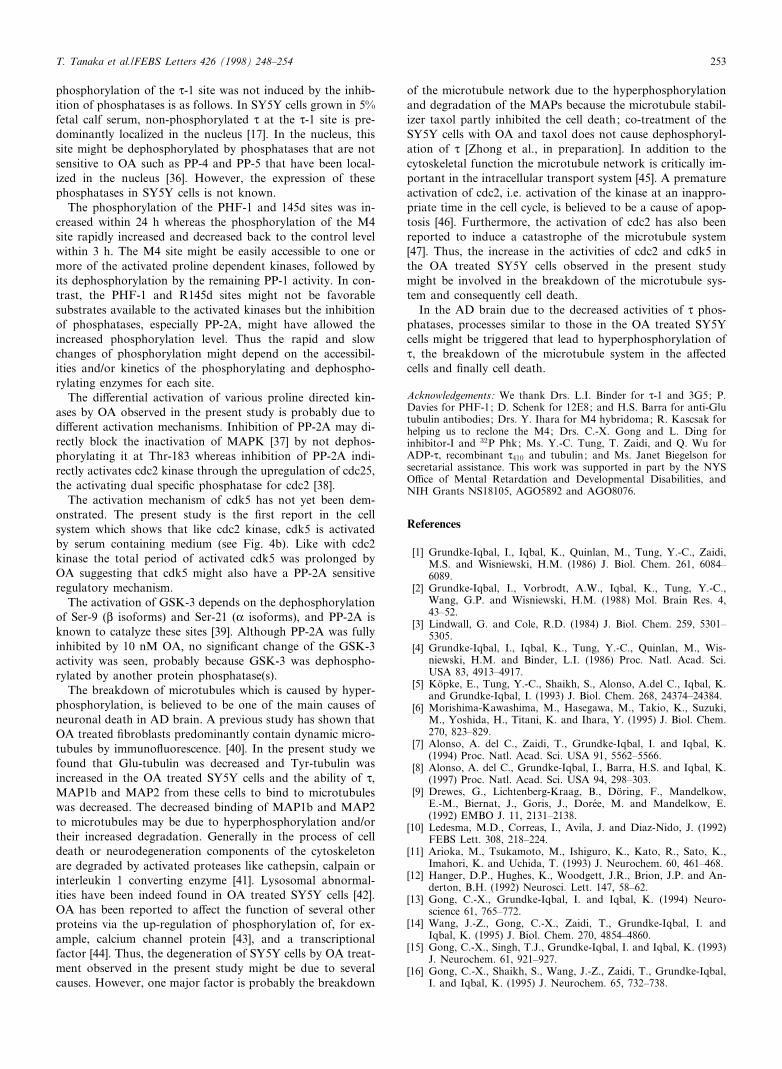
Fig. 6. Inhibition of OA mediated cell death by taxol. SY5Y cellswere treated either with 10 nM (O) or 100 nM (a) OA alone (bro-ken lines) or in the presence of 1 WM taxol (solid lines). After di¡er-ent time periods the viability of the cells was determined as in Fig.1. The cell death that had been induced by 10 nM or 100 nM OAwas signi¢cantly inhibited in the taxol treated cell cultures after12 h.
Table 1Binding of MAPs in SY5Y cell extracts to taxol stabilized microtu-bules
MAPs % MAP bound
Control OA treated
d 9.0 þ 3.6 4.8 þ 1.8MAP1b 81.1 þ 6.1 10.0 þ 1.6***MAP2 76.2 þ 4.6 55.4 þ 4.1**
Cell extracts of SY5Y cells that had been either control treated (Con-trol) or treated with 10 nM OA for 24 h (OA treated) were incubatedwith taxol stabilized microtubules and the levels of MAPs bound tothe microtubules determined as described in Section 2.**P6 0.005; ***P6 0.0005.
T. Tanaka et al./FEBS Letters 426 (1998) 248^254252
phosphorylation of the d-1 site was not induced by the inhib-ition of phosphatases is as follows. In SY5Y cells grown in 5%fetal calf serum, non-phosphorylated d at the d-1 site is pre-dominantly localized in the nucleus [17]. In the nucleus, thissite might be dephosphorylated by phosphatases that are notsensitive to OA such as PP-4 and PP-5 that have been local-ized in the nucleus [36]. However, the expression of thesephosphatases in SY5Y cells is not known.
The phosphorylation of the PHF-1 and 145d sites was in-creased within 24 h whereas the phosphorylation of the M4site rapidly increased and decreased back to the control levelwithin 3 h. The M4 site might be easily accessible to one ormore of the activated proline dependent kinases, followed byits dephosphorylation by the remaining PP-1 activity. In con-trast, the PHF-1 and R145d sites might not be favorablesubstrates available to the activated kinases but the inhibitionof phosphatases, especially PP-2A, might have allowed theincreased phosphorylation level. Thus the rapid and slowchanges of phosphorylation might depend on the accessibil-ities and/or kinetics of the phosphorylating and dephospho-rylating enzymes for each site.
The di¡erential activation of various proline directed kin-ases by OA observed in the present study is probably due todi¡erent activation mechanisms. Inhibition of PP-2A may di-rectly block the inactivation of MAPK [37] by not dephos-phorylating it at Thr-183 whereas inhibition of PP-2A indi-rectly activates cdc2 kinase through the upregulation of cdc25,the activating dual speci¢c phosphatase for cdc2 [38].
The activation mechanism of cdk5 has not yet been dem-onstrated. The present study is the ¢rst report in the cellsystem which shows that like cdc2 kinase, cdk5 is activatedby serum containing medium (see Fig. 4b). Like with cdc2kinase the total period of activated cdk5 was prolonged byOA suggesting that cdk5 might also have a PP-2A sensitiveregulatory mechanism.
The activation of GSK-3 depends on the dephosphorylationof Ser-9 (L isoforms) and Ser-21 (K isoforms), and PP-2A isknown to catalyze these sites [39]. Although PP-2A was fullyinhibited by 10 nM OA, no signi¢cant change of the GSK-3activity was seen, probably because GSK-3 was dephospho-rylated by another protein phosphatase(s).
The breakdown of microtubules which is caused by hyper-phosphorylation, is believed to be one of the main causes ofneuronal death in AD brain. A previous study has shown thatOA treated ¢broblasts predominantly contain dynamic micro-tubules by immuno£uorescence. [40]. In the present study wefound that Glu-tubulin was decreased and Tyr-tubulin wasincreased in the OA treated SY5Y cells and the ability of d,MAP1b and MAP2 from these cells to bind to microtubuleswas decreased. The decreased binding of MAP1b and MAP2to microtubules may be due to hyperphosphorylation and/ortheir increased degradation. Generally in the process of celldeath or neurodegeneration components of the cytoskeletonare degraded by activated proteases like cathepsin, calpain orinterleukin 1 converting enzyme [41]. Lysosomal abnormal-ities have been indeed found in OA treated SY5Y cells [42].OA has been reported to a¡ect the function of several otherproteins via the up-regulation of phosphorylation of, for ex-ample, calcium channel protein [43], and a transcriptionalfactor [44]. Thus, the degeneration of SY5Y cells by OA treat-ment observed in the present study might be due to severalcauses. However, one major factor is probably the breakdown
of the microtubule network due to the hyperphosphorylationand degradation of the MAPs because the microtubule stabil-izer taxol partly inhibited the cell death; co-treatment of theSY5Y cells with OA and taxol does not cause dephosphoryl-ation of d [Zhong et al., in preparation]. In addition to thecytoskeletal function the microtubule network is critically im-portant in the intracellular transport system [45]. A prematureactivation of cdc2, i.e. activation of the kinase at an inappro-priate time in the cell cycle, is believed to be a cause of apop-tosis [46]. Furthermore, the activation of cdc2 has also beenreported to induce a catastrophe of the microtubule system[47]. Thus, the increase in the activities of cdc2 and cdk5 inthe OA treated SY5Y cells observed in the present studymight be involved in the breakdown of the microtubule sys-tem and consequently cell death.
In the AD brain due to the decreased activities of d phos-phatases, processes similar to those in the OA treated SY5Ycells might be triggered that lead to hyperphosphorylation ofd, the breakdown of the microtubule system in the a¡ectedcells and ¢nally cell death.
Acknowledgements: We thank Drs. L.I. Binder for d-1 and 3G5; P.Davies for PHF-1; D. Schenk for 12E8; and H.S. Barra for anti-Glutubulin antibodies; Drs. Y. Ihara for M4 hybridoma; R. Kascsak forhelping us to reclone the M4; Drs. C.-X. Gong and L. Ding forinhibitor-I and 32P Phk; Ms. Y.-C. Tung, T. Zaidi, and Q. Wu forADP-d, recombinant d410 and tubulin; and Ms. Janet Biegelson forsecretarial assistance. This work was supported in part by the NYSO¤ce of Mental Retardation and Developmental Disabilities, andNIH Grants NS18105, AGO5892 and AGO8076.
References
[1] Grundke-Iqbal, I., Iqbal, K., Quinlan, M., Tung, Y.-C., Zaidi,M.S. and Wisniewski, H.M. (1986) J. Biol. Chem. 261, 6084^6089.
[2] Grundke-Iqbal, I., Vorbrodt, A.W., Iqbal, K., Tung, Y.-C.,Wang, G.P. and Wisniewski, H.M. (1988) Mol. Brain Res. 4,43^52.
[3] Lindwall, G. and Cole, R.D. (1984) J. Biol. Chem. 259, 5301^5305.
[4] Grundke-Iqbal, I., Iqbal, K., Tung, Y.-C., Quinlan, M., Wis-niewski, H.M. and Binder, L.I. (1986) Proc. Natl. Acad. Sci.USA 83, 4913^4917.
[5] Koëpke, E., Tung, Y.-C., Shaikh, S., Alonso, A.del C., Iqbal, K.and Grundke-Iqbal, I. (1993) J. Biol. Chem. 268, 24374^24384.
[6] Morishima-Kawashima, M., Hasegawa, M., Takio, K., Suzuki,M., Yoshida, H., Titani, K. and Ihara, Y. (1995) J. Biol. Chem.270, 823^829.
[7] Alonso, A. del C., Zaidi, T., Grundke-Iqbal, I. and Iqbal, K.(1994) Proc. Natl. Acad. Sci. USA 91, 5562^5566.
[8] Alonso, A. del C., Grundke-Iqbal, I., Barra, H.S. and Iqbal, K.(1997) Proc. Natl. Acad. Sci. USA 94, 298^303.
[9] Drewes, G., Lichtenberg-Kraag, B., Doëring, F., Mandelkow,E.-M., Biernat, J., Goris, J., Doreèe, M. and Mandelkow, E.(1992) EMBO J. 11, 2131^2138.
[10] Ledesma, M.D., Correas, I., Avila, J. and Diaz-Nido, J. (1992)FEBS Lett. 308, 218^224.
[11] Arioka, M., Tsukamoto, M., Ishiguro, K., Kato, R., Sato, K.,Imahori, K. and Uchida, T. (1993) J. Neurochem. 60, 461^468.
[12] Hanger, D.P., Hughes, K., Woodgett, J.R., Brion, J.P. and An-derton, B.H. (1992) Neurosci. Lett. 147, 58^62.
[13] Gong, C.-X., Grundke-Iqbal, I. and Iqbal, K. (1994) Neuro-science 61, 765^772.
[14] Wang, J.-Z., Gong, C.-X., Zaidi, T., Grundke-Iqbal, I. andIqbal, K. (1995) J. Biol. Chem. 270, 4854^4860.
[15] Gong, C.-X., Singh, T.J., Grundke-Iqbal, I. and Iqbal, K. (1993)J. Neurochem. 61, 921^927.
[16] Gong, C.-X., Shaikh, S., Wang, J.-Z., Zaidi, T., Grundke-Iqbal,I. and Iqbal, K. (1995) J. Neurochem. 65, 732^738.
FEBS 20113 17-4-98
T. Tanaka et al./FEBS Letters 426 (1998) 248^254 253

[17] Tanaka, T., Iqbal, K., Trenkner, E., Liu, D.J. and Grundke-Iqbal, I. (1995) FEBS Lett. 360, 5^9.
[18] Arias, C., Sharma, N., Davies, P. and Sha¢t-Zagardo, B. (1993)J. Neurochem. 61, 673^682.
[19] Szendrei, V.M., Lee, V.-Y. and Otvos Jr., L. (1993) J. Neurosci.Res. 34, 243^249.
[20] Greenberg, S.G., Davies, P., Schein, J.D. and Binder, L. (1992)J. Biol. Chem. 267, 564^569.
[21] Hasegawa, M., Watanabe, A., Takio, K., Suzuki, M., Arai, T.,Titani, K. and Dhasa, Y. (1995) J. Neurochem. 60, 2068^2077.
[22] Seubert, P., Mawal-Devan, M., Barbour, R., Jakes, R., Goedert,M., Johnson, G.V.W., Literski, J.M., Schenk, D., Lieberburg, I.,Trojanowski, J.Q. and Lee, V.M.-Y. (1995) J. Biol. Chem. 270,18917^18922.
[23] Arregui, C., Buociglio, C., Caceres, A. and Barra, H.S. (1991)J. Neurosci. Res. 28, 171^181.
[24] Pei, J.-J., Gong, C.-X., Iqbal, K., Grundke-Iqbal, I., Wu, Q.-L.,Winblad, B. and Cowburn, R. (1998) J. Neural. Trans. 105, 69^83.
[25] Pei, J.-J., Tanaka, T., Tung, Y.-C., Braak, E., Iqbal, K. andGrundke-Iqbal, I. (1997) J. Neuropathol. Exp. Neurol. 56, 70^78.
[26] Singh, T.J., Haque, N., Grundke-Iqbal, I. and Iqbal, K. (1995)FEBS Lett. 358, 267^272.
[27] Bensadoun, A. and Weinstein, D. (1976) Anal. Biochem. 70, 241^250.
[28] Khatoon, S., Grundke-Iqbal, I. and Iqbal, K. (1992) J. Neuro-chem. 59, 750^753.
[29] Boe, R., Gjertsen, B.T., Vintermyr, O.K., Houge, G., Lanotte,M. and Doskeland, S.O. (1991) Exp. Cell Res. 195, 237^246.
[30] Gundersen, G.G., Kalnoski, M.H. and Bulinski, J.C. (1984) Cell38, 779^789.
[31] Alonso, A. del C., Grundke-Iqbal, I. and Iqbal, K. (1996) NatureMed. 2, 783^787.
[32] Vincent, I., Jicha, G., Rosado, M. and Dickson, D.W. (1997)J. Neurosci. 17, 3588^3598.
[33] Wang, J.-Z., Grundke-Iqbal, I. and Iqbal, K. (1996) Mol. Brain.Res. 38, 200^208.
[34] Baxter, G.D. and Lavin, M.F. (1992) J. Immunol. 148, 1949^1954.
[35] Suganuma, M. and Fujiki, H. (1993) Tanpakushitsu KakusanKoso 38, 1960^1970.
[36] Chen, M.X., McPartlin, A.E., Brown, L., Chen, Y.H., Barker,H.M. and Cohen, P.T. (1994) EMBO J. 13, 4278^4290.
[37] Clarke, P.R., Ho¡mann, I., Draetta, G. and Karsenti, E. (1993)Mol. Biol. Cell 4, 397^411.
[38] Yamashita, K., Yasuda, H., Pines, J., Yasumoto, K., Nishitani,H., Ohtsubo, M., Hunter, T., Sugimura, T. and Nishimoto, T.(1992) EMBO J. 11, 973^982.
[39] Sutherland, C., Leighton, I.A. and Cohen, P. (1993) Biochem. J.296, 15^19.
[40] Gurland, G. and Gundersen, G.G. (1993) Proc. Natl. Acad. Sci.USA 90, 8827^8831.
[41] Cataldo, A.M., Paskevich, P.A., Kominami, E. and Nixon, R.A.(1991) Proc. Natl. Acad. Sci. USA 88, 10998^11002.
[42] Shea, T.B. (1996) Neurosci. Res. Commun. 19, 27^36.[43] Haby, C., Larsson, O., Islam, M.S., Aunis, D., Berggren, P.O.
and Zwiller, J. (1994) Biochem. J. 298, 341^346.[44] Hagiwara, M., Alberts, A., Brindle, P., Meinkoth, J., Feramisco,
J., Deng, T., Karin, M., Shenolikar, S. and Montminy, M. (1992)Cell 70, 105^113.
[45] Hirokawa, N. (1993) Curr. Opin. Neurobiol. 3, 724^731.[46] Shi, L., Nishioka, W.K., Th'ng, J., Bradbury, E.M., Litch¢eld,
D.W. and Greenberg, A.H. (1994) Science 263, 1143^1145.[47] Verde, F., Labbe, J.C., Doree, M. and Karsenti, E. (1990) Nature
343, 233^238.
FEBS 20113 17-4-98
T. Tanaka et al./FEBS Letters 426 (1998) 248^254254