The Plant Journal (2005) 43 High-throughput in planta...
15
High-throughput in planta expression screening identifies a class II ethylene-responsive element binding factor-like protein that regulates plant cell death and non-host resistance Khairun Hisam Bin Nasir 1,2 , Yoshihiro Takahashi 1 , Akiko Ito 1 , Hiromasa Saitoh 1,3 , Hideo Matsumura 1 , Hiroyuki Kanzaki 1 , Takeshi Shimizu 1 , Minako Ito 1 , Shizuko Fujisawa 1 , Prakash C. Sharma 1,4 , Masaru Ohme-Takagi 5 , Sophien Kamoun 6 and Ryohei Terauchi 1,2,* 1 Iwate Biotechnology Research Center, 22-174-4, Narita, Kitakami, Iwate 024-0003, Japan, 2 The United Graduate School of Agricultural Sciences, Iwate University, 3-18-8, Ueda, Morioka, Iwate 020-8550, Japan, 3 Max-Planck-Institute for Plant Breeding Research, Carl-von-Linne ´ -Weg 10, 50829 Cologne, Germany, 4 School of Biotechnology, Guru Gobind Singh Indraprastha University, 110006 New Delhi, India, 5 Gene Function Research Center, National Institute of Advanced Industrial Science and Technology (AIST), Central 4, Tsukuba 305-8562, Japan, and 6 Department of Plant Pathology, Ohio Agricultural Research and Development Center, Ohio State University, Wooster, OH 44691, USA Received 11 April 2005; revised 20 May 2005; accepted 23 May 2005. * For correspondence (fax þ81 197 68 3881; e-mail [email protected]). Summary We performed high-throughput screening using the potato virus X (PVX) system to overexpress Nicotiana benthamiana genes in planta and identify positive regulators of cell death. This screening identified NbCD1,a novel class II ethylene-responsive element binding factor (ERF), as a potent inducer of the hypersensitive response (HR)-like cell death. NbCD1 expression was induced by treatments with INF1 elicitor and a non-host pathogen Pseudomonas cichorii. NbCD1 exhibited transcriptional repressor activity through its EAR motif, and this motif was necessary for NbCD1 to cause cell death. We identified 58 genes that displayed altered transcription following NbCD1 overexpression. NbCD1 overexpression downregulated the expression of HSR203, a negative regulator of hypersensitive death. Conditional expression of NbCD1 in Arabidopsis also caused cell death, indicating that NbCD1 downstream cascades are conserved in dicot plants. To further confirm the role of NbCD1 in defense, we used virus-induced gene silencing to demonstrate that NbCD1 is required for non-host resistance of N. benthamiana to the bacterial pathogen P. cichorii. Our data point to a model of transcriptional regulatory cascades. NbCD1 positively regulates cell death and contributes to non- host resistance, possibly by downregulating the expression of other defense response genes. Keywords: cell death, class II ERF, repressor, SuperSAGE, VIGS, non-host resistance. Introduction Programmed cell death (pcd) plays key roles in plant devel- opment, senescence and defense response against patho- gens (reviewed in Kuriyama and Fukuda, 2002; Lam, 2004). Substantial information on plant pcd came from studies on the hypersensitive response (HR), a rapid death of plant cells that is associated with restriction of pathogen growth (Heath, 2000). Signaling cascades leading to the HR and disease resistance, particularly following R-gene-mediated pathogen recognition, have been the focus of intensive cellular and genetic studies (for recent reviews, see Greenberg and Yao, 2004; Martin et al., 2003; Shirasu and Schulze-Lefert, 2003). Upon R-gene-mediated perception of pathogens, early cellular responses are triggered including changes in ion flux, generation of reactive oxygen species (ROS), and in most cases, rapid development of cell death (HR). The HR seems to require ROS generation mediated by NADPH oxidase activation (Torres et al., 2002). Balance in cellular concen- tration of H 2 O 2 and NO (Delledonne et al., 2001) as well as ª 2005 Blackwell Publishing Ltd 491 The Plant Journal (2005) 43, 491–505 doi: 10.1111/j.1365-313X.2005.02472.x
Transcript of The Plant Journal (2005) 43 High-throughput in planta...

High-throughput in planta expression screening identifiesa class II ethylene-responsive element binding factor-likeprotein that regulates plant cell death and non-hostresistance
Khairun Hisam Bin Nasir1,2, Yoshihiro Takahashi1, Akiko Ito1, Hiromasa Saitoh1,3, Hideo Matsumura1, Hiroyuki Kanzaki1,
Takeshi Shimizu1, Minako Ito1, Shizuko Fujisawa1, Prakash C. Sharma1,4, Masaru Ohme-Takagi5, Sophien Kamoun6 and Ryohei
Terauchi1,2,*
1Iwate Biotechnology Research Center, 22-174-4, Narita, Kitakami, Iwate 024-0003, Japan,2The United Graduate School of Agricultural Sciences, Iwate University, 3-18-8, Ueda, Morioka, Iwate 020-8550, Japan,3Max-Planck-Institute for Plant Breeding Research, Carl-von-Linne-Weg 10, 50829 Cologne, Germany,4School of Biotechnology, Guru Gobind Singh Indraprastha University, 110006 New Delhi, India,5Gene Function Research Center, National Institute of Advanced Industrial Science and Technology (AIST), Central 4, Tsukuba
305-8562, Japan, and6Department of Plant Pathology, Ohio Agricultural Research and Development Center, Ohio State University, Wooster, OH
44691, USA
Received 11 April 2005; revised 20 May 2005; accepted 23 May 2005.*For correspondence (fax þ81 197 68 3881; e-mail [email protected]).
Summary
We performed high-throughput screening using the potato virus X (PVX) system to overexpress Nicotiana
benthamiana genes in planta and identify positive regulators of cell death. This screening identified NbCD1, a
novel class II ethylene-responsive element binding factor (ERF), as a potent inducer of the hypersensitive
response (HR)-like cell death. NbCD1 expression was induced by treatments with INF1 elicitor and a non-host
pathogen Pseudomonas cichorii. NbCD1 exhibited transcriptional repressor activity through its EARmotif, and
this motif was necessary for NbCD1 to cause cell death. We identified 58 genes that displayed altered
transcription following NbCD1 overexpression. NbCD1 overexpression downregulated the expression of
HSR203, a negative regulator of hypersensitive death. Conditional expression of NbCD1 in Arabidopsis also
caused cell death, indicating that NbCD1 downstream cascades are conserved in dicot plants. To further
confirm the role of NbCD1 in defense, we used virus-induced gene silencing to demonstrate that NbCD1 is
required for non-host resistance of N. benthamiana to the bacterial pathogen P. cichorii. Our data point to a
model of transcriptional regulatory cascades. NbCD1 positively regulates cell death and contributes to non-
host resistance, possibly by downregulating the expression of other defense response genes.
Keywords: cell death, class II ERF, repressor, SuperSAGE, VIGS, non-host resistance.
Introduction
Programmed cell death (pcd) plays key roles in plant devel-
opment, senescence and defense response against patho-
gens (reviewed in Kuriyama and Fukuda, 2002; Lam, 2004).
Substantial information on plant pcd came from studies on
the hypersensitive response (HR), a rapid death of plant cells
that is associatedwith restriction of pathogengrowth (Heath,
2000). Signaling cascades leading to the HR and disease
resistance, particularly followingR-gene-mediatedpathogen
recognition, have been the focus of intensive cellular and
genetic studies (for recent reviews, see Greenberg and
Yao, 2004; Martin et al., 2003; Shirasu and Schulze-Lefert,
2003).UponR-gene-mediatedperceptionof pathogens, early
cellular responsesare triggered includingchanges in ionflux,
generation of reactive oxygen species (ROS), and in most
cases, rapid development of cell death (HR). The HR seems
to require ROS generation mediated by NADPH oxidase
activation (Torres et al., 2002). Balance in cellular concen-
tration of H2O2 and NO (Delledonne et al., 2001) as well as
ª 2005 Blackwell Publishing Ltd 491
The Plant Journal (2005) 43, 491–505 doi: 10.1111/j.1365-313X.2005.02472.x

cellular levels of salicylic acid (Gaffney et al., 1993) may
define the rate of the HR. Several positive and negative
regulators of the HR have been identified using genetic
approaches (see Shirasu and Schulze-Lefert, 2000). Genes
required for some, but not all, R-gene-mediated HR have
been identified and include Rar1 (Shirasu et al., 1999), Sgt1
(Azevedo et al., 2002), Hsp90 (Hubert et al., 2003; Takahashi
et al., 2003), Ndr1 (Century et al., 1995), and Eds1 (Parker
et al., 1996). Protein phosphorylation cascades involving the
kinases CDPK and MAPKs also appear to be important for
transducing signals from pathogen recognition to the HR
and disease resistance (Romeis, 2001). Nonetheless, despite
the significant knowledge accumulated in the last decade,
our understanding of the HR and cell death, particularly of
downstream signaling events, remains fragmentary. Many
important players involved in positive and negative regula-
tion of cell death remain to be discovered. The recent finding
of a plant vacuolar protease VPE, necessary for virus-medi-
ated HR, and similar in activity to the animal cell death
executioners caspases, suggests that much remains to be
discovered about plant cell death (Hatsugai et al., 2004).
To date, genetic approaches to explore regulators of plant
defense and cell death have emphasized loss-of-function
mutagenesis (Glazebrook, 2001). However, many genes
cannot be uncovered using this approach because of lethal
effects or functional redundancy, especially in complex
reiterative signal networks. Therefore, Xia et al. (2004)
employed a gain-of-function mutation approach using T-
DNA activation tagging in Arabidopsis thaliana. Screening
of approximately 5000 lines with random insertion of
cauliflower mosaic virus (CaMV) 35S enhancer identified
an aspartic protease, CDR1, as a positive regulator of
defense. An alternative to activation tagging is functional
expression screening of cDNAs. Karrer et al. (1998) con-
structed a tobacco cDNA library in a tobacco mosaic virus
(TMV)-based expression vector. Infectious transcripts were
generated and used to inoculate tobacco plants resulting in
the identification of 12 unique cDNAs that cause HR-like cell
death. The identified cDNA sequences showed similarity to
ubiquitin, a tumor-related protein, and various unknown
proteins. However, there was no clear evidence linking the
observed cell death and overexpression of the cloned cDNA,
and no detailed characterization of the cDNAs was carried
out. The observed cell death may have been caused by
co-suppression (ubiquitin gene) or overexpression (tumor-
related protein gene) (Karrer et al., 1998). In addition, virus
vectors have tremendously improved since the study of
Karrer et al. (1998) and current virus expression systems are
particularly adapted to high-throughput screens (see
Huitema et al., 2004). For example, Takken et al. (2000)
developed a functional cloning strategy based on a binary
potato virus X (PVX)-expression vector and applied it to
expressing pathogen genes in planta. In that study, cDNAs
of the tomato fungal pathogen, Cladosprorium fulvum, were
cloned into a PVX vector, and transferred to Agrobacterium
tumefaciens. A total of 9600 clones were tooth pick-inocu-
lated onto tobacco leaves, among which four cDNAs were
identified to cause necrotic lesion around the inoculation
site. One of the identified cDNAs encoded AVR4, a race-
specific elicitor. Similar high-throughput approaches have
been used to identify cDNAs from the oomycete plant
pathogen Phytophthora that elicit defense responses in
plants (Qutob et al., 2002; Torto et al., 2003).
In this study, we performed high-throughput expression
of plant cDNAs to identify novel factors that positively
regulate cell death. We carried out functional screening of
plant cell death-causing genes by transiently overexpressing
a 40 000 Nicotiana benthamiana cDNA library in planta
using the binary PVX-vector pSfinx (Takken et al., 2000). This
high-throughput functional screening resulted in the iden-
tification of several genes that cause cell death upon
overexpression. Here, we present and characterize one of
these genes, a novel ethylene response factor (ERF) that
functions as a strong cell death-inducing factor.
Results
Functional screening of plant cell death factors in
N. benthamiana identifies NbCD1
To search for plant genes that cause cell death upon over-
expression, we constructed a N. benthamiana cDNA library
in the binary PVX-expression vector pSfinx developed by
Takken et al. (2000). Complementary DNA was synthesized
from mRNA extracted from N. benthamiana leaves infiltra-
ted with the HR elicitor INF1 of Phytophthora infestans
(Kamoun et al., 1998) to activate defense-related genes, and
directionally cloned into pSfinx in the sense orientation. We
screened 40 000 cDNAs by toothpick inoculation of
N. benthamiana leaves with A. tumefaciens carrying
recombinant pSfinx. Four to 14 days post-inoculation, cell
death became visible around the inoculated site if the vector
insert contained a cDNA of a gene for cell death-inducing
protein. Such candidate A. tumefaciens clones were liquid-
cultured, and infiltrated into N. benthamiana leaf blades by a
needleless syringe to test whether cell death can be repro-
ducibly observed. This functional screening identified a total
of 30 cDNA clones that consistently caused cell death. The
cDNA inserts were sequenced, and similarity searches were
carried out with the BLAST program (Altschul et al., 1990) to
annotate the genes. The sequenced cDNAs showed signifi-
cant similarity to ubiquitin-like protein (eight cDNA clones),
RNA recognition motif protein (five), ethylene-responsive
element binding factor (ERF)-like protein (one), and MAPKK-
like protein (one) among others. We elected to focus on the
ERF-like protein cDNA (henceforth named NbCD1) because
it: (i) exhibited the strongest cell death-inducing pheno-
types, causing cell death on N. benthamiana leaves 7 days
492 Khairun Hisam Bin Nasir et al.
ª Blackwell Publishing Ltd, The Plant Journal, (2005), 43, 491–505

after toothpick inoculation and 4 days after infiltration
(Figure 1a); and (ii) encoded a novel putative transcription
factor of the class II ERF family, a family of proteins that have
been previously implicated in defense. Detailed description
of the other identified cDNAs will be reported elsewhere.
Cell death was only induced when the NbCD1 cDNA was
inserted in the vector in the sense, but not in the anti-sense,
orientation (Figure 1b), indicating that overexpression, not
virus-induced gene silencing (VIGS), of NbCD1 induced cell
death. This was further confirmed by agroinfiltration and by
correlating the presence of NbCD1 protein with cell death
(see below).
NbCD1 belongs to class II ERF
The NbCD1 cDNA insert in the pSfinx vector contained 965
nucleotides (GenBank accession no. AB196362). This DNA
sequence had no perfect similarity to known sequences
deposited in public databases, indicating that NbCD1 is a
novel plant gene. This NbCD1 cDNA harbored a single ORF
corresponding to a protein of 231 amino acids in size
(Figure 2a) with a high sequence similarity to the class II
ethylene-responsive element binding factor (ERF, Figure 2b).
Class II ERF proteins are characterized by the presence of
the EAR (ERF-associated amphiphilic repression) motif
(L/FDLNL/F(x)P) near their C-terminus in addition to the
AP2/ERFDNA-bindingdomainandacidicdomaincommonto
all classes of ERFs (Fujimoto et al., 2000; Ohta et al., 2001).
The NbCD1 predicted protein also contains a sequence with
high similarity to the EAR motif (Figure 2a). NtERF3, a mem-
ber of class II ERF, is known to function as a strong tran-
scriptional repressor through itsEARmotif (Ohtaet al., 2001).
NbCD1 functions as an active repressor
To test whether, similar to other class II ERFs, NbCD1 dis-
plays transcriptional repressor activity, we carried out a
transient expression assay (Figure 3a) as described by Fuji-
moto et al. (2000). The reporter plasmid contained a firefly
luciferase gene located downstream of 5· yeast GAL4 se-
quence and 4·GCC box, a target cis-element of ERFs (Ohme-
Takagi and Shinshi, 1995). This reporter plasmid, together
with an effector plasmid(s), was delivered to A. thaliana
leaves by particle bombardment. Effector plasmids con-
tained GAL4 DNA-binding domain (GAL4DB) only, GAL4DB
fused to the transcriptional activation domain of viral protein
16 (VP16: GAL4DB-VP16), NbCD1, or NbCD1 lacking the EAR
motif (NbCD1DEAR), all under the control of the CaMV35S
promoter. Expression of GAL4DB did not induce the tran-
scription of luciferase, whereas GAL4DB-VP16 expression
(a) (b)
Figure 1. Overexpression of NbCD1 causes cell death.
(a) Agrobacterium tumefaciens clones transformed with the pSfinx vector harboring NbCD1 cDNA (left), INF1 cDNA (center), or no insert (right) were toothpick-
inoculated (top) or infiltrated (bottom) to Nicotiana benthamiana leaves. Photographs were taken 14 and 4 days after toothpick inoculation and infiltration to the half
leaf, respectively. Necrotic cell death after toothpick inoculation of A. tumefaciens with pSfinx-NbCD1 became visible as early as 7 days post-inoculation.
(b) Agrobacterium tumefaciens clone harboring the pSfinx vector with NbCD1 in the sense orientation caused cell death, whereas that with NbCD1 in the antisense
orientation did not.
Overexpression of a class II ERF causes plant cell death 493
ª Blackwell Publishing Ltd, The Plant Journal, (2005), 43, 491–505

Figure 2. NbCD1 encoded a protein with similarity to class II ethylene-responsive element binding factor (ERF).
(a) Amino acid sequence alignment of NbCD1, NtERF3 (Ohme-Takagi and Shinshi, 1995), AtERF3, and AtERF4 (Fujimoto et al., 2000).
(b) A phylogenetic tree of ERF proteins. Gene names or species names followed by GenBank accession numbers in parenthesis.
494 Khairun Hisam Bin Nasir et al.
ª Blackwell Publishing Ltd, The Plant Journal, (2005), 43, 491–505

significantly activated luciferase transcription (Figure 3b).
This VP16-mediated transactivation was abrogated by
co-expression of NbCD1, suggesting that NbCD1 has an
intermolecular repressor activity. This repressor activity was
not observed with the construct expressing NbCD1 with a
deleted EAR motif. These results demonstrate that NbCD1,
similar to other class II ERFs, functions as a transcriptional
repressor through its EAR motif.
NbCD1 is a single-copy gene, and its expression is induced
by treatments with INF1 elicitor and Pseudomonas cichorii, a
non-host pathogen
Southern blot analysis indicated that NbCD1 is a single-copy
gene (Figure 4a). To see the expression changes in NbCD1
during defense responses of N. benthamiana, Northern blot
analysis was carried out using RNA extracted from leaves
collected at defined time intervals after infiltration with INF1
HR elicitor (Kamoun et al., 1998), P. cichorii, a non-host bac-
terial pathogen (Hikichi et al., 1998), and water as control
(Figure 4b). Hybridization probe used for Northern blot ana-
lysis (Figure 4b) was derived from NbCD1 cDNA region
showing low sequence similarity to other members of the
ERF gene family, and is the same as the one used for
Southern blot analysis (Figure 4a), suggesting that tran-
scripts detected here are NbCD1 gene specific. NbCD1
expression is absent in intact leaves. One hour after all
treatments, strong transient induction of NbCD1 was
observed, presumably caused by hypo-osmotic stimulus
following infiltration. While NbCD1 expression was shut off
4 h after water treatment, its expression continued in INF1-
and P. cichorii-treated leaves up to 12 h after the treatments,
indicating that NbCD1 expression is induced by INF1 and
P. cichorii treatments.
Inducible expression of NbCD1 protein triggers H2O2
generation and cell death
To correlate NbCD1 protein production and leaf cell death,
NbCD1 cDNA was cloned into the glucocorticoid-inducible
plant expression vector GVG as developed by Aoyama and
Figure 3. NbCD1 is an active repressor.
(a) Schematic diagram of the reporter and effector plasmids used. The GAL4
binding site (open boxes) and GCC box sequence (filled circles) were fused to
a minimal TATA box and the luciferase gene. The effector plasmid contains
either the GAL4 DNA binding domain (GAL4DB), GAL4DB fused with the VP16
activation domain (GAL4DB-VP16), NbCD1, or NbCD1 lacking the EAR motif
(NbCD1DEAR), all under the control of the CaMV 35S promoter. Nos denotes
the terminator signal of the gene for nopaline synthase. X indicates
translational enhancer of tobacco mosaic virus.
(b) NbCD1 represses VP16-mediated transactivation. Values shown are
averages of results from three independent experiments. Error bars indicate
standard deviation. All luciferase activities are expressed relative to the
reporter construct alone (value set to 1).
Figure 4. NbCD1 is a single-copy gene, and its
expression is induced by elicitor and a pathogen.
(a) Southern blot analysis of NbCD1. Genomic
DNA of Nicotiana benthamiana was digested
with EcoRI or HindIII, transferred to a membrane,
and hybridized with a partial fragment of NbCD1
cDNA.
(b) Northern blot analysis of NbCD1 expression
after treatments with Phytophthora infestans
INF1 elicitor protein and a non-host pathogen,
Pseudomonas cichorii. The DNA fragment used
for the probe was the same as the one used in (a).
Numbers indicate hours after infiltration.
Overexpression of a class II ERF causes plant cell death 495
ª Blackwell Publishing Ltd, The Plant Journal, (2005), 43, 491–505

Chua (1999). Insert cDNA was modified so that the C-termi-
nus of the protein was tagged with the triple HA (influenza
haemagglutinin) epitope. This vector GVG-NbCD1-HA was
transferred to A. tumefaciens by electroporation. Liquid
culture of the transformed A. tumefaciens was infiltrated
into N. benthamiana leaves by a needleless syringe to
establish transient transformation of the leaves. Two days
later, a glucocorticoid inducer, dexamethasone (DEX), was
infiltrated to induce NbCD1-HA expression. At a given time
after DEX treatment, the leaf sample was collected and kept
for further analysis. NbCD1-HA protein was detected by
Western blot analysis using an anti-HA antibody. As shown
in Figure 5(a), the NbCD1-HA protein became detectable 4 h
after DEX treatment and abundantly produced at 24 h after
DEX treatment. Leaf cell death became visible at 12 h after
DEX treatment, and complete desiccation of the leaf was
observed 48 h after the treatment. By contrast, in leaves that
were treated with 0.1% ethanol instead of DEX, no produc-
tion of NbCD1-HA protein was detected and no cell death
was observed. This result demonstrates that overproduction
of NbCD1 protein caused leaf cell death. It is noteworthy that
multiple fragments with different molecular sizes were
detected in the Western blot analysis of NbCD1-HA, sug-
gesting possible protein modification (Figure 5a).
We studied in detail the early cellular events leading to
leaf cell death triggered by induction of NbCD1. In NbCD1-
HA-transformed leaves that were treated with DEX, massive
H2O2 generation started at 4 h and continued until 12 h after
the treatment (Figure 5b), exactly corresponding to the
timing of the accumulation of NbCD1-HA protein in the leaf
(a) (b)
(c) (d)
Figure 5. NbCD1 overexpression induces HR-like cell death.
(a) Western blot analysis of NbCD1-triple HA protein after transient transformation of Nicotiana benthamiana leaves with Agrobacterium tumefaciens containing
GVG-NbCD1-triple HA followed by treatment with DEX (þDEX) or 0.1% ethanol ()DEX) (top). Leaf phenotypes of DEX-treated and 0.1% ethanol-treated leaves
(bottom).
(b) Hydrogen peroxide generation in non-transformed leaves treated with DEX (WT), GVG-NbCD1-transformed leaves treated with DEX (þDEX; black bar), or with
0.1% ethanol ()DEX; dark bar).
(c) Ion leakage from NbCD1-transformed leaves treated with DEX (þDEX; black bar) or with 0.1% ethanol ()DEX; dark bar).
(d) Agarose gel (1.0%) electrophoresis of genomic DNAs extracted from N. benthamiana leaves transformed with NbCD1 and treated with DEX.
496 Khairun Hisam Bin Nasir et al.
ª Blackwell Publishing Ltd, The Plant Journal, (2005), 43, 491–505

(Figure 5a). This H2O2 production is specifically induced by
NbCD1-HA protein, as no H2O2 burst was observed in
NbCD1-HA-transformed leaves without DEX treatment nor
in untransformed leaves treated with DEX (Figure 5b). Ion
leakage from NbCD1-HA-transformed cells was detected as
early as 4 h after DEX treatment, indicating that the cellular
membrane became permeable (Figure 5c). Genomic DNA
extracted from leaf tissue 12 h after DEX treatment exhibited
a ladder-like pattern after agarose gel electrophoresis,
indicating that DNA fragmentation, a hallmark of apoptotic
cell death, is taking place in NbCD1-overexpressing cells
(Figure 5d). Taken together, these results suggest that
NbCD1 causes HR-like cell death.
NbCD1 is localized in the plant nucleus
Subcellular localization of NbCD1 was studied by transiently
transforming N. benthamiana leaves with A. tumefaciens
harboring the GVG vector containing NbCD1 fused to GFP at
its N-terminus (GVG-GFP-NbCD1), followed by DEX induc-
tion (Figure 6). GFP fluorescence became visible in nuclei
6 h after DEX treatment. Twenty-four hours after DEX
treatment, GFP fluorescence accumulated in the nuclei of
GVG-GFP-NbCD1-transformed leaves. After this time, nuclei
expressing GFP became deformed and irregular (data not
shown). Leaf cell death became evident between 48 and 72 h
after DEX treatment. Thus, NbCD1 localized to the nucleus
prior to the onset of the HR, and likely functions in the nuc-
leus to trigger cell death.
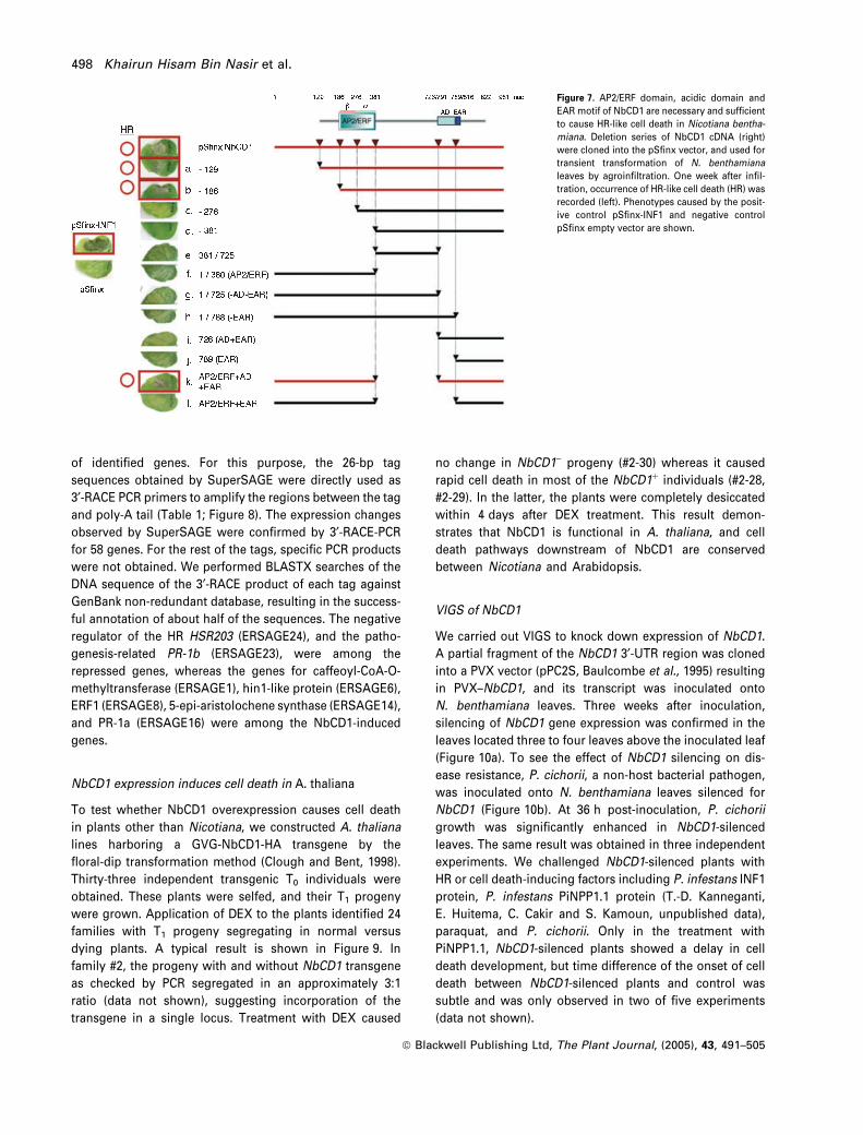
AP2/ERF domain, acidic domain and EAR motif of NbCD1 are
necessary and sufficient to cause cell death
To identify the domains of NbCD1 responsible for induction
of cell death, a series of deletion mutants of NbCD1 were
cloned into the pSfinx vector and tested for their potency in
causing cell death upon transient overexpression by agro-
infiltration (Figure 7). Results show that the N-terminal re-
gion upstream of AP2/ERF domain is dispensable for cell
death (Figure 7a,b). Lack of either the intact AP2/ERF domain
(Figure 7c,d), acidic domain (Figure 7l) or EAR motif (Fig-
ure 7h) abrogates the function of NbCD1 as a cell death in-
ducer. The intervening region between AP2/ERF domain and
acidic domain was dispensable for causing cell death (Fig-
ure 7k). Taken together, these results suggest that the AP2/
ERF domain, acidic domain and EAR motif of NbCD1 are
necessary and sufficient to cause HR-like cell death in
N. benthamiana leaves.
SuperSAGE reveals NbCD1 downstream targets
To gain an insight into the genes regulated by NbCD1,
SuperSAGE (Matsumura et al., 2003) was applied to
N. benthamiana to compare gene expression profiles
of NbCD1-HA-overexpressing and GFP-overexpressing
(control) leaves. GVG-NbCD1-HA and GVG-GFP plasmids
were separately transferred to A. tumefaciens, and used for
transient transformation of N. benthamiana leaves. Forty-
eight hours later, DEX was infiltrated to induce gene
expression from the GVG vectors, and leaf samples
were collected 4 h after DEX treatment. Messenger RNAwas
extracted from the leaf samples, and subjected to Super-
SAGE. For each of GVG-NbCD1-HA- and GVG-GFP-trans-
formed leaves, a total of 10 269 tags corresponding to 6659
genes and 10 884 tags corresponding to 6796 genes were
isolated, respectively. After comparison of frequencies of
each tag in the two samples, 138 tags were identified to be
differentially represented between GVG-NbCD1-HA- and
GVG-GFP-transformed leaves by criteria that either the tag
frequencies are statistically significantly different or the tag
frequencies are different more than fourfold between the
two samples. Of these, 68 tags were overrepresented,
whereas 70 tags were underrepresented in NbCD1-over-
expressing leaves. To further confirm the SuperSAGE
results, RT-PCR was carried out to compare the expression
Figure 6. NbCD1 is localized in nuclei prior to triggering cell death. GVG-
GFP-NbCD1 (left) and GVG-GFP (right) were transiently transformed to
Nicotiana benthamiana leaves. Two days later, leaves were treated with
DEX to induce transgene expression. Leaf tissue was observed under a UV
microscope 6 h (top) and 24 h (middle) after DEX treatment. Leaf phenotypes
72 h after DEX treatment are also shown (bottom).
Overexpression of a class II ERF causes plant cell death 497
ª Blackwell Publishing Ltd, The Plant Journal, (2005), 43, 491–505
of identified genes. For this purpose, the 26-bp tag
sequences obtained by SuperSAGE were directly used as
3¢-RACE PCR primers to amplify the regions between the tag
and poly-A tail (Table 1; Figure 8). The expression changes
observed by SuperSAGE were confirmed by 3¢-RACE-PCRfor 58 genes. For the rest of the tags, specific PCR products
were not obtained. We performed BLASTX searches of the
DNA sequence of the 3¢-RACE product of each tag against
GenBank non-redundant database, resulting in the success-
ful annotation of about half of the sequences. The negative
regulator of the HR HSR203 (ERSAGE24), and the patho-
genesis-related PR-1b (ERSAGE23), were among the
repressed genes, whereas the genes for caffeoyl-CoA-O-
methyltransferase (ERSAGE1), hin1-like protein (ERSAGE6),
ERF1 (ERSAGE8), 5-epi-aristolochene synthase (ERSAGE14),
and PR-1a (ERSAGE16) were among the NbCD1-induced
genes.
NbCD1 expression induces cell death in A. thaliana
To test whether NbCD1 overexpression causes cell death
in plants other than Nicotiana, we constructed A. thaliana
lines harboring a GVG-NbCD1-HA transgene by the
floral-dip transformation method (Clough and Bent, 1998).
Thirty-three independent transgenic T0 individuals were
obtained. These plants were selfed, and their T1 progeny
were grown. Application of DEX to the plants identified 24
families with T1 progeny segregating in normal versus
dying plants. A typical result is shown in Figure 9. In
family #2, the progeny with and without NbCD1 transgene
as checked by PCR segregated in an approximately 3:1
ratio (data not shown), suggesting incorporation of the
transgene in a single locus. Treatment with DEX caused
no change in NbCD1) progeny (#2-30) whereas it caused
rapid cell death in most of the NbCD1þ individuals (#2-28,
#2-29). In the latter, the plants were completely desiccated
within 4 days after DEX treatment. This result demon-
strates that NbCD1 is functional in A. thaliana, and cell
death pathways downstream of NbCD1 are conserved
between Nicotiana and Arabidopsis.
VIGS of NbCD1
We carried out VIGS to knock down expression of NbCD1.
A partial fragment of the NbCD1 3¢-UTR region was cloned
into a PVX vector (pPC2S, Baulcombe et al., 1995) resulting
in PVX–NbCD1, and its transcript was inoculated onto
N. benthamiana leaves. Three weeks after inoculation,
silencing of NbCD1 gene expression was confirmed in the
leaves located three to four leaves above the inoculated leaf
(Figure 10a). To see the effect of NbCD1 silencing on dis-
ease resistance, P. cichorii, a non-host bacterial pathogen,
was inoculated onto N. benthamiana leaves silenced for
NbCD1 (Figure 10b). At 36 h post-inoculation, P. cichorii
growth was significantly enhanced in NbCD1-silenced
leaves. The same result was obtained in three independent
experiments. We challenged NbCD1-silenced plants with
HR or cell death-inducing factors including P. infestans INF1
protein, P. infestans PiNPP1.1 protein (T.-D. Kanneganti,
E. Huitema, C. Cakir and S. Kamoun, unpublished data),
paraquat, and P. cichorii. Only in the treatment with
PiNPP1.1, NbCD1-silenced plants showed a delay in cell
death development, but time difference of the onset of cell
death between NbCD1-silenced plants and control was
subtle and was only observed in two of five experiments
(data not shown).
Figure 7. AP2/ERF domain, acidic domain and
EAR motif of NbCD1 are necessary and sufficient
to cause HR-like cell death in Nicotiana bentha-
miana. Deletion series of NbCD1 cDNA (right)
were cloned into the pSfinx vector, and used for
transient transformation of N. benthamiana
leaves by agroinfiltration. One week after infil-
tration, occurrence of HR-like cell death (HR) was
recorded (left). Phenotypes caused by the posit-
ive control pSfinx-INF1 and negative control
pSfinx empty vector are shown.
498 Khairun Hisam Bin Nasir et al.
ª Blackwell Publishing Ltd, The Plant Journal, (2005), 43, 491–505

Table 1 Differentially expressed genes inNbCD1- versus GFP-overexpressing Nic-otiana benthamiana leaves (4 h after DEXtreatment for the induction of transgeneexpression)
Tag sequencea
Count of tags
Putative encoded proteinb ID no.NbCD1 GFP
ACTCAAATACTTGTGCACGAGG 8 33 PR-1b ERSAGE23TGCCGTCTTGATTGTCACGTTC 8 22 HSR203 ERSAGE24ACAGCAGCAGCAGCGACAGCGA 7 22 No homology ERSAGE25ATAAGCTTTAAGGGATTAGTCG 1 20 Cytosolic aldolase ERSAGE26
CGCCCCCCGTCCGCTTGCCGAC 3 16 Senescence-associated protein ERSAGE27GCCGACTTGCTGCACGTCAACC 1 13 60S ribosomal protein ERSAGE28ACAATATGCTCTGTCTTGTCTG 3 11 No homology ERSAGE29GCTAATGCTGGACCTGGAACCA 2 10 Cyclophilin ERSAGE30CGCCGTTTTGGCTGTAGAATGG 0 7 Hypothetical protein similar to NtPR27 ERSAGE31
ACCGTGGAGCCTTGATCATTTT 0 7 No homology ERSAGE32GATAGTCCTTCACATTGGCACG 0 7 Chaperonin 21 ERSAGE33AGCAGCTAAGTGAAGAAACTTG 1 6 Glutamate-ammonia ligase ERSAGE34ATCAAAATAGATTTCAGTTGGG 1 6 Phospho glycerate kinase ERSAGE35TAATTTCCCAAATCGAACTGTA 1 6 Carbonic anhydrase ERSAGE36GACGCTTCCAGACTACACAGGA 1 6 No homology ERSAGE37
CCAGCTGGGAGAGCTAATCCGC 0 6 Copper chaperone ERSAGE38GGGGTATACCACACTGTCTTTG 0 6 Aspartic proteinase ERSAGE39TGCTGCAGGCAGTGCTTCCGCA 0 6 40S ribosomal protein ERSAGE40GATGAGCTTTTAAGGGGACTAGT 0 6 No homology ERSAGE41ATGCAGCTGGGTTGTGATGGCG 0 5 No homology ERSAGE42
TAATTTGGCGGGGAGTAATGTA 0 5 No homology ERSAGE43TGAAAGAACAGACTGAGCTTGT 0 5 No homology ERSAGE44GATGGTATGTGCCTGCTCCAGT 0 5 Ribosomal protein ERSAGE45CAAAACACTCTCATCCCCCCTA 0 4 Tonoplast intrinsic protein ERSAGE46GAGGCATTCTCCCGTACGTCAT 0 4 Cytosolic acotase ERSAGE47TCTACGGAGGCTGTAACTTTTT 0 4 Calmodulin ERSAGE48
GGTAGAGCCAAAGAGTGTGAAC 0 4 Ribosomal protein ERSAGE49TTCTGCTACTCGACTATGAGAC 0 4 No homology ERSAGE50TGCTTCAAGACGTATCACTTGT 0 4 SAR8.2c protein ERSAGE51TACACTTCAAGAATCCTACTCC 0 4 No homology ERSAGE52GGTAGATGGATGGTTTGCTTAG 0 4 Hypothetical protein (A. thaliana) ERSAGE53
GCACAGTTAAAGGATTCTCTCT 0 4 40S ribosomal protein ERSAGE54GATGAAGAAGCTGCTGGGTTTT 0 4 RNA binding glycine-rich protein ERSAGE55CAAAACACTCTCATCCCCCCTA 0 4 ADP-glucose pyrophosphorylase ERSAGE56ACACGGTCAAGCAAAGATCTGT 0 4 No homology ERSAGE57GAATGCATTGTAGAATACTGTG 0 4 No homology ERSAGE58
GATCATATGATTTCATATTTGT 22 3 Caffeoyl-CoA O-methyltransferase ERSAGE1GGCAGATCAATGGGATCCAGCC 16 2 Hypothetical protein (A. thaliana) ERSAGE2ACGTATTACAAGTACCAAAAGC 15 2 Hypothetical protein similar to NtSA1 ERSAGE3
GAAGAAGCAACTTTAGTGTGGT 14 3 Calmodulin ERSAGE4TACATTGAAAGATGGAGGCGGA 13 0 No homology ERSAGE5GTACCATCTTGTTATATTTGGA 10 1 hin1-like protein ERSAGE6GTGGTGGGTACATCGTTAGAAG 9 1 Epoxide hydrolase ERSAGE7TTGATTATATGACCGGAGGGTA 7 0 ERF1 ERSAGE8
GGGTGTTGACCAAGACGCACTT 7 1 Unknown protein ERSAGE9AGTGCAAGCGTTCGAGGTTCCT 7 1 No homology ERSAGE10GGCAGTGAAACTGGGAAGAAGA 6 0 No homology ERSAGE11ATTACTATTCTATCAAGGGACT 6 1 60S ribosomal protein ERSAGE12GATCGGCAAACAAAGAGATAAT 6 0 No homology ERSAGE13TCGTATAAAGTTGTAACGGAGT 6 0 5-epi-aristolochene synthase ERSAGE14
AGTCTCAACATTAGGTGGATTA 6 0 No homology ERSAGE15CACTAATAATGCTACTTCAAGT 5 0 PR-1 ERSAGE16TGGAGTTAGATCCAAATTTTCC 4 0 No homology ERSAGE17GTACTACTCCTGGAAGATCATT 4 0 No homology ERSAGE18GATTCCAAAAAAGAGCAAAAGC 4 0 Phenylpropanoid:glucosyltransferase ERSAGE19
GATATTGATGATCAGAATAATG 4 0 No homology ERSAGE20CTAATAAGGAAATTGATGCTGC 4 0 No homology ERSAGE21ACTTCTTGGGACTGATGTACAT 4 0 Cysteine proteinase ERSAGE22
aTags represented as a 22-bp sequence excluding the NlaIII site (CATG).bEncoded proteins were deduced by BLAST search with 3¢RACE fragment recovered by using a26-bp-tag primer. No homology indicates that no homologous proteins were identified by BLASTsearch.
Overexpression of a class II ERF causes plant cell death 499
ª Blackwell Publishing Ltd, The Plant Journal, (2005), 43, 491–505

Figure
8.NbCD1
target
gen
esof
Nic
oti
ana
ben
tham
ian
aas
reve
aled
bySuperSAGEan
d3¢-RACEPCR.The26
-bpSuperSAGEtagse
quen
celis
tedin
Tab
le1was
use
das
3¢-RACEPCRprimersto
amplifytheregions
betwee
ntagsan
dpoly-A
tailforNbCD1-ove
rexp
ress
edan
dGFP
-ove
rexp
ressed
leav
es.The3¢-RACEPCRproductswerese
quen
cedan
dan
notatedbyBLA
STse
arch
es.
500 Khairun Hisam Bin Nasir et al.
ª Blackwell Publishing Ltd, The Plant Journal, (2005), 43, 491–505

Discussion
Functional screening of plant genes identifies novel positive
regulators of cell death
To search for plant genes that positively regulate cell death,
we employed a high-throughput overexpression screening
method similar to the one described by Takken et al. (2000).
Overexpression of anonymous cDNAs in vivo followed by
screening for cells showing phenotypic changes is a routine
approach to functionally identify novel genes in prokaryotes,
yeasts, and animal cells (Grimm, 2004; Rine, 1991). The
approach we employed in this study proved useful in iden-
tifying new genes in plants solely based on their function
and is an alternative strategy to activation-tagging screens
(Xia et al., 2004). Use of a binary-PVX vector system allows
reliable transient overexpression and is therefore superior to
the TMV expression system (Karrer et al., 1998). The over-
expression screens we performed in N. benthamiana are
complementary to the VIGS approach taken by Lu et al.
(2003). In that study, approximately 5000 normalized ran-
dom cDNA fragments were used to silence their corres-
ponding genes and identify genes involved in HR caused by
the interaction between the Pto resistance gene and Pseu-
domonas syringae (Lu et al., 2003). This VIGS screening
identified Hsp90 among 79 genes required for R-gene-
mediated HR. In a similar VIGS screen in N. benthamiana,
Del Pozo et al. (2004) identified MAPKKKa as a positive
regulator of HR and disease resistance against P. syringae.
The loss-of-function screens used by Lu et al. (2003) and Del
Pozo et al. (2004), as well as the gain-of-function strategy
described here and elsewhere (Kamoun et al., 2003; Takken
et al., 2000), provide a useful set of high-throughput
techniques for isolating novel factors involved in plant cell
death in particular, and plant defense in general.
NbCD1 positively regulates cell death and contributes to
non-host resistance
NbCD1 encoded a protein with a high sequence similarity to
class II ERFs (Figure 2). ERFs form a distinct family of plant-
specific transcription factors with similarity to AP2
transcription factors (Ohme-Takagi et al., 2000). Although
Figure 9. NbCD1 overexpression causes cell
death in Arabidopsis thaliana. Genomic DNAs
of a wild type (WS) and three T1 progeny (#2-28,
#2-29, and #2-30) of the GVG-NbCD1-HA trans-
genic line were used as templates for PCR
amplification of b-tublin, NbCD1 transgene, and
GVG vector arm (top). These plants were treated
with DEX, and phenotypes observed 4 days after
treatment (bottom).
(a) (b)
Figure 10. VIGS of NbCD1 compromises non-host resistance of Nicotiana
benthamiana to Pseudomonas cichorii.
(a) RT-PCR of NbCD1 and rbcS genes in PVX-GFP-inoculated plant and PVX-
NbCD1-inoculated plant.
(b) Leaves of N. benthamiana exhibiting NbCD1 silencing were inoculated
with P. cichorii and bacterial growth was monitored at 1, 12, 24, and 36 h after
the inoculation. PVX-GFP-inoculated plants were used as control.
Overexpression of a class II ERF causes plant cell death 501
ª Blackwell Publishing Ltd, The Plant Journal, (2005), 43, 491–505
several class I and class III ERFs have been linked to defense,
this study reports a class II ERF that contributes to cell death
and defense. NtERF1, 2, 3 and 4 of tobaccowere first isolated
as genes coding for proteins that specifically bind to a cis-
element, GCC-box, known to be necessary and sufficient for
ethylene-induced responseof promotersof a varietyof genes
(Ohme-Takagi and Shinshi, 1990, 1995). ERFs were subdivi-
ded into three classes according to their aminoacid sequence
similarity (Fujimoto et al., 2000): class I ERFs include tobacco
NtERF1andNtERF2, aswell asA. thalianaAtERF1andAtERF2
and tomato Pti4 and Pti5 (Zhou et al., 1997); class II ERFs
consist of NtERF3, AtERF3, and AtERF4; and class III ERFs
comprise NtERF4 and AtERF5. Members of all ERF classes
were shown to bind to GCC-box sequences (Ohme-Takagi
and Shinshi, 1995; Solano et al., 1998; Zhou et al., 1997),
whereas binding of Pti4 to non-GCC-box cis-element was
recently demonstrated (Chakravarthy et al., 2003). ERFs
belonging to class I and class III function as transcriptional
activators, and plants overexpressing some of these factors
showed enhanced disease resistance (Berrocal-Lobo et al.,
2002; Gu et al., 2002). In contrast, class II ERFs function as
active transcriptional repressors (Fujimoto et al., 2000),
based on their C-terminal EAR motifs (Ohta et al., 2001).
Co-expression assay of NbCD1 and GAL4-VP19 showed
that similar to other class II ERFs, NbCD1 functions as a
transcriptional repressor through its EAR motif (Figure 3).
The EAR motif, as well as the AP2/ERF and acidic domains,
were necessary for NbCD1 to exert cell death (Figure 7). It is
well known that overexpression of some cellular factors
cause titration of interacting proteins (squelching) and this
squelching effect is responsible for the alteration of pheno-
types (Ptashne and Gann, 2002). In such circumstances,
overexpression of isolated domains involved in protein–
protein interaction or protein–DNA interaction alone would
be able to cause the phenotype. However, this was not the
case for NbCD1, which required all three functional domains
to exert cell death (Figure 7). Altogether, these results
suggest that transcriptional repressor activity is necessary
for NbCD1 to cause cell death.
We observed that overexpressed NbCD1 was targeted to
plant nuclei (Figure 6), where it is likely to bind to DNA
regions so far unidentified. We hypothesize that NbCD1
represses the expression of unidentified target genes, some
of which are presumably negative regulators of cell death
(see below). Cell death caused by NbCD1 overexpression
displayed many similarities to pathogen-induced HR
(Figure 5). It was associated with rapid generation of H2O2,
ion leakage, and DNA fragmentation. We envisage that
expression changes in NbCD1 target gene(s) triggered the
generation of H2O2 and ultimately cell death. It is well
established that accumulation of H2O2 to high concentration
leads to the HR (Alvarez et al., 1998; Dat et al., 2003).
Apparently, such a mechanism operates not only in Nicoti-
ana but also in Arabidopsis, suggesting a conserved cell
death-inducing process downstream of NbCD1 in dicot
plants (Figure 9).
NbCD1 expression was induced by HR elicitor INF1 and a
non-host pathogen, P. cichorii (Figure 4). VIGS of NbCD1
partially compromised plant defense against P. cichorii
(Figure 10). These results suggest that NbCD1 positively
regulates defense response, including non-host resistance.
However, VIGS of NbCD1 did not affect the timing of onset of
cell death caused by INF1, paraquat, and P. cichorii. We
hypothesize that either (i) NbCD1 is not involved in the
signaling pathways emanating from tested HR- or cell death-
inducing agents but plays a role in other pathways of cell
death signaling, or (ii) NbCD1 actually is involved in tested
cell death pathways, but because of redundancy of signaling
pathways, VIGS of NbCD1 alone did not affect the final
output. Further studies with other pathogens and cell death-
inducing agents are needed to obtain a clear picture of
function of NbCD1.
SuperSAGE revealed downstream targets of NbCD1
We exploited the SuperSAGE technique to identify down-
stream targets of NbCD1. SuperSAGE (Matsumura et al.,
2003) is an improved version of serial analysis of gene
expression (SAGE, Velculescu et al., 1995) that is
particularly adapted to non-model organisms, such as
N. benthamiana. SuperSAGE, along with RT-PCR vali-
dation, applied to NbCD1-overexpressing and control leaves
identified 58 differentially expressed transcripts between
the two samples (Table 1; Figure 8). Although NbCD1 was
shown to function as an active repressor (Figure 3), its
overexpression caused induction as well as repression
of many genes. Several of the differentially expressed
genes were previously tied to pathogen defense and
hypersensitive death. HSR203 (ERSAGE24) was among the
NbCD1-repressed genes. This gene codes for a serine
hydrolase and is known to be rapidly induced in incom-
patible host–pathogen interactions (Pontier et al., 1994).
Transgenic tobacco plants with antisense-mediated reduced
HSR203 transcript levels exhibited a remarkably accelerated
HR response when inoculated with incompatible patho-
gens, leading the authors to suggest that HSR203 is a
negative regulator of the HR (Tronchet et al., 2001). It is
possible that NbCD1 repression of HSR203 expression
contributes to the rapid development of the HR. The
induced genes included a gene for ERF1 (ERSAGE8), a
class I ERF suggesting that expression of repressor and
activator ERF types is interlinked. Other induced genes
included several HR marker genes (hin1; Gopalan et al.,
1996), genes for acidic PR protein (PR1a), lignin biosynthe-
sis (caffeoyl-CoA-O-methyltransferase) and phytoalexin
biosynthesis (5-epi-aristolochene synthase). Induction of
these defense-related genes could be a secondary effect of
NbCD1-medidated repression of key genes that negatively
502 Khairun Hisam Bin Nasir et al.
ª Blackwell Publishing Ltd, The Plant Journal, (2005), 43, 491–505

regulate defense responses. About half of the genes listed
in Table 1 had no similarities to sequences in public data-
base or code for unknown proteins. In the future, transient
overexpression of NbCD1-induced genes as well as VIGS of
NbCD1-repressed genes will identify the downstream fac-
tors directly involved in NbCD1-mediated HR-like cell death.
In conclusion, gain-of-function screening of plant cDNAs
implicates NbCD1 in cell death signaling and regulation of
non-host resistance. Our data point to a model of transcrip-
tional regulatory cascades. NbCD1 may positively regulate
cell death through its EAR motif-mediated repressor activity
and contributes to non-host resistance by downregulating
the expression of other defense response genes. Future
studies will focus on the key target genes of NbCD1
responsible for cell death and non-host resistance.
Experimental procedures
Plant material and INF1 treatment
Nicotiana benthamiana plants were grown in a glasshouse at 20�C.INF1 elicitor (100 nM) was prepared according to Kamoun et al.(1998) and infiltrated to well-developed leaf blades. Leaves werecollected 0, 15, 30, 60, 120 and 240 min after infiltration and used forisolation of RNA for cDNA library construction.
cDNA library construction in pSfinx vector
Messenger RNA (mRNA) was isolated from total RNA by the mRNApurification kit (Amersham Biosciences, Little Chalfont, UK). Dou-ble-stranded cDNAs were synthesized by a SuperScript PlasmidSystem kit (Gibco BRL, Gaithersburg, MD, USA). These cDNAs withthe SalI site in the 5¢-ends and the NotI site in the 3¢-ends weredirectionally cloned into a modified pSfinx vector (Takken et al.,2000) whereby the original ClaI–SfiA–SfiB–AscI–NotI–SalI cloningcassette was replacedwith the AscI–SalI–NotI cassette using the SalIand NotI sites. Competent Escherichia coli (DH10a) cells weretransformed with the cDNAs by electroporation. From more than500 000 independent E. coli colonies, plasmids were isolated inbulk. Using this bulked cDNA, A. tumefaciens strain MOG101 cellswere transformed by electroporation. From more than 100 000transformed A. tumefaciens colonies, approximately 40 000 cloneswere transferred to 384-well microtiter plates filled with LB-agarmedium, and kept for further use.
Toothpick inoculation of A. tumefaciens
From 384-well microtiter plates, A. tumefaciens clones were trans-ferred to 96-well microtiter plates filled with LB liquid medium, andcultured for 48 h at 28�C. Liquid-cultured cells were lifted by atoothpick, and inoculated to N. benthamiana leaves basically fol-lowing the method of Takken et al. (2000).
Transient assay of NbCD1 repressor activity
Reporter plasmid, construction of effector plasmids, and delivery ofplasmids into A. thaliana leaves by particle bombardment are asdescribed (Fujimoto et al., 2000). For the reporter plasmid, fireflyluciferase gene was used. To monitor cell viability, another plasmid
containing Renilla luciferase under the control of the CaMV35Spromoter was co-bombarded with reporter and effector plasmids.Firefly luciferase activity was normalized as its raw value divided byRenilla luciferase activity.
Northern blot analysis
The probe used was a PCR-amplified partial fragment of NbCD1cDNA corresponding to the amino acid residue nos 85–231, down-stream of the AP2 domain of the NbCD1 protein. This region of theNbCD1 cDNA exhibits low sequence similarity to other ERF genes.After hybridization, the membrane was washed in high-stringencycondition (0.1x SSC, 0.1% SDS at 60�C).
Inducible expression of NbCD1-triple HA and NbCD1-GFP
To the 3¢-end of the ORF of NbCD1, an extension of the DNAsequence (5¢- GCTTCTAGATATCCATATGATGTTCCAGATTATGCT-GGTTATCCATATGATGTTCCAGATTATGCTGGTTCTTATCCATATG-ATGTTCCAGATTATGCTTCTAGATGA-3¢) corresponding to the tri-ple-HA tag (ASRYPYDVPDYAGYPYDVPDYAGSYPYDVPDYASR)wasadded by PCR resulting in NbCD1-triple HA cDNA. This fragmentwas cloned into XhoI and SpeI sites of a GVG-vector, pTA7001(Aoyama and Chua, 1999). Engineered green fluorescent protein(mGFP, Haseloff and Amos, 1995) gene was fused to the 3¢-end ofNbCD1 cDNA, and cloned into pTA7001. These binary vectorswere used for transformation of A. tumefaciens MOG101 by elec-troporation. Nicotiana benthamiana leaves were infiltrated withA. tumefaciens cells to establish transient transformation. Two daysafter A. tumefaciens infiltration, DEX (30 lM in 0.1% ethanol) wasfurther infiltrated to the same leaves to induce gene expression.
Measurement of H2O2 and ion leakage
H2O2 generation was measured by using DCFH-DA as described bySanchez et al. (1990). Ion leakage was measured according to Kimet al. (2003).
SuperSAGE
Four hours after induction of NbCD1 and GFP from the GVG vector,leaf samples (approximately 5 g) were collected. Total RNA wasextracted from them according to the standard method, and mRNAisolated by Oligo-dT columns. SuperSAGE was carried out as des-cribed (Matsumura et al., 2003) using 5 lg mRNA as the startingmaterial. Agroinfiltration causes defense response in plants (Dittet al., 2001). In this experiment, we focused on the genes that aredifferentially transcribed between the NbCD1 and control treat-ments. Using the same RNA, cDNA was synthesized separatelyusing an anchored oligodT primer (5¢-biotin-CTGATCTA-GAGGTACCGGATCCCAGCAGTTTTTTTTTTTTTTTTT-3¢). For 3¢-RACE PCR, 26-bp primers corresponding to the SuperSAGE tagswere used in combination with the primer (polyT anc: 5¢-GGCCACGCGTCGACTAGTACTTTTTTTTTTTTTTTTT-3¢) comple-mentary to the cDNA ends.
Arabidopsis transformation
Arabidopsis thaliana landrace WS was grown for 50 days underlight. Floral dip transformation of A. thaliana plant with the GVG-NbCD1-HA construct was carried out according to Clough andBent (1998). Transformed T0 seeds were selected by 30 lg ml)1
hygromycin and 500 lg ml)1 cefotaxim. DEX (50 nM in 0.1%
Overexpression of a class II ERF causes plant cell death 503
ª Blackwell Publishing Ltd, The Plant Journal, (2005), 43, 491–505

ethanol) was used to induce the expression of NbCD1-HA fromthe GVG vector.
VIGS of NbCD1
The NbCD1 cDNA fragment corresponding to positions 654–834,whereby the first nucleotide of the first codon was set to position 1,was cloned into a PVX vector pPC2S in the antisense orientationresulting in pTXS.NbCD1. pTXS.NbCD1 was linearized by a restric-tion endonuclease SpeI, and in vitro runoff transcripts were syn-thesized by T7 RNA polymerase. The transcripts were inoculated toN. benthamiana plants as described elsewhere (Saitoh et al., 2001).Confirmation of gene silencing of NbCD1 was made by using aprimer pair, 5¢-CCGTCGACTGGTTTTAGAGCAGGAGA-3¢ and 5¢-CCGATATCTGAAGAAATCACTTGCTC-3¢ annealing to position 361and position 656 of NbCD1 cDNA.
Inoculation with P. cichorii and determination of growth
kinetics
Pseudomonas cichorii SPC9001 (Hikichi et al., 1998) was grown at28�C in nutrient broth medium (Difco, Detroit, MI, USA) containingampicillin (10 lg ml)1) overnight. After centrifugation, bacterialcells were resuspended in 10 mM MgCl2 (OD600 ¼ 0.01). Bacterialsuspensions were inoculated onto leaves using a needleless syr-inge. The increase in the numbers of bacteria was estimated in leafdisks. Further details are available in Sharma et al. (2003).
Acknowledgements
We dedicate this work to H. Enei and K. Hinata, the former directorsof IBRC. We acknowledge Mattieu Joosten, Wageningen University,for the provision of pSfinx vector, David Baulcombe, SainsburyLaboratory, John Innes Center for pPC2S, Nam Hai Chua, Rocke-feller University for pTA 7001, and Jim Haseloff, Cambridge Uni-versity for mGFP. This work was carried out in part by support fromthe ‘Program for Promotion of Basic Research Activities for Inno-vative Biosciences’ (Japan) and by the ‘Research for the FutureProgram of the Japan Society for the Promotion of Science’ and bythe ‘Iwate University 21st Century COE Program: Establishment ofThermo-Biosystem Research Program’. SK was supported by theNSF Plant Genome Research Program grant DBI-0211659. This workwas carried out in a containment facility of the Iwate BiotechnologyResearch Center under license no. 13-YokoShoku-965 from theMinistry of Agriculture, Forestry and Fisheries, Japan, and licenseno. 12-Ken-Kyoku-52 from the Ministry of Education, Culture andScience, Japan.
References
Altschul, S.F., Gish, W., Miller, W., Myers, E.W. and Lipman, D.J.
(1990) Basic local alignment search tool. J. Mol. Biol. 215, 403–410.Alvarez, M.E., Pennell, R.I., Meijer, P.J., Ishikawa, A., Dixon, R.A.
and Lamb, C. (1998) Reactive oxygen intermediates mediate asystemic signal network in the establishment of plant immunity.Cell, 92, 773–784.
Aoyama, T. and Chua, N.-H. (1999) A glucocorticoid-mediatedtranscriptional induction system in transgenic plants. Plant J. 11,605–612.
Azevedo, C., Sadanandom, A., Kitagawa, K., Freialdenhoven, A.,
Shirasu, K. and Schulze-Lefert, P. (2002) The RAR1 interactorSGT1, an essential component of R gene-triggered diseaseresistance. Science, 295, 2073–2076.
Baulcombe, D.C., Chapman, S. and Santa Cruz, S. (1995) Jellyfishgreen fluorescent protein as a reporter for virus infections. PlantJ. 7, 1045–1053.
Berrocal-Lobo, M., Molina, A. and Solano, R. (2002) Constitutiveexpression of ETHYLENE-RESPONSE-FACTOR1 in Arabidopsisconfers resistance to several necrotrophic fungi.Plant J. 29, 23–32.
Century, K.S., Holub, E.B. and Staskawicz, B.J. (1995) NDR1, a locusof Arabidopsis thaliana that is required for disease resistance toboth a bacterial and a fungal pathogen. Proc. Natl Acad. Sci. USA,92, 6597–6601.
Chakravarthy, S., Tuori, R.P., D’Ascenzo, M.D., Fobert, P.R., Des-
pres, C. and Martin, G.B. (2003) The tomato transcription factorPti4 regulates defense-related gene expression via GCC box andnon-GCC box cis elements. Plant Cell, 15, 3033–3050.
Clough, S.J. and Bent, A.F. (1998) Floral dip: a simplified method forAgrobacterium-mediated transformation of Arabidopsis thaliana.Plant J. 16, 735–743.
Dat, J.F., Pellinen, R., Beeckman, T., Van De Cotte, B., Langebartels,
C., Kangasjarvi, J., Inze, D. and Van Breusegem, F. (2003) Chan-ges in hydrogen peroxide homeostasis trigger an active cell deathprocess in tobacco. Plant J. 33, 621–632.
Del Pozo, O., Pedley, K.F. and Martin, G.B. (2004) MAPKKKa is apositive regulator of cell death associated with both plantimmunity and disease. EMBO J. 23, 3072–3082.
Delledonne, M., Zeier, J., Marocco, A. and Lamb, C. (2001) Signalinteractions between nitric oxide and reactive oxygen interme-diates in the plant hypersensitive disease resistance response.Proc. Natl Acad. Sci. USA, 98, 13454–13459.
Ditt, R.F., Nester, E.W. and Comai, L. (2001) Plant gene expressionresponse to Agrobacterium tumefaciens. Proc. Natl Acad. Sci.USA, 98, 10954–10959.
Fujimoto, A.Y., Ohta, M., Usui, A., Shinshi, H. and Ohme-Takagi, M.
(2000) Arabidopsis ethylene-responsive element binding factorsact as transcriptional activators or repressors of GCC box-medi-ated gene expression. Plant Cell, 12, 393–404.
Gaffney, T., Friedrich, L., Vernooij, B., Negrotto, D., Nye, G., Uknes,
S., Ward, E., Kessmann, H. and Ryals, J. (1993) Requirement ofsalicylic acid for the induction of systemic acquired resistance.Science, 261, 754–756.
Glazebrook, J. (2001) Genes controlling expression of defense re-sponses in Arabidopsis – 2001 status. Curr. Opin. Plant Biol. 4,301–308.
Gopalan, S., Wei, W. and He, S.Y. (1996) hrp gene-dependentinduction of hin1: a plant gene activated rapidly by both harpinsand the avrPto gene-mediated signal. Plant J. 10, 591–600.
Greenberg, J.T. and Yao, N. (2004) The role and regulation of pro-grammed cell death in plant–pathogen interactions. Cell Micro-biol. 6, 201–211.
Grimm, S. (2004) The art and design of genetic screens: mammalianculture cells. Nat. Rev. Genet. 5, 179–189.
Gu, Y.Q., Wildermuth, M.C., Charkavarthy, S., Loh, Y.T., Yang, C.,
He, X., Han, Y. and Martin, G.B. (2002) Tomato transcription fac-tors Pti4, Pti5, and Pti6 activate defense responses when ex-pressed in Arabidopsis. Plant Cell, 14, 817–831.
Haseloff, J. and Amos, B. (1995) GFP in plant. Trends Genet. 11, 328–329.
Hatsugai, N., Kuroyanagi, M., Yamada, K., Meshi, T., Tsuda, S.,
Kondo, M., Nishimura, M. and Hara-Nishimura, I. (2004) A plantvacuolar protease, VPE, mediates virus-induced hypersensitivecell death. Science, 305, 855–858.
Heath, M.C. (2000) Hypersensitive response-related death. PlantMol. Biol. 44, 321–334.
Hikichi, Y., Suzuki, K., Toyoda, K., Horikoshi, M., Hirooka, T.
and Okuno, T. (1998) Successive observation of growth and
504 Khairun Hisam Bin Nasir et al.
ª Blackwell Publishing Ltd, The Plant Journal, (2005), 43, 491–505
movement of genetically lux-marked Pseudomonas cichorii andthe response of host tissues in the same lettuce leaf. Ann. Phy-topathol. Soc. Jpn, 64, 519–525.
Hubert, D.A., Tornero, P., Belkhadir, Y., Krishna, P., Takahashi, A.,
Shirasu, K. and Dangl, J.L. (2003) Cytosolic HSP90 associates withand modulates the Arabidopsis RPM1 disease resistance protein.EMBO J. 22, 5679–5689.
Huitema, E., Bos, J.I.B., Tian, M., Win, J., Waugh, M.E. and Kamoun,
S. (2004) Linking sequence to phenotype in Phytophthora–plantinteractions. Trends Microbiol. 12, 193–200.
Kamoun, S., van West, P., Vleeshouwers, V.G., de Groot, K.E. and
Govers, F. (1998) Resistance of Nicotiana benthamiana to Phyt-ophthora infestans is mediated by the recognition of the elicitorprotein INF1. Plant Cell, 10, 1413–1426.
Kamoun, S., Hamada, W. and Huitema, E. (2003) Agrosuppression:a bioassay for the hypersensitive response suited to high-throughput screening. Mol. Plant Microbe Interact. 16, 7–13.
Karrer, E.K., Beachy, R.N. and Holt, C.A. (1998) Cloning of tobaccogenes that elicit the hypersensitive response. Plant Mol. Biol. 36,681–690.
Kim, M., Ahn, J.-W., Jin, U.-H., Choi, D., Paek, K.-H. and Pai, H.-S.
(2003) Activation of the programmed cell death pathway byinhibition of proteasome function in plants. J. Biol. Chem. 278,19406–19415.
Kuriyama, H. and Fukuda, H. (2002) Developmental programmedcell death in plants. Curr. Opin. Plant Biol. 5, 568–573.
Lam, E. (2004) Controlled cell death, plant survival and develop-ment. Nat. Rev. Mol. Cell Biol. 5, 305–315.
Lu, R., Malcuit, I., Moffett, P., Ruiz, M.T., Peart, J., Wu, A.J.,
Rathjen, J.P., Bendahmane, A., Day, L. and Baulcombe, D.C.
(2003) High throughput virus-induced gene silencing implicatesheat shock protein 90 in plant disease resistance. EMBO J. 22,5690–5699.
Martin, G., Bogdanove, A.J. and Sessa, G. (2003) Understanding thefunctions of plant disease resistance proteins. Annu. Rev. PlantBiol. 54, 23–62.
Matsumura, H., Reich, S., Ito, A., Saitoh, H., Kamoun, S., Winter, P.,
Kahl, G., Reuter, M., Kruger, D.H. and Terauchi, R. (2003) Geneexpression analysis of plant host–pathogen interactions by Su-perSAGE. Proc. Natl Acad. Sci. USA, 100, 15718–15723.
Ohme-Takagi, M. and Shinshi, H. (1990) Structure and expression ofa tobacco b-1,3-glucanase gene. Plant Mol. Biol. 15, 941–946.
Ohme-Takagi, M. and Shinshi, H. (1995) Ethylene-inducible DNAbinding proteins that interact with an ethylene-responsive ele-ment. Plant Cell, 7, 173–182.
Ohme-Takagi, M., Suzuki, K. and Shinshi, H. (2000) Regulation ofethylene-induced transcription of defense genes. Plant CellPhysiol. 41, 1187–1192.
Ohta, M., Matsui, K., Hiratsu, K., Shinshi, H. and Ohme-Takagi, M.
(2001) Repression domains of class II ERF transcriptional repres-sors share an essential motif for active repression. Plant Cell, 13,1959–1968.
Parker, J.E., Holub, E.B., Frost, L.N., Falk, A., Gunn, N.D. and Dan-
iels, M.J. (1996) Characterization of eds1, a mutation in Arabid-opsis suppressing resistance to Peronospora parasitica specifiedby several different RPP genes. Plant Cell, 8, 2033–2046.
Pontier, D., Godiard, L., Marco, Y. and Roby, D. (1994) hsr203J, atobacco gene whose activation is rapid, highly localized andspecific for incompatible plant/pathogen interactions. Plant J. 5,507–521.
Ptashne, M. and Gann, A. (2002) Genes and Signals. Cold SpringHarbor, NY, USA: Cold Spring Harbor Laboratory Press.
Qutob, D., Kamoun, S. and Gijzen, M. (2002) Expression of aPhytophthora sojae necrosis-inducing protein occurs duringtransition from biotrophy to necrotrophy. Plant J. 32, 361–373.
Rine, J. (1991) Gene overexpression in studies of Saccharomycescerevisiae. Methods Enzymol. 194, 239–251.
Romeis, T. (2001) Protein kinases in the plant defence response.Curr. Opin. Plant Biol. 4, 407–414.
Saitoh, H., Kiba, A., Nishihara, M., Yamamura, S., Suzuki, K. and
Terauchi, R. (2001) Production of antimicrobial defensin inNicotiana benthamiana with a potato virus X vector. Mol. PlantMicrobe Interact. 14, 111–115.
Sanchez, F.A., Santema, J.S., Hilhorst, R. and Visser, A.J. (1990)Fluorescence detection of enzymatically formed hydrogenperoxide in aqueous solution and in reversed micelles. Anal.Biochem. 187, 129–132.
Sharma, P.C., Ito, A., Shimizu, T., Terauchi, R., Kamoun, S. and
Saitoh, H. (2003) Virus-induced silencing of WIPK and SIPK genesreduces resistance to a bacterial pathogen, but has no effect onthe INF1-induced hypersensitive response (HR) in Nicotianabenthamiana. Mol. Genet. Genomics, 269, 583–591.
Shirasu, K. and Schulze-Lefert, P. (2000) Regulators of cell death indisease resistance. Plant Mol. Biol. 44, 371–385.
Shirasu, K. and Schulze-Lefert, P. (2003) Complex formation, pro-miscuity and multi-functionality: protein interactions in disease-resistance pathways. Trends Plant Sci. 8, 252–258.
Shirasu, K., Lahaye, T., Tan, M.W., Zhou, F., Azevedo, C. and
Schulze-Lefert, P. (1999) A novel class of eukaryotic zinc-bindingproteins is required for disease resistance signaling in barley anddevelopment in C. elegans. Cell, 99, 355–366.
Solano, R., Stepanova, A., Chao, Q. and Echer, J.R. (1998) Nuclearevents in ethylene signaling: a transcriptional cascade mediatedby ETHYLENE-INSENSITIVE3 and ETHYLENE-RESPONSE-FACTOR1. Genes Dev. 12, 3703–3714.
Takahashi, A., Casais, C., Ichimura, K. and Shirasu, K. (2003) HSP90interacts with RAR1 and SGT1 and is essential for RPS2-mediateddisease resistance in Arabidopsis. Proc. Natl Acad. Sci. USA, 100,11777–11782.
Takken, F.L.W., Luderer, R., Gabriels, S.J.E.J., Westerink, N., Lu, R.,
de Wit, P.J.G.M. and Joosten, M.H.A.J. (2000) A functional clo-ning strategy, based on a binary PVX-expression vector, to isolateHR-inducing cDNAs of plant pathogens. Plant J. 24, 275–283.
Torres, M.A., Dangl, J.L. and Jones, J.D.G. (2002) Arabidopsisgp91phox homologues AtrbohD and AtrbohF are required foraccumulation of reactive oxygen intermediates in the plantdefense response. Proc. Natl Acad. Sci. USA, 99, 517–522.
Torto, T.A., Li, S., Styer, A., Huitema, E., Testa, A., Gow, N.A.R., van
West, P. and Kamoun, S. (2003) EST mining and functionalexpression assays identify extracellular effector proteins fromthe plant pathogen Phytophthora. Genome Res. 13, 1675–1685.
Tronchet, M., Ranty, B., Marco, Y. and Roby, D. (2001) HSR203antisense suppression in tobacco accelerates development ofhypersensitive cell death. Plant J. 27, 115–127.
Velculescu, V.E., Zhang, L., Vogelstein, B. and Kinzler, K.W.
(1995) Serial analysis of gene expression. Science, 270, 484–487.Xia, Y., Suzuki, H., Borevitz, J., Blount, J., Guo, Z., Patel, K., Dixon,
R.A. and Lamb, C. (2004) An extracellular aspartic proteasefunctions in Arabidopsis disease resistance signaling. EMBO J.23, 980–988.
Zhou, J., Tang, X. and Martin, G.B. (1997) The Pto kinase conferringresistance to tomato bacterial speck disease interacts with pro-teins that bind a cis-element of pathogenesis-related genes.EMBO J. 16, 3207–3218.
Genbank accession no.: AB196362 (NbCD1)
Overexpression of a class II ERF causes plant cell death 505
ª Blackwell Publishing Ltd, The Plant Journal, (2005), 43, 491–505