The effects of PPAR-γ agonist pioglitazone on renal ischemia/reperfusion injury in rats
9
The effects of PPAR-g agonist pioglitazone on renal ischemia/reperfusion injury in rats Buket Reel, PhD, a Mehmet Guzeloglu, MD, b, * Alper Bagriyanik, c Soner Atmaca, MD, c Koray Aykut, MD, b Gokhan Albayrak, MD, b and Eyup Hazan b a Faculty of Pharmacy, Department of Pharmacology, Ege University, Izmir, Turkey b Faculty of Medicine, Department of Cardiovascular Surgery, Izmir University, Izmir, Turkey c Faculty of Medicine, Department of Histology and Embryology, Dokuz Eylul University, Izmir, Turkey article info Article history: Received 28 May 2012 Received in revised form 20 July 2012 Accepted 9 August 2012 Available online 25 August 2012 Keywords: Renal ischemia/reperfusion PPAR-g agonist MMP Oxidative stress Inflammation abstract Background: Acute renal failure due to renal ischemia/reperfusion (IR) injury is a significant clinical problem in cardiovascular surgery. Reactive oxygen species and inflammation play essential roles in the pathophysiology of IR injury. Matrix metalloproteinases (MMPs) are enzymes that play important roles in inflammation and mediate extracellular matrix degradation. It is known that peroxisome proliferatoreactivated receptor-g agonists have antiinflammatory and antioxidant effects. In the present study, we aimed to investigate the effects of pioglitazone, a synthetic peroxisome proliferatoreactivated receptor-g agonist, on MMPs and oxidative stress in a renal IR injury model in rats. Materials and methods: Male Wistar albino rats were divided into three groups: control (n ¼ 7), placebo (n ¼ 7; saline/p.o.), and pioglitazone (n ¼ 7; 5 mg/kg/day/p.o.). In the control group, a right nephrectomy was conducted without left renal IR injury. In the placebo and pioglitazone groups, pretreatments were started 3 d before operation. In both groups, left renal pedicles were clamped for 60 min and then reperfused for 60 min. Paraffinized renal sections were evaluated histopathologically. Furthermore, expressions of MMP-2, MMP-9, tissue inhibitor of metalloproteinase (TIMP)-2, superoxide dismutase 1 (SOD1), and p47-phox/p67-phox subunits of NADPH oxidase were determined by immunostaining and scoring. Results: In the placebo group, renal IR injury induced diffuse tubular necrosis and intense acute inflammation, but pioglitazone inhibited these effects. MMP-2, MMP-9, and TIMP-2 expression increased in the placebo group. However, while MMP-2 and -9 expression decreased, TIMP-2 expression did not change in the pioglitazone group. p47-phox/p67- phox expression increased in the placebo group, but SOD1 expression did not change. Pioglitazone diminished p47-phox/p67-phox expression, whereas it enhanced SOD1 expression. Conclusion: Our results suggest that pioglitazone might be helpful to reduce renal IR injury because of its antiinflammatory and antioxidant effects. ª 2013 Elsevier Inc. All rights reserved. * Corresponding author. Department of Cardiovascular Surgery, Izmir University Medical School, Karsiyaka, Izmir, Turkey. Tel.: þ90 532 472 0220; fax: þ90 232 483 5476. E-mail address: [email protected] (M. Guzeloglu). Available online at www.sciencedirect.com journal homepage: www.JournalofSurgicalResearch.com journal of surgical research 182 (2013) 176 e184 0022-4804/$ e see front matter ª 2013 Elsevier Inc. All rights reserved. http://dx.doi.org/10.1016/j.jss.2012.08.020
Transcript of The effects of PPAR-γ agonist pioglitazone on renal ischemia/reperfusion injury in rats

ww.sciencedirect.com
j o u r n a l o f s u r g i c a l r e s e a r c h 1 8 2 ( 2 0 1 3 ) 1 7 6e1 8 4
Available online at w
journal homepage: www.JournalofSurgicalResearch.com
The effects of PPAR-g agonist pioglitazone on renalischemia/reperfusion injury in rats
Buket Reel, PhD,a Mehmet Guzeloglu, MD,b,* Alper Bagriyanik,c Soner Atmaca, MD,c
Koray Aykut, MD,b Gokhan Albayrak, MD,b and Eyup Hazanb
a Faculty of Pharmacy, Department of Pharmacology, Ege University, Izmir, Turkeyb Faculty of Medicine, Department of Cardiovascular Surgery, Izmir University, Izmir, Turkeyc Faculty of Medicine, Department of Histology and Embryology, Dokuz Eylul University, Izmir, Turkey
a r t i c l e i n f o
Article history:
Received 28 May 2012
Received in revised form
20 July 2012
Accepted 9 August 2012
Available online 25 August 2012
Keywords:
Renal ischemia/reperfusion
PPAR-g agonist
MMP
Oxidative stress
Inflammation
* Corresponding author. Department of CardTel.: þ90 532 472 0220; fax: þ90 232 483 5476
E-mail address: mehmetguzeloglu@hotm0022-4804/$ e see front matter ª 2013 Elsevhttp://dx.doi.org/10.1016/j.jss.2012.08.020
a b s t r a c t
Background: Acute renal failure due to renal ischemia/reperfusion (IR) injury is a significant
clinical problem in cardiovascular surgery. Reactive oxygen species and inflammation play
essential roles in the pathophysiology of IR injury. Matrix metalloproteinases (MMPs) are
enzymes that play important roles in inflammation and mediate extracellular matrix
degradation. It is known that peroxisome proliferatoreactivated receptor-g agonists have
antiinflammatory and antioxidant effects. In the present study, we aimed to investigate the
effects of pioglitazone, a synthetic peroxisome proliferatoreactivated receptor-g agonist,
on MMPs and oxidative stress in a renal IR injury model in rats.
Materials and methods: Male Wistar albino rats were divided into three groups: control
(n ¼ 7), placebo (n ¼ 7; saline/p.o.), and pioglitazone (n ¼ 7; 5 mg/kg/day/p.o.). In the control
group, a right nephrectomy was conducted without left renal IR injury. In the placebo and
pioglitazone groups, pretreatments were started 3 d before operation. In both groups, left
renal pedicles were clamped for 60 min and then reperfused for 60 min. Paraffinized renal
sections were evaluated histopathologically. Furthermore, expressions of MMP-2, MMP-9,
tissue inhibitor of metalloproteinase (TIMP)-2, superoxide dismutase 1 (SOD1), and
p47-phox/p67-phox subunits of NADPH oxidase were determined by immunostaining and
scoring.
Results: In the placebo group, renal IR injury induced diffuse tubular necrosis and intense
acute inflammation, but pioglitazone inhibited these effects. MMP-2, MMP-9, and TIMP-2
expression increased in the placebo group. However, while MMP-2 and -9 expression
decreased, TIMP-2 expression did not change in the pioglitazone group. p47-phox/p67-
phox expression increased in the placebo group, but SOD1 expression did not change.
Pioglitazone diminished p47-phox/p67-phox expression, whereas it enhanced SOD1
expression.
Conclusion: Our results suggest that pioglitazone might be helpful to reduce renal IR injury
because of its antiinflammatory and antioxidant effects.
ª 2013 Elsevier Inc. All rights reserved.
iovascular Surgery, Izmir University Medical School, Karsiyaka, Izmir, Turkey..ail.com (M. Guzeloglu).ier Inc. All rights reserved.
j o u r n a l o f s u r g i c a l r e s e a r c h 1 8 2 ( 2 0 1 3 ) 1 7 6e1 8 4 177
1. Introduction its reduction in oxidative stress and inhibition of proin-
Renal ischemia/reperfusion injury (IRI) induced by cardio-
vascular surgical procedures, such as thoracoabdominal
aortic aneurysm repair or heart transplantation, is the major
cause of acute renal failure, and also may lead to the devel-
opment or progression of some forms of chronic kidney
disease [1,2]. Development of renal IRI induces the generation
of reactive oxygen species (ROS) and the decline of antioxidant
protection [3,4]. Besides, ischemic damage to the renal
vasculature results in a breakdown of barrier function and
increased adhesiveness to inflammatory cells, both of which
are likely to contribute to the loss of function following
ischemic injury [5e7].
Matrix metalloproteinases (MMPs) are a family of at least
23 structurally and functionally related zinc-dependent
endopeptidases, which regulate fundamental cellular activi-
ties such as cell proliferation, migration, and differentiation
by degrading extracellular matrix [8,9]. Many pathologic
processes, including inflammation and oxidative and nitro-
sative stress, are involved in the increase of MMP expression
and activity. Accordingly, MMPs play an important role in
disease progression after IRI [10,11]. Activities of proin-
flammatory MMPs are controlled by endogenous MMP inhib-
itors, including tissue inhibitors of metalloproteinases
(TIMPs-1e4) [10]. The accumulation of inflammatory cells
during IRI leads to an increase in ROS, reactive nitrogen
species, and proinflammatory cytokines in the ischemic
tissues. All these stimuli can induce strong transcriptional
expression and/or posttranslational activation of MMPs after
an ischemic injury [11].
Peroxisome proliferatoreactivated receptor-g (PPAR-g) is
a ligand-activated transcription factor belonging to the
nuclear hormone receptor superfamily. PPAR-gmodulates the
function of many target genes and participates in the regula-
tion of vital processes such as inflammation, proliferation,
migration, matrix remodeling, and differentiation [12,13].
Thiazolidinediones, which are widely used in the treat-
ment of type II diabetics as insulin sensitizers, are selective
activators of PPAR-g [14]. PPAR-g agonists are likely to exert
their antiinflammatory effects by negatively regulating the
expression of proinflammatory genes that are induced during
macrophage differentiation and activation [12,15,16]. Besides,
PPAR-g agonists have been reported to have a potent antiox-
idant effect and ameliorate oxidative injury [17,18].
In relation to these data, recent evidence demonstrated
that PPAR-g agonists reduce the injury caused by ischemia
and reperfusion of kidney [19,20]. In particular, pioglitazone,
which is a synthetic PPAR-g agonist, was reported to inhibit
inflammation and oxidative stress induced by some IRI
models independently from its insulin-sensitizing effect
[21e24].
Although antiinflammatory and antioxidant protective
effects of pioglitazone in atherosclerosis and IRI in some
organs are known, its effects on intracellular and molecular
mechanisms involved in renal IRI have not been fully clari-
fied yet.
In light of the data collected, we proposed that PPAR-g
agonist pioglitazonemay show renoprotective effects through
flammatory MMPs in an experimental renal IRI model in rats.
2. Materials and methods
This study was approved by the Local Ethics Committee of
DokuzEylulUniversity, SchoolofMedicine.All animals received
care in compliance with the “Principles of Laboratory Animal
Care” formulated by the National Society for Medical Research
and the Guide for the Care and Use of Laboratory Animals.
Animalswere kept in a light- and temperature-controlled room.
They were fed regular rat chow and had free access to water.
2.1. Experimental groups
Male Wistar albino rats, (250e300 g, n ¼ 21) were randomly
divided into three groups, each consisting of seven animals, as
control, placebo, and pioglitazone group. The rats in the
placebo and pioglitazone groups were administered vehicle
(saline) or pioglitazone (5 mg/kg/d) through an orogastric tube
for 3 d before the surgery. Thereafter, the rats were anes-
thetized with an intraperitoneal injection of ketamine (75 mg/
kg) and xylazine (8 mg/kg), an upper abdominal midline inci-
sion was made, and renal blood vessels were isolated bilat-
erally with minimal dissection. A right nephrectomy was
performed in the control group. In the placebo and pioglita-
zone groups, a right nephrectomy followed by occlusion of the
left renal pedicle for 60 min and reperfusion for 60 min was
performed. At the end of the experimental procedure, the rats
underwent a left nephrectomy under anesthesia. Then the left
kidneys of the animals were cut into two halves and imme-
diately fixed in 10% neutral buffered formalin solution for
24e48 h for histologic evaluation, or stored at �80�C for
subsequent biochemical analyses.
2.2. Histologic evaluation of renal injury
The fixed kidney samples were dehydrated through graded
ethanol series. Samples were embedded in paraffin by the
standardmethod and 5-mm-thick sectionsweremounted onto
glass slides. Then, sections were stained with hematoxylin-
eosin or periodic acideSchiff, according to standard proto-
cols. Sections were examined under a light microscope (Leica
DM6000; Leica Microsystems, Wetzlar, Germany), which is
connected to an image capture system, and images takenwith
the digital video camera (Leica DC280; Leica Microsystems)
were transferred to the computer.
Histologic analyses of equally distributed cross-sections
were performed by two masked histologists. One hundred
tubules, 25 fromeachof the fourdifferent areasof kidney (outer
cortex, inner cortex, outer stripe of the outer medulla, and
medullary rays), were randomly evaluated. One hundred
tubules were scored for each section. Themean score for each
of the four regions was calculated. An overall renal tubular
score that included all four regionswas also calculated for each
rat. Tubular atrophywasevaluatedby scoringas follows: 0¼no
atrophy; 1 ¼ <25%; 2 ¼ between 25% and 50%; and 3 ¼ >50%.
Similarly, acute inflammationwasgradedbyscoringas follows;
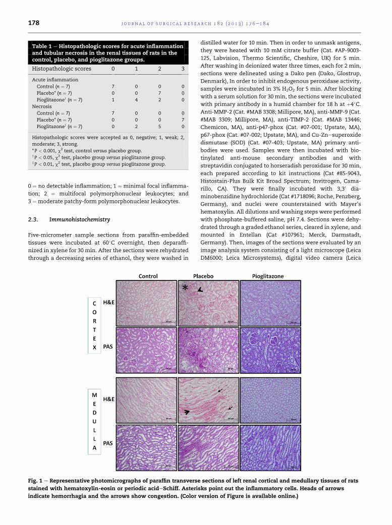
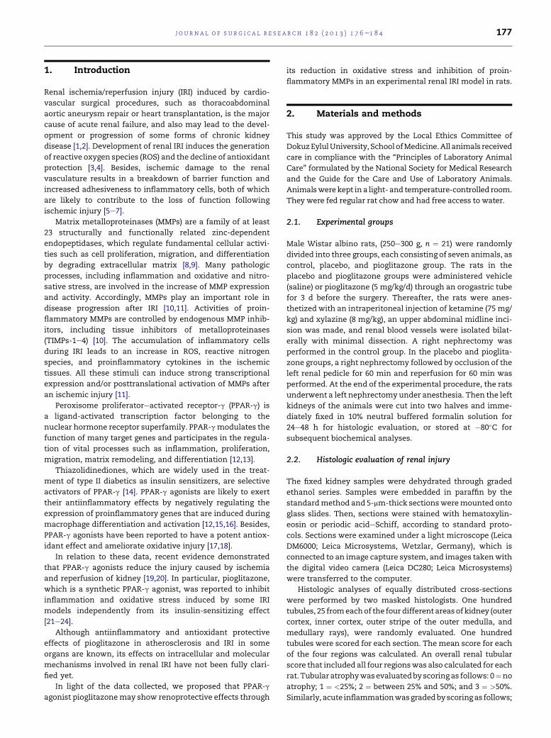
Table 1 e Histopathologic scores for acute inflammationand tubular necrosis in the renal tissues of rats in thecontrol, placebo, and pioglitazone groups.
Histopathologic scores 0 1 2 3
Acute inflammation
Control (n ¼ 7) 7 0 0 0
Placebo* (n ¼ 7) 0 0 7 0
Pioglitazoney (n ¼ 7) 1 4 2 0
Necrosis
Control (n ¼ 7) 7 0 0 0
Placebo* (n ¼ 7) 0 0 0 7
Pioglitazonez (n ¼ 7) 0 2 5 0
Histopathologic scores were accepted as 0, negative; 1, weak; 2,
moderate; 3, strong.
* P < 0.001, c2 test, control versus placebo group.yP < 0.05, c2 test, placebo group versus pioglitazone group.zP < 0.01, c2 test, placebo group versus pioglitazone group.
j o u r n a l o f s u r g i c a l r e s e a r c h 1 8 2 ( 2 0 1 3 ) 1 7 6e1 8 4178
0 ¼ no detectable inflammation; 1 ¼ minimal focal inflamma-
tion; 2 ¼ multifocal polymorphonuclear leukocytes; and
3 ¼ moderate patchy-form polymorphonuclear leukocytes.
2.3. Immunohistochemistry
Five-micrometer sample sections from paraffin-embedded
tissues were incubated at 60�C overnight, then deparaffi-
nized in xylene for 30 min. After the sections were rehydrated
through a decreasing series of ethanol, they were washed in
Fig. 1 e Representative photomicrographs of paraffin transverse
stained with hematoxylin-eosin or periodic acideSchiff. Asteris
indicate hemorrhagia and the arrows show congestion. (Color v
distilled water for 10 min. Then in order to unmask antigens,
they were heated with 10 mM citrate buffer (Cat. #AP-9003-
125, Labvision, Thermo Scientific, Cheshire, UK) for 5 min.
After washing in deionized water three times, each for 2 min,
sections were delineated using a Dako pen (Dako, Glostrup,
Denmark), In order to inhibit endogenous peroxidase activity,
samples were incubated in 3% H2O2 for 5 min. After blocking
with a serum solution for 30 min, the sections were incubated
with primary antibody in a humid chamber for 18 h at þ4�C.Anti-MMP-2 (Cat. #MAB 3308; Millipore, MA), anti-MMP-9 (Cat.
#MAB 3309; Millipore, MA), anti-TIMP-2 (Cat. #MAB 13446;
Chemicon, MA), anti-p47-phox (Cat. #07-001; Upstate, MA),
p67-phox (Cat. #07-002; Upstate, MA), and Cu-Znesuperoxide
dismutase (SOD) (Cat. #07-403; Upstate, MA) primary anti-
bodies were used. Samples were then incubated with bio-
tinylated anti-mouse secondary antibodies and with
streptavidin conjugated to horseradish peroxidase for 30 min,
each prepared according to kit instructions (Cat #85-9043,
Histostain-Plus Bulk Kit Broad Spectrum; Invitrogen, Cama-
rillo, CA). They were finally incubated with 3,30 dia-
minobenzidine hydrochloride (Cat #1718096; Roche, Penzberg,
Germany), and nuclei were counterstained with Mayer’s
hematoxylin. All dilutions and washing steps were performed
with phosphate-buffered saline, pH 7.4. Sections were dehy-
drated through a graded ethanol series, cleared in xylene, and
mounted in Entellan (Cat #107961; Merck, Darmstadt,
Germany). Then, images of the sections were evaluated by an
image analysis system consisting of a light microscope (Leica
DM6000; Leica Microsystems), digital video camera (Leica
sections of left renal cortical and medullary tissues of rats
ks point out the inflammatory cells. Heads of arrows
ersion of Figure is available online.)

Table 2 e Immunoscores for MMP-2, MMP-9, and TIMP-2in the renal tissues from rats in the control, placebo, andpioglitazone groups.
Immunoscores 0 1 2 3
MMP-2
Control (n ¼ 7) 6 1 0 0
Placebo* (n ¼ 7) 0 1 4 2
Pioglitazoney (n ¼ 7) 1 6 0 0
MMP-9
Control (n ¼ 7) 5 2 0 0
Placebo* (n ¼ 7) 0 0 4 3
Pioglitazoney (n ¼ 7) 1 4 2 0
TIMP-2
Control (n ¼ 7) 3 4 0 0
Placeboz (n ¼ 7) 0 4 3 0
Pioglitazone (n ¼ 7) 0 6 1 0
Immunoscores were accepted as 0, negative; 1, weak; 2, moderate;
3, strong.
* P < 0.01, c2test, control versus placebo group.y P < 0.05 c2 test, placebo group versus pioglitazone group.z P < 0.05, c2 test, control versus placebo group.
j o u r n a l o f s u r g i c a l r e s e a r c h 1 8 2 ( 2 0 1 3 ) 1 7 6e1 8 4 179
DC280; Leica Microsystems), and an image capture system,
which is connected to the light microscope. Appropriate
positive controls were also stained.
2.4. Biochemical examinations
Renal samples, which were stored at �80�C previously, were
homogenized in 50 mmol L�1 phosphate buffer (pH ¼ 7.4) by
sonication on ice and then extraction was performed. Total
oxidant status (TOS) and total antioxidant status (TAS) were
determined with commercial spectrophotometric kits (RL0024
and RL0017, respectively; Rel Assay Diagnostics, Gaziantep,
Turkey) as previously described [25e27].
Fig. 2 e Representative photomicrographs of paraffin transverse
Immunohistochemical analysis with MMP-2, MMP-9, and TIMP
immunopositive areas. (Color version of Figure is available onli
2.5. Statistical analysis
Statistical analysis was performed using SPSS version 15.0
for Windows (SPSS Inc, Chicago, IL). A c2 test was used to
evaluate the difference of histologic and immunohisto-
chemical data between control, placebo, and pioglitazone
groups. An unpaired Student t-test was used to compare the
biochemical data. P < 0.05 was considered statistically
significant.
3. Results
3.1. Histology
Renal sections were examined histologically and acute
inflammation and tubular necrosis were evaluated by
scoring.
Histologic examination of renal tissues of the control group
showed normal renal morphology (Table 1, Fig. 1).
Renal tubular injury and acute inflammation scores of the
placebo group were significantly higher compared with those
of the control group (P < 0.001) (Table 1, Fig. 1). Diffuse tubular
necrosis, intracytoplasmic vacuolization, hemorrhage,
congestion, and mononuclear cell infiltration were observed
in the cortical and medullary tissue sections of the placebo
group (Table 1, Fig. 1).
Following pretreatment with pioglitazone, both acute
inflammation and tubular injury scores of the pioglitazone
group were significantly lower compared with those of the
placebo group (P < 0.05 and P < 0.01, respectively) (Table 1,
Fig. 1). Minimal congestion and tubular necrosis, and a slight
mononuclear cell infiltration, were observed in the cortical
and medullary tissue sections of the pioglitazone group
(Table 1, Fig. 1).
sections of left renal cortical tissues of rats.
-2 antibodies. Dark brownestained areas show
ne.)
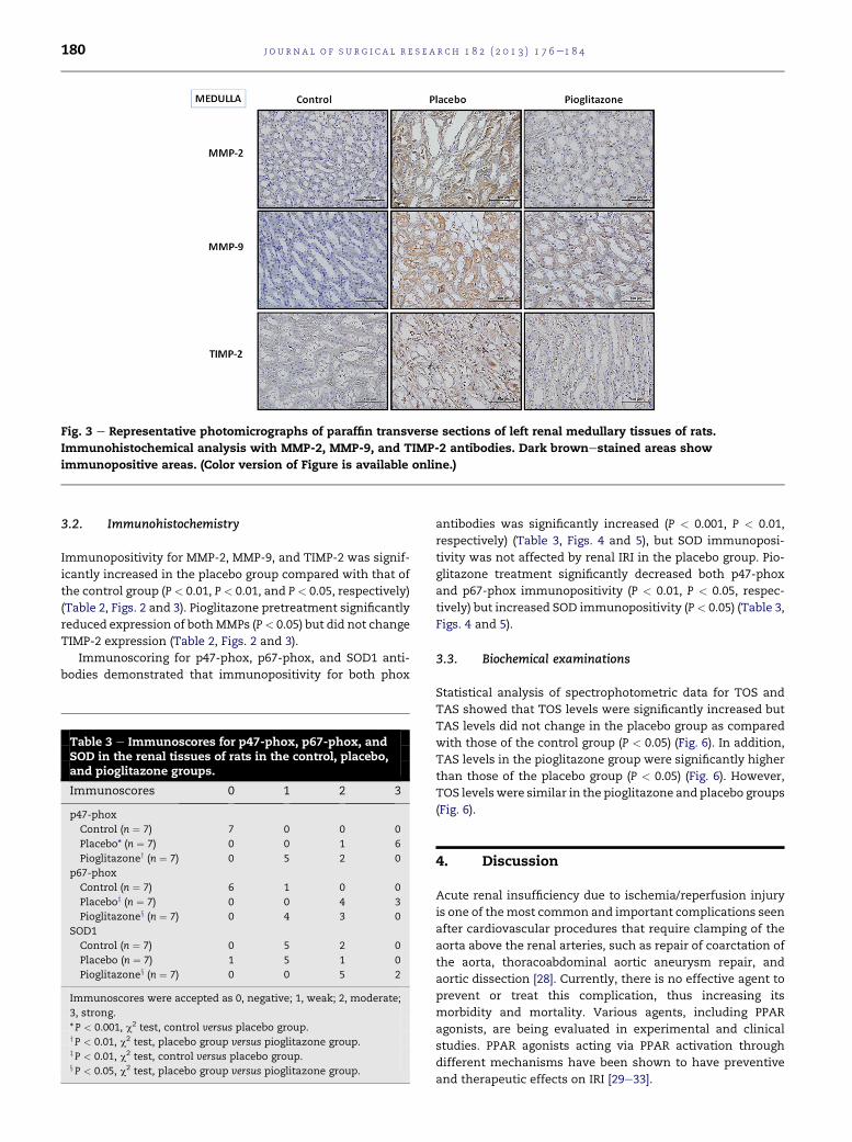
Fig. 3 e Representative photomicrographs of paraffin transverse sections of left renal medullary tissues of rats.
Immunohistochemical analysis with MMP-2, MMP-9, and TIMP-2 antibodies. Dark brownestained areas show
immunopositive areas. (Color version of Figure is available online.)
j o u r n a l o f s u r g i c a l r e s e a r c h 1 8 2 ( 2 0 1 3 ) 1 7 6e1 8 4180
3.2. Immunohistochemistry
Immunopositivity for MMP-2, MMP-9, and TIMP-2 was signif-
icantly increased in the placebo group compared with that of
the control group (P < 0.01, P < 0.01, and P < 0.05, respectively)
(Table 2, Figs. 2 and 3). Pioglitazone pretreatment significantly
reduced expression of bothMMPs (P< 0.05) but did not change
TIMP-2 expression (Table 2, Figs. 2 and 3).
Immunoscoring for p47-phox, p67-phox, and SOD1 anti-
bodies demonstrated that immunopositivity for both phox
Table 3 e Immunoscores for p47-phox, p67-phox, andSOD in the renal tissues of rats in the control, placebo,and pioglitazone groups.
Immunoscores 0 1 2 3
p47-phox
Control (n ¼ 7) 7 0 0 0
Placebo* (n ¼ 7) 0 0 1 6
Pioglitazoney (n ¼ 7) 0 5 2 0
p67-phox
Control (n ¼ 7) 6 1 0 0
Placeboz (n ¼ 7) 0 0 4 3
Pioglitazonex (n ¼ 7) 0 4 3 0
SOD1
Control (n ¼ 7) 0 5 2 0
Placebo (n ¼ 7) 1 5 1 0
Pioglitazonex (n ¼ 7) 0 0 5 2
Immunoscores were accepted as 0, negative; 1, weak; 2, moderate;
3, strong.
* P < 0.001, c2 test, control versus placebo group.yP < 0.01, c2 test, placebo group versus pioglitazone group.zP < 0.01, c2 test, control versus placebo group.xP < 0.05, c2 test, placebo group versus pioglitazone group.
antibodies was significantly increased (P < 0.001, P < 0.01,
respectively) (Table 3, Figs. 4 and 5), but SOD immunoposi-
tivity was not affected by renal IRI in the placebo group. Pio-
glitazone treatment significantly decreased both p47-phox
and p67-phox immunopositivity (P < 0.01, P < 0.05, respec-
tively) but increased SOD immunopositivity (P< 0.05) (Table 3,
Figs. 4 and 5).
3.3. Biochemical examinations
Statistical analysis of spectrophotometric data for TOS and
TAS showed that TOS levels were significantly increased but
TAS levels did not change in the placebo group as compared
with those of the control group (P < 0.05) (Fig. 6). In addition,
TAS levels in the pioglitazone group were significantly higher
than those of the placebo group (P < 0.05) (Fig. 6). However,
TOS levelswere similar in the pioglitazone and placebo groups
(Fig. 6).
4. Discussion
Acute renal insufficiency due to ischemia/reperfusion injury
is one of themost common and important complications seen
after cardiovascular procedures that require clamping of the
aorta above the renal arteries, such as repair of coarctation of
the aorta, thoracoabdominal aortic aneurysm repair, and
aortic dissection [28]. Currently, there is no effective agent to
prevent or treat this complication, thus increasing its
morbidity and mortality. Various agents, including PPAR
agonists, are being evaluated in experimental and clinical
studies. PPAR agonists acting via PPAR activation through
different mechanisms have been shown to have preventive
and therapeutic effects on IRI [29e33].

Fig. 4 e Representative photomicrographs of paraffin transverse sections of left renal cortical tissues of rats.
Immunohistochemical analysis with p47-phox, p67-phox, and SOD1 antibodies. Dark brownestained areas show
immunopositive areas. (Color version of Figure is available online.)
j o u r n a l o f s u r g i c a l r e s e a r c h 1 8 2 ( 2 0 1 3 ) 1 7 6e1 8 4 181
PPARs are ligand-activated nuclear transcription factors
belonging to the nuclear receptor superfamily. So far three
PPAR isoforms, designated as a, d, and g, have been identi-
fied. These three receptor subtypes are expressed in many
tissues in the human body. Improving insulin sensitivity
in diabetes, cellular differentiation and apoptosis, and anti-
inflammatory effects are the main effects of PPAR-g agonists
[14,34].
It has been demonstrated that PPAR-g agonists suppress
the production of inflammatory cytokines interleukin 1b,
Fig. 5 e Representative photomicrographs of paraffin transverse
Immunohistochemical analysis with p47-phox, p67-phox, and
immunopositive areas. (Color version of Figure is available onli
interleukin 6, and tumor necrosis factor a in stimulated
human peripheral blood monocytes [35]. They exert their
antiinflammatory effects through the negative regulation of
proinflammatory gene expression, which occurs in response
to monocyte/macrophage differentiation or activation [15].
PPAR-g agonists also lead to apoptosis in macrophages stim-
ulated by tumor necrosis factor a / interferon g due to the
inhibition of antiapoptotic nuclear factor-kB signaling
pathway [36]. In addition, it has been shown that these
agonists inhibit MMP and enhance PPAR-g expression [37].
sections of left renal medullary tissues of rats.
SOD1 antibodies. Dark brownestained areas show
ne.)

Fig. 6 e TOS (A) and TAS (B) levels in renal tissues of rats.
Values are represented asmean ± SEM. *P< 0.05, unpaired
Student t-test between control (n [ 5) and placebo (n [ 7)
groups. DP < 0.05, unpaired Student t-test between
placebo (n [ 7) and pioglitazone (Pio) (n [ 6) groups.
j o u r n a l o f s u r g i c a l r e s e a r c h 1 8 2 ( 2 0 1 3 ) 1 7 6e1 8 4182
Previous studies reported that pioglitazone, a PPAR-g
agonist, improves ischemia/reperfusion injury, which causes
intestinal damage [38], gastric mucosal damage [39], and
pulmonary damage [40] and also reduces myocardial infarc-
tion area [41], when pretreatment was performed before
ischemia. Rosiglitazone, another PPAR-g agonist, was shown
to improve renal function after IRI [42]. Hu et al. [43], in their
study on the protective effects of pioglitazone on IRI in mice,
observed that blood urea nitrogen and creatinine levels and
histopathologic scores were lower in animals pretreated with
pioglitazone. They concluded that PPAR activation by piogli-
tazone exerts protective effects on renal IRI in mice by
inhibiting renal cell apoptosis. In the present study, we also
detected significant inflammation and tubular necrosis in the
renal tissue after IRI in the placebo-treated rats. However, the
inflammation and necrosis scores were significantly lower in
the rats pretreated with pioglitazone.
It has been demonstrated that MMP-2 and MMP-9 are
enhanced in postischemic renal tissue and are localized in the
renal tubules, interstitial cells, and tubulointerstitial space
[44]. Moreover, experimental studies revealed that inhibition
of MMP-2 and MMP-9 is associated with a decrease in the IRI
level [45,46]. The present study revealed increased MMP-2,
MMP-9, and TIMP-2 levels after renal IRI. Furthermore,
although no significant difference was observed in TIMP-2
expression, MMP-2 and MMP-9 levels in rats that were pre-
treated with pioglitazone were significantly lower than those
of the placebo group.
The NOX family of NADPH oxidases consists of enzymes
that play a role in various physiological and pathologic
processes, and lead to ROS production. During these processes,
p47-phox and p67-phox, which are the cytosolic subunits of
NADPHoxidase, playorganizerandactivator roles, respectively
[47]. As they are likely to be involved in various disease
processes, NOX enzymes are established drug targets as well
[47,48]. We found that p47-phox and p67-phox expression was
significantly enhanced in the ischemia/reperfusion-induced
rats and was significantly reduced in those pretreated with
pioglitazone.
Schneider et al. [49] reported no difference between the
renal IRI and the control group in terms of the expression of
SOD1, which is the cytoplasmic form of SOD. Similarly, we
also found that there is no difference in SOD1 levels between
IRI-induced and control rats.
It has been reported that PPAR-g activation encodes target
genes, including genes that encode antioxidative enzymes,
such as SOD1, catalase, and thioredoxin [50]. Consistent with
this evidence, the present study demonstrated higher SOD1
levels in rats pretreated with pioglitazone as compared with
those of the placebo group. It has been suggested that renal IRI
is more severe in the event of SOD1 deficiency. Moreover, it
has been stated that SOD1 production is not essential for renal
function under normal conditions, but the antioxidant effect
of SOD1 is beneficial in the case of IRI due to increased ROS
production [51]. From this point of view, it can be suggested
that increased SOD1 levels in the rats preatreated with pio-
glitazone might contribute to the improvement of IRI.
It is known that inhibition of ROS production and removal
of ROS after IRI ameliorates renal injury [52]. In the present
study, IRI-induced rats showed increased TOS levels but
showed no significant difference in TAS levels compared with
those of the controls. TAS level was found to be increased in
those pretreated with pioglitazone.
In conclusion, the present study demonstrated that pio-
glitazone has antioxidant effects due to its regulation of SOD1
and TAS levels and inhibitory effects on proinflammatory
MMPs in a renal IRI model in rats. Our results suggest that
pretreatment with pioglitazone may reduce IRI during renal
transplantation and cardiovascular surgical procedures.
r e f e r e n c e s
[1] McCombs PR, Roberts B. Acute renal failure followingresection of abdominal aortic aneurysm. Surg Gynecol Obstet1979;148:175.
[2] Pinney SP, Balakrishnan R, Dikman S, et al. Histopathology ofrenal failure after heart transplantation: a diverse spectrum.J Heart Lung Transplant 2012;31:233.
[3] Nath KA, Norby SM. Reactive oxygen species and acute renalfailure. Am J Med 2000;109:665.

j o u r n a l o f s u r g i c a l r e s e a r c h 1 8 2 ( 2 0 1 3 ) 1 7 6e1 8 4 183
[4] Chatterjee PK. Novel pharmacological approaches to thetreatment of renal ischemia-reperfusion injury: acomprehensive review. Naunyn Schmiedebergs ArchPharmacol 2007;376:1.
[5] Rabb H, Mendiola CC, Dietz J, et al. Role of CD11a andCD11b in ischemic acute renal failure in rats. Am J Physiol1994;267:1052.
[6] Sheridan AM, Bonventre JV. Cell biology and molecularmechanisms of injury in ischemic acute renal failure. CurrOpin Nephrol Hypertens 2000;9:427.
[7] Sutton TA, Mang HE, Campos SB, et al. Injury of the renalmicrovascular endothelium alters barrier function followingischemia. Am J Physiol Renal Physiol 2003;285:191.
[8] Nagase H, Woessner JF Jr. Matrix metalloproteinases. J BiolChem 1999;274:21491.
[9] Newby AC. Matrix metalloproteinases regulate migration,proliferation, and death of vascular smooth muscle cells bydegrading matrix and non-matrix substrates. Cardiovasc Res2006;69:614.
[10] Catania JM, Chen G, Parrish AR. Role of matrixmetalloproteinases in renal pathophysiologies. Am J PhysiolRenal Physiol 2007;292:905.
[11] Dejonckheere E, Vandenbroucke RE, Libert C. Matrixmetalloproteinases as drug targets in ischemia/reperfusioninjury. Drug Discov Today 2011;16:762.
[12] Ricote M, Li AC, Willson TM, et al. The peroxisomeproliferator-activated receptor-gamma is a negativeregulator of macrophage activation. Nature 1998;391:79.
[13] van Bilsen M, van Nieuwenhoven FA. PPARs as therapeutictargets in cardiovascular disease. Expert Opin Ther Targets2010;14:1029.
[14] Robinson E, Grieve DJ. Significance of peroxisomeproliferator-activated receptors in the cardiovascular systemin health and disease. Pharmacol Ther 2009;122:246.
[15] von Knethen A, Brune B. PPARgammaean importantregulator of monocyte/macrophage function. Arch ImmunolTher Exp (Warsz) 2003;51:219.
[16] Li MD, Yang X. A retrospective on nuclear receptor regulationof inflammation: lessons from GR and PPARs. PPAR Res 2011;2011:742785.
[17] Wang X, Wang Z, Liu JZ, et al. Double antioxidant activities ofrosiglitazone against high glucose-induced oxidative stressin hepatocyte. Toxicol In Vitro 2011;25:839.
[18] Giannini S, Serio M, Galli A. Pleiotropic effects ofthiazolidinediones: taking a look beyond antidiabeticactivity. J Endocrinol Invest 2004;27:982.
[19] Sivarajah A, Chatterjee PK, Patel NS, et al. Agonists ofperoxisome-proliferator activated receptor-gamma reducerenal ischemia/reperfusion injury. Am J Nephrol 2003;23:267.
[20] Abdelrahman M, Sivarajah A, Thiemermann C. Beneficialeffects of PPAR-gamma ligands in ischemia-reperfusioninjury, inflammation and shock. Cardiovasc Res 2005;65:772.
[21] Ito H, Nakano A, Kinoshita M, et al. Pioglitazone,a peroxisome proliferator-activated receptor-gammaagonist, attenuates myocardial ischemia/reperfusion injuryin a rat model. Lab Invest 2003;83:1715.
[22] Akahori T, Sho M, Hamada K, et al. Importance ofperoxisome proliferator-activated receptor-gamma inhepatic ischemia/reperfusion injury in mice. J Hepatol 2007;47:784.
[23] Cao Z, Ye P, Long C, et al. Effect of pioglitazone, a peroxisomeproliferator-activated receptor gamma agonist, on ischemia-reperfusion injury in rats. Pharmacology 2007;79:184.
[24] Somi MH, Hajipour B, Asl NA, et al. Pioglitazone attenuatesischemia/reperfusion-induced liver injury in rats. TransplantProc 2009;41:4105.
[25] Rice-Evans C, Miller NJ. Total antioxidant status in plasmaand body fluids. Methods Enzymol 1994;234:279.
[26] Erel O. A novel automated method to measure totalantioxidant response against potent free radical reactions.Clin Biochem 2004;37:112.
[27] Guzeloglu M, Yalcinkaya F, Atmaca S, et al. The beneficialeffects of tadalafil on renal ischemia-reperfusion injury inrats. Urol Int 2011;86:197.
[28] Ellenberger C, Schweizer A, Diaper J, et al. Incidence, riskfactors, and prognosis of changes in serum creatinineearly after aortic abdominal surgery. Intensive Care Med2006;32:1808.
[29] Cheng CF, Lian WS, Chen SH, et al. Protective effects ofadiponectin against renal ischemia-reperfusion injury viaprostacyclin-PPARa-heme oxygenase-1 signaling pathway. JCell Physiol 2012;227:239.
[30] Collino M, Benetti E, Miglio G, et al. Peroxisome proliferator-activated receptor b/d agonism protects the kidney againstischemia/reperfusion injury in diabetic rats. Free Radic BiolMed 2011;50:345.
[31] Matsuyama M, Yoshimura R, Kawahito Y, et al.Relationship between peroxisome proliferator-activatedreceptor-g and renal ischemia-reperfusion injury. Mol MedReport 2008;1:499.
[32] Miglio G, Rosa AC, Rattazzi L, et al. The subtypes ofperoxisome proliferator-activated receptors expressed byhuman podocytes and their role in decreasing podocyteinjury. Br J Pharmacol 2011;162:111.
[33] Patel NS, di Paola R, Mazzon E, et al. Peroxisome proliferator-activated receptor-alpha contributes to the resolution ofinflammation after renal ischemia/reperfusion injury. JPharmacol Exp Ther 2009;328:635.
[34] Bishop-Bailey D, Bystrom J. Emerging roles of peroxisomeproliferator-activated receptor-beta/delta in inflammation.Pharmacol Ther 2009;124:141.
[35] Jiang C, Ting AT, Seed B. PPAR-gamma agonists inhibitproduction of monocyte inflammatory cytokines. Nature1998;391:82.
[36] Chinetti G, Griglio S, Antonucci M, et al. Activation ofproliferator-activated receptors alpha and gamma inducesapoptosis of human monocyte-derived macrophages. J BiolChem 1998;273:25573.
[37] Ricote M, Huang JT, Welch JS, et al. The peroxisomeproliferator-activated receptor (PPARgamma) as a regulatorof monocyte/macrophage function. J Leukoc Biol 1999;66:733.
[38] Naito Y, Takagi T, Uchiyama K, et al. Suppression ofintestinal ischemia-reperfusion injury by a specificperoxisome proliferator-activated receptor-gamma ligand,pioglitazone, in rats. Redox Rep 2002;7:294.
[39] Ichikawa H, Naito Y, Takagi T, et al. A specific peroxisomeproliferator-activated receptor-gamma (PPAR-gamma)ligand, pioglitazone, ameliorates gastric mucosal damageinduced by ischemia and reperfusion in rats. Redox Rep 2002;7:343.
[40] Ito K, Shimada J, Kato D, et al. Protective effects ofpreischemic treatment with pioglitazone, a peroxisomeproliferator-activated receptor-ligand, on lung ischemia-reperfusion injury in rats. Eur J Cardiothorac Surg 2004;25:530.
[41] Wayman NS, Hattori Y, McDonald MC, et al. Ligands of theperoxisome proliferator-activated receptors (PPAR-gammaand PPAR-alpha) reduce myocardial infarct size. FASEB J2002;16:1027.
[42] Betz B, Schneider R, Kress T, et al. Rosiglitazone affects nitricoxide synthases and improves renal outcome in a rat modelof severe ischemia/reperfusion injury. PPAR Res 2012;2012:219319.
[43] Hu H, Zou C, Xi X, et al. Protective effects of pioglitazone onrenal ischemia-reperfusion injury in mice. J Surg Res; 2012.in press.

j o u r n a l o f s u r g i c a l r e s e a r c h 1 8 2 ( 2 0 1 3 ) 1 7 6e1 8 4184
[44] Basile DP, Fredrich K, Weihrauch D, et al. Angiostatin andmatrix metalloprotease expression following ischemic acuterenal failure. Am J Physiol Renal Physiol 2004;286:893.
[45] Novak KB, Le HD, Christison-Lagay ER, et al. Effects ofmetalloproteinase inhibition in a murine model of renalischemia-reperfusion injury. Pediatr Res 2010;67:257.
[46] Kunugi S, Shimizu A, Kuwahara N, et al. Inhibition of matrixmetalloproteinases reduces ischemia-reperfusion acutekidney injury. Lab Invest 2011;91:170.
[47] Bedard K, Krause KH. The NOX family of ROS-generatingNADPH oxidases: physiology and pathophysiology. PhysiolRev 2007;87:245.
[48] Lambeth JD,KrauseKH,ClarkRA.NOXenzymesasnoveltargetsfor drug development. Semin Immunopathol 2008;30:339.
[49] Schneider MP, Sullivan JC, Wach PF, et al. Protective role ofextracellular superoxide dismutase in renal ischemia/reperfusion injury. Kidney Int 2010;78:374.
[50] Fan Y, Wang Y, Tang Z, et al. Suppression of pro-inflammatory adhesion molecules by PPAR-delta in humanvascular endothelial cells. Arterioscler Thromb Vasc Biol2008;28:315.
[51] Yamanobe T, Okada F, Iuchi Y, et al. Deterioration ofischemia/reperfusion-induced acute renal failure in SOD1-deficient mice. Free Radic Res 2007;41:200.
[52] Kim J, Jung KJ, Park KM. Reactive oxygen species differentlyregulate renal tubular epithelial and interstitial cellproliferation after ischemia and reperfusion injury. Am JPhysiol Renal Physiol 2010;298:F1118.