The effect of α and γ interferon on cell growth and histocompatibility antigen expression by human...
7
Anti-leukaemic Effects of RuC&(DMSO)~ Isomers 1879 22. 23. 24. 25. containing dimethylsulphoxide. In Lippard SJ, ed. Platinlcm, Gold, and Other Metal Chemotherapeutic Agents. Washington DC, ACS Symposium Series 209,1983,279-2%. Wesr SJ, Graham-Pole J, Hard&y RM. Factors in the pathogenesis of central nervous system leukemia. Br MedJ 1972,2,311-314. Mestroni G, Alessio E, Sava G, Pacer S, Bregant L, Coluccia M, Fricker SP. Ruthenitm$II)-dimetbylsulphoxide complexes: anti- tumor and microbiological properties. Anticancer Drug Design 1991, 6,214. Sherman SE, Lippard SJ. Structural aspects of platinum anticancer drug interactions with DNA. Cheer Rev 1987,87,1153-1181. Heiger-Bernays WJ, Essigman JM, Lippard SJ. Effect of the antitumor drug cis-diamminedichloroplatintm$II) and related plati- num complexes on eukaryotic DNA replication. Biochencisry 1990, 29,8461-8466. 26. Sava G, Pacer S, Bregant F, et al. Mechanism of tumor inhibition by the metal complex tran.+RuCl,(dimetbylsulphoxide),. Phannacol (Life Sci Adv) 1990,9,79-84. 27. Coluccia M, Alessio E, Mestroni G, et al. Effect of cis- and rrans-RuCl,(DMSO), on the fibrinolytic activity on Lewis lung carcinoma. Anticancer Res 1990,10,1410. AcknowledgementsWork done with contributions from M.U.R.S.T. 40%. Eur~Cancn, Vol. Z9A, No. 13, fq. 187~1885,1993. 0959d0#9l93$6.00 + 0.00 Printed UI Great Bnlnin 0 1993Pergmwon PressLid The Effect of a and y Interferon on Cell Growth and Histocompatibility Antigen Expression by Human Renal Carcinoma Cells in vi&o Rachel Angus, C.M.P. Collins and M.O. Symes Turnout cells were separated from 19 renal carcinomas and cultured in vitro. The effect of interferon (IFN) OL and y on cell proliferation was measured and compared to the effect of IFN on the expression of class I and class II major histocompatibility complex (MHC) antigens. When tested within the 6rst 14 days of culture, IFN-a inhibited protein synthesis in 12 of 15 and IFN-y in four of nine tumours. Reduction in cell counts was in parallel. In six tumours the culture period was extended and in all six the effect of IFN-cy was lost. Exposure to IFN-a induced or enhanced class I antigen expression in eight of 19 tumours and class II expression in two of 19. The analogous figures for IFN-y were five and three tumours. In four of five cases where a comparison could be made there was a correlation between the effects of IFN-cu on cell proliferation and class I antigen expression. The efficacy of IFN in the treatment of renal carcinomas may thus, in part, result from inhibition of cell proliferation and enhancement of antigen expression. EurJ Cancer, Vol. 29A, No. 13, pp. 1879-1885,1993. INTRODUCTION INTERFERONS HAVE a variety of effects on cells which mitigate against the growth of neoplasms. For example, interferon a (IFN-IX) inhibits cell proliferation [l], is cytotoxic [2] and amplifies or induces the expression or re-expression of major histocompatibility complex (MHC) antigens [3, 41. Interferon- y (IFN-y) activates immune cells, for example, mononuclear phagocytes [S, 61, T-lymphocytes [7] and natural killer cells [S]. It is particularly effective in amplifying or inducing the expression of class II MHC antigen (Ag) [9]. MHC Ag molecules play an important role in the presentation of self or non-self Ag leading to the initiation of an immune response and thus their increased expression may be important in potentiating host reactivity against tumour cells. IFN-a and IFN-y have been used to treat metastatic human renal carcinoma with a response rate of approximately 15% [10-l 31. We have recently described a method for the establishment of Correspondence to M.O. Symes. R. Angus and M.O. Symes are at the University Department of Surgery; andC.M.P. Collins is at the University Department of Pathology, Bristol Royal Infirmary, Bristol BS2 8HW, U.K. Revised 14 Apr. 1993; accepted 20 May 1993. human renal carcinoma cells (RCC) in long-term culture [ 141. As the identification of a subset of RCC likely to respond to IFN therapy could be clinically useful it seemed germane to study the effect of interferons on the growth and histocompatibility antigen expression by RCC cells cultured from the primary biopsy. MATERIALS AND METHODS General plan of the experiments Carcinoma cells from 15 RCC were tested in vitro for sensitivity to IFN-a and -y, using the uptake of [‘%e]selenomethionine [‘%eM], as a measure of protein synthesis, and by cell counting. Cells from each tumour were tested within the first 14 days of culture and in some cases were re-examined after longer periods of in vitro growth. At the same time the effect of the interferons on the expression of class I and II MHC Ag was examined. An association was sought between these two effects of interferons. Culture of renal carcinoma cells The methods for separation of carcinoma cells from the RCC [ 14, 151, the purity of the tumour cell suspensions obtained [ 151 and their culture [ 141 have previously been described. A cytospin preparation was made of the cells from the top band on the Nycodenz column for 11 tumours. The cells were stained with
Transcript of The effect of α and γ interferon on cell growth and histocompatibility antigen expression by human...
Anti-leukaemic Effects of RuC&(DMSO)~ Isomers 1879
22.
23.
24.
25.
containing dimethylsulphoxide. In Lippard SJ, ed. Platinlcm, Gold, and Other Metal Chemotherapeutic Agents. Washington DC, ACS Symposium Series 209,1983,279-2%. Wesr SJ, Graham-Pole J, Hard&y RM. Factors in the pathogenesis of central nervous system leukemia. Br MedJ 1972,2,311-314. Mestroni G, Alessio E, Sava G, Pacer S, Bregant L, Coluccia M, Fricker SP. Ruthenitm$II)-dimetbylsulphoxide complexes: anti- tumor and microbiological properties. Anticancer Drug Design 199 1, 6,214. Sherman SE, Lippard SJ. Structural aspects of platinum anticancer drug interactions with DNA. Cheer Rev 1987,87,1153-1181. Heiger-Bernays WJ, Essigman JM, Lippard SJ. Effect of the
antitumor drug cis-diamminedichloroplatintm$II) and related plati- num complexes on eukaryotic DNA replication. Biochencisry 1990, 29,8461-8466.
26. Sava G, Pacer S, Bregant F, et al. Mechanism of tumor inhibition by the metal complex tran.+RuCl,(dimetbylsulphoxide),. Phannacol (Life Sci Adv) 1990,9,79-84.
27. Coluccia M, Alessio E, Mestroni G, et al. Effect of cis- and rrans-RuCl,(DMSO), on the fibrinolytic activity on Lewis lung carcinoma. Anticancer Res 1990,10,1410.
AcknowledgementsWork done with contributions from M.U.R.S.T. 40%.
Eur~Cancn, Vol. Z9A, No. 13, fq. 187~1885,1993. 0959d0#9l93$6.00 + 0.00 Printed UI Great Bnlnin 0 1993 Pergmwon Press Lid
The Effect of a and y Interferon on Cell Growth and Histocompatibility Antigen Expression by
Human Renal Carcinoma Cells in vi&o Rachel Angus, C.M.P. Collins and M.O. Symes
Turnout cells were separated from 19 renal carcinomas and cultured in vitro. The effect of interferon (IFN) OL and y on cell proliferation was measured and compared to the effect of IFN on the expression of class I and class II major histocompatibility complex (MHC) antigens. When tested within the 6rst 14 days of culture, IFN-a inhibited protein synthesis in 12 of 15 and IFN-y in four of nine tumours. Reduction in cell counts was in parallel. In six tumours the culture period was extended and in all six the effect of IFN-cy was lost. Exposure to IFN-a induced or enhanced class I antigen expression in eight of 19 tumours and class II expression in two of 19. The analogous figures for IFN-y were five and three tumours. In four of five cases where a comparison could be made there was a correlation between the effects of IFN-cu on cell proliferation and class I antigen expression. The efficacy of IFN in the treatment of renal carcinomas may thus, in part, result from inhibition of cell proliferation and enhancement of antigen expression. EurJ Cancer, Vol. 29A, No. 13, pp. 1879-1885,1993.
INTRODUCTION INTERFERONS HAVE a variety of effects on cells which mitigate against the growth of neoplasms. For example, interferon a (IFN-IX) inhibits cell proliferation [l], is cytotoxic [2] and amplifies or induces the expression or re-expression of major histocompatibility complex (MHC) antigens [3, 41. Interferon- y (IFN-y) activates immune cells, for example, mononuclear phagocytes [S, 61, T-lymphocytes [7] and natural killer cells [S]. It is particularly effective in amplifying or inducing the expression of class II MHC antigen (Ag) [9]. MHC Ag molecules play an important role in the presentation of self or non-self Ag leading to the initiation of an immune response and thus their increased expression may be important in potentiating host reactivity against tumour cells. IFN-a and IFN-y have been used to treat metastatic human renal carcinoma with a response rate of approximately 15% [10-l 31.
We have recently described a method for the establishment of
Correspondence to M.O. Symes. R. Angus and M.O. Symes are at the University Department of Surgery; andC.M.P. Collins is at the University Department of Pathology, Bristol Royal Infirmary, Bristol BS2 8HW, U.K. Revised 14 Apr. 1993; accepted 20 May 1993.
human renal carcinoma cells (RCC) in long-term culture [ 141. As the identification of a subset of RCC likely to respond to IFN therapy could be clinically useful it seemed germane to study the effect of interferons on the growth and histocompatibility antigen expression by RCC cells cultured from the primary biopsy.
MATERIALS AND METHODS General plan of the experiments
Carcinoma cells from 15 RCC were tested in vitro for sensitivity to IFN-a and -y, using the uptake of [‘%e]selenomethionine [‘%eM], as a measure of protein synthesis, and by cell counting. Cells from each tumour were tested within the first 14 days of culture and in some cases were re-examined after longer periods of in vitro growth. At the same time the effect of the interferons on the expression of class I and II MHC Ag was examined. An association was sought between these two effects of interferons.
Culture of renal carcinoma cells The methods for separation of carcinoma cells from the RCC
[ 14, 151, the purity of the tumour cell suspensions obtained [ 151 and their culture [ 141 have previously been described. A cytospin preparation was made of the cells from the top band on the Nycodenz column for 11 tumours. The cells were stained with

1880 R. Angus et al.
May Grunwald Giemsa stain and a differential count involving 2200 cells was obtained for 10 tumours: K49, K50, K51, K52, K61, K62, K63, K68, K70 and K73. The mean percentage of tumour cells was 98.5 + 2.5 (standard deviation, S.D.): for tumour K65 the percentage of tumour cells was 60. The charac- terisation of the cells from each tumour studied, in the present experiments, was previously detailed by us [ 141.
z?uerfeo?ls IFN-a-N1 (Wellferon) and human natural lymphoblastoid
IFN-a, produced by the cell line Namalva, transformed by exposure to the Sendai virus [ 161, were kindly supplied by Wellcome Biotechnology Ltd. These two types of IFN-a were found to give comparable results in all assay systems. IFN-y (recombinant) was generously donated by Knoll AG.
Assays for the effect of interferons on cell proliferation Aliquots of IO4 renal carcinoma cells in RPM1 1640 + 10% v/
v newborn calf serum and 1% w/v glutamine (Gibco) were exposed for 24 or 48 h to 100, 500 or 1000 U/ml of IFN-o. or -y in round-bottomed polystyrene tubes (Becton Dickinson). Thereafter, the cells were centrifuged (200 g, 5 mitt) and resus- pended with 0.5 pCi [‘?3eM] (CIS) in 0.5 ml methionine-free modified Eagle’s medium (MEM) (Gibco) for 48 h. The cells were then washed three times in phosphate-buffered saline (PBS) and the incorporated radioactivity measured using a Wilj 2001 y Counter. The percentage inhibition of isotope incorporation in comparison to cells maintained in culture medium alone was determined. All comparisons were based on six individual determinations.
Further aliquots of lo4 carcinoma cells were suspended in 2 ml RPM1 (supplemented as above) containing 100, 500 or 1000 U/ml of IFN-a or -y in 35 x 10 mm plastic petri dishes to which the cells adhered. After 4 days the medium was replaced by 2 ml of fresh medium containing the appropriate IFN. At 8 days the medium was replaced by 0.25 ml of PBS containing 0.5 mg/ml trypsin and 0.2 mg/ml EDTA (Gibco). After 10 min at 37”C, 0.25 ml of 0.17% w/v trypan blue was added. After 5 min the number of viable carcinoma cells, recognised by dye exclusion, was estimated by cell counting in a Neubar Chamber (Hawksley). The percentage inhibition in mean cell numbers, compared to cultures maintained in medium alone was calcu- lated. All comparisons were based on three to six individual determinations.
Assay for the effect of ZFN on expression of MHC Ag by renal carcinoma cells
The methods used have previously been reported [ 171. In brief, aliquots of lo4 carcinoma cells in 0.3 ml medium were cultured for 24 h on three-well slides to allow the cells to adhere. The medium was then changed and the cells cultured for 1 or 3 days on three-well slides in medium alone or medium containing 100, 500 or 1000 U IFN-a or -y. The slides were fixed in cold acetone for 10 min. The cells in one well were exposed to monoclonal antibody (MoAb) M736 (Dako). This is derived from clone W6/32 [ 181 and recognises a monomorphic epitope on the 45 kD polypeptide gene product of the HLA, A, B, C loci (class 1 MHC Ag). The cells in a second well were exposed to MoAb M704 (Dako) derived from clone DK22. This recogn- ises all gene products from subregions DP, DX and DQ, except DQwl (class 11 MHC Ag). The cells in the third well were not exposed to a MoAb and served as a negative control. Reaction with antibody was recognised using the indirect immunoperoxi-
dase technique, with 3-amino 9-ethylcarbaxole as the chromo- gen.
Evaluation of staining All preparations were examined by two independent
observers. The number of cells stained were graded as: NIL, 1 (l-25%), 2 (26-50%), 3 (51-75%) and 4 (>76%). The degree of staining of the individual cells was graded: + (minimal), + + (moderate) and +++ (marked), with reference to sections of normal human kidney or tonsil stained as positive controls.
Statistics For a particular tumour, the individual y counts for incorpor-
ation of [75SeM] were subject to a one-way analysis of variance and the significance of differences between individual groups was determined using Student’s c-test based on a pooled estimate of the variance. The significance of differences between mean cell counts was calculated following normalisation of the data, using significance values for the normal distribution.
RESULTS The effect of exposure to ZFN-cu on protein synthesis
After an initial culture period of < 14 days cells from 15 RCC were exposed to increasing concentrations of IFN-a (Wellferon) for 48 h. In six RCC (K49, K50, K61, K67, K70 and K73) protein synthesis, as measured by the uptake of [75SeM], was inhibited in a dose-dependent fashion (Fig. 1). Protein synthesis was also inhibited in six other tumours (K51, K52, K59, K63, K68 and K69) in a non-dose dependent manner (Fig. 1). In the other three tumours (K60, K62 and K65) no effect of IFN-a was seen. The effect of IFN-a decreased as the culture period increased in the six tumours (K42, K49, KSO, K51, K52 and K61) examined (Fig. 2). Exposure to IFN-a for 24 h was without effect on the six tumours examined (K42, K52, K56, K57, K58, K62), data not shown. Tumour K42 showed inhi- bition of protein synthesis when fust exposed to IFN-a for 48 h after 133 days of culture, but the effect was lost as the culture period increased (Fig. 2).
100
C Concentration n 100 aIFN (h/ml) 500
K67 (3) K68 (13) K69 (0) K70 (0) K73 (4)
Tumour (days in culture)
Fig. 1. The effect of 48-h exposure to a-N1 interferon in inhibiting the subsequent uptake of [‘%eM] over 48 h by renal carcinoma cells in vitro. The bars represent the percentage inhibition of isotopic uptake compared to cells maintained in medium without IFN for 48 h
prior to the addition of [‘?3eM].

Effect of o and y Interferon on Human Renal Carcinoma Cells in virr~ 1881
Coucentrrtion n 100 aIFN(lu/ml) 0 500
0 1000
B 8l ” 75
c B .t 1
.*. P < 0.001
.s g 50 . . P < 0.01
. P <0.05 2 2s
05 n ” K42 (169) K49 (27) KS0 (21) KS1 (77) KS2 (116)K61 (155)
Tumour (days in culture)
Fig. 2. A comparison of the action of 48-h exposure to a-N1 iater- feron in inhibiting the uptake of [%eM] by renal carcinoma cells
after increasing periods in culture.
The effect of IFN-cx on cell numbers Following 8 days exposure to IFN-a (Wellferon), nine
recently derived cultures showed a significant reduction in cell numbers, compared to cultures in medium alone (Fig. 3). In four there was a dose response (K42, K57, K68, K73). The other five tumours were inhibited in a dose-independent manner (K56, K58, K61, K63 and K70). In general, the percentage reduction in mean cell count was greater than that seen for inhibition of protein synthesis. However, the significance values for reduction in cell numbers were less due to greater variability in the data.
The effect of exposure to IFN- y on protein synthesis Exposure to IFN-y for 48 h had an inhibitory effect on protein
synthesis in four of nine recently derived cultures. In two of these (K63, K70) the level of inhibition was dose dependent,
but in the other two tumours (K65, K68) it was not dose related (Fig. 4). No inhibition was seen following 24-h exposum to IFN-r.
The effect of IFN-y on cell numbers Exposure to IFN-y for 8 days led to a significant and dose-
dependent reduction in cell numbers in three of eight tumours (K63, K68, K69) and in another tumour (K62), to a non-dose- related decrease (Fig. 4).
The relationship between twwur cell purity and the effect of IFN For 9 of 10 tumour cell suspensions, with a mean tumour cell
content from the Nycodenz column of 98.5%, there was a dose-dependent (five cases) and dose-independent (four cases) inhibition of protein synthesis by IFN-a. In two tumours, K62 (100% tumour cells from the column) and K65 (60% tumour cells), IFN-a was without effect (Fig. 1). The effect of IFN-7 was determined for six of these tumours. There was no effect in three, dose-dependent inhibition in one and dose-independent inhibition in two, including K65 (Fig. 4). Of the six tumours in which the effect of IFN-a declined as the culture period was increased, the cell purity in five where preparations were obtained from the column was 97.2 k 3.0%.
The effect of ZFN+ on MHC Ag expression Four of 19 tumours (K49, K60, K68 and K73) initially
showed absence of class I Ag (Table 1). IFN-a produced a dose- dependent induction or increase in class I Ag expression in three of these tumours (K60, K68 and K73) and in five other tumours (K42, K56, K58, K67 and K70) (Table 2). Expression of class II Ag was induced in one tumour (K73) and increased in another (K61).
9% 80
12 4 $ 60
sFm, 40
zsg 20 “Y E o
s - 100
3.5 c 80 B A 80 l ” P < 0.001
1460 .* P < 0.01 40 l P < 0.05
20
0 ” K67 (3) K68 (13) K69 (0) K’O (0) K73 (4) k6-I (3) K68 (13) K69 (0) K’O (0) K73 (4)
Tumour (days in culture) Tumour (days in culture)
Fig. 3. The effect of g-day exposure to a-N1 interferon on cell counts Fig. 4. A comparison of the inhibition in [“SeM] uptake following of renal carcinoma cells ia vitro. The bars represent the percentage 48-h exposure to IFN-y with the direct inhibition of ceU numbers reduction ia cell numbers compared to that ia aliquots of cells not following %-day exposure to IFNy. The percentage iahibition is
exposed to IFN. calculated with reference to aliquots of cells not exposed to IFN.

1882 R. Angus et al.
Table 1. Expression of class I and class II MHC Ag by renal carcinoma cells in culture
Tumour Class I MHC Ag Class II MHC Ag
K42 4++(8)4+(68)4++(169) 1 + ‘(8) 0(68) 0( 169) 4+(218) O(218)
K49 O(0) O(0) KS0 4+++(o) l++(o) KS1 4+++(o) O(0) K52 4++‘+++(0)4+‘++(116) 1++(0)0(116) K56 4+(12) 2+(106) 0(12)0(106) K57 4+‘+++(11) O(l1) K58 4’(4) l++(4) K59 4++(6) l”(6) KM) O(4) 3+(26) O(4) 0(26) K61 4+++(o) l”(0) K62 4+++(6) l++(6) K63 4+‘++‘+++(4) l+++(4) K65 4”(5) O(5) K67 3++(3) O(3) K68 0(13) l”(13) K69 4”(O) l++(o) K70 3+ ‘(0) l++(o) K73 l”(4) O(4)
Cells from each culture were harvested and grown in droplet culture for 1 day prior to estimation of antigen expression. 0: days in culture, (0): cells from the Nycodenz column. No. of cells stained: 4 = 76lOO%, 3 = 51-75%, 2 = 2650%, 1 = l-25%, 0 = NIL. Degree of staining: + + + = heavy, + + = moderate, + = light.
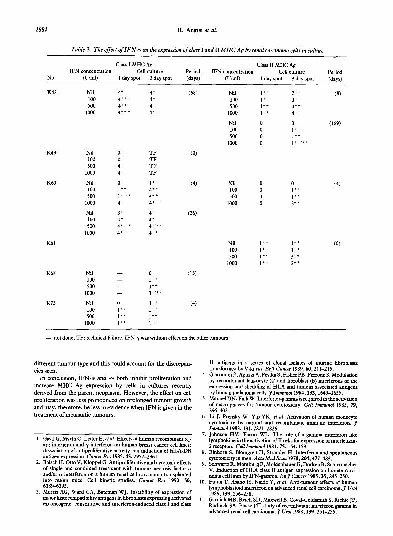
The effect of IFN- y on MHC Ag expression IFN--y induced or increased class I Ag expression in five
tumours (K42, K49, K60, K68 and K73) and class II Ag expression in three tumours (K42, K60 and K61) (Table 3). Of nine tumours (K42, K49, K56, K58, K60, K67, K68, K70 and K73) which initially showed absence or reduced expression of class I antigen, all except K49 showed upregulation by IFN-a but only five by IFN-y. By contrast, tumour K49 showed upregulation of class I Ag by IFN-y but not by IFN-(w.
The relationship between expression of class I and class II MHC Ag by tumour cells in culture and the effect of IFN-a and -yin inhibiting protein synthesis
IFN-cy. There was no relationship between initial expression of MHC Ag by the tumour cells in culture and the effect of IFN-a in inhibiting protein synthesis (Fig. 1, Table 1). Seven tumours were examined after the same culture period for inhibition of protein synthesis and enhancement of MHC class I Ag expression. There was correlation in four tumours (K67, K68, K70 and K73) and no correlation in one (K60). In the other tumours (KS9 and K52), expression of class I Ag was already maximal before the addition of IFN.
ZFN-y. Only one tumour (K68) studied showed a correlation between reduced protein synthesis and increased Ag expression. Otherwise there was no apparent relationship between either initial MHC Ag expression by the tumour cells or its induction by IFN-y and the effect of IFN-1, in inhibiting protein synthesis (Fig. 4, Tables 1,3).
DISCUSSION IFN-a. is an effective form of therapy for metastatic RCC in
approximately 15% of patients [lO-131. The majority of RCC
are, however, unresponsive. IFN-a is both cytotoxic [2] and inhibits cell proliferation [l], in addition to exerting indirect effects via the host immune response, for example, by alteration of MHC Ag expression [3, 41. Identification of a subset of RCC likely to respond to IFN therapy would be diagnostically useful. This paper has shown that both IFN-a and -y have direct antiproliferative action on recently derived RCC cells. IFN-a was effective against more tumours than IFN-y and sensitivity to IFN was not necessarily dose dependent. Thus, in vitro RCC cells can be divided into subsets of those that are sensitive to the effects of IFN and those that are resistant. A study by Nanus et al. [19], involving IFN-cx treatment of RCC cell lines showed comparable results. Furthermore, they showed that the sensitive RCC lines when grown as tumour xenografts in mice were also markedly inhibited by systemically administered IFN-a. This indicates the relevance of such in vitro work in an in vivo situation, although it is difficult to define the exact effects of systemically administered IFN.
Cell selection during tumour growth in vitro [14] may be similar to that which occurs in the formation of metastases [20]. Thus the decline in the effect of IFN-a on cell proliferation, noted after increasing periods of culture for the cells from all six tumours studied, may be a pointer to the reduced efficacy of this agent in treating patients with metastases arising after prolonged tumour growth.
We have also demonstrated the ability of IFN-o. and -y to enhance MHC Ag expression in tumour cells recently derived from RCC. In relation to enhancement of class I Ag expression IFN-a displayed a broader spectrum of activity than IFN-y. However, IFN-r induced class II Ag in only three of 19 tumours in contrast to its usual efficacy [9], but the effect of IFN-?/ on cultures of normal kidney was not investigated in the present study. In theory, any enhancement in an in vivo situation would lead to an increase in both cytotoxic (class I Ag recognition) and helper (class II Ag recognition) T cell involvement in an antitumour immune response. However, MHC Ag have been shown to influence tumour cell growth directly, in addition to their classical role in antitumour immunity. Sunday et al. [21] transfected a class I gene into a human neuroendocrine carcinoma cell line COLO 320. They found an increased ability of the tumour cells expressing the H-2Ld gene to grow as colonies in soft agar and to form metastatic lung colonies in nude mice. Further support for the idea that increased MHC Ag expression may be associated with the neoplastic phenotype comes from the demonstration of increased MHC class I Ag expression on hepatocellular carcinoma cells in comparison to normal hepato- cytes [22, 231; and our demonstration of increased MHC Ag expression in renal carcinoma cells compared to normal renal tubular cells [17]. Several mechanistic pathways have been postulated for these effects. One hypothesis is that an increase in MHC Ag, with its phosphorylated intracellular threonine and serine, could serve as a phosphate donor in tyrosine phosphoryl- ation of growth factor receptors, or otherwise facilitate the signal transduction mechanism triggering DNA synthesis and cell division [21]. Thus, the ability of IFN to increase MHC Ag expression may not be as clinically useful as was originally suggested. However, in a human colon carcinoma cell line HCT- 15, MHC class I gene transfection has been shown to inhibit clonogenic growth [24]. This further illustrates the complexity of the signalling pathways controlling growth.
A number of studies have sought a correlation between the effects of cytokines on MHC Ag expression and on the inhibition of cell proliferation. In agreement with Beniers et al. [25], who

Effect of a and y Interferon on Human Renal Carcinoma Cells in vitro 1883
Table 2. The effect of IFN-a on the expression of class I and class II MHC Ag by the renal carcinoma celki in culture
No.
Class I MHC Ag Class II MHC Ag
IFN-a concentration Cell culture Period IFN-a concentration cell culture Period
WW 1 day spot 3 day spot (days) @JW 1 day spot 3 day spot (days)
K42 Nil 100 500
1000
K56 Nil 100 500
1000
K58 Nil 100 500
1000
K60 Nil 100 500
1000
Nil 100 500
1000
K61
K67
K68
K70
K73
4’ 4”’ 4”’ 4”’
0 0 2’ 4’
4’ 4’ 4++ 4”’
0 1’ 1’ 4’
3’ 4’ 4’ 4++
Nil 2” 1’ (3) 100 4++,+++ 3” 500 4++/+++ 4”’
1000 3” 4++i+++
Nil - 0 (13) 100 - 2”’
500 - 4” 1000 - 4++/+++
Nil 3” 4”” (0) 100 3” 4’
500 4” 4”
1000 4” 4”
Nil 0 1” (4) 100 2” 1” 500 0 1”
1000 2” 2”
4’ (68) 4++ 4” 4”
4’ (12) 4” 4+++ 4+++
4”’ (4) 4”’
4”’ 4”’
1” (4) 4’ 4” 4”’
4‘ (26) 4” 4” 4”
Nil 1++ 1” 100 1++ 2” 500 1” 2”
1000 1++ 2”
Nil 0 0 100 1++ 0 500 1” 1”
1000 TF I”
(0)
(4)
-: not done, TF: technical failure. IFN-a was without effect on the other tumours.
used renal cell carcinoma xenografts, we found no correlation in response to IFN-cl and significant inhibition of protein between base level class I or class II Ag expression and sensitivity synthesis (dose related and dose independent) in four of five to cytokine treatment. Beniers et al. noted that substantial evaluable tumours. The data is insufficient to reach other upregulation of both class I and class II Ag was apparently conclusions, although one of two tumours studied showed indicative of a good response to cytokine treatment; and that inhibition of protein synthesis and an increase in class I MHC upregulation of either class I or class II Ag correlated with Ag in response to IFN-y. In accordance with Beniers, we moderate sensitivity of the tumour cell line. In our studies the found that the greatest inhibition of protein synthesis did not majority of tumours showed increases in either class I or class II necessarily correlate with the maximum rise in class I or class II expression and not in both. To relate changes in MHC Ag Ag expression for a given concentration of IFN-a. In contrast, expression to a decrease in protein synthesis we need to compare Gastl et al. [l] reported a dissociation between antiproliferative equivalent periods of culture for each tumour. We report a activity and induction of HLA-DR Ag expression in response to possible correlation between an increase in class I Ag expression IFN-rr and -y. They studied breast cancer cell lines, i.e. a
1884 R. Angus et al.
Table 3. The effect of IFN-y on the expression of class I and II MHC Ag by renal carcinotna cells in culture
No.
Class I MHC Ag Class II MHC Ag IFN concentration Cell culture Period IFN concentration Cell culture Period
(Um0 1 day spot 3 day spot (days) (U/ml) 1 day spot 3 day spot (days)
K42 Nil 4’ 4’ (68) Nil 1” 2” (8) 100 4”’ 4’ 100 1’ 3’ 500 4”’ 4” 500 1” 4”
1000 4”’ 4” 1000 1++ 4”
Nil 0 0 (169) 100 0 1” 500 0 1”
1000 0 1++/+++
K49
K60
Nil 100 500
1000
Nil 100 500
1000
Nil 100 500
K61
K68
K73
0
0
4’ 4’
0 1++ 1+/++
4’
3’ 4’ 4+/++
4”
TF TF TF TF
1” 4” 4” 4”’
4’ 4’ 4+/++
4++
(4)
(26)
(13)
(4)
Nil 0 0
100 0 1” 500 0 1”
1000 0 3”
Nil 1” 1” 100 1” 1” 500 1” 3++
1000 1” 2++
(4)
(0)
Nil - 0 100 - 1++ 500 - 1++
1000 - 3+/t+
Nil 0 1” 100 1” 1” 500 1” 1++
1000 1” 1”
-: not done, TF: technical failure. IFN-y was without effect on the other tumours.
different tutnour type and this could account for the discrepan- cies seen.
In conclusion, IFN-a and -y both inhibit proliferation and increase MHC Ag expression by cells in cultures recently derived from the parent neoplasm. However, the effect on cell proliferation was less pronounced on prolonged tumour growth and may, therefore, be less in evidence when IFN is given in the treatment of metastatic turnouts.
4.
_
II antigens in a series of clonal isolates of murine fibroblasts transformed by V-hi-ras. BrJ Cancer 1989,60,211-215. Giacomini P, Aguzxi A, Pestka S, Fisher PB, Ferrone S. Modulation by recombinant leukocyte (a) and fibroblast (b) interferons of the expression and shedding of HLA and tumour associated antigens by human melanoma cells.JZmmunoZ 1984,133,1649-1655.
5. Marmel DN, Falk W. Interferon-gamma is required in the activation of macrophages for tumour cytotoxicity. CeZZ Zmmunol 1983, 79, 3wO2.
6. Li J, Prensky W, Yip YK, et 02. Activation of human monocyte cytotoxicity by natural and recombinant immune interferon. 3 Zmmuno11983,131,2821-2826.
1. Gastl G, Marth C, Leiter E, et al. Effects of human recombinant oz- arg-interferon and y interferon on human breast cancer cell lines: dissociation of antiproliferative activity and induction of HLA-DR antigen expression. Cancer Res 1985,45,2957-2961.
2. Baisch H, Otto V, Kloppel G. Antiproliferative and cytotoxic effects of single and combined treatment with tumour necrosis factor a and/or o interferon on a human renal cell carcinoma transplanted into nukm mice. Cell kinetic studies. Cancer Res 1990, 50, 6389-6395.
3. Morris AG, Ward GA, Bateman WJ. Instability of expression of major histocompatibility antigens in fibroblasts expressing activated ras oncogene: constitutive and interferon-induced class I and class
7. Johnson HM, Farrar WL. The role of a gamma interferon like lymphokine in the activation of T cells for expression of interleukin- 2 receptors. CellZmmunoll98l,75,154-159.
8. Einhorn S, Blomgren H, Strander H. Interferon and spontaneous cytotoxicity in men. Acta Med Scan 1978,204,477-483.
9. Schwartz R, Momburg F, Moldenhauer G, Dorken B, Schirrmacher V. Induction of HLA class II antigen expression on human carci- noma cell lines by IFN-gamma. ZntJ Cancer 1985,35,245-250.
10. Fujita T, Asano H, Naide Y, et al. Anti-tumour effects of human lymphoblastoid interferon on advanced renal cell carcinoma.J Ural 1988,139,256-258.
11. Gamick MB, Reich SD, Maxwell B, Coval-Goldsmith S, Richie JP, Rudnick SA. Phase I/II studv of recombinant interferon aamma in advanced renal cell carcinoma.3 Ural 1988,139,251-2551

Effect of (Y and y Interferon on Human Renal Carcinoma Cells in vifro 1885
12.
13.
14.
15.
16.
17.
18.
19.
Sama G, Figlin R, De Kernion JB. Interferon in renal cell carci- 20. noma. Cuncer 1987,59,610-612. Trump DL, Ravdin PM, Borden EC, Magers CF, Whisnant JK. Interferon alpha-n1 and continuous infusion vinblastine for 21. treatment of advanced renal cell carcinoma. J Biol Resp Modif 1990, 9,108-111. Lai T, Collins CMP, Hall P, er 41. Verapamil enhances doxorubicin activity on cultured human renal carcinoma cells. Eur J Cuncer 22. 1993,29A, 378-383. Ford TC, Lai T, Symes MO. Morphological and functional charac- teristics of mouse mammary IX&OXIU cells separated on Nycodenx 23. columns. BrJ Exp Path 1987,@3,453-460. Fames KH, Finter NB. Znterfmon: Research Clinical Applicotim and Regulatory Conderazion. Zoon KC, Noguchi PD, Lin T-Y, eds. 24.
Elsevier, New York, 1984,27-34. Angus RL, Collins CMP, Symes MO. Expression of major histo- compatibility complex antigens and their loss on culture in renal carcinoma. EnrJ Cancer, in press. Barnstable CJ, Bodmer WF, Brown G, er al. Production of mono- 25* clanal antibodies to group A erythrocytes HLA and other human cell surface antigens-new tools for genetic analysis. Cell 1977,14, 9-20.
Alam SM, Whitford P, Cushley W, George WD, Campbell AM. Aneuploid subpopulations in tumour invaded lymph nodes from breast cancer patients. EurJ Cancer 1992,28A, 357-362. Sunday ME, Isselbacher KJ, Gattoni-Celli S, Willett CG. Altered growth of a human neuroendocrine carcinoma line afttr transfeetion of a major h&compatibility complex class I gene. Proc Nd Ad Sci USA 1989,86,4700-4704. Paterson AC, Sciot R, Kew MC, Callea F, Dusheiko GM, Desmet VJ. HLA expression in human hepatocellular carcinoma. Br J Cancer 1988,57,369-373. Fukusato T, Gerber MA, Thung SN, Ferrone S, Schaffner F. Expression of HLA class I antigens on hepatocytes in liver disease. Am9 Path01 1986,123,264-270. Gattoni-Celli S, Willett CG, Rhoads, DB, et al. Partial suppression of anchorage-independent growth and tumorigenicity in immuno- deficient mice by transfection of the H-2 class I gene H-2Ld into a human colon cancer cell (HCT). Prcc Nut1 Acud Sci USA 1988,85, 8543-8547. Beniers AJMC, Peelen WP, Debruyne FMJ, Schalken JA. HLA class I and class II expression on renal tumour xenografts and the relation to sensitivity for a-IFN, y-IFN and TNF. Znr y Cancer 1991,48,709716.
Nanus DM, Pfeffer LM, Bander NH, Bahri S, Albino AP. Anti- proliferative and antitumor effects of o-interferon in renal cell
Acknowledgement-R. Angus is in receipt of a research studentship from the United Bristol Healthcare Trust. We should like to thank Dako
carcinomas: correlation with the expression of a kidney-associated Ltd, Wellcome Biotechnology Ltd and Knoll AG for their generous gifts differentiation glycoprotein. Cuncer Res 1990,50,41~194. of reagents.
EurJ Cancer, Vat. 29A. No. 13, PP. 1885-1890, 1993. 09S9-8049/93$6.W + 0.00 Prinred in Great Erhn 0 I993 Pmgmlm PraS Lid
Radioimmunodetection of Human Small Cell Lung Carcinoma Xenografts in the Nude Rat Using lllIn-labelled Monoclonal Antibody MOC-31
Martinus W.A. de Jonge, jos G.W. Kosterink, Yu Yan Bin, Jeff W.M. Bulte, Roland A.M. Kengen, Do A. Piers, T. Hauw The and Lou de Leij
The applicability of mouse monoclonal antibody MOC-31 for in viva radioimmunodetection of human small cell lung cancer (SCLC) was investigated in a nude rat xenograft modei. MOC-31 is reactive with a 38 kD pan- carcinoma membrane antigen. [“‘In]DTPA-MOC-31 showed good in viva immunolocalisation to xenografted SCLC cells, whereas antigen-related uptake was low in normal rat tissues and in a control antigen-negative, human-derived tumour. Non-antigen-related uptake in the liver could be blocked by pretreatment with irrelevant antibody. It is concluded that MOC-31 can be used for radioimmunodetection of SCLC in viva and may be suitable as a targeting device in patients. EurJ Cancer, Vol. 29A, No. 13, pp. 18851890,1993.
INTRODUCTION MONOCLONAL ANTIBODIES (Mab) provide good opportunities for diagnostic and therapeutic applications in patients [l-3]. Most patients examined so far in radioimmtmodetection trials suffered from melanoma, lymphoma, colon, breast or ovarian carcinoma.
Correspondence to M.W.A. de Jonge. M. W.A. de Jonge, J.W.M. Bulte, T. Hauw The and L. de Leij are at the Department of Clinical Immunology, University of Groningen, Oostersingel59,9713 EZ Groningen, The Netherlands; J.G.W. Koster- ink is at the Department of Pharmacy; R.A.M. Kengen and D.A. Piers are at the Department of Nuclear Medicine, University Hospital Groningen, The Netherlands; and Y.Y. Bin is at the Department of Biochemistry and Immunology, Institute for Medical Research, China-Japan Friendship Hospital, Beijing, China. Received 8 Apr. 1993; accepted 8 June 1993.
Although lung cancer is also an important target for radioimmun- odetection or radioimmunotherapy, few studies have been pub- lished on this type of cancer [4, 53. Based on histoIogid and clinical findings, lung cancer can be divided into small cell lung carcinoma (SCLC) and non-SCLC 161. SCLC is characterised by a high metastatic capacity and a high initial sensitivity to chemo- or radiotherapy. Although good initial responses to chemo- or radiotherapy are obtained in most SCLC patients, almost all patients show therapy-resistant recurrences within 0.5-I year and as a result survival is very low [7, 81. To overcome the bad prognosis for SCLC patients, additional treatment modalities are needed. Mabs might be helpful for such a requirement in the future. A number of Mabs directed against SCLC-associated antigens has been developed [7-141. One of these, MOC-31, which has ken clustered as an SCLC-cIuster 2 antibody during