Tetratricopeptide repeat protein-associated protein s ... file3 1 INTRODUCTION 2 Periodontal...
48
1 Tetratricopeptide repeat protein-associated proteins contribute to virulence 1 of Porphyromonas gingivalis. 2 Yoshio Kondo 1,2 , Naoya Ohara 3 , Keiko Sato 1 , Mamiko Yoshimura 4 , Hideharu Yukitake 1 , 3 Mariko Naito 1 , Taku Fujiwara 2 , and Koji Nakayama 1,5, * 4 5 1 Division of Microbiology and Oral Infection, Department of Molecular Microbiology 6 and Immunology, Nagasaki University Graduate School of Biomedical Sciences, 7 Nagasaki 852-8588, Japan, 2 Department of Pediatric Dentistry, Nagasaki University 8 Graduate School of Biomedical Sciences, Nagasaki 852-8588, Japan, 3 Department of 9 Oral Microbiology, Okayama University Graduate School of Medicine , Dentistry and 10 Pharmaceutical Sciences, Okayama 700-8558, Japan, 4 Department of Bacteriology, 11 Osaka City University Graduate, School of Medicine, Osaka 545-8585, Japan, 5 Global 12 COE Program at Nagasaki University, Nagasaki 852-8588, Japan 13 14 *Corresponding author: Koji Nakayama, D.D.S., Ph.D. 15 Division of Microbiology and Oral Infection, Department of Molecular Microbiology 16 and Immunology, Nagasaki University Graduate School of Biomedical 17 Sciences, 1-7-1 Sakamoto, Nagasaki 852-8588, Japan 18 Phone: 81-95-819-7648 19 Fax: 81-95-819-7650 20 E-mail: [email protected] 21 22 Copyright © 2010, American Society for Microbiology and/or the Listed Authors/Institutions. All Rights Reserved. Infect. Immun. doi:10.1128/IAI.01448-09 IAI Accepts, published online ahead of print on 29 March 2010 on December 31, 2019 by guest http://iai.asm.org/ Downloaded from
Transcript of Tetratricopeptide repeat protein-associated protein s ... file3 1 INTRODUCTION 2 Periodontal...

1
Tetratricopeptide repeat protein-associated proteins contribute to virulence 1
of Porphyromonas gingivalis. 2
Yoshio Kondo1,2
, Naoya Ohara3, Keiko Sato
1, Mamiko Yoshimura
4, Hideharu Yukitake
1, 3
Mariko Naito1, Taku Fujiwara
2, and Koji Nakayama
1,5, * 4
5
1Division of Microbiology and Oral Infection, Department of Molecular Microbiology 6
and Immunology, Nagasaki University Graduate School of Biomedical Sciences, 7
Nagasaki 852-8588, Japan, 2Department of Pediatric Dentistry, Nagasaki University 8
Graduate School of Biomedical Sciences, Nagasaki 852-8588, Japan, 3Department of 9
Oral Microbiology, Okayama University Graduate School of Medicine , Dentistry and 10
Pharmaceutical Sciences, Okayama 700-8558, Japan, 4Department of Bacteriology, 11
Osaka City University Graduate, School of Medicine, Osaka 545-8585, Japan, 5Global 12
COE Program at Nagasaki University, Nagasaki 852-8588, Japan 13
14
*Corresponding author: Koji Nakayama, D.D.S., Ph.D. 15
Division of Microbiology and Oral Infection, Department of Molecular Microbiology 16
and Immunology, Nagasaki University Graduate School of Biomedical 17
Sciences, 1-7-1 Sakamoto, Nagasaki 852-8588, Japan 18
Phone: 81-95-819-7648 19
Fax: 81-95-819-7650 20
E-mail: [email protected] 21
22
Copyright © 2010, American Society for Microbiology and/or the Listed Authors/Institutions. All Rights Reserved.Infect. Immun. doi:10.1128/IAI.01448-09 IAI Accepts, published online ahead of print on 29 March 2010
on Decem
ber 31, 2019 by guesthttp://iai.asm
.org/D
ownloaded from
2
ABSTRACT 1
Porphyromonas gingivalis is one of the most etiologically important microorganisms in 2
periodontal disease. We found in a previous study that PG1385 (TprA) protein, a 3
tetratricopeptide repeat (TPR) protein, was up-regulated in P. gingivalis wild-type cells 4
placed in a mouse subcutaneous chamber and that a tprA mutant was clearly less 5
virulent in the mouse subcutaneous abscess model. In this study, we investigated the 6
gene expression profile of tprA mutant cells placed in a mouse subcutaneous chamber 7
and found that 9 genes, including PG2102 (tapA), PG2101 (tapB) and PG2100 (tapC) 8
genes, were down-regulated in the tprA mutant compared with those in the wild type. 9
Expression of a cluster of tapA, tapB and tapC genes of the mutant was also 10
down-regulated in an in vitro culture with enriched brain heart infusion. TprA protein 11
has three TPR motifs, which are known as a protein-protein interaction module. Yeast 12
two-hybrid system analysis and in vitro protein binding assays with 13
immunoprecipitation and surface plasmon resonance detection revealed that TprA 14
protein could bind to TapA and TapB proteins. TprA and TapB proteins were located 15
in the periplasmic space, whereas TapA, which appeared to be one of the C-terminal 16
domain family proteins, was located at the outer membrane. We constructed tapA, 17
tapB and tapC single mutants and a tapA-tapB-tapC deletion mutant. In the mouse 18
subcutaneous infection experiment, all of the mutants were less virulent than the wild 19
type. These results suggest that TprA, TapA, TapB and TapC are cooperatively 20
involved in P. gingivalis virulence. 21
22
on Decem
ber 31, 2019 by guesthttp://iai.asm
.org/D
ownloaded from

3
INTRODUCTION 1
Periodontal disease, the major cause of tooth loss in the general population of industrial 2
nations (21, 37), is a chronic inflammatory disease of the periodontium that leads to 3
erosion of the attachment apparatus and supporting bone for the teeth (1) and is one of 4
the most common infectious diseases of humans (36). The obligately anaerobic 5
Gram-negative bacterium Porphyromonas gingivalis has become recognized as a major 6
pathogen for chronic periodontitis (7). P. gingivalis has been found to express 7
numerous potential virulence factors, such as fimbriae, hemagglutinins, 8
lipopolysaccharides, and various proteases that are capable of hydrolyzing collagen, 9
immunoglobulins, iron-binding proteins, and complement factors (16, 17). Expression 10
of these virulence factors is thought to be tightly regulated in response to environmental 11
cues. In recent years, the search for virulence factors has been greatly facilitated by 12
molecular genetics (27). Although a number of studies have shown gene expression of 13
P. gingivalis being regulated by environmental stresses (13, 19, 35, 38, 41, 46, 55), gene 14
expression of P. gingivalis cells in in vivo lesions is not completely understood. Our 15
previous study (54) using a subcutaneous chamber model showed that ten P. gingivalis 16
proteins were up-regulated in host tissues, while four proteins were down-regulated. 17
Among the up-regulated proteins, PG1089 (DNA-binding response regulator RprY), 18
PG1385 (TPR domain protein) and PG2102 (immunoreactive 61 kDa antigen PG91) 19
were chosen for further analysis. Mouse abscess model experiments revealed that a 20
mutant strain defective in PG1385 was clearly less virulent and a mutant defective in 21
PG2102 also had a tendency to be less virulent than the wild-type parent strain. These 22
on Decem
ber 31, 2019 by guesthttp://iai.asm
.org/D
ownloaded from

4
results suggest that PG1385 and PG2102 proteins are involved in virulence of P. 1
gingivalis. 2
PG1385 protein has three tetratricopeptide repeat (TPR) motifs. The TPR 3
motif is a protein-protein interaction module found in multiple copies in a number of 4
functionally different proteins that facilitate specific interactions with a partner 5
protein(s) (3, 10). 6
In this study, we found that PG1385 protein bound to each of PG2101 and 7
PG2102 proteins and that these mutant strains were less virulent, suggesting that 8
PG1385, PG2101 and PG2102 proteins are cooperatively involved in P. gingivalis 9
virulence. 10
11
MATERIALS AND METHODS 12
Strains and culture conditions. All P. gingivalis strains and plasmids used are shown 13
in Table 1. P. gingivalis cells were grown anaerobically (10% CO2, 10% H2, 80% N2) 14
in enriched brain heart infusion (BHI) medium and on enriched tryptic soy (TS) agar. 15
For selection and maintenance of the antibiotic-resistant strains, the antibiotics 16
erythromycin (Em) and tetracycline (Tc) were added to the medium at concentrations of 17
10 µg/ml and 0.5 µg/ml, respectively. 18
19
Subcutaneous chamber model experiment. A subcutaneous chamber model 20
experiment was performed according to Yoshimura et al. (54). Bacterial cells were 21
grown at 37°C until an optical density at 550 nm (OD550) of 1.0 was reached. 22
on Decem
ber 31, 2019 by guesthttp://iai.asm
.org/D
ownloaded from

5
Cultures were then concentrated by centrifugation at 10,000 x g for 10 min and cells 1
were collected and resuspended in 1/30 of the original volume in fresh enriched BHI 2
broth. Female BALB/c mice of 8 to 10 weeks in age were used. Coil-shaped 3
subcutaneous chambers were prepared and surgically implanted as previously described 4
by Genco et al. (15). One week after implantation, the chambers were inoculated with 5
0.4 ml of a concentrated suspension of P. gingivalis in enriched BHI broth. Ninety min 6
after inoculation, chamber fluid containing bacterial cells was aseptically removed from 7
each implanted chamber using a hypodermic needle (25-gauge) and syringe. Chamber 8
fluid harvested from three mice was mixed and subjected to isolation of RNA for 9
microarray analysis and real-time qPCR. 10
11
Microarray and data analyses. Bacterial cells were lysed in TRIzol Reagent 12
(Invitrogen). RNA was isolated by TRIzol extraction followed by RNeasy column 13
purification with genomic DNA digestion (DNase I) (Qiagen). Subsequently, 14
synthesis of cDNA, target hybridization, washing and scanning were carried out 15
according to the Affymetrix protocol. Gene chips for P. gingivalis W83 (TI242619 16
60mer), in which numbers of probes per target gene, replicates and total probes per chip 17
were 19, 5 and 192,000, respectively, were purchased from Roche NimbleGen Inc. 18
The gene chips were scanned, and the resulting image files were used to calculate and 19
normalize the hybridization intensity data utilizing the GeneChip Operating Software 20
(Affymetrix). Single microarray analysis measures a relative level of expression of a 21
transcript (signal) and determines whether a transcript is present (P) or absent (A). 22
on Decem
ber 31, 2019 by guesthttp://iai.asm
.org/D
ownloaded from

6
Absolute analysis of each microarray was followed by comparison analysis using 1
GeneSpringGX7 software (Silicon Genetics). The comparison estimates the 2
magnitude of change (i.e., the fold change of the normalized data) and the direction of 3
the change (increase, decrease, or no change) of a transcript across the two arrays. 4
Each experiment was performed twice, and only transcripts showing P/P were included 5
here. Mean data for two sets of replicate samples were used for the comparison 6
analysis. For most data sets, the results are shown as average fold change from the 7
comparisons. A given transcript was designated as “up-regulated” when the average 8
fold change increased at least 1.5 fold in expression level between two sets of replicate 9
samples. A given transcript was designated as “down-regulated” when the average 10
fold change decreased at least 1.5 fold in expression level between two sets of replicate 11
samples. 12
13
Real-time qPCR. Total RNA was reverse-transcribed into cDNA with SuperScript III 14
First Strand Synthesis System (Invitrogen). cDNA was used in real-time qPCR 15
experiments performed in triplicate by using Brilliant○R II Fast SYBR
○R Green QPCR 16
Master Mix (Stratagene) with an Mx3005PTM Real-Time PCR System (Stratagene) 17
according to the manufacturer’s instructions. The primers for the real-time analysis 18
(Table S1 in the supplemental material) were designed using Primer3 software 19
(http://primer3.sourceforge.net/). Real-time qPCR conditions were as follows: 1 cycle at 20
95°C for 2 min and 35 cycles of 95°C for 5 sec and 60°C for 20 sec. At each cycle, 21
accumulation of PCR products was detected by the reporter dye from the 22
on Decem
ber 31, 2019 by guesthttp://iai.asm
.org/D
ownloaded from

7
dsDNA-binding SYBR Green. To confirm that a single PCR product was amplified, 1
after the PCR, a dissociation curve (melting curve) was constructed in the range of 55°C 2
to 95°C. All data were analyzed using Mx3005P software. The expression level of 3
each targeted gene was normalized to that of gyrA (PG1386 encoding DNA gyrase A 4
subunit) (31). All PCR reactions were carried out in triplicate. The efficiency of 5
primer binding was determined by linear regression by plotting the cycle threshold (CT) 6
value versus the log of the cDNA dilution. Relative quantification of the transcript 7
was determined using the comparative CT method (2-∆∆CT
) calibrated to gyrA. qPCR 8
experiments were performed three times independently with comparable results. 9
10
Subcellular fractionation. Subcellular fractionation of P. gingivalis cells was 11
performed essentially according to Murakami et al. (26). P. gingivalis cells were 12
harvested by centrifugation at 10,000 x g for 30 min at 4°C and resuspended with 20 ml 13
of PBS containing 0.1 mM N-p-tosyl-L-lysine chloromethyl ketone (TLCK), 0.1 mM 14
leupeptin, and 0.5 mM EDTA. The cells were disrupted in a French pressure cell at 15
100 MPa by two passes. The remaining intact bacterial cells were removed by 16
centrifugation at 3,000 g for 10 min, and the supernatant was subjected to 17
ultracentrifugation at 100,000 x g for 60 min. The cells were pelleted, and the 18
supernatant was retained as the periplasmic/cytoplasmic fraction. The pellets were 19
treated with 1% Triton X-100 in PBS containing 20 mM MgCl2 for 30 min at 20°C. 20
The outer membrane fraction was recovered as a precipitate by ultracentrifugation at 21
100,000 x g for 60 min at 4°C. The supernatant was obtained as the inner membrane 22
on Decem
ber 31, 2019 by guesthttp://iai.asm
.org/D
ownloaded from

8
fraction. 1
2
Spheroplast formation and proteinase treatment. Spheroplast formation and 3
proteinase treatment of P. gingivalis cells was essentially performed by the method 4
described previously (12). After being suspended in 50 mM Tris acetate buffer (pH 5
7.8) containing 0.75 M sucrose, P. gingivalis cells were treated with lysozyme (final 6
concentration, 0.1 mg/ml) on ice for 2 min. Conversion to spheroplasts was performed 7
by slowly diluting the cell suspension over a period of 10 min with 2 volumes of cold 8
1.5 mM EDTA. After centrifugation at 10,000 x g for 10 min, the resulting 9
precipitates were gently resuspended in 50 mM Tris acetate buffer (pH 7.8) containing 10
0.25 M sucrose and 10 mM MgSO4 (spheroplasts). The supernatants were used as the 11
periplasm fraction and the proteins in this fraction were precipitated with trichloroacetic 12
acid and then subjected to SDS-PAGE and immunoblot analysis. Spheroplasts were 13
treated on ice with proteinase K (final concentration, 1 mg/ml) in the presence or 14
absence of 2% Triton X-100 for 1 h. After quenching proteinase K with 15
phenylmethylsulfonyl fluoride (final concentration, 5 mM) for 5 min, the whole volume 16
of the sample was mixed with 4 volumes of Laemmli sample buffer and subjected to 17
SDS-PAGE and immunoblot analysis. 18
19
Protein electrophoresis and immunoblot analysis. SDS-PAGE was performed by 20
using the method of Laemmli (23). The gels were stained with 0.1% Coomassie 21
brilliant blue (CBB) R-250. For immunoblot analysis, proteins on SDS-PAGE gels were 22
on Decem
ber 31, 2019 by guesthttp://iai.asm
.org/D
ownloaded from

9
electroblotted onto a PVDF membrane. The blots were blocked with 5% skim milk 1
for 1 h at room temperature, probed with anti-PG1385, anti-PG2101, anti-PG2102, or 2
MAb 1B5 overnight at 4°C, washed, incubated with HRP-conjugated secondary 3
antibodies, and finally detected with ECL (GE Healthcare). 4
5
Protein purification and preparation of antisera. P. gingivalis W83 genome 6
sequence data were obtained from the TIGR website (http://www.tigr.org). A genomic 7
region including the PG1385 gene was amplified by PCR from the chromosomal DNA 8
of P. gingivalis W83 with the primer pair 5-rPG1385/BamHI and 3-rPG1385/EcoRI 9
using a PCR kit (Advantage-HF 2 PCR Kit; Clontech). The amplified DNA fragment 10
was cloned into a T-vector (pGEM-T Easy; Promega) and digested with BamHI and 11
EcoRI. The resulting fragment was then inserted into the BamHI-EcoRI region of 12
pGEX-6P-1 (GE Healthcare), and the recombinant expression plasmid was then 13
transformed into E. coli BL21 (DE3). E. coli BL21 (DE3) harboring the recombinant 14
plasmid was inoculated into LB broth for large-scale culture. 15
Isopropyl-β-D-thiogalactoside (IPTG) was added to the culture at a concentration of 0.1 16
mM, and this was followed by incubation for 3 h to overproduce the recombinant 17
protein. The recombinant protein was purified with Glutathione Sepharose beads (GE 18
Healthcare). For removal of the GST tag, the purified recombinant protein was 19
incubated with PreScission proteases (GE Healthcare) at 4°C for 24 h. Then the 20
protein was further purified and concentrated by using Amicon Ultra (Millipore). 21
Genomic regions including the PG2101 and PG2102 genes were amplified by 22
PCR from the chromosomal DNA of P. gingivalis W83 with the primer pair 23
5-rPG2101/KpnI and 3-rPG2101/NotI for the PG2101 gene and with the primer pair 24
5-rPG2102/KpnI and 3-rPG2102/NotI for the PG2102 gene using the PCR kit. The 25
on Decem
ber 31, 2019 by guesthttp://iai.asm
.org/D
ownloaded from

10
amplified DNA fragments were cloned into the T-vector and digested with KpnI and 1
NotI. The resulting fragments were then inserted into the KpnI-NotI region of pET32a 2
(Novagen) and the recombinant expression plasmids were then transformed into 3
Escherichia coli BL21 (DE3). E. coli BL21 (DE3) harboring the recombinant 4
plasmids was inoculated into LB broth for large-scale culture. IPTG was added to the 5
culture at a concentration of 0.1 mM, and this was followed by incubation for 3 h to 6
overproduce the recombinant proteins. The recombinant proteins were purified with a 7
TALON Purification Kit (TAKARA Bio). Then the proteins were further purified and 8
concentrated by using Amicon Ultra (Millipore). 9
Recombinant PG2101-His (rPG2101-His) protein was mixed with TiterMax 10
Gold (TiterMax), and the mixtures were injected into mice (BALB/c) subcutaneously, 11
resulting in anti-2101 antiserum. Anti-PG2101 IgG was purified from the antiserum 12
obtained from mice using nProtein A sepharose (GE Healthcare). Polyclonal rabbit 13
antisera against recombinant PG1385 (rPG1385) and PG2102-His (rPG2102-His) 14
proteins were ordered from Sigma Genosys. Monoclonal antibody (MAb) 1B5 (9) 15
was kindly provided by Michael A. Curtis. 16
17
Yeast two-hybrid system. The yeast two-hybrid system-3 was purchased from 18
Clontech. An Sau3AI-digested genomic DNA of P. gingivalis W83 was inserted into 19
the BamHI site of pGADT7 to yield a genomic plasmid library. A genomic region 20
including the PG1385 gene was amplified by PCR from the chromosomal DNA of 21
P. gingivalis W83 with the primer pair Y2H-PG1385-Fw and Y2H-PG1385-Rv using 22
on Decem
ber 31, 2019 by guesthttp://iai.asm
.org/D
ownloaded from
11
the PCR kit. The amplified DNA fragment was cloned into the T-vector and digested by 1
NcoI and BamHI. The resulting fragment was then inserted into the NcoI-BamHI region 2
of pGBKT7 to yield pKD1019. Competent yeast cells (strain AH109) containing 3
pKD1019 were transformed with 15 µg of genomic plasmid library and were plated 4
onto minimal synthetic dropout (SD) agar lacking tryptophan, leucine, and histidine. 5
The plates were incubated at 30°C for 7 days, and then transformants were streaked 6
onto fresh SD agar and tested further for their ability to hydrolyze X-β-Gal 7
(5-bromo-4-chloro-3-indolyl--D-galactopyranoside). 8
9
Binding assays between rPG1385 and rPG2101-His and between rPG1385 and 10
rPG2102-His. rPG1385 (0 to 1.0 µg) was dissolved in PBS and added to 500 ng of 11
rPG2101-His or rPG2102-His in a final volume of 200 µl and then incubated at 4°C for 12
2 h. Following incubation, Ni2+
-chelate resin (Clontech) was added to the reaction 13
mixture and incubated at 4°C for 2 h. Resin beads were recovered by centrifugation 14
and washed 3 times with 500 µl of PBS. The opposite way for the above reaction was 15
also analyzed. rPG1385 (500 ng) was dissolved in PBS and added to 0 to 1.0 µg of 16
rPG2101-His or rPG2102-His in a final volume of 200 µl and then incubated at 4°C for 17
2 h. The resulting complexes were immunoprecipitated using anti-PG1385 antibody 18
coupled with nProtein A sepharose (GE Healthcare) at 4 °C for 2 h. These precipitates 19
were suspended in SDS-PAGE sample buffer and denatured by heating. Proteins on 20
SDS-PAGE gels were blotted onto PVDF membranes and then subjected to 21
immunoblotting using anti-PG1385, anti-PG2101 or anti-PG2102 antibody. 22
on Decem
ber 31, 2019 by guesthttp://iai.asm
.org/D
ownloaded from

12
1
Surface plasmon resonance (BIACORE). The interaction of rPG1385 with 2
rPG2101-His or rPG2102-His was determined with surface plasmon resonance 3
detection (BIACORE X100, GE Healthcare). rPG1385 (20 µg/ml) in 10 mM sodium 4
acetate (pH 5.5) was immobilized on a CM5 carboxymethyl-dextran sensor chip using 5
the aminecoupling method. rPG2101-His or rPG2102-His (0.016 µM to 10 µM) in 6
HBS-EP buffer (10 mM HEPES (pH 7.4) containing 150 mM NaCl, 3 mM EDTA and 7
0.005 % v/v Surfactant P 20) was passed over the surface of the sensor chip at a flow 8
rate of 30 µl/ min. The interaction was monitored by determining changes in surface 9
plasmon resonance response at 25°C. After 2 min of monitoring, the same buffer was 10
introduced onto the sensor chip in place of the rPG1385 solution to start the dissociation. 11
Both the association rate constant (Ka) and the dissociation rate constant (Kd) were 12
calculated by using BIAevaluation software (GE Healthcare) and the program 1:1 13
(Langmuir) binding model. The dissociation constant (KD) was determined from 14
Kd/Ka. 15
16
Construction of mutant strains. P. gingivalis PG2100 gene deletion mutant was 17
constructed as follows. PG2100-upstream and PG2100-downstream DNA regions 18
were PCR-amplified from P. gingivalis W83 chromosomal DNA using the primer pair 19
5-PG2100up/NotI and 3-PG2100up/BamHI for the PG2100-upstream region and the 20
primer pair 5-PG2100dn/BamHI and 3-PG2100dn/KpnI for the PG2100-downstream 21
region. The primers for the construction of mutant strains are listed in Table S1 in the 22
on Decem
ber 31, 2019 by guesthttp://iai.asm
.org/D
ownloaded from

13
supplemental material. The amplified DNAs were cloned into the T-vector and 1
digested with NotI and BamHI for the PG2100-upstream DNA and with BamHI and 2
KpnI for the PG2100-downstream DNA. The resulting fragments were inserted into the 3
NotI-KpnI region of pBluescript II SK(-) to yield pKD1008. The 1.5-kb BamHI ermF 4
DNA cartridge was inserted into the BamHI site of pKD1008, resulting in pKD1009 5
(∆PG2100::ermF). P. gingivalis W83 was then transformed with the BssHII-linearized 6
pKD1009 DNA to yield strain KDP386. 7
P. gingivalis PG2101, PG2102, and PG2102-PG2100 gene deletion mutants 8
were constructed essentially the same as the PG2100 gene deletion mutant. The 9
primer pair 5-PG2101up/NotI and 3-PG2101up/BamHI and the primer pair 10
5-PG2101dn/BamHI and 3-PG2101dn/KpnI were used for the PG2101-upstream region 11
and the PG2101-downstream region in construction of the PG2101 gene deletion 12
mutant, respectively. The primer pair 5-PG2102up/NotI and 3-PG2102up/BamHI and 13
the primer pair 5-PG2102dn/BamHI and 3-PG2102dn/KpnI were used for the 14
PG2102-upstream region and the PG2102-downstream region in construction of the 15
PG2102 gene deletion mutant, respectively. The primer pair 5-PG2102up/NotI and 16
3-PG2102up/BamHI and the primer pair 5-PG2100dn/BamHI and 3-PG2100dn/KpnI 17
were used for the PG2102-upstream region and the PG2100-downstream region in 18
construction of the PG2102-PG2100 gene deletion mutant, respectively. 19
P. gingivalis porT deletion mutant was constructed as follows. P. gingivalis 20
W83 was transformed with the BssHII-linearized pKD357 (43) DNA to yield strain 21
KDP385. 22
on Decem
ber 31, 2019 by guesthttp://iai.asm
.org/D
ownloaded from

14
1
Immunoprecipitation. P. gingivalis cells were harvested and then dissolved with 2
RIPA buffer (150 mM NaCl, 1% Nonidet P-40, 0.5% deoxycholate, 0.1% SDS and 50 3
mM Tris-HCl, pH 8.0) and immunoprecipitated by nProtein G agarose beads with 5 µg 4
of anti-PG2102 polyclonal antibody. The resulting precipitate was dissolved with the 5
same volume of the sample buffer and loaded on an SDS-10% gel. Immunoblot analysis 6
was performed with the anti-P. gingivalis anionic surface polysaccarides monoclonal 7
antibody MAb 1B5 (9). 8
9
Mouse virulence assay and statistical analysis. Virulence of P. gingivalis W83 and 10
mutant strains was determined by mouse subcutaneous infection experiments (28, 49, 11
53). Bacterial cells were grown at 37°C until an OD550 of 1.0 was reached. The 12
cells were harvested and then resuspended and adjusted to a concentration of 13
approximately 1 x 10¹²
CFU/ml in the enriched BHI broth. Female BALB/c mice (8 to 14
10 weeks of age) were challenged with subcutaneous injections of 0.1 ml of bacterial 15
suspension at two sites on the depilated dorsal surface (0.2 ml per mouse). Injected mice 16
were examined daily for survival. Three sets of experiments were carried out. 17
For data analysis, Kaplan-Meier plots were made and the log-rank test was 18
used to evaluate the difference in mean survival rates in three experiments between 19
mice infected with W83 parent strain and those infected with the mutant strains. 20
21
RESULTS 22
on Decem
ber 31, 2019 by guesthttp://iai.asm
.org/D
ownloaded from

15
Gene expression profiling of the PG1385 mutant (tprA) inoculated into a mouse 1
subcutaneous chamber. 2
We previously found that a strain defective in the PG1385 gene was clearly less virulent 3
than the wild-type strain, indicating that PG1385 protein was involved in P. gingivalis 4
virulence (54). The PG1385 gene encodes a TPR protein and is designated tprA. To 5
investigate whether loss of the tprA gene product (TprA) influences in vivo expression 6
of P. gingivalis genes, we determined the in vivo gene expression profile of the tprA 7
mutant that was inoculated in a mouse subcutaneous chamber by using microarray 8
analysis. Coil-shaped subcutaneous chambers were surgically implanted as previously 9
described by Genco et al. (15). One week after implantation, P. gingivalis cells were 10
inoculated into mouse subcutaneous chambers. Chamber fluid containing bacterial 11
cells was aseptically removed from each implanted chamber 90 min after inoculation 12
and subjected to isolation of total RNA for microarray analysis. 13
Genes of the tprA mutant with an average expression more than 1.5-fold (up 14
or down) relative to those of the wild-type parent were identified since the threshold 15
was reported to be biologically significant (20, 24, 48). In the tprA mutant, 11 genes 16
were up-regulated, while 12 genes were down-regulated (Table 2). We found that the 17
up-regulated genes were related to protein synthesis and hypothetical protein, grouped 18
by TIGR functional role categories. Interestingly, PG1055 (thiol protease) and 19
PG2102 (immunoreactive 61-kDa antigen PG91), which were up-regulated in P. 20
gingivalis wild-type cells placed in a mouse subcutaneous chamber (54), were found to 21
be down-regulated in the tprA mutant. To confirm the results obtained by microarray 22
on Decem
ber 31, 2019 by guesthttp://iai.asm
.org/D
ownloaded from
16
analysis, the RNA samples were subjected to real-time qPCR analysis. The expression 1
of gyrA (gene encoding a putative DNA gyrase A subunit, PG1386) was used for 2
normalization. The 12 down-regulated genes in microarray analysis were examined 3
using real-time qPCR, showing that 9 of the 12 genes were down-regulated (Fig. 1). 4
Genes for PG0162 (RNA polymerase sigma-70 factor, ECF subfamily), PG1055, 5
PG2100 (immunoreactive 63-kDa antigen PG102), PG2101 (hypothetical protein) and 6
PG2102 were markedly down-regulated. Down-regulation of genes for PG2100, 7
PG2101 and PG2102 in the tprA mutant was also observed in cells incubated in 8
enriched BHI medium (Fig. S1 in the supplemental material). 9
To determine whether PG2102-, PG2101- and PG2100-encoding genes, 10
which are designated tapA (TprA-associated protein A gene), tapB and tapC, 11
respectively, are polycistronically transcribed, total RNA of the wild-type strain was 12
subjected to PCR analysis. Total RNA was isolated from the wild-type strain, then 13
cDNA was synthesized, and gene-specific primers were used for amplifying junction 14
DNA among these genes. The junction DNA between the tapA and tapB genes and 15
between the tapB and tapC genes was amplified, suggesting that these genes make up of 16
an operon (Fig. S2 in the supplemental material). 17
18
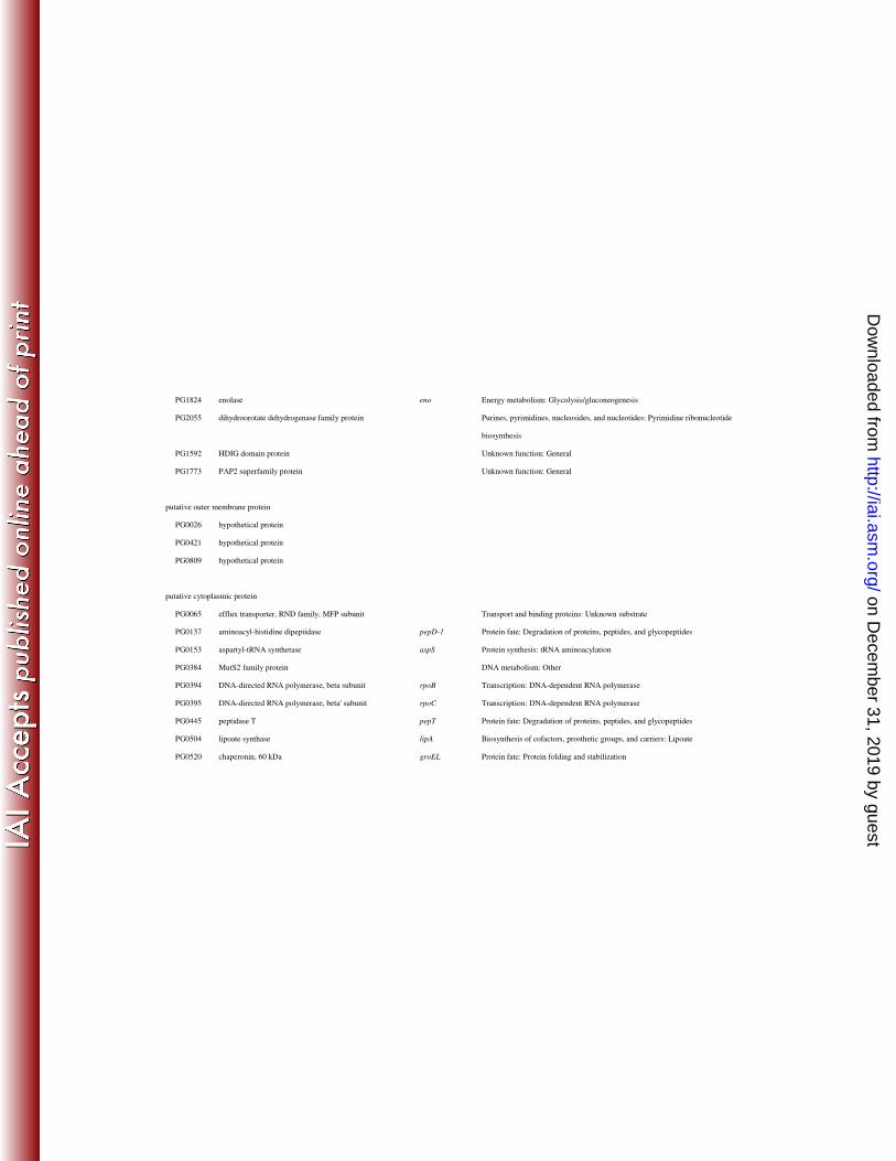
Yeast two-hybrid analysis 19
As another approach to clarify the molecular mechanism of contribution of TprA 20
protein to P. gingivalis virulence, we attempted to find P. gingivalis proteins that 21
interacted with TprA protein since it is likely that TprA protein, which is one of the TPR 22
on Decem
ber 31, 2019 by guesthttp://iai.asm
.org/D
ownloaded from

17
domain-containing proteins (3, 10), is associated with other proteins. Firstly, we 1
investigated the localization of TprA protein. The tprA mutant and its wild-type parent 2
were fractionated into cytoplasm/periplasm, inner membrane and outer membrane 3
fractions and then subjected to SDS-PAGE and immunoblot analysis with anti-TprA 4
antiserum (Fig. 2A). An anti-TprA-immunoreactive protein was found at a molecular 5
mass of 43 kDa in the cytoplasm/periplasm fraction. For further analysis, spheroplast 6
formation and proteinase K treatment was performed (Fig. 2B). The 43-kDa anti-TprA 7
immunoreactive protein band was observed in the periplasmic fraction. The 43-kDa 8
protein, which was degraded by treatment with proteinase K, was also detected in the 9
spheroplast fraction. In addition, TprA protein possessed a signal sequence as revealed 10
by in silico analysis with the software SignalP V3.0 11
(http://www.cbs.dtu.dk/services/SignalP/) (2). These results clearly indicate that TprA 12
protein is located in the periplasmic space, which is consistent with results of a previous 13
study (45). 14
Yeast two-hybrid analysis was then performed using the tprA gene as bait. A 15
number of P. gingivalis protein candidates interacting with TprA protein were found in 16
the analysis (Table 3). They included PG0415 (peptidyl-prolyl cis-trans isomerase, 17
PPIC-type), PG0497 (5’-methylthioadenosine/S-adenosylhomocysteine nucleosidase), 18
PG1334 (band 7/Mec-2 family protein), PG2101 (TapB) and PG2200 (TPR domain 19
protein) as a putative periplasmic protein. 20
21
Specific binding of TprA protein with TapA and TapB proteins 22
on Decem
ber 31, 2019 by guesthttp://iai.asm
.org/D
ownloaded from
18
We examined the interaction of TprA protein with TapA, TapB and TapC proteins in 1
vitro using protein binding assays. We determined the binding activity of TprA protein 2
with TapA and TapB proteins since we could not obtain a TapC soluble recombinant 3
protein (Fig. 3). Poly-histidine-tagged recombinant TapA (rTapA-His) or TapB 4
(rTapB-His) was incubated with Ni-beads and recombinant TprA (rTprA). rTprA itself 5
did not bind to Ni-beads, as revealed by immunoblot analysis using anti-TprA antibody, 6
while rTprA could bind to Ni-beads in the presence of rTapA-His or rTapB-His, and the 7
reaction was observed in a concentration-dependent manner (Fig. 3A, C). Next, rTprA 8
was mixed with rTapA-His or rTapB-His, immunoprecipitated with anti-TprA antibody, 9
and subjected to immunoblot analysis with anti-TapA or anti-TapB antibody. The 10
results were consistent with the above results (Fig. 3B, D). 11
These specific interactions were further confirmed by surface plasmon resonance 12
detection using the BIAcore system. The Kd values of interactions of rTprA with 13
rTapA-His and rTapB-His were 1.89 x 10⁻⁴
M and 1.42 x 10⁻⁶
M, respectively (Fig. 4). 14
15
Localization of TapA and TapB proteins 16
Cells of the wild-type and tapB mutant strains were fractionated into 17
cytoplasm/periplasm, inner membrane and outer membrane fractions and then subjected 18
to SDS-PAGE and immunoblot analysis with anti-TapB antiserum (Fig. 5A). A 19
33-kDa anti-TapB-immunoreactive protein was found in the cytoplasm/periplasm 20
fraction. To determine whether the 33-kDa anti-TapB-immunoreactive protein was 21
located in the cytoplasm or periplasm, cells of the wild-type strain were subjected to 22
on Decem
ber 31, 2019 by guesthttp://iai.asm
.org/D
ownloaded from
19
spheroplast formation and proteinase K treatment followed by immunoblot analysis 1
with anti-TapB (Fig. 5B). The 33-kDa anti-TapB-immunoreactive protein was found 2
in the periplasm fraction. The 33-kDa protein was also found in the spheroplast 3
fraction and disappeared after the proteinase K treatment. TapB protein possessed a 4
signal sequence as revealed by in silico analysis with the software SignalP V3.0. 5
These results indicated periplasmic localization of TapB. 6
To investigate the localization of TapA protein, the wild-type and tapA mutant 7
cells were fractionated into cytoplasm/periplasm, inner membrane and outer membrane 8
fractions and then subjected to SDS-PAGE and immunoblot analysis with anti-TapA 9
(Fig. 5C). A discrete protein band with a molecular mass of 60 kDa and diffuse 10
protein bands with molecular masses of 65-95 kDa were detected in the outer membrane 11
fraction of the wild-type strain but not in that of the tapA mutant. These protein bands 12
were also observed in the cytoplasmi/periplasm fraction with much weaker reaction. 13
These results indicated that TapA protein was located at the outer membrane. 14
15
TapA is one of the CTD proteins. 16
P. gingivalis has been shown to possess a novel family of outer membrane proteins that 17
have a conserved C-Terminal Domain (CTD) of RgpB (45, 51). The CTD has been 18
proposed to play roles in the secretion of the proteins across the outer membrane and 19
their attachment to the cell surface, probably via glycosylation (30, 43, 45). Very 20
recently, we have found a novel secretion system, Por secretion system (PorSS), by 21
which the CTD proteins may be secreted (42). 22
on Decem
ber 31, 2019 by guesthttp://iai.asm
.org/D
ownloaded from

20
TapA (PG2102) was reported to belong to the CTD family of proteins and 1
seemed to be secreted across the outer membrane and attach to the cell surface via 2
glycosylation. TapA protein was immunoprecipitated from the wild-type, porT and 3
tapA mutant strains using anti-TapA. The immunoprecipitates were subjected to 4
SDS-PAGE and immunoblot analysis using a monoclonal antibody, MAb 1B5, which 5
recognizes P. gingivalis anionic surface polysaccharides. MAb 1B5-immunoreactive 6
diffuse protein bands with molecular masses of 65-95 kDa were found in the wild-type 7
strain but not in the porT or tapA mutant strain (Fig. 6). The results suggested that 8
the MAb 1B5-immunoreactive diffuse protein bands with molecular masses of 65-95 9
kDa were glycosylated forms of TapA protein. In the porT mutant, the 10
anti-TapA-immunoreactive protein with a molecular mass of 60 kDa was mainly found 11
in the cytoplasm/periplasm fraction, suggesting that TapA protein is one of the outer 12
membrane proteins secreted via PorSS. 13
14
Contribution of the tapA-tapB-tapC operon to virulence of P. gingivalis 15
BALB/c mice were challenged with subcutaneous injections of bacterial suspension (2 x 16
1011
colony-forming units (CFU) per animal) (28, 49, 53), and their survival was 17
monitored for 9 days. About 66.7% of the mice challenged with W83 died at the end 18
of the experiment (9 days). In contrast, the survival rates of mice inoculated with the 19
tprA, tapA, tapB and tapC single mutants and the tapA-tapB-tapC deletion mutant were 20
significantly higher (P < 0.05, log-rank test) than that of mice inoculated with the 21
wild-type strain (Fig. 7). These results suggested that the tprA, tapA, tapB and tapC 22
on Decem
ber 31, 2019 by guesthttp://iai.asm
.org/D
ownloaded from

21
genes were involved in P. gingivalis virulence. 1
2
DISCUSSION 3
The TPR motif was originally reported for cell division cycle proteins of 4
Saccharomyces cerevisiae (18, 47). This motif is now known to be ubiquitous in 5
nature, as it is found within functionally unrelated proteins from all genera. A TPR is 6
defined as a degenerate 34-residue motif with a consensus amino acid arrangement of 7
alternate large and small residues and high amino acid conservation observed 8
specifically at positions 8, 20, and 27 (47). These conserved residues allow the TPR to 9
create a pair of antiparallel alpha helices. Multiple motifs, ranging from 3 to 16 in 10
number among TPR proteins, lead to the formation of an alpha superhelical structure 11
(11). This complex and unique structure gives rise to distinct substrate grooves that 12
facilitate specific protein-protein interactions. The ability of TPR-containing proteins to 13
interact with other proteins enables them to play a vital role in eukaryotic cell processes, 14
such as mitosis, transcription repression, and protein import (14, 22, 50). Bacteria also 15
utilize TPR proteins for a range of functions, including gene regulation, flagellar motor 16
function, chaperone activity, gliding motility and virulence (4, 5, 8, 29, 34, 44, 52). For 17
example, FrzF from Myxococcus xanthus is a methyltransferase containing three TPR 18
domains and it regulates the Frz chemosensory system, which controls cell reversals and 19
gliding motility. In particular, TPRs in FrzF are involved in site-specific methylation 20
of FrzCD, a metlyl-accepting chemotaxis protein (44). In addition, several chaperones 21
required for the type III secretion system contain a TPR domain, including PcrH from 22
on Decem
ber 31, 2019 by guesthttp://iai.asm
.org/D
ownloaded from
22
Pseudomonas aeruginosa, LcrH from Yersinia species, and CesD from 1
enteropathogenic Escherichia coli (4, 5, 52). 2
P. gingivalis W83 has at least ten TPR proteins. However, none of them 3
have been investigated so far. Structures of the TPR proteins are depicted with their 4
putative subcellular location in Fig. S4 in the supplemental material. TprA protein 5
contains three TPR domains. Okano et al. (35) reported that 19 proteins of P. 6
gingivalis W83 are up-regulated by aeration and that the 19 proteins include PG1385 7
(TprA), PG0045 (HtpG), PG0520 (60-kDa chaperonin), PG1208 (DnaK), PG0762 8
(trigger factor), PG0185 (RagA) and PG0618 (AhpC). Masuda et al. (25) reported that 9
14 proteins including TprA are up-regulated in P. gingivalis ATCC 33277 cells 10
cultivated in a nutrient-poor medium. We have reported that TprA protein was 11
up-regulated in P. gingivalis W83 cells placed in a mouse subcutaneous chamber (54). 12
Mouse subcutaneous infection experiments revealed that the tprA mutant was clearly 13
less virulent. These findings suggest that expression of tprA is influenced by various 14
environmental stresses and that TprA is involved in virulence of P. gingivalis. 15
TprA protein is located at the periplasm (45), implying that TprA binds to 16
other proteins at the periplasm or protruding from the inner or outer membrane. We 17
performed a yeast two-hybrid assay and found TapB (hypothetical protein) as a 18
TprA-associated protein. Taken together with the finding that expression of TapA was 19
induced in P. gingivalis cells placed in a mouse chamber as well as TprA, we assumed 20
that TprA was related with products of a cluster of tapA, tapB and tapC genes. Protein 21
binding assays with immunoprecipitation and plasmon resonance detection revealed the 22
on Decem
ber 31, 2019 by guesthttp://iai.asm
.org/D
ownloaded from

23
interaction of TprA protein with TapA and TapB proteins. We could not examine the 1
interaction between TprA protein and TapC protein in this study; however, TapC protein 2
may interact with TprA protein since TapC protein has 45% identity in amino acid 3
sequence with TapA protein. Mouse subcutaneous infection experiments in this study 4
revealed that the survival rate of mice with the tprA mutant was equivalent to that of 5
mice with the tapA-tapB-tapC deletion mutant, suggesting that TprA protein plays a role 6
in virulence of P. gingivalis in cooperation with TapA, TapB and TapC proteins. 7
We also investigated the gene expression profile of the tprA mutant inoculated 8
into a mouse subcutaneous chamber compared with the wild-type strain by using a 9
microarray. The results of microarray analysis revealed that 12 genes were 10
up-regulated and 11 genes were down-regulated in the tprA mutant. Interestingly, a 11
cluster of tapA, tapB and tapC genes was found to be down-regulated in the tprA mutant 12
strain. Down-regulation of the tapA, tapB and tapC genes was confirmed by real-time 13
qPCR analysis. At present, the mechanism of down-regulation remains to be clarified. 14
TapA and TapC proteins belong to the C-Terminal Domain (CTD) family of 15
proteins (45). The CTD has been proposed to play roles in the secretion of proteins 16
across the outer membrane and their attachment to the surface of the cell, probably via 17
glycosylation (30, 43, 45). Nguyen et al. reported that CTD is specifically conserved in 18
proteins of distinct members of the phylum Bacteroidetes and suggested that CTD 19
proteins are secreted by a novel secretion system. Very recently, we have found a novel 20
secretion system by which CTD proteins are secreted and named it Por secretion system 21
(PorSS) (42). We found in the present study that TapA protein, which was an outer 22
on Decem
ber 31, 2019 by guesthttp://iai.asm
.org/D
ownloaded from
24
membrane protein, was modified by polysaccharides immunoreactive to the MAb 1B5, 1
resulting in production of diffuse protein bands with molecular masses of 65-95 kDa, 2
which were lacking in the porT mutant. These results suggest that TapA protein is 3
secreted by PorSS. 4
As revealed by the mouse subcutaneous infection experiments, the tapA and 5
tapC mutants were less virulent than the wild-type parent strain, suggesting that the 6
CTD proteins TapA and TapC were related to virulence of P. gingivalis. Besides TapA 7
and TapC proteins, P. gingivalis has a number of CTD proteins: gingipain proteinases 8
(RgpA, RgpB and Kgp), CPG70 carboxypeptidase (6), PrtT thiol proteinase, HagA 9
haemagglutinin, Streptococcus gordonii binding protein (55), putative haemagglutinin, 10
putative thiol reductase, putative fibronectin binding protein, putative Lys-specific 11
proteinase and putative von Willebrand factor domain protein. RgpA and Kgp contain 12
C-terminal adhesins that are secreted and processed to form non-covalent complexes on 13
the cell surface and are considered to be the major virulence factors of this bacterium 14
(32, 33, 40). Gingipains have been linked directly to disease pathogenesis due to their 15
ability to degrade host structural and defense proteins and the inability of mutants 16
lacking functional Kgp or RgpA/B to cause alveolar bone loss in murine periodontitis 17
models (39). The majority of these proteins are likely to play important roles in 18
virulence of the bacterium since they are involved in extracellular proteolytic activity, 19
aggregation, heme/iron capture and storage, biofilm formation and resistance to 20
oxidative stress. 21
22
on Decem
ber 31, 2019 by guesthttp://iai.asm
.org/D
ownloaded from

25
1
2
ACKNOWLEDGMENTS 3
This work was supported by Grant-in-Aids (18018032 and 20249073 to K.N.) 4
for Scientific Research from the Ministry of Education, Culture, Sports, Science and 5
Technology of Japan and by the Global COE Program at Nagasaki University to K.N. 6
7
on Decem
ber 31, 2019 by guesthttp://iai.asm
.org/D
ownloaded from

26
REFFERENCES 1
2
1. Armitage, G. C. 1996. Periodontal diseases: diagnosis. Ann Periodontol 3
1:37-215. 4
2. Bendtsen, J. D., H. Nielsen, G. von Heijne, and S. Brunak. 2004. Improved 5
prediction of signal peptides: SignalP 3.0. J Mol Biol 340:783-795. 6
3. Blatch, G. L., and M. Lassle. 1999. The tetratricopeptide repeat: a structural 7
motif mediating protein-protein interactions. Bioessays 21:932-939. 8
4. Broms, J. E., P. J. Edqvist, A. Forsberg, and M. S. Francis. 2006. 9
Tetratricopeptide repeats are essential for PcrH chaperone function in 10
Pseudomonas aeruginosa type III secretion. FEMS Microbiol Lett 256:57-66. 11
5. Broms, J. E., A. L. Forslund, A. Forsberg, and M. S. Francis. 2003. PcrH of 12
Pseudomonas aeruginosa is essential for secretion and assembly of the type III 13
translocon. J Infect Dis 188:1909-1921. 14
6. Chen, Y. Y., K. J. Cross, R. A. Paolini, J. E. Fielding, N. Slakeski, and E. C. 15
Reynolds. 2002. CPG70 is a novel basic metallocarboxypeptidase with 16
C-terminal polycystic kidney disease domains from Porphyromonas gingivalis. J 17
Biol Chem 277:23433-23440. 18
7. Christersson, L. A., C. L. Fransson, R. G. Dunford, and J. J. Zambon. 1992. 19
Subgingival distribution of periodontal pathogenic microorganisms in adult 20
periodontitis. J Periodontol 63:418-425. 21
8. Core, L., and M. Perego. 2003. TPR-mediated interaction of RapC with ComA 22
inhibits response regulator-DNA binding for competence development in 23
Bacillus subtilis. Mol Microbiol 49:1509-1522. 24
9. Curtis, M. A., A. Thickett, J. M. Slaney, M. Rangarajan, J. Aduse-Opoku, P. 25
Shepherd, N. Paramonov, and E. F. Hounsell. 1999. Variable carbohydrate 26
modifications to the catalytic chains of the RgpA and RgpB proteases of 27
Porphyromonas gingivalis W50. Infect Immun 67:3816-3823. 28
10. D'Andrea, L. D., and L. Regan. 2003. TPR proteins: the versatile helix. Trends 29
Biochem Sci 28:655-662. 30
11. Das, A. K., P. W. Cohen, and D. Barford. 1998. The structure of the 31
tetratricopeptide repeats of protein phosphatase 5: implications for 32
on Decem
ber 31, 2019 by guesthttp://iai.asm
.org/D
ownloaded from

27
TPR-mediated protein-protein interactions. EMBO J 17:1192-1199. 1
12. Delgado-Partin, V. M., and R. E. Dalbey. 1998. The proton motive force, 2
acting on acidic residues, promotes translocation of amino-terminal domains of 3
membrane proteins when the hydrophobicity of the translocation signal is low. J 4
Biol Chem 273:9927-9934. 5
13. Diaz, P. I., N. Slakeski, E. C. Reynolds, R. Morona, A. H. Rogers, and P. E. 6
Kolenbrander. 2006. Role of oxyR in the oral anaerobe Porphyromonas 7
gingivalis. J Bacteriol 188:2454-2462. 8
14. Dodt, G., and S. J. Gould. 1996. Multiple PEX genes are required for proper 9
subcellular distribution and stability of Pex5p, the PTS1 receptor: evidence that 10
PTS1 protein import is mediated by a cycling receptor. J Cell Biol 11
135:1763-1774. 12
15. Genco, C. A., C. W. Cutler, D. Kapczynski, K. Maloney, and R. R. Arnold. 13
1991. A novel mouse model to study the virulence of and host response to 14
Porphyromonas (Bacteroides) gingivalis. Infect Immun 59:1255-1263. 15
16. Grenier, D., and D. Mayrand. 1987. Selected characteristics of pathogenic and 16
nonpathogenic strains of Bacteroides gingivalis. J Clin Microbiol 25:738-740. 17
17. Haffajee, A. D., and S. S. Socransky. 1994. Microbial etiological agents of 18
destructive periodontal diseases. Periodontol 2000 5:78-111. 19
18. Hirano, T., N. Kinoshita, K. Morikawa, and M. Yanagida. 1990. Snap helix 20
with knob and hole: essential repeats in S. pombe nuclear protein nuc2+. Cell 21
60:319-328. 22
19. Hosogi, Y., and M. J. Duncan. 2005. Gene expression in Porphyromonas 23
gingivalis after contact with human epithelial cells. Infect Immun 73:2327-2335. 24
20. Hughes, T. R., M. J. Marton, A. R. Jones, C. J. Roberts, R. Stoughton, C. D. 25
Armour, H. A. Bennett, E. Coffey, H. Dai, Y. D. He, M. J. Kidd, A. M. King, 26
M. R. Meyer, D. Slade, P. Y. Lum, S. B. Stepaniants, D. D. Shoemaker, D. 27
Gachotte, K. Chakraburtty, J. Simon, M. Bard, and S. H. Friend. 2000. 28
Functional discovery via a compendium of expression profiles. Cell 29
102:109-126. 30
21. Irfan, U. M., D. V. Dawson, and N. F. Bissada. 2001. Epidemiology of 31
periodontal disease: a review and clinical perspectives. J Int Acad Periodontol 32
3:14-21. 33
on Decem
ber 31, 2019 by guesthttp://iai.asm
.org/D
ownloaded from

28
22. King, R. W., J. M. Peters, S. Tugendreich, M. Rolfe, P. Hieter, and M. W. 1
Kirschner. 1995. A 20S complex containing CDC27 and CDC16 catalyzes the 2
mitosis-specific conjugation of ubiquitin to cyclin B. Cell 81:279-288. 3
23. Laemmli, U. K. 1970. Cleavage of structural proteins during the assembly of 4
the head of bacteriophage T4. Nature 227:680-685. 5
24. Lo, A. W., C. A. Seers, J. D. Boyce, S. G. Dashper, N. Slakeski, J. P. Lissel, 6
and E. C. Reynolds. 2009. Comparative transcriptomic analysis of 7
Porphyromonas gingivalis biofilm and planktonic cells. BMC Microbiol 9:18. 8
25. Masuda, T., Y. Murakami, T. Noguchi, and F. Yoshimura. 2006. Effects of 9
various growth conditions in a chemostat on expression of virulence factors in 10
Porphyromonas gingivalis. Appl Environ Microbiol 72:3458-3467. 11
26. Murakami, Y., M. Imai, H. Nakamura, and F. Yoshimura. 2002. Separation 12
of the outer membrane and identification of major outer membrane proteins 13
from Porphyromonas gingivalis. Eur J Oral Sci 110:157-162. 14
27. Nakayama, K. 2003. Molecular genetics of Porphyromonas gingivalis: 15
gingipains and other virulence factors. Curr Protein Pept Sci 4:389-395. 16
28. Neiders, M. E., P. B. Chen, H. Suido, H. S. Reynolds, J. J. Zambon, M. 17
Shlossman, and R. J. Genco. 1989. Heterogeneity of virulence among strains 18
of Bacteroides gingivalis. J Periodontal Res 24:192-198. 19
29. Newton, H. J., F. M. Sansom, J. Dao, A. D. McAlister, J. Sloan, N. P. 20
Cianciotto, and E. L. Hartland. 2007. Sel1 repeat protein LpnE is a Legionella 21
pneumophila virulence determinant that influences vacuolar trafficking. Infect 22
Immun 75:5575-5585. 23
30. Nguyen, K. A., J. Travis, and J. Potempa. 2007. Does the importance of the 24
C-terminal residues in the maturation of RgpB from Porphyromonas gingivalis 25
reveal a novel mechanism for protein export in a subgroup of Gram-Negative 26
bacteria? J Bacteriol 189:833-843. 27
31. Nguyen, K. A., J. Zylicz, P. Szczesny, A. Sroka, N. Hunter, and J. Potempa. 28
2009. Verification of a topology model of PorT as an integral outer-membrane 29
protein in Porphyromonas gingivalis. Microbiology 155:328-337. 30
32. NM, O. B.-S., P. D. Veith, S. G. Dashper, and E. C. Reynolds. 2003. 31
Porphyromonas gingivalis gingipains: the molecular teeth of a microbial 32
vampire. Curr Protein Pept Sci 4:409-426. 33
on Decem
ber 31, 2019 by guesthttp://iai.asm
.org/D
ownloaded from

29
33. O'Brien-Simpson, N. M., R. A. Paolini, B. Hoffmann, N. Slakeski, S. G. 1
Dashper, and E. C. Reynolds. 2001. Role of RgpA, RgpB, and Kgp proteinases 2
in virulence of Porphyromonas gingivalis W50 in a murine lesion model. Infect 3
Immun 69:7527-7534. 4
34. Okabe, M., T. Yakushi, and M. Homma. 2005. Interactions of MotX with 5
MotY and with the PomA/PomB sodium ion channel complex of the Vibrio 6
alginolyticus polar flagellum. J Biol Chem 280:25659-25664. 7
35. Okano, S., Y. Shibata, T. Shiroza, and Y. Abiko. 2006. Proteomics-based 8
analysis of a counter-oxidative stress system in Porphyromonas gingivalis. 9
Proteomics 6:251-258. 10
36. Oliver, R. C., L. J. Brown, and H. Loe. 1998. Periodontal diseases in the 11
United States population. J Periodontol 69:269-278. 12
37. Papapanou, P. N. 1999. Epidemiology of periodontal diseases: an update. J Int 13
Acad Periodontol 1:110-116. 14
38. Park, Y., O. Yilmaz, I. Y. Jung, and R. J. Lamont. 2004. Identification of 15
Porphyromonas gingivalis genes specifically expressed in human gingival 16
epithelial cells by using differential display reverse transcription-PCR. Infect 17
Immun 72:3752-3758. 18
39. Pathirana, R. D., N. M. O'Brien-Simpson, G. C. Brammar, N. Slakeski, and 19
E. C. Reynolds. 2007. Kgp and RgpB, but not RgpA, are important for 20
Porphyromonas gingivalis virulence in the murine periodontitis model. Infect 21
Immun 75:1436-1442. 22
40. Potempa, J., R. Pike, and J. Travis. 1995. The multiple forms of trypsin-like 23
activity present in various strains of Porphyromonas gingivalis are due to the 24
presence of either Arg-gingipain or Lys-gingipain. Infect Immun 63:1176-1182. 25
41. Rodrigues, P. H., and A. Progulske-Fox. 2005. Gene expression profile 26
analysis of Porphyromonas gingivalis during invasion of human coronary artery 27
endothelial cells. Infect Immun 73:6169-6173. 28
42. Sato, K., M. Naito, H. Yukitake, H. Hirakawa, M. Shoji, M. J. McBride, R. 29
G. Rhodes, and K. Nakayama. 2010. A protein secretion system linked to 30
bacteroidete gliding motility and pathogenesis. Proc. Natl. Acad. Sci. U.S.A. 31
107: 276-281. 32
43. Sato, K., E. Sakai, P. D. Veith, M. Shoji, Y. Kikuchi, H. Yukitake, N. Ohara, 33
on Decem
ber 31, 2019 by guesthttp://iai.asm
.org/D
ownloaded from

30
M. Naito, K. Okamoto, E. C. Reynolds, and K. Nakayama. 2005. 1
Identification of a new membrane-associated protein that influences 2
transport/maturation of gingipains and adhesins of Porphyromonas gingivalis. J 3
Biol Chem 280:8668-8677. 4
44. Scott, A. E., E. Simon, S. K. Park, P. Andrews, and D. R. Zusman. 2008. 5
Site-specific receptor methylation of FrzCD in Myxococcus xanthus is controlled 6
by a tetra-trico peptide repeat (TPR) containing regulatory domain of the FrzF 7
methyltransferase. Mol Microbiol 69:724-735. 8
45. Seers, C. A., N. Slakeski, P. D. Veith, T. Nikolof, Y. Y. Chen, S. G. Dashper, 9
and E. C. Reynolds. 2006. The RgpB C-terminal domain has a role in 10
attachment of RgpB to the outer membrane and belongs to a novel 11
C-terminal-domain family found in Porphyromonas gingivalis. J Bacteriol 12
188:6376-6386. 13
46. Shelburne, C. E., R. M. Gleason, G. R. Germaine, L. F. Wolff, B. H. Mullally, 14
W. A. Coulter, and D. E. Lopatin. 2002. Quantitative reverse transcription 15
polymerase chain reaction analysis of Porphyromonas gingivalis gene 16
expression in vivo. J Microbiol Methods 49:147-156. 17
47. Sikorski, R. S., M. S. Boguski, M. Goebl, and P. Hieter. 1990. A repeating 18
amino acid motif in CDC23 defines a family of proteins and a new relationship 19
among genes required for mitosis and RNA synthesis. Cell 60:307-317. 20
48. Smoot, L. M., J. C. Smoot, M. R. Graham, G. A. Somerville, D. E. 21
Sturdevant, C. A. Migliaccio, G. L. Sylva, and J. M. Musser. 2001. Global 22
differential gene expression in response to growth temperature alteration in 23
group A Streptococcus. Proc Natl Acad Sci U S A 98:10416-10421. 24
49. Tachibana-Ono, M., A. Yoshida, S. Kataoka, T. Ansai, Y. Shintani, Y. 25
Takahashi, K. Toyoshima, and T. Takehara. 2008. Identification of the genes 26
associated with a virulent strain of Porphyromonas gingivalis using the 27
subtractive hybridization technique. Oral Microbiol Immunol 23:84-87. 28
50. Tzamarias, D., and K. Struhl. 1994. Functional dissection of the yeast 29
Cyc8-Tup1 transcriptional co-repressor complex. Nature 369:758-761. 30
51. Veith, P. D., G. H. Talbo, N. Slakeski, S. G. Dashper, C. Moore, R. A. Paolini, 31
and E. C. Reynolds. 2002. Major outer membrane proteins and proteolytic 32
processing of RgpA and Kgp of Porphyromonas gingivalis W50. Biochem J 33
on Decem
ber 31, 2019 by guesthttp://iai.asm
.org/D
ownloaded from
31
363:105-115. 1
52. Wattiau, P., B. Bernier, P. Deslee, T. Michiels, and G. R. Cornelis. 1994. 2
Individual chaperones required for Yop secretion by Yersinia. Proc Natl Acad 3
Sci U S A 91:10493-10497. 4
53. Yoshimura, M., Y. Nakano, Y. Yamashita, T. Oho, T. Saito, and T. Koga. 5
2000. Formation of methyl mercaptan from L-methionine by Porphyromonas 6
gingivalis. Infect Immun 68:6912-6916. 7
54. Yoshimura, M., N. Ohara, Y. Kondo, M. Shoji, S. Okano, Y. Nakano, Y. 8
Abiko, and K. Nakayama. 2008. Proteome analysis of Porphyromonas 9
gingivalis cells placed in a subcutaneous chamber of mice. Oral Microbiol 10
Immunol 23:413-418. 11
55. Zhang, Y., T. Wang, W. Chen, O. Yilmaz, Y. Park, I. Y. Jung, M. Hackett, 12
and R. J. Lamont. 2005. Differential protein expression by Porphyromonas 13
gingivalis in response to secreted epithelial cell components. Proteomics 14
5:198-211. 15
16
FIGURE LEGENDS 17
18
Fig. 1. Real-time qPCR analysis of gene expression in the tprA mutant inoculated 19
into a mouse subcutaneous chamber. 20
Mouse subcutaneous chamber experiments were performed as described in “Materials 21
and Methods.” Total RNA was extracted from W83 (wild type) and KDP159 22
(tprA::ermF). The down-regulated genes revealed by microarray analysis were selected 23
for analysis by real-time qPCR. All PCR reactions were carried out in triplicate. 24
25
Fig. 2. Subcellular localization of TprA protein. 26
(A) Cytoplasm/periplasm, total membrane, inner membrane and outer membrane 27
on Decem
ber 31, 2019 by guesthttp://iai.asm
.org/D
ownloaded from

32
fractions. Cells of W83 (wild type) and KDP159 (tprA::ermF) were fractionated and 1
subjected to SDS-PAGE and immunoblot analysis using anti-TprA antiserum. Lanes: 1, 2
cytoplasm/periplasm fraction; 2, total membrane fraction; 3, inner membrane fraction; 4, 3
outer membrane fraction. (B) Spheroplast and periplasm fractions. Preparation of 4
spheroplasts of P. gingivalis cells was performed as described in “Materials and 5
Methods” and the spheroplasts were subjected to proteinase K treatment (Pro K) in the 6
presence or absence of 2% Triton X-100 (TX-100). Samples were subjected to 7
SDS-PAGE followed by immunoblot analysis with anti-TprA antiserum. Sph, 8
spheroplasts; Peri, periplasm fraction. 9
10
Fig. 3. Binding of TprA protein to TapA and TapB proteins. 11
(A, B) Binding of TprA and TapB. rTprA (0 to 1.0 µg) was mixed with rTapB-His (0.5 12
µg) and was affinity-purified by Ni-beads. The resulting samples were subjected to 13
SDS-PAGE and immunoblot analysis with anti-TprA (A). rTapB-His (0 to 1.0 µg) was 14
mixed with rTprA (0.5 µg) and was immuno-purified by protein G agarose with 15
anti-TprA. The resulting samples were subjected to SDS-PAGE and immunoblot 16
analysis with anti-TapB (B). (C, D) Binding of TprA and TapA. rTprA (0 to 1.0 µg) 17
was mixed with rTapA-His (0.5 µg) and was affinity-purified by Ni-beads. The resulting 18
samples were subjected to SDS-PAGE and immunoblot analysis with anti-TprA (C). 19
rTapA-His (0 to 1.0 µg) was mixed with rTprA (0.5 µg) and was immuno-purified by 20
protein G agarose with anti-TprA. The resulting samples were subjected to SDS-PAGE 21
and immunoblot analysis with anti-TapA (D). 22
on Decem
ber 31, 2019 by guesthttp://iai.asm
.org/D
ownloaded from

33
1
Fig. 4. Surface plasmon resonance detection. 2
Parameters of association and dissociation for rTapA-His and rTapB-His with rTprA 3
were determined by surface plasmon resonance analysis (Biacore). Sensorgrams for the 4
binding of rTapA-His or rTapB-His to rTprA immobilized on a CM5 sensor chip were 5
overlaid at various concentrations of rTapA-His or rTapB-His. rTapA-His, rTapB-His or 6
bovine serum albumin (BSA) was injected at the concentrations of 0.016, 0.08, 0.4, 2 7
and 10 mM. The Kd values of binding of rTprA to rTapA-His and rTapB-His were 1.89 8
x 10-4
M and 1.42 x 10⁻⁶
M, respectively. 9
10
Fig. 5. Localization of TapA and TapB proteins. 11
(A, B) Subcellular localization of TapB protein. Fractions of cytoplasm/periplasm (lane 12
1) and total membrane (lane 2) of W83 (wild type) and KDP382 (∆tapB::ermF) were 13
subjected to SDS-PAGE and immunoblot analysis using anti-TapB (A). Spheroplast 14
and periplasm fractions of P. gingivalis cells were separated as described in “Materials 15
and Methods.” Spheroplasts were subjected to the proteinase K treatment (Pro K) in 16
the presence or absence of 2% Triton X-100 (TX-100). Samples were subjected to 17
SDS-PAGE followed by immunoblot analysis with anti-TapB (B). Sph, spheroplasts; 18
Peri, periplasm fraction. (C) Subcellular localization of the TapA protein. 19
Cytoplasm/periplasm, inner membrane and outer membrane fractions of W83 (wild 20
type), KDP385 (∆porT::erm) and KDP383 (∆tapA::ermF) were subjected to SDS-PAGE 21
and immunoblot analysis using anti-TapA. Lanes: 1, cytoplasm/periplasm fraction; 2, 22
on Decem
ber 31, 2019 by guesthttp://iai.asm
.org/D
ownloaded from

34
inner membrane fraction; 3, outer membrane fraction. 1
2
Fig. 6. Glycosylation of TapA protein. 3
TapA protein was immunoprecipitated from lysates of W83, KDP385 (∆porT::erm) and 4
KDP383 (∆tapA::ermF) cells using anti-TapA. The resulting immunoprecipitates were 5
subjected to SDS-PAGE and immunoblot analysis with MAb 1B5. 6
7
Fig. 7. Survival rates of mice challenged with the wild-type and mutant strains of P. 8
gingivalis. 9
Construction of the tapA, tapB, tapC and tapA tapB tapC mutants is described in the 10
Materials and Methods. Genetic manipulation did not affect the expression of 11
downstream or upstream gene (Fig. S4 in the supplemental material). Female BALB/c 12
mice were inoculated intradermally into the back with P. gingivalis cells (approximately 13
2 x 10¹¹
CFU) and then their survival was monitored daily up to 9 days. The animal 14
experiment in which five mice were used for each bacterial strain was performed three 15
times. *P< 0.05 versus the corresponding values for the wild-type littermates, with the 16
log-rank test. 17
18
Supplemental Material 19
20
Table S1. Oligonucleotides used in this study 21
22
on Decem
ber 31, 2019 by guesthttp://iai.asm
.org/D
ownloaded from
Table 1. Strains and plasmids used in this study
strain / plasmid Description source or ref.
P. gingivalis
W83 wild type
KDP159 tprA::[ermF ermAM], Emr 54
KDP382 ∆tapB::ermF, Emr this study
KDP383 ∆tapA::ermF, Emr this study
KDP384 ∆[tapA tapB tapC]::ermF, Emr this study
KDP385 ∆porT::[ermF ermAM], Emr this study
KDP386 ∆tapC::ermF, Emr this study
E. coli
XL1-Blue General-purpose host strain for cloning stratagene
BL21(DE3) host strain for expression vector pET-32a Nippongene
S. cerevisiae
AH109 host strain for yeast two-hybrid system Clontech
plasmid
pBluescript II SK(-) Apr, cloning vector (pBSSK) stratagene
pET-32a Apr, expression vector Novagen
pGEX-6P-1 Apr, expression vector GE Healthcare
pKD357 Apr Emr, pBSSK contains ermF ermAM cassette located between upstream and 43
downstream of the porT gene
pKD1008 Apr, pBSSK contains 0.3-kb tapC-upstream and 0.5-kb tapC-downstream regions this study
pKD1009 Apr Emr, the ermF casassette was inserted into BamHI site of pKD1008 this study
pKD1010 Apr, pBSSK contains 0.3-kb tapB-upstream and 0.5-kb tapB-downstream regions this study
pKD1011 Apr Emr, the ermF casassette was inserted into BamHI site of pKD1010 this study
pKD1012 Apr, pBSSK contains 0.3-kb tapA-upstream and 0.5-kb tapA-downstream regions this study
pKD1013 Apr Emr, the ermF casassette was inserted into BamHI site of pKD1012 this study
pKD1014 Apr, pBSSK contains 0.3-kb tapA-upstream and 0.5-kb tapC-downstream regions this study
pKD1015 Apr Emr, the ermF casassette was inserted into BamHI site of pKD1014 this study
pKD1016 Apr, pET-32a containing P. gingivalis tapB gene this study
on Decem
ber 31, 2019 by guesthttp://iai.asm
.org/D
ownloaded from

pKD1017 Apr, pET-32a containing P. gingivalis tapA gene this study
pKD1018 Apr, pGEX-6P-1 containing P. gingivalis tprA gene this study
pKD1019 pGBKT7 containing P. gingivalis tprA gene This study
on Decem
ber 31, 2019 by guesthttp://iai.asm
.org/D
ownloaded from
Table 2. P. gingivalis genes differentially expressed in the tprA mutant cells placed in a mouse subcutaneous
chamber compared to the wild-type cells
TIGR ID Identification Avr. fold difference
down-reglulated genes
PG0100 hypothetical protein 0.59
PG0162 RNA polymerase sigma-70 factor, ECF subfamily 0.57
PG0591 ISPg5, transposase Orf2 0.63
PG0756 hypothetical protein 0.47
PG1055 thiol protease 0.36
PG1972 hemagglutinin protein HagB 0.63
PG1975 hemagglutinin protein HagC 0.64
PG2100 (tapC) immunoreactive 63 kDa antigen PG102 0.34
PG2101 (tapB) hypothetical protein 0.25
PG2102 (tapA) immunoreactive 61 kDa antigen PG91 0.51
PG2103 hypothetical protein 0.24
PG2214 hypothetical protein 0.57
up-regulated genes
PG0327 hypothetical protein 1.92
PG0373 hypothetical protein 1.72
PG0546 hypothetical protein 1.83
PG0635 ribosomal protein L11 methyltransferase 1.69
PG0656 ribosomal protein L34 2.02
PG0722 hypothetical protein 2.93
PG0969 S-adenosylmethionine:tRNA ribosyltransferase-isomerase, putative 1.98
PG1532 hypothetical protein 1.92
PG1866 hypothetical protein 1.93
PG2006 hypothetical protein 1.69
PG2225 hypothetical protein 1.63
Mouse subcutaneous chamber experiments were performed as described in “MATERIALS AND
on Decem
ber 31, 2019 by guesthttp://iai.asm
.org/D
ownloaded from

METHODS.” Total RNA was extracted from W83 (wild type) and KDP159 (tprA::erm). Total RNA was
extracted and analyzed by using a microarray as described in “MATERIALS AND METHODS.” The
number in the “Average fold difference” column indicates the gene expression in the tprA mutant versus
the wild-type strain. The cut-off- ratio for the fold difference was 1.5.
on Decem
ber 31, 2019 by guesthttp://iai.asm
.org/D
ownloaded from

Table 3. TprA-associated proteins predicted from yeast two-hybrid system
TIGR ID description Gene symbol TIGR functional role category
putative periplasmic protein
PG0415 peptidyl-prolyl cis-trans isomerase, PPIC-type Protein fate: Protein folding and stabilization
PG0497 5'-methylthioadenosine/S-adenosylhomocysteine
nucleosidase
mtn Central intermediary metabolism: Other
PG1334 band 7/Mec-2 family protein Unknown function: General
PG2101 hypothetical protein tapB
PG2200 TPR domain protein Unknown function: General
putative inner membrane protein
PG0081 hypothetical protein
PG0093 HlyD family secretion protein Transport and binding proteins: Other
PG0255 translation initiation factor IF-2 infB Protein synthesis: Translation factors
PG0512 guanylate kinase gmk Purines, pyrimidines, nucleosides, and nucleotides: Nucleotide and nucleoside
interconversions
PG1068 conserved hypothetical protein Hypothetical proteins: Conserved
PG1271 acetylornithine aminotransferase, putative Amino acid biosynthesis: Glutamate family
PG1490 TraG family protein Mobile and extrachromosomal element functions: Plasmid functions
PG1768 magnesium chelatase, subunit D/I family Transport and binding proteins: Cations and iron carrying compounds
on Decem
ber 31, 2019 by guesthttp://iai.asm
.org/D
ownloaded from
PG1824 enolase eno Energy metabolism: Glycolysis/gluconeogenesis
PG2055 dihydroorotate dehydrogenase family protein Purines, pyrimidines, nucleosides, and nucleotides: Pyrimidine ribonucleotide
biosynthesis
PG1592 HDIG domain protein Unknown function: General
PG1773 PAP2 superfamily protein Unknown function: General
putative outer membrane protein
PG0026 hypothetical protein
PG0421 hypothetical protein
PG0809 hypothetical protein
putative cytoplasmic protein
PG0065 efflux transporter, RND family, MFP subunit Transport and binding proteins: Unknown substrate
PG0137 aminoacyl-histidine dipeptidase pepD-1 Protein fate: Degradation of proteins, peptides, and glycopeptides
PG0153 aspartyl-tRNA synthetase aspS Protein synthesis: tRNA aminoacylation
PG0384 MutS2 family protein DNA metabolism: Other
PG0394 DNA-directed RNA polymerase, beta subunit rpoB Transcription: DNA-dependent RNA polymerase
PG0395 DNA-directed RNA polymerase, beta' subunit rpoC Transcription: DNA-dependent RNA polymerase
PG0445 peptidase T pepT Protein fate: Degradation of proteins, peptides, and glycopeptides
PG0504 lipoate synthase lipA Biosynthesis of cofactors, prosthetic groups, and carriers: Lipoate
PG0520 chaperonin, 60 kDa groEL Protein fate: Protein folding and stabilization
on Decem
ber 31, 2019 by guesthttp://iai.asm
.org/D
ownloaded from

PG0553 extracellular protease, putative Protein fate: Degradation of proteins, peptides, and glycopeptides
PG0629 ATP-NAD kinase ppnK Biosynthesis of cofactors, prosthetic groups, and carriers: Pyridine nucleotides
PG0728 conserved hypothetical protein Hypothetical proteins: Conserved
PG1195 8-amino-7-oxononanoate synthase bioF-2 Biosynthesis of cofactors, prosthetic groups, and carriers: Biotin
PG1242 replicative DNA helicase dnaB DNA metabolism: DNA replication, recombination, and repair
PG1622 DNA topoisomerase IV, A subunit, putative DNA metabolism: DNA replication, recombination, and repair
PG1849 DNA repair protein RecN recN DNA metabolism: DNA replication, recombination, and repair
PG1875 hemolysin Cellular processes: Pathogenesis
PG1926 ribosomal protein L5 rplE Protein synthesis: Ribosomal proteins: synthesis and modification
not detemined
PG0232 zinc carboxypeptidase, putative Protein fate: Degradation of proteins, peptides, and glycopeptides
PG1084 thioredoxin family protein Energy metabolism: Electron transport
on Decem
ber 31, 2019 by guesthttp://iai.asm
.org/D
ownloaded from

Fig. 1
0
0.2
0.4
0.6
0.8
1
1.2
1.4
1.6
1.8
2
PG
0100
PG
0162
PG
0591
PG
0756
PG
1055
PG
1972
PG
1975
PG
2100
PG
2101
PG
2102
PG
2103
PG
2214
Rel
ativ
e Q
uan
tity
W83
KDP159
(tap
A)
(tap
C)
(tap
B)
on Decem
ber 31, 2019 by guesthttp://iai.asm
.org/D
ownloaded from

Fig. 2
55
43
36.5
28
1 2 3 4 1 2 3 4
W83 KDP159
(kDa)
anti-TprA
A
B
anti-TprA
Peri Sph
TX-100
Pro K
- -+ -
+
- +
+
(kDa)
55
43
36.5
--
on Decem
ber 31, 2019 by guesthttp://iai.asm
.org/D
ownloaded from

B
A
C
D
Fig. 3 rTapB-His
rTprA
anti-TprA
(µg)
- - - + + +
0.1 10.5 0 0.1 0.5 1
+
AP: Ni-beads
43
55
rTapB-His
rTprA
(µg)
- - - + + +
0.1 10.5 0 0.1 0.5 1
+
IP: proteinG + anti-TprA
anti-TapB
55
43
(µg)
rTapA-His
rTprA
anti-TprA
- - - + + +
0.1 10.5 0 0.1 0.5 1
+
AP: Ni-beads
55
43
rTapA-His
rTprA
anti-TapA
(µg)
- - - + + +
0.1 10.5 0 0.1 0.5 1
+
IP: ProteinG + anti-TprA
95
70
(kDa)
(kDa)
(kDa)
(kDa)
on Decem
ber 31, 2019 by guesthttp://iai.asm
.org/D
ownloaded from

Fig. 4
0 200 400 600 800 1000 1200 1400 1600
-30
-20
-10
0
10
20
30
40
50
-200
RU
Resp
on
se
Time s
0 200 400 600 800 1000 1200 1400 1600
-30
-20
-10
0
10
20
30
40
50
-200
RU
Time s
0 200 400 600 800 1000 1200 1400 1600
-30
-20
-10
0
10
20
30
40
50
-200
RU
Time s
PG2101 (TapB)
PG2102 (TapA)
BSA
A
B
C
Resp
on
se
Resp
on
se
on Decem
ber 31, 2019 by guesthttp://iai.asm
.org/D
ownloaded from

Fig. 5A
Sph
4336.5
2819.3
(kDa)
Peri
TX-100
Pro K
- -+ -
+- +
+
anti-TapB
B
C W83 KDP385 KDP383
1 2 3 1 2 3 1 2 3
anti-TapA
95
70
55
(kDa)
43
36.5
28
19.3
W83 KDP382
1 2
anti- TapB
(kDa)1 2 1 2
--
on Decem
ber 31, 2019 by guesthttp://iai.asm
.org/D
ownloaded from

210
140
95
70
55
43
36.5
28
wt
W83
(kDa)
MAb 1B5
Fig. 6
KD
P385
KD
P383
on Decem
ber 31, 2019 by guesthttp://iai.asm
.org/D
ownloaded from

W83 ( wild type)
KDP159 (tprA)
KDP386 (tapC)
KDP382 (tapB)
KDP383 (tapA)
KDP384 (tapA tapB tapC)
0 1 2 3 4 5 6 7 8 9
Days after infection
0
10
20
30
40
50
60
70
80
90
100
Su
rviv
al r
ate
(%) *
*
*
**
Fig. 7
on Decem
ber 31, 2019 by guesthttp://iai.asm
.org/D
ownloaded from