Targeting the K-Ras/PDE δ Protein-Protein Interaction: The Solution for...
3
DOI: 10.1002/cmdc.201300311 Targeting the K-Ras/PDEd Protein–Protein Interaction : The Solution for Ras-Driven Cancers or Just Another Therapeutic Mirage? Brendan Frett, [a, b] Yuanxiang Wang, [a, b] and Hong-yu Li* [a, b] Cancer has become a significant, predominate focus in world medicine over the past century. In 2008, a total of 7.6 million deaths internationally were attributed to the disease, giving cancer the dubious honor of being the leading cause of death worldwide. [1] The disease itself is actually a diverse subset of multiple genetic disorders, which leads to the generation of various rogue proteins and tumor suppressor knockouts that interrupt cellular homeostasis. Treatment success is highest when the specific genetic disorder causing a patient’s tumor is targeted through a personalized medicine approach, but this is often difficult because of issues with druggability of the desired target. The most commonly mutated oncogene, at present deemed undruggable, is Ras (rat sarcoma), which has been identified in about 30 % of cancers. [2, 3] Ras comprises a family of guanosine- 5’-triphosphate (GTP) binding proteins (K-Ras, N-Ras, and H-Ras) localized at the inner cell membrane, and these proteins serve as a molecular switch controlling cell survival, differentia- tion, and proliferation. [3] Many cancers utilize Ras proteins as survival pathways to drive and propagate the disease. Efforts thus far to directly target Ras at the GTP binding site have been met with much disappointment, as Ras proteins bind GTP with picomolar affinity. [4] Adding to this difficulty, GTP is present intracellularly at micromolar concentrations, [4] making the GTP pocket on Ras realistically unamenable to small-mole- cule inhibition. The druggability issue is made even more diffi- cult because Ras does not contain any likely pockets where a small molecule can bind with high affinity. [4] Because of these problems, tumors bearing Ras mutations are difficult to treat. Current therapeutic avenues have been addressed and dis- cussed in a comprehensive review by Wang et al. [5] Neverthe- less, there have been a few transient success stories targeting the Ras protein, which primarily rely on inhibiting easily drug- gable kinases upstream and downstream in the Ras signal transduction pathway. This approach has been used to treat a certain subset of non-small-cell lung cancer (NSCLC). The NSCLC subset utilizes epidermal growth factor receptor (EGFR) as a survival pathway, which is upstream of Ras. Therapeutical- ly, EGFR has been drugged with gefitinib (1), and significant tumor remission has been observed (Figure 1). However, effica- cy is temporary, as NCSLC develops resistance to gefitinib over time. Interestingly, resistance has been shown to develop through activated Ras, where Ras is able to signal independent of EGFR stimulation. [6] In another example, vemurafenib (2) has been developed as a Raf (rapidly accelerated fibrosarcoma) in- hibitor to treat late-stage melanoma, specifically inhibiting B-Raf V600E . Raf is downstream of Ras, and treatment with vemur- afenib initially blocks progression of the melanoma. However, treatment eventually becomes ineffective as a second Ras iso- form (N-Ras) reactivates the mitogen-activated protein kinase (MAPK) signaling pathway and bypasses vemurafenib inhibi- tion. [7] Therefore, all efforts to date to disrupt the Ras signaling pathway that show clinical promise have failed because of re- sistance conferred through the Ras protein (Scheme 1). Devel- oping a strategy to target Ras directly has the potential to offer longer disease remission and overall better treatment op- tions for Ras-driven tumors. However, the druggability of Ras at a clinically effective level still remains a major problem. In an effort to reverse the uncertainty of drugging Ras, a recent breakthrough paper by Zimmerman and co-workers has revived the possibility of identifying a druglike inhibitor to Figure 1. Upstream (gefitinib) and downstream (vemurafenib) effectors of Ras signaling. [a] B. Frett, Dr. Y. Wang, Prof.Dr. H.-y. Li Department of Pharmacology & Toxicology College of Pharmacy, University of Arizona Tucson, AZ 85721 (USA) E-mail: [email protected] [b] B. Frett, Dr. Y. Wang, Prof.Dr. H.-y. Li BIO5 Oro Valley (BIO5OV) University of Arizona 1580 E. Hanley Blvd, Oro Valley, AZ 85737 (USA) # 2013 Wiley-VCH Verlag GmbH & Co. KGaA, Weinheim ChemMedChem 2013, 8, 1620 – 1622 1620 CHEMMEDCHEM HIGHLIGHTS
Transcript of Targeting the K-Ras/PDE δ Protein-Protein Interaction: The Solution for...

DOI: 10.1002/cmdc.201300311
Targeting the K-Ras/PDEd Protein–Protein Interaction: TheSolution for Ras-Driven Cancers or Just AnotherTherapeutic Mirage?Brendan Frett,[a, b] Yuanxiang Wang,[a, b] and Hong-yu Li*[a, b]
Cancer has become a significant, predominate focus in worldmedicine over the past century. In 2008, a total of 7.6 milliondeaths internationally were attributed to the disease, givingcancer the dubious honor of being the leading cause of deathworldwide.[1] The disease itself is actually a diverse subset ofmultiple genetic disorders, which leads to the generation ofvarious rogue proteins and tumor suppressor knockouts thatinterrupt cellular homeostasis. Treatment success is highestwhen the specific genetic disorder causing a patient’s tumor istargeted through a personalized medicine approach, but this isoften difficult because of issues with druggability of thedesired target.
The most commonly mutated oncogene, at present deemedundruggable, is Ras (rat sarcoma), which has been identified inabout 30 % of cancers.[2, 3] Ras comprises a family of guanosine-5’-triphosphate (GTP) binding proteins (K-Ras, N-Ras, andH-Ras) localized at the inner cell membrane, and these proteinsserve as a molecular switch controlling cell survival, differentia-tion, and proliferation.[3] Many cancers utilize Ras proteins assurvival pathways to drive and propagate the disease. Effortsthus far to directly target Ras at the GTP binding site havebeen met with much disappointment, as Ras proteins bindGTP with picomolar affinity.[4] Adding to this difficulty, GTP ispresent intracellularly at micromolar concentrations,[4] makingthe GTP pocket on Ras realistically unamenable to small-mole-cule inhibition. The druggability issue is made even more diffi-cult because Ras does not contain any likely pockets wherea small molecule can bind with high affinity.[4] Because of theseproblems, tumors bearing Ras mutations are difficult to treat.
Current therapeutic avenues have been addressed and dis-cussed in a comprehensive review by Wang et al.[5] Neverthe-less, there have been a few transient success stories targetingthe Ras protein, which primarily rely on inhibiting easily drug-gable kinases upstream and downstream in the Ras signaltransduction pathway. This approach has been used to treata certain subset of non-small-cell lung cancer (NSCLC). The
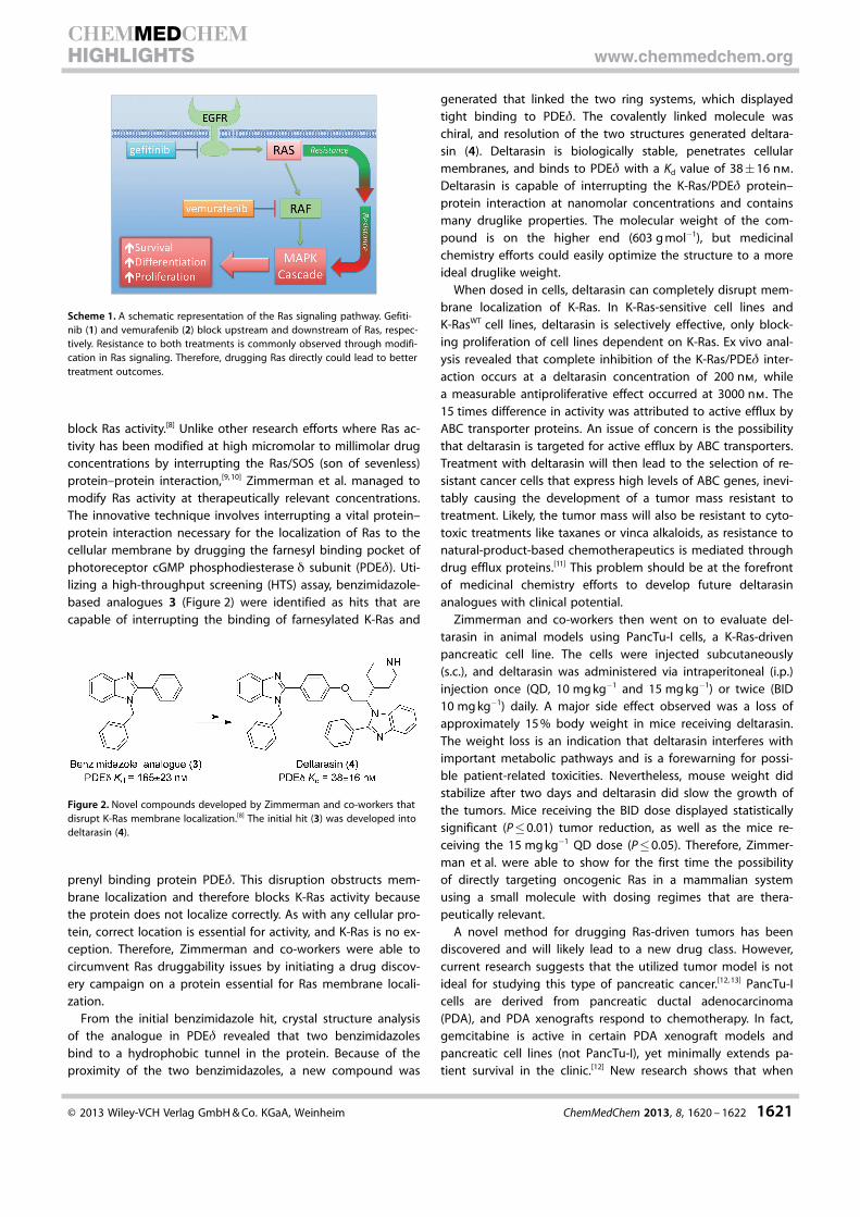
NSCLC subset utilizes epidermal growth factor receptor (EGFR)as a survival pathway, which is upstream of Ras. Therapeutical-ly, EGFR has been drugged with gefitinib (1), and significanttumor remission has been observed (Figure 1). However, effica-
cy is temporary, as NCSLC develops resistance to gefitinib overtime. Interestingly, resistance has been shown to developthrough activated Ras, where Ras is able to signal independentof EGFR stimulation.[6] In another example, vemurafenib (2) hasbeen developed as a Raf (rapidly accelerated fibrosarcoma) in-hibitor to treat late-stage melanoma, specifically inhibitingB-RafV600E. Raf is downstream of Ras, and treatment with vemur-afenib initially blocks progression of the melanoma. However,treatment eventually becomes ineffective as a second Ras iso-form (N-Ras) reactivates the mitogen-activated protein kinase(MAPK) signaling pathway and bypasses vemurafenib inhibi-tion.[7] Therefore, all efforts to date to disrupt the Ras signalingpathway that show clinical promise have failed because of re-sistance conferred through the Ras protein (Scheme 1). Devel-oping a strategy to target Ras directly has the potential tooffer longer disease remission and overall better treatment op-tions for Ras-driven tumors. However, the druggability of Rasat a clinically effective level still remains a major problem.
In an effort to reverse the uncertainty of drugging Ras,a recent breakthrough paper by Zimmerman and co-workershas revived the possibility of identifying a druglike inhibitor to
Figure 1. Upstream (gefitinib) and downstream (vemurafenib) effectors ofRas signaling.
[a] B. Frett, Dr. Y. Wang, Prof. Dr. H.-y. LiDepartment of Pharmacology & ToxicologyCollege of Pharmacy, University of ArizonaTucson, AZ 85721 (USA)E-mail : [email protected]
[b] B. Frett, Dr. Y. Wang, Prof. Dr. H.-y. LiBIO5 Oro Valley (BIO5OV)University of Arizona1580 E. Hanley Blvd, Oro Valley, AZ 85737 (USA)
� 2013 Wiley-VCH Verlag GmbH & Co. KGaA, Weinheim ChemMedChem 2013, 8, 1620 – 1622 1620
CHEMMEDCHEMHIGHLIGHTS
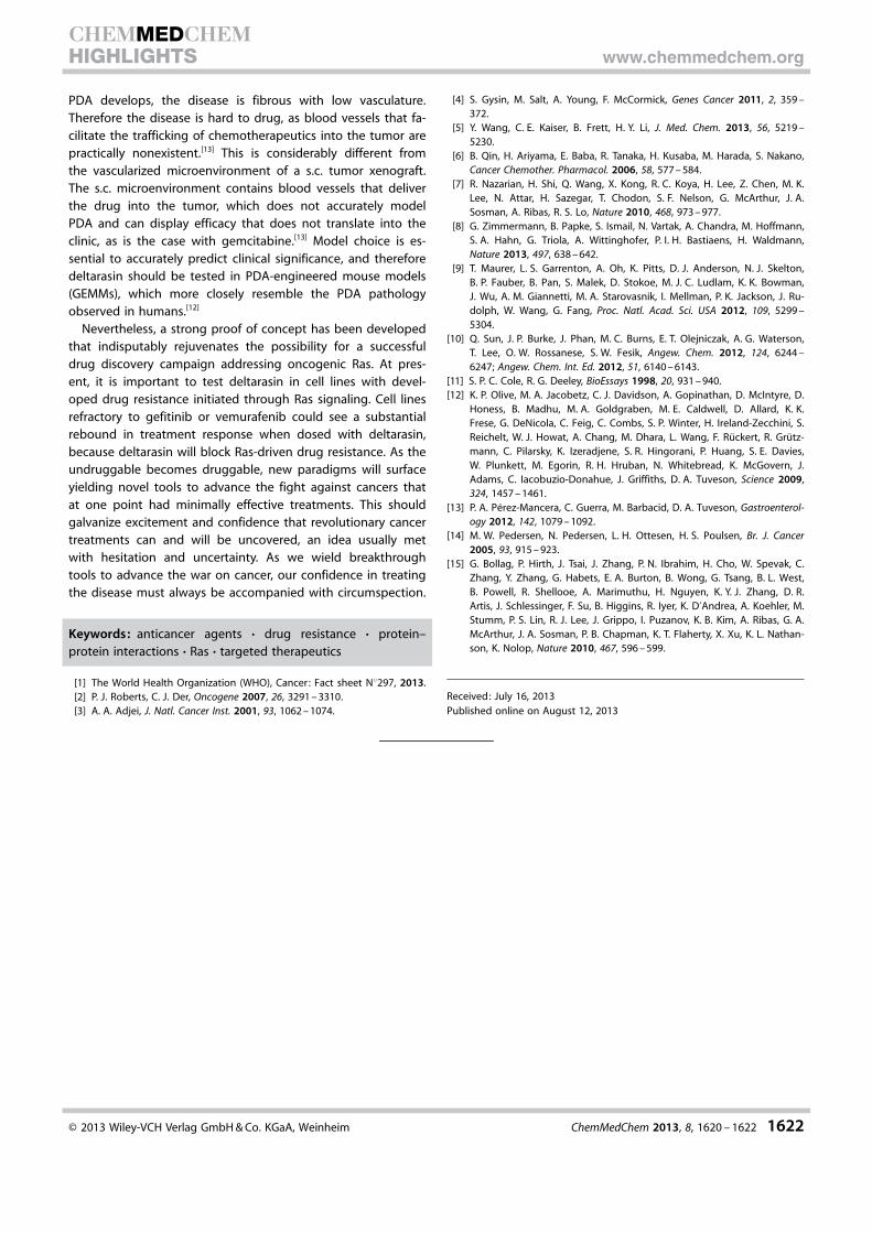
block Ras activity.[8] Unlike other research efforts where Ras ac-tivity has been modified at high micromolar to millimolar drugconcentrations by interrupting the Ras/SOS (son of sevenless)protein–protein interaction,[9, 10] Zimmerman et al. managed tomodify Ras activity at therapeutically relevant concentrations.The innovative technique involves interrupting a vital protein–protein interaction necessary for the localization of Ras to thecellular membrane by drugging the farnesyl binding pocket ofphotoreceptor cGMP phosphodiesterase d subunit (PDEd). Uti-lizing a high-throughput screening (HTS) assay, benzimidazole-based analogues 3 (Figure 2) were identified as hits that arecapable of interrupting the binding of farnesylated K-Ras and
prenyl binding protein PDEd. This disruption obstructs mem-brane localization and therefore blocks K-Ras activity becausethe protein does not localize correctly. As with any cellular pro-tein, correct location is essential for activity, and K-Ras is no ex-ception. Therefore, Zimmerman and co-workers were able tocircumvent Ras druggability issues by initiating a drug discov-ery campaign on a protein essential for Ras membrane locali-zation.
From the initial benzimidazole hit, crystal structure analysisof the analogue in PDEd revealed that two benzimidazolesbind to a hydrophobic tunnel in the protein. Because of theproximity of the two benzimidazoles, a new compound was
generated that linked the two ring systems, which displayedtight binding to PDEd. The covalently linked molecule waschiral, and resolution of the two structures generated deltara-sin (4). Deltarasin is biologically stable, penetrates cellularmembranes, and binds to PDEd with a Kd value of 38�16 nm.Deltarasin is capable of interrupting the K-Ras/PDEd protein–protein interaction at nanomolar concentrations and containsmany druglike properties. The molecular weight of the com-pound is on the higher end (603 g mol�1), but medicinalchemistry efforts could easily optimize the structure to a moreideal druglike weight.
When dosed in cells, deltarasin can completely disrupt mem-brane localization of K-Ras. In K-Ras-sensitive cell lines andK-RasWT cell lines, deltarasin is selectively effective, only block-ing proliferation of cell lines dependent on K-Ras. Ex vivo anal-ysis revealed that complete inhibition of the K-Ras/PDEd inter-action occurs at a deltarasin concentration of 200 nm, whilea measurable antiproliferative effect occurred at 3000 nm. The15 times difference in activity was attributed to active efflux byABC transporter proteins. An issue of concern is the possibilitythat deltarasin is targeted for active efflux by ABC transporters.Treatment with deltarasin will then lead to the selection of re-sistant cancer cells that express high levels of ABC genes, inevi-tably causing the development of a tumor mass resistant totreatment. Likely, the tumor mass will also be resistant to cyto-toxic treatments like taxanes or vinca alkaloids, as resistance tonatural-product-based chemotherapeutics is mediated throughdrug efflux proteins.[11] This problem should be at the forefrontof medicinal chemistry efforts to develop future deltarasinanalogues with clinical potential.
Zimmerman and co-workers then went on to evaluate del-tarasin in animal models using PancTu-I cells, a K-Ras-drivenpancreatic cell line. The cells were injected subcutaneously(s.c.), and deltarasin was administered via intraperitoneal (i.p.)injection once (QD, 10 mg kg�1 and 15 mg kg�1) or twice (BID10 mg kg�1) daily. A major side effect observed was a loss ofapproximately 15 % body weight in mice receiving deltarasin.The weight loss is an indication that deltarasin interferes withimportant metabolic pathways and is a forewarning for possi-ble patient-related toxicities. Nevertheless, mouse weight didstabilize after two days and deltarasin did slow the growth ofthe tumors. Mice receiving the BID dose displayed statisticallysignificant (P�0.01) tumor reduction, as well as the mice re-ceiving the 15 mg kg�1 QD dose (P�0.05). Therefore, Zimmer-man et al. were able to show for the first time the possibilityof directly targeting oncogenic Ras in a mammalian systemusing a small molecule with dosing regimes that are thera-peutically relevant.
A novel method for drugging Ras-driven tumors has beendiscovered and will likely lead to a new drug class. However,current research suggests that the utilized tumor model is notideal for studying this type of pancreatic cancer.[12, 13] PancTu-Icells are derived from pancreatic ductal adenocarcinoma(PDA), and PDA xenografts respond to chemotherapy. In fact,gemcitabine is active in certain PDA xenograft models andpancreatic cell lines (not PancTu-I), yet minimally extends pa-tient survival in the clinic.[12] New research shows that when
Scheme 1. A schematic representation of the Ras signaling pathway. Gefiti-nib (1) and vemurafenib (2) block upstream and downstream of Ras, respec-tively. Resistance to both treatments is commonly observed through modifi-cation in Ras signaling. Therefore, drugging Ras directly could lead to bettertreatment outcomes.
Figure 2. Novel compounds developed by Zimmerman and co-workers thatdisrupt K-Ras membrane localization.[8] The initial hit (3) was developed intodeltarasin (4).
� 2013 Wiley-VCH Verlag GmbH & Co. KGaA, Weinheim ChemMedChem 2013, 8, 1620 – 1622 1621
CHEMMEDCHEMHIGHLIGHTS www.chemmedchem.org
PDA develops, the disease is fibrous with low vasculature.Therefore the disease is hard to drug, as blood vessels that fa-cilitate the trafficking of chemotherapeutics into the tumor arepractically nonexistent.[13] This is considerably different fromthe vascularized microenvironment of a s.c. tumor xenograft.The s.c. microenvironment contains blood vessels that deliverthe drug into the tumor, which does not accurately modelPDA and can display efficacy that does not translate into theclinic, as is the case with gemcitabine.[13] Model choice is es-sential to accurately predict clinical significance, and thereforedeltarasin should be tested in PDA-engineered mouse models(GEMMs), which more closely resemble the PDA pathologyobserved in humans.[12]
Nevertheless, a strong proof of concept has been developedthat indisputably rejuvenates the possibility for a successfuldrug discovery campaign addressing oncogenic Ras. At pres-ent, it is important to test deltarasin in cell lines with devel-oped drug resistance initiated through Ras signaling. Cell linesrefractory to gefitinib or vemurafenib could see a substantialrebound in treatment response when dosed with deltarasin,because deltarasin will block Ras-driven drug resistance. As theundruggable becomes druggable, new paradigms will surfaceyielding novel tools to advance the fight against cancers thatat one point had minimally effective treatments. This shouldgalvanize excitement and confidence that revolutionary cancertreatments can and will be uncovered, an idea usually metwith hesitation and uncertainty. As we wield breakthroughtools to advance the war on cancer, our confidence in treatingthe disease must always be accompanied with circumspection.
Keywords: anticancer agents · drug resistance · protein–protein interactions · Ras · targeted therapeutics
[1] The World Health Organization (WHO), Cancer : Fact sheet N8297, 2013.[2] P. J. Roberts, C. J. Der, Oncogene 2007, 26, 3291 – 3310.[3] A. A. Adjei, J. Natl. Cancer Inst. 2001, 93, 1062 – 1074.
[4] S. Gysin, M. Salt, A. Young, F. McCormick, Genes Cancer 2011, 2, 359 –372.
[5] Y. Wang, C. E. Kaiser, B. Frett, H. Y. Li, J. Med. Chem. 2013, 56, 5219 –5230.
[6] B. Qin, H. Ariyama, E. Baba, R. Tanaka, H. Kusaba, M. Harada, S. Nakano,Cancer Chemother. Pharmacol. 2006, 58, 577 – 584.
[7] R. Nazarian, H. Shi, Q. Wang, X. Kong, R. C. Koya, H. Lee, Z. Chen, M. K.Lee, N. Attar, H. Sazegar, T. Chodon, S. F. Nelson, G. McArthur, J. A.Sosman, A. Ribas, R. S. Lo, Nature 2010, 468, 973 – 977.
[8] G. Zimmermann, B. Papke, S. Ismail, N. Vartak, A. Chandra, M. Hoffmann,S. A. Hahn, G. Triola, A. Wittinghofer, P. I. H. Bastiaens, H. Waldmann,Nature 2013, 497, 638 – 642.
[9] T. Maurer, L. S. Garrenton, A. Oh, K. Pitts, D. J. Anderson, N. J. Skelton,B. P. Fauber, B. Pan, S. Malek, D. Stokoe, M. J. C. Ludlam, K. K. Bowman,J. Wu, A. M. Giannetti, M. A. Starovasnik, I. Mellman, P. K. Jackson, J. Ru-dolph, W. Wang, G. Fang, Proc. Natl. Acad. Sci. USA 2012, 109, 5299 –5304.
[10] Q. Sun, J. P. Burke, J. Phan, M. C. Burns, E. T. Olejniczak, A. G. Waterson,T. Lee, O. W. Rossanese, S. W. Fesik, Angew. Chem. 2012, 124, 6244 –6247; Angew. Chem. Int. Ed. 2012, 51, 6140 – 6143.
[11] S. P. C. Cole, R. G. Deeley, BioEssays 1998, 20, 931 – 940.[12] K. P. Olive, M. A. Jacobetz, C. J. Davidson, A. Gopinathan, D. McIntyre, D.
Honess, B. Madhu, M. A. Goldgraben, M. E. Caldwell, D. Allard, K. K.Frese, G. DeNicola, C. Feig, C. Combs, S. P. Winter, H. Ireland-Zecchini, S.Reichelt, W. J. Howat, A. Chang, M. Dhara, L. Wang, F. R�ckert, R. Gr�tz-mann, C. Pilarsky, K. Izeradjene, S. R. Hingorani, P. Huang, S. E. Davies,W. Plunkett, M. Egorin, R. H. Hruban, N. Whitebread, K. McGovern, J.Adams, C. Iacobuzio-Donahue, J. Griffiths, D. A. Tuveson, Science 2009,324, 1457 – 1461.
[13] P. A. P�rez-Mancera, C. Guerra, M. Barbacid, D. A. Tuveson, Gastroenterol-ogy 2012, 142, 1079 – 1092.
[14] M. W. Pedersen, N. Pedersen, L. H. Ottesen, H. S. Poulsen, Br. J. Cancer2005, 93, 915 – 923.
[15] G. Bollag, P. Hirth, J. Tsai, J. Zhang, P. N. Ibrahim, H. Cho, W. Spevak, C.Zhang, Y. Zhang, G. Habets, E. A. Burton, B. Wong, G. Tsang, B. L. West,B. Powell, R. Shellooe, A. Marimuthu, H. Nguyen, K. Y. J. Zhang, D. R.Artis, J. Schlessinger, F. Su, B. Higgins, R. Iyer, K. D’Andrea, A. Koehler, M.Stumm, P. S. Lin, R. J. Lee, J. Grippo, I. Puzanov, K. B. Kim, A. Ribas, G. A.McArthur, J. A. Sosman, P. B. Chapman, K. T. Flaherty, X. Xu, K. L. Nathan-son, K. Nolop, Nature 2010, 467, 596 – 599.
Received: July 16, 2013
Published online on August 12, 2013
� 2013 Wiley-VCH Verlag GmbH & Co. KGaA, Weinheim ChemMedChem 2013, 8, 1620 – 1622 1622
CHEMMEDCHEMHIGHLIGHTS www.chemmedchem.org