Strain pattern of each ligamentous band of the superficial ...
7
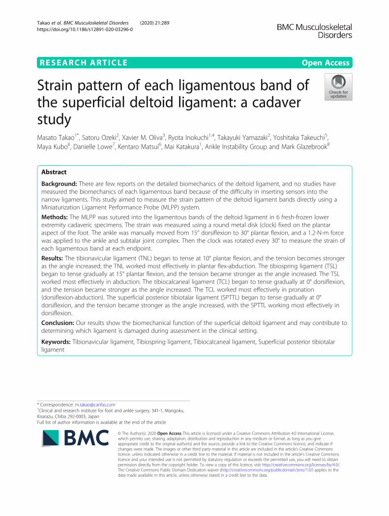
RESEARCH ARTICLE Open Access Strain pattern of each ligamentous band of the superficial deltoid ligament: a cadaver study Masato Takao 1* , Satoru Ozeki 2 , Xavier M. Oliva 3 , Ryota Inokuchi 1,4 , Takayuki Yamazaki 2 , Yoshitaka Takeuchi 5 , Maya Kubo 6 , Danielle Lowe 7 , Kentaro Matsui 6 , Mai Katakura 1 , Ankle Instability Group and Mark Glazebrook 8 Abstract Background: There are few reports on the detailed biomechanics of the deltoid ligament, and no studies have measured the biomechanics of each ligamentous band because of the difficulty in inserting sensors into the narrow ligaments. This study aimed to measure the strain pattern of the deltoid ligament bands directly using a Miniaturization Ligament Performance Probe (MLPP) system. Methods: The MLPP was sutured into the ligamentous bands of the deltoid ligament in 6 fresh-frozen lower extremity cadaveric specimens. The strain was measured using a round metal disk (clock) fixed on the plantar aspect of the foot. The ankle was manually moved from 15° dorsiflexion to 30° plantar flexion, and a 1.2-N-m force was applied to the ankle and subtalar joint complex. Then the clock was rotated every 30° to measure the strain of each ligamentous band at each endpoint. Results: The tibionavicular ligament (TNL) began to tense at 10° plantar flexion, and the tension becomes stronger as the angle increased; the TNL worked most effectively in plantar flex-abduction. The tibiospring ligament (TSL) began to tense gradually at 15° plantar flexion, and the tension became stronger as the angle increased. The TSL worked most effectively in abduction. The tibiocalcaneal ligament (TCL) began to tense gradually at 0° dorsiflexion, and the tension became stronger as the angle increased. The TCL worked most effectively in pronation (dorsiflexion-abduction). The superficial posterior tibiotalar ligament (SPTTL) began to tense gradually at 0° dorsiflexion, and the tension became stronger as the angle increased, with the SPTTL working most effectively in dorsiflexion. Conclusion: Our results show the biomechanical function of the superficial deltoid ligament and may contribute to determining which ligament is damaged during assessment in the clinical setting. Keywords: Tibionavicular ligament, Tibiospring ligament, Tibiocalcaneal ligament, Superficial posterior tibiotalar ligament © The Author(s). 2020 Open Access This article is licensed under a Creative Commons Attribution 4.0 International License, which permits use, sharing, adaptation, distribution and reproduction in any medium or format, as long as you give appropriate credit to the original author(s) and the source, provide a link to the Creative Commons licence, and indicate if changes were made. The images or other third party material in this article are included in the article's Creative Commons licence, unless indicated otherwise in a credit line to the material. If material is not included in the article's Creative Commons licence and your intended use is not permitted by statutory regulation or exceeds the permitted use, you will need to obtain permission directly from the copyright holder. To view a copy of this licence, visit http://creativecommons.org/licenses/by/4.0/. The Creative Commons Public Domain Dedication waiver (http://creativecommons.org/publicdomain/zero/1.0/) applies to the data made available in this article, unless otherwise stated in a credit line to the data. * Correspondence: [email protected] 1 Clinical and research institute for foot and ankle surgery, 341-1, Mangoku, Kisarazu, Chiba 292-0003, Japan Full list of author information is available at the end of the article Takao et al. BMC Musculoskeletal Disorders (2020) 21:289 https://doi.org/10.1186/s12891-020-03296-0
Transcript of Strain pattern of each ligamentous band of the superficial ...
RESEARCH ARTICLE Open Access
Strain pattern of each ligamentous band ofthe superficial deltoid ligament: a cadaverstudyMasato Takao1*, Satoru Ozeki2, Xavier M. Oliva3, Ryota Inokuchi1,4, Takayuki Yamazaki2, Yoshitaka Takeuchi5,Maya Kubo6, Danielle Lowe7, Kentaro Matsui6, Mai Katakura1, Ankle Instability Group and Mark Glazebrook8
Abstract
Background: There are few reports on the detailed biomechanics of the deltoid ligament, and no studies havemeasured the biomechanics of each ligamentous band because of the difficulty in inserting sensors into thenarrow ligaments. This study aimed to measure the strain pattern of the deltoid ligament bands directly using aMiniaturization Ligament Performance Probe (MLPP) system.
Methods: The MLPP was sutured into the ligamentous bands of the deltoid ligament in 6 fresh-frozen lowerextremity cadaveric specimens. The strain was measured using a round metal disk (clock) fixed on the plantaraspect of the foot. The ankle was manually moved from 15° dorsiflexion to 30° plantar flexion, and a 1.2-N-m forcewas applied to the ankle and subtalar joint complex. Then the clock was rotated every 30° to measure the strain ofeach ligamentous band at each endpoint.
Results: The tibionavicular ligament (TNL) began to tense at 10° plantar flexion, and the tension becomes strongeras the angle increased; the TNL worked most effectively in plantar flex-abduction. The tibiospring ligament (TSL)began to tense gradually at 15° plantar flexion, and the tension became stronger as the angle increased. The TSLworked most effectively in abduction. The tibiocalcaneal ligament (TCL) began to tense gradually at 0° dorsiflexion,and the tension became stronger as the angle increased. The TCL worked most effectively in pronation(dorsiflexion-abduction). The superficial posterior tibiotalar ligament (SPTTL) began to tense gradually at 0°dorsiflexion, and the tension became stronger as the angle increased, with the SPTTL working most effectively indorsiflexion.
Conclusion: Our results show the biomechanical function of the superficial deltoid ligament and may contribute todetermining which ligament is damaged during assessment in the clinical setting.
Keywords: Tibionavicular ligament, Tibiospring ligament, Tibiocalcaneal ligament, Superficial posterior tibiotalarligament
© The Author(s). 2020 Open Access This article is licensed under a Creative Commons Attribution 4.0 International License,which permits use, sharing, adaptation, distribution and reproduction in any medium or format, as long as you giveappropriate credit to the original author(s) and the source, provide a link to the Creative Commons licence, and indicate ifchanges were made. The images or other third party material in this article are included in the article's Creative Commonslicence, unless indicated otherwise in a credit line to the material. If material is not included in the article's Creative Commonslicence and your intended use is not permitted by statutory regulation or exceeds the permitted use, you will need to obtainpermission directly from the copyright holder. To view a copy of this licence, visit http://creativecommons.org/licenses/by/4.0/.The Creative Commons Public Domain Dedication waiver (http://creativecommons.org/publicdomain/zero/1.0/) applies to thedata made available in this article, unless otherwise stated in a credit line to the data.
* Correspondence: [email protected] and research institute for foot and ankle surgery, 341-1, Mangoku,Kisarazu, Chiba 292-0003, JapanFull list of author information is available at the end of the article
Takao et al. BMC Musculoskeletal Disorders (2020) 21:289 https://doi.org/10.1186/s12891-020-03296-0
BackgroundThe deltoid ligament has both superficial and deep layersconsisting of up to six ligamentous bands [1]. The superfi-cial layer of the deltoid ligament is composed of four liga-mentous bands, including the tibionavicular (TNL),tibiospring (TSL), tibiocalcaneal (TCL), and superficialposterior tibiotalar (SPTTL) ligaments. Two ligamentousbands comprise the deep layer of the deltoid ligament: thedeep anterior and posterior tibiotalar ligaments. Generally,the deltoid ligament is known to work cooperatively andis primarily responsible for 1) stabilizing the medial side ofthe ankle to limit anterior, posterior, and lateral transla-tion of the talus and 2) restraining talar abduction at thetalocrural joint [2, 3]. Specifically, the superficial deltoidresists eversion of the hindfoot, and the deep deltoid is theprimary restraint to external rotation of the talus [4–7].There are few reports in terms of detailed biomechan-
ics of the deltoid ligament [8–12], and no studies havedirectly measured the biomechanics of each ligamentousband because of the difficulty in inserting sensors intothe narrow ligaments.Biomechanical data regarding each ligamentous band
would contribute to precisely assessing which ligament isdamaged in the clinical setting, leading to the design andperformance of repair and reconstruction procedures beforesurgery. Thus, we used a Miniaturization Ligament Perform-ance Probe (MLPP) system that can be inserted into smallligaments and allows for the precise measurement of thestrain patterns of the deltoid ligament during ankle motion.
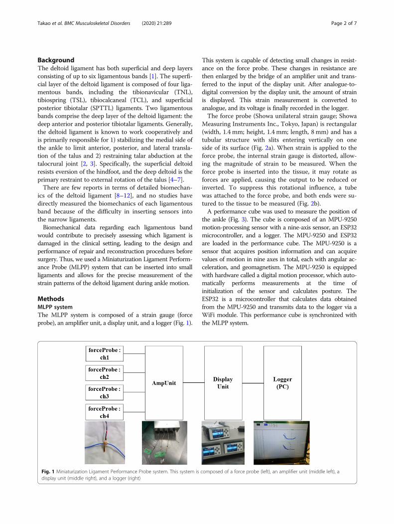
MethodsMLPP systemThe MLPP system is composed of a strain gauge (forceprobe), an amplifier unit, a display unit, and a logger (Fig. 1).
This system is capable of detecting small changes in resist-ance on the force probe. These changes in resistance arethen enlarged by the bridge of an amplifier unit and trans-ferred to the input of the display unit. After analogue-to-digital conversion by the display unit, the amount of strainis displayed. This strain measurement is converted toanalogue, and its voltage is finally recorded in the logger.The force probe (Showa unilateral strain gauge; Showa
Measuring Instruments Inc., Tokyo, Japan) is rectangular(width, 1.4 mm; height, 1.4 mm; length, 8 mm) and has atubular structure with slits entering vertically on oneside of its surface (Fig. 2a). When strain is applied to theforce probe, the internal strain gauge is distorted, allow-ing the magnitude of strain to be measured. When theforce probe is inserted into the tissue, it may rotate asforces are applied, causing the output to be reduced orinverted. To suppress this rotational influence, a tubewas attached to the force probe, and both ends were su-tured to the tissue to be measured (Fig. 2b).A performance cube was used to measure the position of
the ankle (Fig. 3). The cube is composed of an MPU-9250motion-processing sensor with a nine-axis sensor, an ESP32microcontroller, and a logger. The MPU-9250 and ESP32are loaded in the performance cube. The MPU-9250 is asensor that acquires position information and can acquirevalues of motion in nine axes in total, each with angular ac-celeration, and geomagnetism. The MPU-9250 is equippedwith hardware called a digital motion processor, which auto-matically performs measurements at the time ofinitialization of the sensor and calculates posture. TheESP32 is a microcontroller that calculates data obtainedfrom the MPU-9250 and transmits data to the logger via aWiFi module. This performance cube is synchronized withthe MLPP system.
Fig. 1 Miniaturization Ligament Performance Probe system. This system is composed of a force probe (left), an amplifier unit (middle left), adisplay unit (middle right), and a logger (right)
Takao et al. BMC Musculoskeletal Disorders (2020) 21:289 Page 2 of 7
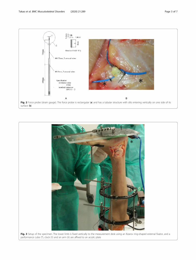
Fig. 3 Setup of the specimen. The lower limb is fixed vertically to the measurement desk using an Ilizarov ring-shaped external fixator, and aperformance cube (*), clock (†) and an arm (‡) are affixed to an acrylic plate
Fig. 2 Force probe (strain gauge). The force probe is rectangular (a) and has a tubular structure with slits entering vertically on one side of itssurface (b)
Takao et al. BMC Musculoskeletal Disorders (2020) 21:289 Page 3 of 7
Cadaveric tests using the MLPP systemSix fresh-frozen through-the-knee lower extremity ca-daveric specimens were used for this study (three rightand three left). Three specimens were from men, andthree were from women. The median age was 64 years(range 46–82 years). These specimens were free of ankleor hind foot deformities, did not undergo surgery or dis-section, and did not have any history of trauma or otherpathology that may alter the anatomy.All cadaveric studies were performed at the University
of Barcelona in Catalonia, Spain. All methods in thisstudy were reviewed and approved by the InstitutionalReview Board of the University of Barcelona. Consentfor the storage and use of the bodies for research pur-poses was given by all body donors before death or bytheir next of kin.
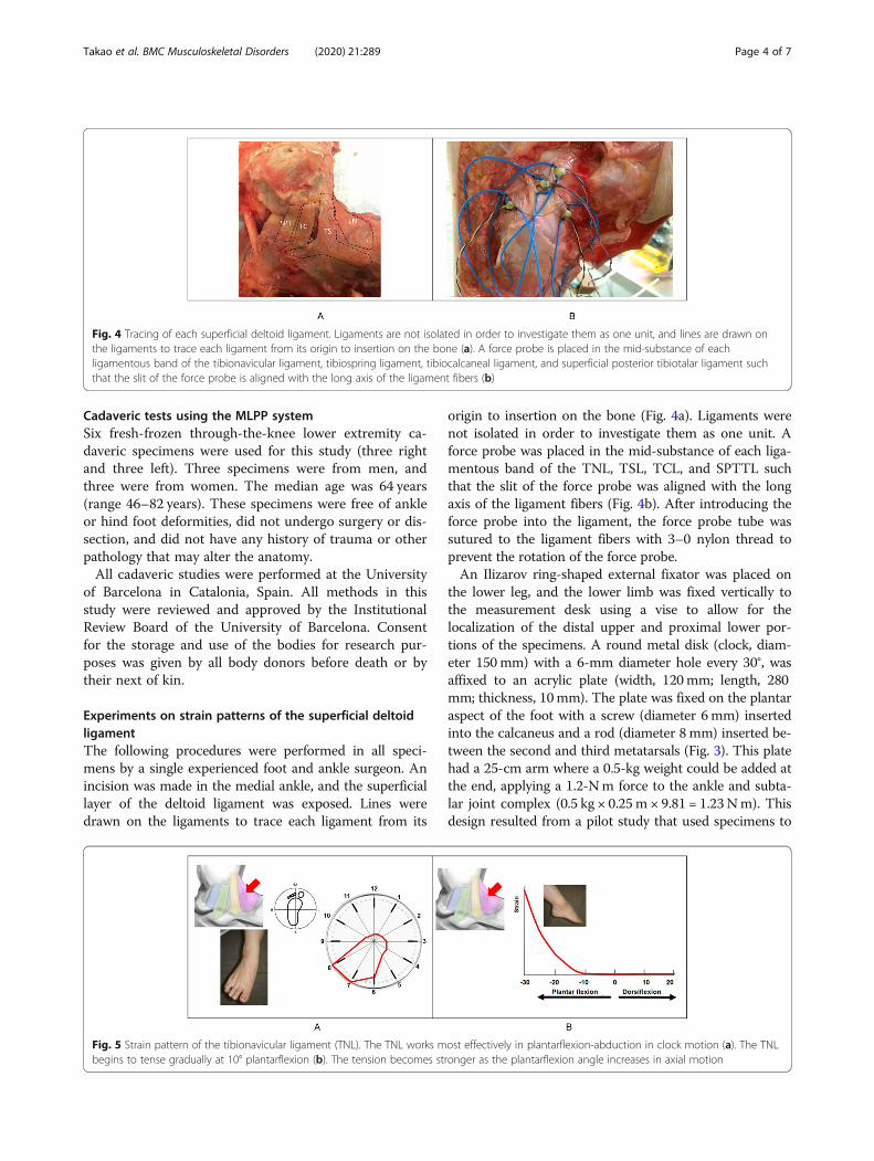
Experiments on strain patterns of the superficial deltoidligamentThe following procedures were performed in all speci-mens by a single experienced foot and ankle surgeon. Anincision was made in the medial ankle, and the superficiallayer of the deltoid ligament was exposed. Lines weredrawn on the ligaments to trace each ligament from its
origin to insertion on the bone (Fig. 4a). Ligaments werenot isolated in order to investigate them as one unit. Aforce probe was placed in the mid-substance of each liga-mentous band of the TNL, TSL, TCL, and SPTTL suchthat the slit of the force probe was aligned with the longaxis of the ligament fibers (Fig. 4b). After introducing theforce probe into the ligament, the force probe tube wassutured to the ligament fibers with 3–0 nylon thread toprevent the rotation of the force probe.An Ilizarov ring-shaped external fixator was placed on
the lower leg, and the lower limb was fixed vertically tothe measurement desk using a vise to allow for thelocalization of the distal upper and proximal lower por-tions of the specimens. A round metal disk (clock, diam-eter 150mm) with a 6-mm diameter hole every 30°, wasaffixed to an acrylic plate (width, 120mm; length, 280mm; thickness, 10mm). The plate was fixed on the plantaraspect of the foot with a screw (diameter 6mm) insertedinto the calcaneus and a rod (diameter 8mm) inserted be-tween the second and third metatarsals (Fig. 3). This platehad a 25-cm arm where a 0.5-kg weight could be added atthe end, applying a 1.2-Nm force to the ankle and subta-lar joint complex (0.5 kg × 0.25m × 9.81 = 1.23Nm). Thisdesign resulted from a pilot study that used specimens to
Fig. 4 Tracing of each superficial deltoid ligament. Ligaments are not isolated in order to investigate them as one unit, and lines are drawn onthe ligaments to trace each ligament from its origin to insertion on the bone (a). A force probe is placed in the mid-substance of eachligamentous band of the tibionavicular ligament, tibiospring ligament, tibiocalcaneal ligament, and superficial posterior tibiotalar ligament suchthat the slit of the force probe is aligned with the long axis of the ligament fibers (b)
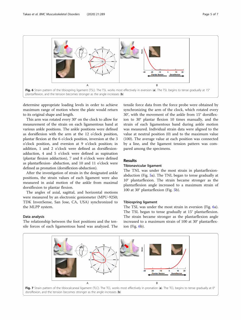
Fig. 5 Strain pattern of the tibionavicular ligament (TNL). The TNL works most effectively in plantarflexion-abduction in clock motion (a). The TNLbegins to tense gradually at 10° plantarflexion (b). The tension becomes stronger as the plantarflexion angle increases in axial motion
Takao et al. BMC Musculoskeletal Disorders (2020) 21:289 Page 4 of 7
determine appropriate loading levels in order to achievemaximum range of motion where the plate would returnto its original shape and length.This arm was rotated every 30° on the clock to allow for
measurement of the strain on each ligamentous band atvarious ankle positions. The ankle positions were definedas dorsiflexion with the arm at the 12 o’clock position,plantar flexion at the 6 o’clock position, inversion at the 3o’clock position, and eversion at 9 o’clock position; inaddition, 1 and 2 o’clock were defined as dorsiflexion-adduction, 4 and 5 o’clock were defined as supination(plantar flexion adduction), 7 and 8 o’clock were definedas plantarflexion- abduction, and 10 and 11 o’clock weredefined as pronation (dorsiflexion-abduction).After the investigation of strain in the designated ankle
positions, the strain values of each ligament were alsomeasured in axial motion of the ankle from maximaldorsiflexion to plantar flexion.The angles of axial, sagittal, and horizontal motions
were measured by an electronic goniometer (MPU-9250;TDK InvenSense, San Jose, CA, USA) synchronized tothe MLPP system.
Data analysisThe relationship between the foot positions and the ten-sile forces of each ligamentous band was analyzed. The
tensile force data from the force probe were obtained bysynchronizing the arm of the clock, which rotated every30°, with the movement of the ankle from 15° dorsiflex-ion to 30° plantar flexion 10 times manually, and thestrain of each ligamentous band during ankle motionwas measured. Individual strain data were aligned to thevalue at neutral position (0) and to the maximum value(100). The average value at each position was connectedby a line, and the ligament tension pattern was com-pared among the specimens.
ResultsTibionavicular ligamentThe TNL was under the most strain in plantarflexion-abduction (Fig. 5a). The TNL began to tense gradually at10° plantarflexion. The strain became stronger as theplantarflexion angle increased to a maximum strain of100 at 30° plantarflexion (Fig. 5b).
Tibiospring ligamentThe TSL was under the most strain in eversion (Fig. 6a).The TSL began to tense gradually at 15° plantarflexion.The strain became stronger as the plantarflexion angleincreased to a maximum strain of 100 at 30° plantarflex-ion (Fig. 6b).
Fig. 6 Strain pattern of the tibiospring ligament (TSL). The TSL works most effectively in eversion (a). The TSL begins to tense gradually at 15°plantarflexion, and the tension becomes stronger as the angle increases (b)
Fig. 7 Strain pattern of the tibiocalcaneal ligament (TLC). The TCL works most effectively in pronation (a). The TCL begins to tense gradually at 0°dorsiflexion, and the tension becomes stronger as the angle increases (b)
Takao et al. BMC Musculoskeletal Disorders (2020) 21:289 Page 5 of 7
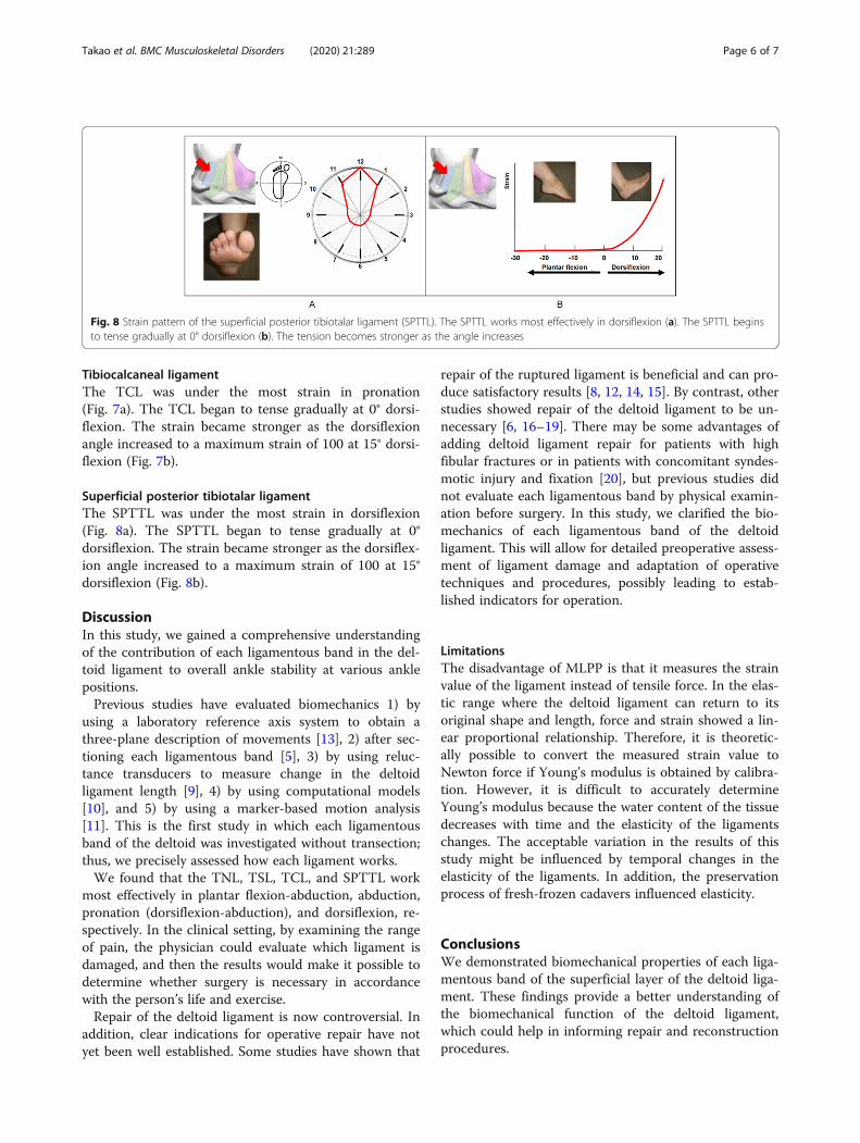
Tibiocalcaneal ligamentThe TCL was under the most strain in pronation(Fig. 7a). The TCL began to tense gradually at 0° dorsi-flexion. The strain became stronger as the dorsiflexionangle increased to a maximum strain of 100 at 15° dorsi-flexion (Fig. 7b).
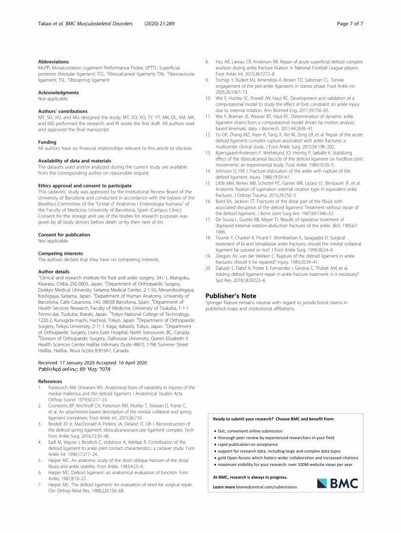
Superficial posterior tibiotalar ligamentThe SPTTL was under the most strain in dorsiflexion(Fig. 8a). The SPTTL began to tense gradually at 0°dorsiflexion. The strain became stronger as the dorsiflex-ion angle increased to a maximum strain of 100 at 15°dorsiflexion (Fig. 8b).
DiscussionIn this study, we gained a comprehensive understandingof the contribution of each ligamentous band in the del-toid ligament to overall ankle stability at various anklepositions.Previous studies have evaluated biomechanics 1) by
using a laboratory reference axis system to obtain athree-plane description of movements [13], 2) after sec-tioning each ligamentous band [5], 3) by using reluc-tance transducers to measure change in the deltoidligament length [9], 4) by using computational models[10], and 5) by using a marker-based motion analysis[11]. This is the first study in which each ligamentousband of the deltoid was investigated without transection;thus, we precisely assessed how each ligament works.We found that the TNL, TSL, TCL, and SPTTL work
most effectively in plantar flexion-abduction, abduction,pronation (dorsiflexion-abduction), and dorsiflexion, re-spectively. In the clinical setting, by examining the rangeof pain, the physician could evaluate which ligament isdamaged, and then the results would make it possible todetermine whether surgery is necessary in accordancewith the person’s life and exercise.Repair of the deltoid ligament is now controversial. In
addition, clear indications for operative repair have notyet been well established. Some studies have shown that
repair of the ruptured ligament is beneficial and can pro-duce satisfactory results [8, 12, 14, 15]. By contrast, otherstudies showed repair of the deltoid ligament to be un-necessary [6, 16–19]. There may be some advantages ofadding deltoid ligament repair for patients with highfibular fractures or in patients with concomitant syndes-motic injury and fixation [20], but previous studies didnot evaluate each ligamentous band by physical examin-ation before surgery. In this study, we clarified the bio-mechanics of each ligamentous band of the deltoidligament. This will allow for detailed preoperative assess-ment of ligament damage and adaptation of operativetechniques and procedures, possibly leading to estab-lished indicators for operation.
LimitationsThe disadvantage of MLPP is that it measures the strainvalue of the ligament instead of tensile force. In the elas-tic range where the deltoid ligament can return to itsoriginal shape and length, force and strain showed a lin-ear proportional relationship. Therefore, it is theoretic-ally possible to convert the measured strain value toNewton force if Young’s modulus is obtained by calibra-tion. However, it is difficult to accurately determineYoung’s modulus because the water content of the tissuedecreases with time and the elasticity of the ligamentschanges. The acceptable variation in the results of thisstudy might be influenced by temporal changes in theelasticity of the ligaments. In addition, the preservationprocess of fresh-frozen cadavers influenced elasticity.
ConclusionsWe demonstrated biomechanical properties of each liga-mentous band of the superficial layer of the deltoid liga-ment. These findings provide a better understanding ofthe biomechanical function of the deltoid ligament,which could help in informing repair and reconstructionprocedures.
Fig. 8 Strain pattern of the superficial posterior tibiotalar ligament (SPTTL). The SPTTL works most effectively in dorsiflexion (a). The SPTTL beginsto tense gradually at 0° dorsiflexion (b). The tension becomes stronger as the angle increases
Takao et al. BMC Musculoskeletal Disorders (2020) 21:289 Page 6 of 7
AbbreviationsMLPP: Miniaturization Ligament Performance Probe; SPTTL: Superficialposterior tibiotalar ligament; TCL: Tibiocalcaneal ligament; TNL: Tibionavicularligament; TSL: Tibiospring ligament
AcknowledgmentsNot applicable.
Authors’ contributionsMT, SO, XO, and MG designed the study; MT, SO, XO, TY, YT, MK, DL, KM, MK,and MG performed the research; and RI wrote the first draft. All authors readand approved the final manuscript.
FundingAll authors have no financial relationships relevant to this article to disclose.
Availability of data and materialsThe datasets used and/or analyzed during the current study are availablefrom the corresponding author on reasonable request.
Ethics approval and consent to participateThis cadaveric study was approved by the Institutional Review Board of theUniversity of Barcelona and conducted in accordance with the bylaws of theBioethics Committee of the “Unitat d’ Anatomia i Embriologia humana” ofthe Faculty of Medicine, University of Barcelona, Spain (Campus Clinic).Consent for the storage and use of the bodies for research purposes wasgiven by all body donors before death or by their next of kin.
Consent for publicationNot applicable.
Competing interestsThe authors declare that they have no competing interests.
Author details1Clinical and research institute for foot and ankle surgery, 341-1, Mangoku,Kisarazu, Chiba 292-0003, Japan. 2Department of Orthopaedic Surgery,Dokkyo Medical University Saitama Medical Center, 2-1-50, Minamikoshigaya,Koshigaya, Saitama, Japan. 3Department of Human Anatomy, University ofBarcelona, Calle Casanova, 143, 08038 Barcelona, Spain. 4Department ofHealth Services Research, Faculty of Medicine, University of Tsukuba, 1-1-1Tenno-dai, Tsukuba, Ibaraki, Japan. 5Tokyo National College of Technology,1220-2, Kunugida-machi, Hachioji, Tokyo, Japan. 6Department of OrthopaedicSurgery, Teikyo University, 2-11-1 Kaga, Itabashi, Tokyo, Japan. 7Departmentof Orthopaedic Surgery, Lions Gate Hospital, North Vancouver, BC, Canada.8Division of Orthopaedic Surgery, Dalhousie University, Queen Elizabeth IIHealth Sciences Center Halifax Infirmary (Suite 4867), 1796 Summer StreetHalifax, Halifax, Nova Scotia B3H3A7, Canada.
Received: 17 January 2020 Accepted: 16 April 2020
References1. Pankovich AM, Shivaram MS. Anatomical basis of variability in injuries of the
medial malleolus and the deltoid ligament. I Anatomical Studies ActaOrthop Scand. 1979;50:217–23.
2. Cromeens BP, Kirchhoff CA, Patterson RM, Motley T, Stewart D, Fisher C,et al. An attachment-based description of the medial collateral and springligament complexes. Foot Ankle Int. 2015;36:710.
3. Brodell JD Jr, MacDonald A, Perkins JA, Deland JT, Oh I. Reconstruction ofthe deltoid-spring ligament: tibiocalcaneonavicular ligament complex. TechFoot Ankle Surg. 2016;15:39–46.
4. Earll M, Wayne J, Brodrick C, Vokshoor A, Adelaar R. Contribution of thedeltoid ligament to ankle joint contact characteristics: a cadaver study. FootAnkle Int. 1996;17:317–24.
5. Harper MC. An anatomic study of the short oblique fracture of the distalfibula and ankle stability. Foot Ankle. 1983;4:23–9.
6. Harper MC. Deltoid ligament: an anatomical evaluation of function. FootAnkle. 1987;8:19–22.
7. Harper MC. The deltoid ligament. An evaluation of need for surgical repair.Clin Orthop Relat Res. 1988;226:156–68.
8. Hsu AR, Lareau CR, Anderson RB. Repair of acute superficial deltoid complexavulsion during ankle fracture fixation in National Football League players.Foot Ankle Int. 2015;36:1272–8.
9. Tochigi Y, Rudert MJ, Amendola A, Brown TD, Saltzman CL. Tensileengagement of the peri-ankle ligaments in stance phase. Foot Ankle Int.2005;26:1067–73.
10. Wei F, Hunley SC, Powell JW, Haut RC. Development and validation of acomputational model to study the effect of foot constraint on ankle injurydue to external rotation. Ann Biomed Eng. 2011;39:756–65.
11. Wei F, Braman JE, Weaver BT, Haut RC. Determination of dynamic ankleligament strains from a computational model driven by motion analysisbased kinematic data. J Biomech. 2011;44:2636–41.
12. Yu GR, Zhang MZ, Aiyer A, Tang X, Xie M, Zeng LR, et al. Repair of the acutedeltoid ligament complex rupture associated with ankle fractures: amulticenter clinical study. J Foot Ankle Surg. 2015;54:198–202.
13. Kjaersgaard-Andersen P, Wethelund JO, Helmig P, Søballe K. Stabilizingeffect of the tibiocalcaneal fascicle of the deltoid ligament on hindfoot jointmovements: an experimental study. Foot Ankle. 1989;10:30–5.
14. Johnson D, Hill J. Fracture-dislocation of the ankle with rupture of thedeltoid ligament. Injury. 1988;19:59–61.
15. Little MM, Berkes MB, Schottel PC, Garner MR, Lazaro LE, Birnbaum JF, et al.Anatomic fixation of supination external rotation type IV equivalent anklefractures. J Orthop Trauma. 2015;29:250–5.
16. Baird RA, Jackson ST. Fractures of the distal part of the fibula withassociated disruption of the deltoid ligament. Treatment without repair ofthe deltoid ligament. J Bone Joint Surg Am. 1987;69:1346–52.
17. De Souza L, Gustilo RB, Meyer TJ. Results of operative treatment ofdisplaced external rotation-abduction fractures of the ankle. JBJS. 1985;67:1066.
18. Tourne Y, Charbel A, Picard F, Montbarbon E, Saragaglia D. Surgicaltreatment of bi-and trimalleolar ankle fractures: should the medial collateralligament be sutured or not? J Foot Ankle Surg. 1999;38:24–9.
19. Zeegers AV, van der Werken C. Rupture of the deltoid ligament in anklefractures: should it be repaired? Injury. 1989;20:39–41.
20. Dabash S, Elabd A, Potter E, Fernandez I, Gerzina C, Thabet AM, et al.Adding deltoid ligament repair in ankle fracture treatment: is it necessary?Syst Rev. 2018;18:30223–6.
Publisher’s NoteSpringer Nature remains neutral with regard to jurisdictional claims inpublished maps and institutional affiliations.
Takao et al. BMC Musculoskeletal Disorders (2020) 21:289 Page 7 of 7


![Craftingeek* pattern [ Corazón básico ] · Craftingeek*pattern [ Corazón básico ] Craftingeek*pattern [ Corazón básico ] Craftingeek*pattern [ Corazón básico ]](https://static.fdocument.pub/doc/165x107/5f04cca07e708231d40fc3ea/craftingeek-pattern-corazn-bsico-craftingeekpattern-corazn-bsico.jpg)