Histone deacetylase inhibitors induce thyroid cancer-specific ...

4 Am J Psychiatry 157:1, January 2000
Special Articles
Cholinesterase Inhibitors:A New Class of Psychotropic Compounds
Jeffrey L. Cummings, M.D.
Objective: This article reviews evidence indicating that acetylcholinesterase inhibitorshave psychotropic properties. Method: The author reviewed the English-language litera-ture pertinent to the response of neuropsychiatric symptoms in Alzheimer’s disease and re-lated conditions to cholinergic agents. Results: The cholinergic system originates in thebasal forebrain and projects diffusely to the cerebral cortex; the limbic and paralimbic re-gions receive the most abundant cholinergic projections. The basal forebrain nuclei are po-sitioned at the interface of the limbic system and cerebral cortex, where they play a role inmediating emotional responses. The basal forebrain nuclei are atrophic in Alzheimer’s dis-ease, leading to a widespread cholinergic deficit. The cholinergic disturbance may contrib-ute to neuropsychiatric manifestations of the disease. The treatment of patients with Alz-heimer’s disease with acetylcholinesterase inhibitors reduces neuropsychiatric symptoms,particularly apathy and visual hallucinations. In some studies, a variety of other neuropsy-chiatric symptoms have been reported to respond to treatment with acetylcholinesteraseinhibitors. Response profiles vary among acetylcholinesterase inhibitors. Conclusions:Acetylcholinesterase inhibitors have psychotropic effects and may play an important role incontrolling neuropsychiatric and behavioral disturbances in patients with Alzheimer’s dis-ease. These agents also may contribute to the management of other disorders with cholin-ergic system abnormalities and neuropsychiatric symptoms. The beneficial response ismost likely mediated through limbic cholinergic structures.
(Am J Psychiatry 2000; 157:4–15)
Acetylcholinesterase inhibitors have recently beenintroduced as cognition-enhancing agents in the treat-ment of patients with mild to moderate Alzheimer’sdisease. Tacrine (1–3) and donepezil (4–6) have beenapproved by the U.S. Food and Drug Administration(FDA); metrifonate (7, 8), and rivastigmine (9–11) areunder review as possible marketable compounds, andlong-acting physostigmine (12), eptastigmine (13), hu-perzine (14), galantamine (15), and velnacrine (16)have been investigated as acetylcholinesterase inhibi-tors with potential therapeutic benefit in Alzheimer’sdisease. These agents exert their beneficial effect onintellectual functioning by blocking acetylcholinest-erase and enhancing cholinergic function. They havebeen developed to improve the neuropsychologicaldeficits of Alzheimer’s disease, as shown in most clini-cal trials by improved scores or superior performancecompared to patients receiving placebo on the cogni-
Received Jan. 19, 1999; revision received July 16, 1999;accepted July 21, 1999. From the Department of Neurology andDepartment of Psychiatry and Biobehavioral Sciences, UCLASchool of Medicine. Address reprint requests to Dr. Cummings,UCLA Department of Neurology, 710 Westwood Plaza, Los Ange-les, CA 90095-1769; [email protected] (e-mail).
Supported by Alzheimer’s Disease Center grant AG-16570 andAlzheimer’s Disease Cooperative study grant AG-10483 from theNational Institute on Aging, an Alzheimer’s Disease ResearchCenter of California grant, the Sidell-Kagan Foundation, GeneralClinical Research Center grant RR-00865 to the University of Cali-fornia at Los Angeles from the NIH National Center for ResearchResources, and Alzheimer’s Disease Cooperative Study grant AG-10483 from the National Institute on Aging. Images provided bythe UCLA Laboratory of Neuroimaging, supported by grant RR-13642 from the NIH National Center for Research Resources.
Dr. Cummings has served as a consultant or performed researchin conjunction with Novartis, Parke-Davis, Pfizer, Inc./Eisai, BayerCorporation, Eli Lilly and Company, Janssen Pharmaceutica, Inc.,and SmithKline Beecham Consumer Healthcare regarding agentsrelevant to this article.

Am J Psychiatry 157:1, January 2000 5
JEFFREY L. CUMMINGS
tive subscale of the Alzheimer’s Disease AssessmentScale (17), the Mini-Mental State examination (18),and a global scale such as the Clinician Interview-Based Impression (19).
Evidence has accrued that acetylcholinesterase inhib-itors ameliorate behavioral disturbances as well as en-hance cognition. These observations have implicationsfor understanding the pathophysiological basis of neu-ropsychiatric symptoms in Alzheimer’s disease (20, 21)and for developing therapeutic options for cliniciansinvolved in managing behavioral alterations in patientswith the disease. This review presents the availabledata concerning the psychotropic effects of acetylcho-linesterase inhibitors and describes the neurobiologicalbasis for these neuropsychiatric alterations.
CENTRAL CHOLINERGIC SYSTEMS
Anatomy of the Cholinergic System
Acetylcholinesterase, the substrate of acetylcho-linesterase inhibitors, is located in the synaptic space(soluble form) and in the synaptic membranes (mem-brane-bound form) of the neurons of the cholinergicsystem (22, 23). Thus, the anatomy of the cholinergicsystem determines the pharmacoanatomy of the re-sponse to acetylcholinesterase inhibitors.
Mesulam (24) identified eight cholinergic cellgroups that form the origins of projections to othercentral nervous system (CNS) structures. The medialseptal nucleus and the vertical nucleus of the diagonalband constitute the major cholinergic projections tothe hippocampal formation, cingulate cortex, olfac-tory bulb, and hypothalamus. The horizontal limb ofthe diagonal band nucleus projects to the olfactorybulb, whereas the nucleus basalis of Meynert providesthe major innervation of the cerebral cortex andamygdala (figure 1). The pedunculopontine nucleusand the laterodorsal tegmental nucleus of the brain-stem project to the thalamus; the medial habenula in-nervates the interpeduncular nucleus; and the para-bigeminal nucleus projects to the superior colliculus.Cholinergic neurons manufacture choline acetyltrans-ferase, which is transported to projection targets,where it catalyzes the synthesis of acetylcholine. All ofthe cholinergic innervation of the human cerebral cor-tex and thalamus arises from these extrinsic cholin-ergic sources.
All layers of the cerebral cortex receive cholinergicinnervation; the density of cholinergic projections ishighest in layers 1 and 2 and the upper regions of layer3. Muscarinic cortical input disinhibits the cortical py-ramidal cells that enhance the intralaminar transfer ofinformation between cortical columns; in contrast,nicotinic input augments inhibition (25). There is re-gional variability in the cholinergic innervation of thehuman cortex (26): limbic areas that include theamygdala and hippocampus have the highest densityof cholinergic axons; paralimbic regions have the next
highest density of cholinergic fibers; the unimodal andheteromodal association cortices have intermediatedensities of cholinergic innervation; and the primaryvisual cortex has the least abundant cortical cholin-ergic projections.
The neurons of the nucleus basalis and basal fore-brain complex do not receive reciprocal projectionsfrom many of the cortical regions that they innervate.The afferent input to the nucleus basalis is largelyfrom limbic brain regions, including the prepyriformcortex, orbitofrontal cortex, anterior insula, tempo-ral pole, medial temporal cortex, entorhinal cortex,septal nuclei, nucleus accumbens, and hypothalamus(27). The virtually unique limbic origin of afferents tothe nucleus basalis, coupled with its widespread cor-tical, limbic, and paralimbic projections, position thisstructure to determine the emotional valence of stim-uli, influence the impact of emotionally relevant in-formation on cortical function, and play a major rolein emotionally relevant brain function (20, 27).
Cholinergic Receptors
Two classes of cholinergic receptors are recognizedon the basis of their responses to specific agonists andantagonists (28)—muscarinic and nicotinic. Threetypes of muscarinic receptors have been identifiedpharmacologically, and five types have been shown toexist on the basis of molecular cloning experiments.Two major nicotinic receptor types have been identi-fied in the CNS by using α-bungarotoxin and neuronalbungarotoxin (29). Muscarinic receptors use G pro-teins for signal transduction (28) and are metabotro-pic; nicotinic receptors are ionotropic and use ligand-gated ion channels for signal transduction (28, 30).
FIGURE 1. Cholinergic Projections From the Nucleus Basalisto the Cerebral Cortex and Related Structuresa
a Image provided by the UCLA Laboratory of Neuroimaging; cour-tesy of Andrew Lee, John Bachelor, and Arthur Toga.

6 Am J Psychiatry 157:1, January 2000
CHOLINESTERASE INHIBITORS
The M1 receptor is the most common muscarinic re-ceptor subtype in the cerebral cortex (figure 2). Itshighest concentrations are found in the dentate gyrus,hippocampus, anterior olfactory nucleus, cerebralcortex, olfactory tubercle, and nucleus accumbens.Moderate concentrations are found in the olfactorybulb and amygdala. The M2 receptor is found in brainareas with abundant cholinergic neurons, includingthe interpeduncular nucleus and basal forebrain. TheM2 receptor is a presynaptic autoreceptor that gov-erns cholinergic release (31). M3 receptors are concen-trated primarily in the diencephalic and brainstem re-gions, and M4 receptors are found mainly in thestriatum and olfactory tubercle (32). Nicotinic recep-tors are most abundant in the thalamus, periaqueduc-tal gray, and substantia nigra (figure 3). Intermediatelevels are found in the cerebral cortex and striatum.Relatively low levels are found in the hippocampusand amygdala (33).
Acetylcholinesterase
There are two cholinesterases in the CNS—acetyl-cholinesterase and butyrylcholine esterase. The latter isfound also in the liver and plasma. The active site ofacetylcholinesterase resides in a deep, narrow gorgewithin the three-dimensional structure of the enzyme(34, 35). Acetylcholinesterase inhibitors interact withthe anionic or the esteratic (catalytic) site of the en-zyme. Monomeric, dimeric, and tetrameric molecularforms of acetylcholinesterase arise from the posttrans-lational modification of the expressed protein. A singlegene at chromosomal location 7q23 encodes acetyl-
cholinesterase in humans (30, 34). The membrane-bound tetrameric form and the soluble monomericform are the predominant enzyme species in humans(22, 23, 36).
CHOLINERGIC ABNORMALITIES IN ALZHEIMER’S DISEASE
Alzheimer’s disease is a complex neurodegenerativedisease with characteristic histological changes, includ-ing neuritic plaques, neurofibrillary tangles, and a va-riety of neurochemical deficits that affect the seroton-ergic, noradrenergic, and cholinergic systems (37). Thecholinergic deficit of Alzheimer’s disease is well docu-mented; there is a marked loss of neurons in the basalforebrain nuclei that usually exceeds 75% of the totalneuronal population at the time of an autopsy (38, 39).There are abundant neurofibrillary tangles in the re-maining neurons of the basal nucleus (40). The deathof cholinergic neurons leads to reductions in cholineacetyltransferase of 80%–90% in the hippocampusand temporal cortex and 40%–75% in the parietalcortex and frontal convexity (figure 4 and figure 5).Less marked reductions occur in the mammillary bod-ies, cingulate gyrus, priming motor and sensory corti-ces, and caudate. Normal levels are present in thepons, midbrain, thalamus, hypothalamus, nucleus ac-cumbens, and cerebellum (41, 42).
M1 receptors are preserved or modestly reduced inAlzheimer’s disease, presynaptic M2 autoreceptors aredecreased, and M3 receptor levels are normal or up-
FIGURE 2. Regional Concentrations of M1 Muscarinic Cholin-ergic Receptorsa
a High=green, low=blue. Image provided by the UCLA Laboratoryof Neuroimaging; courtesy of Andrew Lee, John Bachelor, andArthur Toga.
FIGURE 3. Regional Concentrations of Nicotinic CholinergicReceptorsa
a High=green, medium=pink, low=blue. Image provided by theUCLA Laboratory of Neuroimaging; courtesy of Andrew Lee,John Bachelor, and Arthur Toga.

Am J Psychiatry 157:1, January 2000 7
JEFFREY L. CUMMINGS
regulated (31, 43). The decreased M1 receptor immu-noreactivity suggests that the M1 receptor functionmay be altered despite near-normal receptor numbers(31). Nicotinic receptors also are decreased in Alzhei-mer’s disease (44). Studies of acetylcholinesterase re-veal that there is a preferential loss of the tetramericmembrane-bound form compared to the monomericsoluble form (36).
CLASSES OF CHOLINESTERASE INHIBITORS
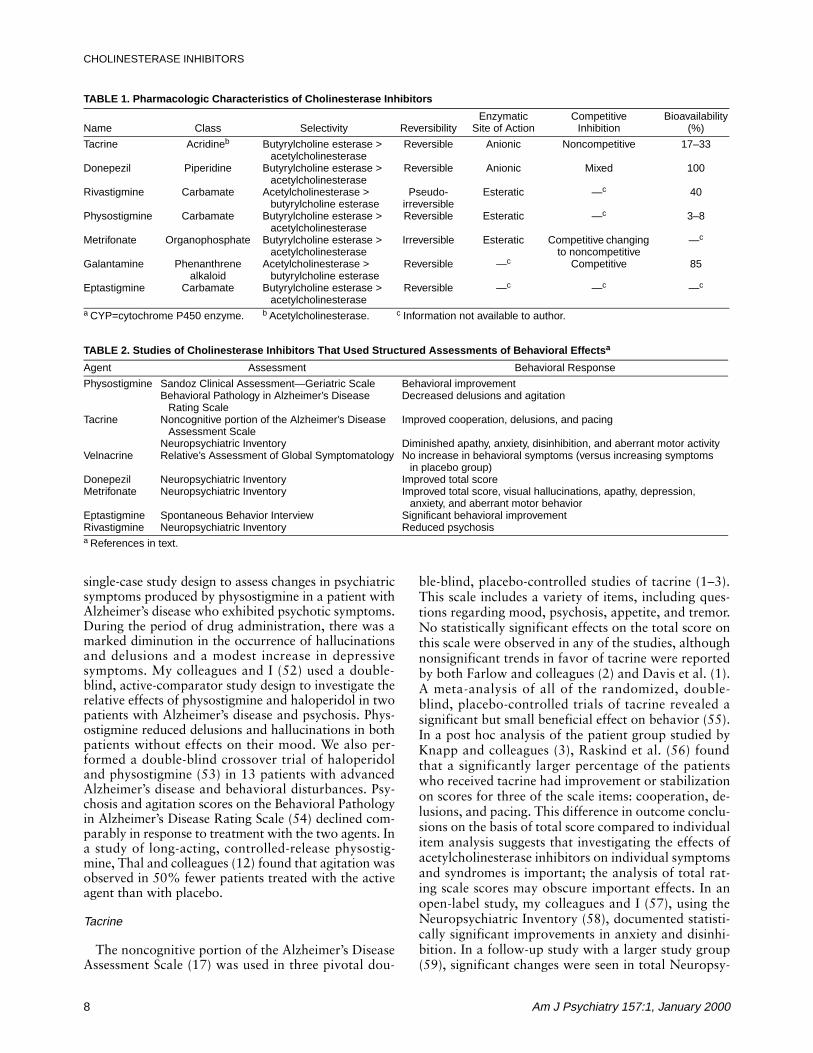
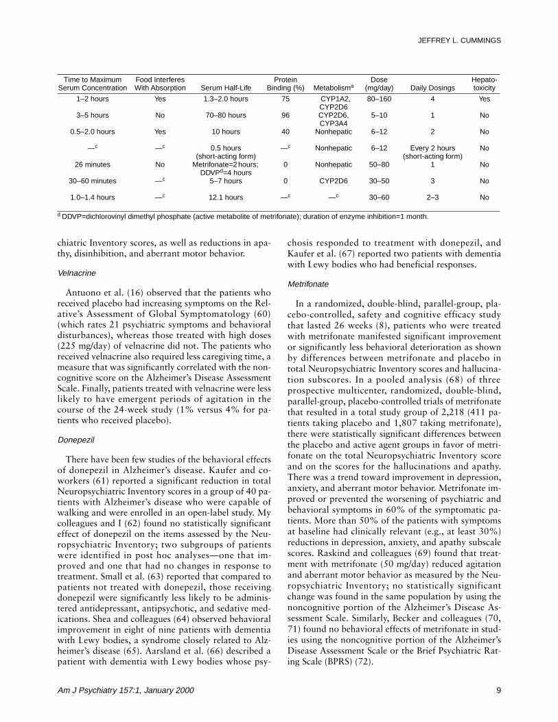
Table 1 provides comparative information on theprincipal acetylcholinesterase inhibitors that are cur-rently approved or under investigation. These drugsrepresent different classes of agents: physostigmine is acarbamate, tacrine and velnacrine are acridines, done-pezil is a piperidine, rivastigmine and eptastigmine arecarbamates, metrifonate is an organophosphate, andgalantamine is a phenanthrene alkaloid. They differprincipally in the type of bond they form with acetyl-cholinesterase. Tacrine, velnacrine, donepezil, and hu-perzine are high-affinity, noncovalent inhibitors; metri-fonate forms an irreversible covalent bond with thesubstrate. Tacrine and velnacrine are noncompetitiveinhibitors, donepezil has noncompetitive and competi-tive properties, galantamine is a competitive inhibitor,and metrifonate begins with competitive inhibitionand becomes a noncompetitive inhibitor over time(34). Differences in the duration of action and metab-olism determine the dosing regimen and the likelihoodof drug interactions. Galantamine is an acetylcho-linesterase inhibitor and an allosteric modulator of nic-otinic cholinergic receptors (45). The last activity mayincrease acetylcholine release by activating presynapticnicotinic receptors. Tacrine and velnacrine are associ-ated with a high frequency of hepatotoxicity; eptastig-mine produces neutropenia (46).
BEHAVIORAL RESPONSES TO CHOLINESTERASE INHIBITORS
Table 2 summarizes the available information de-rived from standardized rating scales on the neuropsy-chiatric changes observed after treatment with acetyl-cholinesterase inhibitors.
Physostigmine
Physostigmine was the acetylcholinesterase inhibitormost studied in the early phases of the development ofantidementia drugs. Many studies did not include be-havioral measures, but Harrell and colleagues (47) in-cluded the Sandoz Clinical Assessment—GeriatricScale (48) and reported that in the group of patientswho had responded to physostigmine, as evidenced bycognitive improvement or correct identification ofgroup membership by the patient’s family or physician,there was significantly greater behavioral improve-ment than in patients receiving placebo or those desig-nated as nonresponders. Schwartz and Kohlstaedt (49)reported that four of 11 patients who received intra-muscular physostigmine had dramatic behavioral im-provements lasting up to 3 days after drug administra-tion. There was decreased restlessness and depressionand improved sociability and initiative. In contrast,Wirkowski and colleagues (50) reported two cases of“physostigmine syndrome” in which patients becameanxious, irritable, and uncooperative and one experi-enced extreme fatigue. The doses in this study ex-ceeded considerably those administered by Schwartzand Kohlstaedt (49).
Four studies have specifically investigated the neurop-sychiatric effects of physostigmine. Molchan and col-leagues (51) used a double-blind, placebo-controlled,
FIGURE 4. Regional Reductions of Cholinergic Markers inAlzheimer’s Disease in the Lateral (Left) Brain Region a
a Marked=blue, medium=green. Image provided by the UCLA Lab-oratory of Neuroimaging; courtesy of Andrew Lee, John Bachelor,and Arthur Toga.
FIGURE 5. Regional Reductions of Cholinergic Markers inAlzheimer’s Disease in the Medial (Right) Brain Region a
a Modest cholinergic reductions in areas shown in purple. Imageprovided by the UCLA Laboratory of Neuroimaging; courtesy ofAndrew Lee, John Bachelor, and Arthur Toga.
8 Am J Psychiatry 157:1, January 2000
CHOLINESTERASE INHIBITORS
single-case study design to assess changes in psychiatricsymptoms produced by physostigmine in a patient withAlzheimer’s disease who exhibited psychotic symptoms.During the period of drug administration, there was amarked diminution in the occurrence of hallucinationsand delusions and a modest increase in depressivesymptoms. My colleagues and I (52) used a double-blind, active-comparator study design to investigate therelative effects of physostigmine and haloperidol in twopatients with Alzheimer’s disease and psychosis. Phys-ostigmine reduced delusions and hallucinations in bothpatients without effects on their mood. We also per-formed a double-blind crossover trial of haloperidoland physostigmine (53) in 13 patients with advancedAlzheimer’s disease and behavioral disturbances. Psy-chosis and agitation scores on the Behavioral Pathologyin Alzheimer’s Disease Rating Scale (54) declined com-parably in response to treatment with the two agents. Ina study of long-acting, controlled-release physostig-mine, Thal and colleagues (12) found that agitation wasobserved in 50% fewer patients treated with the activeagent than with placebo.
Tacrine
The noncognitive portion of the Alzheimer’s DiseaseAssessment Scale (17) was used in three pivotal dou-
ble-blind, placebo-controlled studies of tacrine (1–3).This scale includes a variety of items, including ques-tions regarding mood, psychosis, appetite, and tremor.No statistically significant effects on the total score onthis scale were observed in any of the studies, althoughnonsignificant trends in favor of tacrine were reportedby both Farlow and colleagues (2) and Davis et al. (1).A meta-analysis of all of the randomized, double-blind, placebo-controlled trials of tacrine revealed asignificant but small beneficial effect on behavior (55).In a post hoc analysis of the patient group studied byKnapp and colleagues (3), Raskind et al. (56) foundthat a significantly larger percentage of the patientswho received tacrine had improvement or stabilizationon scores for three of the scale items: cooperation, de-lusions, and pacing. This difference in outcome conclu-sions on the basis of total score compared to individualitem analysis suggests that investigating the effects ofacetylcholinesterase inhibitors on individual symptomsand syndromes is important; the analysis of total rat-ing scale scores may obscure important effects. In anopen-label study, my colleagues and I (57), using theNeuropsychiatric Inventory (58), documented statisti-cally significant improvements in anxiety and disinhi-bition. In a follow-up study with a larger study group(59), significant changes were seen in total Neuropsy-
TABLE 1. Pharmacologic Characteristics of Cholinesterase Inhibitors
Name Class Selectivity ReversibilityEnzymatic
Site of ActionCompetitive
InhibitionBioavailability
(%)
Tacrine Acridineb Butyrylcholine esterase > acetylcholinesterase
Reversible Anionic Noncompetitive 17–33
Donepezil Piperidine Butyrylcholine esterase > acetylcholinesterase
Reversible Anionic Mixed 100
Rivastigmine Carbamate Acetylcholinesterase > butyrylcholine esterase
Pseudo-irreversible
Esteratic —c 40
Physostigmine Carbamate Butyrylcholine esterase > acetylcholinesterase
Reversible Esteratic —c 3–8
Metrifonate Organophosphate Butyrylcholine esterase > acetylcholinesterase
Irreversible Esteratic Competitive changing to noncompetitive
—c
Galantamine Phenanthrenealkaloid
Acetylcholinesterase > butyrylcholine esterase
Reversible —c Competitive 85
Eptastigmine Carbamate Butyrylcholine esterase > acetylcholinesterase
Reversible —c —c —c
a CYP=cytochrome P450 enzyme. b Acetylcholinesterase. c Information not available to author.
TABLE 2. Studies of Cholinesterase Inhibitors That Used Structured Assessments of Behavioral Effectsa
Agent Assessment Behavioral Response
Physostigmine Sandoz Clinical Assessment—Geriatric Scale Behavioral improvementBehavioral Pathology in Alzheimer’s Disease
Rating ScaleDecreased delusions and agitation
Tacrine Noncognitive portion of the Alzheimer’s Disease Assessment Scale
Improved cooperation, delusions, and pacing
Neuropsychiatric Inventory Diminished apathy, anxiety, disinhibition, and aberrant motor activityVelnacrine Relative’s Assessment of Global Symptomatology No increase in behavioral symptoms (versus increasing symptoms
in placebo group)Donepezil Neuropsychiatric Inventory Improved total scoreMetrifonate Neuropsychiatric Inventory Improved total score, visual hallucinations, apathy, depression,
anxiety, and aberrant motor behaviorEptastigmine Spontaneous Behavior Interview Significant behavioral improvementRivastigmine Neuropsychiatric Inventory Reduced psychosisa References in text.
Am J Psychiatry 157:1, January 2000 9
JEFFREY L. CUMMINGS
chiatric Inventory scores, as well as reductions in apa-thy, disinhibition, and aberrant motor behavior.
Velnacrine
Antuono et al. (16) observed that the patients whoreceived placebo had increasing symptoms on the Rel-ative’s Assessment of Global Symptomatology (60)(which rates 21 psychiatric symptoms and behavioraldisturbances), whereas those treated with high doses(225 mg/day) of velnacrine did not. The patients whoreceived velnacrine also required less caregiving time, ameasure that was significantly correlated with the non-cognitive score on the Alzheimer’s Disease AssessmentScale. Finally, patients treated with velnacrine were lesslikely to have emergent periods of agitation in thecourse of the 24-week study (1% versus 4% for pa-tients who received placebo).
Donepezil
There have been few studies of the behavioral effectsof donepezil in Alzheimer’s disease. Kaufer and co-workers (61) reported a significant reduction in totalNeuropsychiatric Inventory scores in a group of 40 pa-tients with Alzheimer’s disease who were capable ofwalking and were enrolled in an open-label study. Mycolleagues and I (62) found no statistically significanteffect of donepezil on the items assessed by the Neu-ropsychiatric Inventory; two subgroups of patientswere identified in post hoc analyses—one that im-proved and one that had no changes in response totreatment. Small et al. (63) reported that compared topatients not treated with donepezil, those receivingdonepezil were significantly less likely to be adminis-tered antidepressant, antipsychotic, and sedative med-ications. Shea and colleagues (64) observed behavioralimprovement in eight of nine patients with dementiawith Lewy bodies, a syndrome closely related to Alz-heimer’s disease (65). Aarsland et al. (66) described apatient with dementia with Lewy bodies whose psy-
chosis responded to treatment with donepezil, andKaufer et al. (67) reported two patients with dementiawith Lewy bodies who had beneficial responses.
Metrifonate
In a randomized, double-blind, parallel-group, pla-cebo-controlled, safety and cognitive efficacy studythat lasted 26 weeks (8), patients who were treatedwith metrifonate manifested significant improvementor significantly less behavioral deterioration as shownby differences between metrifonate and placebo intotal Neuropsychiatric Inventory scores and hallucina-tion subscores. In a pooled analysis (68) of threeprospective multicenter, randomized, double-blind,parallel-group, placebo-controlled trials of metrifonatethat resulted in a total study group of 2,218 (411 pa-tients taking placebo and 1,807 taking metrifonate),there were statistically significant differences betweenthe placebo and active agent groups in favor of metri-fonate on the total Neuropsychiatric Inventory scoreand on the scores for the hallucinations and apathy.There was a trend toward improvement in depression,anxiety, and aberrant motor behavior. Metrifonate im-proved or prevented the worsening of psychiatric andbehavioral symptoms in 60% of the symptomatic pa-tients. More than 50% of the patients with symptomsat baseline had clinically relevant (e.g., at least 30%)reductions in depression, anxiety, and apathy subscalescores. Raskind and colleagues (69) found that treat-ment with metrifonate (50 mg/day) reduced agitationand aberrant motor behavior as measured by the Neu-ropsychiatric Inventory; no statistically significantchange was found in the same population by using thenoncognitive portion of the Alzheimer’s Disease As-sessment Scale. Similarly, Becker and colleagues (70,71) found no behavioral effects of metrifonate in stud-ies using the noncognitive portion of the Alzheimer’sDisease Assessment Scale or the Brief Psychiatric Rat-ing Scale (BPRS) (72).
Time to MaximumSerum Concentration
Food InterferesWith Absorption Serum Half-Life
Protein Binding (%) Metabolisma
Dose(mg/day) Daily Dosings
Hepato-toxicity
1–2 hours Yes 1.3–2.0 hours 75 CYP1A2, CYP2D6
80–160 4 Yes
3–5 hours No 70–80 hours 96 CYP2D6, CYP3A4
5–10 1 No
0.5–2.0 hours Yes 10 hours 40 Nonhepatic 6–12 2 No
—c —c 0.5 hours(short-acting form)
—c Nonhepatic 6–12 Every 2 hours (short-acting form)
No
26 minutes No Metrifonate=2 hours; DDVPd=4 hours
0 Nonhepatic 50–80 1 No
30–60 minutes —c 5–7 hours 0 CYP2D6 30–50 3 No
1.0–1.4 hours —c 12.1 hours —c —c 30–60 2–3 No
d DDVP=dichlorovinyl dimethyl phosphate (active metabolite of metrifonate); duration of enzyme inhibition=1 month.
10 Am J Psychiatry 157:1, January 2000
CHOLINESTERASE INHIBITORS
Eptastigmine
Imbimbo and colleagues (13) assessed the safety andefficacy of two doses (30 mg/day and 45 mg/day) ofeptastigmine in a double-blind, placebo-controlledstudy. Using the Spontaneous Behavior Interview (73),a caregiver-based instrument that assesses a wide rangeof neuropsychiatric symptoms, they documented sig-nificant behavioral improvement in those who received30 mg/day.
Rivastigmine
Jan and McKeith (74) reported that Neuropsychiat-ric Inventory scores that reflected the presence of psy-chosis were reduced by treatment with rivastigmine.
DISCUSSION
The principal conclusion of this review is that thereis substantial and growing evidence that acetylcho-linesterase inhibitors exert beneficial psychotropic ef-fects in patients with Alzheimer’s disease. Changes inneuropsychiatric symptoms should be among the clin-ical outcomes assessed in clinical trials of cholinergicagents, and clinicians should monitor psychiatric andbehavioral responses in patients as an indication ofdrug effect when prescribing acetylcholinesterase in-hibitors. Limbic and paralimbic cortices normally re-ceive robust cholinergic innervation and have cholin-ergic deficits in Alzheimer’s disease. Restoration offunction in these brain regions that are critical to themediation of emotion may underlie the behavioral re-sponse to acetylcholinesterase inhibitors.
Visual hallucinations and apathy are the most pre-dictably responsive symptoms in most investigations.Anxiety, disinhibition, agitation, depression, delu-sions, and aberrant motor behavior have improved insome studies but not in others. The observation thatseveral acetylcholinesterase inhibitors have similareffects on behavior suggests that this may be a classeffect that reflects cholinergic enhancement in behav-iorally relevant areas of the brain. However, acetylcho-linesterase inhibitors may differ in their neuropsychiat-ric potency, and assessments of the psychotropic effectsof individual agents are necessary.
Neurobiological Basis of the Response to Cholinergic Agents
Similarities between the neuropsychiatric symptomsof Alzheimer’s disease and anticholinergic toxicity,the response of these symptoms to acetylcholines-terase inhibitors in conditions with cholinergic defi-cits, and the anatomic distribution of the cholinergicdeficits all link cholinergic abnormalities to neuropsy-chiatric disturbances (20).
Anticholinergic agents induce changes in mental sta-tus that are similar to the neuropsychiatric symptomsof Alzheimer’s disease, including thought disorganiza-
tion, visual hallucinations, and variable mood changes.Patients with Alzheimer’s disease are unusually sensi-tive to the adverse effects of anticholinergic compounds(75). Neuropsychiatric symptoms induced by anticho-linergics can be ameliorated by acetylcholinesterase in-hibitors, including tacrine and physostigmine (76).
Patients with neurologic disorders with concomitantcholinergic deficits have been reported to respond toacetylcholinesterase inhibitors with reduced neuropsy-chiatric symptoms. Patients with dementia with Lewybodies improve behaviorally in response to treatmentwith acetylcholinesterase inhibitors (64, 66, 67), andpatients with Parkinson’s disease and dementia withdelusions and hallucinations may exhibit a beneficialneuropsychiatric response to therapy with acetylcho-linesterase inhibitors (77). Cholinergic disturbancesare present in the limbic and paralimbic cortices,which mediate functions critical to emotion (24, 27,41) this regional deficiency may provide the substratefor some of the emotional disturbances of Alzheimer’sdisease and their response to cholinergic therapy.
Neurobiological Basis of Variations in Response to Cholinergic Therapy
Variations in the cholinergic deficit may account forsome of the observed neuropsychiatric heterogeneityof Alzheimer’s disease and the differences in responseto treatment with cholinergic agents. A few patientswith histopathological changes typical of Alzheimer’sdisease do not show a loss of neurons in the nucleusbasalis or a cortical cholinergic deficit at autopsy (78).Some investigators have found greater preservation ofthe nucleus basalis neurons in patients with onset ofthe disease after age 65—the most typical age at on-set—than in those whose symptoms began earlier inlife (79). Davis and colleagues (80) reported that corti-cal cholinergic deficits were not present in elderly pa-tients with mild to moderate Alzheimer’s disease, al-though they were notable in patients with advanceddisease. Patients with the apolipoprotein E-4 genotypehave less brain choline acetyltransferase and nicotinicreceptor binding than patients with the E-3 or E-2 gen-otype (81, 82). In one study (83), women with Alz-heimer’s disease had more cytoskeletal changes in thenucleus basalis than men with Alzheimer’s disease.Moreover, women who receive estrogen replacementtherapy and those without the E-4 genotype have beenshown to respond most favorably to tacrine (84, 85).Thus, differences in age, disease severity, or genotypemay influence the cholinergic deficit and the responseto therapy with acetylcholinesterase inhibitors.
Additional variability in the behavioral symptoms ofAlzheimer’s disease and the responsiveness to cholin-ergic therapy may reflect dynamic interactions betweenthe cholinergic changes and other transmitter systemsinvolved in Alzheimer’s disease (20, 37). The cholin-ergic system interacts with a variety of other transmit-ters or neuromodulators, including norepinephrine,

Am J Psychiatry 157:1, January 2000 11
JEFFREY L. CUMMINGS
dopamine, serotonin, γ-aminobutyric acid, opioid pep-tides, galanin, substance P, and angiotensin II (86).
Relationship of Behavioral and Cognitive Changes
Acetylcholinesterase inhibitors were developed to en-hance cognition in patients with Alzheimer’s disease. Inpatients with mild to moderate cognitive impairment,they temporarily improve, stabilize, or reduce the rateof decline in memory and other intellectual functionsrelative to the results with placebo. A few studies haveinvestigated the relationship between cognitive and be-havioral responses. My colleagues and I (57), in a studyof the neuropsychiatric effects of tacrine, noted that of10 subjects who had at least a 3-point improvement inMini-Mental State examination scores, all also had animprovement in neuropsychiatric symptoms as re-flected by lower scores on the Neuropsychiatric Inven-tory. Of the nine patients in the study who met thestringent criteria of at least a 9-point reduction inscores on the Neuropsychiatric Inventory, six met thecriteria for improved cognitive function; three patientsshowed a substantial behavioral benefit from therapywithout a coincident improvement in cognition. In afollow-up article (59), we noted that patients withmoderate cognitive impairment (Mini-Mental State ex-amination scores between 11 and 20) had the mostconsistent improvement in behavior; mildly affectedpatients exhibited fewer behaviors and had less robusttreatment effects; and patients with severe dementia(Mini-Mental State examination scores of 10 or lower)exhibited an improvement in some symptoms (delu-sions, anxiety, apathy, and disinhibition) and a worsen-ing of others (hallucinations, agitation, dysphoria, eu-phoria, irritability, and aberrant motor behavior). Theavailable data suggest that behavioral improvement isnot contingent upon cognitive changes, and some pa-tients exhibit behavioral responses without concurrentimprovements in cognition.
Cholinergic agents affect many aspects of cognition,which suggests that the primary effect may be on an at-tentional or executive system with a secondary, pan-in-tellectual modulating influence on memory, language,and visuospatial skills (87). Conversely, anticholin-ergics have disproportionately adverse effects on atten-tion, immediate memory, and executive processes (88–90). Improvement in attention may underlie the reduc-tions in apathy that are commonly associated with ace-tylcholinesterase inhibitors, possibly explaining whyapathy is among the most responsive of neuropsychiat-ric symptoms to cholinergic therapy and why it corre-lates highly with cognitive improvement.
Regulatory Issues
FDA guidelines specify that a claim of efficacy in de-mentia can arise only 1) if a drug beneficially affects acore symptom or sign of dementia (i.e., has cognitiveeffects) or 2) if the effect of the drug is expressed onlyor is differentially expressed in patients with dementiawho exhibit the symptom, sign, or behavior (91). It is
likely that cholinergic agents will benefit only patientswho have disturbances of cholinergic function, and,thus, a specific indication for the treatment of neuro-psychiatric symptoms in Alzheimer’s disease may beallowable under the second FDA criterion. These con-siderations are important since cholinergic agentssometimes have emotional benefits for patients who donot improve cognitively; cholinergic agents may reduceneuropsychiatric symptoms late in the course of thedisease, when cognitive enhancement may be limited;subpopulations of patients with behaviors that arespecifically responsive to cholinergic agents may beidentified; and agents may differ substantially in theirefficacy regarding the treatment of neuopsychiatricsymptoms. Thus, acetylcholinesterase inhibitors mayhave a psychotropic role that is independent of theircognitive effects and deserves regulatory recognition.
Clinical Importance
Reducing behavioral disturbances in patients withAlzheimer’s disease is an important treatment goal.Neuropsychiatric disturbances are distressing to thepatient who experiences fear, sadness, or anxiety; theyare a source of marked distress for the caregiver (92);and they may precipitate institutionalization (93).Thus, the treatment of behavioral disturbances inAlzheimer’s disease may ameliorate a patient’s emo-tional distress and have secondary beneficial effects onthe caregiver and on the opportunity for the patient toremain at home. Cholinergic agents may play an im-portant role in these treatment goals.
Determining the magnitude of the psychotropic ef-fect of acetylcholinesterase inhibitors is critical to es-tablishing the clinical importance of these agents astreatments for neuropsychiatric symptoms. The psy-chotropic effects of individual cholinergic agents mustbe determined by prospective, randomized, controlledtrials in which specific behavioral criteria are used forthe selection of participants. The magnitude of the be-havioral response can be anticipated by reviewing thechange of symptoms in patients who were includedfrom existing trials who were symptomatic at baseline.For example, of the patients treated with metrifonate,more than 50% had at least a 30% reduction in de-pression, anxiety, apathy, and total NeuropsychiatricInventory scores (68). A 30% reduction in such symp-toms is clinically relevant and comparable in magni-tude to the changes observed with conventional psy-chotropic agents.
Role of Cholinergic Therapy in Other Conditions
Cholinergic treatments were developed for use in pa-tients with Alzheimer’s disease but may be applicableto patients with other disorders with cholinergic ab-normalities. As already noted, preliminary evidencesuggests that patients with dementia with Lewy bodies(64, 67) or Parkinson’s disease with dementia (77) mayexhibit a behavioral response to treatment with acetyl-cholinesterase inhibitors. Cortical cholinergic deficits

12 Am J Psychiatry 157:1, January 2000
CHOLINESTERASE INHIBITORS
have been identified in a variety of other neurologicaldisorders, including some cases of Pick’s disease (94),olivopontocerebellar atrophy (95), progressive supra-nuclear palsy (96), the parkinsonism dementia com-plex of Guam (97), alcoholism with Wernicke’s en-cephalopathy (98), Creutzfeldt-Jakob disease (99),subacute sclerosing panencephalitis (99), dementia pu-gilistica (100, 101), and traumatic brain injury (102).Patients with vascular dementia may have lesions thatinterrupt projections from the nucleus basalis and pro-duce a cortical cholinergic deficit, or they may havemixed Alzheimer’s disease plus cerebrovascular dis-ease, which render them potentially responsive to ace-tylcholinesterase inhibitors (103). In addition, thereare age-related changes in the nucleus basalis; cellnumbers decline from approximately 450,000 in chil-dren to approximately 150,000 in elderly control sub-jects (104). Neuropsychiatric disturbances in theseconditions might be reduced with the use of acetylcho-linesterase inhibitors.
Open-label studies suggest that acetylcholinesteraseinhibitors are of potential benefit in bipolar disorder(105), and cholinergic dysfunction may be present insome patients with schizophrenia(106). These findingsraise the possibility that these disorders could betreated with cholinergic agents. Cholinesterase inhibi-tors also might benefit other disorders that putativelyaffect cholinergic systems, including attention deficithyperactivity disorder, autism, and sleep disorders.
Behavioral Effects of Cholinergic Agonists
The hypothesis that cholinergic enhancement haspsychotropic effects is strengthened by the observationthat cholinergic agonists, as well as acetylcholinest-erase inhibitors, have been reported to relieve neuro-psychiatric symptoms. Xanomeline, an M1- and M4-selective muscarinic receptor agonist, was studied in a6-month randomized, double-blind, placebo-con-trolled, parallel-group, multiple-dose trial (107). Theagent exhibited substantial behavioral effects and sig-nificant dose-dependent reductions in vocal outbursts,suspiciousness, delusions, agitation, hallucinations,wandering, fearfulness, compulsiveness, tearfulness,mood swings, and threatening behaviors. The emer-gence of neuropsychiatric symptoms in patients inwhom these were absent at baseline was suppressed.SB-202026, an M1 partial agonist, produced an im-provement in behavior, compared to the deteriorationin behavior in the placebo group, when assessed withthe noncognitive portion of the Alzheimer’s DiseaseAssessment Scale (108).
The BPRS was used to assess behavioral responses tointravenous administration of the cholinergic agonistarecoline to patients with Alzheimer’s disease (109).The anergia subscale of the BPRS showed a biphasicresponse, with decreases following low-dose infusionsand increases following high-dose infusions. Similarly,the score on the thought disorder subscale increasedwith high-dose infusions.
Penn and colleagues (110) noted a trend toward de-creased abnormal behavior scores during intraventric-ular administration of the cholinergic agonist be-thanechol, whereas my colleagues and I (111) observedthat two of five patients treated in the course of a dose-finding study exhibited distress, restlessness, agitation,and depression. Oxotremorine, a long-acting, directcholinergic agonist, was administered to seven patientswith Alzheimer’s disease and was noted to precipitatedepressive reactions in five (112). Intravenous nicotinehas been used to assess the effect of nicotinic receptorstimulation in Alzheimer’s disease, and Newhouse andcolleagues (113) found that at the highest doses, pa-tients exhibited significant elevations in scores for anx-iety and depression.
Thus, whereas cholinergic agonists have producedimprovement in neuropsychiatric symptoms in somestudies of patients with Alzheimer’s disease, the infor-mation available is limited, and the responses reportedare less consistent than those observed with acetylcho-linesterase inhibitors.
Summary
Acetylcholinesterase inhibitors have psychotropic aswell as cognition-enhancing effects. The ameliorationof apathy and reduction of visual hallucinations arethe most reproducible effects on neuropsychiatricsymptoms in Alzheimer’s disease, but other neuropsy-chiatric symptoms have responded in some studies.There may be variations among acetylcholinesteraseinhibitors in their psychotropic properties. The benefi-cial effects of acetylcholinesterase inhibitors on emo-tion and behavior are most likely mediated throughcholinergic influences on limbic and paralimbic brainstructures. Patients who exhibit substantial cognitiveimprovement usually have a concomitant behavioralresponse, but behavioral and cognitive responses maybe dissociated. Clinical trials should be designed toclarify the neuropsychiatric effects of acetylcholinest-erase inhibitors in Alzheimer’s disease and to explorethe potential use of these agents in other conditions.
REFERENCES
1. Davis KL, Thal LJ, Gamzu ER, Davis CS, Woolson RF, Gra-con SI, Drachman DA, Schneider LS, Whitehouse PJ, HooverTM, Morris JC, Kawas CH, Knopman DS, Earl NL, Kumar V,Doody RS (the Tacrine Collaborative Study Group): A double-blind, placebo-controlled multicenter study of tacrine forAlzheimer’s disease. N Engl J Med 1992; 327:1253–1259
2. Farlow M, Gracon SI, Hershey LA, Lewis KW, Sadowsky CH,Dolan-Ureno J (Tacrine Study Group): A controlled trial oftacrine in Alzheimer’s disease. JAMA 1992; 268:2523–2529
3. Knapp MJ, Knopman DS, Solomon PR, Pendlebury WW,Davis CS, Gracon SI (Tacrine Study Group): A 30-week ran-domized controlled trial of high-dose tacrine in patients withAlzheimer’s disease. JAMA 1994; 271:985–994
4. Rogers SL, Farlow MR, Doody RS, Mohs R, Friedhoff LT(Donepezil Study Group): A 24-week, double-blind, placebo-controlled trial of donepezil in patients with Alzheimer’s dis-ease. Neurology 1998; 50:136–145
5. Rogers SL, Doody RS, Mohs RC, Friedhoff LT (DonepezilStudy Group): Donepezil improves cognition and global func-

Am J Psychiatry 157:1, January 2000 13
JEFFREY L. CUMMINGS
tion in Alzheimer disease: a 15-week, double-blind, placebo-controlled study. Arch Intern Med 1998; 158:1021–1031
6. Rogers SL, Friedhoff LT (Donepezil Study Group): The effi-cacy and safety of donepezil in patients with Alzheimer’s dis-ease: results of a US multicentre, randomized, double-blind,placebo-controlled trial. Dementia 1996; 7:293–303
7. Cummings JL, Cyrus PA, Bieber F, Mas J, Orazem J, GulanskiB (Metrifonate Study Group): Metrifonate treatment of thecognitive deficits of Alzheimer’s disease. Neurology 1998; 50:1214–1221
8. Morris JC, Cyrus PA, Orazem J, Mas J, Bieber F, Ruzicka BB,Gulanski B: Metrifonate benefits cognitive, behavioral, andglobal function in patients with Alzheimer’s disease. Neurol-ogy 1998; 50:1222–1230
9. Corey-Bloom J, Anand R, Veach J (ENA 713 B352 StudyGroup): A randomized trial evaluating the efficacy and safetyof ENA 713 (rivastigmine tartrate), a new acetylcholinesteraseinhibitor, in patients with mild to moderately severe Alzhei-mer’s disease. Int J Geriatr Psychopharmacol 1998; 1:55–65
10. Sramek JJ, Anand R, Wardle TS, Irwin P, Hartman RD, CutlerNR: Safety/tolerability trial of SDZ ENA 713 in patients withprobable Alzheimer’s disease. Life Sci 1996; 58:1201–1207
11. Rosler M, Anand R, Cicin-Sain A, Gauthier S, Agid Y, Dal-Bi-anco P, Stahelin H, Hartmann R, Gharabawi M: B303 Exelonstudy group: efficacy and safety of rivastigmine in patientswith Alzheimer’s disease: international randomised controlledtrial. Br Med J 1999; 318:633–640
12. Thal LJ, Ferguson JM, Mintzer J, Raskin A, Targum SD: A 24-week randomized trial of controlled-release physostigmine inpatients with Alzheimer’s disease. Neurology 1999; 52:1146–1152
13. Imbimbo BP, Lucca U, Lucchelli F, Alberoni M, Thal LJ(Eptastigmine Study Group): A 25-week placebo-controlledstudy of eptastigmine in patients with Alzheimer’s disease.Alzheimer Dis Assoc Disord 1998; 12:313–322
14. Tariot PN, Schneider L: Contemporary treatment approachesto Alzheimer’s disease. Consultant Pharmacist 1996; 11(supplE):16–24
15. Rainer M: Galantamine in Alzheimer’s disease: a new alterna-tive to tacrine? CNS Drugs 1997; 2:89–97
16. Antuono PG (Mentane Study Group): Effectiveness andsafety of velnacrine for the treatment of Alzheimer’s disease:a double-blind, placebo-controlled study. Arch Intern Med1995; 155:1766–1772
17. Rosen WG, Mohs RC, Davis KL: A new rating scale for Alzhei-mer’s disease. Am J Psychiatry 1984; 141:1356–1364
18. Folstein MF, Folstein SE, McHugh PR: “Mini-Mental State”: apractical method for grading the cognitive state of patients forthe clinician. J Psychiatr Res 1975; 12:189–198
19. Knopman DS, Knapp MJ, Gracon SI, Davis CS: The ClinicianInterview-Based Impression (CIBI): a clinician’s globalchange rating scale in Alzheimer’s disease. Neurology 1994;44:2315–2321
20. Cummings JL, Kaufer D: Neuropsychiatric aspects of Alzhei-mer’s disease: the cholinergic hypothesis revisited. Neurology1996; 47:876–883
21. Cummings JL, Back C: The cholinergic hypothesis of neuro-psychiatric symptoms in Alzheimer’s disease. Am J GeriatrPsychiatry 1998; 6(suppl 1):S64–S78
22. Schegg KM, Harrington LS, Nielsen S, Zwieg RM, PeacockJH: Soluble and membrane-bound forms of brain acetylcho-linesterase in Alzheimer’s disease. Neurobiol Aging 1992; 13:697–704
23. Younkin SG, Goodridge B, Katz J, Lockett G, Nafziger D,Usiak MF, Younkin LH: Molecular forms of acetylcholinest-erase in Alzheimer’s disease. Fed Proc 1986; 45:2982–2988
24. Mesulam M-M: Structure and function of cholinergic pathwaysin the cerebral cortex, limbic system, basal ganglia, and thal-amus of the human brain, in Psychopharmacology: TheFourth Generation of Progress. Edited by Bloom FE, KupferDJ. New York, Raven Press, 1995, pp 135–146
25. Xiang Z, Huguenard JR, Prince DA: Cholinergic switchingwithin neocortical inhibitory networks. Science 1998; 281:985–988
26. Guela C: Abnormalities of neural circuitry in Alzheimer’s dis-ease: hippocampus and cortical cholinergic innervation. Neu-rology 1998; 51(suppl 1):S18–S29
27. Mesulam M-M, Mufson EJ: Neural inputs into the nucleusbasalis of the substantia innominata (Ch4) in the rhesus mon-key. Brain 1984; 107:253–274
28. Richelson E: Cholinergic transduction, in Psychopharmacol-ogy: The Fourth Generation of Progress. Edited by Bloom FE,Kupfer DJ. New York, Raven Press, 1995, pp 125–134
29. Arneric SP, Sullivan JP, Williams M: Neuronal nicotinic acetyl-choline receptors: novel targets for central nervous systemtherapeutics. Ibid, pp 995–1010
30. Nicholls DG: Proteins, Transmitters and Synapses. London,Blackwell Science, 1994
31. Flynn DD, Ferrari-DiLeo G, Mash DC, Levery AL: Differentialregulation of modular subtypes of muscarinic receptors inAlzheimer’s disease. J Neurochem 1995; 64:1888–1891
32. Schliebs R, Robner S: Distribution of muscarinic acetylcholinereceptors in the CNS, in CNS Neurotransmitters and Neuro-modulators: Acetylcholine. Edited by Stone TW. Boca Raton,Fla, CRC Press, 1995, pp 67–83
33. Court JA, Perry EK: Distribution of nicotinic receptors in theCNS. Ibid, pp 85–104
34. Taylor P: Development of acetylcholinesterase inhibitors inthe therapy of Alzheimer’s disease. Neurology 1998; 51(suppl1):S30–S35
35. Zhou H-X, Wlodek ST, McCammon JA: Conformation gatingas a mechanism for enzyme specificity. Proc Natl Acad SciUSA 1998; 95:9280–9283
36. Fishman EB, Siek GC, MacCallum RD, Bird ED, Volicer L,Marquis JK: Distribution of the molecular forms of acetylcho-linesterase in human brain: alterations in dementia of theAlzheimer type. Ann Neurol 1986; 19:246–252
37. Cummings JL, Vinters HV, Cole GM, Khachaturian ZS: Alzhei-mer’s disease: etiologies, pathophysiology, cognitive reserve,and treatment opportunities. Neurology 1998; 51(suppl 1):S2–S17
38. Whitehouse PJ, Price DL, Clark AW, Coyle JT, DeLong MR:Alzheimer’s disease: evidence for selective loss of cholinergicneurons in the nucleus basalis. Ann Neurol 1981; 10:122–126
39. Whitehouse PJ, Price DL, Struble RG, Clark AW, Coyle JT:Alzheimer’s disease and senile dementia: loss of neurons inthe basal forebrain. Science 1982; 215:1237–1239
40. Rasool CG, Svendsen CN, Selkoe DJ: Neurofibrillary degen-eration of cholinergic and noncholinergic neurons of the basalforebrain in Alzheimer’s disease. Ann Neurol 1986; 20:482–488
41. Davies P: Studies on the neurochemistry of central cholin-ergic systems in Alzheimer’s disease, in Alzheimer’s Disease:Senile Dementia and Related Disorders. Edited by KatzmanR, Terry RD, Bick KL. New York, Raven Press, 1978, pp 453–459
42. Guela C, Mesulam M-M: Cholinergic systems and relatedneuropathological predilection patterns in Alzheimer disease,in Alzheimer Disease. Edited by Terry RD, Katzman R, BickKL. New York, Raven Press, 1994, pp 263–291
43. Rodriguez-Puertas R, Pascual J, Vilaro T, Pazos A: Autorad-iographic distribution of M1, M2, M3, and M4 muscarinic re-ceptor subtypes in Alzheimer’s disease. Synapse 1997; 26:341–350
44. Perry EK, Perry RH, Smith CJ, Purohit D, Bonham J, Dick DJ,Candy JM, Edwardson JA, Fairbairn A: Cholinergic receptorsin cognitive disorders. Can J Neurol Sci 1986; 13:521–527
45. Pontecorvo MJ, Parys W: Clinical development of galan-tamine: evaluation of a compound with possible acetylcho-linesterase inhibiting and nicotinic modulating activity (ab-stract). Neurobiol Aging 1998; 19(suppl 7):57
46. Imbimbo BP, Martelli P, Troetel WM, Lucchelli F, Lucca U, ThalLJ (Eptastigmine Study Group): Efficacy and safety of

14 Am J Psychiatry 157:1, January 2000
CHOLINESTERASE INHIBITORS
eptastigmine for the treatment of patients with Alzheimer’sdisease. Neurology 1999; 52:700–708
47. Harrell LE, Calloway R, Morere D, Falgout J: The effect oflong-term physostigmine administration in Alzheimer’s dis-ease. Neurology 1990; 40:1350–1354
48. Shader RI, Harmatz JS, Salzman C: A new scale for clinicalassessment in geriatric populations: Sandoz Clinical Assess-ment—Geriatric (SCAG). J Am Geriatr Soc 1974; 22:107–113
49. Schwartz AS, Kohlstaedt EV: Physostigmine effects in Alzhei-mer’s disease: relationship to dementia severity. Life Sci 1986;38:1021–1028
50. Wirkowski E, Prohovnik I, Young WL: Observations on thephysostigmine syndrome in patients with Alzheimer’s disease.J Neuropsychiatry Clin Neurosci 1991; 3:73–75
51. Molchan SE, Vitiello B, Minichiello M, Sunderland T: Recipro-cal changes in psychosis and mood after physostigmine in apatient with Alzheimer’s disease (letter). Arch Gen Psychiatry1991; 48:1113
52. Cummings JL, Gorman DG, Shapira J: Physostigmine ame-liorates the delusions of Alzheimer’s disease. Biol Psychiatry1993; 33:536–541
53. Gorman DG, Read S, Cummings JL: Cholinergic therapy ofbehavioral disturbances in Alzheimer’s disease. Neuropsychi-atry Neuropsychol Behav Neurol 1993; 6:229–234
54. Reisberg B, Borenstein J, Salob SP, Ferris SH, Franssen E,Georgotas A: Behavioral symptoms in Alzheimer’s disease:phenomenology and treatment. J Clin Psychiatry 1987;48(suppl 5):9–15
55. Qizilbash N, Whitehead A, Higgins J, Wilcock G, Schneider L,Farlow M (Dementia Trialists’ Collaboration): Cholinesteraseinhibition for Alzheimer disease: a meta-analysis of the tacrinetrials. JAMA 1998; 280:1777–1782
56. Raskind MA, Sadowsky CH, Sigmund WR, Beitler PJ, AusterSB: Effect of tacrine on language, praxis, and noncognitivebehavioral problems in Alzheimer’s disease. Arch Neurol1997; 54:836–840
57. Kaufer DI, Cummings JL, Christine D: Effect of tacrine on be-havioral symptoms in Alzheimer’s disease: an open-labelstudy. J Geriatr Psychiatry Neurol 1996; 9:1–6
58. Cummings JL, Mega M, Gray K, Rosenberg-Thompson S,Carusi DA, Gornbein J: The Neuropsychiatric Inventory: com-prehensive assessment of psychopathology in dementia.Neurology 1994; 44:2308–2314
59. Kaufer D, Cummings JL, Christine D: Differential neuropsychi-atric symptom responses to tacrine in Alzheimer’s disease:relationship to dementia severity. J Neuropsychiatry Clin Neu-rosci 1998; 10:55–63
60. Raskin A, Crook T: Relative’s Assessment of Global Symp-tomatology (RAGS). Psychopharmacol Bull 1988; 24:759–763
61. Kaufer D, Catt K, Pollack B, Lopez O, DeKosky S: Donepezilin Alzheimer’s disease: relative cognitive and neuropsychiat-ric responses and impact on caregiver distress (abstract).Neurology 1998; 50:A89
62. Mega M, Masterman DM, O’Connor SM, Barclay TR, Burzyn-ski MJ, Cummings JL: The spectrum of behavioral responsesin cholinesterase inhibitor therapy in Alzheimer’s disease.Arch Neurol (in press)
63. Small G, Donohue J, Brooks R: An economic evaluation ofdonepezil in the treatment of Alzheimer’s disease. Clin Ther1998; 20:838–850
64. Shea C, MacKnight C, Rockwood K: Aspects of dementia:donepezil for treatment of dementia with Lewy bodies: a caseseries of nine patients. Int Psychogeriatr 1998; 10:229–238
65. Perry EK, Kerwin J, Perry RH, Irving D, Blessed G, FairbairnA: Cerebral cholinergic activity is related to the incidence of vi-sual hallucinations in senile dementia of Lewy body type. De-mentia 1990; 1:2–4
66. Aarsland D, Bronnick K, Karlsen K: Donepezil for dementiawith Lewy bodies: a case study. Int J Geriatr Psychiatry 1999;14:69–72
67. Kaufer DI, Catt KE, Lopez OL, DeKosky ST: Dementia withLewy bodies: response of delirium-like features to donepezil.Neurology 1998; 51:1512
68. Cummings JL, Cyrus PA, Ruzicka BB, Gulanski B: The effi-cacy of metrifonate in improving the behavioral disturbancesof Alzheimer’s disease patients (abstract). Neurology 1999;524:4
69. Raskind M, Cyrus PA, Ruzicka BB, Gulanski B: MetrifonateStudy Group: the effects of metrifonate on the cognitive, be-havioral, and functional performance of Alzheimer’s diseasepatients. J Clin Psychiatry 1999; 60:318–325
70. Becker R, Colliver J, Elble R, Feldman E, Giacobini E, KumarV, Markwell S, Moriearty P, Parks R, Shillcutt S, Unni L, VicariS, Womack C, Zec R: Effects of metrifonate, a long-actingcholinesterase inhibitor, in Alzheimer disease: report of anopen trial. Drug Dev Res 1990; 19:425–434
71. Becker RE, Colliver JA, Markwell SJ, Moriearty PL, Unni LK,Vicari S: Double-blind, placebo-controlled study of metri-fonate, an acetylcholinesterase inhibitor, for Alzheimer’s dis-ease. Alzheimer Dis Assoc Disorder 1996; 10:124–131
72. Overall JE, Gorham DR: The Brief Psychiatric Rating Scale.Psychol Rep 1962; 10:799–812
73. Spagnoli A, Lucca U, Menasce G: Long-term acetyl-L-car-nitine treatment in Alzheimer’s disease. Neurology 1991; 41:1726–1732
74. Jan G, McKeith IG: Special workshop in dementia with Lewybodies (abstract). Neurobiol Aging 1998; 19:45
75. Sunderland T, Tariot PN, Cohen RM, Weingartner H, MuellerEA III, Murphy DL: Anticholinergic sensitivity in patients withdementia of the Alzheimer type and age-matched controls: adose-response study. Arch Gen Psychiatry 1987; 44:418–426
76. Gershon S, Olariu J: JB 329—a new psychotomimetic, its an-tagonism by tetrahydroaminoacridine and its comparison withLSD, mescaline and serynl. J Neuropsychiatry 1960; 1:283–292
77. Hutchinson M, Fazzini E: Cholinesterase inhibition in Parkin-son’s disease. J Neurol Neurosurg Psychiatry 1996; 61:324–325
78. Zweig RM, Schegg K, Peacock JH, Melarkey D: A case ofAlzheimer’s disease and hippocampal sclerosis with normalcholinergic activity in basal forebrain, neocortex, and hippo-campus. Neurology 1989; 39:288–290
79. Whitehouse PJ, Hedreen JC, White CL III, Price DL: Basalforebrain neurons in the dementia of Parkinson disease. AnnNeurol 1983; 13:243–248
80. Davis K, Mohs R, Marin D, Purohit D, Perl D, Lantz M, AustinG, Haroutunian V: Cholinergic markers in elderly patients withearly signs of Alzheimer disease. JAMA 1999; 281:1401–1406
81. Poirier J, Delisle M-C, Quirion R, Aubert I, Farlow M, Lahiri D,Hui S, Bertrand P, Nalbantoglu J, Gilfix BM, Gauthier S: Apo-lipoprotein E4 allele as a predictor of cholinergic deficits andtreatment outcome in Alzheimer’s disease. Proc Natl Acad SciUSA 1995; 92:12260–12264
82. Allen SJ, MacGowan SH, Tyler S, Wilcock GK, RobertsonAGS, Holden PH, Smith SKF, Dawbarn D: Reduced cholin-ergic function in normal and Alzheimer’s disease brain is as-sociated with apolipoprotein E4 genotype. Neurosci Lett1997; 239:33–36
83. Salehi A, Gonzalez Martinez V, Swaab DF: A sex differenceand no effect of ApoE type on the amount of cytoskeletal alter-ations in the nucleus basalis of Meynert in Alzheimer’s dis-ease. Neurobiol Aging 1998; 19:505–510
84. Farlow MR, Lahiri DK, Poirier J, Davignon J, Schneider L, HuiSL: Treatment outcome of tacrine therapy depends on apoli-poprotein genotype and gender of the subjects with Alzhei-mer’s disease. Neurology 1998; 50:669–677
85. Schneider LS, Farlow MR, Henderson VW, Pogoda JM: Ef-fects of estrogen replacement therapy on response to tacrinein patients with Alzheimer’s disease. Neurology 1996; 46:1580–1584

Am J Psychiatry 157:1, January 2000 15
JEFFREY L. CUMMINGS
86. Decker MW, McGaugh JL: The role of interactions betweenthe cholinergic system and other neuromodulatory systems inlearning and memory. Synapse 1991; 7:151–168
87. Lawrence AD, Sahakian BJ: Alzheimer disease, attention,and the cholinergic system. Alzheimer Dis Assoc Disord1995; 9(suppl 2):43–49
88. Cooper JA, Sagar HJ, Doherty SM, Jordan N, Tidswell P, Sul-livan EV: Different effects of dopaminergic and anticholinergictherapies on cognitive and motor function in Parkinson’s dis-ease. Brain 1992; 115:1701–1725
89. Dubois B, Pillon B, Lhermitte F, Agid Y: Cholinergic deficiencyand frontal dysfunction in Parkinson’s disease. Ann Neurol1990; 28:117–121
90. Van Spaendonck KP, Berger HJ, Horstink MW, BuytenhuijsEL, Cools AR: Impaired cognitive shifting in parkinsonian pa-tients on anticholinergic therapy. Neuropsychologia 1993; 31:407–411
91. Leber P: Guidelines for the Clinical Evaluation of Antidemen-tia Drugs. Washington, DC, Food and Drug Administration,1990
92. Kaufer DI, Cummings JL, Christine D, Bray T, Castellon S,Masterman D, MacMillan A, Ketchel P, DeKosky ST: Assess-ing the impact of neuropsychiatric symptoms in Alzheimer’sdisease: the Neuropsychiatric Inventory Caregiver DistressScale. J Am Geriatr Soc 1998; 46:210–215
93. Steele C, Rovner B, Chase GA, Folstein M: Psychiatric symp-toms and nursing home placement of patients with Alzhei-mer’s disease. Am J Psychiatry 1990; 147:1049–1051
94. Uhl GR, Hilt DC, Hedreen JC, Whitehouse PJ, Price DL:Pick’s disease (lobar sclerosis): depletion of neurons in thenucleus basalis of Meynert. Neurology 1983; 33:1470–1473
95. Tagliavini F, Pilleri G: Neuronal loss in the basal nucleus ofMeynert in a patient with olivopontocerebellar atrophy. ActaNeuropathol 1985; 66:127–133
96. Tagliavini F, Pilleri G, Gemignani F, Lechi A: Neuronal loss inthe basal nucleus of Meynert in progressive supranuclearpalsy. Acta Neuropathol 1983; 61:157–160
97. Nakano I, Hirano A: Neuron loss in the nucleus basalis ofMeynert in parkinsonism-dementia complex of Guam. AnnNeurol 1983; 13:87–91
98. Cullen KM, Halliday GM: Mechanisms of cell death in cholin-ergic basal forebrain neurons in chronic alcoholics. MetabBrain Dis 1995; 10:81–91
99. Rogers JD, Brogan D, Mirra SS: The nucleus basalis of Mey-nert in neurological disease: a quantitative morphologicalstudy. Ann Neurol 1985; 17:163–170
100. Tagliavini F, Pilleri G: The basal nucleus of Meynert in cerebralaging and degenerative dementias, in Brain Pathology, 1. Ed-ited by Pilleri G, Tagliavini F. Berne, Switzerland, Brain Anat-omy Institute, 1984, pp 181–218
101. Cummings JL, Benson DF: The role of the nucleus basalis ofMeynert in dementia: review and reconsideration. AlzheimerDis Assoc Disord 1987; 1:128–145
102. Murdoch I, Perry EK, Court JA, Graham DI, Dewar D: Corticalcholinergic dysfunction after human head injury. J Neu-rotrauma 1998; 15:295–305
103. Mendez M, Younesi F, Perryman K: Use of donepezil for vas-cular dementia: preliminary clinical experience. J Neuropsy-chiatry Clin Neurosci 1999; 11:268–270
104. McGeer PL, McGeer EG, Suzuki J, Dolman CE, Nagai T: Ag-ing, Alzheimer’s disease, and the cholinergic system of thebasal forebrain. Neurology 1984; 34:741–745
105. Burt T, Sachs G, Demopulos C: Donepezil in treatment-resis-tant bipolar disorder. Biol Psychiatry 1999; 45:959–964
106. White K, Cummings JL: Schizophrenia and Alzheimer’s dis-ease: clinical and pathophysiologic analogies. Compr Psychi-atry 1996; 37:188–195
107. Bodick NC, Offen WW, Levey AI, Cutler NR, Gauthier SG, Sat-lin A, Shannon HE, Tollefson GD, Rasmussen K, BymasterFP, Hurley DJ, Potter WZ, Paul SM: Effects of xanomeline, aselective receptor agonist, on cognitive function and behav-ioral symptoms in Alzheimer’s disease. Arch Neurol 1997; 4:465–473
108. Kumar R, Orgogozo J: Efficacy and safety of SB 202026 as asymptomatic treatment for Alzheimer’s disease, in Alzhei-mer’s Disease: Biology, Diagnosis, and Therapeutics. Editedby Iqbal K, Winbald B, Nishimura T, Takeda M, WisniewskiHM. New York, John Wiley & Sons, 1997, pp 677–685
109. Tariot PN, Cohen RM, Welkowitz JA, Sunderland T, New-house PA, Murphy DL, Weingarten H: Multiple-dose arecolineinfusions in Alzheimer’s disease. Arch Gen Psychiatry 1988;45:901–905
110. Penn RD, Martin EM, Wilson RS, Fox JH, Savoy SM: Intraven-tricular bethanechol infusion for Alzheimer’s disease: resultsof double-blind and escalating-dose trials. Neurology 1988;38:219–222
111. Read SL, Frazee J, Shapira J, Smith C, Cummings JL, Tomi-yasu U: Intracerebroventricular bethanechol for Alzheimer’sdisease: variable dose-related responses. Arch Neurol 1990;47:1025–1030
112. Davis KL, Hollander E, Davidson M, Davis BM, Mohs RC,Horvath TB: Induction of depression with oxotremorine in pa-tients with Alzheimer’s disease. Am J Psychiatry 1987; 144:468–471
113. Newhouse PA, Sunderland T, Tariot PN, Blumhardt CL, Wein-garten H, Mellow A, Murphy DL: Intravenous nicotine inAlzheimer’s disease: a pilot study. Psychopharmacology(Berl) 1988; 95:171–175