SKELETAL SYSTEM. SKELETAL SYSTEM FUNCTIONS Support (Primary function) Movement (Passive) Protection...
47
SKELETAL SYSTEM
-
Upload
elmer-matthews -
Category
Documents
-
view
219 -
download
2
Transcript of SKELETAL SYSTEM. SKELETAL SYSTEM FUNCTIONS Support (Primary function) Movement (Passive) Protection...
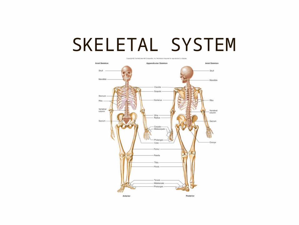
SKELETAL SYSTEM
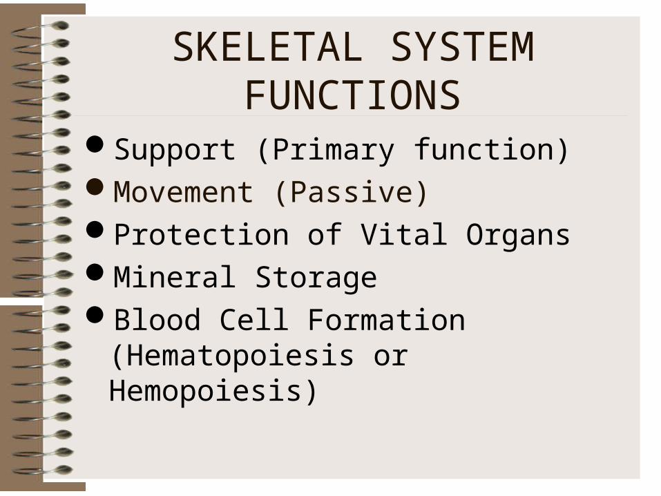
SKELETAL SYSTEM FUNCTIONS
Support (Primary function)Movement (Passive)Protection of Vital OrgansMineral StorageBlood Cell Formation (Hematopoiesis or
Hemopoiesis)

OSSEOUS TISSUE
Cancellous (spongy) BoneCompact (dense) BoneBone Cells
- Osteoblasts – Secrete to form bone- Osteocytes
* Mature bone cells* “Trapped” osteoblasts
- Osteoclasts – destroy bone* Enzymes digest protein* Acids dissolve minerals* Forms Marrow Cavity; Involved in Remodelling
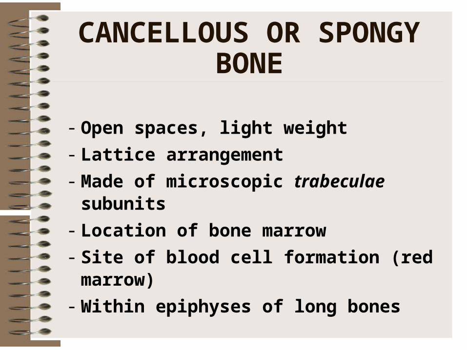
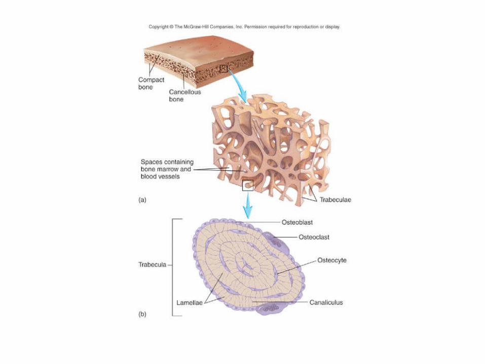
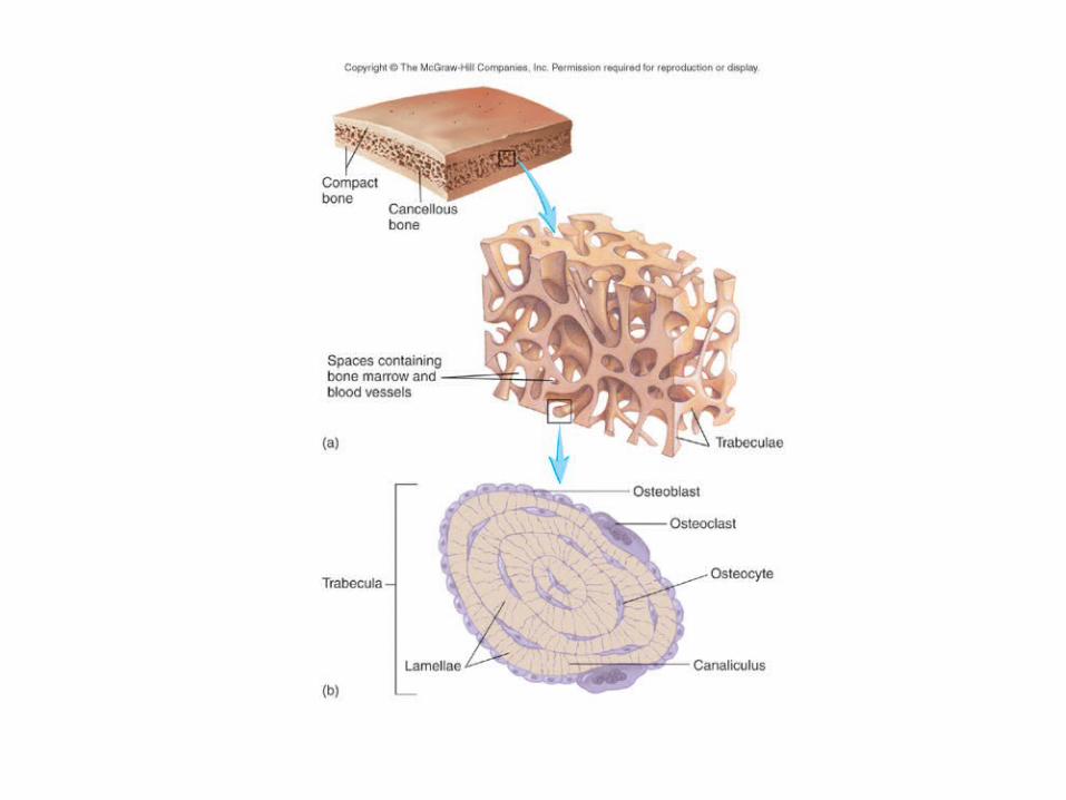
CANCELLOUS OR SPONGY BONE
- Open spaces, light weight
- Lattice arrangement
- Made of microscopic trabeculae subunits
- Location of bone marrow
- Site of blood cell formation (red marrow)
- Within epiphyses of long bones
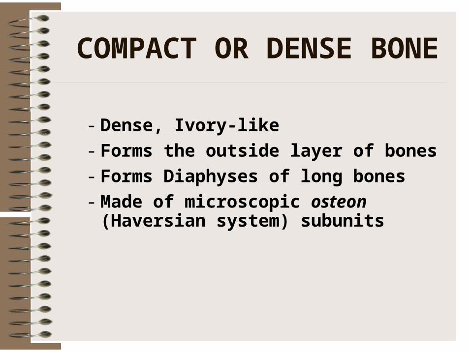
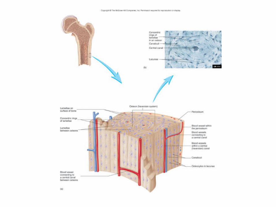
COMPACT OR DENSE BONE
- Dense, Ivory-like
- Forms the outside layer of bones
- Forms Diaphyses of long bones
- Made of microscopic osteon (Haversian system) subunits

Axial skeletonAxial skeleton
Appendicular skeletonAppendicular skeleton
STRUCTURAL CLASSIFICATION:
APPENDICULAR AND AXIAL
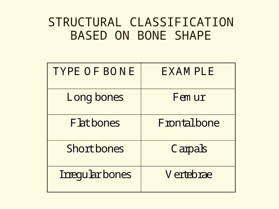
STRUCTURAL CLASSIFICATION BASED ON BONE SHAPE
TYPE OF BONE EXAMPLE
Long bones Femur
Flat bones Frontal bone
Short bones Carpals
Irregular bones Vertebrae
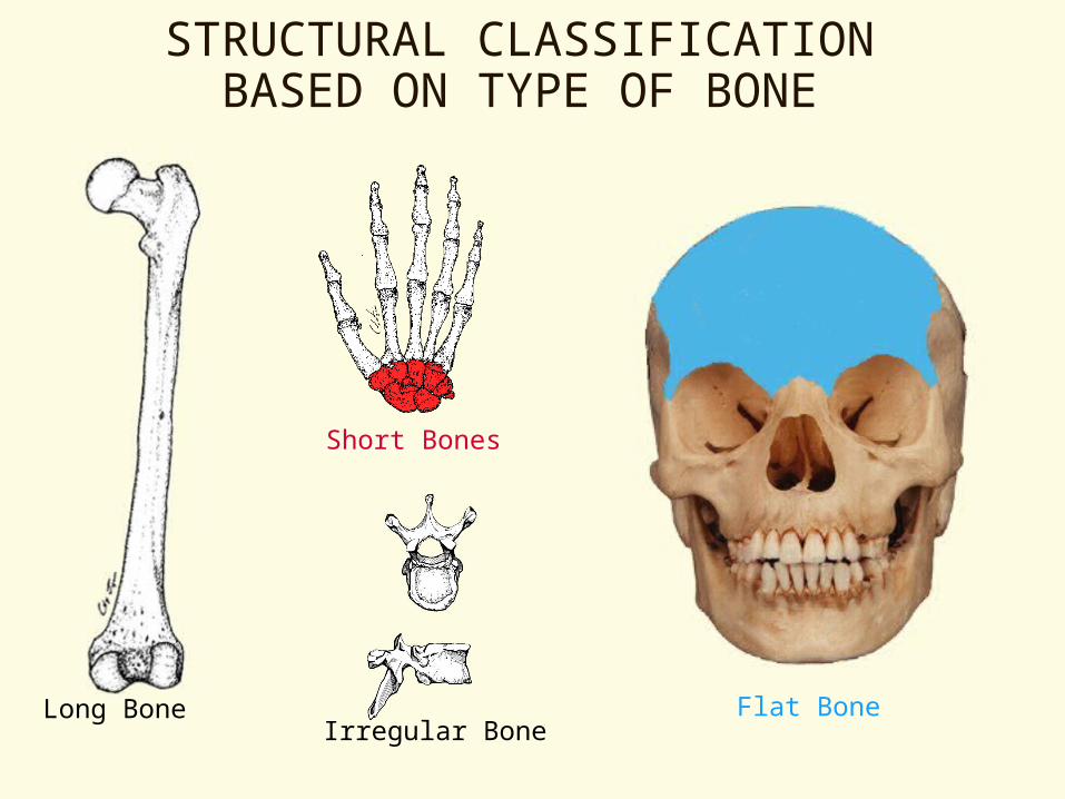
Long BoneIrregular Bone
Flat Bone
Short Bones
STRUCTURAL CLASSIFICATION BASED ON TYPE OF BONE
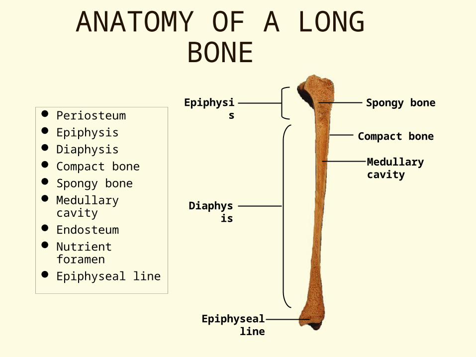
ANATOMY OF A LONG BONE
Periosteum Epiphysis Diaphysis Compact bone Spongy bone Medullary cavity Endosteum Nutrient foramen Epiphyseal line
Epiphysis
Diaphysis
Spongy bone
Compact bone
Medullary cavity
Epiphyseal line

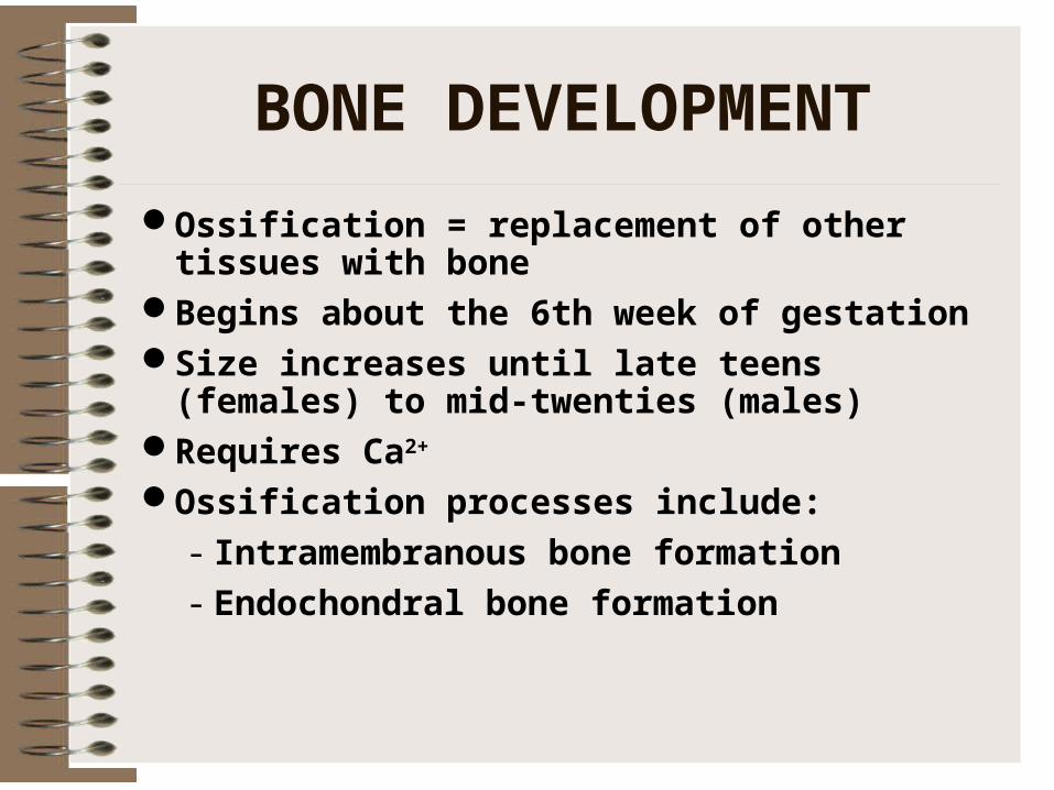
BONE DEVELOPMENT
Ossification = replacement of other tissues with bone
Begins about the 6th week of gestationSize increases until late teens (females) to mid-
twenties (males)Requires Ca2+
Ossification processes include:
- Intramembranous bone formation
- Endochondral bone formation
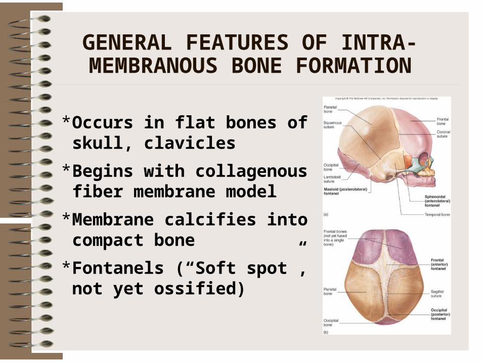
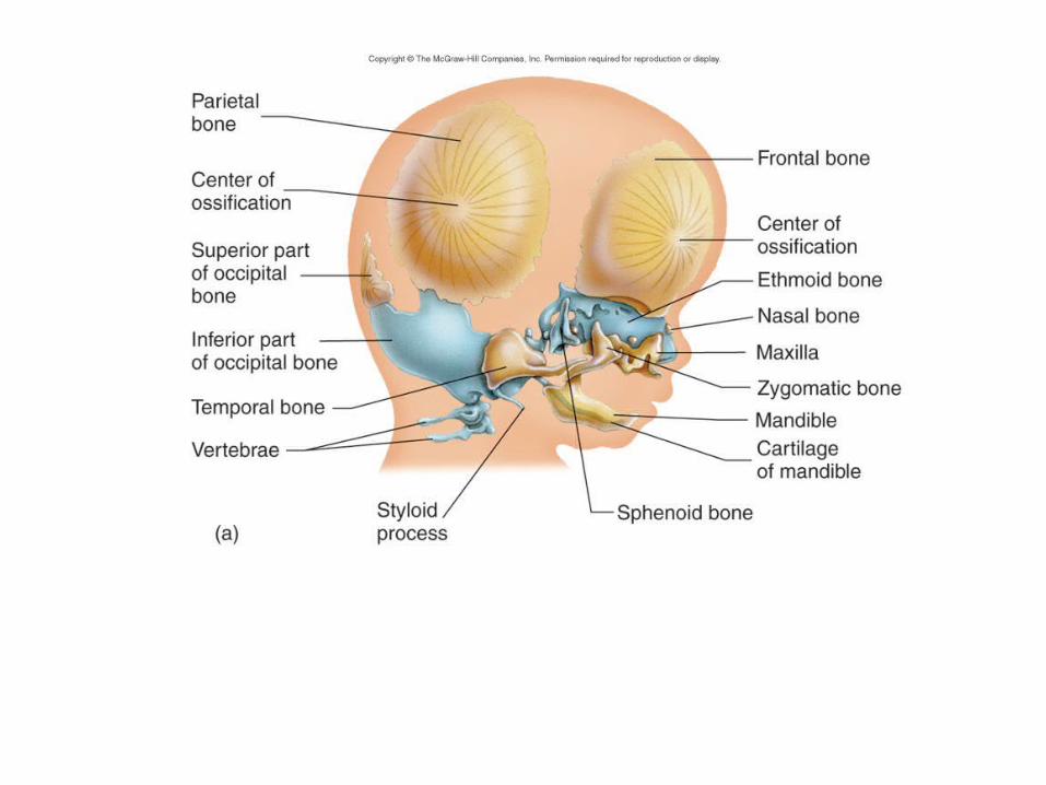
GENERAL FEATURES OF INTRA-MEMBRANOUS BONE FORMATION
* Occurs in flat bones of skull, clavicles
* Begins with collagenous fiber membrane model
* Membrane calcifies into compact bone
* Fontanels (“Soft spot”, not yet ossified)
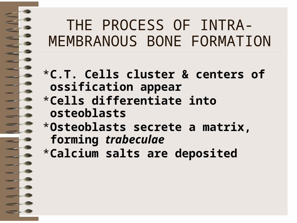
THE PROCESS OF INTRA-MEMBRANOUS BONE FORMATION
* C.T. Cells cluster & centers of ossification appear
* Cells differentiate into osteoblasts* Osteoblasts secrete a matrix, forming
trabeculae* Calcium salts are deposited
* Trabeculae fuse into spongy bone lattice
* Lattice fills with red bone marrow
* Eventually, peripheral trabeculae thicken into compact bone (periosteal ossification)
THE PROCESS OF INTRA-MEMBRANOUS BONE FORMATION
CONTINUED
* Occurs in remainder of skeleton
* Begins with hyaline cartilage model
* Cartilage is replaced by bony tissue
GENERAL FEATURES OF ENDOCHONDRAL BONE FORMATION
THE PROCESS OF ENDOCHONDRAL BONE FORMATION
FORMATION OF BONE COLLAR
- Cartilage model is covered by perichondrium
- Perichondrium becomes periosteum
- A “collar” of bone is produced around the diaphysis
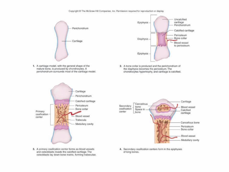

THE PROCESS OF ENDOCHONDRAL BONE FORMATION
CALCIFICATION OF DIAPHYSEAL CARTILAGE- Hypertrophy of chondrocytes
- Surrounding matrix calcifies
- Diffusion disabled, chondrocytes die
- Cartilaginous matrix disintegrates

THE PROCESS OF ENDOCHONDRAL BONE FORMATION CONTINUED
FORMATION OF PRIMARY OSSIFICATION CENTER- Diaphysis penetrated by blood vessels,
osteoblasts, osteoclasts- Marrow cavity formed by osteoclasts- Trabeculae form (Spongy bone) - Cartilage model grows at ends,
elongating bone

THE PROCESS OF ENDOCHONDRAL BONE FORMATION CONTINUED
FORMATION OF SECONDARY CENTER OF OSSIFICATION- Blood vessels reach epiphyses- Secondary ossification centers develop- Spongy bone is formed- Cartilage is replaced by bone, except at
articular surfaces- Cartilage remains at epiphyseal plate
(metaphysis) until growth is complete

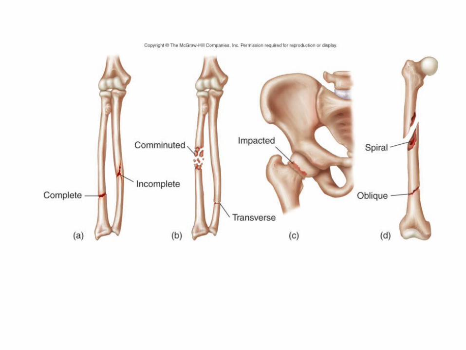
FRACTURES AND THEIR REPAIR
Definition: Any break in a boneRepair may take monthsTypes include
- Simple (skin not broken)
- Compound (bone protrudes through skin)
- Greenstick (shaft bent/broken)
- Spiral (twisting force, ragged break)
- Comminuted (shattered into fragments)
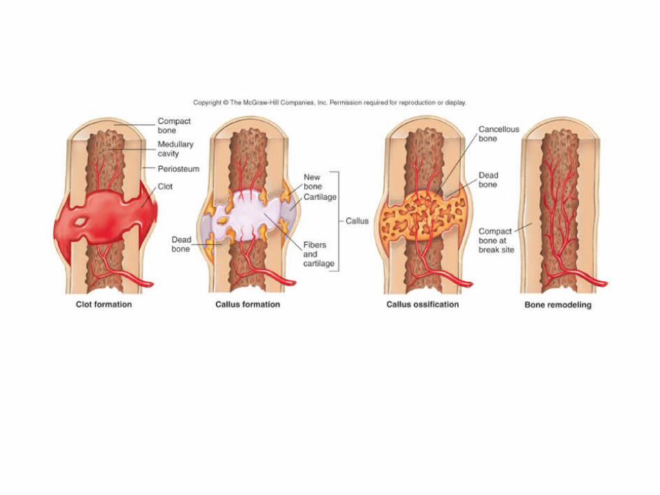
STEPS IN FRACTURE REPAIR
- Broken blood vessels form a fracture hematoma
- C.T. and Capillaries invade site, form fibrocartilage callus
- Repair cells (osteoblasts) are activated in about 48 hours
- Bony callus replaces fibrocartilage callus- Bony callus is remodeled by osteoclasts
BONES AS LEVERS
Lever: A rigid rod that moves about a fixed point
Fulcrum: The fixed point around which a lever moves (joints)
Forces: Act to move levers at two points- Resistance: Force to be overcome- Effort or Work: Force required to overcome
resistance; supplied by skeletal muscles
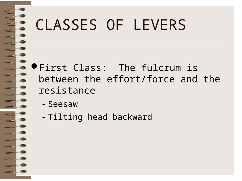
CLASSES OF LEVERS
First Class: The fulcrum is between the effort/force and the resistance- Seesaw
- Tilting head backward
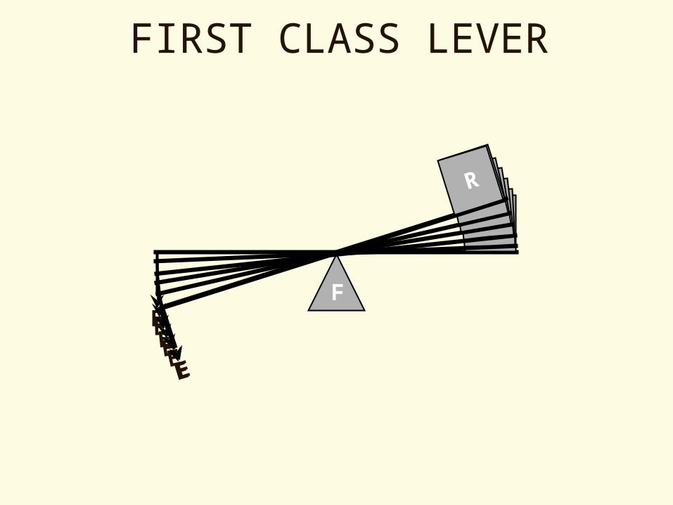
FIRST CLASS LEVER
F
R
E
R
E
R
E
R
E
R
E
R
E
R
E
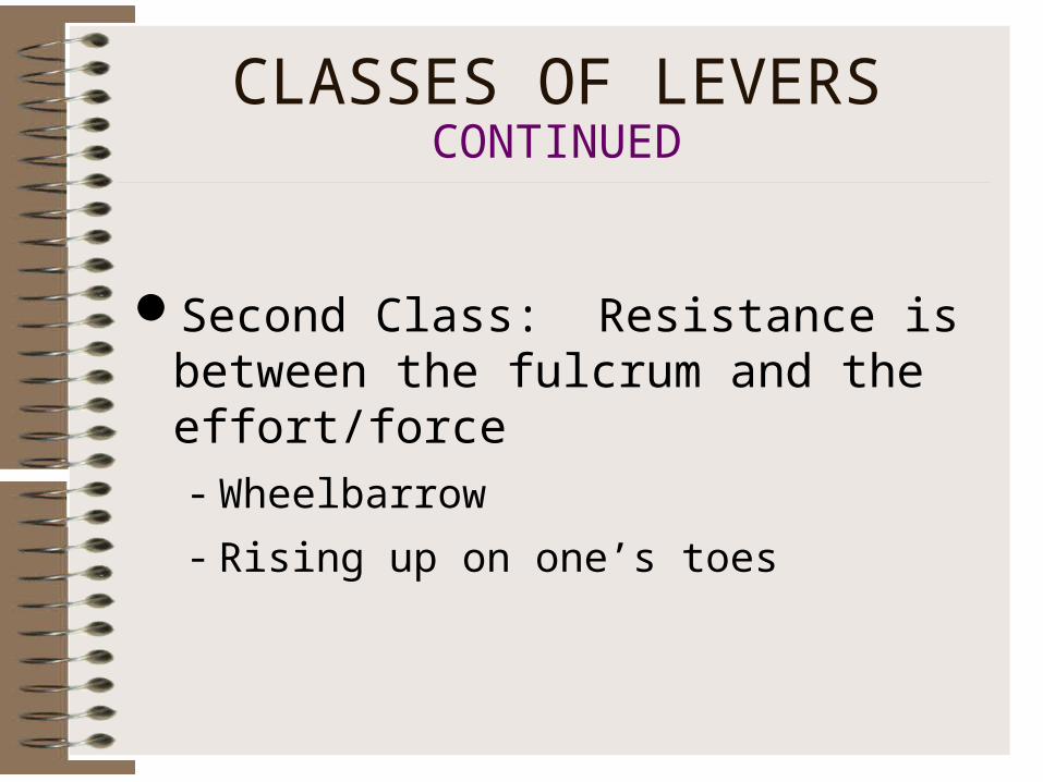
CLASSES OF LEVERS CONTINUED
Second Class: Resistance is between the fulcrum and the effort/force- Wheelbarrow
- Rising up on one’s toes
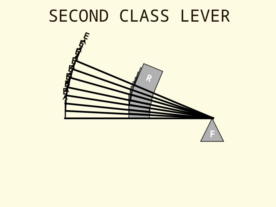
SECOND CLASS LEVER
F
R
ER
E
R
E
R
E
R
E
R
E
R
E
R
E

Third Class: The effort/force is between the fulcrum and the resistance- Most common type in the human body
- Flexing the elbow
CLASSES OF LEVERS CONTINUED
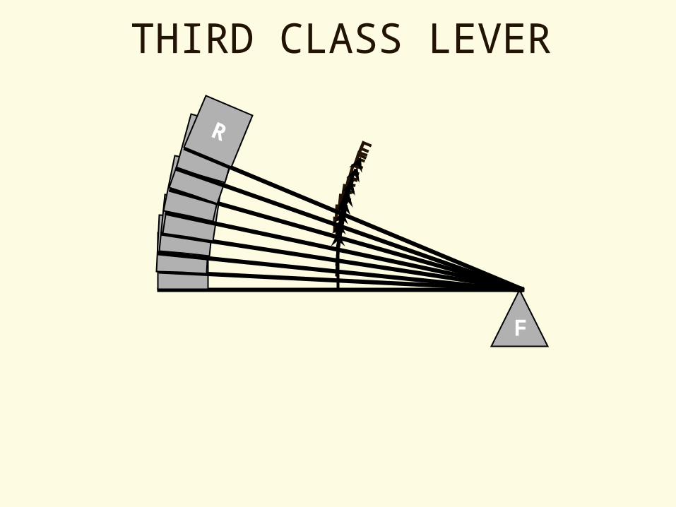
THIRD CLASS LEVER
F
R
ER
ERERER EER
R ER
E
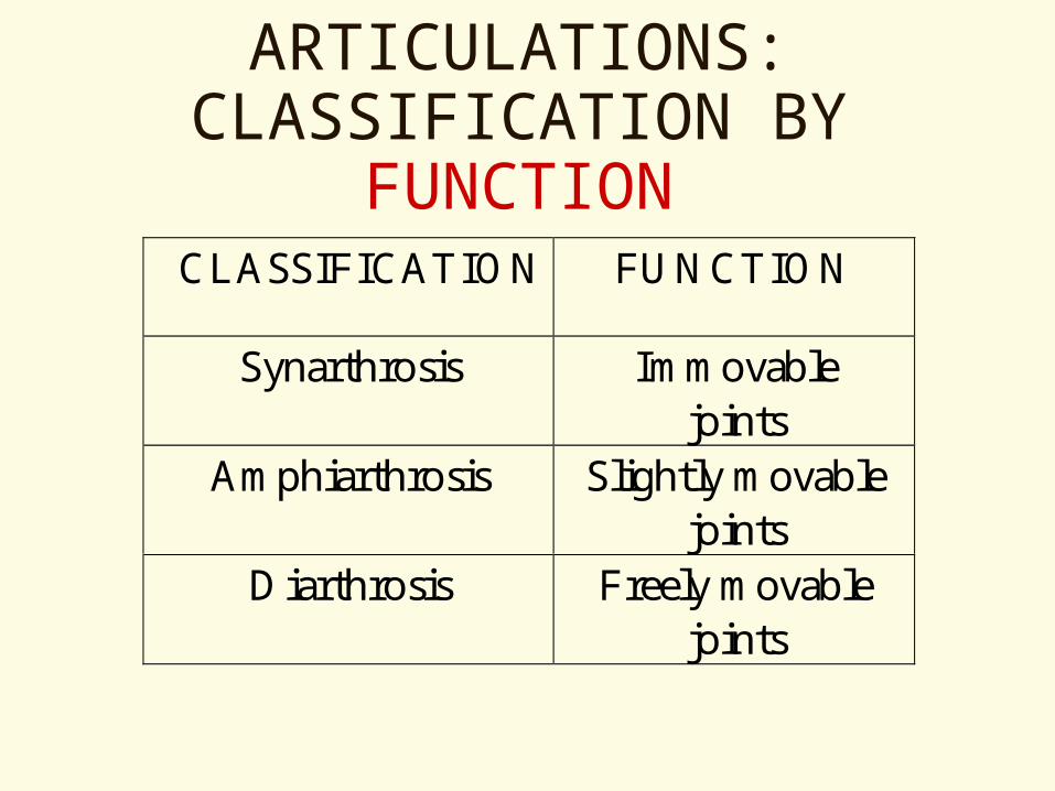
ARTICULATIONS: CLASSIFICATION BY
FUNCTIONCLASSIFICATION FUNCTION
Synarthrosis Immovablejoints
Amphiarthrosis Slightly movablejoints
Diarthrosis Freely movablejoints
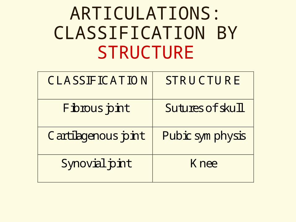
ARTICULATIONS: CLASSIFICATION BY
STRUCTURECLASSIFICATION STRUCTURE
Fibrous joint Sutures of skull
Cartilagenous joint Pubic symphysis
Synovial joint Knee
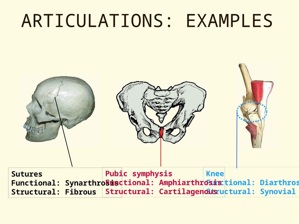
ARTICULATIONS: EXAMPLES
SuturesFunctional: SynarthrosisStructural: Fibrous
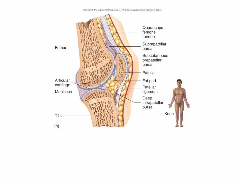
KneeFunctional: DiarthrosisStructural: Synovial
Pubic symphysisFunctional: AmphiarthrosisStructural: Cartilagenous

STRUCTURE OF A SYNOVIAL JOINT
Articular cartilage – cover bone endsSynovial membrane – lines joint capsuleSynovial fluid – lubricates & nourishes
cartilageSynovial cavityJoint capsule – fibrous C.T.Ligaments – reinforce jointBursae – synovial sacs at other sites of
friction

TYPES OF SYNOVIAL JOINTS
Classified based on shape of articular surfaces
Gliding (plane)HingePivotEllipsoidal (condyloid)SaddleBall-and-socket