Siva Raman CV
22
CURRICULUM VITAE Dr. Siva P Raman, MD 8887 Purple Iris Lane Cell Phone: 916-342-6687 Elkridge, MD 21075 [email protected] Work Experience: 2011 – Present Assistant Professor of Radiology and Radiological Sciences, Johns Hopkins University School of Medicine, Baltimore, MD 2012 – 2014 Associate Program Director for Johns Hopkins Radiology Residency Other Major Experiences: 2013 – Present Amirsys Publishing Consultant and Author Co-Lead editor and author working with Dr. Michael Federle to edit and create content for StatDx, Radprimer, and Diagnostic Imaging Series (Gastrointestinal). Co-editor of "Diagnostic Imaging: Gastrointestinal" 3 rd Edition textbook due to be released in June 2015 2014 – Present Journal of the American College of 1
-
Upload
siva-raman -
Category
Documents
-
view
258 -
download
1
Transcript of Siva Raman CV
CURRICULUM VITAEDr. Siva P Raman, MD
8887 Purple Iris Lane Cell Phone: 916-342-6687Elkridge, MD 21075
Work Experience:
2011 – Present Assistant Professor of Radiology and Radiological Sciences, Johns Hopkins University School of Medicine, Baltimore, MD
2012 – 2014 Associate Program Director for Johns Hopkins Radiology Residency
Other Major Experiences:
2013 – Present Amirsys Publishing Consultant and Author
Co-Lead editor and author working with Dr. Michael Federle to edit and create content for StatDx, Radprimer, and Diagnostic Imaging Series (Gastrointestinal).
Co-editor of "Diagnostic Imaging: Gastrointestinal" 3rd Edition textbook due to be released in June 2015
2014 – Present Journal of the American College of Radiology Feature Co-editorFeature co-editor (along with Elliot Fishman) of a series of opinion articles published in JACR starting in 2015
Education:
Undergraduate
1
1997 - 2000 Bachelor of Arts, Psychobiology, Los Angeles, CA (Member of eight year BA-MD program)
2000-2001 Study in European History, University of Cambridge (UK)
Doctoral
2001 - 2005 Doctor of Medicine (MD), Johns Hopkins University School of Medicine, Baltimore, MD
Postdoctoral
2005 - 2006 Preliminary Internal Medicine Intern, University of California at Davis, Sacramento, CA
2006 – 2010 Resident, Diagnostic Radiology, University of Washington, Seattle, WA
2010 – 2011 Abdominal Imaging Fellowship, Department of Radiology, Stanford University, Stanford, CA
Board Certification:
2007 ABR Physics Examination - Passed2008 ABR Written Examination – Passed2010 ABR Oral Examination – Passed (Scored 72 in neuroradiology; 71 in all other sections)
Medical Examinations:
2003 USMLE Step I: Three Digit Score - 254 Percentile - 99 2005 USMLE Step II: Three Digit Score - 244 Percentile - 992004 USMLE Step II CS (Clinical Skills) - Pass2006 USMLE Step III: Three digit Score - 233 Percentile - 961999 MCAT: Total Score 41 (15/15 biology; 15/15 chemistry/physics; 11/13 Verbal)
Honors and Awards:
2015 Herbert M. Stauffer award (Association of University Radiologists/AUR) for best basic science paper Award given for “CT texture analysis of renal masses: Pilot study using random forest classification for prediction of pathology” in Academic Radiology
2
2013 Ahmad A. Fakhri Johns Hopkins research award2010 Fred Mann Award for outstanding research project at Harborview Medical Center2008 University of Washington: Award of excellence in thoracic radiology1999 University of Cambridge Overseas Trust Scholarship1999 Phi Kappa Phi1999 Golden Key1998-99 Outstanding organic chemistry student award1997 Trustee Scholarship (Full tuition paid for undergraduate education)1997 Gencorp National Merit Scholar1997 High-School Valedictorian
Other Experience and Professional Memberships
2012 – Present American Roentgen Ray Society member2013 – Present ARRS SHERPA committee chairperson2012 – Present Reviewer for Radiology2012 – Present Reviewer for Emergency Radiology2012 – Present Reviewer and contributor for Radiologyinfo.org2014 – Present Feature co-editor (along with Elliot Fishman) for ‘Rethinking the patient
experience’ series in JACR2013 – Present Consultant and author for Amirsys Publishing
Peer-reviewed publications (current h-index: 12; i10-index: 13)
1. Gallia GL, Scuibba DM, Hann CL, Raman SP, Westra WH, Tufaro AP, and Olivi A. Synovial sarcoma of the frontal sinus. Case Report. J Neurosurgery. 2005; 103: 1077-1080
2. Cherry CL, Skolasky RL, Lal L, Creighton J, Hauer, Raman SP, et al. Antiretroviral use and other risks for HIV-associated neuropathies in an international cohort. Neurology. 2006; 66(6): 867-73
3. Prabhakaran V, Raman SP, Grunwald MR, Mahadevia A, Hussain N, Lu H, Van Zijl PCM, Hillis AE. Neural substrates of word generation during stroke recovery: the influence of cortical hypoperfusion. Behav Neurol. 2007; 18(1): 45-52
4. Are C, Raman SP, Ravipatti N, and Talamini M. Decreased porcine uterine blood flow during laparoscopic Nissen fundoplication. Journal of Gynecologic Surgery. 2004; 20(4): 113-118 (NOT IN PUB-MED)
5. Zhou L, Kitch W, Evans SR, Hauer P, Raman S, Ebenezer GJ, Gerschenson M, Marra CM, Valcour V, DiazArrastia R, Goodkin K, Millar L, Schriver S, Asmuth DM, Clifford DB, Simpson DM, McArthur JC, NARC and ACTG A5117 Study Group. Correlates of Epidermal Nerve Fiber Densities in HIV associated Distal Sensory Polyneuropathy. Neurology. 2007; 68(24): 2113-9
6. Raman SP, Lehnert B, and Pruthi S. “Unusual presentation of ACE-inhibitor induced pharyngeal angioedema. AJNR. Jan 2009. 30(1): 77-78
7. Chen YH, Carrino JA, Raman SP, Morrison WB, and Fayad LM. “Atraumatic lateral 3
collateral ligament complex signal abnormalities by magnetic resonance imaging in patients with osteoarthritis of the knee. Journal of computer assisted tomography. 2008. 32(6): 982-986
8. Raman SP and Pipavath S. “Images in clinical medicine: Asymmetric edema of the upper lung due to mitral valvular dysfunction.” New England Journal of Medicine. 2009. 361(5): e6
9. Raman SP, Pipavath S, Raghu G, Schmidt R, and Godwin D. “Imaging of thoracic lymphatic diseases” American Journal of Radiology. December 2009. 193(6)
10. Raman SP and Fishman EK. “Imaging: Multidetector CT in the diagnosis of suspected appendicitis.” Nat Rev Gastroenterol Hepatol. September 20, 2011; 8(11): 607-609
11. Raman SP, Horton KM, and Fishman EK. “Transitional Cell Carcinoma of the Upper Urinary Tract: Optimizing Image Interpretation with 3-D Reconstructions.” 2011 Dec 30 [Epub ahead of print]
12. Raman SP, Neyman EG, Horton KM, Eckhauser FE, and Fishman EK. Superior mesenteric artery syndrome: Spectrum of CT findings with multiplanar reconstructions and 3-D imaging. Abdom Imaging (2012); 37(6): 1079-88
13. Raman SP, Hruban RH, Cameron JL, Wolfgang CL, Kawamoto S, and Fishman EK. Acinar cell carcinoma of the pancreas: Computed tomography features – A study of 15 patients. Abdom Imaging (2013); 38(1): 137-143
14. Raman SP, Kamaya A, Federle M, and Fishman EK. Aortoenteric Fistulas: Spectrum of CT findings. Abdom Imaging (2013); 38(2): 367-375
15. Raman SP, Hruban RH, and Fishman EK. Beyond renal cell carcinoma: Rare and unusual renal masses. Abdom Imaging (2012); 37(5): 873-84
16. Raman SP, Hruban RH, and Fishman EK. Hepatic adenomatosis: Spectrum of imaging findings. Abdom Imaging (2013); 38(3): 474-81
17. Raman SP, Hruban RH, Cameron JL, Wolfgang CL, and Fishman EK. Pancreatic imaging mimics: Part II, Pancreatyic Neuroendocrine tumors and their mimics. AJR. 2012 Aug; 199(2): 309-318
18. Smith JA, Wild AT, Singhi A, Raman SP, et al. Clinicopathologic comparison of high-dose-rate endorectal brachytherapy versus conventional chemoradiotherapy in the neoadjuvant setting for resectable stages II and III low rectal cancer. Int J Surg Oncol. 2012; 2012: 406568 [Epub 2012 July 8].
19. Raman SP and Fishman EK. Advances in CT imaging of GI malignancies. Gastrointest Cancer Res. 2012 May; 5(3 Suppl 1): S4-9
20. Raman SP, Singhi A, Horton KM, Hruban RH, and Fishman EK. Sclerosing angiomatoid nodular transformation (SANT): Multimodality imaging appearance of five cases with radiology-pathology correlation. Abdom Imaging (2013); 38(4): 827-234
21. Raman SP, Horton KM, Fishman EK. Multimodality imaging of pancreatic cancer – CT, MRI, and PET. The Cancer Journal (2012); 8(6): 511-522
22. Raman SP, Johnson PT, Deshmukh S, Mahesh M, Grant KL, and Fishman EK. CT dose reduction applications: Available tools on the latest generation of CT scanners. JACR (2013); 10(1): 37-41.
23. Raman SP, Raminpour S, Horton KM, and Fishman EK. Informatics in radiology: CT contrast protocols application on the iPad: A new resource for technologists, nurses, and radiologists. Radiographics (2013); 33(3): 913-21
4
24. Raman SP, Kawamoto S, Blackford A, Hruban RH, Wolfgang CL, O’Brien-Lennon AM, Edil B, and Fishman EK. Histopathologic findings of multifocal pancreatic mucinous neoplasms on CT. AJR (2013); 200(3): 563-569
25. Raman SP, Fishman EK, and Lennon AM. Endoscopic ultrasound and pancreatic applications: What the radiologist needs to know. Abdominal Imaging (2013) Jan 19 [Epub ahead of print].
26. Raman SP, Horton KM, Cameron JL, and Fishman EK. Computed tomography after pancreaticoduodenectomy: Spectrum of normal findings and complications. AJR (2013); 201(1): 2-13
27. Raman SP, Salaria S, Hruban RH, and Fishman EK. Groove pancreatitis: Spectrum of imaging findings and radiology-pathology correlation. AJR (2013); 201(1): W29-39
28. Raman SP and Fishman EK. State of the art multidetector CT imaging for hepatobiliary malignancies. 2013 Gastrointestinal Cancers Symposium Proceedings. (NOT IN PUB-MED)
29. Raman SP, Horton KM, and Fishman EK. MDCT/CTA evaluation of rectal bleeding: The role of volume visualization. AJR (2013); 201(3): 589-97
30. Northcutt B, Raman SP, and Johnson PT. MDCT of adrenal masses: can dual-phase enhancement patterns be used to distinguish between adenoma and pheochromocytoma? AJR (2013); 201(4): 834-9
31. Raman SP, Law J, Kawamoto S, et al. Institutional experience with solid-pseudopapillary neoplasms: Focus on CT, MRI, conventional ultrasound, and predictors of aggressive histology. JCAT (2013); 37(5): 824-33
32. Raman SP, Horton KM, and Fishman EK. Computed tomography of Crohn’s disease: The role of 3D technique. World J Radiol (2013); 25; 5(5): 193-20
33. Dalal PS, Raman SP, Horton KM, and Fishman EK. Portal vein aneurysms: Imaging manifestations and clinical significance. Emergency Radiology (2013); 20(5): 453-7
34. Raman SP, Johnson PT, Allaf M, Netto G, and Fishman EK. Chromophobe renal cell carcinoma: Multiphase MDCT enhancement patterns and morphology. AJR (2013); 201(6):1268-76.
35. Raman SP, Horton KM, and Fishman EK. MDCT evaluation of ureteral tumors: Advantages of 3-D reconstruction and volume visualization. AJR (2013); 201(6): 1239-1247
36. Raman SP, Mahadevappa M, Blasko RV, and Fishman EK. CT scan parameters and radiation dose: Practical advice for radiologists. JACR (2013); 10(11): 840-846
37. Raman SP and Fishman EK. Mycotic aneurysms: A critical diagnosis in the emergency setting. Emergency Radiology (2013) [Epub ahead of print]
38. Dholakia AS, Kumar R, Raman SP, et al. Mapping Patterns of Local Failure Following Pancreaticoduodenectomy for Pancreatic Cancer: A New Approach to Adjuvant Radiation Fields. Internation Journal of Radiation Oncology, Biology, and Physics (2013); 87(5): 1007-1015
39. Mitchell C, Johnson PT, Fishman EK, and Raman SP. Features suggestive of gallbladder malignancy: Analysis of T1, T2, and T3 tumors on cross-sectional imaging. JCAT (2014) [Epub].
40. Law JK, Ahmed A, Singh VK, Akshintala VS, Htfless SM, Olsen M, Raman SP, et al. A systematic review of solid pseudopapillary neoplasms: Are these rare lesions?
5
Pancreas (2014); 43(3): 331-33741. Dreizin D, Infante J, Tirada N, Raman SP, Madrazo B. Focal nodular hyperplasia within
accessory liver: Imaging findings at computed tomography and magnetic resonance imaging. JCAT (2014); 38(2): 235-241
42. Dreizin D, Bordegaray M, Tirada N, Raman SP, Kadakia K, and Munera F. Evaluating blunt pancreatic trauma at whole body CT: Current practices and future directions. Emerg Radiol (2013); 20(6): 517-527
43. Wild AT, Chang DT, Goodman KA, Laheru DA, Zheng L, Raman SP, et al. A phase 2 multi-institutional study to evauate gemcitabine and fractionated stereoteactic radiotherapy for unresectable, locally advanced pancreatic adenocarcinoma. Pract Radiat Oncol (2013); 3(2 Suppl 1): S4-5
44. Dholakia AS, Chaudhry M, Leal JP, Chang DT, Raman SP, et al. Baseline Metabolic Tumor Volume and Total Lesion Glycolysis is Associated With Improved Survival Outcomes in Patients with Locally Advanced Pancreatic Cancer Receiving Stereotactic Body Radiation Therapy. International Journal of Radiation Oncology, Biology, Physics (2014); S0360-3016(14)-275-2
45. Lennon AM, Manos LL, Hruban RH, Ali SZ, Fishman EK, Kamel IR, Raman SP, et al. Role of a multidisciplinary clinic in the management of patients with pancreatic cysts: A single center cohort study. Ann Surg Oncol 2014 May 8 [Epub ahead of print]
46. Raman SP and Fishman EK. Abnormalities of the distal common bile duct and ampulla: Diagnostic approach and differential diagnosis using multiplanar reformats and 3-D imaging. AJR 2014; 203(1): 17-28
47. Raman SP and Fishman EK. Bladder malignancies on computed tomography: The underrated role of CT in diagnosis. AJR 2014; 203(2): 347-354
48. Raman SP, Chen Y, Schroeder JL, Huang P, and Fishman EK. CT texture analysis of renal masses: Pilot study utilizing random forest classification for prediction of pathology. Academic Radiology. 2014 Dec; 21(12): 1587-96
49. Sinha A, Singh VK, Cruise M, Afghani E, Matsukuma K, Ali S, Anderson DK, Makary M, Raman SP, et al. Abdominal CT predictors of fibrosis in patients with chronic pancreatitis undergoing surgery. European Radiology. Dec 4 2014 [Epub ahead of print]
50. Raman SP, Reddy S, Weiss MJ, Manos LL, et al. The impact of the time interval between MDCT imaging and surgery on the accuracy of identifying metastatic disease in patients with pancreatic cancer. AJR. 2015 Jan; 204(1): W37-42
51. Herman JM, Chang DT, Goodman KA, Dholakia AS, Raman SP, et al. Phase II multi-institutional trial evaluating gemcitabine and stereotactic body radiation therapy for locally advanced unresectable pancreatic adenocarcinoma. Cancer. 2014 Dec 23 [Epub ahead of print]
52. Raman SP, Horton KM, and Fishman EK. Introduction to the ‘Rethinking the patient experience’ series. JACR. 2015 Jan; 12(1): 16
53. Schulze HH, Fishman EK, Horton KM, and Raman SP. The pursuit of excellence: From hotels to hospitals. JACR. 2015 Jan; 12(1): 17-8
54. Moningi S, Dholakia AS, Raman SP, Blackford A, et al. The role of stereotactic body radiation therapy for pancreatic cancer: A single institution experience. Ann Surg Oncol 2015 Jan 7 [Epub ahead of print]
55. Raman SP, Lessne M, Kawamoto S, Chen Y, Salvatori R, Prescott JD, and Fishman EK.
6
Diagnostic performance of multidetector computed tomography in distinguishing unilateral from bilateral abnormalities in primary hyperaldosteronism: Comparison of MDCT with adrenal vein sampling. JCAT. Jan 2015 [Epub ahead of print]
56. Zember W, Fishman EK, Horton KM, and Raman SP. How social media can impact medicine and radiology. JACR. 2015 Jan 28 (Epub ahead of print]
57. Raman SP, Schroeder JL, Huang P, Chen Y, Coquia SF, et al. Preliminary data using CT texture analysis for the classification of hypervascular liver lesions: Generation of a predictive model on the basis of quantitative spatial frequency measurements – A work in progress. JCAT. 2015 Feb 13 [Epub ahead of print]
58. Catmull E, Fishman EK, Horton KM, and Raman SP. From ‘Toy Story’ to CT scans: Lessons from Pixar for radiology. JACR. 2015 Feb 19 [Epub ahead of print]
59. Raman SP, Chen Y, and Fishman EK. Cross-sectional imaging and the role of PET in pancreatic cancer evaluation. Seminars in Oncology. 2015 Feb; 42(1): 40-58
60. Wolf C, Fishman EK, Horton KM, and Raman SP. “Stories from the Kitchen: Lessons for Radiology from the Restaurant Business.” JACR. 2015 Mar; 12(3): 307-308
61. Raman SP, Chen Y, and Fishman EK. Evolution of imaging in rectal cancer: Multimodality imaging with MDCT, MRI, and PET. Journal of Gastrointestinal Oncology. 2015 Apr; 6(2): 172-184
62. Greenberg P, Fishman EK, Horton KM, and Raman SP. Management in the media age: Lessons for radiology from the world of television and music. JACR. 2015 May; 12(5): 491-2
63. Wells T, Fishman EK, Horton KM, and Raman SP. Millenial mindset: Pursuing the next generation of consumers. JACR. 2015 June 1 [Epub ahead of print]
64. Raman SP. Reply to ‘Concerning preoperative evaluation of pancreatic adenocarcinoma.’ AJR. 2015 Aug; 205(2): W226
65. Kawamoto S, Raman SP, Blackford A, Hruban RH, Fishman EK. CT detection of symptomatic and asymptomatic Meckel’s diverticulum. AJR. 2015 Aug; 205(2): 281-91
66. Kaplowitz M, Fishman EK, Horton KM, and Raman SP. Improving patient care through inspiring happiness. JACR. 2015 July 22 [Epub ahead of print]
67. Springer S, Wang Y, Dal Molin M, Masica DL, Jiao Y, Kinde I, Blackford A, Raman SP, Wolfgang CL, Tomita T, et al. A combination of molecular markers and clinical features improve the classification of pancreatic cysts. Gastroenterology. 2015 Aug 4 [Epub ahead of print].
68. Hrabowski FA 3rd, Fishman EK, Horton KM, and Raman SP. Inclusive excellence and institutional culture change. JACR. 2015 Sep 1 [Epub ahead of print]
69. Bonecamp D, Raman SP, Horton KM, and Fishman EK. Role of computed tomography angiography in detection and staging of small bowel carcinoid tumors. World J Radiol. 2015 Sep 28; 7(9): 220-235
Peer-Reviewed Manuscripts Accepted and In Press.
1. Dholakia AS, Hacker-Prietz A, Wild AT, Raman SP, et al. Resection of borderline resectable pancreatic cancer after neoadjuvant chemoradiation does not depend on improved radiographic appearance tumor-vessel relationships: A retrospective review.
7
Radiation oncology. IN PRESS.2. Raman SP and Fishman EK. CT Angiography of the small bowel and mesentery.
Radiologic Clinics of North America. IN PRESS.3. Phillips B, Fishman EK, Horton KM, and Raman SP. The Men’s Health special sauce:
ingredients revealed. JACR. IN PRESS.
Book Chapters:
1. Are C, Raman SP, Talamini MA. Port Site Closure Methods and Hernia Prevention. Chapter in ”The SAGES Manual of Peri-Operative Care in Minimally Invasive Surgery.” Edited by Richard Whelan. 2004.
2. Are C, Raman SP, and Talamini M. Physiologic consequences of laparoscopic surgery. “Mastery of endoscopic and laproscopic surgery, 2nd edition.” Lipincott Williams and Wilkens
3. Raman SP, Horton KM, and Fishman EK. Vascular disorders of the small bowel. Textbook of Gastrointestinal Radiology (4th Edition) by Gore & Levine.
4. Raman SP and Gayer G. Post-surgical and traumatic lesions of the biliary tract. Textbook of Gastrointestinal Radiology (4th Edition) by Gore & Levine.
5. Raman SP and Fishman EK. Computed Tomography of the Chest: Evaluation of the Solitary Pulmonary Nodule. Current Therapy in Thoracic and Cardiovascular Surgery (2nd edition). IN PRESS.
6. Raman SP, Horton KM, Johnson PT, Megibow A, and Fishman EK. Applications of computed tomography to the gastrointestinal tract. Textbook of Gastroenterology (6 th
edition) by Kalloo et al. IN PRESS.7. Raman SP and Fishman EK. Multidetector computed tomography in the evaluation of
cystic tumors of the pancreas. Cystic tumors of the pancreas: Diagnosis and Treatment. Del Chairo M, Haas, SL, and Schulick R. IN PRESS.
8. Raman SP and Fishman EK. Multidetector computed tomography and magnetic resonance imaging of distal cholangiocarcinoma. Cholangiocarcinoma: From diagnosis to treatment. Nova Science Publishers. Pawlik T and Ribero D. IN PRESS.
9. Raman SP. Aortoenteric fistula. Pearls and Pitfalls in Cardiovascular Imaging. Zimmerman S and Fishman EK. Cambridge University Press, Cambridge UK. IN PRESS
10. Raman SP and Fishman EK. Computed tomography of urologic malignancies: The role of MDCT in renal cell carcinoma and transitional cell carcinoma. Management of urologic cancer: Focal therapy and tissue preservation. Wiley, Oxford UK. IN PRESS.
11. Raman SP, Horton KM, Johnson PT, Fishman EK, Megobow AJ. Applications of computed tomography to the gastrointestinal tract. Atlas of Gastroenterology 4 th Edition. Yamada T, Alpers DH, Laine L, Kaplowitz N, Owyang C, Powell DW. Lipincott Williams & Wilkins. IN PRESS.
Textbooks:
8
1. Federle M and Raman SP. Diagnostic Imaging: Gastrointestinal. Elsevier (Amirsys). IN PRESS (2015) (1st author of 108 chapters in textbook)
Scientific Abstracts and Presentations:
1. Prabhakaran V, Raman SP, Grunwald MR, Mahadevia A, Werner JK, Philipose LE, Hussain N, Alphs HH, Sun P, Lu H, Barker PB, Wityk RJ, Rypma B, Hillis AE. Neural substrates involved in cognition for stroke patients and control subjects. Cognitive Neuroscience. San Francisco, CA. April 2004 (Presenter of Scientific Poster)
2. Prabhakaran V, Raman SP, Grunwald MR, Mahadevia A, Werner JK, Philipose LE, Hussain N, Alphs HH, Sun P, Lu H, Rypma B, Van Zijl PCM, Hillis AE. Localization of cognitive processes using stroke patients and fMRI. Cognitive Science. Chicago, IL. September 2004.
3. Philipose LE, Alphs HH, Grunwald MR, Raman SP, Mahadevia A, Prabhakaran V, Hillis AE. Validating fMRI results using acute stroke patients. Auditory Brain Imaging Conference. December 2004.
4. Philipose LE, Alphs HH, Grunwald MR, Raman SP, Mahadevia A, Prabhakaran V, Hillis AE. Validating fMRI results using acute stroke patients. Sfn abstracts. San Diego, CA. November 2004.
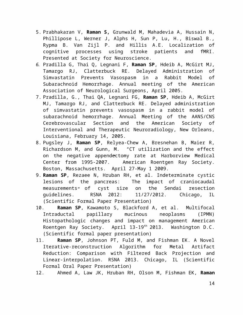
5. Prabhakaran V, Raman S, Grunwald M, Mahadevia A, Hussain N, Phillipose L, Werner J, Alphs H, Sun P, Lu, H., Biswal B., Rypma B. Van Zijl P. and Hillis A.E. Localization of cognitive processes using stroke patients and fMRI. Presented at Society for Neuroscience.
6. Pradilla G, Thai Q, Legnani F, Raman SP, Hdeib A, McGirt MJ, Tamargo RJ, Clatterbuck RE. Delayed Administration of Simvastatin Prevents Vasospasm in a Rabbit Model of Subarachnoid Hemorrhage. Annual meeting of the American Association of Neurological Surgeons, April 2005.
7. Pradilla, G., Thai QA, Legnani FG, Raman SP, Hdeib A, McGirt MJ, Tamargo RJ, and Clatterbuck RE. Delayed administration of simvastatin prevents vasospasm in a rabbit model of subarachnoid hemorrhage. Annual Meeting of the AANS/CNS Cerebrovascular Section and the American Society of Interventional and Therapeutic Neuroradiology, New Orleans, Louisiana, February 14, 2005.
8. Pugsley J, Raman SP, Relyea-Chew A, Bresnehan B, Maier R, Richardson M, and Gunn, M. “CT utilization and the effect on the negative appendectomy rate at Harborview Medical Center from 1995-2007.” American Roentgen Ray Society. Boston, Massachusetts. April 27-May 1 2009.
9. Raman SP, Rezaee N, Hruban RH, et al. Indeterminate cystic lesions of the pancreas: The impact of craniocaudal measurements of cyst size on the Sendai resection guidelines.” RSNA 2012: 11/27/2012. Chicago, IL (Scientific Formal Paper Presentation)
10. Raman SP, Kawamoto S, Blackford A, et al. Multifocal Intraductal papillary mucinous neoplasms (IPMN) Histopathologic changes and impact on management American
9
Roentgen Ray Society. April 13-19th 2013. Washington D.C. (Scientific formal paper presentation)
11. Raman SP, Johnson PT, Fuld M, and Fishman EK. A Novel Iterative-reconstruction Algorithm for Metal Artifact Reduction: Comparison with Filtered Back Projection and Linear-interpolation. RSNA 2013. Chicago, IL (Scientific Formal Oral Paper Presentation)
12. Ahmed A, Law JK, Hruban RH, Olson M, Fishman EK, Raman SP, et al. The epidemiology and clinical outcome of pancreatic solid pseudopapillary neoplasms: A systematic review. Digestive Disease Week (AGA) 2013
13. Mitchell CH, Johnson PT, Fishman EK, Hruban RH, and Raman SP. Early Gallbladder Cancer: CT and MR Findings with Pathologic Correlation. RSNA 2013. Chicago, IL. (Scientific Formal Oral Paper Presentation)
14. Raman SP, Kawamoto S, Law JK, Blackford A, et al. Solid-pseudopapillary Tumors of the Pancreas: Multimodality Imaging Predictors of Aggressive Behavior. RSNA 2013. Chicago, IL. (Scientific poster presentation)
15. Northcutt BG, Zingarelli EN, Trakhtenbroit MA, Raman SP, Fishman EK, and Johnson PT. MDCT of the indeterminate adrenal mass: Identification of a venous enhancement level to distinguish pheochromocytoma from adenoma. RSNA 2013. Chicago, IL. (Scientific formal paper presentation)
16. Raman SP, Schroeder J, Chen Y, Huang P, and Fishman EK. Classification of hypervascular liver lesions using CT texture analysis: Generation of a predictive model using spatial frequency measurements. Society of Abdominal Radiology March 23-28, 2014. Boca Raton, FL. (Scientific poster presentation)
17. Raman SP, Schroeder J, Chen Y, Huang P, and Fishman EK. Classification of hypervascular liver lesions using CT texture analysis: Generation of a predictive model using spatial frequency measurements. American Roentgen Ray Society May 4-9, 2014 (Formal Scientific oral presentation)
18. Rezaee N, Manos LL, Raman SP, et al. Main pancreatic duct size and risk of malignancy in intraductal papillary mucinous neoplasm. Pancreas Club May 2-3 2014 (Chicago, IL)
19. Moningi A, Raman SP, Dholakia AS, et al. Stereotactic body radiation therapy for pancreatic cancer: The Johns Hopkins experience. Pancreas Club May 2-3 2014 (Chicago, IL)
20. Dholakia AS, Kumar R, Wild AT, Hacker-Prietz A, Ellsworth A, Raman SP, et al. Patterns of failure following Whipple procedure for resectable pancreatic ductal adenocarcinoma (oral presentation) Pancreas Club May 17-18 2013. Lake Buena Vista, FL (oral presentation)
21. Dholakia AS, Chang DT, Raman SP, et al. A phase 2 multicenter study to evaluate gemcitabine and fractionated stereotactic body radiation therapy for locally advanced pancreatic adenocarcinoma. American Society for Radiation Oncology September 22-25, 2013. Atlanta, GA (oral presentation).
22. Dholakia AS, Chang DT, Goodman KA, Raman SP, et al. 18Flurodeoxyglucose-PET Baseline Avidity Predicts for Inferior Outcomes in Patients with Locally Advanced Pancreatic Cancer Treated with Gemcitabine and Stereotactic Body Radiotherapy. American Society for Radiation Oncology September 22-25, 2013 Atlanta, GA (poster
10
presentation)23. Dholakia AS, Hacker-Prietz A, Wild A, Raman S, et al. Radiographic changes
associated with surgical resection following neoadjuvant radiation therapy for locally advanced pancreatic cancer American Hepato-Pancreato-Biliary Association February 19-23 2013. Miami, FL (oral presentation).
24. Dholakia A, Hacker-Prietz A, Wild AT, Raman SP, et al. Prognostic factors for achieving resection following neoadjuvant radiation therapy for borderline resectable pancreatic adenocarcinoma. Gastrointestinal Cancers Symposium January 24-26, 2013. San Francisco, CA (Scientific poster)
25. Kumar R, Dholakia AS, Smith JA, Leal JP, Chaudhry MA, Wild AT, Hacker-Prietz A, Raman SP, et al. First report of the correlation of PERCIST criteria and pathologic change in rectal cancer patients treated with neoadjuvant radiation. Gastrointestinal Cancers Symposium January 24-26, 2013. San Francisco, CA (Scientific poster)
26. Moningi S, Raman SP, Dholakia AS, et al. Sterotactic body radiation therapy for pancreatic cancer: Single institution experience American Society of Clinical Oncology (ASCO) GI Cancers Symposium. January 16-18 2014 San Francisco, CA (Scientific poster)
27. Sinha A, Ali A, Cruise M, Matsukuma K, Raman SP, et al. Can abdominal computed tomography (CT) scan predict fibrosis in chronic pancreatitis (CP): A radio-pathologic diagnosis. Digestive Disease Week (AGA) May 3-6, 2014 Chicago, IL
28. Verde F, Raman SP, Chu LC, Chen Y, Huang P, and Fishman EK. Texture analysis with predictive modeling of solid appearing pancreatic serous cystadenoma versus neuroendocrine tumors. RSNA 2014 Chicago, IL (scientific poster)
29. Raman SP, Kawamoto S, Chen Y, Johnson PT, Lessne ML, and Fishman EK. Diagnostic imaging in patients with primary hyperaldosteronism: Correlation of MDCT findings with adrenal vein sampling. RSNA 2014 Chicago, IL (scientific poster)
30. Moningi S, Dholakia AS, Leal JP, Rosati L, Fishman EK, Raman SP, et al. Interpreting baseline and follow-up 18Fluorodeoxyglucose-PET parameters in patients with locally advanced and borderline resectable pancreatic cancer. RSNA 2014 Chicago, IL (scientific paper)
31. Raman SP, Chen Y, Schroeder JL, Huang P, and Fishman EK. Classification of renal masses using CT texture analysis: Generation of a predictive model on the basis of quantitative spatial frequency measurements and random forest modeling. RSNA 2014 Chicago, IL (scientific paper)
32. Kawamoto S, Raman SP, Blackford A, Hruban RH, and Fishman EK. CT detection of complicated and uncomplicated Meckel’s diverticulum. RSNA 2014 Chicago, IL (scientific paper)
Educational Abstracts and Exhibits:
1. Raman SP, Hruban RH, and Fishman EK. Lymphangiomas: Spectrum of Imaging Findings, Differential Diagnosis, and Radiology-Pathology Correlation. RSNA 2012. Chicago, IL. (Educational exhibit)
2. Raman SP, Horton KM, and Fishman EK. The Whipple Procedure: Spectrum of normal post-operative CT appearances and imaging diagnosis of common complications. RSNA
11
2012. Chicago, IL. (Educational exhibit)3. Raman SP, Hruban RH, and Fishman EK. Groove Pancreatitis: Imaging Appearance and
Radiology-Pathology Correlation. RSNA 2012. Chicago, IL. (Educational exhibit)4. Raman SP and Fishman EK. Aortoenteric Fistulas: Spectrum of CT Findings.”
American Roentgen Ray Society (ARRS). Vancouver, Canada. April 29 – May 4, 2012.5. Mitchell CH, Johnson PT, Fishman EK, and Raman SP. Early gallbladder cancer:
Multimodality imaging findings and the identification of subtle lesions. RSNA 2013. Chicago, IL. (Educational exhibit)
6. Raman SP and Fishman EK. Diagnosis of pancreatic lesions and pseudolesions: Pearls, pitfalls, and challenges. RSNA 2013. Chicago, IL. (Educational exhibit)
7. Kawamoto S, Raman SP, Hruban RH, and Fishman EK. Complicated and uncomplicated Meckel’s diverticulum: Spectrum of CT appearance. RSNA 2013. Chicago, IL. (Educational exhibit)
8. Nicholas K, Raman SP, Horton KM, Fishman EK, and Johnson PT. Resident primer in recognizing abdominal and pelvic free air on CT: IT’s all about being in the right place at the right time. RSNA 2013. Chicago, IL. (Educational exhibit)
9. Zingarelli EN, Raman SP, Horton KM, Fishman EK, and Johnson PT. Resident primer in recognizing acute hemorrhage on noncontrast and IV contrast enhanced CT. RSNA 2013. Chicago, IL. (Educational exhibit)
10. Tang L, Raman SP, Horton KM, Fishman EK, and Johnson PT. Resident primer in identifying acute abdominal and pelvic arterial and venous thrombus and associated complications on CT. RSNA 2013. Chicago, IL. (Educational exhibit)
11. Northcutt BG, Dreizin D, Johnson PT, Kawamoto S, Zimmerman SL, Tirada N, Raman SP, Zaheer A, and Fishman EK. Diagnosis of biliary stones and strictures: Unique pearls and pitfalls of MRCP and 64-section and higher MDCT. RSNA 2013. Chicago, IL. (Educational exhibit)
12. Raminpour S, Fishman EK, Rowell MR, Raman SP, and Corl FM. iBook vs standard publication: Is it worth the effort? RSNA 2013. Chicago, IL. (Educational exhibit)
13. Fishman EK, Horton KM, Raman SP, Johnson PT, and Raminpour S. An iPad application designed for self-paced learning in in body CT: The lecture series. RSNA 2013. Chicago, IL. (Educational exhibit)
14. Fishman EK, Raman SP, Johnson PT, and Horton KM. PEalrs and pitfalss in the evaluation of renal pathology: What we miss, misinterpret and mistake for pathology and how to avoid errors. RSNA 2013. Chicago, IL. (Educational exhibit)
15. Raman SP, Fishman EK, and Lennon AM. Endoscopic ultrasound of the pancreas: What the radiologist needs to know. ARRS 2013.
16. Ahmed S, Raman SP, and Fishman EK. Arteriovenous (AV) grafts and fistulas for hemodialysis access - The role of MDCT with CT angiography and 3-D reconstructions in delineating anatomy and identifying complications. RSNA 2014. Chicago, IL. (Educational exhibit)
17. Fishman EK, Johnson PT, and Raman SP. CTA angiography with 3D mapping of the renal arteries: Normal anatomy, pathology, and pitfalls – What the radiologist needs to know. RSNA 2014. Chicago, IL. (Educational exhibit)
18. Schroeder JL, Raman SP, Chen Y, Johnson PT, and Fishman EK. Texture analysis of abdominal tumors: The next frontier in CT diagnosis and characterization. RSNA 2014.
12
Chicago, IL. (Educational exhibit)
Invited Lectures and CME Instruction:
1. CT Bootcamp 2012: Principles, pearls, and protocol. November 3-6 th, 2012. Las Vegas, NV
a. “CT Evaluation of primary liver masses”b. “CT evaluation of parenchymal liver disease”c. “CT evaluation of pancreatitis: Current concepts”
2. Computed Tomography 2013: The cutting edge. February 14-17th, 2013. Orlando, FL.a. “CT Evaluation of primary liver masses”b. “CT evaluation of parenchymal liver disease”
3. American Roentgen Ray Society. April 13-19th. Washington D.C.a. “How to succeed in academics . . .” (Clinician educator development/CEDP
Course)b. Endoscopic ultrasound of the pancreas: What the radiologist needs to know
(Power-Hour presentation).4. CT Bootcamp 2013: Principles, pearls, and protocol. May 29 – June 2nd, 2013. Las
Vegas, NVa. “CT Evaluation of primary liver masses”b. “CT evaluation of parenchymal liver disease”c. “CT evaluation of pancreatitis: Current concepts”
5. Oncology Imaging of the Abdomen: Diagnosis, Staging and Management (Educational Symposia). October 18-19th, 2013. Las Vegas, NV
a. “The role of MRI in GI/GU oncology”b. “Transitional cell carcinoma: Challenges in detection”c. “CT imaging of solid pancreatic tumors”
6. CT Bootcamp 2013: Principles, pearls, and protocol. October 25-27, 2013. Las Vegas, NV
a. “CT Evaluation of primary liver masses”b. “CT evaluation of parenchymal liver disease”c. “CT evaluation of pancreatitis: Current concepts”d. “CT evaluation of inflammatory small bowel diseases”e. “CT dose reduction”
7. Moderator of Scientific Session (‘Potpourri session’) at Association of University Radiologists (AUR), Baltimore, MD, April 3rd, 2014
8. CT Bootcamp 2014: Principles, pearls, and protocol. June 26-29, 2014. Las Vegas, NVa. “CT Evaluation of primary liver masses”b. “CT evaluation of parenchymal liver disease”c. “CT evaluation of pancreatitis: Current concepts”d. “CT evaluation of GI bleeding”
9. CT Bootcamp 2014: Principles, pearls, and protocol. October 16-19, 2014. Las Vegas, NV
a. “CT Evaluation of primary liver masses”
13
b. “CT evaluation of pancreatitis: Current concepts”c. “CT dose reduction”d. “CT evaluation of GI bleeding”
10. Computed Tomography 2013: The cutting edge. February 12-15th, 2015. Orlando, FL.a. “CT evaluation of pancreatitis: Current concepts”b. “CT Evaluation of primary liver masses”c. “CT evaluation of parenchymal liver disease”
11. Body CT 2015-2016: Back to Basics and Beyond CME Course (recorded CME video lectures). July 2015. Baltimore, MD.
a. “CT evaluation of pancreatitis: Current concepts”b. “Transitional cell carcinoma: Challenges in detection”c. “CT evaluation of inflammatory small bowel diseases”d. “CT imaging of solid pancreatic tumors”e. “CT evaluation of GI bleeding”f. “CT after pancreaticoduodenectomy”
Online Lectures:
1. “CT evaluation of pancreatitis.” CTisus.com. November 11/18/25 2013.2. “Transitional cell carcinoma: Challenges in detection.” CTisus.com. October 18, November
4/11 2013.3. “CT imaging of solid pancreatic tumors.” CTisus.com. August 26, September 2/9 2013.4. “Computed tomography after pancreaticoduodenectomy: Spectrum of normal findings and
complications.” CTisus.com. June 24, July1/8 2013.5. “Diffuse liver disease.” CTisus.com. April 15/22/29 2013.6. “Focal liver lesions.” Ctisus.com. March 4/11/18 20137. “CT of acute GI bleeding.” CTisus.com. October 6/13/20/27 20148. “CT of small bowel inflammatory diseases.” CTisus.com. January 12/19/26 2015
OTHER PROFESSIONAL ACCOMPLISHMENTS:
April 2012 Course attendee, American Roentgen Ray Society (ARRS) – Clinician educator development course (CEDP), Vancouver, Canada
April 2013 Course Attendee, Association of University Radiologists (AUR)-Philips Academic Faculty Development Program, AUR 60th Annual Meeting, Boston, MA
14


![Siva K. Balasubramanian [aka Siva Balas · 1 Siva K. Balasubramanian [aka Siva Balas] EDUCATION PhD (1986) State University of New York (SUNY) at Buffalo Major: Marketing Minors:](https://static.fdocument.pub/doc/165x107/5edcc69bad6a402d66679717/siva-k-balasubramanian-aka-siva-balas-1-siva-k-balasubramanian-aka-siva-balas.jpg)