Site specific differential activation of ras/raf/ERK signaling in rabbit isoproterenol-induced left...
9
Site specific differential activation of ras/raf/ERK signaling in rabbit isoproterenol-induced left ventricular hypertrophy Nari Kim, Hyunju Kim, Jae Boum Youm, Won Sun Park, Mohamad Warda, Jae-Hong Ko, Jin Han ⁎ Department of Physiology and Biophysics, Mitochondrial Signaling Laboratory, Mitochondria Research Group, College of Medicine, Cardiovascular and Metabolic Diseases Center, Inje University, 633-165 Gaegeum-Dong, Busanjin-Gu, Busan 614-735, Korea Received 10 May 2006; received in revised form 14 July 2006; accepted 1 August 2006 Available online 5 August 2006 Abstract To understand better the mediating role of ras/raf/ERK signaling pathway in development of cardiac hypertrophy and cerebrovascular events in vivo, the molecular mechanism of the pathway in heart and cerebral arteries after isoproterenol (ISO) induced β-adrenergic receptor (βAR) stimulation was examined in rabbit as animal model. Compared with the heart, our findings indicate that ISO-stimulation results in increase in mRNA levels of ras, raf, and immediate-early genes in the cerebral arteries. Conversely, the ras and raf protein expression levels (determined by Western blot) and the ras-GTP level (determined by pull-down assay) in the heart, but not the cerebral arteries, are markedly elevated after treatment. In addition, despite constant ERK1/2 abundance, phosphorylated ERK (pERK) activity was elevated at both sites with prominent effect on heart following stimulation. Opposing to the PKA and PKC, as upstream contributors in the pathway, which seem to be similarly affected at both sites following ISO-stimulation, the results imply that the downstream candidates ras and raf, as well as immediate-early genes, have different responses at both sites post-stimulation. The results provide an evidence of site-dependent differential response of ras/raf/ERK pathway after cardiac hypertrophy-induced by ISO-stimulation. This varied response may account for underlying mechanisms of development of cardiac hypertrophy and cerebrovascular events in heart and cerebral arteries, respectively. © 2006 Elsevier B.V. All rights reserved. Keywords: Cardiac hypertrophy; β-adrenergic receptor; Cerebrovascular event; ras/raf/MAPK pathway 1. Introduction Although the contribution of cardiac hypertrophy as prog- nostic clues in aggravation of cerebral damage still equivocal, several experimental and clinical data [1–6] suggest that left ventricular hypertrophy (LVH) is associated with cerebral damage even in the absence of clinical symptoms. Cardiac remodeling associated with cardiac hypertrophy involves alterations in specific signaling molecules and their respective downstream pathways in individual myocytes [7–9]. These hypertrophic stimuli affect the necessary alterations in gene expression by receptor-mediated activation of downstream kinase cascades [9,10]. Norepinephrine, as one of neurohor- mones, has been implicated as mediators of cardiac hypertrophy and failure. In cardiac hypertrophy and failure, the G-dependent pathways activated by this neurotransmitter stimulate mitogen- activated protein kinases (MAPK); the elusive signaling pathway responsible for many cerebral and brain injury [11– 13]. Despite of that the common link between cardiac hypertrophy endothelial dysfunction, MAPK activation and cerebrovascular events still speculative [14]. Moreover, there is possible cross-talk between the mitogen-activated protein kinase (MAPK) and protein kinase C (PKCα) during the ISO- induced hypertrophic response [15,16]. In addition, the target of both protein kinase A (PKA) and PKCα is thought to be the ras/ raf/MEK/ERK cascade in a variety of cell types [17–20]. Although this diverse array of βAR stimulated interacting cascades was shown to induce cardiac remodeling, it is not determined yet whether chronic βAR stimulation alters genes or proteins expression in remote vascular bed, such as the cerebral arteries, with possible contribution in development of cere- brovascular events [5,6]. Biochimica et Biophysica Acta 1763 (2006) 1067 – 1075 www.elsevier.com/locate/bbamcr ⁎ Corresponding author. Tel.: +82 52 890 6727; fax: +82 51 894 5714. E-mail address: [email protected] (J. Han). 0167-4889/$ - see front matter © 2006 Elsevier B.V. All rights reserved. doi:10.1016/j.bbamcr.2006.08.002
Transcript of Site specific differential activation of ras/raf/ERK signaling in rabbit isoproterenol-induced left...

1763 (2006) 1067–1075www.elsevier.com/locate/bbamcr
Biochimica et Biophysica Acta
Site specific differential activation of ras/raf/ERK signaling in rabbitisoproterenol-induced left ventricular hypertrophy
Nari Kim, Hyunju Kim, Jae Boum Youm, Won Sun Park, Mohamad Warda, Jae-Hong Ko, Jin Han ⁎
Department of Physiology and Biophysics, Mitochondrial Signaling Laboratory, Mitochondria Research Group, College of Medicine, Cardiovascular and MetabolicDiseases Center, Inje University, 633-165 Gaegeum-Dong, Busanjin-Gu, Busan 614-735, Korea
Received 10 May 2006; received in revised form 14 July 2006; accepted 1 August 2006Available online 5 August 2006
Abstract
To understand better the mediating role of ras/raf/ERK signaling pathway in development of cardiac hypertrophy and cerebrovascular eventsin vivo, the molecular mechanism of the pathway in heart and cerebral arteries after isoproterenol (ISO) induced β-adrenergic receptor (βAR)stimulation was examined in rabbit as animal model. Compared with the heart, our findings indicate that ISO-stimulation results in increase inmRNA levels of ras, raf, and immediate-early genes in the cerebral arteries. Conversely, the ras and raf protein expression levels (determined byWestern blot) and the ras-GTP level (determined by pull-down assay) in the heart, but not the cerebral arteries, are markedly elevated aftertreatment. In addition, despite constant ERK1/2 abundance, phosphorylated ERK (pERK) activity was elevated at both sites with prominent effecton heart following stimulation. Opposing to the PKA and PKC, as upstream contributors in the pathway, which seem to be similarly affected atboth sites following ISO-stimulation, the results imply that the downstream candidates ras and raf, as well as immediate-early genes, havedifferent responses at both sites post-stimulation. The results provide an evidence of site-dependent differential response of ras/raf/ERK pathwayafter cardiac hypertrophy-induced by ISO-stimulation. This varied response may account for underlying mechanisms of development of cardiachypertrophy and cerebrovascular events in heart and cerebral arteries, respectively.© 2006 Elsevier B.V. All rights reserved.
Keywords: Cardiac hypertrophy; β-adrenergic receptor; Cerebrovascular event; ras/raf/MAPK pathway
1. Introduction
Although the contribution of cardiac hypertrophy as prog-nostic clues in aggravation of cerebral damage still equivocal,several experimental and clinical data [1–6] suggest that leftventricular hypertrophy (LVH) is associated with cerebraldamage even in the absence of clinical symptoms. Cardiacremodeling associated with cardiac hypertrophy involvesalterations in specific signaling molecules and their respectivedownstream pathways in individual myocytes [7–9].
These hypertrophic stimuli affect the necessary alterations ingene expression by receptor-mediated activation of downstreamkinase cascades [9,10]. Norepinephrine, as one of neurohor-mones, has been implicated as mediators of cardiac hypertrophy
⁎ Corresponding author. Tel.: +82 52 890 6727; fax: +82 51 894 5714.E-mail address: [email protected] (J. Han).
0167-4889/$ - see front matter © 2006 Elsevier B.V. All rights reserved.doi:10.1016/j.bbamcr.2006.08.002
and failure. In cardiac hypertrophy and failure, the G-dependentpathways activated by this neurotransmitter stimulate mitogen-activated protein kinases (MAPK); the elusive signalingpathway responsible for many cerebral and brain injury [11–13]. Despite of that the common link between cardiachypertrophy endothelial dysfunction, MAPK activation andcerebrovascular events still speculative [14]. Moreover, there ispossible cross-talk between the mitogen-activated proteinkinase (MAPK) and protein kinase C (PKCα) during the ISO-induced hypertrophic response [15,16]. In addition, the target ofboth protein kinase A (PKA) and PKCα is thought to be the ras/raf/MEK/ERK cascade in a variety of cell types [17–20].Although this diverse array of βAR stimulated interactingcascades was shown to induce cardiac remodeling, it is notdetermined yet whether chronic βAR stimulation alters genes orproteins expression in remote vascular bed, such as the cerebralarteries, with possible contribution in development of cere-brovascular events [5,6].

1068 N. Kim et al. / Biochimica et Biophysica Acta 1763 (2006) 1067–1075
Repeated or continuous injection of the βAR agonistisoproterenol (ISO) in animals induces marked cardiachypertrophy within days without systolic hypertension [21–25], making it a useful tool in experimental model. ISO interactswith βARs of cardiomyocyte sarcolemmal membrane, leadingto the activation of different intracellular signaling pathways[26,27]. β-Adrenergic stimulation leads to activation of manysignals that modulate the expression of proto-oncogenes, suchas c-fos, c-jun, and c-myc, with subsequent induction of cardiachypertrophy [28]. Previously, we provided evidence thatprolonged ISO infusion in a rabbit model leads to functionalimpairment of the coronary arteries, which implies a novelmechanism for reduced coronary reserve during cardiachypertrophy [29]. Since full understanding of this signalingpathway is the first step for better intervention of βAR-inducedcardiac hypertrophy and reliable interpretation of possibleinvolvement in cerebrovascular events. We therefore postulatepossible central role of ras/raf/MEK/ERK cascade in thedevelopment of both cardiac hypertrophy and cerebrovascularevents after β-adrenergic receptor (βAR) stimulation usingβAR agonist isoproterenol (ISO) in rabbit model.
This study addresses questions about the in situ local effectof ISO-stimulation, with cardiac hypertrophy end result, on theras/raf/MAPK pathway in heart and its remote impact on thecerebral arteries. The contribution of PKA and PKCα in thiscascade and the downstream activation of c-fos, c-jun, and c-myc, as early response genes, at both sites were also tested.
2. Materials and methods
2.1. Induction of cardiac hypertrophy in an animal model
White male New Zealand rabbits (0.8–1.0 kg) were used in the study.Cardiac hypertrophy was induced in the tested group by daily isoproterenoladministration (10 mg/kg body weight, i.v.) for 7 days [29]. The control groupwas treated similarly with 0.9% saline (1 ml/kg body weight). At the end ofexperimental period cardiac hypertrophy was confirmed as previously described[29] in the tested group. Only those proved to exhibit cardiac hypertrophy wereconsidered in tested group. At the end of the experimental period, samples ofheart and cerebral arteries were collected. To improve the protein yield ofcerebral arteries total cerebral arteries, e.g., anterior, middle and posteriorcerebral arteries were included in the study.
2.2. PKA and PKCα assay activities
The Pep Tag assay (Promega, Madison, WI) was used to measure PKA andtotal PKC activity, according to the manufacturer's instructions. Briefly, PKAand PKC were isolated by tissue homogenization in extraction buffer A (10 mMHEPES [pH 7.9], 10 mM KCL, 0.1 mM EDTA, 1 mM DTT, 0.5 mM
Table 1The forward and reverse primers used for quantitative real-time PCR
Gene GenBankAccession no.
Primer sequence
Forward
H-ras X57125 5′-TGAAAGACTCGGraf-1 NM012639 5′-ATGTCCACATGGc-fos AB020214 5′-TCCGAGGAGCCc-jun AB020219 5′-TTCTTGGGGCACc-myc AB019241 5′-TCAGAGAAGCTG
phenylmethylsulfonyl fluoride (PMSF), 0.4% IGEPAL) at 4 °C using coldPolytron homogenizer. The homogenate was then centrifuged (14,000×g) for5 min at 4 °C. The recovered supernatant was passed through 1 ml DEAEcellulose column that was pre-equilibrated with extraction buffer. The columnwas then washed and eluted with 200 mM NaCl. In the reaction buffer, thepurified PKA and PKC samples were incubated for 30 min at 37 °C with thespecific substrate in the presence of activating solution. The reaction wasstopped in a heating block at 95 °C for 10 min. The Pep Tag assay utilizesbrightly colored, fluorescent peptides as alternative substrates that are highlyspecific for the kinases in question: L-R-R-A-S-L-G peptide for PKA and P-L-S-R-T-L-S-V-A-A-K peptide for PKC. Therefore, the activity was measured as afunction of the phosphorylation of alternative substrates used by the assay. ThePKA or PKC-induced phosphorylation specifically changes the net charge of thefluorescent peptide substrate from +1 to −1. Consequently peptides wereseparated according to their net charges via electrophoresis in 0.8% agarosehorizontal gel at 100 V for 15 min. The phosphorylated species migrated towardthe anode whilst non-phosphorylated to the cathode. The ratio betweenphosphorylated to non-phosphorylated peptides was quantified using adensitometer (Gel Doc 1000 Darkroom; Bio-Rad, Hercules, CA).
To quantify the concentration of heart PKCα soluble (cytoplasm) andparticulate (membrane) fractions, the previously frozen tissues were homo-genized in double volume ice-cold extraction buffer A, using a liquid nitrogenpre-cooled mortar and pestle. A handheld micro-tissue grinder was ultimatelyused to homogenize the cerebral arteries. The homogenates were thencentrifuged (15,000×g) for 5 min at 4 °C. The recovered supernatant wasused to determine the soluble PKCα (cytosol) concentration. The resulting pelletwas re-suspended by stirring for 1 h at 4 °C in extraction buffer B (20 mMHEPES [pH 7.9], 0.4 M NaCl, 1 mM EDTA, 10% glycerol, 1 mMDTT, 0.5 mMPMSF), and re-centrifuged at 30,000×g at 4 °C. The resulting supernatant wasused to determine the particulate PKCα (membrane) concentration. Expressedprotein in each fraction was determined by Western blot using specific anti-PKCα Ab.
2.3. Real-time quantitative PCR
Total RNA was extracted from the heart and cerebral arteries using RNA-Bee reagent (Tel-Test; Iso-Tex Diagnostics, Houston, TX). Changes in themRNA levels of H-ras, raf-1, c-fos, c-jun, and c-myc were examined usingquantitative real-time PCR. After treatment with deoxyribonuclease I (Invitro-gen, Carlsbad, CA), total RNA was reverse transcribed to cDNA usingSuperScript II reverse transcriptase (Invitrogen). Real-time quantitative PCRwas performed using the iCycler iQ system (Bio-Rad, Hercules, CA). Theprimers and probes for each gene were designed using Beacon Designer 2.06software (Premier Biosoft International, CA). All primers and probes weredesigned using gene-specific sequences deposited in GenBank. The geneexpression data were normalized to mammalian GAPDH [30]. The primersequences used for each gene are shown in Table 1.
2.4. Western blot analysis
Twenty micrograms of protein from each tissue were immunoblotted usingantibodies against H-ras (Santa Cruz Biotechnology, Santa Cruz, CA), raf-1(Santa Cruz Biotechnology), PKCα (Upstate Biotechnology, Lake Placid, NY),ERK1/2 (Abcam, Cambridge, UK), phospho-ERK1/2 (Upstate Biotechnology),
Reverse
ACGACGTG-3′ 5′-TCGATATAGGGGACGCCGTA-3′TCAGCACC-3′ 5′-GGCTGAAGGTGAGGCTGATT-3′TTTCAGTCT-3′ 5′-GCTCCCATCTCGGCATAGAA-3′AGGAACT-3′ 5′-ACAGAGCATGACCCTGAACC-3′GCCTCCTA-3′ 5′-TCGTTGGAGGAGAGCAGAGA-3′

Fig. 1. Effect of prolonged ISO-stimulation on PKA activity in the heart andcerebral arteries. PKA activities of heart (A) and cerebral artery (B) showing thetime course of PKA activity following ISO infusion. Upper panels: horizontalelectrophoresis of PKA-specific non-phosphorylated and phosphorylatedsubstrate (Pep Tag assay) in 0.8% agarose gel. Densitometric analysis of bandintensities at each time point was determined using Multi Gauge V2.2. Lowerpanels: The percentage ratio of phosphorsubstrate/non-phosphosubstrate at eachtime point vs. that of the control value at day zero (100% residual activity) wasused as function of PKA activity. (Data represent the mean±SE, n=3 per groupat each time point; *P<0.05). Over time, there is a significant decrease in PKAactivity in both the heart and cerebral arteries when compared with the control(day 0).
Fig. 2. Effect of prolonged ISO-stimulation on PKC activity (A) and PKCαconcentration (B), in heart and cerebral arteries. (A) Horizontal electrophoresisof PKC-specific non-phosphorylated and phosphorylated substrate (Pep Tagassay) in 0.8% agarose gel (upper panel). Densitometric analysis at each timepoint was determined using Multi Gauge V2.2. Lower panel: The percentageratio of phosphorsubstrate/non-phosphosubstrate after ISO-stimulation vs.control (100% residual activity) was used as direct function of PKC activity.There was no significant change in PKC activity in either heart or cerebralarteries following ISO-stimulation. (B) Upper panel: PKCα abundance asdetermined by Western blotting for either the cytosol or membrane fractions inheart and cerebral arteries. Lower panel: presented data as the correspondingband intensity. It is clear that the soluble fraction of the cerebral arteries has amarked elevation in PKCα abundance above the control value, which wasalready significantly higher compared to all the other fractions. The datarepresent the mean±SE of five independent experiments; *P<0.05.
1069N. Kim et al. / Biochimica et Biophysica Acta 1763 (2006) 1067–1075
and the housekeeping reference protein β-tubulin (Sigma Chemicals, St. Louis,MO). Immunoreactivity was visualized with an ECL detection kit (AmershamBiosciences, Buckinghamshire, UK).
2.5. ras activity assay
The ras activity was analyzed with a ras activation (ras pull-down) assay kit(Upstate Biotechnology), according to the manufacturer's instructions. In brief,each tissue extract was incubated with 15 μl of a 50% slurry of raf-1 bindingdomain (RBD) conjugated with agarose beads. After extensive wash, the activeRas protein (ras-GTP) bound by the RBD was eluted with sample buffer, andsubjected to Western blot analysis for ras. The ras-GTP levels were related tothe total ras protein levels as determined by anti-H-ras immunoblotting in thecorresponding total-cell lysates.
2.6. Determination of ERK1/2 activity
To measure the ERK1/2 activity, the concentration of phosphorylated ERK(pERK) from tissue lysates was used as a function of ERK activity. The anti-phospho-p44/42 ERK monoclonal antibody was used in Western blot.
2.7. Statistical analysis
All results are expressed as the mean±SE. Differences between the ISOinfusion test group and the control were analyzed using an unpaired t-test. Thetime course data were compared using a repeated measures one-way analysis ofvariance (ANOVA). Values of P<0.05 were considered statistically significant.
3. Results
3.1. PKA and PKCα assays
The time course of PKA activity changes in the heart andcerebral arteries after prolonged ISO infusion are shown in Fig.1A and B, respectively. The results clearly show that the PKAactivity in both the heart and cerebral arteries decreasedgradually with time. In contrast, the PKC activity did notchange in either the heart or cerebral arteries following ISO-stimulation (Fig. 2A). To evaluate whether any relationship

Table 2Quantitative analysis of the relative changes in H-ras mRNA expression levels using real-time quantitative PCR during isoproterenol-induced cardiac hypertrophy
H-ras Gene CTa ΔCT
b ΔΔCTc Expression relative
to control d
HeartCardiac Hypertrophy GAPDH 12.78±0.62
H-ras 22.65±0.49 10.16±0.12 −0.31±0.19 1.24Control GAPDH 12.01±0.31
H-ras 22.09±0.25 10.01±0.16
Cerebral arteriesCardiac Hypertrophy GAPDH 16.87±0.78
H-ras 24.29±0.71 7.43±0.26 −0.95±0.26 1.93Control GAPDH 14.82±0.27
H-ras 23.18±0.25 8.37±0.07a The average of the CT data for each sample.b The ΔCT value is calculated by the subtraction of the GAPDH CT from each sample CT.c The ΔΔCT value is calculated by subtraction of the control ΔCT from each ISO-treated sample ΔCT.d The expression relative to control is calculated using the equation 2−ΔΔCT.
1070 N. Kim et al. / Biochimica et Biophysica Acta 1763 (2006) 1067–1075
exists between PKC activity and PKCα abundance in thecytosol and membrane fractions, we determined the level ofPKCα expression. Interestingly, despite the stability of PKCαexpression in the heart cytosol and membrane fractions afterISO-stimulation (Fig. 2B), the cerebral arteries soluble fractionshowed a significant increase after stimulation above its basalcontrol value. This basal value, however, was relatively higherthan other measured basal values of other fractions in heart andcerebral arteries.
3.2. H-ras and raf-1 assay
Chronically enhanced catecholaminergic stimulation of themyocardium has been linked to MAPK activation. Toinvestigate the upstreamMAPK cascade regulatory mechanism,the H-ras and raf-1 mRNA expression level and theircorresponding protein concentration were analyzed using real-time PCR and Western blot, respectively. We performed real-time PCR using GAPDH as a reference housekeeping gene.Using real-time PCR, the H-ras and raf-1 mRNA expression
Table 3Quantitative analysis of the relative changes in raf-1 mRNA expression levels using
raf-1 Gene CTa
HeartCardiac Hypertrophy GAPDH 12.86±0.73
raf-1 20.63±0.88Control GAPDH 11.54±0.36
raf-1 19.75±0.46
Cerebral arteriesCardiac Hypertrophy GAPDH 15.59±0.65
raf-1 21.36±0.72Control GAPDH 14.27±0.38
raf-1 21.00±0.42
a The average of the CT data for each sample.b The ΔCT value is calculated by the subtraction of the GAPDH CT from each sac The ΔΔCT value is calculated by subtraction of the control ΔCT from each ISOd The expression relative to control is calculated using the equation 2−ΔΔCT.
levels in the cerebral arteries were significantly elevated afterstimulation (Tables 2 and 3). Contrastingly, the final outcome ofthis mRNA elevation was significant decrease in H-ras and raf-1 protein levels as shown by Western blot (Fig. 3A and B) thatconfirmed by decreased ras activity, demonstrated by thelowered H-ras-GTP level (Fig. 3C). This was not the case in theheart after ISO-stimulation, where the expressed H-ras and raf-1 protein concentrations were markedly elevated (Fig. 3A andB) with increased ras activity (Fig. 3C) without an initialmRNA induction (Tables 2 and 3).
3.3. ERK1/2 and phospho-ERK1/2
The classical ERKs, ERK2 or p42 and ERK1 or p44, arepositioned downstream from raf-1 and MEK1. Together, thesecomprise an orderly signaling cascade in response to a variety ofextracellular stimuli. To examine the possible ISO-evoked H-ras and raf-1 activation, the concentration and activity ofERK1/2 has to be determined in both heart and cerebral arteries.Therefore, Western blot was performed using ERKs p42 and
real-time quantitative PCR during isoproterenol-induced cardiac hypertrophy
ΔCTb ΔΔCT
c Expression relativeto control d
7.77±0.33 −0.45±0.33 1.37
8.22±0.16
5.76±0.25 −0.92±0.25 1.89
6.68±0.07
mple CT.-treated sample ΔCT.

Fig. 3. The protein expression levels of H-ras and raf-1 (A, B), and H-ras-GTPlevel in ISO-stimulated heart and cerebral arteries (C). A and B represent theWestern blot data of H-ras and raf-1 expressed proteins, respectively. (C)Western blot of affinity-precipitated active Ras. Active Ras chemiluminescentimmunoblot data in densitometry units. The intensity was determined usingMulti Gauge V2.2. Columns represent the mean±SE, n=5 independentexperiments. □, control group;▪, ISO group. *P<0.05 compared to control.
1071N. Kim et al. / Biochimica et Biophysica Acta 1763 (2006) 1067–1075
p44 antibodies for heart and cerebral arteries. To determine thedegree of ERK1/2 phosphorylation as a direct function ofERK1/2 activity, immunoblotting was conducted using anti-bodies against phospho-ERKs p42 and p44. However, ISO-stimulation had little impact on the level of ERK1/2 expression(Fig. 4A and B), the activity of ERK1/2 markedly increasedafter ISO-stimulation (Fig. 4A and B). Note that there was amajor shift in ERK1/2 activity in the heart that increased as aconsequence of ISO-stimulation compared to the basal controlvalue before stimulation.
3.4. c-fos, c-jun, and c-myc mRNA
The above actions stimulate, at least in part, the translocationof activated MAPK into the nucleus, where it phosphorylatesand thereby activates nuclear transcription factors and conse-quently mediates transcription of the immediate-early responseoncogenes c-fos and c-jun. The mRNA levels of c-fos, c-jun,and c-myc in heart and cerebral arteries are shown in Tables 4, 5,and 6, respectively. However there is no change in mRNA level
of c-jun or c-myc, the level of c-fos mRNAwas increased afterprolonged ISO-stimulation in heart. In contrast, markedincrease in the mRNA levels of c-fos and c-myc mRNAgenes was observed in the cerebral arteries after prolongedISO-stimulation.
4. Discussion
Although the ISO-stimulated activation of the ras/raf/ERKspathway has been previously investigated [27,31], its detailedrole in development of cardiac hypertrophy with possible invivo contribution to cerebrovascular events remains unclear.This work was done to understand better the effect of long termISO-stimulation with cardiac hypertrophy end result on ras/raf/ERKs signaling pathway in both heart and cerebral arteries.
As with many Gs protein-coupled receptors, long term ISO-stimulation of βAR receptor results in PKA-dependent receptorphosphorylation with consequent desensitization after previousactivation [32]. Therefore there is observed decrease, rather thanincrease, in PKA activity in both heart and cerebral arteries afterseveral days of receptor activation. Previous peaking, however,was possibly too short to be recognized one day of ISO-stimulation. It is not clear whether this decrease in PKA activity;as innate inhibitor of Ras /raf pathway [33], indirectlycontributes to their activation. Moreover, recent findingprovides evidence that PKA activation pathway is unlikelycontribute to ras/raf activation pathway [34]. Ras and raf aretwo proto-oncogenes that activated during hypertrophy[27,31,35] and involve in growth modulation, cell proliferation,and transformation. The increase in their protein expression andactivity (ras-GTP) in heart rather than cerebral arteries confirmstheir site specific differential response to ISO stimulation. Theirimportant roles in organization of cytoskeletal proteins [36] andmodulation of actin cytoskeleton in many cell types [37] viaRho-GTPase activation afford an explanation of their elevatedlevels in heart; the place where cardiac hypertrophy occurs.
Another major finding of this study is that the mismatch ondifferent molecular levels, e.g. the mRNA and protein levelbetween ras and raf at both sites after ISO-stimulation. SinceRT-PCR technique was used to estimate the mRNA copynumbers on transcriptional base regardless to translational, post-translational or “translation on demand” [38] regulations level,this discrepancy between constitutive mRNA expression and itscorresponding final protein outcome is not unlikely to be found[39,40]. On the other hand, prenylation and subsequentmethylation are essential for posttranslational modificationevents affecting many signaling regulatory proteins, thereforeras, like many guanine nucleotide-binding proteins, is pre-nylated and undergoes methylation [41,42]. Prenylated methy-lated protein methyl esterase (PMPMEase) readily hydrolyzesits protein substrate esters as a reversible and possiblyregulatory step. Since PMPMEase is recently isolated fromthe rat brain [43], its presence can account for acceleratedhydrolysis of preformed, prenylated active ras in cerebralarteries with consequent cellular autodegradation. Moreover,the role of GTPase activating proteins that terminates rassignaling by stimulating intrinsic GTPase activity [44] cannot

Fig. 4. Abundance and activity of ERK1/2 in the heart and cerebral arteries during isoproterenol-induced cardiac hypertrophy. (A) Whole ERK1/2 abundance in wholeextracts was measured using Western blot analysis with anti-ERK1/2 antibody using β-tubulin as house keeping reference protein. (B) The intensity of the ERK1/2bands was determined using Multi Gauge V2.2. The columns represent the mean±SE, n=5 independent experiments. □, control groups;▪, hypertrophic groups.*P<0.05 compared to control. (C) Phospho-ERK1/2 (as a function of activity) in whole extracts was measured using Western blot analysis with phospho-specificERK1/2 antibody. (D) The intensity of the phospho-ERK1/2 bands was determined using Multi Gauge V2.2. The columns represent the mean±SE, n=5 independentexperiments. □, control groups;▪, hypertrophic groups. *P<0.05 compared to control.
1072 N. Kim et al. / Biochimica et Biophysica Acta 1763 (2006) 1067–1075
be ruled out in cerebral arteries. If this is the case it can afford areasonable explanation of the decreased activity and concentra-tion of ras, despite of previous high mRNA expression incerebral arteries. These assumptions, however, remain to beresolved.
It was proved that the effect of β-adrenoceptor activation onERKs activity is generally stimulatory [45,46]. In agreementwith that ERKs activation is the final stimulatory outcomeobserved in both heart and cerebral arteries. Ras-GTP loadingtranslocates raf family mitogen-activated protein kinase(MAPK) kinase kinases to the membrane, and initiates the
Table 4Quantitative analysis of the relative changes in c-fos mRNA expression levels using
c-fos Gene CTa
HeartCardiac hypertrophy GAPDH 14.18±1.34
c-fos 23.10±1.48Control GAPDH 13.87±1.17
c-fos 24.16±1.02
Cerebral arteriesCardiac hypertrophy GAPDH 20.76±1.85
c-fos 27.65±1.95Control GAPDH 13.83±0.39
c-fos 25.23±0.54a The average of the CT data for each sample.b The ΔCT value is calculated by the subtraction of the GAPDH CT from each sac The ΔΔCT value is calculated by subtraction of the control ΔCT from each ISOd The expression relative to control is calculated using the equation 2−ΔΔCT.
activation of raf, thereby activating the extracellular signal-regulated kinase 1/2 (ERK1/2) cascade. The increased con-centration of ras and raf as well as ras activity observed inheart assist their consequent downstream activation of signaltransducing ERKs protein in heart rather than cerebral arteries.Therefore, the increase in the phosphorylation rate of ERK, wasmuch higher in the heart, where the regulatory role of thecytoskeleton is prominent, than in the cerebral arteries, whichmay be more vulnerable to mitogenic effects.
Nevertheless, the higher basal level of pEKR in cerebralarteries evidently exposes the constitutive role of pEKR in
real-time quantitative PCR during isoproterenol-induced cardiac hypertrophy
ΔCTb ΔΔCT
c Expression relativeto control d
8.92±0.33 −1.35±0.33 2.55
10.27±0.35
11.40±0.28 −4.51±0.32 22.76
6.89±0.32
mple CT.-treated sample ΔCT.
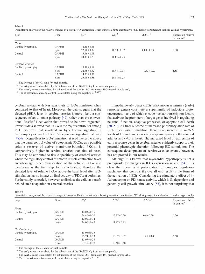
Table 5Quantitative analysis of the relative changes in c-jun mRNA expression levels using real-time quantitative PCR during isoproterenol-induced cardiac hypertrophy
c-jun Gene CTa ΔCT
b ΔΔCTc Expression relative
to control d
HeartCardiac hypertrophy GAPDH 12.15±0.15
c-jun 22.96±0.32 10.78±0.37 0.03±0.23 0.98Control GAPDH 13.66±1.09
c-jun 24.44±1.23 10.81±0.23
Cerebral arteriesCardiac hypertrophy GAPDH 15.38±0.68
c-jun 22.88±0.62 11.44±0.34 −0.63±0.23 1.55Control GAPDH 14.35±0.30
c-jun 25.79±0.58 10.81±0.23a The average of the CT data for each sample.b The ΔCT value is calculated by the subtraction of the GAPDH CT from each sample CT.c The ΔΔCT value is calculated by subtraction of the control ΔCT from each ISO-treated sample ΔCT.d The expression relative to control is calculated using the equation 2−ΔΔCT.
1073N. Kim et al. / Biochimica et Biophysica Acta 1763 (2006) 1067–1075
cerebral arteries with less sensitivity to ISO-stimulation whencompared to that of heart. Moreover, the data suggest that theelevated pEKR level in cerebral arteries is more likely a con-sequence of an ultimate pathway [47] rather than the conven-tional Ras/Raf-1 activation that proved to be down regulated.Previous data showed that PKCα is the major contributor amongPKC isoforms that involved in hypertrophic signaling incardiomyocytes via the ERK1/2-dependent signaling pathway[48,49]. Regardless to ISO-stimulation, it is of interest to noticethat the basal control value of cytoplasmic PKCα, as a possiblesoluble reserve of active membrane-bounded PKCα, iscomparatively higher in cerebral arteries than that of heart.This may be attributed to tissue specificity of cerebral arterieswhere the regulatory control of smooth muscle contraction takesan advantage. Since translocation of the soluble PKCα intomembrane is the first step for its activation, therefore theelevated level of soluble PKCα above the basal level after ISO-stimulation has no impact on final activity of PKCα at both sites.Further study is needed, however, to disclose the cellular benefitbehind such adaptation in cerebral arteries.
Table 6Quantitative analysis of the relative changes in c-myc mRNA expression levels usin
c-myc Gene CTa
HeartCardiac hypertrophy GAPDH 12.03±0.15
c-myc 24.40±0.20Control GAPDH 12.09±0.34
c-myc 24.06±0.47
Cerebral arteryCardiac hypertrophy GAPDH 15.06±0.35
c-myc 25.74±0.53Control GAPDH 13.97±0.28
c-myc 27.35±0.38a The average of the CT data for each sample.b The ΔCT value is calculated by the subtraction of the GAPDH CT from each sac The ΔΔCT value is calculated by subtraction of the control ΔCT from each ISOd The expression relative to control is calculated using the equation 2−ΔΔCT.
Immediate-early genes (IEGs; also known as primary (early)response genes) constitute a superfamily of inducible proto-oncogenes, many of which encode nuclear transcription factorsthat activate the promoters of target genes involved in regulatingneuronal function, adaptive processes, or apoptotic cell death[50–53]. As final outcome of increased phosphorylation rate ofERK after βAR stimulation, there is an increase in mRNAlevels of fos and c-myc (as early response genes) in the cerebralarteries and c-fos in heart. The increased level of expression ofearly response genes in cerebral arteries evidently supports theirpotential phenotypic alteration following ISO-stimulation. Theconsequent development of cerebrovascular events, however,has not proved in our results.
Although it is known that myocardial hypertrophy is not aprerequisite for changes in IEGs expression in vivo [54], it isclear that there is a participation of complex regulatorymachinery that controls the overall end result in the form ofthe activation of IEGs. Considering the stimulatory effect of β-Adrenoceptor on PI3 kinase activity, which is Gi-dependent andgenerally cell growth stimulatory [55], it is not surprising that
g real-time quantitative PCR during isoproterenol-induced cardiac hypertrophy
ΔCTb ΔΔCT
c Expression relativeto control d
12.37±0.29 0.4±0.29 0.76
11.97±0.45
13.37±0.32 −2.7±0.48 6.50
10.68±0.48
mple CT.-treated sample ΔCT.

1074 N. Kim et al. / Biochimica et Biophysica Acta 1763 (2006) 1067–1075
PKCα in a parallel pathway to PI3Kγ [56] converge tocontribute to fos and c-myc genes activation.
In conclusion the increased concentration and activity of rasand raf in heart with consequent downstream pERK elevationreflect the importance of ras/raf/ERK signaling pathway duringISO-stimulation with cardiac hypertrophy end-result. Thedecrease in their concentration in cerebral arteries, however,clearly disproves their local impact on ERK activation withpossible contribution of ultimate regulators. These data give thefirst insight about site-specific diversity of ras/raf/ERKsignaling pathway as consequence of ISO-stimulation.Increased expression of IEGs in cerebral arteries raises thespeculation of ISO-stimulation involvement in cerebrovascularevents that warrants further investigation to elucidate the exactmechanism. The mismatch noticed between mRNA transcrip-tion level and its final protein outcome suggests post-translationmultidisciplinary fine tuning that leads to this discrepancy.From the study attention should be taken to the local finaloutcome rather than remote intermediate results for betteraddressing pathophysiological interpretation on molecularbasis.
Acknowledgments
This work was supported by the 2003 Inje UniversityResearch Grant.
References
[1] E.J. Benjamin, R.B. D'Agostino, A.J. Belanger, P.A. Wolf, D. Levy, Leftatrial size and the risk of stroke and death. The Framingham Heart Study,Circulation 92 (1995) 835–841.
[2] M. Bikkina, D. Levy, J.C. Evans, M.G. Larson, E.J. Benjamin, P.A. Wolf,W.P. Castelli, Left ventricular mass and risk of stroke in an elderly cohort.The Framingham heart study, JAMA 272 (1994) 33–36.
[3] R. Marco, M.R. Di Tullio, R. Donna, D.R. Zwas, R.L. Sacco, R.R. Sciacca,S. Homma, Left ventricular mass and geometry and the risk of ischemicstroke, Stroke 34 (2003) 2380.
[4] C.J. Rodriguez, L. Ralph, R.L. Sacco, R.R. Sciacca, B. Boden-Albala, S.Homma, M.R. Di Tullio, Physical activity attenuates the effect of increasedleft ventricular mass on the risk of ischemic stroke, J. Am. Coll. Cardiol. 39(2002) 1482–1488.
[5] G. Selvetella, A. Notte, A. Maffei, V. Calistri, V. Scamardella, G. Frati, B.Trimarco, C. Colonnese, G. Lembo, Left ventricular hypertrophy isassociated with asymptomatic cerebral damage in hypertensive patients,Stroke 34 (2003) 1766–1770.
[6] P. Verdecchia, C. Porcellati, G. Reboldi, R. Gattobigio, C. Borgioni, T.A.Pearson, G. Ambrosio, Left ventricular hypertrophy as an independentpredictor of acute cerebrovascular events in essential hypertension,Circulation 104 (2001) 2039–2044.
[7] J.W. Adams, Y. Sakata, M.G. Davis, V.P. Sah, Y. Wang, S.B. Liggett, K.R.Chien, J.H. Brown, G.W. Dorn II, Enhanced Galphaq signaling: a commonpathway mediates cardiac hypertrophy and apoptotic heart failure, Proc.Natl. Acad. Sci. 95 (1998) 10140–10145.
[8] M.R. Bristow, Why does the myocardium fail? Insights from basic science,Lancet 352 (1998) S18–S114.
[9] P.H. Sugden, Signaling in myocardial hypertrophy: life after calcineurin?Circ. Res. 84 (1999) 633–646.
[10] M.A. Bogoyevitch, P.H. Sugden, The role of protein kinases in adapta-tional growth of the heart, Int. J. Biochem. Cell Biol. 28 (1996) 1–12.
[11] M. Henriksson, C.B. Xu, L. Edvinsson, Importance of ERK1/2 in
upregulation of endothelin type B receptors in cerebral arteries, Br. J.Pharmacol. 42 (2004) 1155–1161.
[12] F. Lennmyr, S. Karlsson, P. Gerwins, K.A. Ata, A. Terent, Activation ofmitogen-activated protein kinases in experimental cerebral ischemia, ActaNeurol. Scand. 106 (2002) 333–340.
[13] J.H. Zhang, Role of MAPK in cerebral vasospasm, Drug News Perspect.14 (2001) 261–267.
[14] T.M. Behr, S.S. Nerurkar, A.H. Nelson, R.W. Coatney, T.N. Woods, A.Sulpizio, S. Chandra, D.P. Brooks, S. Kumar, J.C. Lee, E.H. Ohlstein, C.E.Angermann, J.L. Adams, J. Sisko, J.D. Sackner-Bernstein, R.N. Willette,Hypertensive end-organ damage and premature mortality are p38 mitogen-activated protein kinase-dependent in a rat model of cardiac hypertrophyand dysfunction, Circulation 104 (2001) 1292–1298.
[15] M. Braun, G. Simonis, K. Birkner, B. Pauke, R.H. Strasser, Regulation ofprotein kinase C isozyme and calcineurin expression in isoproterenolinduced cardiac hypertrophy, J. Cardiovasc. Pharmacol. 41 (2003)946–954.
[16] L.J. De Windt, H.W. Lim, S. Haq, T. Force, J.D. Molkentin, Calcineurinpromotes protein kinase C and c-Jun NH2-terminal kinase activation in theheart. Cross-talk between cardiac hypertrophic signaling pathways, J. Biol.Chem. 275 (2000) 13571–13579.
[17] B.M.T. Burgering, G.J. Pronk, P.C. Weeren, P. Chardin, J.L. Bos, cAMPantagonizes p21 ras-directed activation of extracellular signal-regulatedkinase 2 and phosphorylation of mSos nucleotide exchange factor, EMBOJ. 12 (1993) 4211–4220.
[18] S.J. Cook, F. McCormick, Inhibition by cAMP of Ras-dependentactivation of Raf, Science 262 (1993) 1069–1072.
[19] G. D'Angelo, H. Lee, R.I. Weiner, cAMP-dependent protein kinaseinhibits the mitogenic action of vascular endothelial growth factor andfibroblast growth factor in capillary endothelial cells by blocking Rafactivation, J. Cell Biochem. 67 (1997) 353–366.
[20] W. Wen-Sheng, Protein kinase C alpha triggers Ras and Raf-independentMEK/ERK activation for TPA-induced growth inhibition of humanhepatoma cell HepG2, Cancer Lett. 239 (2006) 27–35.
[21] I.L. Ennis, E.M. Escudero, G.M. Console, G. Camihort, C.G. Dumm, R.W.Seidler, M.C. Camilion de Hurtado, H.E. Cingolani, Regression ofisoproterenol-induced cardiac hypertrophy by Na+/H+ exchanger inhibi-tion, Hypertension 41 (2003) 1324–1329.
[22] T. Fujita, H. Noda, Y. Ito, M. Isaka, Y. Sato, E. Ogata, Increasedsympathoadrenomedullary activity and left ventricular hypertrophy inyoung patients with borderline hypertension, J. Mol. Cell. Cardiol. 21(1989) 31–38.
[23] O.B. Marvin, L. Xilin, E. Thomas, Isoproterenol infusion inducesalterations in expression of hypertrophy-associated genes in rat heart,Am. J. Physiol. 269 (1995) 638–647.
[24] T. Omura, S. Kim, K. Takeuchi, H. Iwao, T. Takeda, Transforming growthfactor β1 and extracellular matrix gene expression in isoprenaline inducedcardiac hypertrophy: effects of inhibition of the renin-angiotensin system,Cardiovasc. Res. 28 (1994) 1835–1842.
[25] J. Polonia, L. Martins, D. Bravo-Faria, F. Macedo, J. Coutinho, H. Simoes,Higher left ventricle mass in normotensives with exaggerated bloodpressure responses to exercise associated with higher ambulatory bloodpressure load and sympathetic activity, Eur. Heart J. 13 (1992) 30–36.
[26] D.J. Choi, W.J. Koch, J.J. Hunter, H.A. Rockman, Mechanism of β-adrenergic receptor desensitization in cardiac hypertrophy is increased β-adrenergic receptor kinase, J. Biol. Chem. 272 (1997) 17223–17229.
[27] H.G. Zimmer, Catecholamine-induced cardiac hypertrophy: significanceof proto-oncogene expression, J. Mol. Med. 75 (1997) 849–859.
[28] P.H. Sugden, M.A. Bogoyevitch, Intracellular signaling through proteinkinases in the heart, Cardiovasc. Res. 30 (1995) 478–492.
[29] N. Kim, J. Chung, E. Kim, J. Han, Changes in the Ca2+-activated K+
channels of the coronary artery during left ventricular hypertrophy, Circ.Res. 93 (2003) 541–547.
[30] K. Kizaki, M. Okada, R. Ito, K. Yoshioka, K. Hashizume, K. Mutoh, Y.Hara, Induction of heparanase gene expression in ventricular myocardiumof rats with isoproterenol-induced cardiac hypertrophy, Biol. Pharm. Bull.28 (2005) 2331–2334.
[31] Y. Zou, A. Yao, W. Zhu, S. Kudoh, Y. Hiroi, M. Shimoyama, H. Uozumi,

1075N. Kim et al. / Biochimica et Biophysica Acta 1763 (2006) 1067–1075
O. Kohmoto, T. Takahashi, F. Shibasaki, R. Nagai, Y. Yazaki, I. Komuro,Isoproterenol activates extracellular signal-regulated protein kinases incardiomyocytes through calcineurin, Circulation 104 (2001) 102–108.
[32] H. Lodish, A. Berk, P. Matsudaira, C.A. Kaiser, M. Krieger, M.P. Scott,L. Zipursky, J. Darnell, Molecular Cell Biology, Signaling at the CellSurface, 5th ed., W.H. Freeman Company, New York, 2004, pp. 534–565,Chap. 13.
[33] W. Kolck, Meaningful relationships: the regulation of the Ras/Raf/Erkpathway by protein interactions, Biochem. J. 351 (2000) 289–305.
[34] L. Chen, P. Wang, C.F. Andrade, I.Y. Zhao, P.E. Dubé, P.L. Brubaker, M.Liu, T. Jin, PKA independent and cell type specific activation of theexpression of caudal homeobox gene Cdx-2 by cyclic AMP, FEBS J. 272(2005) 2746–2759.
[35] W. Zierhut, H.G. Zimmer, Significance of myocardial alpha- and beta-adrenoceptors in catecholamine-induced cardiac hypertrophy, Circ. Res.65 (1989) 1417–1425.
[36] K. Rajalingam, C. Wunder, V. Brinkmann, Y. Churin, M. Hekman, C.Sievers, U.R. Rapp, T. Rudel, Prohibitin is required for Ras-induced Raf-MEK-ERK activation and epithelial cell migration, Nat. Cell Biol. 7(2005) 837–843.
[37] A. Pozzi, S. Coffa, N. Bulus, W. Zhu, D. Chen, X. Chen, G. Mernaugh, Y.Su, S. Cai, A. Singh, M. Brissova, R. Zent, H-Ras, R-Ras, and TC21Differentially Regulate Ureteric Bud Cell Branching Morphogenesis, Mol.Biol. Cell 17 (2006) 2046–2056.
[38] J.F. Hancock, Ras proteins: different signals from different locations, Nat.Rev., Mol. Cell Biol. 4 (2003) 373–384.
[39] A. Beyer, J. Hollunder, H.P. Nasheuer, T. Wilhelm, Post-transcriptionalexpression regulation in the yeast Saccharomyces cerevisiae on a genomicscale, Mol. Cell Proteomics 11 (2004) 1083–1092.
[40] D. Greenbaum, C. Colangelo, K. Williams, M. Gerstein, Comparingprotein abundance and mRNA expression levels on a genomic scale,Genome Biol. 4 (2003) 117.
[41] R.J.R. Roskoski, Protein prenylation: a pivotal posttranslational process,Biochem. Biophys. Res. Commun. 303f (2003) 1–7.
[42] S. Yalovsky, M. Rodr Guez-Concepcion, W. Gruissem, Lipid modifica-tions of proteins—Slipping in and out of membranes, Trends Plant Sci. 4(1999) 439–445.
[43] N.S. Lamango, Liver prenylated methylated protein methyl esterase is anorganophosphate-sensitive enzyme, J. Biochem. Mol. Toxicol. 19 (2005)347–357.
[44] J.F. Hancock, Ras proteins: different signals from different locations, Nat.Rev., Mol. Cell Biol. 4 (2003) 373–384.
[45] L.M. Luttrell, T. van Biesen, B.E. Hawes, W.J. Koch, K.M. Krueger, K.Touhara, R.J. Lefkowitz, G-protein-coupled receptors and their regula-tion: activation of the MAP kinase signaling pathway by G-protein-coupled receptors, Adv. Second Messenger Phosphoprot. Res. 31 (1997)263–277.
[46] G. Selvetella, A. Notte, A. Maffei, V. Calistri, V. Scamardella, G. Frati, B.Trimarco, C. Colonnese, G. Lembo, Left ventricular hypertrophy isassociated with asymptomatic cerebral damage in hypertensive patients,Stroke 34 (2003) 1766–1770.
[47] D. Mizuchi, T. Kurosu, A. Kida, Z.H. Jin, A. Jin, A. Arai, O. Miura, BCR/ABL activates Rap1 and B-Raf to stimulate the MEK/Erk signalingpathway in hematopoietic cells, Biochem. Biophys. Res. Commun. 326(2005) 645–651.
[48] J.C. Braz, O.F. Bueno, L.J. DeWindt, J.D. Molkentin, PKC alpha regulatesthe hypertrophic growth of cardiomyocytes through extracellular signal-regulated kinase1/2 (ERK1/2), J. Cell Biol. 156 (2002) 905–919.
[49] R. Kerkela, M. Ilves, S. Pikkarainen, H. Tokola, J. Ronkainen, O.Vuolteenaho, J. Leppaluoto, H. Ruskoaho, Identification of PKCalphaisoform-specific effects in cardiac myocytes using antisense phosphor-othioate oligonucleotides, Mol. Pharmacol. 62 (2002) 1482–1491.
[50] M. Dragunow, K. Preston, The role of inducible transcription factors inapoptotic nerve cell death, Brain Res. Rev. 21 (1995) 1–28.
[51] P. Hughes, M. Dragunow, Induction of immediate-early genes and thecontrol of neurotransmitter-regulated gene expression within the nervoussystem, Pharmacol. Rev. 47 (1995) 133–178.
[52] K.A. Krown, M.T. Page, C. Nguyen, D. Zechner, V. Gutierrez, K.L.Comstock, C.C. Glembotski, P.J. Quintana, R.A. Sabbadini, Tumornecrosis factor alpha-induced apoptosis in cardiac myocytes. Involvementof the sphingolipid signaling cascade in cardiac cell death, J. Clin. Invest.98 (1996) 2854–2865.
[53] M. Sheng, M.E. Greenberg, The regulation and function of c-fos and otherimmediate-early genes in the nervous system, Neuron 4 (1990) 477–485.
[54] N. Dzimiri, K. Al-Bahnasi, Z. Al-Halees, Myocardial hypertrophy is not aprerequisite for changes in early gene expression in left ventricular volumeoverload, Fund. Clin. Pharmacol. 18 (2004) 39–44.
[55] A. Chesley, M.S. Lundberg, T. Asai, R.P. Xiao, S. Ohtani, E.G. Lakatta,M.T. Crow, The β2-adrenergic receptor delivers an antiapoptotic signal tocardiac myocytes through Gi-dependent coupling to phosphatidylinositol3′-kinase, Circ. Res. 87 (2000) 1172–1179.
[56] E.K. Schmidt, S. Fichelson, S.M. Feller, PI3 kinase is important for Ras,MEK and Erk activation of Epo-stimulated human erythroid progenitors,BMC Biol. 2 (2004) 7.