Short- and medium-chain fatty acids in the energy … · Short- and medium-chain fatty acids in the...
32
1 Short- and medium-chain fatty acids in the energy metabolism – the cellular perspective Peter Schönfeld* 1 , Lech Wojtczak 2 1 Institute of Biochemistry and Cell Biology, Otto-von-Guericke University, Magdeburg, Leipziger Str. 44, 39120 Magdeburg, Germany; 2 Nencki Institute of Experimental Biology; Pasteura 3, 02-093 Warsaw, Poland Running title Short –and medium-chain fatty acids energy metabolism Abbreviations: AMPK, AMP-dependent kinase; LCFAs, long-chain fatty acids; MCFAs, medium- chain fatty acids; ROS, reactive oxygen species; SCFAs, short-chain fatty acids *Author to whom correspondence should be addressed E-mail: [email protected] Tel.: +49-391-67-15362 Fax.: +49-391-67-14365 by guest, on August 4, 2018 www.jlr.org Downloaded from
-
Upload
nguyenmien -
Category
Documents
-
view
215 -
download
0
Transcript of Short- and medium-chain fatty acids in the energy … · Short- and medium-chain fatty acids in the...

1
Short- and medium-chain fatty acids in the energy metabolism – the cellular perspective
Peter Schönfeld*1, Lech Wojtczak2
1Institute of Biochemistry and Cell Biology, Otto-von-Guericke University, Magdeburg, Leipziger Str.
44, 39120 Magdeburg, Germany; 2Nencki Institute of Experimental Biology; Pasteura 3, 02-093
Warsaw, Poland
Running title
Short –and medium-chain fatty acids energy metabolism
Abbreviations: AMPK, AMP-dependent kinase; LCFAs, long-chain fatty acids; MCFAs, medium-
chain fatty acids; ROS, reactive oxygen species; SCFAs, short-chain fatty acids
*Author to whom correspondence should be addressed
E-mail: [email protected]
Tel.: +49-391-67-15362
Fax.: +49-391-67-14365
by guest, on August 4, 2018
ww
w.jlr.org
Dow
nloaded from

2
Abstract:
Short- and medium-chain fatty acids (SCFAs and MCFAs), independently of their cellular
signalling functions, are important substrates of the energy metabolism and anabolic processes in
mammals. SCFAs are mostly generated by colonic bacteria and are predominantly metabolized by
enterocytes and liver, whereas MCFAs arise mostly from dietary triglycerides, among them milk and
dairy products.
A common feature of SCFAs and MCFAs is their carnitine-independent uptake and
intramitochondrial activation to acyl-CoA thioesters. Contrary to long-chain fatty acids, the cellular
metabolism of SCFAs and MCFAs depends in a lesser extent on fatty acid-binding proteins. SCFAs
and MCFAs modulate tissue metabolism of carbohydrates and lipids as manifested by mostly
inhibitory effect on glycolysis and stimulation of lipogenesis or gluconeogenesis.
SCFAs and MCFAs exert in mitochondria no or only weak protonophoric and lytic activities
and do not significantly impair the electron transport in the respiratory chain. SCFAs and MCFAs
modulate mitochondrial energy production by two mechanism: they provide reducing equivalents to
the respiratory chain and partly decrease efficacy of the oxidative ATP synthesis.
Supplementary keywords: short chain fatty acids, medium chain fatty acids, mitochondria, energy
metabolism
by guest, on August 4, 2018
ww
w.jlr.org
Dow
nloaded from
3
Short- and medium-chain fatty acids (SCFAs and MCFAs), along with more abundant long-
chain fatty acids (LCFAs), are natural compounds present in both animal and plant tissues that
participate in cell metabolism. SCFAs and MCFAs are also important food constituents, where they
are mostly in the form of triglycerides in some plant oils and milk (1). Nevertheless, bacterial
fermentation of amylase-resistant starch and non-starch polysaccharides in the gut is probably the
most important source of SCFAs in human and most mammalian species (2-4).
Along with their role as energy-supplying fuel, SCFAs and MCFAs exhibit various regulatory
and signalling functions. Butyrate and other SCFAs are known to induce apoptosis under specific
conditions and thus to control cell proliferation (5,6). Currently, increasing attention is given to
SCFAs with respect to their putative role in the pathogenesis of allergies as well as autoimmune,
metabolic and neurological diseases [reviewed in (7)]. In the last two decades, the role of MCFAs as
agonists of peroxisome proliferator-activated receptors has also been characterized (8). Moreover,
accumulating evidence indicates that SCFAs generated by the gut microbiota exert influence on food
intake, thereby regulating energy homeostasis and body weight [reviewed in (9-11)]. SCFAs and
MCFAs also play an important role in the intracellular signalling and contribute to the regulation of
cell metabolism [reviewed in (12-16)]. Finally, MCFAs and SCFAs can control cell death and survival
(17-20). These important regulatory functions of MCFAs and SCFAs and their implications to human
health and pathologies are subject of a number of excellent comprehensive reviews (1,7,21,22). Here,
we want to concentrate on some peculiarities of the metabolic features of SCFAs and MCFAs that
differ from those of LCFAs and to sum up the current understanding of their role in the cellular energy
metabolism. Some aspects of SCFA and MCFA participation in energy-dependent mitochondrial
processes have already been briefly reviewed by ourselves previously (23).
DEFINITION AND PHYSICOCHEMICAL PROPERTIES
SCFAs and MCFAs, being monocarboxylic acids with hydrocarbon chain length of 1 to 12
total carbon atoms, are abundant in nature, although they are present in plant and animal material at
much smaller quantities than LCFAs (24,25). Fatty acids of total carbon atom numbers from 1 to 6 are
usually classified as SCFAs, whereas those of 7 to 12 carbon atoms are defined as MCFAs. Fatty acids
by guest, on August 4, 2018
ww
w.jlr.org
Dow
nloaded from
4
with shorter chain, up to 9 total carbon atoms, are liquid at room temperature [Table 1, (26)]. The odor
of the first members is pungent, whereas that of the higher members is rancid or none. The
lipophilicity of SCFAs and MCFAs measured as partition of the free acid between water and heptane
gradually increases with increasing the carbon atom chain length so that MCFAs become comparable
in this aspect to LCFAs (27-30). Due to their lower lipophilicity as compared to LCFAs, SCFAs do
not form micellar structures and do not participate in the formation of biological membranes (31).
SCFAs and MCFAs are weak acids, with pKa values around 4.8, except of formic acid whose pKa is
by about one unit lower (Table 1). Thus, their alkali metal salts are considerably hydrolyzed in
aqueous solutions. Water-soluble members of the family have a high tendency to form bimolecular
associates in water solution. Interestingly, incorporation of SCFAs and MCFAs into bilayer
membranes is known to increase their pKa values similarly as in case of LCFAs (32,33).
ORIGIN OF MCFAS AND SCFAS
In human, the major source of SCFAs is the fermentation of dietary fiber and undigested
saccharides in the gut by colonic anaerobic bacteria [(2); reviewed in (3,4,7)]. Acetate is mainly
formed by reductive methylation of CO2 (34). There are two main routes producing propionate by
colonic bacteria. According to the methylmalonic-CoA pathway (also called dicarboxylic pathway),
propionate is generated from lactate, supplied by lactate fermenting bacteria. In short, lactate is taken
up by propionic bacteria and thereafter dehydrogenated to pyruvate, which becomes carboxylated by
methylmalonyl-CoA-carboxyl transferase to oxaloacetate. Subsequently, the latter is converted to
propionate through a four-carbon pathway, consisting of the intermediates malate, fumarate, succinate
and methylmalonyl-CoA (34). It should be noted, that this pathway generates, in addition to
propionate, one acetate molecule per two molecules of propionate. Other bacteria, such as Clostridium
propionicum and Megasphaera elsdenii, produce propionate easily from lactate (35,36). According to
this route, the CoA ester of lactate (lactoyl-CoA) is converted via acryloyl-CoA to propionyl-CoA,
which becomes subsequently hydrolysed to propionic acid (the acrylate pathway) (34). Butyrate is
formed by the condensation of two acetyl-CoA molecules to form acetoacetyl-CoA, followed by the
reductive conversion of acetoacetyl-CoA to butyryl-CoA (37). According to an estimation, acetate,
by guest, on August 4, 2018
ww
w.jlr.org
Dow
nloaded from
5
propionate, and butyrate are formed in the human colon at a ratio of about 3:1:1 (38,39). In an in vivo
study, the rate of SCFAs release by the gut to the circulatory system amounts to about 35 µmol/kg
body weight/h (40). The highest concentrations (70-140 mM) were found in the proximal colon (2).
SCFAs may contribute to approximately 10% of the total human caloric uptake (4).
For newborn mammals, including human babies, mother’s milk constitutes an important
source of MCFAs and SCFAs that are present mainly in the form of triglycerides and phospholipids.
For example, the content of MCFAs (C7:0 – C12:0) amounts to 6% - 17% and to 9% - 28% of all fatty
acids in bovine (1) and human (41,42) milk, respectively. Cow milk and milk products remain the
main dietary source of SCFAs, mainly butyric acid, in adult humans. Other natural sources of MCFAs
and SCFAs are coconut oil and palm kernel oil [(1) and references therein]. In comparison to
triglycerides containing LCFAs, those containing MCFAs are more rapidly hydrolyzed in the
intestinal tract and do not become incorporated into chylomicrons. SCFAs and MCFAs are transported
by portal bloodstream to the liver, where they are readily metabolized (21).
SCFAs and MCFAs can also be formed in mammalian and human tissues, mainly liver. Thus,
the peroxisomal β-oxidation of LCFAs produces chain-shortened acyl-CoAs (43) that can be
hydrolyzed inside peroxisomes by distinct acyl-CoA thioesterases and released into the cytosol. In
addition, peroxisomes are also equipped with carnitine-acetyl and carnitine-octanoyl transferases and
thus shortened acyl-CoAs are converted into carnitine esters for the supply to mitochondria [reviewed
in (44)].
Under pathological conditions, for example in inborn medium-chain acyl-CoA dehydrogenase
deficiency, octanoate and decanoate accumulate to considerable amounts in tissues (45) resulting in an
impairment of functioning of the mitochondrial respiratory complexes (46). These disorders are often
accompanied by elevated urinary excretion of dicarboxylic MCFAs (mainly adipic, suberic and
sebacic acids) that apparently originate from microsomal ω-oxidation of corresponding medium-chain
acyl-CoAs (47,48). Based on their rapid absorption, triglycerides of MCFAs were introduced as a
quickly available energy source in clinical nutrition in the middle of the last century. Emulsions
enriched with MCFA-containing triglycerides were applied to patients suffering from various forms of
impaired digestion of normal LCFA-containing triglycerides (1). In addition, due to the rapid transport
by guest, on August 4, 2018
ww
w.jlr.org
Dow
nloaded from

6
of MCFAs from the gut to the liver, breath tests were developed for non-invasive clinical diagnosis
using 13C-labeled octanoate (49,50). Thereby, specific hepatic pathways as well as the speed of gastric
emptying can be measured [reviewed in (51)].
PRINCIPLES OF METABOLISM IN ANIMAL TISSUES
Transport from the gut to the liver
Butyrate is taken up by enterocytes presumably by means of the monocarboxylate transporter
1 (MCT-1) and the sodium-coupled monocarboxylate transporter 1 (SMCT-1) [reviewed in (52,53)].
Butyrate is used by these cells mostly as fuel. According to other authors, SCFAs are also absorbed
from the intestine lumen by an exchange with Cl¯ (54) and/or HCO3¯ (55). More recently, however, the
non-ionic diffusion of protonated SCFAs and MCFAs across the colon epithelium has been favored
(56). The latter mechanism is also supported by studies in model systems (30). According to this
mechanism the intestinal absorption of SCFAs depends on pH, while slight acidification of luminal
pH, possibly by bacterial metabolic activity, increases the prevalence of the protonated form of
SCFAs. Otherwise, the transport of SCFAs from enterocytes into the blood might be driven by anion
exchange. Thus, it seems likely that the transport across the basolateral membrane is based on the
anionic form of SCFAs against HCO3¯ (57). Butyrate as well as other SCFAs and MCFAs that have
not been utilized by enterocytes are transported by the portal vein to liver (40,58) and metabolized by
hepatocytes.
In contrast to LCFAs, which are esterified to triglycerides in enterocytes, incorporated into
chylomicrons and then enter the lymphatic system, SCFAs and MCFAs from the intestinal tract enter
the portal vein as free acids. There, MCFAs become partly bound to plasma albumin. The proportion
between albumin-bound and free MCFAs increases with increasing chain length, so that the first
equilibrium constant (i.e. for the strongest binding site) between the albumin-bound and the free forms
increases from 1.5 x 10⁴ for hexanoate, through 3.4 x 10⁴ for octanoate and 105 for decanoate, up to
2.4 x 10⁶ for laureate (59,60). It has to be remembered that LCFAs are present in circulating blood
practically totally bound to plasma albumin with the first equilibrium constant of the order of 107 to
109 (60). The subsequent uptake of SCFAs and MCFAs, at least the lower members of that group, by
by guest, on August 4, 2018
ww
w.jlr.org
Dow
nloaded from
7
liver and muscle cells as well as other tissues is independent of fatty acid binding proteins (1).
Similarly, their uptake by cells requires neither fatty acid transport proteins, nor plasma membrane-
embedded fatty acid translocase, nor cytosolic fatty acid-binding proteins. These observations provide
a likely explanation why octanoate oxidation by isolated hepatocytes is about five times faster than
that of oleate (61). Moreover, the intracellular metabolism of SCFAs and MCFAs seems to require no
or much less fatty acid-binding proteins (62,63). In contrast, LCFAs require fatty acid-binding proteins
for their cellular uptake, intracellular transport, regulatory functions and metabolism [reviewed in
(63)]. Binding of free LCFAs or LCFA-CoA-esters to fatty acid binding proteins also minimizes their
toxic effects, for example such as the lytic property or enzyme inhibition (63). Interestingly, in rats
deficient in one of the fatty acid transport proteins (CD36 protein), feeding with SCFA- and MCFA-
rich diet eliminated the increased glucose uptake, hyperinsulinemia and heart hypertrophy (64).
Similarly, in CD36-deficient mice octanoate alleviated poor heart ischemic tolerance (65). These
observations may have important implications to human medicine.
Fuelling the tissue energy metabolism
The utilization by different tissues of acetate formed by intestinal bacteria greatly differs
between ruminants and non-ruminants [(4) and references therein]. Acetate is also endogenously
generated in adult humans by ethanol oxidation, which operates mainly in liver (66). Thus, it has been
shown that ethanol oxidation could result in a 20-fold increase of the acetate level in peripheral blood
(67). In addition, net acetate generation has been found during fatty acid oxidation in perfused rat liver
(68). Formed acetate results mainly from the operation of acetyl-CoA hydrolase (acetyl-CoA
deacylase), which in rat liver has been found predominantly in the mitochondrial matrix (69, 70).
Because acetyl-CoA hydrolase is inhibited by free CoASH (Ki=17 µM), the level of free CoASH has
to be strongly lowered before hydrolase can produce free acetate from acetyl-CoA (70). On the other
hand, free acetate produced in liver by oxidation of ethanol or as byproduct of ketogenesis is barely
oxidized in this organ. This is because of high Km of hepatic mitochondrial acetyl-CoA synthetase (71)
or its absence in these organelles [(72), see also the next paragraph]. Acetate can be, however,
by guest, on August 4, 2018
ww
w.jlr.org
Dow
nloaded from

8
transported by circulation to other organs, e.g. the heart and skeletal muscles, where Km of
mitochondrial acetyl-CoA synthetase is much lower and where it can be utilized as fuel (70).
Activation of SCFAs occurs in the liver and several other tissues by acyl-CoA synthetases (72).
These enzymes are located in the cytosol as well as in the mitochondrial matrix, where they are
loosely bound to the inner mitochondrial membrane. In mammals, acetate is activated to acetyl-CoA
by two different acetyl-CoA synthetases, of which one (AceCS1) is cytosolic (78 kDa, Km=0.11 mM)
and the other one (AceCS2) mitochondrial (71 kDa, Km=0.06) (73). According to these authors (73),
AceCS2 is present in a wide range of tissues, with the highest level in heart (bovine and rodent), but
essentially absent in liver. In contrast, earlier investigations (74-76) demonstrated the presence of
acetyl-CoA synthetase activity in hepatocytes in both mitochondria (20-50%) and the cytosol (50-
80%).
Acetate is an important fuel during fasting as evidenced by the observation that in skeletal
muscles of mice lacking AceCS2 the ATP content declined to 50% in comparison to wild-type mice
(77). Interestingly, the activity of cytosolic and mitochondrial acetyl-CoA synthetases are regulated by
a reversible acetylation. Furthermore, this process is under the control of NAD+-dependent
deacetylases sirtuin 1 and sirtuin 3 [reviewed in (78)]. Sirtuin 1 is a nuclear and cytoplasmic enzyme,
whereas sirtuin 3 is predominantly located within mitochondria. In summary, in mammals acetate
plays not only an important role in energy homeostasis but also, as a substrate for sirtuins, it is also
involved in the regulation of gene silencing and aging processes (78).
In contrast to LCFAs that are activated to acyl-CoAs in the cytosol and must be transferred to
the mitochondrial interior via the carnitine shuttle, SCFAs and MCFAs, at least those of carbon atom
number up to C8, permeate the inner mitochondrial membrane in the non-esterified form and are
activated to their CoA-derivatives in the mitochondrial matrix. Localization of medium chain acyl-
CoA synthetase in the mitochondrial matrix has been first described already in the late 60’s of the past
century [(79); for more recent reports see (80, 81)]. The latter property may have important metabolic
consequences under specific conditions as will be discussed further. SCFAs and MCFAs activated
inside mitochondria are used as substrates in mitochondrial β-oxidation and the citric acid cycle.
Interestingly, it has been demonstrated using the perfusion technique that in rat liver and heart
by guest, on August 4, 2018
ww
w.jlr.org
Dow
nloaded from

9
octanoate can also undergo peroxisomal β-oxidation, thereby delivering acetyl-CoA to the cytosol (82,
83).
As the energy source for tissue metabolism, triglycerides of MCFAs have several advantages
compared to those of LCFAs. Firstly, they are more rapidly digested and the liberated MCFAs are
more quickly absorbed in the intestinal lumen (21,84). Secondly, tissue metabolism of SCFAs and
partly of MCFAs does not depend on proteins for binding, transport and transmembrane translocation
(see the preceding sub-chapter). Therefore, they can serve as better energy-providing fuel than LCFAs,
especially under pathological conditions, as exemplified by severe inflammation (85). Finally,
MCFAs, having a slightly lower energy content than LCFAs (8.4 instead of 9.2 kcal/g), reduce body
fat mass and enhance the insulin sensitivity of tissues [reviewed in (1,21,22,62)].
As said before, SCFAs and MCFAs are transported by blood from the alimentary tract to the
liver where they are metabolized, and therefore they are not stored in the adipose tissue. Only by
prolonged feeding rats with portacaval anastomoses (blood circulation overpassing the liver) diets
containing MCFAs, the group of van Itallie (86) succeeded to significantly enrich the tissue depot
lipids in triglycerides containing higher MCFAs (C8, C9 and C10). General features of the whole-body
metabolism and physiological functions of MCFAs, in particular octanoate, the most abundant
MCFAs, have been recently summarized (87,88). Like other MCFAs, and in contrast to LCFAs,
octanoate is rapidly degraded and is stored as triglyceride in the adipose or other tissues only in a very
low extent. Octanoate as a fuel for the energy metabolism in mammals has been studied in high-
energy requiring tissues such as skeletal muscle, heart, liver and brain (89-94). Concerning the latter
organ, it is important to remember that SCFAs and MCFAs are able to permeate the blood-brain
barrier (95).
The effects of SCFAs and MCFAs on the hepatic energy metabolism were studied mostly
either by the perfusion technique of isolated rat liver (89,91-93,96-98) or by incubation experiments
with isolated hepatocytes (99-101). In summary, these studies have shown that addition of octanoate
and, to a lesser extent, butyrate enhances oxygen consumption compared to incubations with the
Krebs-Henseleit buffer supplemented with pyruvate or lactate as energy supplying substrates. Both the
stimulation of oxygen consumption and the associated increase of the cellular level of NAD(P)H (102)
by guest, on August 4, 2018
ww
w.jlr.org
Dow
nloaded from
10
indicate that these fatty acids effectively supply reducing equivalents (NADH, FADH2) to the
mitochondrial respiratory chain. In addition, octanoate raised the mitochondrial energization, an
observation based on the in situ measurement of the mitochondrial membrane potential (∆Ψm) (101).
Energization by octanoate of hepatocytes oxidizing pyruvate plus lactate was also manifest (100).
However, in contrast to LCFAs (e.g. oleate), octanoate significantly raised the AMP level in the tissue
(89,100). It has also been reported that feeding rats with MCFA-rich diet enhances skeletal muscle
mitochondrial oxidative capacity (62,103), an observation which is partly attributed to an increased
activity of citrate synthase (62).
Because MCFA-containing triglycerides are rapidly digested in the intestine, rapidly taken up
by enterocytes and are not incorporation into chylomicrons, they are ideal energy-delivering nutrients
in clinical situations, where the digestion and/or absorption of LCFA-containing triglycerides is
impaired or a rapid energy uptake by the body is required. For this reason, MCFA-containing
triglycerides have been used for the nutrition of patients with inherited LCFA β-oxidation disorders
(104). While increasing evidence indicates that the diseased heart suffers from energy deficiency,
fuelling the myocardial energy metabolism with MCFAs has been proposed as metabolic therapy for
treating patients suffering from certain cardiomyopathies (87). For this treatment, MCFAs with odd
carbon-atom numbers appeared superior compared to those with even carbon-atom numbers (105).
Modulation of carbohydrate and lipid metabolism
Contrary to LCFAs, the oxidation of MCFAs is not affected by the carbohydrate content in the
diet. Thus, it has been reported that adaptation of adult rats to low-fat high-carbohydrate or high-fat
low-carbohydrate diet does not change the rate of octanoate oxidation measured in hepatocytes. In
contrast, oleate oxidation declined by 50% in rats adapted to low-fat high-carbohydrate diet (61).
Furthermore, MCFAs derived from digestion of MCFA-containing triglycerides are predominantly
degraded by the hepatic mitochondrial β-oxidation. Excess of formed acetyl-CoA is used for the
synthesis of ketone bodies (mostly acetoacetate and β-hydroxybutyrate), which are delivered as fuel to
non-hepatic tissues (21,22). MCFA-containing triglycerides are preferentially hydrolyzed compared to
those containing LCFAs and liberated MCFAs are also preferentially oxidized in organs, mostly heart,
by guest, on August 4, 2018
ww
w.jlr.org
Dow
nloaded from

11
muscles, kidneys, and liver (93, 106). In vitro studies on isolated hepatocytes and perfused rat liver
have shown that SCFAs and MCFAs modulate the hepatic metabolism of carbohydrates and lipids.
Thus, butyrate and octanoate inhibit glycolysis (107, 108) and thereby exert the “glucose sparing
activity”. In contrast, anabolic pathways of glucose formation (100,109-111) and lipogenesis
(107,112) become stimulated. For illustration, glucose formation by hepatocytes fed with pyruvate
plus lactate as gluconeogenic precursors is about 2-fold stimulated by octanoate (100). In contrast to
inhibiting glycolysis in hepatocytes, decanoate, but not octanoate, has been found to stimulate
glycolysis in astrocytes, thus resulting in an enhanced release of lactate into the extracellular space
(113). Since lactate is considered a key energy source for neurons, the astrocyte/neuron lactate shuttle
supplies this substrate to neighbouring neurons.
Generally, it has been discussed that mitochondrial matrix enzymes, pyruvate carboxylase and
the pyruvate dehydrogenase complex, are regulated by the ratios of acetyl-CoA/CoA, ATP/ADP and
NADH/NAD+ and, in addition, by pyruvate concentration. On the other hand, however, the fatty acid
(octanoate, palmitate)-induced increase of pyruvate flux through both enzymes has been explained
exclusively by an increased uptake of pyruvate into the mitochondrial matrix compartment (109,111).
It has been argued that the formation of acetoacetate from fatty acids drives pyruvate uptake across the
inner mitochondrial membrane. Therefore, there is reason to hypothesize that SCFAs and MCFAs play
a supporting role in the utilization of physiological low concentrations of pyruvate or lactate for
glucose generation (104). Nevertheless, acceleration of pyruvate uptake is not sufficient to explain the
huge stimulation by fatty acids of glucose generation with aspartate plus glycerol as gluconeogenic
precursors (110). Such stimulation is generally attributed to the generation of acetyl-CoA (allosteric
effector of pyruvate carboxylase) and reducing equivalents (114), the latter promoting formation of
glyceraldehyde-3-phosphate. It is also worthy to remember that pyruvate carboxylation in isolated rat
liver mitochondria is strongly stimulated by L-octanoylcarnitine, whereas non-esterified butyrate and
octanoate exert a strong inhibition (100). In addition, octanoate exerts a short-term dual regulatory
effect on the hepatic fatty acid synthesis, namely stimulation in the low concentration range (up to 1
mM) and inhibition at higher concentrations (107,112). The stimulation of lipogenesis has been
attributed to the activation of acetyl-CoA carboxylase, presumably by a covalent modification of the
by guest, on August 4, 2018
ww
w.jlr.org
Dow
nloaded from

12
enzyme. Moreover, studies with digitonin-permeabilized hepatocytes have shown that stimulation of
the acetyl-CoA carboxylase activity depends on the chain length of the fatty acid (112). The
stimulation magnitude increased from capronic (C6:0) to lauric (C12:0) acids, but decreased with fatty
acids of longer chain length. Malonyl-CoA, the product of the cytosolic acetyl-CoA-carboxylase
reaction, acts as a substrate for fatty acid synthesis but also as an inhibitor of carnitine
palmitoyltransferase I.
There is an ongoing discussion that SCFAs and MCFAs activate hepatic AMP-dependent
kinase (AMPK) [reviewed in (115) and references therein]. Generally, AMPK activation inhibits ATP-
utilizing processes in the cell and stimulates those that produce ATP. Being a cytosolic enzyme,
AMPK is activated by elevation of cytosolic AMP. Consequently, the mechanism underlying the
SCFA/MCFA activation of AMPK is not clear, since the activation of SCFAs and MCFAs to their
acyl-CoA esters raises the intramitochondrial AMP level. Interestingly, a recent study with mouse L6
myotubes suggests that AMPK can also be activated without alteration of the cytosolic AMP/ATP
ratio. According to this suggestion the activation mechanism of AMPK by MCFAs is mediated by
extracellular Ca2+-dependent Ca2+/calmodulin-dependent kinase kinase β (116). Other reported effects
of SCFAs and MCFAs on the anabolic pathways are the inhibition of triglyceride synthesis in
adipocytes (117) and a sparing effect on the hepatic glycogen storage (118). The latter activity is
attributed to the competition between fatty acid and glucose oxidation.
The fact that MCFAs with odd-chain and even-chain hydrocarbon skeletons exert different
effects on the cell energy metabolism is of particular interest and practical importance (119, 120). In
contrast to even-chain MCFAs, β-oxidation of odd-chain MCFAs generates acetyl-CoA and, in
addition, propionyl-CoA, which is anaplerotic for the citric acid cycle. Propionyl-CoA can enter the
citric acid cycle after its conversion into succinate. The anaplerotic function of odd-chain MCFAs
seems to be crucial for maintenance of the level of citric acid cycle metabolites in various tissues. This
biochemical background explains the proposed use of the MCFA-derived triheptanoin (glycerol
triheptanoate) as an anaplerotic drug (121, 122) for the treatment of cardiomyopathies in long-chain fat
oxidation disorders (105) and pyruvate carboxylase deficiency (123). This anaplerotic function of odd-
chain MCFAs is also important during episodes of epilepsy when the neurons become excessively
by guest, on August 4, 2018
ww
w.jlr.org
Dow
nloaded from

13
excited and thereby release increased amounts of glutamate (124,125). It is assumed that glutamate
release is likely to decrease the level of citric acid cycle metabolites and thereby declines the oxidation
of acetyl-CoA by mitochondria. Indeed, it has been recently shown that triheptanoin partially restores
the level of citric acid cycle metabolites in an epileptic animal model (126). Triheptanoin is also able
to attenuate harmful side effects associated with ischemic stroke (127). For illustration, when mice
were exposed to transient ischemia, triheptanoin reduced the extracellular level of glutamate released
in the mouse striatum, maintained the cellular ATP content at the desired level and prevented a decline
of the respiratory activity of isolated brain mitochondria. The latter findings strongly suggest that the
mitochondrial ATP regeneration is a target of the trihepatoin action (127). It is also worthwhile to
mention that, in sharp contrast to butyrate and octanoate, the odd-chain SCFA propionate has no
inhibitory effect on glycolysis and does not stimulate ketogenesis (108). Similarly to propionyl-CoA
formed by the cellular degradation of odd-chain fatty acids, external propionate supplies the
gluconeogenic pathway with its hydrocarbon skeleton, an activity that is mostly observed in ruminants
[reviewed in (128)].
POTENTIAL ADVERS EFFECTS
Energy coupling
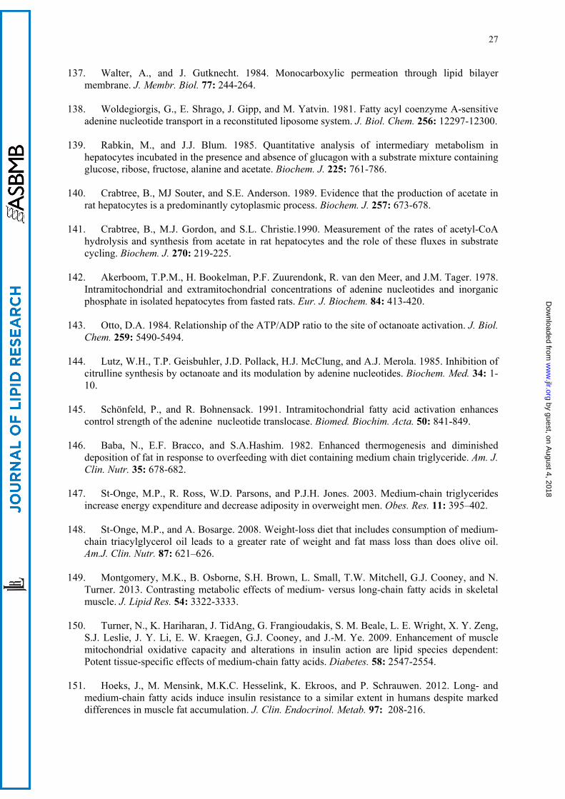
LCFAs are long known as mild uncouplers of oxidative phosphorylation due to their
protonophoric effect on the inner mitochondrial membrane [reviewed in (23)]. The mechanism of this
effect has been comprehensively explained by Skulachev (129,130) who showed that LCFA anions
can be transferred across the inner mitochondrial membrane by adenine nucleotide translocase,
whereas the non-dissociated fatty acid molecules can move across the membrane by flip-flop
mechanism. As effect, a net proton transfer occurs. Subsequent studies have shown that LCFA anions
can also be transferred across the mitochondrial inner membrane by a number of other mitochondrial
inner membrane anion carriers [reviewed in (131,132)]. This protonophoric effect decreases the
electrochemical proton gradient across the inner membrane, thereby decreasing the efficiency of
oxidative phosphorylation. Such activity of LCFAs has been repeatedly reported in vitro with isolated
mitochondria and there is evidence that protonophoric uncoupling can also operate in vivo after
by guest, on August 4, 2018
ww
w.jlr.org
Dow
nloaded from

14
hypoxia/reperfusion or high fat diet (133,134). With isolated mitochondria, uncoupling by LCFAs can
easily be quantified as an increase in the resting state respiration by micromolar concentrations of
these acids. In contrast to LCFAs, the ability to stimulate the resting state respiration by SCFAs or
MCFAs is either oligomycin-sensitive (C4 to C8) or weaker, even when applied at millimolar
concentrations (100,132,135,136). Furthermore, addition of octanoate or decanoate (at 100 or 300 µM
concentration) to cultured neurons or astrocytes did not stimulate their respiration (113).
Based on the fatty acid cycling hypothesis, this difference between LCFAs on one side and
MCFAs and SCFAs on the other side can be discussed in two aspects, namely in terms of (i) varying
permeation rate of fatty acids across the mitochondrial inner membrane and (ii) various affinities of
fatty acid anions to mitochondrial anionic carriers depending on the fatty acid chain length. In this
context it can be expected that SCFAs and MCFAs exhibit lower solubility in the lipid core of the
inner mitochondrial membrane because of their lower lipophilicity. In fact, it has been shown (30) that
the permeation of fatty acids across phosphatidylethanolamine bilayers depends on their partition
coefficient between hexadecane and water and the latter decreases with decreasing hydrocarbon chain
length (137). In addition, there is reason to speculate that the binding of the anionic forms of SCFAs
and MCFAs to the mitochondrial anion carries is lower than those of LCFAs. Such a view is supported
by the observation that inhibition of the adenine nucleotide carrier by acyl-CoA thioesters declines
with their hydrocarbon chain length (138). Nevertheless, there is evidence, mostly from studies on
isolated liver cells and perfused liver, that SCFAs and MCFAs can initiate ATP wastage
(89,96,97,99,100). It has been speculated (100) that this effect may be associated with a futile cycling
between the esterified forms of SCFAs or MCFAs and their acyl-CoA thioesters. This would result in
a net ATP utilization. Thus, a high rate of acetyl-CoA hydrolysis in rat hepatocytes, its stimulation by
increasing acetate concentrations and substrate cycling between acetate and acetyl-CoA have been
suggested (139-141). In the case of acetate this cycling is likely to occur in the cytoplasm of
hepatocytes and may account for as much as 1% of the total heat production (141).
Butyrate and octanoate are known to stimulate oxygen uptake in perfused liver and isolated
hepatocytes, to raise the energization of mitochondria and to support ATP-dependent glucose
generation (100,101,109). However, it has been repeatedly observed that these two fatty acids increase
by guest, on August 4, 2018
ww
w.jlr.org
Dow
nloaded from
15
oxygen uptake and dramatically lower the ATP/O ratio when added to the incubation or perfusion
media supplemented with pyruvate plus lactate or lactate alone (89,96,97,100), thus pointing to an
impairment of the energy generation. The mechanism of this effect could not be explained by
protonophoric uncoupling of oxidative phosphorylation by these SCFAs and MCFAs, as the latter was
characteristic for LCFAs (see the preceding chapter). The main argument against such a mechanism
was the observation that oligomycin, a well known inhibitor of mitochondrial ATPase/ATP synthase,
abolished most of the enhanced hepatic oxygen consumption (89,99) as well as the octanoate-
stimulated oxygen uptake by isolated rat liver mitochondria (100). Other authors attributed the
impairment of energy metabolism by butyrate and octanoate to the stimulation of Na+/K+-ATPase (99)
or to an increase in the FADH2/NADH ratio due to β-oxidation (96). However, these explanations
seem unlikely since a similarly enhanced oxygen uptake in the presence of LCFAs was not sensitive to
oligomycin. A further clue seemed to be a stationary elevated AMP level, which was not observed
with LCFAs (89,100,142-144). This pointed to an increased turnover of ATP within mitochondria
rather than to its impaired production. In addition, this putative ATP turnover competed with
intramitochondrial ATP-dependent reactions, i.e., pyruvate carboxylation (100) and citrulline
synthesis (144). We have shown (100) that this phenomenon is due to enhanced activation of
octanoate within the inner mitochondrial compartment, accompanied by utilization of two high-energy
bonds per each molecule of octanoyl-CoA formed. Because both octanoyl-AMP and octanoyl-CoA
could be partly hydrolyzed within the mitochondrial matrix, a futile cycle appeared that was
responsible for the increased intramitochondrial ATP consumption that resulted in lowering the
mitochondrial membrane potential and thus increasing oxygen uptake (Fig. 1). This mechanism may
prevent a drastic depletion of intramitochondrial free CoA under high supply of SCFAs and MCFAs
with the portal vein. In addition, the octanoate activation-associated increased AMP level decreases
the intramitochondrial pool of exchangeable adenine nucleotides ATP and ADP, an event that slows
down the operation rate of the adenine nucleotide translocase and thereby enhances the control
strength of this transporter on the total flux of oxidative ATP generation (145).
These specific properties of SCFAs and MCFAs may explain the well-known facts that diets
rich in these fatty acids increase energy expenditure and decrease adiposity (146-148). It has been
by guest, on August 4, 2018
ww
w.jlr.org
Dow
nloaded from
16
reported (149,150) that MCFAs, in contrast to LCFAs, contribute to maintain a high sensitivity of
muscle cells to insulin. This view has, however, not been confirmed by other authors (151). It is also
worthy to note that CoA esters of SCFAs and MCFAs accumulate in tissues at various pathological
situations such as the Reye syndrome (152). Furthermore, it has been reported (153) that octanoyl-
CoA at low millimolar concentrations exerts a strong inhibition on complex III activity of the
respiratory chain.
Generation of reactive oxygen species
Oxygen consumption by mitochondria is accompanied by the generation of reactive oxygen
species (ROS), of which β-oxidation is the most important source (154-157). Theoretically, a one-
electron transfer to molecular oxygen, thereby forming superoxide, can take place from complex I of
the respiratory chain as well as from acyl-CoA dehydrogenase, the electron transfer flavoprotein
(ETF), the ETF-ubiquinone oxidoreductase, and complex III (Fig. 2, more details are given in the
legend). Indeed, it has been demonstrated for skeletal muscle mitochondria that several sites are
involved in the β-oxidation-linked superoxide generation (158). H2O2 release by mitochondria from
(rat) skeletal muscle, heart, and liver was measured with carnitine derivatives of palmitate, octanoate,
and butyrate as substrates (157,159). These studies have shown that the β-oxidation-associated ROS
generation is similar with LCFAs and MCFAs (157,159). In contrast, it has been reported that C2C12
myotubes treated with capric (C10) or lauric (C12) acid generate less ROS than treated with LCFAs
myristic or palmitic acid, whereas the oxygen consumption is higher with MCFAs than with LCFAs
(149). These authors speculate that this decrease in ROS production might be attributed to an
increased expression of uncoupling protein-3 by MCFAs. However, other authors (160) did not
observe increased expression of uncoupling protein-3 in hearts of rats fed MCFA-rich diet.
Along with their direct role in ROS generation as electron donors to the respiratory chain in β-
oxidation, fatty acids also play an indirect effect on superoxide production due to modifying both the
rate of the electron flux along the respiratory chain and the degree of energy coupling. As discussed by
ourselves in detail elsewhere (161), LCFAs potentiate ROS generation due to their weak inhibition of
the electron flow at the levels of complexes I and III, most likely by interaction within the complex
by guest, on August 4, 2018
ww
w.jlr.org
Dow
nloaded from

17
subunit structure, and between complexes III and IV due to the release of cytochrome c from the inner
membrane. These effects occur in ROS generation accompanying the so called forward mode of
electron transport. On the other hand, due to the protonophoric action of LCFAs on the inner
mitochondria membrane (“mild uncoupling effect”), they strongly decrease ROS generation in the
reverse mode of electron transport (161). Contrary to this, SCFAs and MCFAs, at least lower members
of the latter, at low physiological levels neither affect functioning of the electron transport chain nor
exert a protonophoric effect on the inner mitochondrial membrane. On the other side, however,
excessive accumulation of MCFAs that occurs under inborn medium-chain acyl-CoA dehydrogenase
deficiency (45) and is connected with an impairment of the mitochondrial respiratory chain complexes
(46) may lead to increased ROS production. This results in an increased lipid peroxidation, generation
of protein carbonyls (as peroxidation products) and a decrease in the non-enzymatic antioxidant
defense (162).
CONCLUDING REMARKS
Although SCFAs and MCFAs, compared to LCFAs, constitute a minor component of human
and most mammalian diets, they play important roles both as nutrients and metabolic regulators. In
addition to their content in food, a large proportion of SCFAs is contributed by the intestinal
microflora by fermentation of otherwise undecomposed food constituents, mostly undigested
carbohydrates. As conclusion, a proper maintenance of gut microflora is important both for better
utilization of food constituents and as a source of molecules important as metabolic regulators.
During the past three or four decades, multiple roles of SCFAs and MCFAs have been
uncovered within the cellular and whole body metabolism. Along with their function as “fuels” for the
oxidative generation of ATP, SCFAs and MCFAs supply anabolic pathways (gluconeogenesis and
lipogenesis) with carbon-containing precursor molecules and contribute to the regulation of the cell
metabolism by triggering signalling pathways. Thus, MCFAs and, in particular, SCFAs play an
important role in a proper balance between lipogenesis and oxidative degradation of fatty acids. Many
of these multiple mechanisms of SCFAs and MCFAs still await full elucidation.
by guest, on August 4, 2018
ww
w.jlr.org
Dow
nloaded from
18
REFERENCES
1. Marten, B., M. Pfeuffer, and J. Schrezenmeir. 2006. Medium-chain triglycerides. Int. Dairy J. 16:
1374-1382. 2. Cummings, J.H., E.W.J. Pomare, H.W.J. Branch, C.P.E. Naylor, and G.T. MacFarlane. 1987.
Short chain fatty acids in human large intestine, portal, hepatic and venous blood. Gut. 28: 1221-1227.
3. Wong, J.M., R. de Souza, C.W. Kendall, A. Emam, and D.J. Jenkins. 2006. Colonic health:
fermentation and short chain fatty acids. J. Clinical Gastroenterol. 40: 235-243. 4. Bergman, E.N. 1990. Energy contributions of volatile fatty acids from the gastrointestinal tract in
various species. Physiol. Rev. 70: 567-590. 5. Trock, B., E. Lanza, and P. Greenwald. 1990. Dietary fiber, vegetables, and colon cancer: critical
review and meta-analyses of the epidemiologic evidence. J. Natl. Cancer Inst. 82: 650–661. 6. Hague, A., D.J. Elder, D.J. Hicks, and C. Paraskeva. 1995. Apoptosis in colorectal tumour cells:
induction by the short chain fatty acids butyrate, propionate and acetate and by the bile salt deoxycholate. Int. J. Cancer. 60: 400–406.
7. Tan, J., C. McKenzie, M. Potamitis, A.N. Thorburn, C.R. Mackay, and L. Macia. 2014. The role
of short-chain fatty acids in health and disease. Adv. Immunol. 121: 91-119. 8. Liberato, M.V., A.S. Nascimento, S.D. Ayers, J.Z. Lin, A. Cvoro, R.L. Silveira, L. Martínez, P.C.
Souza, D. Saidemberg, T. Deng, A.A. Amato, M. Togashi, W.A. Hsueh, K. Phillips, M.S. Palma, F.A. Neves, M.S. Skaf, P. Webb, and I. Polikarpov. 2012. Medium chain fatty acids are selective peroxisome proliferator activated receptor (PPAR)γ activators and pan-PPAR partial agonists.
PLoS One. 7 (5): e36297. 9. Byrne, C.S., E.S. Chambers, D.J. Morrison, and G. Frost. 2015. The role of short chain fatty acids
in appetite regulation and energy homeostasis. Int. J. Obes. 39: 1311-1338. 10. Bäckhed, F., H. Ding, T. Wang, L.V. Hooper, G.Y. Koh, A. Nagy, C.F. Semenkovich, and J.L.
Gordon. 2004. The gut microbiota as an environmental factor that regulates fat storage. Proc. Natl.
Acad. Sci. USA. 101: 15718-15723. 11. Ramakrishna, B.S. 2013. Role of the gut microbiota in human nutrition and metabolism. J.
Gastroenterol. Hepatol. 28: Suppl. 4, 9-17. 12. Georgiadi, A., and S. Kersten. 2012. Mechanism of gene regulation by fatty acids. Adv. Nutr. 3:
127-134. 13. Poulsen, L.I., M. Siersbæk, and S. Mandrup. 2012. PPARs: Fatty acid sensors controlling
metabolism. Semin. Cell Dev. Biol. 23: 631-639.
14. Nakamura, M.T., B.E. Yudell, and J.J. Loor. 2014. Regulation of the energy metabolism by long-chain fatty acid. Progr. Lipid Res. 53: 124-144.
15. Hara, T., I. Kimura, D. Inoue, A. Ichimura, and A. Hirasawa. 2013. Free fatty acid receptors and
their role in regulation of energy metabolism. Rev. Physiol. Biochem. Pharmacol. 164: 77-116.
by guest, on August 4, 2018
ww
w.jlr.org
Dow
nloaded from

19
16. Hara, T., D. Kashihara, A. Ichimura, I. Kimura, G. Tsujimoto, and A. Hirasawa. 2014. Role of free fatty acid receptors in the regulation of energy metabolism. Biochim. Biophys. Acta. 1841: 1292 -1300.
17. Chen, J.S., D.V. Faller, and R.A. Spanjaard. 2002. Short-chain fatty acid inhibitors of histone
deacetylases: promising anticancer therapeutics? Curr. Cancer Drug Targets. 3: 219-236. 18. Tang, Y., Y. Chen, H. Jiang, and D. Nie. 2011. Short-chain fatty acids induced autophagy serves
as an adaptive strategy for retarding mitochondria-mediated apoptotic cell death. Cell Death
Differ. 18: 602–618.
19. Fauser, J.K., G.M. Matthew, A.G. Cummins, and G.S. Howart. 2013. Induction of apoptosis by
the medium-chain length fatty acid lauric acid in colon cancer cells due to induction of oxidative stress. Chemotherapy. 59: 214-224.
20. Walsh, M.E., A. Bhattacharya, K. Sataranataran, R. Qaisar, L. Sloane, M.M. Rahman, M. Kinter,
and H. van Remmen. 2015. The histone deacetylase inhibitor butyrate improves metabolism and reduces muscle atrophy during aging. Aging Cell. 14: 957-970.
21. Bach, A.C., and V.K. Babayan. 1982. Medium-chain triglycerides – An update. Am. J. Clin. Nutr.
36: 950-962. 22. Papamandjaris, A.A., D.E. MacDougall, and P.J.H. Jones. 1998. Medium chain fatty acid
metabolism and energy expenditure: obesity treatment implications. Life Sci. 62: 1203-1215. 23. Wojtczak, L., and P. Schönfeld. 1993. Effect of fatty acids on the energy coupling processes in
mitochondria. Biochim. Biophys. Acta. 1183: 41-57. 24. Spector, A.A., and M.A.Yorek. 1985. Membrane lipid composition and cellular function. J. Lipid
Res. 26: 1015-1035. 25. Seidelin, K.N. 1995. Fatty acid composition of adipose tissue in humans. Implications for the
dietary fat-serum cholesterol-CHD issue. Prog. Lipid Res. 34: 199-217. 26. Rauen, H.M. ed. 1964. Biochemisches Taschenbuch. Springer, Berlin-Göttingen-Heidelberg. 27. Mukerjee, P. 1965. Dimerization of anions of long-chain fatty acids in aqueous solutions and the
hydrophobic properties of the acids. J. Phys. Chem. 69: 2821-2828.
28. Smith, R., and C. Tanford. 1973. Hydrophobicity of long chain n-alkyl carboxylic acids as measured by their distribution between heptane and aqueous solutions. Proc. Natl. Acad. Sci. USA. 70: 289-293.
29. Evtodienko, V.Y., O.N. Kovbasnjuk, Y.N. Antonenko ,and L.S. Yaguzhinsky. 1996. Effect of the alkyl chain length of monocarboxylic acid on the permeation through bilayer lipid membranes. Biochim. Biophys. Acta. 1281: 245-251.
30. Kamp, F., and J.A. Hamilton. 2006. How fatty acids of different chain length enter and leave cells
by free diffusion. Prostaglandins, Leukotr. Essential Fatty Acids. 75: 149-159. 31. Apel, C.L., D.W. Deamer, and M.N. Mautner. 2002. Self-assembled vesicles of monocarboxylic
acids and alcohols: conditions for stability and for encapsulation of biopolymers. Biochim.
Biophys. Acta. 1559: 1-9. 32. Small, D.M., D.J. Cabral, D.P. Cistola, J.S. Parks, and J.A. Hamilton. 1984. The ionization
behavior of fatty acids and bile acids in micelles and membranes. Hepatology. 4: Suppl. 5, 77S-79S.
by guest, on August 4, 2018
ww
w.jlr.org
Dow
nloaded from
20
33. Lieckfeldt, R., J. Villalaín, J.C. Gómez-Fernández, and G. Lee. 1995. Apparent pKa of the fatty
acids within ordered mixtures of model human stratum corneum lipids. Pharm. Res. 12 : 1614-1617.
34. Miller, T.L., and M.J. Wolin. 1996. Pathways of acetate, propionate, and butyrate formation by the
human fecal microbial flora. Appl. Environ. Microbiol. 62: 1589-1592. 35. Schweiger, G., and W. Buckel. 1984. On the dehydration of (R)-lactate in the fermentation of
alanine to propionate by Clostridium propionicum. FEBS Lett. 171:79-84. 36. Counotte, G.H., R.A. Prins, R.H.A.M. Janssen, and M.J.A Debie. 1981. Role of Megasphaera
elsdenii in the fermentation ofDLl-[2-13C]lactate in the rumen of dairy cattle. Appl. Environ.
Microbiol. 42: 649-655.
37. Pryde, S.E., S.H. Duncan, G.L. Hold, C.S. Stewart, and H.J. Flint. 2002. The microbiology of butyrate formation in the human colon. FEMS Microbiol. Lett. 217: 133-139.
38. Cummings, J.H., M.J. Hill, E.S. Bone, W.J. Branch, and D.J. Jenkins. 1979. The effect of meat
protein and dietary fiber on colonic function and metabolism. II. Bacterial metabolites in feces and urine. Am. J. Clin. Nutr. 32: 2094-2101.
39. Tazoe, H., Y. Otomo, I. Kaji, R. Tanaka, S.I. Karaki, and A. Kuwahara. 2008. Roles of short-chain
fatty acid receptors, GPR41 and GPR43, on colonic functions. J. Physiol. Pharm. 59: Suppl. 2, 251-262.
40. Bloemen, J.G., K. Venema, M.C. van de Poll, S.W. Olde Damink, W.A. Buurman, and C.H.
Dejong. 2009. Short chain fatty acids exchange across the gut and the liver in humans measured at surgery. Clin. Nutr. 28: 657-661.
41. van Beusekom, S., I.A. Martini, H.M. Rutgers, E.R. Boersma, and F.A. Muskiet. 1990. A
carbohydrate-rich diet not only leads to incorporation of medium-chain fatty acids (6:0-14:0) in milk triglycerides but also in each milk-phospholipid subclass. Am. J. Clin. Nutr. 52: 326-334.
42. Bahrami, G., and Z. Rahimi. 2005. Fatty acid composition of human milk in Western Iran. Eur. J.
Clin. Nutr. 59: 494-497. 43. Masters, C., and D. Crane. 1996. Principles of Medical Biology. Vol. 3, Chapter 3 –The
Peroxisome, (1996) pp. 39-58. E.E. Bittar and N. Bittar, editors, Elsevier, Amsterdam. 44. Hunt, M.C., M.I. Siponen, and S.H.E. Alexson. 2012. The emerging role of acyl-CoA
thioesterases and acyltransferases in regulating peroxisomal lipid metabolism. Biochim. Biophys.
Acta. 1822: 1397-1410. 45. Onkenhout, W., V. Venizelos, P.F.H. van der Poel, M.P.M. van den Heuvel, and B.J.H.M.
Poorthuis. 1995. Identification and quantification of intermediates of unsaturated fatty acid metabolism in plasma of patients with fatty acid oxidation disorders. Clin. Chem. 41: 1467-1474.
46. Scaini, G., K.R. Simon, A.M. Tonin, E.N. Busanello, A.O. Moura, G.C. Ferreira, M. Wajner, E.L.
Streck, and P.F. Schuck. 2012. Toxicity of octanoate and decanoate in rat peripheral tissues: evidence of bioenergetic dysfunction and oxidative damage induction in liver and skeletal muscle. Mol. Cell. Biochem. 361: 329-335.
47. Gregersen, N., P.B. Mortensen, and S. Kølvraa. 1983. On the biologic origin of C6-C10-
dicarboxylic C6-C10-ω-1-hydroxy monocarboxylic acids in human and rat with acyl-CoA
by guest, on August 4, 2018
ww
w.jlr.org
Dow
nloaded from
21
dehydrogenation deficiencies: in vitro studies on the ω- and ω-1-oxidation of medium-chain (C6-C12) fatty acids in human and rat liver. Pediatr. Res. 17: 828-834.
48. Gregersen, N. 1985. The acyl-CoA dehydrogenation deficiencies. Recent advances in the enzymic
characterization and understanding of the metabolic and pathophysiological disturbances in patients with acyl-CoA dehydrogenation deficiencies. Scand. J. Clin. Lab. Invest. Suppl. 174: 1-60.
49. Grattagliano, I., B.H. Lautenburg, G. Palasciano, and P. Portincasa. 2013. 13C-Breath tests for
clinical investigation of liver mitochondrial function. Eur. J. Clin. Invest. 40: 843-850. 50. Bruno, G., L.R. Lopetuso, G. Ianiro, L. Laterza, V. Gerardi, V. Petito, A. Poscia, A. Gasbarrini, V.
Ojetti, and F. Scaldafferri. 2013. 13C-Octanoic acid breath test to study gastric emptying time. Eur.
Rev. Med. Pharm. Sci. 17: Suppl. 2, 59-64. 51. Bonfrate, L., I. Grattagliano, G. Palsciano, and P. Portincasa. 2015. Dynamic carbon 13 breath
tests for the study of liver function and gastric emptying. Gastroenterol. Reports. 3: 12-21. 52. Enerson, B.E., and L.R. Drewes. 2003. Molecular features, regulation, and function of
monocarboxylate transporters: Implications for drug delivery. J. Pharmaceutical Sci. 92: 1531-1544.
53. Gonçalves, P., and F. Martel. 2013. Butyrate and colorectal cancer: the role of butyrate transport.
Curr. Drug Metabol. 14: 994-1008. 54. Rajendran, V.M., and H..J. Binder. 1994. Apical membrane Cl-butyrate exchange mechanism of
short chain fatty acid stimulation of active chloride absorption in rat distal colon. J. Membr. Biol. 141: 51-58.
55. Mascolo, N., V.M. Rajendran, and H.J. Binder. 1991. Mechanism of short-chain fatty acid uptake
by apical membrane vesicles of rat distal colon. Gastroenterol. 301: 331-338. 56. Charney, A.N., L. Micic, and R.W. Egnor. 1998. Nonionic diffusion of short-chain fatty acids
across rat colon. Am. J. Physiol. (Gastrointest. Liver Physiol.) 274: G518–G524. 57. Sellin, J.H., and R. DeSoignie. 1990. Short-chain fatty acid absorption in the human colon in vitro.
Gastroenterol. 99: 676-683. 58. Demigné, C., C. Yacoub, and C. Rémésy. 1986. Effects of absorption of large amounts of volatile
fatty acids on rat liver metabolism, J. Nutr. 116: 77-86. 59. Ashbrook, J.D., A.A. Spector, and J.E. Fletcher. 1972. Medium chain fatty acid binding to human
plasma albumin. J. Biol. Chem. 247: 7038-7042. 60. Ashbrook, J.D., A.A. Spector, E.C. Santos, and J.E. Fletcher. 1975. Long chain fatty acid binding
to human plasma albumin. J. Biol. Chem. 250: 2333-2338. 61. Pégorier, J.P., P.H. Duée, C. Herbin, P.Y. Laulan, C. Bladée, J. Peret, and J. Girard. 1988. Fatty
acid metabolism in hepatocytes isolated from rats adapted to high-fat diets containing long- or medium-chain triacylglycerols. Biochem. J. 249: 801-806.
62. Ishizawa, R., K. Masuda, S. Sakata, and A. Nakatani. 2015. Effects of different fatty acid chain
lengths on fatty acid oxidation-related protein expression levels in rat skeletal muscles. J. Oleo
Sci. 64: 415-421.
by guest, on August 4, 2018
ww
w.jlr.org
Dow
nloaded from
22
63. Atshaves, B.P., G.G. Martin, H.A. Hostetler, A.L. McIntosh, A.B. Kier, and F. Schroeder. 2010. Liver fatty acid-binding proteins and obesity. J. Nutr. Biochem. 21:1015-1032.
64. Hajri, T., A. Ibrahimi, C. T. Coburn, F. F. Knapp, Jr., T. Kurtz, M. Praveneci, and N.A. Abumrad.
2001. Defective fatty acid uptake in the spontaneously hypertensive rat is a primary determinant of altered glucose metabolism, hyperinsulinemia, and mocardial hypertrophy. J. Biol. Chem. 276: 23661–23666.
65. Irie, H., I.B. Krukenkamp, J.F.F. Brinkmann, G.R. Gaudette, A.E. Saltman, W. Jou, J.F.C. Glatz,
N.A. Abumrad, and A. Ibrahimi. 2003. Myocardial recovery from ischemia is impaired in CD36-null mice and restored by myocyte CD36 expression or medium-chain fatty acids. Proc. Natl.
Acad. Sci. USA. 100: 6819–6824. 66. Lundquist, F., N. Tygstrup, K. Winkler, K. Mellemgaard, and S. Munck-Petersen. 1962. Ethanol
metabolism and production of free acetate in the human liver. J. Clin. Invest. 41: 955-961.
67. Lundquist, F. 1960. The concentration of acetate in blood during alcohol metabolism in man. Acta
physiol. Scand. 175: 97 -105. 68. Seufert, C.D., M. Graf, G. Janson, A. Kuhn, and H.D. Söling. 1974. Formation of free acetate by
isolated perfused livers from normal, starved and diabetic rats. Biochem. Biophys. Res. Commun. 57: 901-909.
69. Grigat, K.P., K. Koppe, C.D. Seufert, and H.D. Söling.1979. Acetyl-coenzyme A deacylase
activity in liver is not an artefact. Subcellular distribution and substrate specificity of acetyl-coenzyme A deacylase activities in rat liver. Biochem. J. 177: 71-79.
70. Yamashita, H., T. Kaneyuki, and K. Tagawa. 2001. Production of acetate in the liver and its
utilization in peripheral tissues. Biochim. Biophys. Acta. 1532: 79-87. 71. Groot, P.H.E. 1975. The activation of short-chain fatty acids by the soluble fraction of guinea-pig
heart and liver mitochondria. The search for distinct propionyl-CoA synthase. Biochim. Biophys.
Acta. 380: 12-20.
72. Scholte, H.R., and P.H.E. Groot. 1975. Organ and intracellular localization of short-chain acyl-CoA synthetases in rat and guinea-pig. Biochim. Biophys. Acta 409: 283-296.
73. Fujino, T., J. Kondo, M. Ishikawa, K. Morikawa, and T.T. Yamamoto. 2001. Acetyl-CoA
synthase 2, a mitochondrial matrix enzyme involved in the oxidation of acetate. J. Biol. Chem.
276:11420-11426. 74. Barth, C, M. Sladek, and K. Decke.r 1971. The subcellular distribution of short-chain acyl-CoA
synthetase activity in rat tissues. Biochim. Biophys. Acta. 248: 24-33.
75. Goldberg, R.P., and H. Brunengraber. 1980. Contribution of cytosolic and mitochondrial acetyl-CoA synthetases to the activation of lipogenic acetate in rat liver. Adv. Exp. Med. Biol. 132: 413-418.
76. Endemann, G., P.G. Goetz, J.F. Tomera, W.M. Rand, S. Desrochers, and H. Brunengraber. 1987. Lipogenesis from ketone bodies in the perfused rat liver: effects of acetate and ethanol. Biochem.
Cell Biol. 65: 989-996.
77. Sakakibara, I., T. Fujino, M. Ishii, T. Tanaka, T. Shimosawa, S. Miura, W. Zhang, Y. Tokutake, J. Yamamoto, M. Awano, S. Iwasaki, T. Motoike, M. Okamura, T. Inagaki, K. Kita, O. Ezaki, M. Naito, T. Kuwaki, S. Chohnan, T.T. Yamamoto, R.E. Hammer, T. Kodama, M. Yanagisawa, and
by guest, on August 4, 2018
ww
w.jlr.org
Dow
nloaded from
23
J. Sakai. 2009. Fasting-induced hypothermia and reduced energy production in mice lacking acetyl-CoA synthetase 2. Cell Metab. 9: 191-202.
78. Shimazu, T., M.D. Hirschey, J.-Y. Huang, L.T.Y. Ho, and E. Verdin. 2010. Acetate metabolism
and aging: An emerging connection. Mech. Aging Dev. 131: 511-516. 79. Aas, M., and J. Bremer. 1968 Short-chain fatty acid activation in rat liver. A new assay procedure
for the enzymes and studies on their intracellular localization. Biochim. Biophys. Acta. 164: 157-166.
80. Vessey, D.A., M. Kelley, and R.S. Warren.1999. Characterization of the CoA ligases of human
liver mitochondria catalyzing the activation of short- and medium-chain fatty acids and xenobiotic carboxylic acids. Biochim. Biophys. Acta. 1428: 455-463.
81. Boomgaarden, I., C. Vock, M. Klapper, and F. Döring. 2009. Comparative analyses of disease risk
genes belonging to the acyl-CoA synthetase medium-chain (ACSM) family in human liver and cell lines. Biochem. Genet. 47: 739-748.
82. Kasumov, T., J.E. Adamas, F. Bian, F. David, K.R. Thomas, K.A. Jobbins, P.E. Minkler, C.L.
Hoppel, and H. Brunengraber. 2005. Probing peroxisomal β-oxidation and the labelling of acetyl-CoA proxies with [1-13C]octanoate and [3-13C]octanoate in the perfused rat liver. Biochem. J. 389: 397-401.
83. Bian, F., T. Kasumov, K.R. Thomas, K.A. Jobbins, F. David, P.E. Minkler, C.L. Hoppel, and H.
Brunengraber. 2005. Peroxisomal and mitochondrial oxidation of fatty acids in the heart, assessed from the 13C labeling of malonyl-CoA and the acetyl moiety of citrate. J. Biol. Chem. 280: 9265-9271.
84. Jeppesen, P.B., and P.B. Mortensen. 1999. Colonic digestion and absorption of energy from
carbohydrates and medium-chain fat in small bowel failure. J. Parenter. Enteral Nutr. 23: 5 Suppl. S101-S105.
85. Hecker, M., N. Sommer, H. Voigtmann, O. Pal, A. Mor, M. Wolf, I. Vadasz, S. Herold, N.
Weissmann, R.E. Morty, W. Seeger, and K. Mayer. 2014. Impact of short- and medium-chain fatty acids on mitochondrial function in severe inflammation. J. Parent. Enteral Nutr. 38: 587-594.
86. Zurier, R.B., R.G. Campbell, S.A. Hashim, and T.B. van Itallie. 1967. Enrichment of depot fat
with odd and even numbered medium chain fatty acids. Am. J. Physiol. 212: 291-294. 87. Labarthe, F., R. Gélinas, and C. Des Rosiers. 2008. Medium-chain fatty acids as metabolic therapy
in cardiac disease. Cardiovasc. Drug Ther. 22: 97-106. 88. Lemarie, F., E. Beauchamp, P. Legrand, and V. Rioux. (2016). Revisiting the metabolism and
physiological functions of caprylic acid (C8:0) with special focus on ghrelin octanoylation, Biochimie. 120: 40-48.
89. Debeer, L.J., G. Mannaerts, and P.J. de Scheppe. 1974. Effects of octanoate and oleate on energy
metabolism in the perfused rat liver. Eur. J. Biochem. 47: 591-600. 90. Kingsley-Hickman, P.B., E.Y. Sako, K. Uğurbil, A.H.L From, and J.E. Foker. 1990. 31P NMR
measurement of mitochondrial uncoupling in isolated rat hearts. J. Biol. Chem. 265: 1545-1550. 91. Scholz, R., U. Schwabe, and S. Soboll. 1984. Influence of fatty acids on energy metabolism. 1.
Stimulation of oxygen consumption, ketogenesis and CO2 production following addition of octanoate and oleate in perfused rat liver. Eur. J. Biochem. 141: 223-230.
by guest, on August 4, 2018
ww
w.jlr.org
Dow
nloaded from
24
92. Soboll, S., S. Gründel, and R. Scholz.1984. Influence of fatty acids on energy metabolism. 2.
Kinetics of changes in metabolic rates and changes in subcellular adenine nucleotide contents and pH gradients following addition of octanoate and oleate in perfused rat liver. Eur. J. Biochem. 141: 231-236.
93. Walton, M.E., D. Ebert, and R.G. Haller. 2003. Octanoate oxidation measured by 13C-NMR
spectroscopy in rat skeletal muscle, heart, and liver. J. Appl. Physiol. 95: 1908-1916. 94. Ebert, D., R.G. Haller, and M.E. Walton. 2003. Energy contribution of octanoate to intact rat brain
metabolism measured by 13C nuclear magnetic resonance spectroscopy. J. Neurosci. 23: 5928-5935.
95. Spector, R. 1988. Fatty acid transport through the blood-brain barrier. J. Neurochem. 50: 639-643. 96. Beauvieux, M.-C., P. Tissier, H. Gin, P. Canioni, and J.-L. Gallis. 2001. Butyrate impairs energy
metabolism in isolated perfused liver of fed rats, J. Nutr. 31: 1986-1992. 97. Gallis, J.-L., P. Tissier, H. Gin, and M.-C. Beauvieux. 2007. Decrease in oxidative
phosphorylation yield in presence of butyrate in perfused liver isolated from fed rats. BMC
Physiology. 7: article No. 8. 98. Gallis, J.-L., H. Gin, H. Roumes, and M.-C. Beauvieux. 2011. A metabolic link between
mitochondrial ATP synthesis and liver glycogen metabolism: NMR study in rats re-fed with butyrate and/or glucose. Nutr. Metabolism. 8: article No. 38.
99. Plomp, P.J.A.M., C.W.T. van Roermund, A.K. Groon, A.J. Meijer, and J.M. Tager. 1985.
Mechanism of the stimulation of respiration by fatty acids in rat liver. FEBS Lett. 193: 243-246. 100. Schönfeld, P., A.B. Wojtczak, M.J.H. Geelen, W. Kunz, and L. Wojtczak. 1988. On the
mechanism of the so-called uncoupling effect of medium- and short-chain fatty acids. Biochim.
Biophys. Acta. 936: 280-288. 101. Nobes, C.D., W.W. Hay, Jr, and M.D. Brand. 1990. The mechanism of stimulation of
respiration by fatty acids in isolated hepatocytes. J. Biol. Chem. 265: 12910-12915. 102. Hassinen, I., K. Ito, S. Nioka, and B. Chance. 1990. Mechanism of fatty acid effect on
myocardial oxygen consumption. A phosphorus NMR study. Biochim. Biophys. Acta 1019: 73-80. 103. Turner, N., K. Hariharan, J. TidAng, G. Frangioudakis, S.M. Beale, L.E. Wright, X.Y. Zeng,
S.J. Leslie, J.Y. Li, E.W. Kraegen, G.J. Cooney, and J.M. Ye. 2009. Enhancement of muscle mitochondrial oxidative capacity and alterations in insulin action are lipid species dependent: potent tissue-specific effects of medium-chain fatty acids. Diabetes. 58: 2547-2554.
104. Saudubray, J.M., D.E. Martin, P. de Lonlay . G. Touati, F. Poggi-Travert, D. Bonnet, P.
Jouvet, M. Boutron, A. Slama, C. Vianey-Saban, J.P. Bonnefont, D. Rabier, P. Kamoun, and M. Brivet. 1999. Recognition and management of fatty acid oxidation defects: a series of 107 patients. J. Inherit. Metab. Dis. 22: 488-502.
105. Roe, C.R., L. Sweetman, D.S. Roe, F. David, and H. Brunengraber. 2002. Treatment of
cardiomyopathy and rhabdomyolysis in long-chain fat oxidation disorders using an anaplerotic odd-chain triglyceride. J. Clin. Invest. 110: 259-269.
106. Metges C.C., and G. Wolfram. 1991. Medium- and long-chain triglycerides labelled with 13C:
A comparison of oxidation after oral or parenteral administration in humans. J. Nutr. 121: 31-36.
by guest, on August 4, 2018
ww
w.jlr.org
Dow
nloaded from
25
107. Nomura, T., A. Iguchi, N. Sakamoto, and R.A. Harris. 1983. Effects of octanoate and acetate upon hepatic glycolysis and lipogenesis. Biochim. Biophys. Acta. 754: 315-320.
108. Morand, C., C. Besson, C. Demigne, and C. Remesy. 1994. Importance of the modulation of
glycolysis in the control of lactate metabolism by fatty acids in isolated hepatocytes from fed rats. Arch. Biochem. Biophys. 309: 254-250.
109. Gonzalez-Manchon, C., M.S. Ayuso, and R. Parrilla. 1989. Control of hepatic
gluconeogenesis: Role of fatty acid oxidation. Arch. Biochem. Biophys. 271: 1-9. 110. Winiarska, K., J. Drożak, M. Węgrzynowicz, A.K. Jagielski, and J. Bryła. 2003 Relationship
between gluconeogenesis and glutathione redox state in rabbit kidney-cortex tubules. Metabolism. 52: 739-746.
111. Agius, L., and G.M.M. Alberti. 1985. Regulation of flux through pyruvate dehydrogenase and
pyruvate carboxylase in rat hepatocytes. Eur. J. Biochem. 152: 699-707. 112. Geelen, M.J. 1994. Medium-chain fatty acids as short-term regulators of hepatic lipogenesis.
Biochem. J. 302: 141-146. 113. Thevenet, J., U. DeMarchi, J.S. Domingo, N. Christinat, L. Bultot, G. Lefebvre, K. Sakamoto,
P. Descombes, M. Masoodi, and A. Wiederkehr. 2016. Medium-chain fatty acids inhibit mitochondrial metabolism in astrocytes promoting astrocyte-neuron lactate and ketone body shuttle systems. FASEB J. doi: 10.1096/fg.201500182.
114. Williamson, J.R., E.T. Browing, and M.S. Olson. 1968. Interrelationships between fatty acid
oxidation and the control of gluconeogenesis in perfused rat liver. Adv. Enzyme Regul. 6: 67-100.
115. Hu, G.-X., G.-R. Chen, H. Xu, R.-S. Ge, and J. Lin. 2010. Activation of the AMP activated protein kinase by short-chain fatty acids is the main mechanism underlying the beneficial effect of a high fiber diet on the metabolic syndrome. Medical Hypotheses. 74: 123-126.
116. Takikawa, M., A. Kumagai, H. Hirata, M. Soga, Y. Yamashita, M. Ueda, H. Ashida, and T.
Tsuda. 2013. 10-Hydroxy-2-decenoic acid, a unique medium-chain fatty acid, activates 5´-AMP-activated protein kinase in L6 myotubes and mice. Mol. Nutr. Food Res. 57: 1794-1802.
117. Guo, W., T. Lei, B.E. Corkey, and J. Han. 2003. Octanoate inhibits triglyceride synthesis in
3T3-LI and human adipocytes. J. Nutr. 133: 2512-2518.
118. Beauvieux, M.C., H. Roumes, N. Robert, H. Gin, V. Rigalleau, and J.L. Gallis. 2008. Butyrate ingestion improves hepatic glycogen storage in the re-fed rat. BMC Physiology. 8: article No. 19.
119. .Okere. I.C., T.A. McElfresh, D.Z. Brunengraber, W. Martini, J.P. Sterk, H. Huang, M.P.
Chandler, H. Brunengraber, and W.C. Stanley. 2006. Differential effects of heptanoate and hexanoate on myocardial citric acid cycle intermediates following ischemia-reperfusion. J. Appl.
Physiol. 100: 76-82. 120. Kajimoto, M., D.R. Ledee, A.K. Olson, N.G. Isern, C. Des Rosiers, and M.P. Portman. 2015.
Differential effects of octanoate and heptanoate on myocardial metabolism during extracorporal membrane oxygenation in infant swine model. Am. J. Physiol. (Heart Circ. Physiol.) 309: H1157-H1165.
121. Brunengraber, H., and C.R. Roe. 2006. Anaplerotic molecules: current and Future. J. Inherit.
Metab. Dis. 29: 327-331.
by guest, on August 4, 2018
ww
w.jlr.org
Dow
nloaded from

26
122. Marin-Valencia, I., L.B. Good, Q. Ma, C.R. Malloy, and J.M. Pascual. 2013. Heptanoate as neural fuel: Energetic and neurotransmitter precursors in normal and glucose transporter I-deficient G1D) brain. J. Cereb. Blood Flow Metab. 33: 175-182.
123. Mochel, F., P. DeLonlay, G Touati, H. Brunengraber, R.P. Kinman, D. Rabier,C.R. Roe, and
J.M. Saudubray. 2005. Pyruvate carboxylase deficiency: clinical and biochemical response to anaplerotic diet therapy. Mol. Genet. Metab. 84: 305-312.
124. Willis, S., J. Stoll, L. Sweetman, and K. Borges. 2010. Anticonvulsant effects of triheptanoin
diet in two mouse chronic seizure models. Neurobiol. Dis. 40: 565-572. 125. Borges, K., and U. Sonnewald. 2012. Triheptanoin – a medium-chain triglyceride with odd
chain fatty acids: a new anaperotic anticonvulsant treatment. Epilepsy Res.100: 239-244. 126. Hadera, M.G., O.B. Smeland, T.S. McDonald, K. Ni Tan, U. Sonnewald, and K. Borges.
2014. Triheptanoin partially restores levels of tricarboxylic cycle intermediates in the mouse pilocarpine model of epilepsy. J. Neurochem. 129: 107-119
127. Schwarzkopf, T.M., K. Koch, and J. Klein. 2015. Reduced severity of ischemic stroke and
improvement of mitochondrial function after dietary treatment with the anaplerotic substance triheptanoin. Neuroscience. 300: 201-209.
128. Al-Lahham, S.H., M.P. Peppelenbosch, H. Roelofsen, R.J. Vonk, and K. Venema. 2010.
Biological effects of propionic acid in humans; metabolism, potential applications and underlying mechanisms. Biochim. Biophys. Acta. 1801: 1175-1183.
129. Skulachev, V.P. 1991. Fatty acid circuit as a physiological mechanism of uncoupling of
oxidative phosphorylation. FEBS Lett. 294:158-162. 130. Skulachev, V.P. 1999. Anion carriers in fatty acid-mediated physiological uncoupling. J.
Bioenerg. Biomembr. 31: 431-435. 131. Wojtczak, L., and M.R. Więckowski. 1999. The mechanisms of fatty acid-induced proton
permeability of the inner mitochondrial membrane. J. Bioenerg. Biomembr. 31: 447-455. 132. Bernardi, P., D. Penzo, and L. Wojtczak. 2002. Mitochondrial energy dissipation by fatty
acids: Mechanisms and implications for cell death. Vitam. Horm. 65: 97-126. 133. Feldkamp, T., J.M. Weinberg, M. Hörbelt, C. von Kropff, O. Witzke, J. Nürnberger, and A.
Kribben. 2009. Evidence for involvement of nonesterified fatty acid protonophoric uncoupling during mitochondrial dysfunction caused by hypoxia and reoxygenation. Nephrol. Dial.
Transplant. 24: 43-51. 134. Cole, M.A., A.J. Murray, L.E. Cochlin, L.C. Heather, S.McAleese; N.S. Knight, E. Sutton,
A.A. Jamil, N. Parassol, and K. Clarke. 2011. A high fat diet increases mitochondrial fatty acid oxidation and uncoupling to decrease efficiency in rat heart. Basic Res. Cardiol. 106: 447-457.
135. Schönfeld, P., L. Schild, and W. Kunz. 1989. Long-chain fatty acids act as protonophoric
uncouplers of oxidative phosphorylation in rat liver mitochondria. Biochim. Biophys. Acta. 977: 266-272.
136. Shabalina, I.G., W.C. Backlund, J. Bar-Tana, B. Cannon, and J. Nedergaard. 2008. Within
brown-fat cells, UCP1-mediated fatty acid-induced uncoupling is independent of fatty acid metabolism. Biochim. Biophys. Acta. 1777: 642-650.
by guest, on August 4, 2018
ww
w.jlr.org
Dow
nloaded from
27
137. Walter, A., and J. Gutknecht. 1984. Monocarboxylic permeation through lipid bilayer membrane. J. Membr. Biol. 77: 244-264.
138. Woldegiorgis, G., E. Shrago, J. Gipp, and M. Yatvin. 1981. Fatty acyl coenzyme A-sensitive
adenine nucleotide transport in a reconstituted liposome system. J. Biol. Chem. 256: 12297-12300. 139. Rabkin, M., and J.J. Blum. 1985. Quantitative analysis of intermediary metabolism in
hepatocytes incubated in the presence and absence of glucagon with a substrate mixture containing glucose, ribose, fructose, alanine and acetate. Biochem. J. 225: 761-786.
140. Crabtree, B., MJ Souter, and S.E. Anderson. 1989. Evidence that the production of acetate in
rat hepatocytes is a predominantly cytoplasmic process. Biochem. J. 257: 673-678. 141. Crabtree, B., M.J. Gordon, and S.L. Christie.1990. Measurement of the rates of acetyl-CoA
hydrolysis and synthesis from acetate in rat hepatocytes and the role of these fluxes in substrate cycling. Biochem. J. 270: 219-225.
142. Akerboom, T.P.M., H. Bookelman, P.F. Zuurendonk, R. van den Meer, and J.M. Tager. 1978.
Intramitochondrial and extramitochondrial concentrations of adenine nucleotides and inorganic phosphate in isolated hepatocytes from fasted rats. Eur. J. Biochem. 84: 413-420.
143. Otto, D.A. 1984. Relationship of the ATP/ADP ratio to the site of octanoate activation. J. Biol.
Chem. 259: 5490-5494. 144. Lutz, W.H., T.P. Geisbuhler, J.D. Pollack, H.J. McClung, and A.J. Merola. 1985. Inhibition of
citrulline synthesis by octanoate and its modulation by adenine nucleotides. Biochem. Med. 34: 1-10.
145. Schönfeld, P., and R. Bohnensack. 1991. Intramitochondrial fatty acid activation enhances
control strength of the adenine nucleotide translocase. Biomed. Biochim. Acta. 50: 841-849. 146. Baba, N., E.F. Bracco, and S.A.Hashim. 1982. Enhanced thermogenesis and diminished
deposition of fat in response to overfeeding with diet containing medium chain triglyceride. Am. J.
Clin. Nutr. 35: 678-682. 147. St-Onge, M.P., R. Ross, W.D. Parsons, and P.J.H. Jones. 2003. Medium-chain triglycerides
increase energy expenditure and decrease adiposity in overweight men. Obes. Res. 11: 395–402. 148. St-Onge, M.P., and A. Bosarge. 2008. Weight-loss diet that includes consumption of medium-
chain triacylglycerol oil leads to a greater rate of weight and fat mass loss than does olive oil. Am.J. Clin. Nutr. 87: 621–626.
149. Montgomery, M.K., B. Osborne, S.H. Brown, L. Small, T.W. Mitchell, G.J. Cooney, and N.
Turner. 2013. Contrasting metabolic effects of medium- versus long-chain fatty acids in skeletal muscle. J. Lipid Res. 54: 3322-3333.
150. Turner, N., K. Hariharan, J. TidAng, G. Frangioudakis, S. M. Beale, L. E. Wright, X. Y. Zeng,
S.J. Leslie, J. Y. Li, E. W. Kraegen, G.J. Cooney, and J.-M. Ye. 2009. Enhancement of muscle mitochondrial oxidative capacity and alterations in insulin action are lipid species dependent: Potent tissue-specific effects of medium-chain fatty acids. Diabetes. 58: 2547-2554.
151. Hoeks, J., M. Mensink, M.K.C. Hesselink, K. Ekroos, and P. Schrauwen. 2012. Long- and
medium-chain fatty acids induce insulin resistance to a similar extent in humans despite marked differences in muscle fat accumulation. J. Clin. Endocrinol. Metab. 97: 208-216.
by guest, on August 4, 2018
ww
w.jlr.org
Dow
nloaded from

28
152. Corkey B.E., D.E. Hale, M.C. Glennon, R.I. Kelley, P.M. Coates, L. Kilpatrick, and C.A. Stanley. 1988. Relationship between unusual hepatic acyl coenzyme A profiles and the pathogenesis of Reye syndrome. J. Clin. Invest. 82: 782-788.
153. Sauer, S.W., J.G. Okub, G.F. Hoffmann, S. Koelker, and M.A. Morath. 2008. Impact of short-
and medium-chain organic acids, acylcarnitines, and acyl-CoAs on mitochondrial energy metabolism. Biochim. Biophys. Acta. 1777: 1276-1282.
154. St-Pierre, J., J.A. Buckingham, S.J. Roebuck, and M.D. Brand. 2002. Topology of superoxide
production from different sites in the mitochondrial electron transport chain. J. Biol. Chem. 277: 44784–44790.
155. Hoffman, D.L., and P.S. Brookes. 2009. Oxygen sensitivity of mitochondrial reactive oxygen
species generation depends on metabolic conditions. J. Biol. Chem. 284: 16236–16245. 156. Seifert, E.L., C. Estey, J.Y. Xuan, and M.E. Harper. 2010. Electron transport chain-dependent
and independent mechanisms of mitochondrial H2O2 emission during long-chain fatty acid oxidation. J. Biol. Chem. 285: 5748–5758.
157. Schönfeld, P., M.R. Więckowski, M. Lebiedzińska, and L. Wojtczak. 2010. Mitochondrial
fatty acid oxidation and oxidative stress: Lack of reverse electron transfer-associated production of reactive oxygen species. Biochim. Biophys. Acta. 1797: 829-938.
158. Prevoshchikova, I.V., C.L. Quinlan, A.L. Orr, A.A. Gerencser, and M.D. Brand. 2013. Sites of
superoxide and hydrogen peroxide production during fatty acid oxidation in rat skeletal muscle mitochondria. Free Radic Biol. & Med. 61: 298-309.
159. Schönfeld, P., and L. Wojtczak. 2012. Brown adipose tissue mitochondria oxidizing fatty
acids generate high levels of reactive oxygen species irrespective of the uncoupling protein-1 activity state. Biochim. Biophys. Acta. 1817: 410-418.
160. Murray, A.J., N.S. Knight, S.E. Little, L.E. Cochlin, M. Clements, and K. Clarke. 2011.
Dietary long-chain, but not medium-chain triglycerides, impair exercise performance and uncouple cardiac mitochondria in rats. Nutr. Metabolism. 8: article No. 55.
161. Schönfeld, P., and L. Wojtczak. 2008. Fatty acids as modulators of the cellular production of
reactive oxygen species. Free Rad. Biol. Med. 45: 231-241. 162. Schuck, P.F., G.C. Ferreira, A. P. Moura, E.N.B. Busanello, A.M. Tonin, C.S. Dutra-Filho,
and M. Wajner. 2009. Medium-chain fatty acids accumulating in MCAD deficiency elicit lipid and protein oxidative damage and decrease non-enzymatic antioxidant defenses in rat brain. Neurochem. Int. 54: 519-525.
by guest, on August 4, 2018
ww
w.jlr.org
Dow
nloaded from
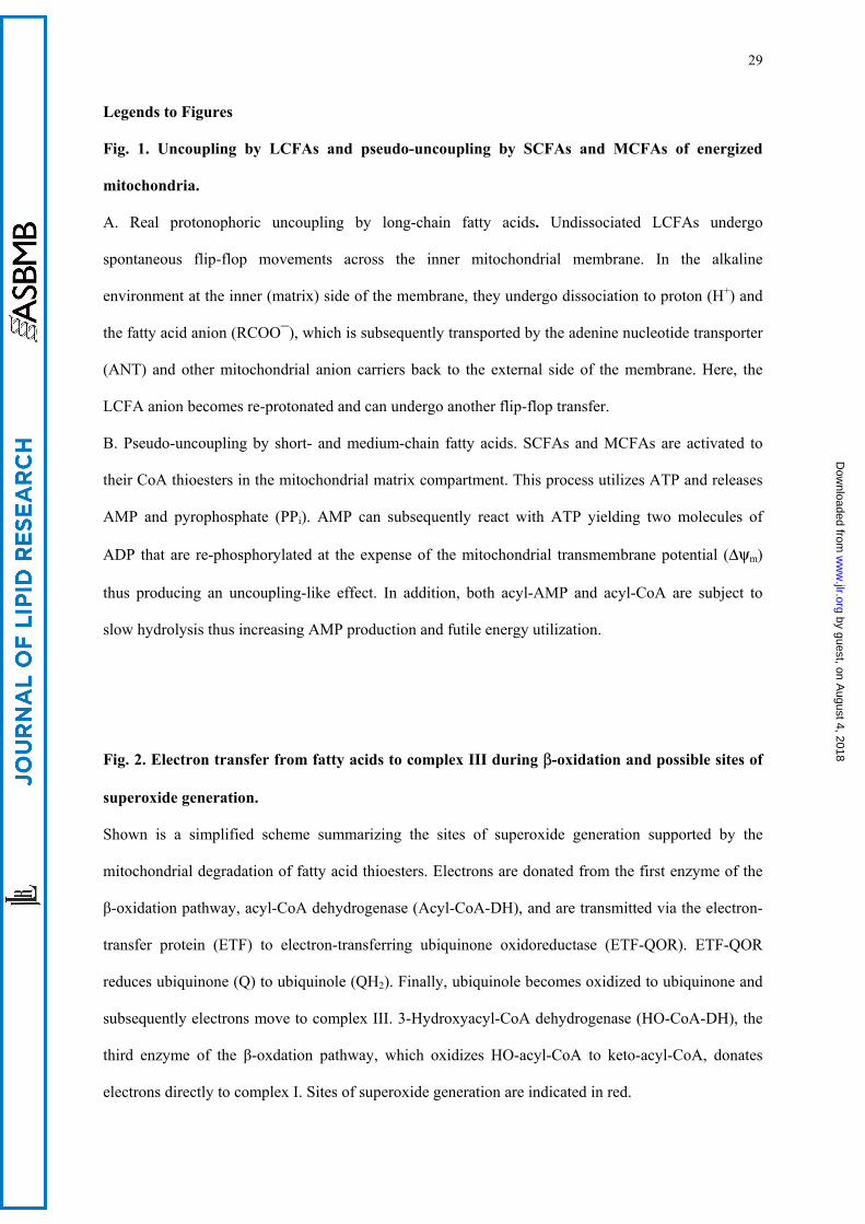
29
Legends to Figures
Fig. 1. Uncoupling by LCFAs and pseudo-uncoupling by SCFAs and MCFAs of energized
mitochondria.
A. Real protonophoric uncoupling by long-chain fatty acids. Undissociated LCFAs undergo
spontaneous flip-flop movements across the inner mitochondrial membrane. In the alkaline
environment at the inner (matrix) side of the membrane, they undergo dissociation to proton (H+) and
the fatty acid anion (RCOO¯), which is subsequently transported by the adenine nucleotide transporter
(ANT) and other mitochondrial anion carriers back to the external side of the membrane. Here, the
LCFA anion becomes re-protonated and can undergo another flip-flop transfer.
B. Pseudo-uncoupling by short- and medium-chain fatty acids. SCFAs and MCFAs are activated to
their CoA thioesters in the mitochondrial matrix compartment. This process utilizes ATP and releases
AMP and pyrophosphate (PPi). AMP can subsequently react with ATP yielding two molecules of
ADP that are re-phosphorylated at the expense of the mitochondrial transmembrane potential (∆ψm)
thus producing an uncoupling-like effect. In addition, both acyl-AMP and acyl-CoA are subject to
slow hydrolysis thus increasing AMP production and futile energy utilization.
Fig. 2. Electron transfer from fatty acids to complex III during β-oxidation and possible sites of
superoxide generation.
Shown is a simplified scheme summarizing the sites of superoxide generation supported by the
mitochondrial degradation of fatty acid thioesters. Electrons are donated from the first enzyme of the
β-oxidation pathway, acyl-CoA dehydrogenase (Acyl-CoA-DH), and are transmitted via the electron-
transfer protein (ETF) to electron-transferring ubiquinone oxidoreductase (ETF-QOR). ETF-QOR
reduces ubiquinone (Q) to ubiquinole (QH2). Finally, ubiquinole becomes oxidized to ubiquinone and
subsequently electrons move to complex III. 3-Hydroxyacyl-CoA dehydrogenase (HO-CoA-DH), the
third enzyme of the β-oxdation pathway, which oxidizes HO-acyl-CoA to keto-acyl-CoA, donates
electrons directly to complex I. Sites of superoxide generation are indicated in red.
by guest, on August 4, 2018
ww
w.jlr.org
Dow
nloaded from
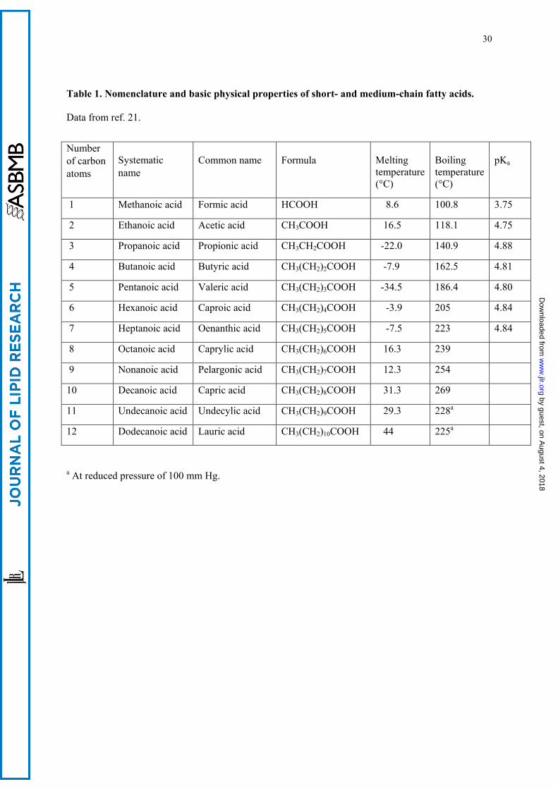
30
Table 1. Nomenclature and basic physical properties of short- and medium-chain fatty acids.
Data from ref. 21.
a At reduced pressure of 100 mm Hg.
Number
of carbon
atoms
Systematic
name
Common name
Formula
Melting temperature (°C)
Boiling temperature (°C)
pKa
1 Methanoic acid Formic acid HCOOH 8.6 100.8 3.75
2 Ethanoic acid Acetic acid CH3COOH 16.5 118.1 4.75
3 Propanoic acid Propionic acid CH3CH2COOH -22.0 140.9 4.88
4 Butanoic acid Butyric acid CH3(CH2)2COOH -7.9 162.5 4.81
5 Pentanoic acid Valeric acid CH3(CH2)3COOH -34.5 186.4 4.80
6 Hexanoic acid Caproic acid CH3(CH2)4COOH -3.9 205 4.84
7 Heptanoic acid Oenanthic acid CH3(CH2)5COOH -7.5 223 4.84
8 Octanoic acid Caprylic acid CH3(CH2)6COOH 16.3 239
9 Nonanoic acid Pelargonic acid CH3(CH2)7COOH 12.3 254
10 Decanoic acid Capric acid CH3(CH2)8COOH 31.3 269
11 Undecanoic acid Undecylic acid CH3(CH2)9COOH 29.3 228a
12 Dodecanoic acid Lauric acid CH3(CH2)10COOH 44 225a
by guest, on August 4, 2018
ww
w.jlr.org
Dow
nloaded from
31
Fig. 1
B Pseudo-uncoupling by ATP utilization
oxidation RCOOH Acyl-AMP Acyl-CoA
ATP
PPi
CoA
AMP
2 ADP
+ ATP
Partial hydrolysi
s
Partial hydrolysi
s
∆
A Uncoupling by ANT-mediated fatty acid
cycling
H+
H+
COOH
-
+
COOH
COO-
COO-
Intermembrane compartment
Matrix compartment
A N
T
by guest, on August 4, 2018
ww
w.jlr.org
Dow
nloaded from
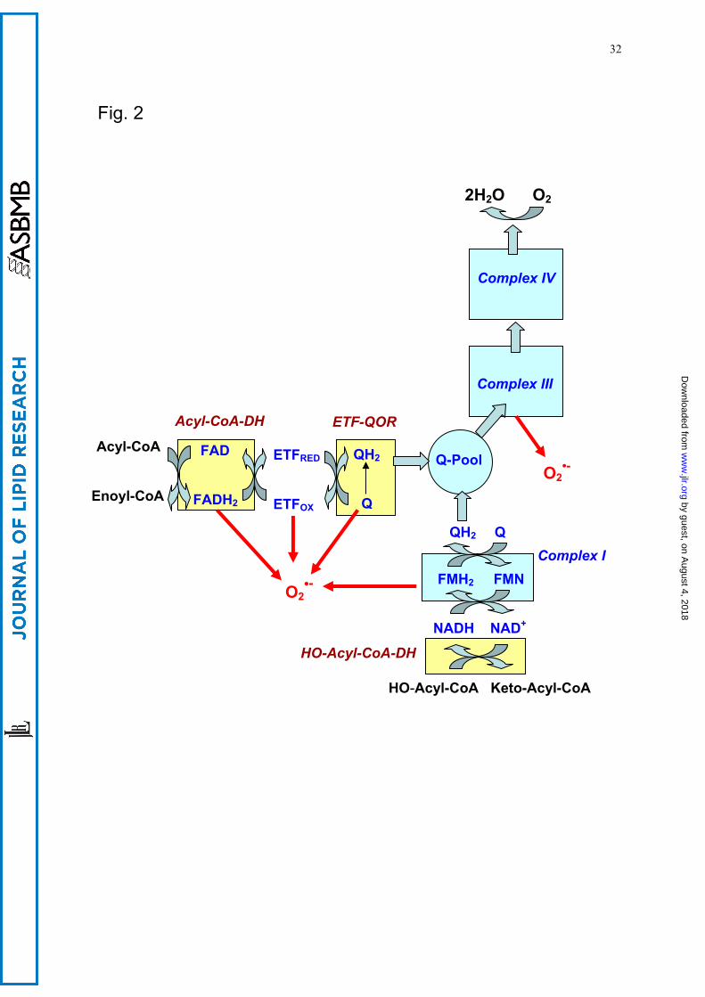
32
Fig. 2
Acyl-CoA
Enoyl-CoA
FAD FADH2
ETFRED
ETFOX
Acyl-CoA-DH
ETF-QOR
QH2 Q
O2•-
O2•-
•-
HO-Acyl-CoA Keto-Acyl-CoA
NADH NAD+
FMH2 FMN
QH2 Q
Q-Pool
Complex I
Complex III
HO-Acyl-CoA-DH
Complex IV
2H2O O2
by guest, on August 4, 2018
ww
w.jlr.org
Dow
nloaded from