Retrospective analysis of antitumor effects and biomarkers ...
12
RESEARCH ARTICLE Retrospective analysis of antitumor effects and biomarkers for nivolumab in NSCLC patients with EGFR mutations Miyuki Sato 1 , Satoshi Watanabe ID 1 *, Hiroshi Tanaka 2 , Koichiro Nozaki 1 , Masashi Arita 1 , Miho Takahashi 1 , Satoshi Shoji 1 , Kosuke Ichikawa 1 , Rie Kondo 1 , Nobumasa Aoki 1 , Masachika Hayashi 1 , Yasuyoshi Ohshima 1 , Toshiyuki Koya 1 , Riuko Ohashi 3 , Yoichi Ajioka 3,4 , Toshiaki Kikuchi 1 1 Department of Respiratory Medicine and Infectious Diseases, Niigata University Graduate School of Medical and Dental Sciences, Niigata City, Niigata, Japan, 2 Department of Internal Medicine, Niigata Cancer Center Hospital, Niigata City, Niigata, Japan, 3 Histopathology Core Facility, Niigata University Faculty of Medicine, Niigata City, Niigata, Japan, 4 Division of Molecular and Diagnostic Pathology, Niigata University Graduate School of Medical and Dental Sciences, Niigata City, Niigata, Japan * [email protected] Abstract Although the blockade of programmed cell death 1 (PD-1)/PD-ligand (L) 1 has demon- strated promising and durable clinical responses for non-small-cell lung cancers (NSCLCs), NSCLC patients with epidermal growth factor receptor (EGFR) mutations responded poorly to PD-1/PD-L1 inhibitors. Previous studies have identified several predictive biomarkers, including the expression of PD-L1 on tumor cells, for PD-1/PD-L1 blockade therapies in NSCLC patients; however, the usefulness of these biomarkers in NSCLCs with EGFR muta- tions has not been elucidated. The present study was conducted to evaluate the predictive biomarkers for PD-1/PD-L1 inhibitors in EGFR-mutated NSCLCs. We retrospectively ana- lyzed 9 patients treated with nivolumab for EGFR-mutated NSCLCs. All but one patient received EGFR-tyrosine kinase inhibitors before nivolumab treatment. The overall response rate and median progression-free survival were 11% and 33 days (95% confidence interval (CI); 7 to 51), respectively. Univariate analysis revealed that patients with a good perfor- mance status (P = 0.11; hazard ratio (HR) 0.183, 95% CI 0.0217 to 1.549), a high density of CD4 + T cells (P = 0.136; HR 0.313, 95% CI 0.045 to 1.417) and a high density of Foxp3 + cells (P = 0.09; HR 0.264, 95% CI 0.0372 to 1.222) in the tumor microenvironment tended to have longer progression-free survival with nivolumab. Multivariate analysis revealed that a high density of CD4 + T cells (P = 0.005; HR<0.001, 95% CI <0.001 to 0.28) and a high den- sity of Foxp3 + cells (P = 0.003; HR<0.001, 95% CI NA) in tumor tissues were strongly corre- lated with better progression-free survival. In contrast to previous studies in wild type EGFR NSCLCs, PD-L1 expression was not associated with the clinical benefit of anti-PD-1 treat- ment in EGFR-mutated NSCLCs. The current study indicated that immune status in the tumor microenvironment may be important for the effectiveness of nivolumab in NSCLC patients with EGFR mutations. PLOS ONE | https://doi.org/10.1371/journal.pone.0215292 April 12, 2019 1 / 12 a1111111111 a1111111111 a1111111111 a1111111111 a1111111111 OPEN ACCESS Citation: Sato M, Watanabe S, Tanaka H, Nozaki K, Arita M, Takahashi M, et al. (2019) Retrospective analysis of antitumor effects and biomarkers for nivolumab in NSCLC patients with EGFR mutations. PLoS ONE 14(4): e0215292. https://doi. org/10.1371/journal.pone.0215292 Editor: Hiroyoshi Nishikawa, National Cancer Center, JAPAN Received: September 5, 2018 Accepted: March 31, 2019 Published: April 12, 2019 Copyright: © 2019 Sato et al. This is an open access article distributed under the terms of the Creative Commons Attribution License, which permits unrestricted use, distribution, and reproduction in any medium, provided the original author and source are credited. Data Availability Statement: All relevant data are within the manuscript and its Supporting Information files. Funding: The authors received no specific funding for this work. Competing interests: The authors have declared that no competing interests exist.
Transcript of Retrospective analysis of antitumor effects and biomarkers ...
RESEARCH ARTICLE
Retrospective analysis of antitumor effects
and biomarkers for nivolumab in NSCLC
patients with EGFR mutations
Miyuki Sato1, Satoshi WatanabeID1*, Hiroshi Tanaka2, Koichiro Nozaki1, Masashi Arita1,
Miho Takahashi1, Satoshi Shoji1, Kosuke Ichikawa1, Rie Kondo1, Nobumasa Aoki1,
Masachika Hayashi1, Yasuyoshi Ohshima1, Toshiyuki Koya1, Riuko Ohashi3,
Yoichi Ajioka3,4, Toshiaki Kikuchi1
1 Department of Respiratory Medicine and Infectious Diseases, Niigata University Graduate School of
Medical and Dental Sciences, Niigata City, Niigata, Japan, 2 Department of Internal Medicine, Niigata Cancer
Center Hospital, Niigata City, Niigata, Japan, 3 Histopathology Core Facility, Niigata University Faculty of
Medicine, Niigata City, Niigata, Japan, 4 Division of Molecular and Diagnostic Pathology, Niigata University
Graduate School of Medical and Dental Sciences, Niigata City, Niigata, Japan
Abstract
Although the blockade of programmed cell death 1 (PD-1)/PD-ligand (L) 1 has demon-
strated promising and durable clinical responses for non-small-cell lung cancers (NSCLCs),
NSCLC patients with epidermal growth factor receptor (EGFR) mutations responded poorly
to PD-1/PD-L1 inhibitors. Previous studies have identified several predictive biomarkers,
including the expression of PD-L1 on tumor cells, for PD-1/PD-L1 blockade therapies in
NSCLC patients; however, the usefulness of these biomarkers in NSCLCs with EGFR muta-
tions has not been elucidated. The present study was conducted to evaluate the predictive
biomarkers for PD-1/PD-L1 inhibitors in EGFR-mutated NSCLCs. We retrospectively ana-
lyzed 9 patients treated with nivolumab for EGFR-mutated NSCLCs. All but one patient
received EGFR-tyrosine kinase inhibitors before nivolumab treatment. The overall response
rate and median progression-free survival were 11% and 33 days (95% confidence interval
(CI); 7 to 51), respectively. Univariate analysis revealed that patients with a good perfor-
mance status (P = 0.11; hazard ratio (HR) 0.183, 95% CI 0.0217 to 1.549), a high density of
CD4+ T cells (P = 0.136; HR 0.313, 95% CI 0.045 to 1.417) and a high density of Foxp3+
cells (P = 0.09; HR 0.264, 95% CI 0.0372 to 1.222) in the tumor microenvironment tended to
have longer progression-free survival with nivolumab. Multivariate analysis revealed that a
high density of CD4+ T cells (P = 0.005; HR<0.001, 95% CI <0.001 to 0.28) and a high den-
sity of Foxp3+ cells (P = 0.003; HR<0.001, 95% CI NA) in tumor tissues were strongly corre-
lated with better progression-free survival. In contrast to previous studies in wild type EGFR
NSCLCs, PD-L1 expression was not associated with the clinical benefit of anti-PD-1 treat-
ment in EGFR-mutated NSCLCs. The current study indicated that immune status in the
tumor microenvironment may be important for the effectiveness of nivolumab in NSCLC
patients with EGFR mutations.
PLOS ONE | https://doi.org/10.1371/journal.pone.0215292 April 12, 2019 1 / 12
a1111111111
a1111111111
a1111111111
a1111111111
a1111111111
OPEN ACCESS
Citation: Sato M, Watanabe S, Tanaka H, Nozaki K,
Arita M, Takahashi M, et al. (2019) Retrospective
analysis of antitumor effects and biomarkers for
nivolumab in NSCLC patients with EGFR
mutations. PLoS ONE 14(4): e0215292. https://doi.
org/10.1371/journal.pone.0215292
Editor: Hiroyoshi Nishikawa, National Cancer
Center, JAPAN
Received: September 5, 2018
Accepted: March 31, 2019
Published: April 12, 2019
Copyright: © 2019 Sato et al. This is an open
access article distributed under the terms of the
Creative Commons Attribution License, which
permits unrestricted use, distribution, and
reproduction in any medium, provided the original
author and source are credited.
Data Availability Statement: All relevant data are
within the manuscript and its Supporting
Information files.
Funding: The authors received no specific funding
for this work.
Competing interests: The authors have declared
that no competing interests exist.
Introduction
Lung cancer is the most common cause of cancer death worldwide [1, 2], and non-small-cell
lung cancer (NSCLC) accounts for the most cases. Immunotherapy for NSCLCs has recently
evolved into a new stage of a novel modality with immune-checkpoint inhibitors (ICIs) [3].
For example, anti-programmed-cell death-1 (PD-1) and anti-PD-ligand (L) 1 antibodies have
demonstrated promising and durable responses across a broad range of solid tumors, includ-
ing NSCLCs [4].
Recent studies have reported the possible predictive biomarkers for PD-1/PD-L1 blockade
therapies. The expression of PD-L1 on tumor cells is the most commonly examined bio-
marker. Subgroup analyses in a large phase III study investigating nivolumab in nonsquamous
lung cancer showed a correlation between overall survival (OS) and PD-L1 expression on
tumor cells [5]. Compared to platinum-doublet chemotherapy, pembrolizumab significantly
prolonged progression-free survival (PFS) and OS in NSCLC patients with a high expression
of PD-L1 [6]. Other predictive biomarkers, such as tumor-mutation burden, tumor-infiltrating
lymphocytes (TILs) including CD8+ T cells and regulatory T cells (Tregs), neutrophil-to-lym-
phocyte ratio (NLR) in peripheral blood, and frequency of immune-suppressive cells in
peripheral blood and tumor tissues have been evaluated to select patients who are more likely
to respond to ICIs [7–12].
Excellent therapeutic effects of epidermal growth factor receptor-tyrosine kinase inhibitors
(EGFR-TKIs) have been reported in EGFR mutation-positive NSCLCs [13–20]. However,
EGFR-TKIs do not cure NSCLCs. All treated patients eventually develop resistance to
EGFR-TKIs, and the illness advances. New therapeutic strategies need to be established for
EGFR-mutated patients. In therapy with ICIs, a clinical study showed no survival benefit of
nivolumab in patients with EGFR mutations [5]. Similarly, compared with docetaxel, pembro-
lizumab did not show any survival advantage in EGFR-mutated NSCLCs [21]. NSCLCs har-
boring EGFR mutations are associated with the low effectiveness of treatments with PD-1/
PD-L1 inhibitors [22, 23]. Possible mechanisms could be the poor antigenicity of tumors due
to a low tumor mutation burden and the immunosuppressive microenvironment in tumor tis-
sues; however, the reasons why PD-1/PD-L1 blockade therapies failed to show a survival bene-
fit in EGFR-mutated NSCLCs are not fully understood [8, 24, 25]. Furthermore, the
effectiveness of PD-1/PD-L1 blockade therapies in EGFR-mutated NSCLC patients with pre-
dictive biomarkers for ICIs remains to be elucidated.
This study aimed to evaluate the potential predictive biomarkers for nivolumab in NSCLC
patients with EGFR mutations.
Materials and methods
Patients
We retrospectively analyzed the data of consecutive patients who received nivolumab for
advanced NSCLC in the Niigata Cancer Center Hospital and Niigata University Medical and
Dental Hospital between January 2016 and December 2017. EGFR mutation testing was per-
formed using the peptide nucleic acid–locked nucleic acid polymerase chain reaction clamp
method or the PCR-invader method [26, 27]. Patients received nivolumab (3 mg/kg) intrave-
nously every 2 weeks until disease progression or unacceptable toxic effect. The present study
was conducted in accordance with the Helsinki Declaration of the World Medical Association.
The protocol was approved by the institutional review board of the Niigata University Medical
and Dental Hospital and the Niigata Cancer Center Hospital and written informed consent
was waived because of the retrospective design.
Effectiveness of nivolumab in EGFR-mutated NSCLCs
PLOS ONE | https://doi.org/10.1371/journal.pone.0215292 April 12, 2019 2 / 12
Immunohistochemistry
In this study, tumor tissues that were adequate for immunohistochemistry analyses were
required for all patients. Formalin-fixed, paraffin embedded tissue (FFPE) sections of 4-μm
thickness were stained for PD-L1 using an automated immunohistochemistry assay (PD-L1
IHC 28–8 pharmDx, Agilent Technologies, Santa Clara, CA). PD-L1 expression on the tumor
cell membrane was evaluated in sections including at least 100 tumor cells. To evaluate the
expression of CD3, CD4, CD8 and Foxp3 in tumor-infiltrating lymphocytes, FFPE sections
were deparaffinized and heated in an antigen retrieval solution at pH 9.0 (Nichirei Biosciences,
Inc., Tokyo, Japan) for 15 min at 121˚C. Endogenous peroxidase activity was quenched using
3% H2O2-methanol for 15 min, and then the sections were blocked with 10% normal goat
serum. Next, sections were incubated with the primary antibodies for CD3 (clone PS1,
Nichirei Corporation Tokyo, Japan), CD4 (clone 4B12, Nichirei Corporation, Tokyo, Japan),
CD8 (clone C8/144B, Nichirei Corporation, Tokyo, Japan) and Foxp3 (clone 236A/E7,
Abcam, Cambridge, UK) overnight incubation at 4˚C. As the second step, a Histofine Simple
Stain MAX-PO (multi) kit (Nichirei Corporation, Tokyo, Japan) was reacted for 30 min. The
samples were carefully washed three times with phosphate-buffered saline (pH 7.4) in each
step. To visualize antigen-antibody complex, a Histofine DAB substrate kit (Nichirei Corpora-
tion, Tokyo, Japan) was used. Nuclear staining was performed with hematoxylin. The numbers
of CD4-, CD8-, Foxp3- and CD3-positive T cells were counted at 1 mm2 magnification in
three different regions of the tumor and averaged, and the standard deviation calculated. The
cell count was performed by using ImageJ software (National Institutes of Health) [28].
Statistical analysis
Kaplan-Meier survival curves were constructed for PFS and OS, and differences between
groups were identified using the log-rank test. Analysis was two-sided, with a 5% significance
level and a 95% confidence interval (CI). All statistical analyses were performed using JMP
9.0.2 statistical software (SAS Institute, Cary, NC, USA).
Results
Patients’ characteristics
We retrospectively identified 9 patients with EGFR-mutated NSCLCs treated with nivolumab
between March 2016 and September 2017. The patient characteristics are listed in Table 1.
There were 6 females and the median age of all the patients was 62 years old (range, 37–72
years). Seven and 2 patients had a performance status of 1 and 2, respectively, and all patients
had adenocarcinoma in histology. Five patients had an exon 19 deletion, one had an exon 19
deletion with T790M, one had L858R with T790M, one had an exon 20 insertion, and one had
a S768I point mutation.
Clinical efficacy of nivolumab in EGFR-mutated NSCLC patients
During treatment with nivolumab, one patient achieved a partial response; however, 7 patients
had progressive disease. The patient who responded to nivolumab had an exon 20 insertion
and had not received EGFR-TKI treatment before nivolumab. We could not evaluate antitu-
mor effects in one patient because the patient discontinued nivolumab treatment due to ileus
and received osimertinib immediately (Table 2).
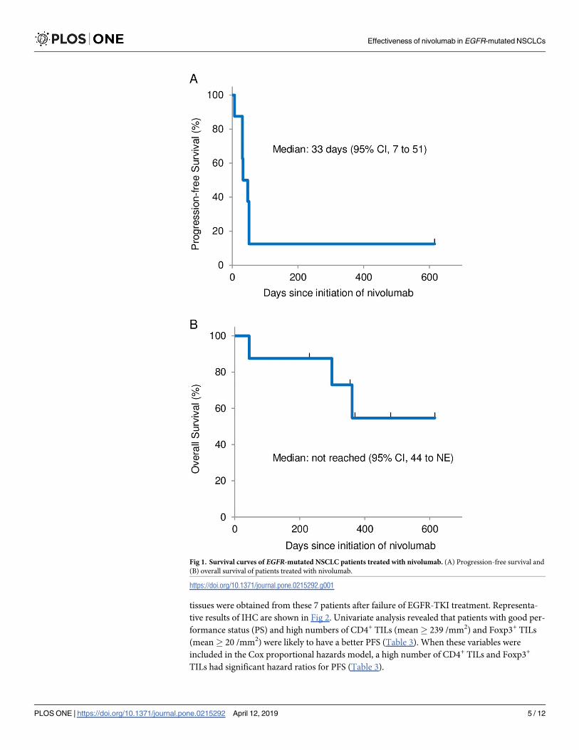
The median number of treatment cycles was 3. The median PFS from the beginning of
nivolumab was 33 days (95% CI 7 to 51), and the median OS was not reached (95% CI 44 to N.
E.) (Fig 1).
Effectiveness of nivolumab in EGFR-mutated NSCLCs
PLOS ONE | https://doi.org/10.1371/journal.pone.0215292 April 12, 2019 3 / 12
Biomarkers for nivolumab in EGFR-mutated patients
Next, we investigated whether the existence of potential biomarkers for immune-checkpoint
inhibitors were associated with the therapeutic effects of nivolumab in NSCLC patients with
EGFR mutations. In the current study, tumor tissues were obtained from all 9 patients and
stained for IHC. Seven out of 9 patients received EGFR-TKIs before nivolumab and tumor
Table 2. Summary of responses.
N = 9 %
CR 0 0
PR 1 11
SD 0 0
PD 7 78
NE 1 11
ORR 11
DCR 11
Cycles received median (range) 3 (2–41)
CR, complete response; PR, partial response; SD, stable disease; PD, progressive disease; NE, not evaluable; ORR,
overall response rate; DCR, disease control ratio
https://doi.org/10.1371/journal.pone.0215292.t002
Table 1. Base line characteristics of all study patients (n = 9).
Parameter Number of patients %
Gender
Female 6 67
Male 3 33
Median age (range), years 62 (37–72)
ECOG PS at initiation of nivolumab
0 / 1 / 2 0 / 7 / 2 0 / 78 / 22
Smoking status
Never smoked 7 78
Current or former 2 22
Histology
Adenocarcinoma 9 100
Clinical stage
IV 7 78
Post operative 2 22
Type of EGFR mutation
Exon19 deletion 5 56
Exon19 deletion + T790M 1 11
L858R + T790M 1 11
Exon 20 insertion 1 11
S768I 1 11
Biopsy site
Primary lesions 7 78
Lymph nodes 2 22
No. of prior regimens before nivolumab
1 / 2 /�3 2 / 4 / 3 22 / 44 / 33
PS, performance status; EGFR, epidermal growth factor receptor.
https://doi.org/10.1371/journal.pone.0215292.t001
Effectiveness of nivolumab in EGFR-mutated NSCLCs
PLOS ONE | https://doi.org/10.1371/journal.pone.0215292 April 12, 2019 4 / 12
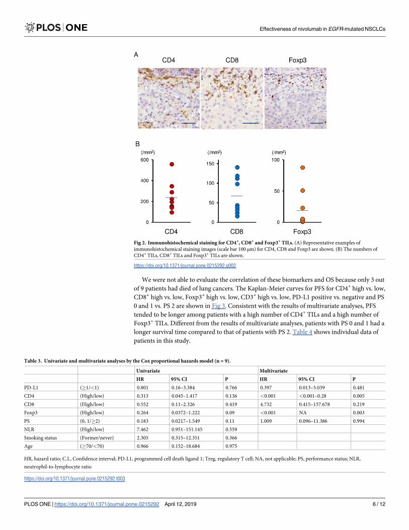
tissues were obtained from these 7 patients after failure of EGFR-TKI treatment. Representa-
tive results of IHC are shown in Fig 2. Univariate analysis revealed that patients with good per-
formance status (PS) and high numbers of CD4+ TILs (mean� 239 /mm2) and Foxp3+ TILs
(mean� 20 /mm2) were likely to have a better PFS (Table 3). When these variables were
included in the Cox proportional hazards model, a high number of CD4+ TILs and Foxp3+
TILs had significant hazard ratios for PFS (Table 3).
Fig 1. Survival curves of EGFR-mutated NSCLC patients treated with nivolumab. (A) Progression-free survival and
(B) overall survival of patients treated with nivolumab.
https://doi.org/10.1371/journal.pone.0215292.g001
Effectiveness of nivolumab in EGFR-mutated NSCLCs
PLOS ONE | https://doi.org/10.1371/journal.pone.0215292 April 12, 2019 5 / 12
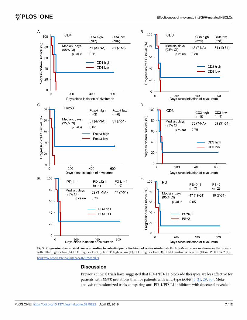
We were not able to evaluate the correlation of these biomarkers and OS because only 3 out
of 9 patients had died of lung cancers. The Kaplan-Meier curves for PFS for CD4+ high vs. low,
CD8+ high vs. low, Foxp3+ high vs. low, CD3+ high vs. low, PD-L1 positive vs. negative and PS
0 and 1 vs. PS 2 are shown in Fig 3. Consistent with the results of multivariate analyses, PFS
tended to be longer among patients with a high number of CD4+ TILs and a high number of
Foxp3+ TILs. Different from the results of multivariate analyses, patients with PS 0 and 1 had a
longer survival time compared to that of patients with PS 2. Table 4 shows individual data of
patients in this study.
Fig 2. Immunohistochemical staining for CD4+, CD8+ and Foxp3+ TILs. (A) Representative examples of
immunohistochemical staining images (scale bar 100 μm) for CD4, CD8 and Foxp3 are shown. (B) The numbers of
CD4+ TILs, CD8+ TILs and Foxp3+ TILs are shown.
https://doi.org/10.1371/journal.pone.0215292.g002
Table 3. Univariate and multivariate analyses by the Cox proportional hazards model (n = 9).
Univariate Multivariate
HR 95% CI P HR 95% CI P
PD-L1 (�1/<1) 0.801 0.16–3.384 0.766 0.397 0.013–5.039 0.481
CD4 (High/low) 0.313 0.045–1.417 0.136 <0.001 <0.001–0.28 0.005
CD8 (High/low) 0.552 0.11–2.326 0.419 4.732 0.415–157.678 0.219
Foxp3 (High/low) 0.264 0.0372–1.222 0.09 <0.001 NA 0.003
PS (0, 1/�2) 0.183 0.0217–1.549 0.11 1.009 0.096–11.386 0.994
NLR (High/low) 7.462 0.951–151.145 0.559
Smoking status (Former/never) 2.305 0.315–12.351 0.366
Age (�70/<70) 0.966 0.152–18.684 0.975
HR, hazard ratio; C.I., Confidence interval; PD-L1, programmed cell death ligand 1; Treg, regulatory T cell; NA, not applicable; PS, performance status; NLR,
neutrophil-to-lymphocyte ratio
https://doi.org/10.1371/journal.pone.0215292.t003
Effectiveness of nivolumab in EGFR-mutated NSCLCs
PLOS ONE | https://doi.org/10.1371/journal.pone.0215292 April 12, 2019 6 / 12
Discussion
Previous clinical trials have suggested that PD-1/PD-L1 blockade therapies are less effective for
patients with EGFR mutations than for patients with wild-type EGFR [5, 21, 29, 30]. Meta-
analysis of randomized trials comparing anti-PD-1/PD-L1 inhibitors with docetaxel revealed
Fig 3. Progression-free survival curves according to potential predictive biomarkers for nivolumab. Kaplan-Meier curves are shown for the patients
with CD4+ high vs. low (A), CD8+ high vs. low (B), Foxp3+ high vs. low (C), CD3+ high vs. low (D), PD-L1 positive vs. negative (E) and PS 0, 1 vs. 2 (F).
https://doi.org/10.1371/journal.pone.0215292.g003
Effectiveness of nivolumab in EGFR-mutated NSCLCs
PLOS ONE | https://doi.org/10.1371/journal.pone.0215292 April 12, 2019 7 / 12
that patients with EGFR mutations did not benefit from PD-1/PD-L1 blockade therapies in
terms of OS, and PFS was even worse [31]. Thus, predictive biomarkers are required to
improve the outcomes of PD-1/PD-L1 blockade therapies in EGFR-mutated NSCLC patients.
The expression of PD-L1 in the tumor microenvironment is the most commonly investigated
biomarker for anti-PD-1/PD-L1 treatments. The association of clinical benefits with PD-L1
expression has been demonstrated in PD-1/PD-L1 blockade therapies [32]. However, it is con-
troversial whether PD-L1 expression is also useful to predict the antitumor effects of PD-1/
PD-L1 inhibitors in EGFR-mutated NSCLCs. In a prospective phase II trial, pembrolizumab,
which is an anti-PD-1 antibody, failed to show clinical benefits in PD-L1 positive EGFR-
mutated NSCLC patients, even in those with a high expression of PD-L1 [33]. In the current
study, 4 out of 9 patients were PD-L1 positive, and a correlation between PD-L1 expression
and clinical efficacy was not observed (Table 3 and Fig 3). Further, there was no statistical dif-
ference of PFS with nivolumab between patients with PD-L1 tumor proportion score of 50%
or greater and patients with PD-L1 tumor proportion score of less than 50% (data not shown).
Tumor cells express PD-L1 in response to inflammatory cytokines, such as IFN-γ, to escape
from attack by effector T cells. Recent studies reported that PD-L1 expression is induced by
signaling through EGFR [34, 35]. This finding may be the reason why PD-L1 expression is not
a reliable predictive biomarker in patients with EGFR mutations.
Accumulating evidence suggests that an inflamed tumor microenvironment may predict
clinical benefits for PD-1/PD-L1 blockade therapies. Considering the mechanisms of PD-1/
PD-L1 blockade therapies, the existence of effector T cells that are suppressed through the PD-
1/PD-L1 axis could be a good predictive biomarker for PD-1/PD-L1 inhibitors. Indeed, several
studies reported that the density of CD8+ T cell infiltration in tumor tissues was associated
with the effectiveness of PD-1/PD-L1 targeted therapies [9, 11]. The high expression of gene
signatures, which were associated with effector T cells and IFN-γ, were also correlated with the
effectiveness of anti-PD-L1 treatment [36]. Wu et al. further demonstrated that the high fre-
quency of PD-L1+CD4+CD25+ Tregs predicted better outcomes in patients treated with PD-1/
PD-L1 blockade therapies [11]. Because the expression of PD-1 on Tregs has a critical role in
maintaining their suppressive function, anti-PD-1 treatment may improve immune responses
in the tumor microenvironment by inhibiting the function of Tregs [37, 38]. In the current
Table 4. Individual data of all study patients (n = 9).
Case Age
(years)
Sex Smoking
status
Types of
EGFR
mutation
Prevoius
treatmet
lines before
nivolumab
EGFR-TKIs PD-L1
expression
No. of
CD4+ T
cells
(/mm2)
N0. of
CD8+ T
cells
(/mm2)
No. of
Foxp3+ T
cells
(/mm2)
PS NLR Response to
nivolumab
PFS
(days)
OS
(days)
1 37 F Never 19del.
+T790M
5 G!E!A 0% 2882 156 44 1 2.4 PD 19 469
2 40 M Never 19del. 2 Gefitinib 100% 2248 978 8 1 1.96 PD 33 370
3 62 F Never 19del. 3 Gefitinib 0% 3452 434 22 1 1.96 PD 51 480
4 66 F Never 20
insertion
4 None 30–40% 5567 1405 874 1 1.51 PR 616 616
5 62 F Never 19del. 2 Afatinib 0% 951 1117 508 1 2.13 PD 51 355
6 72 M Former 19del. 2 Afatinib 0% 1548 236 227 1 2.17 PD 47 299
7 62 F Never L858R
+T790M
3 Afatinib 5–9% 1502 373 11 0 9.875 NE 31 361
8 40 M Former S768I 2 None 0% 1942 1186 22 0 4.94 PD 7 44
9 62 F Never 19del. 1 Afatinib 1–4% 1407 309 54 1 3.44 PD 31 230
F, female; M, male; G, gefitinib; E, erlotinib; A, afatinib; PS, performancde status; NLR, netrophil to lymphocyte ratio; PFS, progression-free survival; OS, overall survival
https://doi.org/10.1371/journal.pone.0215292.t004
Effectiveness of nivolumab in EGFR-mutated NSCLCs
PLOS ONE | https://doi.org/10.1371/journal.pone.0215292 April 12, 2019 8 / 12
study, the high density of CD4+ T cells and Foxp3+ Treg cells, but not CD8+ T cells, in the
tumor microenvironment was positively correlated with better PFS (Table 3 and Fig 3). As dis-
cussed above, EGFR-mutated NSCLCs might express PD-L1 by signaling through EGFR and/
or in response to inflammatory cytokines from effector T cells. The population of immune
cells infiltrating tumor tissues may be good predictive determinants of PD-1/PD-L1 blockade
therapies.
After failure of EGFR-TKI treatment, the mechanisms of immune escape in NSCLCs with
EGFR mutations might be different from those in NSCLCs with EGFR mutations prior
EGFR-TKI treatment. Epithelial-mesenchymal transition and cMET amplification, which
were reported to be acquired resistance suppressed CD8+ T cells [39, 40]. Haratani et al also
demonstrated that compared to T790M-positive NSCLCs, T790M-negative NSCLCs had a
higher level of PD-L1 expression and tended to be benefit from anti-PD-1 treatment [41]. The
information about acquired resistance to EGFR-TKIs may be helpful to guide the administra-
tion of PD-1/PD-L1 inhibitors for EGFR-mutated NSCLCs. In our study, only one patient
who had an exon 20 insertion and was previously untreated with EGFR-TKIs showed a dura-
ble response to nivolumab. Adequate timing of PD-1/PD-L1 blockade therapies for EGFR-
mutated NSCLCs and the association between types of EGFR mutation, resistance mecha-
nisms to EGFR-TKIs and response to PD-1/PD-L1 blockade therapies remain to be
elucidated.
The limitations of the present study include its retrospective design and small sample size.
Because only one patient responded to nivolumab in this study, the results of univariate and
multivariate analyses could be strongly affected by the immune status of this patient. Further
studies are necessary to confirm the correlation between clinical efficacy of PD-1/PD-L1 anti-
bodies and the density of CD4+ T cells and Foxp3+ Treg cells in the tumor microenvironment.
In addition, we only analyzed EGFR-mutated NSCLC patients. Now we are evaluating surface
markers of TILs in EGFR wild-type NSCLC patients to clarify whether the density of CD4+ T
cells and Foxp3+ T cells is correlated with the efficacy of anti-PD-1/PD-L1 treatment.
In conclusion, our findings demonstrated that patients with EGFR mutations poorly
responded to nivolumab treatment regardless of PD-L1 expression on tumor cells. The
immune status of tumor microenvironment may predict antitumor effects of nivolumab in
patients with EGFR mutations. Further studies are warranted to identify predictive biomarkers
for anti-PD-1/PD-L1 antibodies in EGFR-mutated NSCLC patients.
Supporting information
S1 File. Individual data of all study patients.
(XLSX)
Acknowledgments
We thank the patients, their families and all the investigators who are participating in this
study. We also thank Kenji Ohyachi for his expert assistance with histopathology.
Author Contributions
Conceptualization: Satoshi Watanabe, Toshiaki Kikuchi.
Formal analysis: Miyuki Sato, Hiroshi Tanaka, Koichiro Nozaki.
Funding acquisition: Satoshi Watanabe, Toshiaki Kikuchi.
Effectiveness of nivolumab in EGFR-mutated NSCLCs
PLOS ONE | https://doi.org/10.1371/journal.pone.0215292 April 12, 2019 9 / 12
Investigation: Miyuki Sato, Satoshi Shoji, Kosuke Ichikawa, Rie Kondo, Nobumasa Aoki,
Masachika Hayashi, Yasuyoshi Ohshima.
Methodology: Riuko Ohashi.
Project administration: Satoshi Watanabe.
Software: Masashi Arita, Miho Takahashi.
Supervision: Satoshi Watanabe, Toshiyuki Koya, Yoichi Ajioka, Toshiaki Kikuchi.
Validation: Riuko Ohashi, Yoichi Ajioka.
Visualization: Miyuki Sato, Riuko Ohashi, Yoichi Ajioka.
Writing – original draft: Miyuki Sato.
Writing – review & editing: Satoshi Watanabe, Toshiaki Kikuchi.
References1. Global Burden of Disease Cancer C, Fitzmaurice C, Allen C, Barber RM, Barregard L, Bhutta ZA, et al.
Global, Regional, and National Cancer Incidence, Mortality, Years of Life Lost, Years Lived With Disabil-
ity, and Disability-Adjusted Life-years for 32 Cancer Groups, 1990 to 2015: A Systematic Analysis for
the Global Burden of Disease Study. JAMA Oncol. 2017; 3(4):524–48. https://doi.org/10.1001/
jamaoncol.2016.5688 PMID: 27918777.
2. Ferlay J, Soerjomataram I, Dikshit R, Eser S, Mathers C, Rebelo M, et al. Cancer incidence and mortal-
ity worldwide: sources, methods and major patterns in GLOBOCAN 2012. Int J Cancer. 2015; 136(5):
E359–86. https://doi.org/10.1002/ijc.29210 PMID: 25220842.
3. Malhotra J, Jabbour SK, Aisner J. Current state of immunotherapy for non-small cell lung cancer. Transl
Lung Cancer Res. 2017; 6(2):196–211. Epub 2017/05/23. https://doi.org/10.21037/tlcr.2017.03.01
PMID: 28529902; PubMed Central PMCID: PMCPMC5420529.
4. Gong J, Chehrazi-Raffle A, Reddi S, Salgia R. Development of PD-1 and PD-L1 inhibitors as a form of
cancer immunotherapy: a comprehensive review of registration trials and future considerations. Journal
for immunotherapy of cancer. 2018; 6(1):8. Epub 2018/01/24. https://doi.org/10.1186/s40425-018-
0316-z PMID: 29357948; PubMed Central PMCID: PMCPMC5778665.
5. Borghaei H, Paz-Ares L, Horn L, Spigel DR, Steins M, Ready NE, et al. Nivolumab versus Docetaxel in
Advanced Nonsquamous Non-Small-Cell Lung Cancer. N Engl J Med. 2015; 373(17):1627–39. https://
doi.org/10.1056/NEJMoa1507643 PMID: 26412456; PubMed Central PMCID: PMCPMC5705936.
6. Reck M, Rodriguez-Abreu D, Robinson AG, Hui R, Csoszi T, Fulop A, et al. Pembrolizumab versus
Chemotherapy for PD-L1-Positive Non-Small-Cell Lung Cancer. N Engl J Med. 2016; 375(19):1823–
33. Epub 2016/10/11. https://doi.org/10.1056/NEJMoa1606774 PMID: 27718847.
7. Carbone DP, Reck M, Paz-Ares L, Creelan B, Horn L, Steins M, et al. First-Line Nivolumab in Stage IV
or Recurrent Non-Small-Cell Lung Cancer. N Engl J Med. 2017; 376(25):2415–26. Epub 2017/06/22.
https://doi.org/10.1056/NEJMoa1613493 PMID: 28636851.
8. Rizvi NA, Hellmann MD, Snyder A, Kvistborg P, Makarov V, Havel JJ, et al. Cancer immunology. Muta-
tional landscape determines sensitivity to PD-1 blockade in non-small cell lung cancer. Science (New
York, NY). 2015; 348(6230):124–8. Epub 2015/03/15. https://doi.org/10.1126/science.aaa1348 PMID:
25765070; PubMed Central PMCID: PMCPMC4993154.
9. Tumeh PC, Harview CL, Yearley JH, Shintaku IP, Taylor EJ, Robert L, et al. PD-1 blockade induces
responses by inhibiting adaptive immune resistance. Nature. 2014; 515(7528):568–71. Epub 2014/11/
28. https://doi.org/10.1038/nature13954 PMID: 25428505; PubMed Central PMCID:
PMCPMC4246418.
10. Kamphorst AO, Pillai RN, Yang S, Nasti TH, Akondy RS, Wieland A, et al. Proliferation of PD-1+ CD8 T
cells in peripheral blood after PD-1-targeted therapy in lung cancer patients. Proc Natl Acad Sci U S A.
2017; 114(19):4993–8. Epub 2017/04/28. https://doi.org/10.1073/pnas.1705327114 PMID: 28446615;
PubMed Central PMCID: PMCPMC5441721.
11. Wu SP, Liao RQ, Tu HY, Wang WJ, Dong ZY, Huang SM, et al. Stromal PD-L1-Positive Regulatory T
cells and PD-1-Positive CD8-Positive T cells Define the Response of Different Subsets of Non-Small
Cell Lung Cancer to PD-1/PD-L1 Blockade Immunotherapy. J Thorac Oncol. 2018; 13(4):521–32. Epub
2017/12/23. https://doi.org/10.1016/j.jtho.2017.11.132 PMID: 29269008.
Effectiveness of nivolumab in EGFR-mutated NSCLCs
PLOS ONE | https://doi.org/10.1371/journal.pone.0215292 April 12, 2019 10 / 12
12. Bagley SJ, Kothari S, Aggarwal C, Bauml JM, Alley EW, Evans TL, et al. Pretreatment neutrophil-to-
lymphocyte ratio as a marker of outcomes in nivolumab-treated patients with advanced non-small-cell
lung cancer. Lung Cancer. 2017; 106:1–7. Epub 2017/03/14. https://doi.org/10.1016/j.lungcan.2017.01.
013 PMID: 28285682.
13. Maemondo M, Inoue A, Kobayashi K, Sugawara S, Oizumi S, Isobe H, et al. Gefitinib or chemotherapy
for non-small-cell lung cancer with mutated EGFR. N Engl J Med. 2010; 362(25):2380–8. Epub 2010/
06/25. https://doi.org/10.1056/NEJMoa0909530 PMID: 20573926.
14. Mitsudomi T, Morita S, Yatabe Y, Negoro S, Okamoto I, Tsurutani J, et al. Gefitinib versus cisplatin plus
docetaxel in patients with non-small-cell lung cancer harbouring mutations of the epidermal growth fac-
tor receptor (WJTOG3405): an open label, randomised phase 3 trial. Lancet Oncol. 2010; 11(2):121–8.
Epub 2009/12/22. https://doi.org/10.1016/S1470-2045(09)70364-X PMID: 20022809.
15. Zhou C, Wu YL, Chen G, Feng J, Liu XQ, Wang C, et al. Erlotinib versus chemotherapy as first-line
treatment for patients with advanced EGFR mutation-positive non-small-cell lung cancer (OPTIMAL,
CTONG-0802): a multicentre, open-label, randomised, phase 3 study. Lancet Oncol. 2011; 12(8):735–
42. Epub 2011/07/26. https://doi.org/10.1016/S1470-2045(11)70184-X PMID: 21783417.
16. Rosell R, Carcereny E, Gervais R, Vergnenegre A, Massuti B, Felip E, et al. Erlotinib versus standard
chemotherapy as first-line treatment for European patients with advanced EGFR mutation-positive non-
small-cell lung cancer (EURTAC): a multicentre, open-label, randomised phase 3 trial. Lancet Oncol.
2012; 13(3):239–46. Epub 2012/01/31. https://doi.org/10.1016/S1470-2045(11)70393-X PMID:
22285168.
17. Sequist LV, Yang JC, Yamamoto N, O’Byrne K, Hirsh V, Mok T, et al. Phase III study of afatinib or cis-
platin plus pemetrexed in patients with metastatic lung adenocarcinoma with EGFR mutations. J Clin
Oncol. 2013; 31(27):3327–34. Epub 2013/07/03. https://doi.org/10.1200/JCO.2012.44.2806 PMID:
23816960.
18. Wu Y-L, Zhou C, Hu C-P, Feng J, Lu S, Huang Y, et al. Afatinib versus cisplatin plus gemcitabine for
first-line treatment of Asian patients with advanced non-small-cell lung cancer harbouring EGFR muta-
tions (LUX-Lung 6): an open-label, randomised phase 3 trial. The Lancet Oncology. 2014; 15(2):213–
22. https://doi.org/10.1016/S1470-2045(13)70604-1 PMID: 24439929
19. Soria JC, Ohe Y, Vansteenkiste J, Reungwetwattana T, Chewaskulyong B, Lee KH, et al. Osimertinib in
Untreated EGFR-Mutated Advanced Non-Small-Cell Lung Cancer. N Engl J Med. 2018; 378(2):113–
25. Epub 2017/11/21. https://doi.org/10.1056/NEJMoa1713137 PMID: 29151359.
20. Wu Y-L, Cheng Y, Zhou X, Lee KH, Nakagawa K, Niho S, et al. Dacomitinib versus gefitinib as first-line
treatment for patients with EGFR -mutation-positive non-small-cell lung cancer (ARCHER 1050): a ran-
domised, open-label, phase 3 trial. The Lancet Oncology. 2017; 18(11):1454–66. https://doi.org/10.
1016/S1470-2045(17)30608-3 PMID: 28958502
21. Herbst RS, Baas P, Kim DW, Felip E, Perez-Gracia JL, Han JY, et al. Pembrolizumab versus docetaxel
for previously treated, PD-L1-positive, advanced non-small-cell lung cancer (KEYNOTE-010): a rando-
mised controlled trial. Lancet. 2016; 387(10027):1540–50. https://doi.org/10.1016/S0140-6736(15)
01281-7 PMID: 26712084.
22. Ahn MJ, Sun JM, Lee SH, Ahn JS, Park K. EGFR TKI combination with immunotherapy in non-small
cell lung cancer. Expert Opin Drug Saf. 2017; 16(4):465–9. https://doi.org/10.1080/14740338.2017.
1300656 PMID: 28271729.
23. Gainor JF, Shaw AT, Sequist LV, Fu X, Azzoli CG, Piotrowska Z, et al. EGFR Mutations and ALK Rear-
rangements Are Associated with Low Response Rates to PD-1 Pathway Blockade in Non-Small Cell
Lung Cancer: A Retrospective Analysis. Clin Cancer Res. 2016; 22(18):4585–93. https://doi.org/10.
1158/1078-0432.CCR-15-3101 PMID: 27225694; PubMed Central PMCID: PMCPMC5026567.
24. Vogelstein B, Papadopoulos N, Velculescu VE, Zhou S, Diaz LA Jr., Kinzler KW. Cancer genome land-
scapes. Science (New York, NY). 2013; 339(6127):1546–58. Epub 2013/03/30. https://doi.org/10.1126/
science.1235122 23539594; PubMed Central PMCID: PMCPMC3749880. PMID: 23539594
25. Soo RA, Lim SM, Syn NL, Teng R, Soong R, Mok TSK, et al. Immune checkpoint inhibitors in epidermal
growth factor receptor mutant non-small cell lung cancer: Current controversies and future directions.
Lung Cancer. 2018; 115:12–20. Epub 2018/01/02. https://doi.org/10.1016/j.lungcan.2017.11.009
PMID: 29290252.
26. Nagai Y, Miyazawa H, Huqun, Tanaka T, Udagawa K, Kato M, et al. Genetic heterogeneity of the epi-
dermal growth factor receptor in non-small cell lung cancer cell lines revealed by a rapid and sensitive
detection system, the peptide nucleic acid-locked nucleic acid PCR clamp. Cancer research. 2005; 65
(16):7276–82. Epub 2005/08/18. https://doi.org/10.1158/0008-5472.CAN-05-0331 PMID: 16105816.
27. Naoki K, Soejima K, Okamoto H, Hamamoto J, Hida N, Nakachi I, et al. The PCR-invader method
(structure-specific 5’ nuclease-based method), a sensitive method for detecting EGFR gene mutations
in lung cancer specimens; comparison with direct sequencing. International journal of clinical oncology.
2011; 16(4):335–44. Epub 2011/02/12. https://doi.org/10.1007/s10147-011-0187-5 PMID: 21311943.
Effectiveness of nivolumab in EGFR-mutated NSCLCs
PLOS ONE | https://doi.org/10.1371/journal.pone.0215292 April 12, 2019 11 / 12
28. Schneider CA, Rasband WS, Eliceiri KW. NIH Image to ImageJ: 25 years of image analysis. Nat Meth-
ods. 2012; 9(7):671–5. PMID: 22930834; PubMed Central PMCID: PMCPMC5554542.
29. Lee CK, Man J, Lord S, Links M, Gebski V, Mok T, et al. Checkpoint Inhibitors in Metastatic EGFR-
Mutated Non-Small Cell Lung Cancer-A Meta-Analysis. J Thorac Oncol. 2017; 12(2):403–7. https://doi.
org/10.1016/j.jtho.2016.10.007 PMID: 27765535.
30. Rittmeyer A, Barlesi F, Waterkamp D, Park K, Ciardiello F, von Pawel J, et al. Atezolizumab versus doc-
etaxel in patients with previously treated non-small-cell lung cancer (OAK): a phase 3, open-label, multi-
centre randomised controlled trial. Lancet. 2017; 389(10066):255–65. Epub 2016/12/17. https://doi.org/
10.1016/S0140-6736(16)32517-X PMID: 27979383.
31. Dong ZY, Zhang JT, Liu SY, Su J, Zhang C, Xie Z, et al. EGFR mutation correlates with uninflamed phe-
notype and weak immunogenicity, causing impaired response to PD-1 blockade in non-small cell lung
cancer. Oncoimmunology. 2017; 6(11):e1356145. Epub 2017/11/18. https://doi.org/10.1080/
2162402X.2017.1356145 PMID: 29147605; PubMed Central PMCID: PMCPMC5674946.
32. Melosky B, Chu Q, Juergens RA, Leighl N, Ionescu D, Tsao MS, et al. Breaking the biomarker code:
PD-L1 expression and checkpoint inhibition in advanced NSCLC. Cancer treatment reviews. 2018;
65:65–77. Epub 2018/03/24. https://doi.org/10.1016/j.ctrv.2018.02.005 PMID: 29567557.
33. Lisberg A, Cummings A, Goldman JW, Bornazyan K, Reese N, Wang T, et al. A Phase II Study of Pem-
brolizumab in EGFR-Mutant, PD-L1+, Tyrosine Kinase Inhibitor Naive Patients With Advanced NSCLC.
J Thorac Oncol. 2018; 13(8):1138–45. Epub 2018/06/07. https://doi.org/10.1016/j.jtho.2018.03.035
PMID: 29874546; PubMed Central PMCID: PMCPMC6063769.
34. Chen N, Fang W, Zhan J, Hong S, Tang Y, Kang S, et al. Upregulation of PD-L1 by EGFR Activation
Mediates the Immune Escape in EGFR-Driven NSCLC: Implication for Optional Immune Targeted
Therapy for NSCLC Patients with EGFR Mutation. J Thorac Oncol. 2015; 10(6):910–23. Epub 2015/02/
07. https://doi.org/10.1097/JTO.0000000000000500 PMID: 25658629.
35. Zhang N, Zeng Y, Du W, Zhu J, Shen D, Liu Z, et al. The EGFR pathway is involved in the regulation of
PD-L1 expression via the IL-6/JAK/STAT3 signaling pathway in EGFR-mutated non-small cell lung can-
cer. Int J Oncol. 2016; 49(4):1360–8. Epub 2016/08/09. https://doi.org/10.3892/ijo.2016.3632 PMID:
27499357.
36. Fehrenbacher L, Spira A, Ballinger M, Kowanetz M, Vansteenkiste J, Mazieres J, et al. Atezolizumab
versus docetaxel for patients with previously treated non-small-cell lung cancer (POPLAR): a multicen-
tre, open-label, phase 2 randomised controlled trial. The Lancet. 2016; 387(10030):1837–46. https://
doi.org/10.1016/s0140-6736(16)00587-0
37. Stathopoulou C, Gangaplara A, Mallett G, Flomerfelt FA, Liniany LP, Knight D, et al. PD-1 Inhibitory
Receptor Downregulates Asparaginyl Endopeptidase and Maintains Foxp3 Transcription Factor Stabil-
ity in Induced Regulatory T Cells. Immunity. 2018. Epub 2018/07/29. https://doi.org/10.1016/j.immuni.
2018.05.006 PMID: 30054205.
38. Zhang B, Chikuma S, Hori S, Fagarasan S, Honjo T. Nonoverlapping roles of PD-1 and FoxP3 in main-
taining immune tolerance in a novel autoimmune pancreatitis mouse model. Proc Natl Acad Sci U S A.
2016; 113(30):8490–5. Epub 2016/07/14. https://doi.org/10.1073/pnas.1608873113 PMID: 27410049;
PubMed Central PMCID: PMCPMC4968716.
39. Balan M, Mier y Teran E, Waaga-Gasser AM, Gasser M, Choueiri TK, Freeman G, et al. Novel roles of
c-Met in the survival of renal cancer cells through the regulation of HO-1 and PD-L1 expression. J Biol
Chem. 2015; 290(13):8110–20. Epub 2015/02/04. https://doi.org/10.1074/jbc.M114.612689 PMID:
25645920; PubMed Central PMCID: PMCPMC4375468.
40. Chen L, Gibbons DL, Goswami S, Cortez MA, Ahn YH, Byers LA, et al. Metastasis is regulated via
microRNA-200/ZEB1 axis control of tumour cell PD-L1 expression and intratumoral immunosuppres-
sion. Nat Commun. 2014; 5:5241. Epub 2014/10/29. https://doi.org/10.1038/ncomms6241 PMID:
25348003; PubMed Central PMCID: PMCPMC4212319.
41. Haratani K, Hayashi H, Tanaka T, Kaneda H, Togashi Y, Sakai K, et al. Tumor immune microenviron-
ment and nivolumab efficacy in EGFR mutation-positive non-small-cell lung cancer based on T790M
status after disease progression during EGFR-TKI treatment. Ann Oncol. 2017; 28(7):1532–9. https://
doi.org/10.1093/annonc/mdx183 PMID: 28407039.
Effectiveness of nivolumab in EGFR-mutated NSCLCs
PLOS ONE | https://doi.org/10.1371/journal.pone.0215292 April 12, 2019 12 / 12


![Lee Bontecou : a retrospective : [brochure] July 30 ... Bontecou : a retrospective : [brochure] July 30-September 27, ... Welded steel, canvas, and wire, ... a retrospective : [brochure]](https://static.fdocument.pub/doc/165x107/5b3bd4cc7f8b9a5e1f8cf4d4/lee-bontecou-a-retrospective-brochure-july-30-bontecou-a-retrospective.jpg)