Purine and pyrimidine compounds in murine peritoneal macrophages cultured in vitro
6
Journal of Chromatography, 553 (1991) 205-210 Elsevier Science Publishers B.V.. Amsterdam CHROMSYMP. 2119 Purine and pyrimidine compounds in murine peritoneal macrophages cultured in vitro ANDREAS WERNER* Institute qf Biochemistry, CharitP, Hess&he Strasse 3-4, O-1040 Berlin (Germany) RYOUTA MAEBA, HIROYUKI SHIMASAKI and NOBUO UETA Department of Biochemistry, Teikyo University School of Medicine, Tokyo (Japan) and GERHARD GERBER Instirure of Biochemistry. CharitP, Hess&he Stra.s.se 3-4, O-1040 Berlin (Cermuny) ABSTRACT Extracts of murine peritoneal macrophages were analysed by ion-pair reversed-phase high-perform- ance liquid chromatography during incubation at 37°C in vitro. Four-step gradient elution was applied to an ODS column (250 x 4.6 mm I.D.) at a flow-rate of 1.3 ml/mitt, allowing the separation of hypoxan- thine, inosine, guanosine, adenosine, IMP, CDP, AMP, GDP, UDP, ADP. CTP, GTP. U II and ATP within 50 min. Samples of 0.4 lOha. IO6 cells were washed twice with RPM1 1640 medium and extracted with perchloric acid. Nucleotide concentrations of murine peritoneal macrophages did not change during incubation for 4 days in vitro. _ INTRODUCTION Purine and pyrimidine compounds are of major importance for a multitude of cellular functions. Synthesis and catabolism of nucleotides are well understood and reviewed [l-3]. Extensive metabolic studies have been performed on liver, heart, and red blood cells. However, only a limited number of investigations have been made on nucleotide metabolism in white blood cells, although there are relationships between nucleotide metabolism and several specific functions of these cells, such as the following: phagocytosis is accompaied by a decline in ATP [4]; the activation of lymphocytes is promoted by ATP [5]; guanine nucleotides activate the O2 --generating NADPH-dependent oxidase [6] and adenosine inhibits 02- generation [7]; an ecto-5’-nucleotidase cleaves external AMP to adenosine, which is then transported into the macrophage by a purine nucleoside carrier [8]; and the activity of this enzyme characterizes functionally different cell populations [9]. A genetic defect of adenosine deaminase disturbs lymphocyte proliferation and causes immunodeficiency [IO]. A thorough study of the nucleotide profile in lymphocytes and macrophages has not been published. This work was aimed at the determination of a wide range of purine metabolites in macrophages by gradient ion-pair reversed-phase high-per- 0021-9673/91/$03.50 ((3 1991 Elsevier Science Publishers B.V
-
Upload
andreas-werner -
Category
Documents
-
view
212 -
download
0
Transcript of Purine and pyrimidine compounds in murine peritoneal macrophages cultured in vitro
Journal of Chromatography, 553 (1991) 205-210
Elsevier Science Publishers B.V.. Amsterdam
CHROMSYMP. 2119
Purine and pyrimidine compounds in murine peritoneal macrophages cultured in vitro
ANDREAS WERNER*
Institute qf Biochemistry, CharitP, Hess&he Strasse 3-4, O-1040 Berlin (Germany)
RYOUTA MAEBA, HIROYUKI SHIMASAKI and NOBUO UETA
Department of Biochemistry, Teikyo University School of Medicine, Tokyo (Japan)
and
GERHARD GERBER
Instirure of Biochemistry. CharitP, Hess&he Stra.s.se 3-4, O-1040 Berlin (Cermuny)
ABSTRACT
Extracts of murine peritoneal macrophages were analysed by ion-pair reversed-phase high-perform- ance liquid chromatography during incubation at 37°C in vitro. Four-step gradient elution was applied to an ODS column (250 x 4.6 mm I.D.) at a flow-rate of 1.3 ml/mitt, allowing the separation of hypoxan- thine, inosine, guanosine, adenosine, IMP, CDP, AMP, GDP, UDP, ADP. CTP, GTP. U II and ATP within 50 min. Samples of 0.4 lOha. IO6 cells were washed twice with RPM1 1640 medium and extracted with perchloric acid. Nucleotide concentrations of murine peritoneal macrophages did not change during incubation for 4 days in vitro.
_
INTRODUCTION
Purine and pyrimidine compounds are of major importance for a multitude of cellular functions. Synthesis and catabolism of nucleotides are well understood and reviewed [l-3]. Extensive metabolic studies have been performed on liver, heart, and red blood cells. However, only a limited number of investigations have been made on nucleotide metabolism in white blood cells, although there are relationships between nucleotide metabolism and several specific functions of these cells, such as the following: phagocytosis is accompaied by a decline in ATP [4]; the activation of lymphocytes is promoted by ATP [5]; guanine nucleotides activate the O2 --generating NADPH-dependent oxidase [6] and adenosine inhibits 02- generation [7]; an ecto-5’-nucleotidase cleaves external AMP to adenosine, which is then transported into the macrophage by a purine nucleoside carrier [8]; and the activity of this enzyme characterizes functionally different cell populations [9]. A genetic defect of adenosine deaminase disturbs lymphocyte proliferation and causes immunodeficiency [IO].
A thorough study of the nucleotide profile in lymphocytes and macrophages has not been published. This work was aimed at the determination of a wide range of purine metabolites in macrophages by gradient ion-pair reversed-phase high-per-
0021-9673/91/$03.50 ((3 1991 Elsevier Science Publishers B.V

206 A. WERNER et al.
formance liquid chromatography (HPLC), allowing the separation of purine nucleo- tides, nucleosides and nucleobases in a single run.
EXPERIMENTAL
Mucrophage culture A 2-ml volume of 10% proteose peptone (Difco Labs., Detroit, MI, U.S.A.) was
injected into the peritoneal cavities of male mice (age 2 months). After 4 days, 10 ml of RPM1 1640 (Flow Labs., Irvine, U.K.) were injected into the peritoneal cavity and the suspended macrophages were aspirated. The cells were washed twice with that medium. Then 10. lo6 cells were suspended in RPM1 1640 containing 10% foetal calf serum. The suspension was placed in plastic culture dishes of 35 mm diameter (Terumo, Tokyo, Japan). The cells were allowed to adhere at 37°C under 5% carbon dioxide during a 3-h incubation. The non-adhering cells were removed by rinsing twice with RPM1 1640. The incubation was continued and after 24 h the medium was exchanged [l 11. At this time and at the fourth day of incubation aliquots were sampled for nucleotide analysis.
Sample extraction Macrophages were scraped off, washed twice with RPM1 medium and
nucleotides were immediately extracted with 6% perchloric acid; after centrifugation, the supernatant was neutralized with potassium hydroxide. The samples were cleaned from insoluble potassium chlorate by centrifugation at 900 g for 3 min and in addition by percolation through a 0.2-pm membrane filter (Sartorius, Germany).
HPLC equipment The HPLC system consisted of equipment from Waters Assoc. (Milford, MA,
U.S.A.), with two Model 510 HPLC pumps, an automated gradient controller, a programmable multi-wavelength detector (peak identification by UV absorbance measurement at 254 and 280 nm), a Model 745 data module and Rheodyne Model 7125 injector with a 50-~1 sample loop.
Chromatographic conditions Buffer A [lo mM NH4H2P04 (Merck, Darmstadt, Germany) + 2 mM
tetrabutylammonium phosphate (PIC Reagent A; Waters Assoc.)] and buffer B [buffer A containing 20% (v/v) acetonitrile (LiChrosolv, Merck)] were used. The flow-rate was 1.3 ml/min. A Supelcosil ODS column (250 x 4.6 mm I.D.) was applied.
The following gradient was used: 5 min isocratic with 100% buffer A, followed by a 12-min linear gradient up to 80% B, then 30 min isocratic with 80% B-20% A followed by a 2-min linear gradient to 100% B, returning in 5 min to 100% buffer A.
QuantiJication and peak identlyication In perchloric acid extracts, recoveries of 81 f. 12% for ATP and 76 f 14% for
hypoxanthine were obtained. The recoveries for the other compounds were between these values. Different amounts of each standard compound were injected and the linearity of the calibration graph for the particular concentration range was checked (nucleosides, nucleobases, 50-500 pmol; nucleotides, 200-l 500 pmol). Peak identi-
RP-HPLC OF PURINE METABOLITES 207
fication was performed by coelution of the macrophage extracts with known standards.
RESULTS AND DISCUSSION
Ion-pair reversed-phase separation Ion-pair methods were used initially to enhance the selectivity in solvent
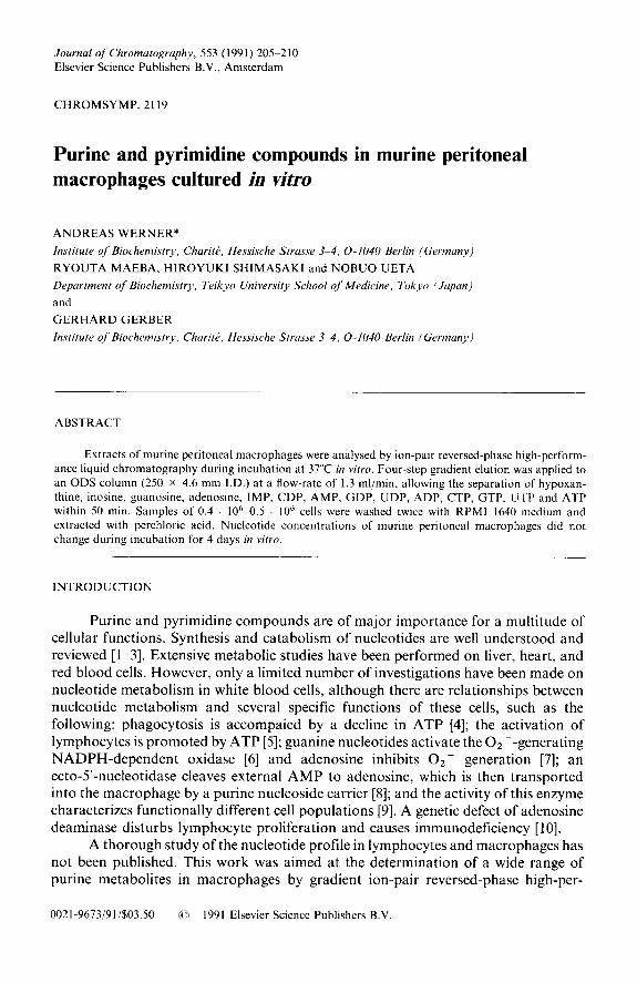
extraction and later they were introduced to improve reversed-phase LC separations [12]. The ion-pair reversed-phase (IP-RP) mode is applied nowadays to separate bases and acids, pharmaceuticals, amino acids, peptides, proteins and nucleic acids and for chiral separations [ 12,131. Nucleotides interact with cationic ion-pair reagents owing to their ionic properties and in this way a good retention of these compounds on Cl8 columns is possible. In the experiments performed here, tetrabutylammonium phosphate was chosen. The pH of the buffers was 5.8. Hypoxanthine, inosine, guanosine, adenosine, IMP, CDP, AMP, GDP, UDP, ADP, CTP, GTP, UTP and ATP eluted as shown in Fig. 1. c-AMP elutes after ATP (not shown in Fig. 1). Similar IP-RP separations of nucleotide standards have been reported [14].
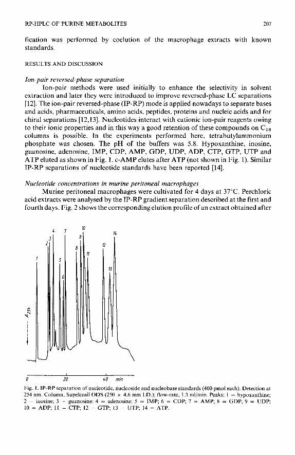
Nucleotide concentrations in murine peritoneal macrophages Murine peritoneal macrophages were cultivated for 4 days at 37°C. Perchloric
acid extracts were analysed by the IP-RP gradient separation described at the first and fourth days. Fig. 2 shows the corresponding elution profile of an extract obtained after
10 47 I
1.
14
3
fi
i
I I
0 20 40 min
Fig. 1. IP-RP separation of nucleotide, nucleoside and nucleobase standards (400 pmol each). Detection at 254 nm. Column, Supelcosil ODS (250 x 4.6 mm I.D.); flow-rate, 1.3 ml/min. Peaks: 1 = hypoxanthine; 2 = inosine; 3 = guanosine; 4 = adenosine; 5 = IMP; 6 = CDP; 7 = AMP; 8 = GDP; 9 = UDP; 10 = ADP; 11 = CTP; 12 = GTP; 13 = UTP; 14 = ATP.
208 A. WbKNCK et al.
L I I
0 20 40 min
Fig. 2. Chromatographic elution profile of an extract of murine peritoneal macrophages after incubation in vitro for 4 days. 0.4. IO6 cells were used for the extraction.
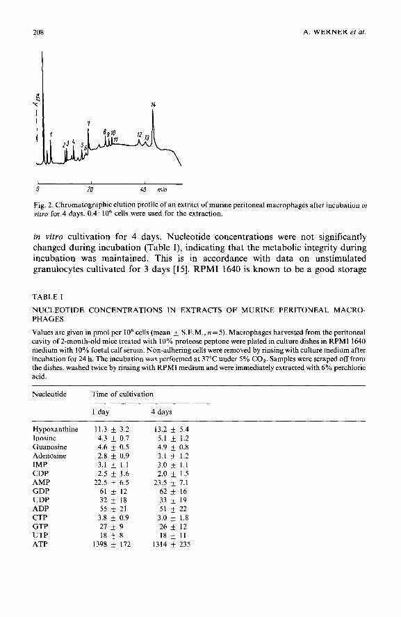
in vitro cultivation for 4 days. Nucleotide concentrations were not significantly changed during incubation (Table I), indicating that the metabolic integrity during incubation was maintained. This is in accordance with data on unstimulated granulocytes cultivated for 3 days [15]. RPM1 1640 is known to be a good storage
TABLE I
NUCLEOTIDE CONCENTRATIONS IN EXTRACTS OF MURINE PERITONEAL MACRO-
PHAGES
Values are given in pmol per IO6 cells (mean k S.E.M., n = 5). Macrophages harvested from the peritoneal cavity of 2-month-old mice treated with 10% proteose peptone were plated in culture dishes in RPM1 1640 medium with 10% foetal calf serum. Non-adhering cells were removed by rinsing with culture medium after incubation for 24 h. The incubation was performed at 37°C under 5% COz. Samples were scraped off from the dishes, washed twice by rinsing with RPM1 medium and were immediately extracted with 6% perchloric acid.
Nucleotide Time of cultivation
1 day 4 days
Hypoxanthine Inosine Guanosine Adenosine IMP CDP AMP GDP UDP ADP CTP GTP UTP ATP
11.3 + 3.2
4.3 + 0.7
4.6 f 0.5
2.8 + 0.9 3.1 * 1.1
2.5 k 1.6
22.5 + 6.5 61 _+ 12
32 + 18 55 _+ 21
3.8 + 0.9
27 _+ 9
18 + 8 1398 f 172
13.2 rf: 5.4
5.1 + 1.2
4.9 + 0.8
3.1 * 1.2
3.0 + 1.1
2.0 + 1.5
23.5 + 7.1 62 + 16
33 + 19 51 _+ 22
3.0 + 1.8
26 _t 12
18 + 11
1314 + 235
RP-HPLC OF PURINE METABOLITES 209
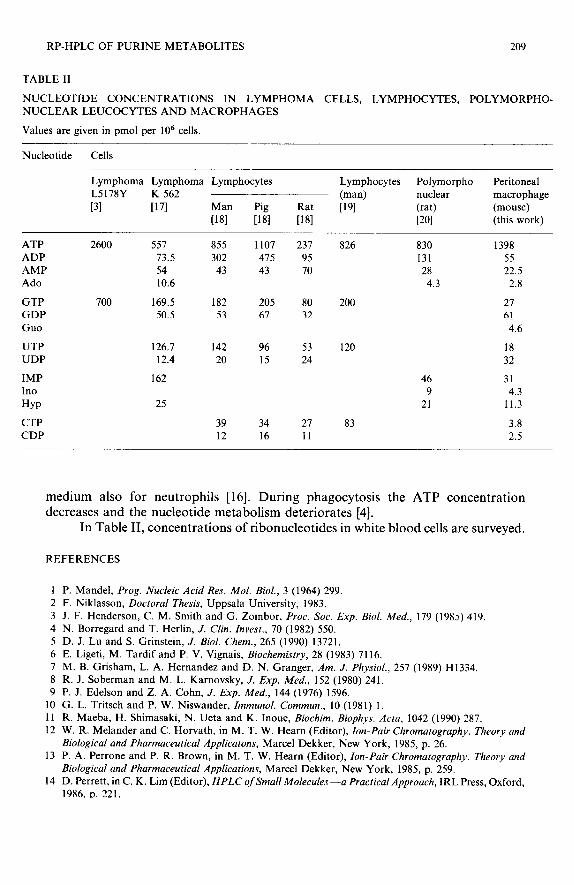
TABLE II
NUCLEOTIDE CONCENTRATIONS IN LYMPHOMA CELLS, LYMPHOCYTES, POLYMORPHO- NUCLEAR LEUCOCYTES AND MACROPHAGES
Values are given in pm01 per lo6 cells.
Nucleotide Cells
Lymphoma Lymphoma Lymphocytes L5178Y K 562
131 P71 Man Pig
1181 1181 Rat
[I81
Lymphocytes Polymorpho Peritoneal
(man) nuclear macrophage
1191 (rat) (mouse)
WI (this work)
ATP 2600 557 ADP 73.5 AMP 54 Ado 10.6
GTP GDP
Guo
700 169.5 182 205 80 200 50.5 53 67 32
UTP UDP
IMP In0
HYP
CTP CDP
126.7 12.4
162
25
855 1107 231 826 302 475 95
43 43 70
142 96 53 120 18 20 15 24 32
39 34 27 83 12 16 11
830 1398 131 55 28 22.5
4.3 2.8
27
h! 4.6
46 31 9 4.3
21 11.3
3.8 2.5
medium also for neutrophils [16]. During phagocytosis the ATP concentration decreases and the nucleotide metabolism deteriorates [4].
In Table II, concentrations of ribonucleotides in white blood cells are surveyed.
REFERENCES
1 P. Mandel, Prog. Nucleic Acid Res. Mol. Biol., 3 (1964) 299. 2 F. Niklasson, Doctoral Thesis, Uppsala University, 1983.
3 J. F. Henderson, C. M. Smith and G. Zombor, Proc. Sot. Exp. Biol. Med., 179 (198~) 419. 4 N. Borregard and T. Herlin, J. Clin. Invest., 70 (1982) 550. 5 D. J. Lu and S. Grinstein, J. Biol. Chem., 265 (1990) 13721. 6 E. Ligeti, M. Tardif and P. V. Vignais, Biochemistry, 28 (1983) 7116. 7 M. B. Grisham, L. A. Hernandez and D. N. Granger, Am. J. Physiol., 257 (1989) H1334. 8 R. J. Soberman and M. L. Karnovsky, J. Exp. Med., 152 (1980) 241. 9 P. J. Edelson and Z. A. Cohn, J. Exp. Med., 144 (1976) 1596.
10 G. L. Tritsch and P. W. Niswander, Immunol. Commun., 10 (1981) 1. 11 R. Maeba, H. Shimasaki, N. Ueta and K. Inoue, Biochim. Biophys. Acta, 1042 (1990) 287. 12 W. R. Melander and C. Horvath, in M. T. W. Hearn (Editor), Ion-Pair Chromatography. Theory and
Biological and Pharmaceutical Applicatons, Marcel Dekker, New York, 1985, p. 26. 13 P. A. Perrone and P. R. Brown, in M. T. W. Hearn (Editor), Ion-Pair Chromatography. Theory and
Biological and Pharmaceutical Applications, Marcel Dekker, New York, 1985, p. 259. 14 D. Perrett, in C. K. Lim (Editor), HPLC of Small Molecules -a Practical Approach, IRL Press, Oxford,
1986. D. 221.

210 A. WERNER et al.
15 T. A. Lane and G. E. Lamkin, Transjirsion, 22 (1982) 368.
16 L. Glasser, R. L. Fiederlein and D. W. Huestis, Blood, 66 (1985) 267. 17 A. Werner, W. Siems, G. Gerber, H. Schmidt, S. Gruner and H. Becker, Chromatographia, 25 (1988)
237.
18 G. J. Peters, R. A. De Abreu, A. Oosterhof and J. H. Veerkamp, Biochim. Riophys. Acta, 759 (1983) 7. 19 A. Cohen, J. Barankiewicz, H. M. Lederman, and E. W. Gelfand, Can. J. Biochem. Cell. Biol., 62 (1983)
577. 20 A. C. Newby and C. A. Holmquist, Biochem. J., 200 (1981) 399.