PROTEIN PHOSPOHATASE 95 Regulates Phosphate Homeostasis … · 1 RESEARCH ARTICLE PROTEIN...
37
1 RESEARCH ARTICLE PROTEIN PHOSPOHATASE 95 Regulates Phosphate Homeostasis by Affecting Phosphate Transporter Trafficking in Rice Zhili Yang a, # , Jian Yang a,b,#, *, Yan Wang a , Fei Wang a , Wenxuan Mao a , Qiuju He a , Jiming Xu a , Zhongchang Wu a , Chuanzao Mao a, * a State Key Laboratory of Plant Physiology and Biochemistry, College of Life Sciences, Zhejiang University, Hangzhou 310058, China b Key Laboratory of Bio-Resource and Eco-Environment of Ministry of Education, College of Life Sciences, Sichuan University, Chengdu 610065, China # These authors contributed equally to this work. *Corresponding authors: Chuanzao Mao Tel: +86-571-88206108 Email: [email protected] Jian Yang Tel: +86-18111249086 E-mail: [email protected] Short title: OsPP95 Regulates Phosphate Transporter Trafficking One-sentence summary: A protein phosphatase positively regulates the trafficking of phosphate transporters in response to phosphate levels. The author(s) responsible for distribution of materials integral to the findings presented in this article in accordance with the policy described in the Instructions for Authors (www.plantcell.org) is: Chuanzao Mao ([email protected]) Abstract Phosphate (Pi) uptake in plants depends on plasma membrane (PM)-localized Pi transporters (PTs). OsCK2 phosphorylates PTs and inhibits their trafficking from the endoplasmic reticulum (ER) to the PM in rice (Oryza sativa), but how PTs are dephosphorylated is unknown. We demonstrate that the protein phosphatases type-2C (PP2C) protein phosphatase OsPP95 interacts with OsPT2 and OsPT8 and dephosphorylates OsPT8 at Ser-517. Rice plants overexpressing OsPP95 reduced OsPT8 phosphorylation and promoted OsPT2 and OsPT8 trafficking from the ER to the PM, resulting in Pi accumulation. Under Pi-sufficient conditions, Pi levels were lower in young leaves and higher in old leaves in ospp95 mutants than in those of the wild type, even though the overall shoot Pi levels were the same in the mutant and wild type. In the wild type, OsPP95 accumulated under Pi starvation but was rapidly degraded under Pi- sufficient conditions. We show that OsPHO2 interacts with and induces the degradation of OsPP95. We conclude that OsPP95, a protein phosphatase negatively regulated by OsPHO2, positively regulates Pi homeostasis and remobilization by dephosphorylating PTs and affecting their trafficking to the PM, a reversible process required for adaptation to variable Pi conditions. Plant Cell Advance Publication. Published on January 9, 2020, doi:10.1105/tpc.19.00685 ©2020 American Society of Plant Biologists. All Rights Reserved
Transcript of PROTEIN PHOSPOHATASE 95 Regulates Phosphate Homeostasis … · 1 RESEARCH ARTICLE PROTEIN...
1
RESEARCH ARTICLE
PROTEIN PHOSPOHATASE 95 Regulates Phosphate Homeostasis by Affecting
Phosphate Transporter Trafficking in Rice
Zhili Yang a, #, Jian Yanga,b,#,*, Yan Wanga, Fei Wanga, Wenxuan Maoa, Qiuju Hea, Jiming Xua, Zhongchang Wua, Chuanzao Maoa,*
a State Key Laboratory of Plant Physiology and Biochemistry, College of Life Sciences, Zhejiang University, Hangzhou 310058, China b Key Laboratory of Bio-Resource and Eco-Environment of Ministry of Education, College of Life Sciences, Sichuan University, Chengdu 610065, China #These authors contributed equally to this work.
*Corresponding authors:Chuanzao MaoTel: +86-571-88206108Email: [email protected]
Jian Yang Tel: +86-18111249086 E-mail: [email protected]
Short title: OsPP95 Regulates Phosphate Transporter Trafficking
One-sentence summary: A protein phosphatase positively regulates the trafficking of phosphate transporters in response to phosphate levels.
The author(s) responsible for distribution of materials integral to the findings presented in this article in accordance with the policy described in the Instructions for Authors (www.plantcell.org) is: Chuanzao Mao ([email protected])
Abstract Phosphate (Pi) uptake in plants depends on plasma membrane (PM)-localized Pi transporters (PTs). OsCK2 phosphorylates PTs and inhibits their trafficking from the endoplasmic reticulum (ER) to the PM in rice (Oryza sativa), but how PTs are dephosphorylated is unknown. We demonstrate that the protein phosphatases type-2C (PP2C) protein phosphatase OsPP95 interacts with OsPT2 and OsPT8 and dephosphorylates OsPT8 at Ser-517. Rice plants overexpressing OsPP95 reduced OsPT8 phosphorylation and promoted OsPT2 and OsPT8 trafficking from the ER to the PM, resulting in Pi accumulation. Under Pi-sufficient conditions, Pi levels were lower in young leaves and higher in old leaves in ospp95 mutants than in those of the wild type, even though the overall shoot Pi levels were the same in the mutant and wild type. In the wild type, OsPP95 accumulated under Pi starvation but was rapidly degraded under Pi-sufficient conditions. We show that OsPHO2 interacts with and induces the degradation of OsPP95. We conclude that OsPP95, a protein phosphatase negatively regulated by OsPHO2, positively regulates Pi homeostasis and remobilization by dephosphorylating PTs and affecting their trafficking to the PM, a reversible process required for adaptation to variable Pi conditions.
Plant Cell Advance Publication. Published on January 9, 2020, doi:10.1105/tpc.19.00685
©2020 American Society of Plant Biologists. All Rights Reserved

2
Introduction
Phosphorus (P) is an essential mineral nutrient for plant development and reproduction and an integral
component of biomacromolecules such as phospholipids and nucleic acids. The levels of phosphate (Pi), the
only form of P that can be absorbed by plants, are commonly limited in soil due to chemical fixation and
microbial activity (Raghothama, 1999). Plants have developed a series of adaptive responses that allow them
to withstand suboptimal Pi conditions, such as enhancing Pi scavenging capacity from the external
environment by modifying root system architecture, secreting acid phosphatases, inducing Pi transport and
symbiosis with mycorrhizal fungi, and recycling and remobilizing internal Pi via RNase activity, and lipid
remodeling (Raghothama, 1999; Lin et al., 2009).
Pi uptake in plants is largely mediated by plasma membrane (PM)-localized Pi transporters (PTs) that
belong to the PHOSPHATE TRANSPORTER1 (PHT1) symporter family. A sequence similarity comparison
with the high-affinity yeast (Saccharomyces cerevisiae) phosphate transporter PHO84 revealed 13 and 9 PHT1
genes have been identified in rice (Oryza sativa) and Arabidopsis thaliana, respectively (Goff et al., 2002;
Karthikeyan et al., 2002). Rice PHT1 genes show different tissue-specific expression patterns and responses
to Pi starvation, and encode enzymes with different affinities for Pi. For instance, the high-affinity Pi
transporter OsPT8 (PHOSPHATE TRANSPORTER 8) is not responsive to Pi starvation at the transcriptional
level but is universally expressed in rice tissues and plays an important role in Pi uptake (Chen et al., 2011;
Jia et al., 2011). Knockdown of OsPT8 in shoots inhibits the redistribution of Pi from source to sink organs
(Li et al., 2015). OsPT1 and OsPT4 are also constitutively expressed in rice, and function in Pi uptake and
redistribution (Sun et al., 2012; Zhang et al., 2015). The low-Pi-induced transporter OsPT2, which is localized
in the stele of roots, plays important roles in Pi uptake and root-to-shoot translocation under Pi-deficient
conditions (Ai et al., 2009). Other functionally characterized PHT1 genes including OsPT3/6/9/10 are also
induced by low Pi and play diverse roles in Pi uptake and translocation (Ai et al., 2009; Sun et al., 2012; Wang
et al., 2014; Chang et al., 2019).
Although most PHT1 genes in rice are induced at the transcript level by Pi starvation or mycorrhizal
symbiosis (Yang et al., 2012; Secco et al., 2013), posttranslational regulation of PHT1 family proteins is also
important for their activities (Wang et al., 2018). In Arabidopsis, several PHT1 members are degraded by the
ubiquitin E2 conjugase PHOSPHATE 2 (AtPHO2) and the ubiquitin E3 ligase NITROGEN LIMITATION
ADAPTATION (AtNLA) (Huang et al., 2013; Lin et al., 2013; Park et al., 2014). Although rice OsPHO2 does
not interact with PHT1 family members (at least not OsPT2/6/8), it does interact with PHOSPHATE
3
TRANSPORTER TRAFFIC FACILITATOR 1 (OsPHF1) (Ying et al., 2016). PHF1 is localized to the ER and
plays an important role in regulating the exit of PTs from the ER and their trafficking to the PM (Gonzalez et
al., 2005; Bayle et al., 2011; Chen et al., 2011). Notably, the phosphorylation of PHT1 family transporters
affects their ER exit (Bayle et al., 2011; Chen et al., 2015). We previously revealed that OsPT2 and OsPT8
can be phosphorylated by casein kinase 2 (CK2) under Pi-sufficient conditions and that phosphorylated PTs
cannot interact with OsPHF1, resulting in the ER retention of PTs, allowing fewer PTs to target the PM to
absorb Pi from the rhizosphere (Chen et al., 2015).
Protein phosphorylation is a reversible reaction catalyzed by two types of antagonistic enzymes: protein
kinases and protein phosphatases (Uhrig et al., 2013). Although PTs are phosphorylated by CK2 in response
to Pi levels in rice, how PTs are dephosphorylated in plants is currently unknown. Here, using yeast-two-
hybrid (Y2H) screening, we identified a PP2C protein phosphatase, OsPP95, that interacts with OsPT2 and
OsPT8. OsPP95 dephosphorylates OsPT8, promoting its ER exit and trafficking to the PM. OsPP95 plays an
important role in Pi uptake and redistribution. In addition, OsPP95 is targeted by OsPHO2 under Pi-sufficient
conditions, resulting in its more rapid degradation under Pi-sufficient vs. Pi-starvation conditions. These
results provide a mechanistic understanding of a pathway in which OsPP95 acts antagonistically with CK2 to
regulate the reversible phosphorylation of PTs, thereby modulating their ER exit and trafficking to the PM,
ultimately regulating plant Pi homeostasis and distribution.
RESULTS OsPP95 Physically Interacts with PTs
To investigate whether protein phosphatase is responsible for the dephosphorylation of PTs and affects their
ER exit and trafficking to the PM, we investigated the subcellular localizations of GFP (green fluorescent
protein)-tagged OsPT2 and OsPT8 (driven by the 35S promoter) treated with or without a general protein
phosphatase inhibitor (cocktail II; Sigma-Aldrich) (Supplemental Figure 1). When transiently expressed in
rice protoplasts, both OsPT2-GFP and OsPT8-GFP were mainly detected in the PM (Supplemental Figure 1A),
as previously reported (Chen et al., 2011). However, both OsPT2-GFP and OsPT8-GFP were retained in the
ER when treated with protein phosphatase inhibitor cocktail II (Supplemental Figure 1A). To further test the
effects of cocktail II on PT localization, we examined the ProPT8:PT8-GFP line (Supplemental Figure 1B).
In contrast to untreated plants, in which most OsPT8-GFP was localized to the PM, OsPT8-GFP was also
detected in the ER after treatment with protein phosphatase inhibitor, resembling the localization of OsPT8-
GFP in the OsCK2α3 overexpression line. These results suggest that protein phosphatase is required for the

4
dephosphorylation of PTs for their trafficking from the ER to the PM. OsPT8-GFP was mainly localized to
the PM of root epidermal cells in ProPT8:PT8-GFP treated with or without okadaic acid (Supplemental Figure
1B), an inhibitor of PPP but not PP2C protein phosphatases, suggesting that the protein phosphatase(s) that
specifically dephosphorylate PTs belong to the PP2C family.
To narrow down the potential candidate PP2Cs, we examined the transcript abundance of PP2C family
genes (90 members) in rice root according to previous data reported by Secco et al. (2013). Fifty-nine out of
the 90 PP2C family genes were filtered out due to low transcript abundances [fragment per kilobase of exon
per million fragments mapped (FPKM) < 10]. The remaining 31 candidates were tested for interaction with
OsPT8 using a split-ubiquitin membrane-based yeast two-hybrid (Y2H) assay. OsPP95 was identified as the
candidate protein phosphatase interacting with OsPT8, using the experimental workflow presented in
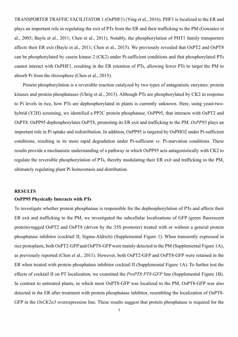
Supplemental Figure 2. OsPP95 interacted with both OsPT8 and OsPT2 in a Y2H assay (Figure 1A and 1B).
To identify the PT-interacting domain of OsPP95, the OsPP95 protein was divided into OsPP95NT (amino
acids 1 to 36, containing a unique OsPP95 sequence) and OsPP95CT (amino acids 37 to 290, including the
conserved PP2C catalytic domain), as shown in Figure 1A and Supplemental Figure 3. Both OsPT2 and OsPT8
interacted with the N-terminus but not the C-terminus of OsPP95 (Figure 1B). Further co-IP assays using rice
protoplasts transiently expressing OsPP95-NT-FLAG together with GFP or OsPT8-GFP confirmed that
OsPP95NT-FLAG could be co-immunoprecipitated by OsPT8-GFP fusion protein but not the GFP control
(Figure 1C). Because the C-terminus of OsPT8 can be phosphorylated by CK2, which impairs its interaction
with OsPHF1, resulting in the retention of OsPT8 in the ER (Chen et al., 2015), we speculated that OsPP95
also interacts with the C-terminus of OsPT8 (OsPT8-CT) (Figure 1D). To test this interaction, we performed
co-IP assays using tobacco leaves transiently expressed OsPP95-FLAG together with GFP or OsPT8-CT-GFP.
OsPP95-FLAG was detected in OsPT8CT-GFP immunoprecipitated proteins but not in the GFP control
(Figure 1E). Further GST pulldown assay confirmed that OsPT8CT interacts with OsPP95 in vitro (Figure
1F), suggesting that OsPP95 interacts with the C-terminus of OsPT8. All these results suggest that OsPP95
plays a specific role in regulating PTs among PP2C proteins.
To validate the interaction between OsPP95 and PTs and to investigate the subcellular location of their
interaction in planta, we conducted bimolecular fluorescence complementation (BiFC) assays with OsPT8 or
OsPT2 fused to the N-terminus of YFP (yellow fluorescent protein; OsPT8-YFPn or OsPT2-YFPn) and
OsPP95 fused to the C-terminus of YFP (OsPP95-YFPc). When OsPT8-YFPn or OsPT2-YFPn were co-
expressed with OsPP95-YFPc in N. benthamiana leaves, YFP fluorescence was well aligned with the red
fluorescence from the ER marker (Figure 1G), suggesting that OsPP95 interacts with PTs in the ER. Together,

5
these results demonstrate that OsPP95 interacts with PTs in vivo and in vitro.
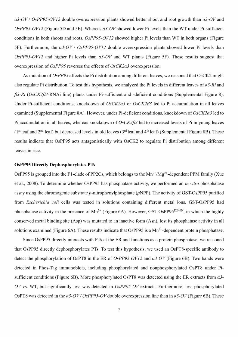
OsPP95 is Expressed in Roots and Shoots, and Its Protein is Localized in All Subcellular Compartments
We examined the subcellular localization of OsPP95 by transiently expressing OsPP95 fused with GFP (green
fluorescent protein) or CFP (cyan fluorescent protein) in rice protoplasts (Figure 2A and 2B; Supplemental
Figure 4). Unlike the ER localization of the interaction between OsPP95 and PTs, OsPP95-GFP and CFP-
OsPP95 fluorescent signals were ubiquitously expressed in all subcellular components, including the nucleus,
cytoplasm, and ER, as the signals aligned with Pro35S:mCherry fluorescence and partially also with that of
an ER marker (ER-rk) (Figure 2A and 2B; Supplemental Figure 4).
To determine the tissue-specific expression patterns of OsPP95 in rice, we generated transgenic plants
harboring the GUS (β-glucuronidase) reporter gene driven by a putative OsPP95 promoter (2334-bp region
immediately upstream of the ATG translation initiation codon) and performed histochemical staining. OsPP95
was expressed in roots, nodes, and leaves (Figure 2C–2I). In roots, GUS activity was detected in the primary
root epidermis, exodermis, xylem, and both the cortex and stele of the lateral root (Figure 2C–2F). In leaf
blades, GUS staining was observed in all cell types except bulliform cells (Figure 2G). In the stem, GUS
staining was detected in vascular tissue (Figure 2H). OsPP95 was expressed in transit vascular bundles and
diffuse vascular bundles, but not in enlarged vascular bundles (Figure 2I).
OsPP95 regulates Pi homeostasis
To examine the role of OsPP95 in regulating Pi homeostasis in rice, we generated OsPP95 overexpression
transgenic lines in the wild-type background (WT; cv. Nipponbare). Three independent representative
overexpression lines (OsPP95-OV3, OsPP95-OV7, and OsPP95-OV12) were chosen for further analysis
(Figure 3A). For phenotyping, 10-d-old seedlings were transferred into Pi-sufficient (200 μM Pi) and Pi-
deficient (10 μM Pi) solutions and grown for 20 more days. Under Pi-sufficient conditions, both the shoots
and roots were smaller in OsPP95 overexpression plants than in wild-type plants (Figure 3B and 3D). The
OsPP95-OV plants displayed leaf tip necrosis in old leaves, a typical Pi toxicity symptom in rice (Figure 3C).
In addition, the Pi concentrations in OsPP95-OV plants were ~1.6-fold that of the WT in roots and 1.8-fold
that in shoots under Pi-sufficient conditions (Figure 3E). However, under Pi-deficient conditions, both shoot
and root growth in OsPP95-OV plants were comparable to that of WT, although two of the overexpression
lines showed lower root fresh weight than the WT (Figure 3B and 3D). Under Pi-deficient conditions, leaf tip
necrosis was not observed in old leaves of OsPP95-OV (Figure 3C), although Pi levels were 0.5-fold greater
in OsPP95-OV shoots than in WT shoots (Figure 3E). In addition, Pi levels in various leaves were higher in

6
OsPP95-OV than those in the WT under both Pi-sufficient and -deficient conditions (Supplemental Figure 5).
To further investigate the physiological function of OsPP95 in rice, we created ospp95 knockout mutants
in the Nipponbare background using the CRISPR-Cas9 system. Two independent mutants, pp95-10 and pp95-
25, were identified by sequencing analysis, which contained a 1-bp insertion and a 10-bp deletion in the second
exon of OsPP95, respectively (Supplemental Figure 6). To rule out the possibility of off-target effects of
CRISPR/cas9, we backcrossed the pp95 mutants to wild-type plants and used homozygous pp95 plants from
the F3 populations for further analysis. The growth of the pp95 mutants was similar to that of WT plants under
both Pi-sufficient and -deficient conditions (Figure 4A and 4B). Under Pi-sufficient conditions, the Pi
concentrations in pp95 shoot and roots were not significantly different from WT (Figure 4C). However, under
Pi-deficient conditions, Pi levels in pp95 roots decreased by 30% compared to the WT, whereas Pi levels in
shoots did not significantly differ among lines (Figure 4C).
To examine whether the mutation of OsPP95 affects the distribution of Pi among leaves in pp95, we
measured Pi levels in different leaves. Under Pi-sufficient conditions, Pi levels were decreased in the youngest
leaf of pp95 compared to WT, while Pi levels were increased in older leaves (leaf 3 and leaf 4). However,
under Pi-deficient conditions, Pi levels were decreased in the youngest leaf but not in old leaves (leaf 2 to leaf
4) of pp95 compared to WT (Figure 4D). No significant differences in the dry weights of different tissues were
detected between the WT and pp95 under Pi-sufficient conditions. However, the dry weight of the youngest
leaf was lower in pp95 than that in WT under Pi-deficient conditions (Supplemental Figure 7). These results
are consistent with those of a previous report showing that the spdt mutant has a reduced Pi concentration and
inhibited growth of the youngest leaf, but not of the whole rice plant, under Pi-deficient conditions (Yamaji et
al., 2017). These results indicate that OsPP95 is involved in regulating Pi homeostasis and distribution under
different Pi conditions in rice.
OsPP95 Acts Antagonistically with CK2 to Regulate Pi Homeostasis
As both OsCK2 and OsPP95 interacted with the C-termini of PTs, we reasoned that OsPP95 might act
antagonistically with OsCK2. To test the genetic interaction between OsPP95 and OsCK2, we crossed
OsCK2α3-RNAi (α3-Ri) and OsCK2α3 overexpression (α3-OV) plants with OsPP95 overexpression (PP95-
OV12) plants, generating α3-Ri / OsPP95-OV and α3-OV / OsPP95-OV plants, respectively. Pi levels were
higher in roots and shoots of a3-Ri than in the WT under Pi-sufficient conditions. However, the α3-Ri /
OsPP95-OV line did not exhibit an additive effect on Pi levels compared to a3-Ri (Figure 5A–C), suggesting
that OsPP95 and OsCK2α3 function in the same genetic pathway to regulate Pi homeostasis. Furthermore, the
7
α3-OV / OsPP95-OV12 double overexpression plants showed better shoot and root growth than α3-OV and
OsPP95-OV12 (Figure 5D and 5E). Whereas α3-OV showed lower Pi levels than the WT under Pi-sufficient
conditions in both shoots and roots, OsPP95-OV12 showed higher Pi levels than WT in both organs (Figure
5F). Furthermore, the α3-OV / OsPP95-OV12 double overexpression plants showed lower Pi levels than
OsPP95-OV12 and higher Pi levels than α3-OV and WT plants (Figure 5F). These results suggest that
overexpression of OsPP95 reverses the effects of OsCK2α3 overexpression.
As mutation of OsPP95 affects the Pi distribution among different leaves, we reasoned that OsCK2 might
also regulate Pi distribution. To test this hypothesis, we analyzed the Pi levels in different leaves of α3-Ri and
b3-Ri (OsCK2b3-RNAi line) plants under Pi-sufficient and -deficient conditions (Supplemental Figure 8).
Under Pi-sufficient conditions, knockdown of OsCK2α3 or OsCK2b3 led to Pi accumulation in all leaves
examined (Supplemental Figure 8A). However, under Pi-deficient conditions, knockdown of OsCK2α3 led to
Pi accumulation in all leaves, whereas knockdown of OsCK2b3 led to increased levels of Pi in young leaves
(1st leaf and 2nd leaf) but decreased levels in old leaves (3rd leaf and 4th leaf) (Supplemental Figure 8B). These
results indicate that OsPP95 acts antagonistically with OsCK2 to regulate Pi distribution among different
leaves in rice.
OsPP95 Directly Dephosphorylates PTs
OsPP95 is grouped into the F1-clade of PP2Cs, which belongs to the Mn2+/Mg2+-dependent PPM family (Xue
et al., 2008). To determine whether OsPP95 has phosphatase activity, we performed an in vitro phosphatase
assay using the chromogenic substrate p-nitrophenylphosphate (pNPP). The activity of GST-OsPP95 purified
from Escherichia coli cells was tested in solutions containing different metal ions. GST-OsPP95 had
phosphatase activity in the presence of Mn2+ (Figure 6A). However, GST-OsPP95D240N, in which the highly
conserved metal binding site (Asp) was mutated to an inactive form (Asn), lost its phosphatase activity in all
solutions examined (Figure 6A). These results indicate that OsPP95 is a Mn2+-dependent protein phosphatase.
Since OsPP95 directly interacts with PTs at the ER and functions as a protein phosphatase, we reasoned
that OsPP95 directly dephosphorylates PTs. To test this hypothesis, we used an OsPT8-specific antibody to
detect the phosphorylation of OsPT8 in the ER of OsPP95-OV12 and α3-OV (Figure 6B). Two bands were
detected in Phos-Tag immunoblots, including phosphorylated and nonphosphorylated OsPT8 under Pi-
sufficient conditions (Figure 6B). More phosphorylated OsPT8 was detected using the ER extracts from α3-
OV vs. WT, but significantly less was detected in OsPP95-OV extracts. Furthermore, less phosphorylated
OsPT8 was detected in the α3-OV / OsPP95-OV double overexpression line than in α3-OV (Figure 6B). These

8
findings support the notion that OsPP95 plays a critical role in dephosphorylating OsPT8.
We previously reported that Ser-517 at the C-terminus of OsPT8 is phosphorylated by OsCK2α3 (Chen
et al., 2015). We therefore examined whether OsPP95 is involved in dephosphorylating OsPT8 at Ser-517
using the C-terminus of OsPT8 (OsPT8CT) as the substrate in an in vitro phosphorylation assay. OsPT8CT,
but not the nonphosphorylatable form (PT8CTS517A), was phosphorylated by OsCK2α3 (Figure 6C), which is
consistent with previous results (Chen et al., 2015). However, a striking reduction in PT8CT phosphorylation
was detected in samples treated with OsPP95 but not the mutated version, OsPP95D240N (Figure 6C),
suggesting that OsPP95 dephosphorylates OsPT8 at Ser-517.
OsPP95 Acts Antagonistically with CK2 to Regulate the ER Exit of PTs
Since OsPP95 acts antagonistically with CK2 to regulate the phosphorylation status of OsPT8 at Ser-517, we
reasoned that OsPP95 might regulate the ER exit of PTs. To test this possibility, we measured PT8 protein
levels in the ER and PM of the OsPP95-OV lines and pp95 mutants. Consistent with the Pi accumulation
patterns in the OsPP95-OV lines (Figure 3E), immunoblotting revealed that more OsPT8 accumulated at the
PM, but less accumulated in the ER, in OsPP95-OV vs. the WT under Pi-sufficient conditions (Supplemental
Figure 9A and 9B). Immunoblot analysis further indicated that less OsPT8 accumulated in the PM in the shoot
base of pp95 compared to WT under both Pi-sufficient and -deficient conditions (Supplemental Figure 9C);
however, the OsPT8 levels in the ER of the pp95 mutants were increased in Pi-sufficient conditions, but not
in Pi-deficient conditions, compared to the WT (Supplemental Figure 9D). Consistently, less OsPT8
accumulated in the PM but more accumulated in the ER in α3-OV vs. WT; more OsPT8 accumulated at the
PM but less accumulated in the ER in OsPP95-OV12 vs. WT; and the levels of both PM- and ER-localized
OsPT8 in α3-OV / OsPP95-OV12 were between those of α3-OV and PP95-OV12 (Figure 7A).
We examined the subcellular localization of GFP-tagged OsPT2/OsPT8 in rice protoplasts co-transformed
with or without OsPP95 and OsCK2α3 overexpression constructs. OsPT2-GFP and OsPT8-GFP fusion
proteins were mainly observed on the PM in control and OsPP95-overexpressing protoplasts; overexpressing
OsCK2α3 resulted in the ER retention of OsPT2-GFP and OsPT8-GFP (Figure 7B; Supplemental Figure 10A–
10D), which is consistent with a previous report (Chen et al., 2015). When OsCK2α3 and OsPP95 were co-
expressed in protoplasts, OsPT2-GFP and OsPT8-GFP were mainly detected in the PM (Figure 7B). By
contrast, PT8S517D-GFP (phosphorylation-mimic version of OsPT8) and PT8S517A-GFP (mimicking the
nonphosphorylatable form of OsPT8) were mainly detected in the ER and PM, respectively, regardless of co-
expression with OsPP95 (Supplemental Figure 10E–L).
9
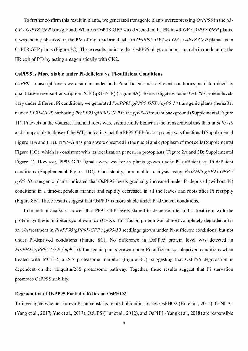
To further confirm this result in planta, we generated transgenic plants overexpressing OsPP95 in the α3-
OV / OsPT8-GFP background. Whereas OsPT8-GFP was detected in the ER in α3-OV / OsPT8-GFP plants,
it was mainly observed in the PM of root epidermal cells in OsPP95-OV / α3-OV / OsPT8-GFP plants, as in
OsPT8-GFP plants (Figure 7C). These results indicate that OsPP95 plays an important role in modulating the
ER exit of PTs by acting antagonistically with CK2.
OsPP95 is More Stable under Pi-deficient vs. Pi-sufficient Conditions
OsPP95 transcript levels were similar under both Pi-sufficient and -deficient conditions, as determined by
quantitative reverse-transcription PCR (qRT-PCR) (Figure 8A). To investigate whether OsPP95 protein levels
vary under different Pi conditions, we generated ProPP95:gPP95-GFP / pp95-10 transgenic plants (hereafter
named PP95-GFP) harboring ProPP95:gPP95-GFP in the pp95-10 mutant background (Supplemental Figure
11). Pi levels in the youngest leaf and roots were significantly higher in the transgenic plants than in pp95-10
and comparable to those of the WT, indicating that the PP95-GFP fusion protein was functional (Supplemental
Figure 11A and 11B). PP95-GFP signals were observed in the nuclei and cytoplasm of root cells (Supplemental
Figure 11C), which is consistent with its localization pattern in protoplasts (Figure 2A and 2B; Supplemental
Figure 4). However, PP95-GFP signals were weaker in plants grown under Pi-sufficient vs. Pi-deficient
conditions (Supplemental Figure 11C). Consistently, immunoblot analysis using ProPP95:gPP95-GFP /
pp95-10 transgenic plants indicated that OsPP95 levels gradually increased under Pi-deprived (without Pi)
conditions in a time-dependent manner and rapidly decreased in all the leaves and roots after Pi resupply
(Figure 8B). These results suggest that OsPP95 is more stable under Pi-deficient conditions.
Immunoblot analysis showed that PP95-GFP levels started to decrease after a 4-h treatment with the
protein synthesis inhibitor cycloheximide (CHX). This fusion protein was almost completely degraded after
an 8-h treatment in ProPP95:gPP95-GFP / pp95-10 seedlings grown under Pi-sufficient conditions, but not
under Pi-deprived conditions (Figure 8C). No difference in OsPP95 protein level was detected in
ProPP95:gPP95-GFP / pp95-10 transgenic plants grown under Pi-sufficient vs. -deprived conditions when
treated with MG132, a 26S proteasome inhibitor (Figure 8D), suggesting that OsPP95 degradation is
dependent on the ubiquitin/26S proteasome pathway. Together, these results suggest that Pi starvation
promotes OsPP95 stability.
Degradation of OsPP95 Partially Relies on OsPHO2
To investigate whether known Pi-homeostasis-related ubiquitin ligases OsPHO2 (Hu et al., 2011), OsNLA1
(Yang et al., 2017; Yue et al., 2017), OsUPS (Hur et al., 2012), and OsPIE1 (Yang et al., 2018) are responsible
10
for the degradation of OsPP95 in rice, we performed Y2H assays using OsPP95 as bait. Only OsPHO2
interacted with OsPP95 in yeast, which was confirmed by BiFC assays (Figure 9A and 9B). No YFP signal
was detected when OsPP95-YFPn and OsPHO2-YFPc were co-expressed in tobacco leaves (Figure 9A).
However, YFP signals were detected when OsPP95-YFPn was co-expressed with OsPHO2C719A-YFPc (the
catalytic residue Cys-719 of OsPHO2 was substituted with Ala) (Figure 9A). These results indicate that
OsPHO2 interacts with OsPP95, and they suggest that OsPHO2 might degrade OsPP95.
To determine whether OsPHO2 degrades OsPP95, we crossed PP95-GFP plants with pho2 to obtain
PP95-GFP / pho2 plants. Immunoblot analysis showed that under Pi-sufficient conditions, OsPP95 protein
levels were higher in PP95-GFP / pho2 than in PP95-GFP. However, Under Pi-deprived conditions, OsPP95
levels were similar in PP95-GFP and PP95-GFP / pho2 plants (Figure 9C). After CHX treatment, PP95-GFP
was degraded more rapidly in PP95-GFP than in PP95-GFP / pho2 under Pi-sufficient conditions (Figure
9D). Finally, we examined the levels of ubiquitin-conjugated OsPP95 in PP95-GFP and PP95-GFP / pho2
plants using anti-ubiquitin antibody and found that the levels of ubiquitinated PP95-GFP were greatly reduced
in PP95-GFP/pho2 compared to PP95-GFP (Figure 9E). These results indicate that OsPHO2 is involved in
degrading OsPP95 through the ubiquitination-mediated degradation pathway in rice.
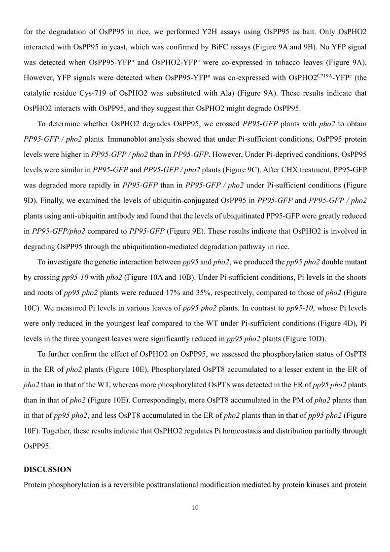
To investigate the genetic interaction between pp95 and pho2, we produced the pp95 pho2 double mutant
by crossing pp95-10 with pho2 (Figure 10A and 10B). Under Pi-sufficient conditions, Pi levels in the shoots
and roots of pp95 pho2 plants were reduced 17% and 35%, respectively, compared to those of pho2 (Figure
10C). We measured Pi levels in various leaves of pp95 pho2 plants. In contrast to pp95-10, whose Pi levels
were only reduced in the youngest leaf compared to the WT under Pi-sufficient conditions (Figure 4D), Pi
levels in the three youngest leaves were significantly reduced in pp95 pho2 plants (Figure 10D).
To further confirm the effect of OsPHO2 on OsPP95, we assessed the phosphorylation status of OsPT8
in the ER of pho2 plants (Figure 10E). Phosphorylated OsPT8 accumulated to a lesser extent in the ER of
pho2 than in that of the WT, whereas more phosphorylated OsPT8 was detected in the ER of pp95 pho2 plants
than in that of pho2 (Figure 10E). Correspondingly, more OsPT8 accumulated in the PM of pho2 plants than
in that of pp95 pho2, and less OsPT8 accumulated in the ER of pho2 plants than in that of pp95 pho2 (Figure
10F). Together, these results indicate that OsPHO2 regulates Pi homeostasis and distribution partially through
OsPP95.
DISCUSSION
Protein phosphorylation is a reversible posttranslational modification mediated by protein kinases and protein

11
phosphatases that plays critical roles in protein localization, stability, and activity (Uhrig et al., 2013). Several
protein kinases are involved in Pi homeostasis (Fragoso et al., 2009; Lei et al., 2014; Chen et al., 2015; Zhang
et al., 2016), but to date, no protein phosphatase was shown to function in Pi acquisition and homeostasis in
plants. Here, we identified the PP2C protein phosphatase OsPP95, which regulates Pi acquisition and
homeostasis via dephosphorylating PTs. The identification of this reversible PT phosphorylation mechanism
for plant adaptation to variable Pi environments should facilitate the genetic improvement of Pi-efficient crops.
OsPP95 is a PP2C that Positively Regulates Pi Homeostasis
The rice and Arabidopsis genomes contain 90 and 80 PP2C genes, respectively, which are divided into 11
clades, with OsPP95 grouped into the F1 clade (Xue et al., 2008; Singh et al., 2010; Fuchs et al., 2013). PP2C
activity relies on the binding of two metal ions, Mg2+ and Mn2+ (Shi, 2009). In the current study, the protein
phosphatase OsPP95 was activated in vitro by Mn2+, but not by Mg2+, suggesting that OsPP95 is slightly
different from other PP2Cs, although it contains a conserved PP2C domain (Supplemental Figure 3). A version
of OsPP95 mutated in the conserved metal binding site (Asp-240 to Asn) lacked phosphatase activity,
indicating that OsPP95 is a typical PP2C protein phosphatase (Figure 6A). In addition to having a conserved
PP2C catalytic domain, many PP2C proteins also contain N- and/or C-terminal domains, which may
contribute to substrate specificity, cellular localization, and/or stability (Shi, 2009). In the current study,
OsPP95 interacted with OsPT8 through its non-conserved N-terminal sequence (Figure 1A–1C). The rice
genome encodes four F1-clade PP2Cs (Singh et al., 2010). Only OsPP95 interacted with OsPT8 in our Y2H
screen, which is consistent with the finding that the N-termini of these PP2Cs are quite different from that of
OsPP95 (Supplemental Figure 3). However, we cannot exclude the possibility that other PP2C proteins
interact with other PTs.
PP2Cs are involved in regulating plant growth and development, plant immunity, biotic and abiotic stress
responses, and potassium nutrient signaling (Schweighofer et al., 2007; Ma et al., 2009; Spartz et al., 2014;
Sun et al., 2016). Despite this, no PP2C protein was previously shown to regulate plant Pi acquisition or Pi
starvation responses. We determined that OsPP95 is degraded more rapidly under Pi-sufficient vs. Pi-deprived
conditions, although OsPP95 transcription is not responsive to Pi starvation, resulting in OsPP95
accumulation under Pi starvation (Figure 8). In addition, our results suggest that OsPP95 is degraded by
OsPHO2 via the 26S proteasome pathway under Pi-sufficient conditions, suggesting that OsPP95 is involved
in the response to Pi starvation in rice.
Pi levels were higher in roots (~1.6-fold) and shoots (1.8-fold) of the OsPP95-OV lines compared to WT
12
under Pi-sufficient conditions, and Pi levels in shoots were also higher (0.5-fold) in these lines than in the WT
under Pi-deficient conditions (Figure 3E). By contrast, the Pi concentration in roots was 30% lower in ospp95
compared to the WT, although there was no significant difference in Pi concentration in the shoots of ospp95
vs. WT under Pi-deficient conditions (Figure 4C). These results suggest that OsPP95 positively regulates Pi
homeostasis.
OsPP95 Acts Antagonistically with CK2 to Regulate PT trafficking
Pi acquisition in plants is mainly mediated by PM-localized PTs. PTs are initially targeted to the ER to ensure
their correct folding and modification, followed by vesicle trafficking to the PM (Gonzalez et al., 2005; Bayle
et al., 2011). Trafficking of PTs from the ER to the PM requires PHF1 (PHOSPHATE TRANSPORTER
TRAFFIC FACILITATOR 1), a Sec12-related protein that facilitates the exit of PTs from the ER (Bayle et al.,
2011; Chen et al., 2011). PTs are subject to phosphorylation, which affects their trafficking from the ER to the
PM in response to Pi status in Arabidopsis and rice (Bayle et al., 2011; Chen et al., 2015). Rice CK2
phosphorylates OsPT2 and OsPT8, impairing their interaction with OsPHF1 and resulting in their retention in
the ER (Chen et al., 2015). In this study, we identified OsPP95, a Mn2+-dependent PP2C phosphatase, which
interacts with OsPT2 and OsPT8 and affects their ER and PM localization (Figure 1 and Figure 7). An in vitro
kinase assay indicated that OsPP95 dephosphorylates OsPT8 at Ser-517 (Figure 6), the same amino acid that
is phosphorylated by OsCK2 (Chen et al., 2015). The phosphorylation-mimic OsPT8 was retained in the ER,
whereas non-phosphorylatable OsPT8 was localized to the PM (Supplemental Figure 10). Furthermore, the
phosphorylation level of OsPT8 was lower in OsPP95-OV plants (Figure 6B), and more OsPT8 in the PM and
less in the ER was detected in these plants than in the WT (Supplemental Figure 9A and 9B). These findings
suggest that OsPP95 dephosphorylates PTs and affects their ER exit and trafficking to the PM. In addition,
overexpressing OsPP95 reversed the ER retention of PTs caused by the overexpression of OsCK2 (Figure 7),
suggesting that OsPP95 functions antagonistically with OsCK2 to regulate PT trafficking and therefore Pi
homeostasis.
Notably, overexpressing OsPP95 led to Pi accumulation in shoots, even under Pi-deficient conditions
(Figure 3), suggesting that PTs might also be phosphorylated under Pi-deficient conditions. The
phosphorylation and protein levels of OsCK2b3 decrease in response to Pi deficiency (Chen et al., 2015).
Although OsCK2b3 protein levels were lower under Pi-deficient vs. Pi-sufficient conditions, OsCK2b3 was
detectable by immunoblotting under Pi-deficient conditions (Supplemental Figure 12), suggesting that CK2
might also function under Pi-deficient conditions. Moreover, RNAi lines of both OsCK2α3 and OsCK2b3

13
accumulated Pi under both Pi-sufficient and -deficient conditions (Supplemental Figure 8), further supporting
the notion that CK2 also functions under Pi-deficient conditions to maintain Pi homeostasis.
OsPP95 Regulates Pi translocation and Distribution
Plants tend to remobilize P from senescing leaves (Aerts, 1996), representing an important P source for plant
growth, especially during the later stages of development and in situations where soil P is limited (Veneklaas
et al., 2012). However, how Pi remobilization is regulated between old leaves and young leaves at the
molecular level is unknown.
Under Pi-sufficient conditions, more Pi accumulated in old vs. young leaves, and Pi was remobilized to
young leaves under Pi-starvation conditions in the WT. However, in the pp95 mutants, Pi levels were lower
in the youngest leaf and higher in older leaves (3rd and 4th) than in WT under Pi-sufficient conditions (Figure
4D). Moreover, Pi levels were lower in pp95 pho2 than in pho2 in young leaves (1st – 3rd leaf) but not in older
leaves (Figure 10D). These findings support the notion that OsPP95 is involved in Pi remobilization from old
to young leaves. By contrast, under Pi-deficient conditions, β3-Ri showed higher Pi concentrations in young
leaves, but lower Pi concentrations in old leaves, compared to the WT (Supplemental Figure 8B). Consistently,
OsCK2β3 levels were higher in young vs. old leaves (Supplemental Figure 12).
OsPT3 and OsPT8 regulate Pi remobilization between old and young leaves (Gu et al., 2015; Wang et al.,
2018). Under Pi-deficient conditions, less Pi accumulated in the youngest leaf in ospt3 and ospt8 compared to
the wild type (Li et al., 2015; Chang et al., 2019). Furthermore, our results indicate that OsPP95 acts
antagonistically with CK2 to regulate OsPT8 trafficking, although whether OsPP95 regulates OsPT3
trafficking requires further investigation. These observations indicate that the regulation of the
phosphorylation levels of PTs by OsCK2 and OsPP95 in leaves is essential for proper Pi distribution and
remobilization. As the transcripts of OsPP95 and its protein levels in different leaves are stable (Supplemental
Figure 13), whereas the protein levels of OsCK2β3 decreases in the old leaves (Supplemental Figure 12), the
balance between OsCK2 and OsPP95 affects Pi distribution and remobilization. Interestingly, the Pi
concentration in α3-Ri was higher in older leaves than in young leaves under both Pi-sufficient and -deficient
conditions, which is not consistent with that of β3-Ri under Pi-deficient conditions (Supplemental Figure 8B),
perhaps because PT phosphorylation requires a CK2 holoenzyme, and OsCK2β3 but not OsCK2α3 is unstable
under Pi-deprived conditions (Chen et al., 2015). The rice genome contains two CK2β subunits, OsCK2β1
and OsCK2β3. OsCK2β3 was found to be necessary for the interaction between OsCK2α3 and PTs (Chen et
al., 2015). However, OsCK2β1 did not interact with PTs (Chen et al., 2015), suggesting only OsCK2β3
14
functioned together with OsCK2α3 to regulate PT trafficking. Recently, results from phosphoproteomic
profiling under Pi-sufficient vs. -deficient conditions revealed that OsCK2 might also phosphorylate other
substrates, such as OsSIZ1 (SAP and Miz 1), to regulate Pi homeostasis in rice (Yang et al., 2019). Although
OsCK2α3 functioned together with OsCK2β3, not OsCK2β1, to regulate PT trafficking, it remains to be
determined whether OsCK2α3 functions with OsCK2β1 to regulate Pi homeostasis through other substrates.
The node is a well-organized vascular system in graminaceous plants that distributes mineral nutrients,
including Pi (Yamaji and Ma, 2014; Clemens and Ma, 2016; Yamaji et al., 2016). OsPP95 was expressed in
the transit vascular bundles and diffuse vascular bundles of nodes (Figure 2I), suggesting that OsPP95 is
involved in regulating Pi distribution in nodes. OsPT3 is expressed in nodes under Pi deficiency, and OsPT8
is expressed throughout the plant (Li et al., 2015; Chang et al., 2019), suggesting that OsPP95 regulates the
phosphorylation status of OsPT3, OsPT8, or other PTs in nodes to affect Pi distribution.
In conclusion, we identified the PT-interacting protein phosphatase OsPP95, which positively regulates
Pi homeostasis and distribution by acting antagonistically with OsCK2 to regulate the phosphorylation status
of PTs and their trafficking to the PM. As summarized in our working model (Figure 11), under Pi-sufficient
conditions, OsPP95 protein is degraded by OsPHO2, and PTs are phosphorylated by the OsCK2 holoenzyme.
These processes weaken the interaction between OsPHF1 and PTs and enhance the ER retention of PTs,
allowing less PT to traffic to the PM to absorb Pi. Under Pi-deficient conditions, OsCK2b3 is degraded, while
OsPP95 and OsPHF1 are more stable, and OsPHO2 is cleaved by OsmiR399. Therefore, more
dephosphorylated PTs exit from the ER with the help of OsPHF1 and traffic to the PM to enhance Pi absorption.
Methods
Plant materials and growth conditions. All rice materials used in the study were generated in the Nipponbare
(japonica rice) background. OsPP95 overexpression lines, pp95 mutants (generated via CRISPR/Cas9),
ProPP95:gPP95-GUS, and ProPP95:gPP95-GFP / pp95-10 were generated by Agrobacterium tumefaciens
(strain EHA105)-mediated transformation by transforming callus induced from mature Nipponbare or pp95-
10 embryos with the corresponding vectors (described below) as described (Zhou et al., 2008). Other plant
materials, including the ProPT8:PT8-GFP line, α3-OV, α3-Ri, β3-Ri, and pho2, were described previously
(Liu et al., 2010; Chen et al., 2015). The α3-OV line was crossed with PP95-OV12 to generate the α3-
OV/PP95-OV double overexpression line. The ospp95-10 line was crossed with pho2 to generate the pp95
pho2 double mutant. The α3-Ri line was crossed with OsPP95-OV12 to generate the α3-Ri / OsPP95-OV line.
Pi-sufficient (HP or +P), Pi-deficient (LP), and Pi-deprived (–P) treatments were performed via
15
hydroponics using rice nutrient solution (Yang et al., 2018) containing 200 μM Pi, 10 μM Pi, and 0 μM Pi,
respectively. The nutrient solution was adjusted to pH 5.5 with 1 M HCl or NaOH. Plants were grown in a
growth chamber at 30°C/22°C (day/night) and 60 % to 70 % humidity, bulb type light with a photon density
of ~300 μmol m–2 s–1, and a photoperiod of 14 h as previously described (Shao et al., 2019).
Construction of vectors to produce transgenic plants. For the OsPP95 overexpression vector construct, the
OsPP95 coding region (870 bp) was amplified by PCR and introduced into the binary expression vector
pF3PZPY122 using BamHI and SmaI enzyme recognition sites downstream of the 35S promoter. To generate
the ProOsPP95:gOsPP95-GUS vector, a 2519-bp fragment, including the promoter and 5' UTR, and the 3889-
bp genomic sequence of OsPP95 without the stop codon were amplified by PCR from Nipponbare DNA and
inserted into the GUS-pBI101.3 vector between the SalI and BamHI sites and fused in-frame to GUS. The
GUS fragment of the ProOsPP95:gPP95-GUS vector was replaced with GFP to generate the
ProOsPP95:gPP95-GFP vector. The CRISPR/Cas9-OsPP95 vector was constructed as previously described
(Ma et al., 2015). All constructs were sequenced to confirm the presence of the correct expression cassette
prior to transformation.
Yeast two-hybrid assays. A DUAL Membrane Pairwise Interaction Kit (Dualsystems Biotech) was used for
the Y2H assays. The full-length (1−873 bp), N-terminal region (1−108 bp), and C-terminal region (109−873
bp) of OsPP95 were cloned into the pDHB1 vectors to generate Cub-OsPP95, Cub-OsPP95NT, and Cub-
OsPP95CT, respectively. The full-length OsPT2 and OsPT8 sequences were cloned into the pPR3-STE
vectors to generate NubG-OsPT2 and NubG-OsPT8, respectively. The full-length OsPHO2 sequence was
cloned into the pPR3-N vector to generate NubG-OsPHO2. Primers are listed in Supplemental Table 1. The
constructs were co-transformed into yeast strain NMY51 and plated onto medium without Trp and Leu to
select positive clones. Protein–protein interactions were indicated by the growth of yeast colonies on medium
without Leu, Trp, His and Ade.
Bimolecular fluorescence complementation (BiFC) assays. The bimolecular fluorescence complementation
assays were performed using a split YFP system as previously described (Lv et al., 2014). The coding sequence
(CDS) of OsPP95 was cloned into the N-terminal and C-terminal fragments of YFP vectors to generate
OsPP95-YFPN and OsPP95-YFPC. The coding sequences of OsPT2 and OsPT8 were cloned into the N-
terminal fragments of YFP vectors to generate OsPT2-YFPN and OsPT8-YFPN. The coding sequences of
OsPHO2 and OsPHO2C719A (Cys-719 changed to Ala) were cloned into the C-terminal fragments of YFP
16
vectors to generate OsPHO2-YFPC and OsPHO2C719A-YFPC, respectively. Primers are listed in Supplemental
Table 1. The constructs and ER marker gene ER-rk were transiently expressed in Nicotiana benthamiana
leaves. YFP fluorescence in N. benthamiana leaves was imaged 3 days after infiltration under a Zeiss LSM710
laser-scanning confocal microscope.
Co-immunoprecipitation assays. The co-immunoprecipitation (co-IP) assays were performed as previously
described (Lv et al., 2014). The full-length and the 1516−1626 bp CDS of OsPT8 were amplified by PCR and
cloned in-frame into the modified binary vector pCAMBIA1300-GFP (Pro35S:GFP) to generate
Pro35S:OsPT8-GFP and Pro35S:OsPT8CT-GFP. The full-length and the 1−108 bp CDS of OsPP95 were
amplified by PCR and introduced into the binary expression vector pF3PZPY122 to generate Pro35S:OsPP95-
FLAG and Pro35S:OsPP95NT-FLAG. The related primers are listed in Supplemental Table 1. To assess the
interaction between PP95NT and OsPT8, the Pro35S:OsPP95NT-FLAG vector was co-transformed with
Pro35S:OsPT8-GFP or Pro35S:GFP into rice protoplasts as previously described (Chen et al., 2011). To
assess the interaction between PP95 and OsPT8CT, the Pro35S:OsPP95-FLAG vector was transiently
expressed in =N. benthamiana leaves together with Pro35S:OsPT8CT-GFP or Pro35S:GFP. GFP-Trap
Magnetic Agarose (Chromotek) was used to immunoprecipitate proteins, which was further analyzed by
immunoblotting with anti-FLAG produced in mouse (Sigma-Aldrich, F1804; 1:5000)and anti-GFP antibodies
produced in rabbit (Sigma-Aldrich, G1544; 1:5000). Proteins were resolved on 12% SDS-PAGE, transferred
to polyvinylidene fluoride membranes, and detected by immunoblot analysis using an ECL reagent (Millipore)
and the ChemDoc XRS system (Bio-Rad).
Pull-Down Assays
Full-length OsPP95 and the 1516−1626 bp CDS of OsPT8 were cloned into the C-terminus of 6xHis and GST
in pET28a (Invitrogen) and pGEX-4T-1 (GE Healthcare), respectively. Primers used are listed in Supplemental
Table 1. The GST-OsPT8CT and His-OsPP95 proteins were expressed in Escherichia coli TransB (DE3)
(Transgen) and purified by Ni-NTA Agarose (Qiagen) and GST-affinity chromatography (GE Healthcare),
respectively. GST pull-down was conducted as previously reported (Lv et al., 2014). Proteins were further
analyzed by immunoblotting with anti-His produced in mouse (Abcam, ab18184; 1:5000) and anti-GST
produced in mouse (TransGen Biotech, HT601-01; 1:5000) antibodies.
Protein expression and phosphatase assays. OsGST-PP95 and OsGST-PP95D240N fusion constructs were
generated by inserting the PCR-amplified CDS (OsPP95 or OsPP95D240N) in-frame into pGEX-4T-1. The
17
coding sequence of OsPP95D240N was generated with specific primers using gene splicing via overlap
extension PCR. Primers are listed in Supplemental Table 1. GST-OsCK2α3, GST-OsPT8CT, and GST-
OsPT8CTS517A construction was previously described (Chen et al., 2015). The GST-tagged proteins were
expressed in Escherichia coli TransB (DE3) (Transgen) and purified via GST-affinity chromatography (GE
Healthcare).
The phosphatase assay was performed as described (Spartz et al., 2014). Briefly, 0.2 μg OsPP95 was
incubated in 100 μL assay buffer containing 75 mM Tris, pH 7.6, 0.5 mM EDTA, 100 mM NaCl, and 5 mM
pNPP supplemented with 10 mM MnCl2, ZnCl2, CaCl2, or MgCl2. Absorbance at 405 nm was recorded after
20 min using a Spectroquant NOVA 60 spectrophotometer (Merck, Darmstadt, Germany). A standard curve
was generated using 4-nitrophenol to calculate phosphatase activity.
For the in vitro dephosphorylation assays, GST-OsPT8CT was labeled with 32P by the catalytic subunit
OsCK2α3 in the presence of [γ-32P]ATP at 30°C for 30 min as previously reported (Chen et al., 2015).
Following the addition of 1 μg GST-OsPP95 or GST-OsPP95D240N and 50 μM 5,6-dichloro-1-b-D-
ribofuranosylbenzimidazole (DRB; a CK2 kinase inhibitor), the reactions were incubated at 30°C for 1 h. The
samples were separated by electrophoresis in a 10% SDS-PAGE gel, followed by staining and visualization
via exposure of x-ray film.
For the in vivo dephosphorylation assays, microsomal fractions were isolated from the shoot bases of 14-
d-old plants grown under Pi-sufficient conditions using a Plant Microsomal Membrane Extraction Kit (Invent
Biotechnologies) according to the manufacturer’s instructions. Protein samples were separated on 12%
acrylamide gels with or without Phos-Tag and analyzed by immunoblotting with anti-OsPT8 antibody (Chen
et al., 2015). The relative intensity value of each band was analyzed using ImageJ software
(https://imagej.en.softonic.com/).
Subcellular localization of OsPT2 and OsPT8 in rice protoplasts. Rice protoplasts were isolated and
transformed as previously described (Chen et al., 2011). For drug treatment, 1x phosphatase inhibitor cocktail
II (Sigma-Aldrich) or 20 μM okadaic acid was added to the samples, followed by incubation for 3 h.
Fluorescence in the protoplasts was imaged under a confocal laser-scanning microscope (Zeiss LSM710).
Excitation/emission wavelengths were 488 nm/495–540 nm for GFP, 458 nm/465−500 nm for CFP, 488
nm/515−545 nm for YFP, and 543 nm/575−630 nm for mCherry.
Isolation of endoplasmic reticulum proteins and plasma membrane proteins. Endoplasmic reticulum
proteins were isolated from the shoot bases of plants grown under Pi-sufficient or -deficient conditions for 14
18
d with an Endoplasmic Reticulum Isolation Kit (Sigma-Aldrich) with some modifications. Briefly, the
materials were ground to a powder and resuspended in extraction buffer (10 mM HEPES, pH 7.8, 1 mM EGTA,
25 mM KCl, and 250 mM sucrose, supplemented with 1 mM PMSF, 20 μM MG132, and 1x protease inhibitor
cocktail [Roche]). The resulting homogenate was centrifuged at 12,000 g for 15 min at 4°C, and 7.5 x volume
of 8 mM CaCl2 was added dropwise to the resulting supernatant. The mixture was stirred for 15 min and then
centrifuged at 8,000 g for 10 min at 4°C. The pellet (containing enriched ER proteins) was resuspended in
extraction buffer. Plasma membrane proteins were isolated from the shoot bases of plants grown under Pi-
sufficient and -deficient conditions as previously described (Chen et al., 2015). The proteins were analyzed
by immunoblotting with anti-OsPHF1 produced in rabbit (Chen et al., 2015), anti-OsPIP1;3 produced in rabbit
(PM marker antibody, Agrisera, AS09504; 1:5000), anti-OsBip produced in rabbit (ER marker antibody,
Agrisera, AS09481; 1:5000), and anti-OsPT8 (Chen et al., 2015) antibodies produced in rabbit.
Histochemical localization of GUS expression. T2 ProOsPP95:gOsPP95-GUS seedlings were grown under
Pi-sufficient conditions. The leaves and roots of 14-d-old seedlings and the stems (internode II/III) and nodes
I of about 60-d-old seedlings were collected for histochemical GUS staining as described (Chen et al., 2011).
Briefly, the leaves were stained with GUS for 8 h and then washed with 80% (v/v) ethanol to remove the
chlorophyll. All materials were embedded in ~3% (w/v) agar and cut into 40−60 μm sections using a vibrating
microtome (Leica). Cross-sections of roots, leaves, stems, and nodes were photographed under a microscope
(Nikon).
In vivo degradation assay. Fourteen-d-old ProOsPP95:gOsPP95-GFP / ospp95-10 and
ProOsPP95:gOsPP95-GFP / ospho2 plants grown under +P and/or –P conditions were treated with 200 µM
CHX (cycloheximide) for 2, 4 and 8 h. Proteins were extracted from roots using extraction buffer containing
25 mM Tris, pH 7.5, 1 mM EDTA, 150 mM NaCl, 1% Triton X-100, and 5% glycerol plus 1 mM PMSF, 20
mM MG132, and 1x protease inhibitor cocktail (Roche). Proteins were separated on 12% SDS-PAGE,
transferred to polyvinylidene fluoride membranes, and detected by immunoblot analysis using an ECL reagent
(Millipore) and the ChemDoc XRS system (Bio-Rad). Anti-GFP antibody was used to detect PP95-GFP fusion
proteins, and anti-Actin antibody (Sigma-Aldrich; A3853; 1:5000) was used as a loading control.
RNA isolation and qRT-PCR analysis. Total RNA was isolated from the samples using an RNA Extraction
Kit (NucleoSpin RNA Plant; Macherey-Nagel, Germany). cDNA was synthesized from 1 μg total RNA using
a SuperScript II Reverse Transcriptase Kit (Invitrogen) with oligo(dT) primer, and qRT-PCR was performed
19
as described (Yang et al., 2017). The primers are listed in Supplemental Table 1.
In vivo ubiquitination assays. Seventeen-d-old ProOsPP95:gOsPP95-GFP/ospp95-10 (OsPP95-GFP) and
ProOsPP95-gOsPP95-GFP / ospp95-10 / ospho2 (OsPP95-GFP / ospho2) plants grown under Pi-sufficient
conditions were treated with 20 µM MG132 for 7 days. Proteins were extracted from roots in extraction buffer
containing 25 mM Tris, pH 7.5, 1 mM EDTA, 150 mM NaCl, 1% Triton X-100, and 5% glycerol plus 1 mM
PMSF, 20 µM MG132, 20 mM DTT, and 1x protease inhibitor cocktail (Roche).
GFP-Trap Magnetic Agarose (Chromotek) was used to immunoprecipitate GFP fusion proteins. The
immunoprecipitation assays were performed as previously described (Zhang et al., 2017). The ubiquitin
modifications of immunoprecipitated proteins were detected using the P4D1 mouse monoclonal anti-ubiquitin
antibody produced in mouse (Santa Cruz Biotechnology, sc-8017; 1:500). Anti-GFP antibody produced in
rabbit (Sigma-Aldrich, G1544; 1:5000) was used as an internal control.
Measurement of Pi concentration. Pi levels in shoots, leaves, and roots were measured as described
previously (Chen et al., 2011).
Statistical analysis. Statistical analysis was conducted using SPSS Statistics v. 22.0 (IBM). Data were
analyzed by a Duncan’s multiple range test (P < 0.05 as the level of significance) or by a Student’s t test.
Accession Numbers. Sequence data in this article can be found in the GenBank/EMBL databases under the
following accession numbers: OsPP95 (Os07g0507000), OsPT2 (Os03g0150800), OsPT8 (Os10g0444700),
OsPHO2 (Os05g0557700), OsCK2α3 (Os03g0207300), OsCK2β3 (Os07g0495100).
Supplemental Data
Supplemental Figure 1. Protein phosphatase inhibitor affects the subcellular localization of PT2 and
PT8.
Supplemental Figure 2. Schematic representation of the experimental workflow for identifying protein
phosphatase that interacts with PTs.
Supplemental Figure 3. Alignment of PP2C F1-clade protein homologs in rice and Arabidopsis.
Supplemental Figure 4. Subcellular localization of the PP95-GFP fusion protein in rice protoplast.
Supplemental Figure 5. PP95 Overexpression transgenic lines showed Pi accumulation in different
leaves under HP (A) or LP (B) conditions.
Supplemental Figure 6. Identification of pp95 mutants.
20
Supplemental Figure 7. Growth phenotype of pp95 mutant under HP and LP conditions.
Supplemental Figure 8. Pi Concentration in different leaves of Ck2α3-RNAi (α3-Ri) and Ck2β3-RNAi
(β3-Ri) plants.
Supplemental Figure 9. PT8 protein levels in the PM and ER of PP95-OV and pp95 plants.
Supplemental Figure 10. OsPP95 affects the subcellular localization of PT8 but not PT8S517A or PT8S517D.
Supplemental Figure 11. Pi and PP95 levels in WT, pp95-10, and ProPP95:gPP95-GFP / pp95-10
transgenic plants under different Pi conditions.
Supplemental Figure 12. Immunoblot analysis of CK2β3 protein levels in different leaves.
Supplemental Figure 13. PP95 distribution in different leaves under HP or LP conditions.
Supplemental Table 1. Primers used in this study.
Supplemental File 1. The results of statistical analyses.
Acknowledgements
We dedicate this work to Prof. Ping Wu. This work was supported by the National Key Research and
Development Program of China (2016YFD0100700), the National Natural Science Foundation (31701984,
31570244), the Ministry of Agriculture of China (2016ZX08001003-009), and the Ministry of Education and
Bureau of Foreign Experts of China (B14027).
Author Contributions
C.M., J.Y., and Z.Y. conceived and designed the experiments. J.Y., Z.Y., Y.W., F. W., W. M., Q. H. and J.X.
performed the experiments. C.M., J.Y., Z.Y. and Z.W. analyzed the data. J.Y., C.M. and Z.Y. wrote the
manuscript.
Competing interests
The authors declare no competing interests.
References
Ai, P., Sun, S., Zhao, J., Fan, X., Xin, W., Guo, Q., Yu, L., Shen, Q., Wu, P., Miller, A.J., and Xu, G. (2009). Two rice phosphate transporters, OsPht1;2 and OsPht1;6, have different functions and kinetic properties in uptake and translocation. Plant J. 57, 798-809.
Bayle, V., Arrighi, J.F., Creff, A., Nespoulous, C., Vialaret, J., Rossignol, M., Gonzalez, E., Paz-Ares, J., and Nussaume, L. (2011). Arabidopsis thaliana high-affinity phosphate transporters exhibit multiple levels of posttranslational regulation. Plant Cell 23, 1523-1535.
Chang, M.X., Gu, M., Xia, Y.W., Dai, X.L., Dai, C.R., Zhang, J., Wang, S.C., Qu, H.Y., Yamaji, N., Feng Ma, J., and Xu, G.H.

21
(2019). OsPHT1;3 mediates uptake, translocation, and remobilization of phosphate under extremely low phosphate regimes. Plant Physiol. 179, 656-670.
Chen, J., Liu, Y., Ni, J., Wang, Y., Bai, Y., Shi, J., Gan, J., Wu, Z., and Wu, P. (2011). OsPHF1 regulates the plasma membrane localization of low- and high-affinity inorganic phosphate transporters and determines inorganic phosphate uptake and translocation in rice. Plant Physiol. 157, 269-278.
Chen, J., Wang, Y., Wang, F., Yang, J., Gao, M., Li, C., Liu, Y., Yamaji, N., Ma, J.F., Paz-Ares, J., Nussaume, L., Zhang, S., Yi, K., Wu, Z., and Wu, P. (2015). the rice CK2 kinase regulates trafficking of phosphate transporters in response to phosphate levels. Plant Cell 27, 711-723.
Clemens, S., and Ma, J.F. (2016). Toxic heavy metal and metalloid accumulation in crop plants and foods. Annu. Rev. Plant. Biol. 67, 489-512.
Fragoso, S., Espindola, L., Paez-Valencia, J., Gamboa, A., Camacho, Y., Martinez-Barajas, E., and Coello, P. (2009). SnRK1 isoforms AKIN10 and AKIN11 are differentially regulated in Arabidopsis plants under phosphate starvation. Plant Physiol. 149, 1906-1916.
Fuchs, S., Grill, E., Meskiene, I., and Schweighofer, A. (2013). Type 2C protein phosphatases in plants. FEBS J. 280, 681-693. Goff, S.A., Ricke, D., Lan, T.-H., Presting, G., Wang, R., Dunn, M., Glazebrook, J., Sessions, A., Oeller, P., Varma, H., Hadley,
D., Hutchison, D., Martin, C., Katagiri, F., Lange, B.M., Moughamer, T., Xia, Y., Budworth, P., Zhong, J., Miguel, T., Paszkowski, U., Zhang, S., Colbert, M., Sun, W.-l., Chen, L., Cooper, B., Park, S., Wood, T.C., Mao, L., Quail, P., Wing, R., Dean, R., Yu, Y., Zharkikh, A., Shen, R., Sahasrabudhe, S., Thomas, A., Cannings, R., Gutin, A., Pruss, D., Reid, J., Tavtigian, S., Mitchell, J., Eldredge, G., Scholl, T., Miller, R.M., Bhatnagar, S., Adey, N., Rubano, T., Tusneem, N., Robinson, R., Feldhaus, J., Macalma, T., Oliphant, A., and Briggs, S. (2002). A draft sequence of the rice genome (Oryza sativa L. ssp. japonica). Science 296, 92-100.
Gonzalez, E., Solano, R., Rubio, V., Leyva, A., and Paz-Ares, J. (2005). PHOSPHATE TRANSPORTER TRAFFIC FACILITATOR1 is a plant-specific SEC12-related protein that enables the endoplasmic reticulum exit of a high-affinity phosphate transporter in Arabidopsis. Plant Cell 17, 3500-3512.
Gu, M., Chen, A., Sun, S., and Xu, G. (2015). Complex regulation of plant phosphate transporters and the gap between molecular mechanisms and practical application: what is missing? Mol Plant 9, 396-416.
Hu, B., Zhu, C., Li, F., Tang, J., Wang, Y., Lin, A., Liu, L., Che, R., and Chu, C. (2011). LEAF TIP NECROSIS1 plays a pivotal role in the regulation of multiple phosphate starvation responses in rice. Plant Physiol. 156, 1101-1115.
Huang, T.K., Han, C.L., Lin, S.I., Chen, Y.J., Tsai, Y.C., Chen, Y.R., Chen, J.W., Lin, W.Y., Chen, P.M., Liu, T.Y., Chen, Y.S., Sun, C.M., and Chiou, T.J. (2013). Identification of downstream components of ubiquitin-conjugating enzyme PHOSPHATE2 by quantitative membrane proteomics in Arabidopsis roots. Plant Cell. 25, 4044-4060
Hur, Y.J., Yi, Y.B., Lee, J.H., Chung, Y.S., Jung, H.W., Yun, D.J., Kim, K.M., Park, D.S., and Kim, D.H. (2012). Molecular cloning and characterization of OsUPS, a U-box containing E3 ligase gene that respond to phosphate starvation in rice (Oryza sativa). Mol. Biol. Rep. 39, 5883-5888.
Jia, H., Ren, H., Gu, M., Zhao, J., Sun, S., Zhang, X., Chen, J., Wu, P., and Xu, G. (2011). The phosphate transporter gene OsPht1;8 is involved in phosphate homeostasis in rice. Plant Physiol. 156, 1164-1175.
Karthikeyan, A.S., Varadarajan, D.K., Mukatira, U.T., D'Urzo, M.P., Damsz, B., and Raghothama, K.G. (2002). Regulated expression of Arabidopsis phosphate transporters. Plant Physiol. 130, 221-233.
Lei, L., Li, Y., Wang, Q., Xu, J., Chen, Y., Yang, H., and Ren, D. (2014). Activation of MKK9-MPK3/MPK6 enhances phosphate acquisition in Arabidopsis thaliana. New Phytol. 203, 1146-1160.
Li, Y., Zhang, J., Zhang, X., Fan, H., Gu, M., Qu, H., and Xu, G. (2015). Phosphate transporter OsPht1;8 in rice plays an important role in phosphorus redistribution from source to sink organs and allocation between embryo and endosperm of seeds. Plant Sci. 230, 23-32.
Lin, W.Y., Lin, S.I., and Chiou, T.J. (2009). Molecular regulators of phosphate homeostasis in plants. J. Exp. Bot. 60, 1427-1438. Lin, W.Y., Huang, T.K., and Chiou, T.J. (2013). NITROGEN LIMITATION ADAPTATION, a Target of MicroRNA827, mediates
degradation of plasma membrane-localized phosphate transporters to maintain phosphate homeostasis in Arabidopsis. Plant Cell. 25, 4061-4074.
22
Liu, F., Wang, Z., Ren, H., Shen, C., Li, Y., Ling, H.Q., Wu, C., Lian, X., and Wu, P. (2010). OsSPX1 suppresses the function of OsPHR2 in the regulation of expression of OsPT2 and phosphate homeostasis in shoots of rice. Plant J. 62, 508-517.
Luan, S. (2003). Protein phosphatases in plants. Annu. Rev. Plant. Biol. 54, 63-92. Lv, Q., Zhong, Y., Wang, Y., Wang, Z., Zhang, L., Shi, J., Wu, Z., Liu, Y., Mao, C., Yi, K., and Wu, P. (2014). SPX4 negatively
regulates phosphate signaling and homeostasis through its interaction with PHR2 in rice. Plant Cell 26, 1586-1597. Ma, X., Zhang, Q., Zhu, Q., Liu, W., Chen, Y., Qiu, R., Wang, B., Yang, Z., Li, H., Lin, Y., Xie, Y., Shen, R., Chen, S., Wang,
Z., Chen, Y., Guo, J., Chen, L., Zhao, X., Dong, Z., and Liu, Y.G. (2015). A robust CRISPR/Cas9 system for convenient, high-efficiency multiplex genome editing in monocot and dicot plants. Mol. Plant 8, 1274-1284.
Ma, Y., Szostkiewicz, I., Korte, A., Moes, D., Yang, Y., Christmann, A., and Grill, E. (2009). Regulators of PP2C phosphatase activity function as abscisic acid sensors. Science 324, 1064-1068.
Park, B.S., Seo, J.S., and Chua, N.H. (2014). NITROGEN LIMITATION ADAPTATION recruits PHOSPHATE2 to target the phosphate transporter PT2 for degradation during the regulation of Arabidopsis phosphate homeostasis. Plant Cell 26, 454-464.
Raghothama, K.G. (1999). Phosphate acquisition. Annu. Rev. Plant. Biol. 50, 665-693. Schweighofer, A., Kazanaviciute, V., Scheikl, E., Teige, M., Doczi, R., Hirt, H., Schwanninger, M., Kant, M., Schuurink, R.,
Mauch, F., Buchala, A., Cardinale, F., and Meskiene, I. (2007). The PP2C-type phosphatase AP2C1, which negatively regulates MPK4 and MPK6, modulates innate immunity, jasmonic acid, and ethylene levels in Arabidopsis. Plant Cell 19, 2213-2224.
Secco, D., Jabnoune, M., Walker, H., Shou, H., Wu, P., Poirier, Y., and Whelan, J. (2013). Spatio-temporal transcript profiling of rice roots and shoots in response to phosphate starvation and recovery. Plant Cell 25, 4285-304
Shao, Y., Zhou, H., Wu, Y.R., Zhang, H., Lin, J., Jiang, X., He, Q., Zhu, J., Li, Y., Yu, H., and Mao, C. (2019). OsSPL3, a SBP-domain protein, regulates crown root development in rice. Plant Cell 31, 1257-1275
Shi, Y. (2009). Serine/threonine phosphatases: mechanism through structure. Cell 139, 468-484. Singh, A., Giri, J., Kapoor, S., Tyagi, A.K., and Pandey, G.K. (2010). Protein phosphatase complement in rice: genome-wide
identification and transcriptional analysis under abiotic stress conditions and reproductive development. BMC Genomics 11, 435.
Spartz, A.K., Ren, H., Park, M.Y., Grandt, K.N., Lee, S.H., Murphy, A.S., Sussman, M.R., Overvoorde, P.J., and Gray, W.M. (2014). SAUR inhibition of PP2C-D phosphatases activates plasma membrane H+-ATPases to promote cell expansion in Arabidopsis. Plant Cell 26, 2129-2142
Sun, N., Wang, J., Gao, Z., Dong, J., He, H., Terzaghi, W., Wei, N., Deng, X.W., and Chen, H. (2016). Arabidopsis SAURs are critical for differential light regulation of the development of various organs. Proc. Natl. Acad. Sci. USA 113, 6071-6076.
Sun, S., Gu, M., Cao, Y., Huang, X., Zhang, X., Ai, P., Zhao, J., Fan, X., and Xu, G. (2012). A constitutive expressed phosphate transporter, OsPht1;1, modulates phosphate uptake and translocation in phosphate-replete rice. Plant Physiol. 159, 1571-1581.
Uhrig, R.G., Labandera, A.M., and Moorhead, G.B. (2013). Arabidopsis PPP family of serine/threonine protein phosphatases: many targets but few engines. Trends Plant Sci. 18, 505-513.
Veneklaas, E.J., Lambers, H., Bragg, J., Finnegan, P.M., Lovelock, C.E., Plaxton, W.C., Price, C.A., Scheible, W.R., Shane, M.W., White, P.J., and Raven, J.A. (2012). Opportunities for improving phosphorus-use efficiency in crop plants. New Phytol.195, 306-320.
Wang, F., Deng, M., Xu, J., Zhu, X., and Mao, C. (2018). Molecular mechanisms of phosphate transport and signaling in higher plants. Semin Cell Dev Biol. 74, 114-122.
Wang, X., Wang, Y., Pineros, M.A., Wang, Z., Wang, W., Li, C., Wu, Z., Kochian, L.V., and Wu, P. (2014). Phosphate transporters OsPHT1;9 and OsPHT1;10 are involved in phosphate uptake in rice. Plant Cell Environ. 37, 1159-1170.
Xue, T., Wang, D., Zhang, S., Ehlting, J., Ni, F., Jakab, S., Zheng, C., and Zhong, Y. (2008). Genome-wide and expression analysis of protein phosphatase 2C in rice and Arabidopsis. BMC Genomics 9, 550.
Yamaji, N., and Ma, J.F. (2014). The node, a hub for mineral nutrient distribution in graminaceous plants. Trends Plant Sci. 19, 556-563.
Yamaji, N., Takemoto, Y., Miyaji, T., Mitani-Ueno, N., Yoshida, K.T., and Ma, J.F. (2017). Reducing phosphorus accumulation in rice grains with an impaired transporter in the node. Nature 541, 92-95

23
Yang, J., Wang, L., Mao, C., and Lin, H. (2017). Characterization of the rice NLA family reveals a key role for OsNLA1 in phosphate homeostasis. Rice (N Y) 10, 52.
Yang, J., Xie, M.Y., Wang, L., Yang, Z.L., Tian, Z.H., Wang, Z.Y., Xu, J.M., Liu, B.H., Deng, L.W., Mao, C.Z., and Lin, H.H. (2018). A phosphate-starvation induced RING-type E3 ligase maintains phosphate homeostasis partially through OsSPX2 in rice. Plant Cell Physiol. 59, 2564-2575
Yang J., Xie M.Y., Yang X.L., Liu B.H, Lin H.H. (2019) Phosphoproteomic profiling reveals the importance of CK2, MAPKs and CDPKs in response to phosphate starvation in rice, Plant Cell Physiol 60: 2785–2796.
Yang, S.Y., Gronlund, M., Jakobsen, I., Grotemeyer, M.S., Rentsch, D., Miyao, A., Hirochika, H., Kumar, C.S., Sundaresan, V., Salamin, N., Catausan, S., Mattes, N., Heuer, S., and Paszkowski, U. (2012). Nonredundant regulation of rice arbuscular mycorrhizal symbiosis by two members of the PHOSPHATE TRANSPORTER1 Gene Family. Plant Cell 24, 4236-4251.
Ying, Y., Yue, W., Wang, S., Li, S., Wang, M., Zhao, Y., Wang, C., Mao, C., Whelan, J., and Shou, H. (2017). Two h-type thioredoxins interact with the PHO2 ubiquitin-conjugating E2 enzyme to fine-tune phosphate homeostasis. Plant Physiol. 173, 812-824
Yue, W., Ying, Y., Wang, C., Zhao, Y., Dong, C., Whelan, J., and Shou, H. (2017). OsNLA1, a RING-type ubiquitin ligase, maintains phosphate homeostasis in Oryza sativa via degradation of phosphate transporters. Plant J. 90, 1040-1051.
Zhang, F., Sun, Y., Pei, W., Jain, A., Sun, R., Cao, Y., Wu, X., Jiang, T., Zhang, L., Fan, X., Chen, A., Sun, S., and Xu, G. (2015). Involvement of OsPht1;4 in phosphate acquisition, and mobilization facilitates embryo development in rice. Plant J. 82, 556-569
Zhang, H., Huang, L., Hong, Y., and Song, F. (2016). BOTRYTIS-INDUCED KINASE1, a plasma membrane-localized receptor-like protein kinase, is a negative regulator of phosphate homeostasis in Arabidopsis thaliana. BMC Plant Biol. 16, 152.
Zhang, J., Wei, B., Yuan, R., Wang, J., Ding, M., Chen, Z., Yu, H., and Qin, G. (2017). The Arabidopsis RING-type E3 ligase TEAR1 controls leaf development by targeting the TIE1 transcriptional repressor for degradation. Plant Cell 29, 243-259.
Zhou, J., Jiao, F., Wu, Z., Li, Y., Wang, X., He, X., Zhong, W., and Wu, P. (2008). OsPHR2 is involved in phosphate-starvation signaling and excessive phosphate accumulation in shoots of plants. Plant Physiol. 146, 1673-1686.

37
PP95NT
PP95
PP95CT
PP2C
PP2C
G
A
1 290
1 36
290
1
PT8
PT8CT
506 541
506 541
Figures and Figure Legends
PT8-YFPN
+PP95-YFPC
PT2-YFPN
+PP95-YFPC
+PP95-YFPC
YFPN
ER-rkYFP Bright Field Merged
Input: α-FLAG
GFP
PT8CT-GFP
PP95-FLAG
IP:α-GFP
+ +
– +
+ –
- 25
WB: α-FLAG
WB:α-GFP
- 35
- 40
- 40
kD
E
- 25
- 35
- 40
- 40GST
GST-PT8CT
His-PP95 + +– +
+ –
Input: α-His
Pulldown: α-GST
WB: α-His
WB: α-GST
F
kDInput: α-FLAG
GFP
PT8-GFP
PP95NT-FLAG
IP:α-GFP
+ +– +
+ –
WB: α-FLAG
WB:α-GFP
- 70
- 30
- 20
- 20
C
kD
PT2
NubG
SD–LW
PT8
SD–LWHA
BD
Figure 1
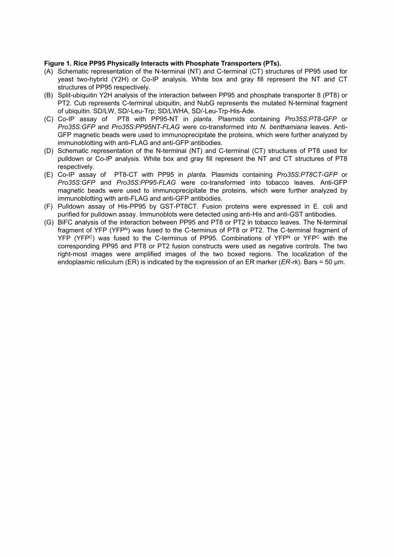
Figure 1. Rice PP95 Physically Interacts with Phosphate Transporters (PTs).(A) Schematic representation of the N-terminal (NT) and C-terminal (CT) structures of PP95 used for
yeast two-hybrid (Y2H) or Co-IP analysis. White box and gray fill represent the NT and CTstructures of PP95 respectively.
(B) Split-ubiquitin Y2H analysis of the interaction between PP95 and phosphate transporter 8 (PT8) orPT2. Cub represents C-terminal ubiquitin, and NubG represents the mutated N-terminal fragmentof ubiquitin. SD/LW, SD/-Leu-Trp; SD/LWHA, SD/-Leu-Trp-His-Ade.
(C) Co-IP assay of PT8 with PP95-NT in planta. Plasmids containing Pro35S:PT8-GFP orPro35S:GFP and Pro35S:PP95NT-FLAG were co-transformed into N. benthamiana leaves. Anti-GFP magnetic beads were used to immunoprecipitate the proteins, which were further analyzed byimmunoblotting with anti-FLAG and anti-GFP antibodies.
(D) Schematic representation of the N-terminal (NT) and C-terminal (CT) structures of PT8 used forpulldown or Co-IP analysis. White box and gray fill represent the NT and CT structures of PT8respectively.
(E) Co-IP assay of PT8-CT with PP95 in planta. Plasmids containing Pro35S:PT8CT-GFP orPro35S:GFP and Pro35S:PP95-FLAG were co-transformed into tobacco leaves. Anti-GFPmagnetic beads were used to immunoprecipitate the proteins, which were further analyzed byimmunoblotting with anti-FLAG and anti-GFP antibodies.
(F) Pulldown assay of His-PP95 by GST-PT8CT. Fusion proteins were expressed in E. coli andpurified for pulldown assay. Immunoblots were detected using anti-His and anti-GST antibodies.
(G) BiFC analysis of the interaction between PP95 and PT8 or PT2 in tobacco leaves. The N-terminalfragment of YFP (YFPN) was fused to the C-terminus of PT8 or PT2. The C-terminal fragment ofYFP (YFPC) was fused to the C-terminus of PP95. Combinations of YFPN or YFPC with thecorresponding PP95 and PT8 or PT2 fusion constructs were used as negative controls. The tworight-most images were amplified images of the two boxed regions. The localization of theendoplasmic reticulum (ER) is indicated by the expression of an ER marker (ER-rk). Bars = 50 µm.

Merged
Merged
mCherry
ER-rk
CFP-PP95Bright Field
CFP-PP95Bright Field
A
C
H I
B
EVB
DVBTVB
epcorsc
exep
ep
xy
ph
xy
xyphedo
ph
D E F G
Figure 2. Subcellular Localization and Tissue-specific Expression of PP95.(A) and (B) Subcellular localization of CFP-PP95 fusion proteins. Pro35S:CFP-PP95 constructs
together with Pro35S:mCherry (A) or ER marker ER-rk (B) were expressed in rice protoplasts.Bar = 5 µm.
(C) to (I) Tissue-specific expression of PP95. GUS staining in the root maturation zone (C),cross-sections of a primary root (D), amplified image of the root central cylinder shown in theboxed region in panel D (E) , lateral root (F), leaf blade (G), stem (H), and node (I) ofProPP95:gPP95-GUS transgenic plants. ep, epidermis; ex, exodermis; sc, sclerenchyma; xy,xylem; ph, phloem; bc, bulliform cells; edo, endodermis; EVB, enlarged vascular bundles;DVB, diffuse vascular bundles; TVB, transit vascular bundles. Bars = 100 μm in (C) and (H),25 μm in (D) and (G), 15 μm in (E) and (F), and 200 μm in (I).
Figure 2
bc

Shoot Root Shoot Root0
2
4
6
8
10PP95-OV3PP95-OV7
WT
PP95-OV12
Fre
sh w
eigh
t (g
/ pla
nt )
HP
0
5
10
15
20
Rel
ativ
e ex
pres
sion
leve
l
WTPP95-OV3PP95-OV7PP95-OV12
HP
LP
A B
D E
LP
LP
C
***
**
HP
Figure 3. Overexpression of PP95 Results in Pi Accumulation in Rice.(A) Expression levels of PP95 in PP95 overexpression lines grown under Pi-sufficient conditions (200 µM).
Error bars represent SD (n = 3).(B) and (C) Phenotypes of wild type (WT) and three PP95 overexpression transgenic lines (PP95-OV3,
PP95-OV7, and PP95-OV12). Photographs of whole seedlings (B) and leaf blades (C) were taken ofplants grown under Pi-sufficient (HP; 200 µM) or Pi-deficient (LP; 10 µM) conditions for 20 days (10-d-old plants were used for treatment). Bars = 10 cm in (B) and 1 cm in (C).
(D) Shoot and root fresh weight (FW) of the plants described in (B). Error bars represent SD (n = 9independent plants). Asterisks indicate significant difference from the WT control (*P < 0.05 and ***P <0.001; Student’s t test).
(E) Pi levels in shoots and roots of wild type (WT) plants and three PP95 overexpression transgenic lines(PP95-OV3, PP95-OV7 and PP95-OV12) grown under Pi-sufficient (HP; 200 µM) or Pi-deficient (LP;10 µM) conditions for 20 days (10-d-old plants were used for treatment). Error bars represent SD (n =3 independent plants). Asterisks indicate significant difference from the WT control (*P < 0.05, **P <0.01 and ***P < 0.001; Student’s t test).
Pi c
on
cen
tra
tion
(m
g /
g F
W)
*
HP LP
***
**
***
Figure 3

HP LP
WT pp95-25 WT
HP LP
A B
C
HP LP
pp95-10 pp95-25pp95-10
**
Young leaf Old leaf
**
*
*****
HP LP
D
Figure 4. Mutation of PP95Alters Pi Homeostasis in Rice.(A) Phenotypes of the wild type (WT) and two independent pp95 mutants. Photographs of whole
seedlings were taken of plants grown under Pi-sufficient (HP; 200 µM) or Pi-deficient (LP; 10 µM)conditions for 20 days (10-d-old plants were used for treatment). Bars = 10 cm.
(B) Shoot and root fresh weight (FW) of plants described in (A). Error bars represent SD (n = 9).(C) Pi levels in shoots and roots of WT and two independent pp95 mutants grown under HP or LP
conditions.(D) Pi levels in different leaves of WT and two independent pp95 mutants under HP or LP conditions.
Error bars represent SD (n =3). Asterisks indicate significant difference from the WT control (*P <0.05, **P < 0.01 and ***P < 0.001; Student’s t test).
Figure 4
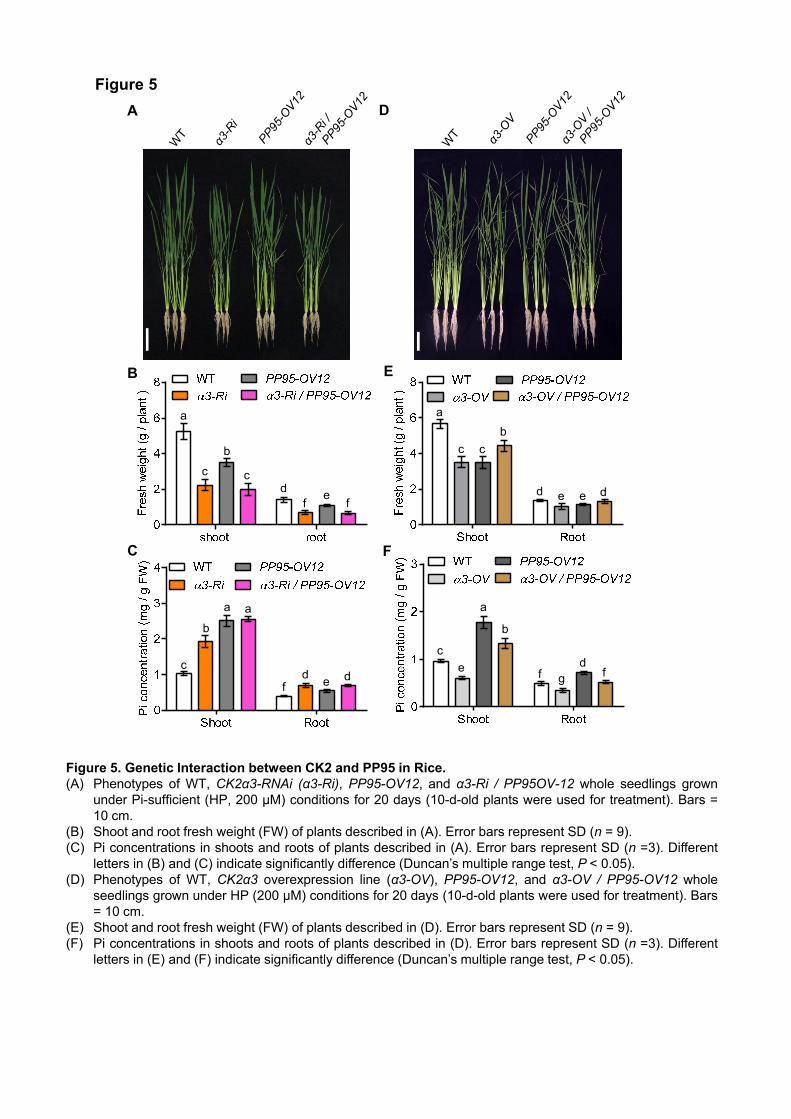
a
b
c c
d e e d
f
a
b
c
e dfg
A
B
C
a
b
c
de
f
c
f
D
E
F
f
a a
b
cd de
Figure 5. Genetic Interaction between CK2 and PP95 in Rice.(A) Phenotypes of WT, CK2α3-RNAi (α3-Ri), PP95-OV12, and α3-Ri / PP95OV-12 whole seedlings grown
under Pi-sufficient (HP, 200 µM) conditions for 20 days (10-d-old plants were used for treatment). Bars =10 cm.
(B) Shoot and root fresh weight (FW) of plants described in (A). Error bars represent SD (n = 9).(C) Pi concentrations in shoots and roots of plants described in (A). Error bars represent SD (n =3). Different
letters in (B) and (C) indicate significantly difference (Duncan’s multiple range test, P < 0.05).(D) Phenotypes of WT, CK2α3 overexpression line (α3-OV), PP95-OV12, and α3-OV / PP95-OV12 whole
seedlings grown under HP (200 µM) conditions for 20 days (10-d-old plants were used for treatment). Bars= 10 cm.
(E) Shoot and root fresh weight (FW) of plants described in (D). Error bars represent SD (n = 9).(F) Pi concentrations in shoots and roots of plants described in (D). Error bars represent SD (n =3). Different
letters in (E) and (F) indicate significantly difference (Duncan’s multiple range test, P < 0.05).
Figure 5

GST-PT8CTGST
GST-α3GST-PP95
GST-PT8CT
1 10.1
1 1
GST-PP95D240N
11
1
1
S517A
GST-α3GST-PP95
or GST-PP95D240N
GST-PT8CT or GST-PT8CTS517A
GST
GST
1 1 1 10.1
11
1
1
S517A
1 2 3
A B
C
- 70
kD
- 35
- 25
kD
CBB Autoradiography
α-PT8
α-PHF1
p-PT8PT8
- 40
- 55
1 2.35 0.14 0.81 p-PT8
0.56 0.29 0.62 0.60 PT8
Figure 6. PP95 Directly Dephosphorylates PTs.(A) Analysis of the phosphatase activity of PP95 and its mutated version. GST fusion proteins were expressed in E.
coli and purified for p-nitrophenylphosphate-based phosphatase assays. Dephosphorylated pNPP levels werecalculated based on absorbance at 405 nm after the dephosphorylation reaction was complete. The insetshows purified GST (lane 1), GST-PP95 (lane 2), and GST-PP95D240N (lane 3) in a Coomassie brilliant blue(CBB) stained SDS-PAGE gel.
(B) In vivo dephosphorylation of PT8 by PP95. Phosphorylated and dephosphorylated PT8 proteins extracted fromendoplasmic reticulum (ER) in α3-OV, PP95-OV, α3-OV/PP95-OV, and wild-type (WT) plants grown under Pi-sufficient conditions were detected by immunoblotting after Phos-tag SDS-PAGE using an anti-OsPT8 antibody.PHF1 detected using anti-PHF1 antibody was used as ER protein control. Values represent relativequantification of PT8 proteins in the indicated plants. kD, kilo Dalton.
(C) In vitro dephosphorylation of GST-PT8CT by GST-PP95. GST-PT8CT proteins were phosphorylated by GST-CK2α3 in the presence of [γ-32P]ATP, followed by the addition of GST-PP95 or GST-PP95D240N and 1 h ofincubation. Proteins were visualized by Coomassie blue staining (left panel), and phosphorylated proteins werevisualized by autoradiography (right panel). GST and GST-PT8CTS517A were used as controls. GST-CK2α3,GST-PP95, and GST-PP95D240N protein levels (in μg) used in the assays are indicated by numbers (0.1 and 1).
Figure 6

PT2-GFP PT8-GFP
Control
α3-OV
α3-OV
PP95-OV
Figure 7. PP95 Acts Antagonistically with CK2 to Modulate the ER Exit of PTs.(A) PT8 protein levels at the plasma membrane (PM) and the endoplasmic reticulum (ER) in plants.
Proteins were isolated from plants grown under Pi-sufficient conditions. Anti-PT8 antibody was used todetect PT8 protein levels. PHF1 and PIP1;3 were used as ER and PM protein controls, respectively.
(B) Subcellular localization of PT2-GFP and PT8-GFP fusion proteins in protoplasts. Pro35S:PT2 / 8-GFP(control), Pro35S:PT2-GFP, or Pro35S:PT8-GFP constructs together with Pro35S:CK2α3 (a3-OV) orPro35S:CK2α3 and Pro35S:PP95 (a3-OV / PP95-OV) were transformed into rice protoplasts. Bar = 5µm.
(C) Subcellular localization of PT8-GFP in root epidermal cells of seven-d-old ProPT8:PT8-GFP,ProPT8:PT8-GFP / a3-OV, and ProPT8:PT8-GFP / a3-OV / PP95-OV transgenic plants. Bars = 20 µm.
B
C
A PM ER
α-PT8
α-PHF1
α-PIP1;3
- 55
- 40
- 25
kD
- 55
- 35
PT8-GFPα3-OV / PP95-OV /
PT8-GFPα3-OV /
PT8-GFP
Figure 7
Bright Field Bright Field
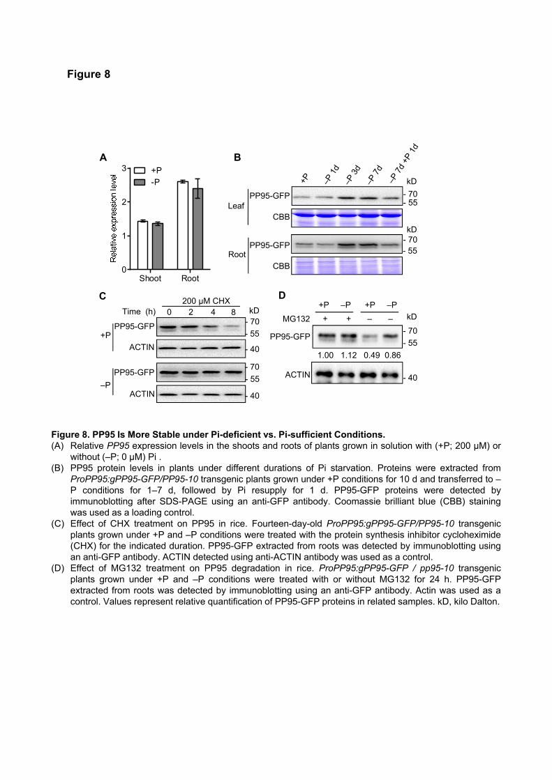
Shoot Root0
1
2
3 +P-P
A
D
MG132
+P –P +P –P
+ + – –
ACTIN
PP95-GFP- 70
kD
- 55
PP95-GFP
B
CBB
PP95-GFP
CBB
Leaf
Root
- 70
kD
- 55
- 70kD
- 55
ACTIN
PP95-GFP
ACTIN
0 2 4 8 200 µM CHX
+P
C
PP95-GFP- 70kDTime (h)
- 55
–P
- 70
- 55
- 40
- 40
- 40
1.00 1.12 0.49 0.86
Figure 8. PP95 Is More Stable under Pi-deficient vs. Pi-sufficient Conditions.(A) Relative PP95 expression levels in the shoots and roots of plants grown in solution with (+P; 200 µM) or
without (–P; 0 µM) Pi .(B) PP95 protein levels in plants under different durations of Pi starvation. Proteins were extracted from
ProPP95:gPP95-GFP/PP95-10 transgenic plants grown under +P conditions for 10 d and transferred to –P conditions for 1–7 d, followed by Pi resupply for 1 d. PP95-GFP proteins were detected byimmunoblotting after SDS-PAGE using an anti-GFP antibody. Coomassie brilliant blue (CBB) stainingwas used as a loading control.
(C) Effect of CHX treatment on PP95 in rice. Fourteen-day-old ProPP95:gPP95-GFP/PP95-10 transgenicplants grown under +P and –P conditions were treated with the protein synthesis inhibitor cycloheximide(CHX) for the indicated duration. PP95-GFP extracted from roots was detected by immunoblotting usingan anti-GFP antibody. ACTIN detected using anti-ACTIN antibody was used as a control.
(D) Effect of MG132 treatment on PP95 degradation in rice. ProPP95:gPP95-GFP / pp95-10 transgenicplants grown under +P and –P conditions were treated with or without MG132 for 24 h. PP95-GFPextracted from roots was detected by immunoblotting using an anti-GFP antibody. Actin was used as acontrol. Values represent relative quantification of PP95-GFP proteins in related samples. kD, kilo Dalton.
Figure 8
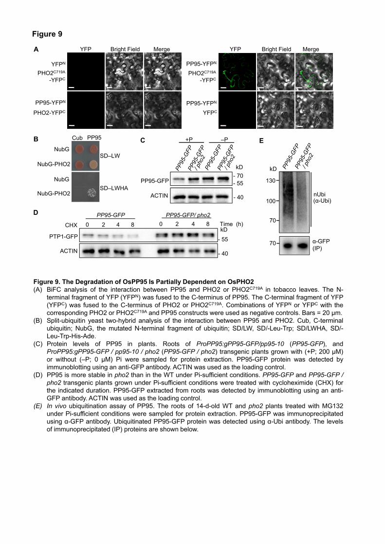
YFP Bright Field
YFPC
PHO2C719A
-YFPC
PP95-YFPN
PP95-YFPN
Merge YFP Bright Field Merge
NubG
NubG-PHO2
Cub PP95
SD–LWHA
SD–LW
PHO2-YFPC
PHO2C719A
-YFPC
YFPN
PP95-YFPN
A
B
D
E
CHX
+P –PC
ACTIN
PP95-GFP - 55- 70
kD
- 40
NubG
NubG-PHO2
α-GFP(IP)
70
70
(α-Ubi)nUbi
100
130
kD
PP95-GFP/ pho2
PTP1-GFP
ACTIN
0 2 4 8 Time (h)
PP95-GFP
0 2 4 8kD
- 55
- 40
Figure 9. The Degradation of OsPP95 Is Partially Dependent on OsPHO2(A) BiFC analysis of the interaction between PP95 and PHO2 or PHO2C719A in tobacco leaves. The N-
terminal fragment of YFP (YFPN) was fused to the C-terminus of PP95. The C-terminal fragment of YFP(YFPC) was fused to the C-terminus of PHO2 or PHO2C719A. Combinations of YFPN or YFPC with thecorresponding PHO2 or PHO2C719A and PP95 constructs were used as negative controls. Bars = 20 µm.
(B) Split-ubiquitin yeast two-hybrid analysis of the interaction between PP95 and PHO2. Cub, C-terminalubiquitin; NubG, the mutated N-terminal fragment of ubiquitin; SD/LW, SD/-Leu-Trp; SD/LWHA, SD/-Leu-Trp-His-Ade.
(C) Protein levels of PP95 in plants. Roots of ProPP95:gPP95-GFP/pp95-10 (PP95-GFP), andProPP95:gPP95-GFP / pp95-10 / pho2 (PP95-GFP / pho2) transgenic plants grown with (+P; 200 µM)or without (–P; 0 µM) Pi were sampled for protein extraction. PP95-GFP protein was detected byimmunoblotting using an anti-GFP antibody. ACTIN was used as the loading control.
(D) PP95 is more stable in pho2 than in the WT under Pi-sufficient conditions. PP95-GFP and PP95-GFP /pho2 transgenic plants grown under Pi-sufficient conditions were treated with cycloheximide (CHX) forthe indicated duration. PP95-GFP extracted from roots was detected by immunoblotting using an anti-GFP antibody. ACTIN was used as the loading control.
(E) In vivo ubiquitination assay of PP95. The roots of 14-d-old WT and pho2 plants treated with MG132under Pi-sufficient conditions were sampled for protein extraction. PP95-GFP was immunoprecipitatedusing α-GFP antibody. Ubiquitinated PP95-GFP protein was detected using α-Ubi antibody. The levelsof immunoprecipitated (IP) proteins are shown below.
Figure 9

B
1 1.58 0.39 0.75
α-PHF1
p-PT8PT8
α-PT8
0.46 0.28 0.17 0.19
α-Bip
p-PT8PT8
kD- 75
- 40- 55
PM ER
α-PHF1
α-PIP1;3
α-PT8 - 55
- 40
- 25
kD
- 55
- 35
1 0.55 2.01 1.75 1 1.36 0.31 0.57
a
b
c
d d
e
c
d
D a
aa
bc
d
ef
ghghj
jk jkl m
A
E
C
F
Figure 10
Figure 10. Genetic Interaction between PHO2 and PP95 in Rice.(A) Phenotype of wild-type (WT), pp95-10, pho2 and pp95 pho2 double mutant whole seedlings after plants
were grown under Pi-sufficient (HP; 200 µM) condition for 45 days. Bars = 10 cm.(B) Shoot and root fresh weight (FW) of plants described in (A). Error bars represent SD (n = 9).(C, D) Pi concentrations in shoot, root (C), and different leaves (D) of WT, pp95 mutant, pho2 mutant and
pp95 pho2 double mutant plants grown under Pi sufficient conditions. Error bars represent SD (n = 3).Different letters indicate significant difference (Duncan’s multiple range test; P < 0.05).
(E) Phosphorylated and dephosphorylated PT8 proteins extracted from the endoplasmic reticulum (ER) inWT, pp95 mutant, pho2 mutant and pp95 pho2 double mutant plants grown in Pi-sufficient condition. PT8proteins were detected by immunoblotting in Phos-tag SDS-PAGE using an anti-OsPT8 antibody. Bip andPHF1 detected using anti-Bip and anti-PHF1 antibody were used as controls. Values represent relativequantification of phosphorylated PT8 and dephosphorylated PT8 proteins in related plants.
(F) PT8 protein levels at the plasma membrane (PM) and the endoplasmic reticulum (ER) in related plants.Proteins were isolated from plants grown under Pi-sufficient (200 µM) conditions. Anti-PHF1 antibody andAnti-PIP1;3 antibody were used as ER and PM protein controls, respectively. Values represent relativequantification of PT8 proteins in the indicated plants. The relative intensity of PM OsPT8 proteins or EROsPT8 proteins in the WT was set to 1, respectively.
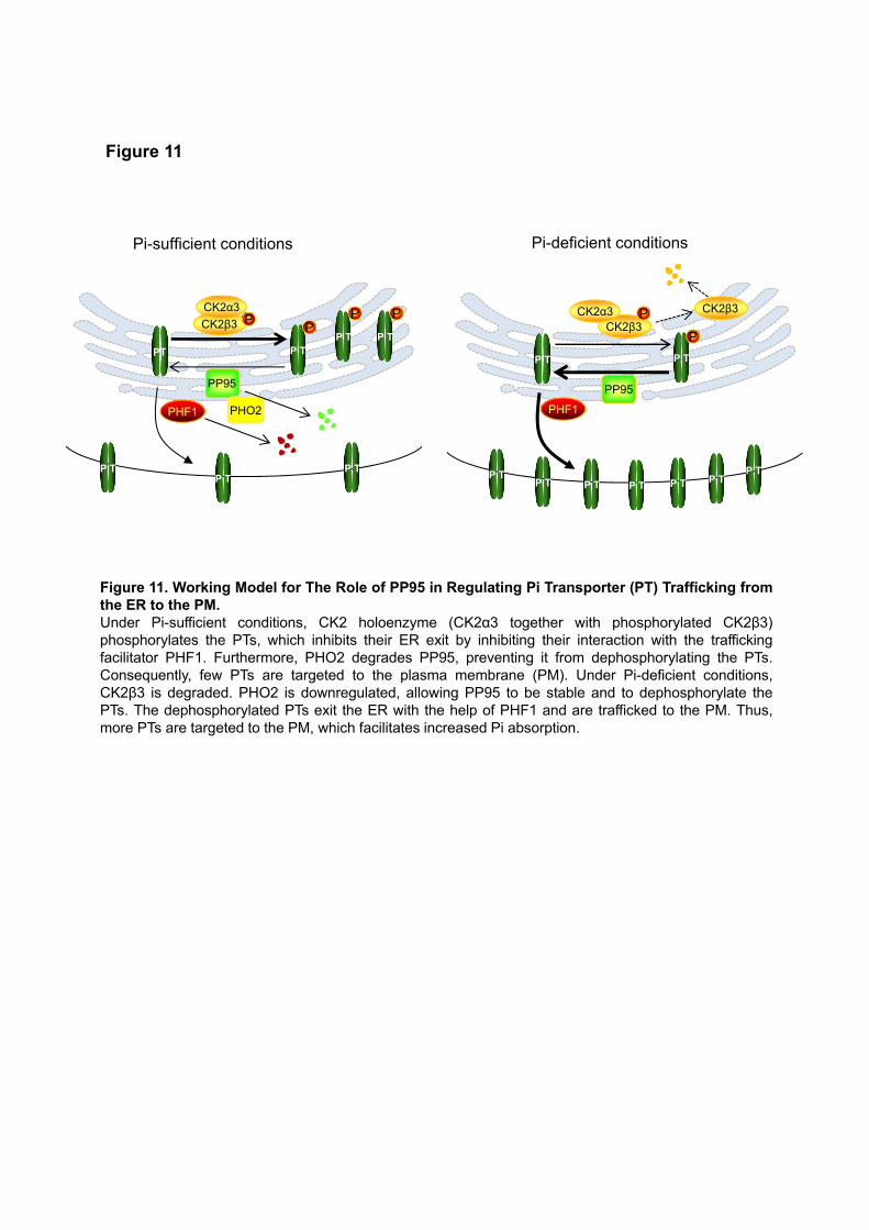
CK2β3
CK2α3P
Pi-sufficient conditions
PP95
PHF1 PHO2
CK2β3CK2α3
Pi-deficient conditions
PP95
PHF1
PT P T
P TP T
P T
P T P T
P TP T
P TP T P TP T P T
PPP T
P
P T
P CK2β3P
Figure 11
Figure 11. Working Model for The Role of PP95 in Regulating Pi Transporter (PT) Trafficking fromthe ER to the PM.Under Pi-sufficient conditions, CK2 holoenzyme (CK2α3 together with phosphorylated CK2β3)phosphorylates the PTs, which inhibits their ER exit by inhibiting their interaction with the traffickingfacilitator PHF1. Furthermore, PHO2 degrades PP95, preventing it from dephosphorylating the PTs.Consequently, few PTs are targeted to the plasma membrane (PM). Under Pi-deficient conditions,CK2β3 is degraded. PHO2 is downregulated, allowing PP95 to be stable and to dephosphorylate thePTs. The dephosphorylated PTs exit the ER with the help of PHF1 and are trafficked to the PM. Thus,more PTs are targeted to the PM, which facilitates increased Pi absorption.
DOI 10.1105/tpc.19.00685; originally published online January 9, 2020;Plant Cell
and Chuanzao MaoZhili Yang, Jian Yang, Yan Wang, Fei Wang, Wenxuan Mao, Qiuju He, Jiming Xu, Wu Zhongchang
Transporter Trafficking in RicePROTEIN PHOSPOHATASE 95 Regulates Phosphate Homeostasis by Affecting Phosphate
This information is current as of March 9, 2020
Supplemental Data /content/suppl/2020/01/31/tpc.19.00685.DC2.html /content/suppl/2020/01/09/tpc.19.00685.DC1.html
Permissions X
https://www.copyright.com/ccc/openurl.do?sid=pd_hw1532298X&issn=1532298X&WT.mc_id=pd_hw1532298
eTOCs http://www.plantcell.org/cgi/alerts/ctmain
Sign up for eTOCs at:
CiteTrack Alerts http://www.plantcell.org/cgi/alerts/ctmain
Sign up for CiteTrack Alerts at:
Subscription Information http://www.aspb.org/publications/subscriptions.cfm
is available at:Plant Physiology and The Plant CellSubscription Information for
ADVANCING THE SCIENCE OF PLANT BIOLOGY © American Society of Plant Biologists