PROGRESSION OF AVASCULAR NECROSIS OF FEMORAL HEAD … · nagoya j. med. sci. 54. 00 - 00, 1992...
13
Nagoya J. Med. Sci. 54. 00 - 00, 1992 Invited Article PROGRESSION OF AVASCULAR NECROSIS OF FEMORAL HEAD AND CHOICE OF TREATMENT HISASHI IWATA, YUKIHARU HASEGAWA, MASANORI MIZUNO, EIICHI GENDA, YUJI KATAOKA and AKIHIDE KADA Department of Orthopedic Surgery, Nagoya University School of Medicine, Nagoya 466, Japan ABSTRACf Observations of the disease course mainly by X-ray were made in 52 patients (85 joints) with avascular necrosis of the femoral head to determine the prognosis and to decide on treatment. The progression of the necrotic area is related to the activity of the original disease, to the size of the necrotic area in the early stages of the disease, and to whether or not the patient received steroid treatment. On the basis of the size and location of the necrotic area, the disease process is divided into four stages, I to IV. The affected heads are also classified into six types according to their site and extent, the degree of flatness of their weightbear- ing surface, and the presence of cystic lesion. Preventive treatment and conservative observation or transtro- chanteric anterior rotational osteotomy and vascularized pedicle bone graft are applicable to cases in Stages I and II. Total hip joint arthroplasties and salvage procedures are performed in Stages III and IV. The useful- ness of 99mTc bone scintigraphy was unexpectedly disappointing for diagnosis of the stages of the disease. However, MRI was sensitive for the diagnosis in the early stages of the disease. INTRODUCfION Cases of avascular necrosis of the femoral head (ANFH) are steadily increasing, but their eti- ology and pathogenesis are still unknown. The determination of what stage and which treatment should be taken for the necrotic area is also controversial because it depends on such a variety of factors including the diverse progress of the necrosis, original disease activity, course of treat- ment, prognosis, and patient's age and occupation.!) In hope of being able to suggest appropri- ate treatment for future cases, we observed mainly the transitional radiographic features of the necrotic lesion in the process of its natural course and discussed the choices of treatment method. We also considered the role of steroids in the etiology2,3,4) and the significance of 99mTc bone scintigraphy and magnetic resonance imaging (MRI) for the early diagnosis of femoral head necrosis. 5 ,6) MATERIALS AND OBSERVATION METHOD The 52 subjects used in our study were selected from among the patients with ANFH treated at Nagoya University Hospital since 1971 who had been followed up for more than one year. Most operative subjects were excluded from this study. The 52 subjects can be categorized as Correspondence: Dr. Hisashi Iwata, Department of Orthopedic Surgery, Nagoya University School of Medicine, 65 Tsuruma-cho, Showa-ku, Nagoya 466 Japan 27
Transcript of PROGRESSION OF AVASCULAR NECROSIS OF FEMORAL HEAD … · nagoya j. med. sci. 54. 00 - 00, 1992...

Nagoya J. Med. Sci. 54. 00 - 00, 1992 Invited Article
PROGRESSION OF AVASCULAR NECROSISOF FEMORAL HEAD ANDCHOICE OF TREATMENT
HISASHI IWATA, YUKIHARU HASEGAWA, MASANORI MIZUNO, EIICHI GENDA,YUJI KATAOKA and AKIHIDE KADA
Department of Orthopedic Surgery, Nagoya University School of Medicine,Nagoya 466, Japan
ABSTRACf
Observations of the disease course mainly by X-ray were made in 52 patients (85 joints) with avascularnecrosis of the femoral head to determine the prognosis and to decide on treatment. The progression of thenecrotic area is related to the activity of the original disease, to the size of the necrotic area in the earlystages of the disease, and to whether or not the patient received steroid treatment. On the basis of the sizeand location of the necrotic area, the disease process is divided into four stages, I to IV. The affected headsare also classified into six types according to their site and extent, the degree of flatness of their weightbearing surface, and the presence of cystic lesion. Preventive treatment and conservative observation or transtrochanteric anterior rotational osteotomy and vascularized pedicle bone graft are applicable to cases in Stages Iand II. Total hip joint arthroplasties and salvage procedures are performed in Stages III and IV. The usefulness of 99mTc bone scintigraphy was unexpectedly disappointing for diagnosis of the stages of the disease.However, MRI was sensitive for the diagnosis in the early stages of the disease.
INTRODUCfION
Cases of avascular necrosis of the femoral head (ANFH) are steadily increasing, but their etiology and pathogenesis are still unknown. The determination of what stage and which treatmentshould be taken for the necrotic area is also controversial because it depends on such a varietyof factors including the diverse progress of the necrosis, original disease activity, course of treatment, prognosis, and patient's age and occupation.!) In hope of being able to suggest appropriate treatment for future cases, we observed mainly the transitional radiographic features of thenecrotic lesion in the process of its natural course and discussed the choices of treatmentmethod. We also considered the role of steroids in the etiology2,3,4) and the significance of99mTc bone scintigraphy and magnetic resonance imaging (MRI) for the early diagnosis offemoral head necrosis. 5,6)
MATERIALS AND OBSERVATION METHOD
The 52 subjects used in our study were selected from among the patients with ANFH treatedat Nagoya University Hospital since 1971 who had been followed up for more than one year.Most operative subjects were excluded from this study. The 52 subjects can be categorized as
Correspondence: Dr. Hisashi Iwata, Department of Orthopedic Surgery, Nagoya University School of
Medicine, 65 Tsuruma-cho, Showa-ku, Nagoya 466 Japan
27

28
Hisashi Iwata et al.
follows: 1) those with bilateral osteonecrosis who were operated on only one side; 2) those whowere under conservative treatment before operation; 3) those in whom surgical treatment hadbeen proposed but was not performed because of basic underlying disease; and 4) those inwhom surgical treatment was not given because pain was mild. Of the 52 subjects (85 joints) observed, there were 40 cases that were bilateral and 12 that were unilateral, 27 males and 25 females, and 25 patients who received steroid treatment or are now receiving it and 27 who didnot. As to the original disease, there were 12 cases of systemic lupus erythematosus (SLE) twocases of nephrose syndrome, two cases of idiopathic thrombocytopenic purpula, two cases of facial nerve palsy, and one case each of hemolytic anemia, Behc;et's disease, Addison's disease,kidney transplantation, asthma brochiale, Harada's disease and sympathic occulitis. On theother hand, of 135 joints that received surgical treatment, 36 joints received endoprosthesis; 33,total hip joint replacement; 33, transtrochanteric anterior rotational osteotomy (Sugioka);7,8)and 33 joints, vascularized pedicle bone graft.9) The progression of ANFH was related to the severity of the original disease and to the stage and extent of the necrotic lesion at the time of theinitial diagnosis. The affected femoral heads were classified into the following four stages recently reported by Ohzono et al. lO) as established by the Japanese Investigation Committeell )
under the auspices of the Ministry of Health and Welfare: Stage I) No radiographic abnormality,but bone scintigram or MRI reveals abnormality; Stage II) Femoral head is spherical with radiolucent area, or collapse is seen to a minor degree of less than 2mm. Bone graft or rotationalosteotomy seems appropriate as the choice of treatment, given careful consideration of the expansion of the necrotic region; Stage III) Remarkable collapse is seen; Stage IV) Arthropathicdeformation is seen.
Based on the site and the extent of the necrotic lesion in the femoral head, the affected femoral heads were also classified into six types as shown in Fig. 1. 10) The necrotic changes of ourcases were further divided into two groups: those that had been treated with steroids and thosethat had not.
Type I -A
~i~
Type I -B Type I -C Type 11 Type m: -A Type ill-B
~~~~Fig. 1. Radiographic classification of ANFH. Type is characterised by the presence of a demarcation line in the
femoral head and is divided into three subtypes, I-A, I-B and I-C, according to its relationship to theweightbearing surface. Type II shows early flattening of the weightbearing surface but has no demarcationline around the necrotic area. Type III has cystic lesions and is divided into two subtypes according to theirsite in the femoral head. (Ohzono, K. et al. 10)
RESULTS
As shown in Table 1, the incidence of type II (32 joints) and type 1-C (7 joints) was muchhigher in patients with steroid-induced ANFH. The distribution of hips according to stage isshown in Table 2. Sixteen joints were classified as Stage I in the patients with steroid-inducedANFH, and 7 joints in those without steroid treatment. Fourteen joints were classified as Stage

29
PROGRESSION OF AVASCULAR NECROSIS AND CHOICE OF TREATMENT
II in the steroid-induced group, and 22 joints in the non-steroid group. In Stage III there were10 joints of the patients with steroid-induced ANFH, and 10 joints of those without steroids. Finally, 4 joints were of Stage IV in the steroid-induced group, and 2 in the non-steroid group.From evaluation of the following cases, the natural course of the necrotic area and the operativeindication for preserving the femoral head were discussed according to the stage and type classifications stated above.
Table 1. Type Classification of ANFH
Type Steroid-induced Non-steroid
I-A 0 2
I-B 5 7
I-C 7 17
II 32 14
III-B 0 1
Total 44 41 (Joints)
Table 2. Stage Classification of ANFH
Stage Steroid-induced Non-steroid
I 16 7
II 14 22III 10 10
IV 4 2
Total 44 41 (Joints)
Case ReportsCase 1. A 40-year-old man with bilateral ANFH.
The patient had a drinking habit of 300 to 500 ml sake per day and suffered from hip pain.X-ray films showed sclerotic changes surrounding a radiolucent area in the bilateral femoralheads (Fig. 2a) together with a slight collapse in the right side. The right side was in Stage II.The left side was also in Stage II. Transtrochanteric anterior rotational osteotomy was performed only on the right side (Fig. 2b) and the left side remained to be observed. Five yearsafter the operation, no recollapse in the right femoral head and no pathological enlargement ofthe necrotic area were seen and the radiolucent lesion was repaired. On the left, unoperatedside, no further progression was seen and even a diminution of the radiolucent finding was apparent (Fig. 2c). At present, the patient has no pain in either side and is in excellent post-operative condition.
30
Hisashi Iwata et af.
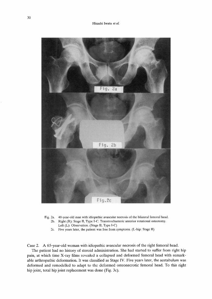
Fig. 2a. 40-year-old man with idiopathic avascular necrosis of the bilateral femoral head.2b. Right (R): Stage II, Type I-C. Transtrochanteric anterior rotational osteotomy.
Left (L): Observation. (Stage II, Type I-C)2c. Five years later, the patient was free from symptoms. (L-hip: Stage II)
Case 2. A 65-year-old woman with idiopathic avascular necrosis of the right femoral head.The patient had no history of steroid administration. She had started to suffer from right hip
pain, at which time X-ray films revealed a collapsed and deformed femoral head with remarkable arthropathic deformation. It was classified as Stage IV. Five years later, the acetabulum wasdeformed and remodelled to adapt to the deformed osteonecrotic femoral head. To this righthip joint, total hip joint replacement was done (Fig. 3c).

31
PROGRESSION OF AVASCULAR NECROSIS AND CHOICE OF TREATMENT
Fig. 3a. 65-year-old woman with idiopathic avascular necrosis of the right femoral head.3b. R: Acetabulum is remodeled and adapted to the collapsed femoral head (Stage
IV, Type II) eight years later.3c. Nine years later, total hip joint replacement has been done.
Case 3. A 34-year-old man with belateral ANFH.The patient had had a short-term history of steroid administration. X-ray films of the femoral
head showed no collapse nor deformation, but a radiolucent finding and sclerotic changes surrounding the radiolucent are were recognized (Fig. 4a). Twelve years later, he suffered from increasing hip joint pain and visited our clinic. From radiologic evaluation, both his right and leftsides were regarded to be in Stage II, (Fig. 4b). After six months, since no improvement was observed by only eliminating weightbearing, transtrochanteric anterior rotational osteotomy wasperformed on the right side whose osteonecrotic area was small (Fig. 4c). The post-operativecourse was excellent. The left side, which had shown pronounced clinical signs as well as radiographic change, showed no further collapse or deformation (Fig. 4d). The patient no longercomplains, at present, of any arthritic pain in either hip joint.
32
Hisashi Iwata et al.
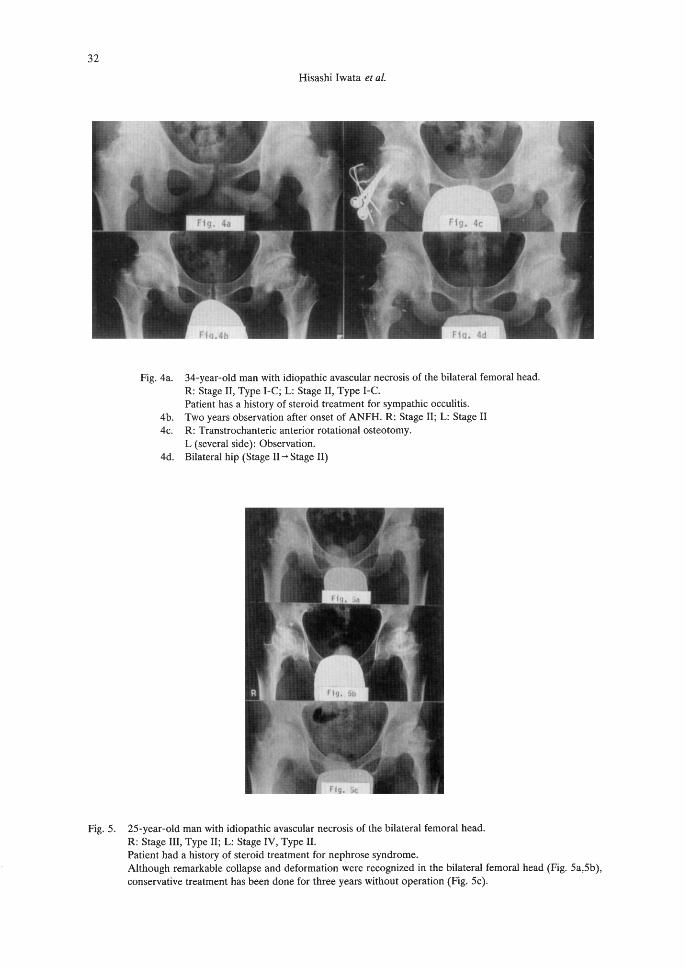
Fig. 4a. 34-year-old man with idiopathic avascular necrosis of the bilateral femoral head.R: Stage II, Type I-C; L: Stage II, Type I-C.Patient has a history of steroid treatment for sympathic occulitis.
4b. Two years observation after onset of ANFH. R: Stage II; L: Stage II4c. R: Transtrochanteric anterior rotational osteotomy.
L (several side): Observation.4d. Bilateral hip (Stage II - Stage II)
Fig. 5. 25-year-old man with idiopathic avascular necrosis of the bilateral femoral head.R: Stage III, Type II; L: Stage IV, Type II.Patient had a history of steroid treatment for nephrose syndrome.Although remarkable collapse and deformation were recognized in the bilateral femoral head (Fig. 5a,5b),conservative treatment has been done for three years without operation (Fig. 5c).
33
PROGRESSION OF AVASCULAR NECROSIS AND CHOICE OF TREATMENT
Case 4. A 25-year-old man with bilateral ANFH.The patient had had a short-term history of steroid treatment for nephrose syndrome. X-ray
films revealed large necrotic areas including collapsed femoral heads in both sides. Taking thepatient's young age and the wide bilateral area into account, non-operative treatment with nonweightbearing two-crutch gait was recommended. As shown in Fig. 5, three years later, progressof the necrotic area remains almost unchanged and can be clinically regarded as satisfactory.Case 5. A 47-year-old woman with SLE and bilateral ANFH.
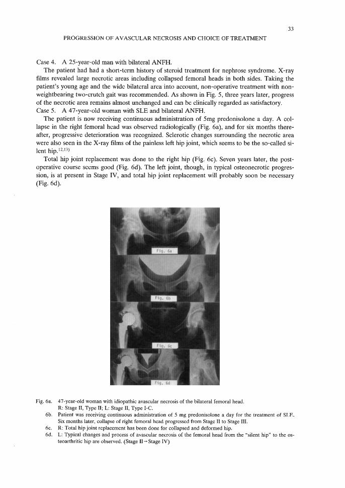
The patient is now receiving continuous administration of 5mg predonisolone a day. A collapse in the right femoral head was observed radiologically (Fig. 6a), and for six months thereafter, progressive deterioration was recognized. Sclerotic changes surrounding the necrotic areawere also seen in the X-ray films of the painless left hip joint, which seems to be the so-called silent hip.12,13)
Total hip joint replacement was done to the right hip (Fig. 6c). Seven years later, the postoperative course seems good (Fig. 6d). The left joint, though, in typical osteonecrotic progression, is at present in Stage IV, and total hip joint replacement will probably soon be necessary(Fig.6d).
Fig.6a, 47-year-old woman with idiopathic avascular necrosis of the bilateral femoral head.R: Stage II, Type II; L: Stage II, Type I-C.
6b. Patient was receiving continuous administration of 5 mg predonisolone a day for the treatment of SLE.Six months later, collapse of right femoral head progressed from Stage II to Stage III.
6c. R: Total hip joint replacement has been done for collapsed and deformed hip.6d. L: Typical changes and process of avascular necrosis of the femoral head from the "silent hip" to the os
teoarthritic hip are observed. (Stage II - Stage IV)
34
Hisashi Iwata el at.
Progression of Osteonecrotic Lesion and its Relation to SteroidsPresenting some representive cases from among our cases, which were classified into steroid
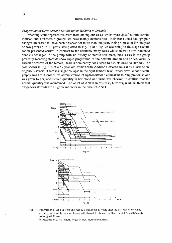
induced and non-steroid groups, we have mainly demonstrated their transitional radiographicchanges. In cases that have been observed for more than one year, their progression for one yearor two years up to 11 years, was plotted in Fig. 7a and Fig. 7b according to the stage classification presented earlier. In contrast to the relatively many cases whose necrotic area remainedalmost unchanged in the group with no history of steroid treatment, most cases in the grouppresently receiving steroids show rapid progression of the necrotic area in one or two years. Avascular necrosis of the femoral head is dominantly considered to owe its onset to steroids. Thecase shown in Fig. 8 is of a 59-year-old woman with Addison's disease caused by a lack of endogenous steroid. There is a slight collapse in the right femoral head, where 99mTc bone scintigraphy was hot. Consecutive administration of hydrocortisone equivalent to 5mg predonisolonewas given to her, and steroid quantity in her blood and urine was checked to confirm that thenormal quantity was maintained. The onset of ANFH in this case, however, made us think thatexogenous steroids are a significant factor in the onset of ANFH.
diagnosis 1
Stage I l-----
diagnosis 1
Fig. 7.
6
Fig.7b
10 Ilyear
10 llyear.
Fig. 7. Progression of ANFH from one year to a maximum 11 years after the first visit to the clinic.a: Progression of 44 femoral heads with steroid treatment for short period or continuouslyfor original disease.b: Progression of 41 femoral heads without steroid treatment.

35
PROGRESSION OF AVASCULAR NECROSIS AND CHOICE OF TREATMENT
Fig. 8. 59-year-old woman with avascular necrosis of the right femoral head. Patient was receiving continuous administration of 5 mg predonisolone for Addison's disease. Steroid quantity in blood and urine was maintained at normal levels with exogenous steroid.
Fig. 9. 26-year-old woman with bilateral ANFH. (Bilateral: Stage II, Type I-C) Patient was receiving continuoussteroid administration for SLE. In bone scintigraphy, an obvious accumulation of 99mTc in the right hipjoint as well as a cold area in the accumulation of the left hip joint are seen.

36
Hisashi Iwata et ai.
Significance ofbone scintigraphy for early diagnosis ofANFH:99mTc bone scintigraphy seems efficient to pursue the necrotic change and the repair process
of the necrotic area. 5,6) Fig. 9 shows a case of SLE in a 26-year-old woman with bilateralANFH. Her right femoral head is collapsed and a radiolucent finding is seen in her left side. Inbone scintigraphy an obvious accumulation of 99mTc in the right hip joint as well as a cold areain the accumulation of the left hip joint are recognized to prove that the center of the radiolucent area is in necrotic condition. In 35 cases of ANFH diagnosed by their clinical symptomsand X-ray findings, 99mTc bone scintigraphies showed an obvious accumulation of 99mTc forall but one case whose bone scintigraphy was normal. However, bone scintigraphies done comparatively to 8 joints of SLE or Sjogren syndrome, cases which were receiving consecutive steroid administration, were normal, that is, no obvious 99mTc take-in was seen. Judging fromthese results, bone scintigraphic diagnosis for ANFH seems to be difficult in the stage where noradiographic change is seen. On the other hand, MRI was very useful for early diagnosis of socalled minimal necrotic lesions, or Stage I, of ANFH as shown in Table 2,
DISCUSSION
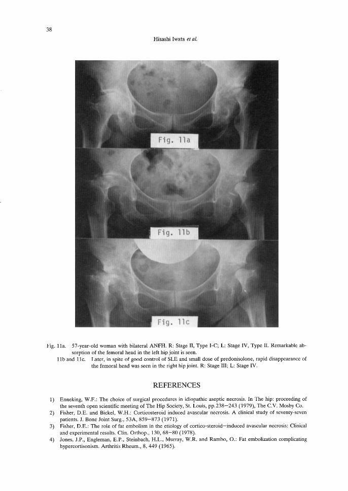
As stated previously, progression of ANFH depends on various aspects of its pathologicalcondition. These include 1) age, 2) sex, 3) occupation, 4) the etiology, stage and type of the lesion and its activity, 5) receipt of steroid treatment or not, 6) quantity and duration of steroidadministration, 7) duration from onset of disease, and 8) size and location of lesion at onset.The choice of treatment, therefore, should be decided through careful observation of each case,with reference to the natural process of similar past cases. Hasty decisions should be avoided,especially in the selection of operative treatment, but appropriate treatment should be taken toprevent further progress of the necrotic area. Sugioka's transtrochanteric anterior rotational osteotomy7,8) and Wagner's osteotomy14) or varus osteotomy9) reduce the weightbearing forces onthe necrotic area and transfer shear forces to the healthy cartilage of the femoral head to avoidrecollapse; consequently, the necrotic area may be expected to be absorbed and remodelled. Incase there is no possibility of recollapse, these methods are apparently effective. However, thisosteotomy is not indicated in cases where the necrotic area of the femoral head is so large thatthe healthy part is less than one third, even though there is no collapse. For such cases, the vascularized pedicle bone graft is well indicated. Fig, 10 shows an example of the indication for different operative methods in a recent bilateral ANFH case. But in patients who have had kidneytransplantation and who suffer from ANFH caused by SLE, whose necrotic areas have beenrapidly absorbed, the application of transtrochanteric anterior rotational osteotomy or vascularized pedicle bone graft should be carefully considered even in the early stages (Stage I or II)when the necrotic areas are still small. Fig. 11 shows X-ray films of a 57-year-old woman withANFH caused by SLE. On her first visit to our clinic remarkable absorption of the femoral headin the left hip joint was seen. Later, SLE was well controlled with slight steroid administration(2.5mg of predonisolone every two days), but rapid disappearance of the femoral head in theright hip joint surprised us and urged us to pay more attention to the decision of operation.
99mTc bone scintigraphy as a method for early diagnosis seems insufficiently effective indefining the necrotic area before radiographic changes can be seen. Further technical developments and devices such as X-ray projective position and focal projection with pin-holecolimeter, and further screening of necrotic areas and their range, are required. It is most important to diagnose ANFH as early as possible in order to decide the treatment method beforecollapse. MRI is more sensitive than other diagnostic tools in the early stages of ANFH.

PROGRESSION OF AVASCULAR NECROSIS AND CHOICE OF TREATMENT
Fig, 10c
Fig. lOa. 31-year-old woman with bilateral ANFH. R: Stage II, Type I-C; L: Stage II, Type III-B.lOb. Vascularized pedicle bone graft has been done to the right hip joint because of largeness of
necrotic area.IDe. On the other hand, transtrochanteric rotational osteotomy has been done to the left hip
joint because of limited necrotic area.
37
38
Hisashi Iwata et al.
Fig. 11a. 57-year-old woman with bilateral ANFH. R: Stage II, Type I-C; L: Stage IV, Type II. Remarkable absorption of the femoral head in the left hip joint is seen.
11b and Hc. Later, in spite of good control of SLE and small dose of predonisolone, rapid disappearance ofthe femoral head was seen in the right hip joint. R: Stage III; L: Stage IV.
REFERENCES
I) Enneking, W.F.: The choice of surgical procedures in idiopathic aseptic necrosis. In The hip: proceeding ofthe seventh open scientific meeting of The Hip Society, SI. Louis, pp.238-243 (1979), The C.V. Mosby Co.
2) Fisher, D.E. and Bickel, W.H.: Corticosteroid induced avascular necrosis. A clinical study of seventy-sevenpatients. J. Bone Joint Surg., 53A, 859-873 (1971).
3) Fisher, D.E.: The role of fat embolism in the etiology of cortico-steroid-induced avascular necrosis: Clinicaland experimental results. Clin. Orthop., 130,68-80 (1978).
4) Jones, J.P., Engleman, E.P., Steinbach, H.L., Murray, W.R. and Rambo, 0.: Fat embolization complicatinghypercortisonism. Arthritis Rheum., 8, 449 (1965).

39
PROGRESSION OF AVASCULAR NECROSIS AND CHOICE OF TREATMENT
5) Bauer, G.C.H.: Radiopharmaceutical imaging of osteonecrosis of the femoral head. In The hip: proceeding ofthe seventh open scientific meeting of The Hip Society, St. Louis, pp.201-217 (1979), The C.V. Mosby Co.
6) D'Ambrosia, R.D., Shoji, H., Riggins, R.S., Satdalnik, R.C. and DeNardo, G.L.: Scintigraphy in the diagnosis of osteonecrosis. Clin. Orthop., 130, 139-143 (1978).
7) Sugioka, Y.: Transtrochanteric anterior rotational osteotomy of the femoral head in the treatment of osteonecrosis affecting the hip: a new osteotomy operation. Clin. Orthop., 130, 191-201 (1978).
8) Sugioka, Y.: Transtrochanteric rotational osteotomy of the femoral head. In The hip: proceeding of theeighth open scientific meeting of The Hip Society, St. Louis, pp.3-23 (1980), The C.V. Mosby Co.
9) Iwata, H., Torii, S., Hasegawa, Y., Itoh, H. and Mizuno, M: Vascularized pedicle iliac bone graft as an operation preserving the femoral head of avascular necrosis, Submitted to Clin. Orthop. (1992).
10) Ohzono, K., Saito, M., Takaoka, K., Ono, K., Saito, S., Nishina, T. and Kadowaki, T.: Natural history ofnontraumatic avascular necrosis of the femoral head. J. Bone Joint Surg., 73B, 68-72, (1991).
11) Ono, K. et al.: Annual report of Japanese investigation committee for avascular necrosis of the femoral head(In Japanese) pp.331-336 (1986).
12) Lee, C.K., Hansen, H.T. and Weiss, A.B.: The "Silent Hip" of idiopathic ischemic necrosis of the femoralhead in adults. J. Bone Joint Surg., 62A, 795-800, (1980).
13) Marcus, N.D., Enneking, W.F. and Massam, R.A.: The silent hip in idiopathic aseptic necrosis. J. Bone JointSurg., 55A, 1351-1366, (1973).
14) Wagner, H.: Prinzipien der Korrekturosteomie am Bein. Orthopade, 6, 145, (1977).