Program ve Bildiri Kitabı (A4)vod2019.org/uploads/bildiri-kitabi.pdf · AKUT SPİNAL KORD...
80
PROGRAM VE BİLDİRİ KİTABI PROGRAM VE BİLDİRİ KİTABI
Transcript of Program ve Bildiri Kitabı (A4)vod2019.org/uploads/bildiri-kitabi.pdf · AKUT SPİNAL KORD...
PROGRAM VE BİLDİRİ KİTABIPROGRAM VE BİLDİRİ KİTABI
KURULLAR
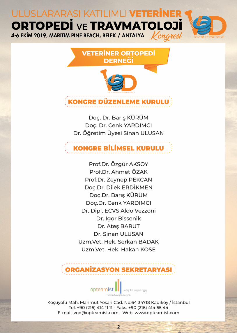
KONGRE DÜZENLEME KURULU
Doç. Dr. Barış KÜRÜMDoç. Dr. Cenk YARDIMCI
Dr. Öğretim Üyesi Sinan ULUSAN
KONGRE BİLİMSEL KURULU
Prof.Dr. Özgür AKSOYProf.Dr. Ahmet ÖZAK
Prof.Dr. Zeynep PEKCANDoç.Dr. Dilek ERDİKMEN
Doç.Dr. Barış KÜRÜMDoç.Dr. Cenk YARDIMCI
Dr. Dipl. ECVS Aldo VezzoniDr. Igor BissenikDr. Ateş BARUT
Dr. Sinan ULUSANUzm.Vet. Hek. Serkan BADAK
Uzm.Vet. Hek. Hakan KÖSE
ORGANİZASYON SEKRETARYASI
Koşuyolu Mah. Mahmut Yesari Cad. No:64 34718 Kadıköy / İstanbulTel: +90 (216) 414 11 11 - Faks: +90 (216) 414 65 44
E-mail: [email protected] - Web: www.opteamist.com
VETERİNER ORTOPEDİDERNEĞİ
VETERİNER ORTOPEDİDERNEĞİ
VETERİNER ORTOPEDİ DERNEĞİ
2
BİLİMSELPROGRAM
4 EKİM 2019 - CUMA14:00 - 14:30 AÇILIŞ SEROMONİSİ
Barış Kürüm, Cenk Yardımcı
14:30 - 15:30 VETERİNER ORTOPEDİDE ANALJEZİ VE ANESTEZİ UYGULAMALARIOturum Başkanı: Cenk Yardımcı
14:30 - 15:00 Veteriner Ortopedide Multimodal AnaljeziZeynep Pekcan
15:00 - 15:30 Ortopedik Cerrahilerde AnesteziZeynep Pekcan
15:30 - 16:00 UYDU SEMPOZYUMUPrevicox ile Köpekleri Özgür BırakınMerve Oyan
16:00 - 16:30 Kahve Molası
16:30 - 18:00 SPİNAL TRAVMALAR VE KOMPLİKASYONLARIOturum Başkanı: Barış Kürüm
16:30 - 17:00 Spinal Travma Hastasına Yaklaşım (Spinal Travma Acilleri, Ne Zaman Ne Yapmalı?)Ateş Barut
17:00 - 17:30 Vertebra Kırıkları ve Sağaltım SeçenekleriAteş Barut
17:30 - 18:00 Postoperatif İskelet-Kas Lezyonlarında Ftr ile Hızlı İyileşmeSara Ece Ulutürk
4
5 EKİM 2019 - CUMARTESİ09:00 - 10:30 PLAK İLE OSTEOSENTEZ UYGULAMALARI
Oturum Başkanı: Ateş Barut
09:00 - 09:30 Neden Plak Tercih Etmeliyim? Avantaj & Dezavantajları Nelerdir?Igor Bissenik
09:30 - 10:00 Minimal Invaziv Plak OsteosenteziBarış Kürüm
10:00-10:30 Açık Kırıkların Yönetimi: Plak Koysam mı? Koymasam mı?Sinan Ulusan
10.30 - 11.00 Kahve Molası
11:00 - 12:30 VEZZONİ TECRÜBESİOturum Başkanı: Ahmet Özak
11:00 - 11:30 Patella Luksasyonlarına Modern YaklaşımAldo Vezzoni
11:30 - 12:00 Yavru Köpeklerde Dirsek Displazisinin Erken Tanısı: Neden & Nasıl?Aldo Vezzoni
12:00 - 12:30 Juvenil Kalça Displazisinin Tedavisinde Çift Pelvik Osteotomi İle Konservatif Yaklaşımın Uzun Dönem Sonuçlarının KarşılaştırılmasıAldo Vezzoni
12:30 - 13:30 Öğle Yemeği
13:30 - 15:00 EKLEM HASTALIKLARIOturum Başkanı: Murat Şaroğlu
13:30 - 14:00 Omuz ve Dirsek Eklemi Tendinopatileri: Semptomlar, Tanı ve Sağaltım.Igor Bissenik
14:00 - 14:30 İnternal Fiksasyon KomplikasyonlarıSinan Ulusan
14:30 - 15:00 Dokunmadan Bilemezsin: Ortopedide Klinik MuayeneBarış Kürüm
15:00 - 15:30 Kahve Molası
5
5 EKİM 2019 - CUMARTESİ15:30 - 17:00 DEJENERATİF EKLEM HASTALIĞI
Oturum Başkanı: Zeynep Pekcan
15:30 - 16:00 Baş bölgesi travmalarında gözden kaçanlarMurat Şaroğlu
16:00 - 16:30 Sıradışı Ortopedik Hastalıklar: HOD, HOP, SPS, SFO, CMO, FHD, POCenk Yardımcı
16:30 - 17:00 Dejeneratif Eklem Hastalığında Predispozisyon, Görütüleme ve TanıIgor Bissenik
17:00 - 17:15 Post-Op İyileşmeyi Hızlandıran ÜrünlerEfe Yörük
17:15 - 18:15 PLAKET TÖRENİ
P A R T Y
DJMARITIM PINE BEACH OTEL
CLUB PINE BAR21:30
5 EKİM CUMARTESİ
6
6 EKİM 2019 - PAZAR09:00 - 11:00 AYIN KARANLIK YÜZÜ: KOMPLİKASYONLAR
Oturum Başkanı: Sinan Ulusan
09:00 - 09:30 Geriatrik Hastalara Gençlik Aşısı: Fizik Tedavi Mucizesi Sara Ece Ulutürk
09:30 - 10:00 Başarılı Bir Operasyon Yaptım ama Kırık Kaynamıyor, Neden?Cenk Yardımcı
10:00 - 10:30 Ön Çapraz Bağ Kopuklarına Güncel YaklaşımlarBarış Kürüm
10:30 - 11:00 Bandaj: Ekstremite Kurtarıcı mı Yoksa Kabus mu?Cenk Yardımcı
11:00 - 11:30 Kahve Molası
11:30 - 12:40 KATILIMCI SUNUMLARIOturum Başkanı: Zeynep Pekcan
11:30 - 11:40 S-01 Köpeklerde Uzun Kemik Kırıklarında Minimal Invaziv Osteosentez Yöntemi İlk SonuçlarıCansu İstim
11:40 - 11:50 S-02 Distal Abtebrachium Ampütasyonu Geçirmiş 12 Köpekte Dış Protez Uygulamaları ve SonuçlarıGürkan Gülanber
11:50 - 12:00 S-03 Sığırlarda Hiperekstensiyon Deformitelerinin Girift Tenorafi Tekniği ile Sağaltımı:Yeni Bir YöntemCelal Şahin Ermutlu
12:00 - 12:10 S-04 Buzağılarda Suprakondüler Femur Kırıklarının Üçlü Pin Tekniği ile Sağaltımının Klinik veRadyografik Olarak Değerlendirilmesi (Yeni Bir Yöntem)Uğur Aydın
12:10 - 12:20 S-05 Köpek Yavrularında Diafizer Femur Kırığının Sağaltımı için Kaz Radiusu KullanımıUğur Yıldız
12:20 - 12:30 S-06 Yüksekten Düşen Dedilerin Retrospektif Değerlendirilmesi (2017-2019): 330 OlguKamil Serdar İnal
12:30 - 12:40 S-07 Distal Abtebrachium Ampütasyonu Geçirmiş 12 Köpekte Dış Protez Uygulamaları ve SonuçlarıBirkan Karslı
7

KONUŞMAÖZETLERİ
VETERİNER ORTOPEDİDE MULTİMODAL ANALJEZİ
Prof. Dr. Zeynep PEKCAN Kırıkkale Üniversitesi Veteriner Fakültesi
Günümüzde hayvan ırklarının çeşitliliği ve buna bağlı eklem sorunları da artmaktadır. Ayrıca iyi bakım ve düzenli veteriner kontrolleri ile hayvanların yaşam süreleri uzamakta ve yaşlanmaya bağlı problemlerin de sayısında artış olmaktadır. Veteriner hekimlerin görevi hayvan sahiplerini bilinçlendirmek ve hastalık durumlarında gerekli müdahaleyi yapmaktır. İleri yaşlarda hayvanların yaşayabileceği belli başlı sorunların arasında osteoartritis ve organ fonksiyon bozuklukları gelir. Osteoartritis olan hayvanlardaki eklem ağrısının azaltılması amacı ile kullanılabilecek ilaçların başında nonsteroidal antiinflamatuvar ilaçlar gelmektedir. Bu amaçla ticari olarak üretilen çok farklı etken maddeye sahip birçok ilaçlar bulunmaktadır. Ancak bunların uzun süreli ve yüksek dozlarda kullanılması hayvanlarda gastritis, midede ülser, hepatitis ve böbrek sorunlarına yol açabilmektedir. Bu gibi durumlarda yapılması gereken farklı analjezik ilaçları ağrı tedavi protokollerinin içerisine koymak olmalıdır. Böylece yüksek doz ilaç kullanımından kaçınılarak yan etkiler en aza indirilmiş olur. Küçük hayvan hekimliğinde analjezik ilaç olarak osteoartritislerde kullanılan ilaçların içerisinde gabapentin, amantadin, tramadol ve amitriptilin yer almaktadır. Gabapentin epilepsi hastalarında krizlerin önlenmesi amacı ile üretilen bir preparattır, ancak son yıllarda insan ve hayvan hekimliğinde kronik ağrılarda, özellikle de nöropatik ağrılarda, ağrı kesilmesi amacı ile kullanılmaktadır. Amantadin ise, aslında Parkinson hastalarının diskinezilerini önlemek amacı ile üretilen bir ilaç olmasına rağmen son yıllarda kronik ağrıda da etkinliği kanıtlanmıştır. Tramadol ve amitriptilin ise, osteoartrit hastalarında ağrı tedavisi amacı ile kullanılabilecek serotonin geri alınımını engelleyen ilaçların başında gelmektedir.
9
MULTIMODAL ANALGESIA IN VETERINARY ORTOPEDICS
Prof. Dr. Zeynep PEKCAN Kırıkkale University Faculty of Veterinary Medicine
Nowadays, the variety of animal breeds and related joint problems are increasing. The life expectancy of the animals is prolonged with good care and regular veterinary checks, and the number of aging related problems increases. The task of veterinarians is to raise awareness of animal owners and to treat the animals. Osteoarthritis and organ dysfunction are the main problems that animals may experience in advanced ages. Nonsteroidal antiinflammatory drugs are the main drugs that can be used to treat the joint pain in animals with osteoarthritis. For this purpose there are many commercially produced drugs with very different active ingredients. However, their long‐term and high doses may cause gastritis, ulcers, hepatitis and kidney problems in animals. Different analgesic drugs should be included in the analgesia treatment protocols in older animals. Thus, side effects can be minimized by avoiding the use of high‐dose drugs. Gabapentin, amantadine, tramadol and amitriptyline can be used for osteoartritic pain treatment in companian animals. Although gabapentin is produced for the treatment of seizures of epileptic patients, its usage for analgesic treatment is popular in recent years. Although amantadine was originally produced to prevent dyskinesias in Parkinson's patients, it has been proven to be effective in chronic pain in recent years. Both tramadol and amitriptiline are serotonin uptake inhibitors and can be used for the treatment of osteoartritic pain.
10
ORTOPEDİK CERRAHİLERDE ANESTEZİ
Prof. Dr. Zeynep PEKCAN Kırıkkale Üniversitesi Veteriner Fakültesi
Ortopedi operasyonları pekçok operasyondan çok daha ağrılı operasyonlardır; bu nedenle mutlaka ağrı kesicilerin kombinasyonu yapılmalıdır. Günümüzde multimodal analjezi kavramı gelişmeye başladığından beri operasyonlardaki ölüm oranları azalmış ve postoperastif süreçte hayvanlar daha rahat uyanmaya ve ayaklanmaya başlanmıştır. Ağrı yalnızca ağrı kesicilerle engellenebilir; bundan dolayı operasyon sırasında kullanılacak anestezik ve analjezik kombinasyonlarının doğru ve bilinçli yapılması önemlidir. Operasyon başlamadan önce analjezik ve/veya lokal anestezik ilaçların uygulanması ile operasyon sırasında ve sonrasında uygulanacak olan total analjezik ilac miktarı azaltılabilir. Hiperaljeziyi önleme açısından analjezi uygulamasının en az 12‐24 saat devam ettirilmesi gereklidir. Anestezi prosedürüne analjezik ilaç eklemesi ile totalde kullanılacak anestezik ilaç miktarı düşer ve anestezinin yüksek doz kullanımına bağlı gelişen yan etkiler azalır.
Opioidler başta olmak üzere, lokal anestezikler, dissosiyatif anestezikler ve alfa‐2 agonistler gibi ağrı kesici özelliği olan ilaçlar anestezi komplikasyonlarının azaltılması, hastanın konforu ve kronik ağrı yollarının engellenmesi amacı ile kombine edilebilir. Ön ve arka bacakların anestezisi amacı ile regionel anesteziden faydalanılabilir. Bu amaçla ön bacakların anestezisinde en çok brakiyel pleksus, arka bacakların anestezisinde ise epidural anestezi uygulamaları tercih edilir. Ön bacakların sinir blokajı ile sadece uzun kemik kırıklarının osteosentezi değil aynı zamanda omuz cerrahisi, dirsek cerrahisi, ön bacak amputasyonu da yapılabilir. Bu amaçla %2’lik lidokain ve %0.5’lik bupivakain kombinasyonu kullanılır. Köpeklerde lidokainin total dozunun 8 mg/kg’ı, bupivakainin de 2 mg/kg’ı geçmemesine, kedilerde ise toplam dozun köpeklerin yarısı kadar olmasına dikkat edilmelidir.
Arka bacakların sinir blokajı için çoğunlukla epidural uygulama tercih edilir. Bu amaçla lokal anestezikler, opioidler ya da bunların kombinasyonu uygulanabilir. 0.2 ml/kg hacminde verilen ilaçlarla epidural uygulamalar yapılır. Bu yolla verilen lidokainin etkisi 10 dk içinde başlar etki süresi yaklaşık 90‐120 dk arasında sürer, bupivakainin etkisi ise biraz daha geç başlar (15‐20 dk) ve etki süresi daha uzun sürer (2‐4 saat). Epidural analjezi amacı ile opioidler de uygulanabilir. Bu amaçla en çok tercih edilen etki süresi uzun olduğu için morfindir. 0.1 mg/kg uygulanan morfinin etkisi 30‐60 dk içinde etkisi başlar ve 12‐24 kadar sürer.
11
ANESTHESIA IN ORTHOPAEDİC SURGERIES
Prof. Dr. Zeynep PEKCAN Kırıkkale University Faculty of Veterinary Medicine
Orthopedic operations are much more painful than many operations, therefore, combination of analgesic drugs should be performed. Since the concept of multimodal analgesia has started to develop, the mortality rates have decreased and animals have started to wake up and move more easily in the postoperative period. Pain can only be prevented by analgesics; therefore, it is important that the anesthetic and analgesic combinations should be used consciously during the operations. The administration of analgesics or local anesthetics before the start of surgery reduces the total usage of analgesic drugs during surgery and postoperatively. It is important to continue the analgesic theraphy at least the first 12 to 24 hours to prevent hyperalgesia. The addition of analgesic agents to the anesthesia procedure alleviates the total usage of anesthetic drugs and reduces the risk of high doses of anesthesia. Pain medications such as opioids, local anesthetics, dissociative anesthetics and alpha‐2 agonists can be combined to reduce the anesthesia complications, provide patient comfort and for preventing chronic pain pathways. Regional anesthesia can be used for anesthesia of the thoracic and hind legs. For this purpose, brachial plexus and epidural anesthesia are preferred. With the brachial plexus blockage, not only the osteosynthesis of long bone fractures, but also shoulder surgery, elbow surgery, and front leg amputation can be done. For this purpose, a combination of 2% lidocaine and 0.5% bupivacaine is used. In dogs, the total dose of lidocaine and bupivacaine should not exceed 8 mg/kg and 2 mg/kg, respectively. In cats the total dose should not exceed the half of the dogs. Epidural administration is preferred for nerve blockage of hind legs. For this purpose, local anesthetics, opioids or a combination thereof can be administered. Epidural applications are made with the drugs given in a volume of 0.2 ml / kg. The effect of epidural lidocaine starts within 10 minutes, and lasts for approximately 90‐120 minutes, whereas the effect of bupivacaine begins a little later (15‐20 minutes) and lasts longer (2‐4 hours). Opioids may also be administered for epidural analgesia. For this purpose, morphine is the most preferred drug because of its long duration of action. The effect of 0.1 mg/kg morphine starts in 30‐60 minutes and lasts up to 12‐24.
12
AKUT SPİNAL KORD HASARLARI; NE ZAMAN NE YAPMALI? Dr. Ateş BARUT Petcode Hayvan Hastanesi, ANKARA
Kedi ve köpeklerde akut spinal kord hasarı doğal ya da travmatik sebeplerle gerçekleşebilir. Bu olayın gerçekleşmesi sonrası spinal kordda primer ve sekonder hasar mekanizmaları gelişmeye başlar. Primer hasar; direk travmatik kontüzyon, laserasyon, hücre hasarı, kanama, hematom, kompresif iskemi olarak sıralanabilir. Sekonder hasarlar ise intraselüler iyon konsantrasyon değişimleri, excitotoxicity (aşırı nörotransmiter salınımına bağlı sinir hücresi ölümü), serbest radikal üretimi, yangıdır.
Fibrokartilaginöz emboli ve travmatik vertebral kırık ve luksasyonlarla sıklıkla karşılaşılabilse bile köpeklerde spinal kord hasarlarının en sık rastlanan sebebi intervertebral disk hastalığıdır. Akut intervertebral disk herniasyonları ya da travmatik vertebra kırık ve luksasyonları sonucu gelişen spinal kord hasarları hastayı hayat boyu sürecek parapleji ve idrar retensiyonu tablosuna mahkum eder. Parapleji ve idrar retensiyonu hem hastanın hem de ailesinin yaşam kalitesini düşürür. İşte bu sebeple tüm akut spinal kord hasarlarında doğru yaklaşım, doğru medikal tedavi ve doğru cerrahi teknikler kullanılarak yapılacak dekompresyon ve stabilizasyon büyük bir sorumluluktur ve hayati önem taşır.
Hastanın medikal ve fiziksel olarak hareketsizliğinin sağlanması hekimin ilk sorumluluğudur. Spinal kord hasarlarında etkin medikal tedaviden bahsetmek doğru olmayacaktır ve cerrahi dekompresyon travma sonrası oluşan iskeminin tek çözümüdür. Medikal tedavinin en önemli aşaması hipotansiyon ve hipoksinin önlenmesine dayanır. Böylece spinal kord perfüzyonu tekrar sağlanır. Nimodipine, riluzole, gacylidine gibi ilaçlarla ilgili insan çalışmaları halan sürmektedir.
Yapılan çalışmalarla tüm spinal kord travmalarında geleneksel olarak kullanılan kortikosteroidlerin tahmin edilen faydaları sağlamaktan çok uzakta olduğu ortaya konmuştur. Özellikle metil prednizolon sodyum süksinat serbest oksijen radikallerini yok ettiği varsayımı ile spinal kord travmalarında uzun yıllar standart medikal tedavi olarak uygulanmıştır. Oysa yapılan son çalışmalar kortikosteroidlerin hastalara yarardan çok daha fazla zarar verdiğini göstermiştir ve geleneksel yüksek doz mpss ve dekzametazon tedavisi bugun tamamen terk edilmiştir. Kortikosteroidler cerrahi dekompresyona kadar kısa dönem ve sadece antiinflamatorik dozlarda uygulanabilir. Siklosporin ve takrolimus kullanımı ile ümit verici tedaviler rapor edilmiştir. Polietilen glikol (PEG) hasar gören membranları onarır, iyon sızıntısını ve aksonal ayrılmayı önler. PEG(%30,3500dalton) ‘nun 2ml/kg (45dk aralıkla 2 defa) şekilde uygulanması olumlu etkiler yaratmıştır. Nimodipine, riluzole, gacylidine gibi ilaçlarla ilgili insan çalışmaları halan sürmektedir. Spinal kord travmalarında tek prognostik faktör derin ağrı duyumudur ve derin ağrı duyumu kaybı kesinlikle yukarı motor nöron semptomları ile karıştırılmamalıdır.
Torakolumbal disk hastalığı köpeklerin en sık rastlanan nörolojik hastalığı dolayısıyla çok önemli bir spinal kord hasarı sebebidir. Tip 1 disk hastalığı ve buna bağlı herniasyonlar özellikle kondrodistrofik ırklarda akut spinal kord hasarı ve paraplejiye sebep olurlar. Disk dejenerasyonu, olgu gençken gelişir (ortalama 2-9 ay) ve klinik bulgular genellikle 2-6 yaşlarda ortaya çıkar. Disk dejenerasyonu temel olarak nukleus pulposusun kondroid metaplazisi ile anulus fibrozusun dejenerasyonu ve zayıflaması ile şekillenir. Dejenere diskin kalsifikasyonu ve radyografik olarak görünür hale gelmesi 6‐18 aylık
13

dönemde şekillenir. Zayıflayan anulusun dejenere nukleusu kontrol edememesi ile columna vertebralisin normal hareketleri bile akut disk prolapsusu için yeterli kuvvet oluşturabilir. Semptomlar akut ve agresiftir. Parezi ve parapleji klinik tablosu çok hızlı şekillenebilir. Bir başka akut spinal kord hasarı yaratan disk herniasyonu ise tip 3 disk hastalığından kaynaklanır. Tip 3 disk hastalığında spinal korda çok yüksek bir hızla çarpan küçük ve sıvı karakterde disk materyali bulunur. Nörolojik hasar darbe momentine bağlı olarak hızlı ve ciddidir ve MR ya da tomografide kompresif materyal görülmez ve bu yüzden cerrahi tedavi endikasyonu yoktur. İster disk hastalığına bağlı olsun isterse de omurga travmalarına hasta değerlendirilmesinde nörolojik muayene (spinal refleks muayenesi, parezi, parapleji, proprioseptif pozisyonladırma tesbiti ve derin ağrı duyumu varlığı) büyük önem taşır. Parezi motor fonksiyonksiyonlarda zayıflama demektir ve istemli adım atma bozulsa da hala korunur. Paralizi ya da parapleji ise motor fonksiyonların nörolojik ya da muskuler sebeplere bağlı olarak kaybolması olarak tanımlanır. Proprioseptif pozisyonlandırma bir refleks değildir ve beynin ekstremitenin nerede olduğunu bilmesini tanımlar. Derin ağrı duyumu kaybı tüm spinal kord hasarlarında tek prognostik faktördür ve kesinlikle yukarı motor nöron semptomları ile karıştırılmamalıdır.
14

ACUTE SPINAL TRAUMA; WHAT TO DO, WHEN TO DO? Dr. Ateş BARUT Petcode Animal Hospital, ANKARA
Acute spinal cord injuries can happen with natural or traumatic causes and although vascular events like fibrocartilaginous embolism can cause this injury intervertebral disc herniations or traumatic vertebral fractures and luxations are the most common two causes of acute spinal cord injury and lead the patient to life long paraplegia, and urinary retention. This paraplegia and urine retention will decrease the life quality of patient and its owners. That is why handling a patient with acute spinal cord injury in a good way, and surgical decompression and stabilization with the proper surgical techniques is essential and a big responsibility.
After the event causing spinal cord injury a primary and secondary damage will start. We can list the primary spinal damages as direct traumatic contusion, laceration, cell damage, hemorrhage, hematoma and compressif ischemia. Secondary injuries are intracellular ion concentration changes, excitotoksicity, free oxygen radical produce, and inflamation.
Medical or physical restrain of the patient must be the first goal of the veterinary surgeon. There is no sufficient medical treatment after spinal injury and surgical decompression and stabilization is the only succesfull treatment option. The most important step of the medical treatment is preventation of hypotension and hypoxia. Spinal cord blood perfusion is always essential. There are still continuing human studies on nipodipine, riluzone, gacylidine. Benefits of traditional corticosteroid use on almost all spinal traumas are far from expection. Several studies showed that corticosteroids has more negative effects than their benefits and high dose of MPSS and dexamethasone are completely out of suggection for the moment. Corticosteroids must be use just in antiinflamatoric dosages just for short periods till the surgery. Hopeful results with cyclosporin and tacrolimus use are reported. Polyethilen glycol (PEG) repair damaged membranes, stop ion leak, and axonal detachments. PEG (%30, 3500 dalton) with a dosage of 2 ml/kg (twice in 45 minutes) gave positive results. The only prognostic factor for spinal cord traumas is deep pain perception and clinician has to differensiate deep pain perception and upper motor neuron symptoms.
Thoracolumbal disc disease is the most common neurological problem of the dogs and that means it is a very important cause of spinal injury. Type 1 disc degeneration and related disc herniations cause acute spinal cord injury and paraplegia especially in chondrtodystrophic breeds. Usually chondrodystrophic breeds (hypochondroplastic). Degeneration begins in early ages like 2‐9 months and clinical signs occur between 2‐6 years of age. Spinal cord damage is not just due to the amount of herniated disc material but also impact momentum during herniation moment.
Disc degeneration basicly represents chondroid metaplazia of nucleus pulposus and degeneration and weakening of annulus fibrosis. During 6‐18months of age calcification and some degeneration can be visualised radiographicaly. Degenerated and weak annulus fibrosis can not control degenerated nucleus and normal movements of columna vertebralis can produce enough force for herniation. The other acute spinal cord injury cause is type III disc disease. In type III disc disease, just small amount of herniated disc material hit to spinal cord with a high pressure. Neurologic damage is so quick and serious because of the momentum of the hit. No obvious compressive lesion in the magnetic resonans images or computered tomography. No surgical treatment.
15
Neurological examination findings are so important for the evaluation of spinal trauma patients either because of disc diasese or a spine trauma. Spinal reflexes, paresis, paraplegia, proprioceptive positioning and presence of deep pain perception are the most important critaria for this evaluation. Paresis describes a decrease in motor functions but in paralisis or paraplegia there is complete loss of moptor functions. Differentiation of deep pain perception and upper motor neuron symptoms is essential for the prognosis. Proprioceptive positioning is not a reflex. In normal proprioceptive positioning brain knows where the extremities are. Deep pain perception is the only prognostic factor for all spinal cord traumas.
16
VERTEBRA KIRIK VE LUKSASYONLARINDA CERRAHİ SAĞALTIM Dr. Ateş BARUT Petcode Hayvan Hastanesi, ANKARA
Spinal kord hasarları sadece travmatik vertebra kırık ve luksasyonları ile sınırlı değildir. Torakolumbal disk hastalığı köpeklerde görülen en önemli nörolojik hastalıktır ve dolayısıyla köpeklerin en önemli felç sebebidir. Diğer vertebra kırık ve luksasyon sebepleri ise yüksekten düşmeler, trafik kazaları, sıkışmalar, ezilmeler, vertebral malformasyonlar, insan travmaları ve köpek saldırıları olarak sıralanabilir.
Dekompresif cerrahinin amacı kesinlikle sadece kanalis vertebralis dorsal laminasını kaldırıp spinal kordu rahatlatmak değildir ve kompresyon yaratan disk materyalinin tamamen uzaklaştırılması şarttır. Bu amaçla yapılabilecek en başarılı cerrahi teknik hemilaminektomidir çünkü ancak hemilaminektomi ile vertebral kanalın tabanına ve dolayısıyla kompresif disk materyaline ulaşım mümkündür. Ayrıca ilgili diskin lateral fenestrasyonu da gereklidir aksi takdirde disk içerisinde kalan nukleus pulpozusun fıtıklaşması kaçınılmaz olacaktır. Kanalis vertebralis içerisindeki kompresif disk materyalinin uzaklaştırılması için en ideal teknik hemilaminektomi olarak kabul edilir. Pedükülektomi ve mikrohemilaminektomi de yaratılan minimum kas travması sebebi ile gittikçe popülarite kazanmaktadır. Bu tekniğin vertebral stabiliteye minimum etkisi vardır skolyoz yaratabilir ancak klinik sorun yaratmaz. Lateralizayon yapılamadığında çift taraflı hemilaminektomi dahi dorsal laminektomiye tercih edilmelidir. Hemilaminektomi sorunları kontralateral tarafa ulaşımın sınırlı olması ve bilateral yapıldığında instabiliteye yol açabilmesi tekniğin dezavantajıdır ayrıca disk sert ve yapışmışsa ventral maniplasyon zordur. Puglarda hemilaminekteomi T10‐11‐12’de instabiliteye yol açabilir.
Servikal vertebra kırık ve luksasyonları kedilerde çok nadir görünürken köpeklerin vertebral kırık ve luksasyonlarının %10‐20’sini oluşturur ve bu kırıkların neredeyse yarısı aksisi etkiler. Tüm servikal vertebra kırık ve luksasyonları kesinlikle instabil kabul edilmelidir. Tetraparezi ya da tetraplejiye neden olur, şiddetli ağrı yapar ve solunum merkezlerini etkiler. Nörolojik tablo çoğu zaman hızla kötüleşir. Bu travmalarda SOP plaklar, kilitli plaklar, pin ve PMMA kombinasyonları gibi birçok farklı stabilizasyon yöntemi kullanılabilir. Stabilizayon her zaman korpus vertebrayı kullanmalı ve ventralden yapılmalıdır. Eğer ayrıca dekompresyon gerekiyorsa ventral stabilizasyon sonrası dorsal dekompresyon yapılabilir. Her vertebraya en az iki pin ya da vida uygulanmalıdır ve güvenli implantasyon koridoru median hattan 30‐40 derece açılı düzleme denk gelir. Iatrojenik hata riski yüksektir. Vertebral kanala ya da intervertebral disk aralığına penetrasyon riskini azaltmak için monokortikal implant uygulamaları ile azaltılır. Bu teknikle hem monokortikal pin/vida ve pmma kombinasyonu hem de kilitli plaklar ve monokortikal vidalar uygulanabilir. Standart ortopedik plaklar bikortikal vidalamaya ve güçlü plak kemik yüzey kontağına dayanır ki bu vertebral kanal için uygun değildir. Kilitli plaklarda plağın bükülmesine gerek yoktur ve vidalar monokortikal uygulanabilir. Vertebral kanal penetrasyonu kontrolü için BT çekilmelidir
Lumbal kırık ve lukasyonlar tüm vertebra kırıkların %25’ini oluşturur. Kaudal lumbal vertebra kırıkları ise hem ala osis iliumların varlığı hem de arka bacak innervasyonundan sorumlu sinir köklerinin korunması gerektiğinden oldukça zordur. Lumbosakral travmalarda kauda equina sinir lifleri kompresyonu ve hasarı oluşur. L6‐L7‐S1 bölgesinde göreceli olarak geniş bir kanal vardır. Spinal kord değil kauda equina yani perifer sinir hasarı söz konusudur o yüzden kesinlikle ataksi olmaz.
17
Hastalarda eş zamanlı ortopedik hasarlar da görülebilir. Pin/vida penetrasyon noktaları L7’nin kranial artikuler proc’in kaudalinde dik şekilde, kranial artikuler proc’in kauda lateralinden 5‐15derece açıyla olacak şekilde kabul edilen düzlemlerle örtüşür. Kranial ve orta torasik vertebralar oldukça stabildir ve bu bölge kırıkları nadir görülür. Bu bölge spinal kord kompresyonları çoğu zaman kongenital vertebral malformasyonların dislokasyonuna bağlı gelişir. Bu bölgede gerçekleştirilecek stabilizasyon ve fiksasyonlar vertebraların değişken şekilleri ve doğru implantasyon koridorlarının bulunmasındaki zorluk sebebi ile zordur. Torakolumbal travmalar en sık rastlanan hasar bölgesidir hem kedi hem de köpeklerde vertebral kırık ve luksasyonların %50’sini oluşturur. Bu kırık ve luksasyonlar hemen her zaman stabilizasyon gerektirir. Üç kompartman kuralı yani vertebranın 3 kompartmanında 2’si etkilendiğinde stabilizasyonun şart olduğu düşünülür. Spinoz proses stabilizasyonu sadece küçük köpekler ve kedilerde, 2 kompartman korunuyorsa ve spinoz prosesler sağlamsa uygulanır (20kg ve aşağısı). Pin/vida ve PMMA tüm dünyada en sık kullanılan tekniktir. Başarılı bir stabilizasyon sağlar. Pin penetrasyon noktalarının bilinmesi çok önemlidir. Her zaman yivli şanz pinleri kullanılmalıdır.
Giriş açısı torasik vertebral için vertikal düzlemden 25‐35 derece (kostanın çıkıntısı üzerinden), lumbal vertebralar için vertikal düzlemden 60 derece (procesus transversus ve proc accesorius arasından) olarak kabul edilir. Kilitli plaklar ve monokortikal vidalama önemli bir avantajdır. Plağın bükülmesi şart değildir. Plağın düzleminden emin olunmadan hiçbir vida sıkılmamalıdır.
18
SURGICAL TREATMENT OF VERTEBRAL FRACTURES AND LUXATIONS
Dr. Ateş BARUT Petcode Animal Hospital, ANKARA Spinal cord injuries are not limited to vertebral fractures and luxations. Thoraco‐lumbal disc disease is the most common neurological disease of dogs and so it is one of the most common cause of paraplegia. The other causes of vertebral fractures and luxations can be listed as falling from highes, traffic accidents, contusions, vertebral malformations, spine tractions, and dog bites. The aim of the decompressive surgery is not only removing the dorsal lamina of canalis vertebralis and complete removal of compressive disc material is essential for success. The best surgical procedure for this purpose is hemilaminectomy because it give direct access to the bottom of canalis vertebralis and all herniated disc material. Disc fenestration is also necessary to prevent recurrence. Peduculectomy and microhemilaminectomy is becoming more popular with the minimum muscle disection and trauma. Hemilaminectomy has a minimum effect on vertebral stability it can cause some degree of scoliosis with no clinical disturbance. In case of lateralization diffuculties bilateral hemilaminectomy still has advantages on dorsal laminectomy even it can cause instability. Altough it give access to the bottom of spinal channel, aproach to contralateral side is limited and bilateral surgery cause instability. In pugs T10‐11‐12 hemilaminectomy can cause instability too. Cervical VFLs are the least commonly encountered anatomic category in dogs and cats, comprising about 10% to 20% of these VFLs in dogs (very uncommon in cats). In dogs, about half of these injuries involve the C2 vertebra. All cervical vertebral fractures and luxations are instable. They cause tetraparesis and tetraplegia, severe pain, and respiratoric compramise. Usually neurological status of the patient get worse so quickly. In these fractures and luxations SOP plates, locking plates, pins/PMMA combinations can be use safely. Two screws or pins has to be implanted to one vertebra and safe implantation window is 30‐40 degree from median plane. If decompression is required, surgeon has to be stabilize ventrally first, then decompress dorsally. The risk of iatrogenic mistakes are high. Monocortical pin/screw placement techniques reduce the risk of vertebral canaşl or spinal cord penetration. With this technique both monocortical pins/screws and PMMA combinations and locking plates and monocortical screw implantations are possible. Standart orhtopedic plates work with bicortical screw placement and strong plate bone surface contact that is why they are not suitable for vertebral fractures. There is no need of plate bending for locking plates and all screws can be use monocorticaly. Vertebral canal penetrations can be check by CT. The cranial to mid‐thoracic vertebral column is inherently stable, so VFLs in this area are uncommon. Stabilization of this region is usually associated with congenital problems. The lumbar region is affected in about 25% to 30% of VFL cases. There is more leeway in the angle of insertion for pins and screws in this area (especially cranial to mid‐lumbar) of the vertebral column than in the thoracic spine. The methods of fixation used for the thoracolumbar junction region are all applicable in this area, but plate application is a bit less technically demanding (no rib heads in the way). In the caudal lumbar region, it can be difficult to apply plates, and the angle of pin/screw insertion is more limited, due to the presence of the ilium laterally. Also, the lumbar spinal nerve and nerve roots need to be protected more carefully in the caudal lumbar region (innervation of pelvic limb musculature). Cauda equine nerve roots compression happens in lumbosacral traumas. Spinal canal is a little wider between L6‐L7‐S1. Lumbosacralş tarumas cause cauda equine nerve damage that is why patients
19
will not have ataxia. So many patients can also have concurrent orthopedic problems. Pin/screw placement must be done just caudal and perpendicular to L7 cranial articular process.
The thoracolumbar junction region is a very common site for VFLs in dogs and cats, comprising about 50% of these injuries in both species. Thoracolumbal fractures are almost always require stabilization. If there is an effect on two of three vertebral compartments there is an obvious need for stabilization. Stabilization using spinal processes are just effective in small breeds of dogs and cats , if 2 of 3 vertebral compartments are safe, and all the spinal prosesses are healthy. Pİn/screw and PMMA use is so popular all over the world. Pin placemet points are again so important for safety. Not normal only positive threatened pins must be used. Penetration point is 25‐35 degree from vertical plane for thorasic vertebrae, and 60 degree from vertical plane for lumbal vertebra.
20

POSTOPERATİF İSKELET-KAS LEZYONLARINDA FTR İLE HIZLI İYİLEŞME Sara Ece ULUTÜRK FizyoVET Veteriner Kliniği, İSTANBUL
Pet fizik tedavi ve rehabilitasyon; vücuda dışarıdan uygulanan sıcak ve soğuk uygulamalar, elektrik akımları, masaj ve egzersiz ile ağrıyı kesmeye yönelik uygulamaları içeren ve kas iskelet sistemi hastalıklarında ya da yararlanmalarında uygulanan ilaç dışı bir tedavi şeklidir.
Hayvanlarda fizik tedavi ve rehabilitasyon çok uzun yıllardan beri uygulanmakla beraber bu alanda 90’lı yıllardan bugüne ciddi ölçüde ilerlemeler kaydedilmiştir. Amerika’da 110 fakültede fizyoterapi ve rehabilitasyon ile ilgili bölümler bulunmaktadır. Köpeklerde ve atlarda, çok spesifik ve sık görülen hastalıkların sağaltımında ciddi sayılabilecek faydaları belirlenmiştir.
Fizik tedavinin amacı nörolojik ve ortopedik operasyonlar sonrasında hastaların eklem açılarını normal değer aralıklarına ulaştırmak ve post‐operatif süreçte ağrı yönetimini yapıp, kas gruplarında atrofi oluşumu, dekubitis oluşumlarının engellenmesi ile hastanın normal mobilitesini geri kazandırmaktır.
Hastalar ilk geldiklerinde, kaygan olmayan bir zemini olan, ilaç kokuları olmayan ve kendilerini güvende hissedecekleri bir muayene odasına alınırlar. Dikkatli bir şekilde yapılan ortopedik ve nörolojik muayene sonucunda; topallama skoru, ağrı değerleri, kas yoğunlukları ve eklem açıları, basış analiz sistemleri ile goniometrik yöntemler kullanılarak belli ölçümler yapılır ve kayıt altına alınır. Hastalarımıza 12 basamaklı tedavi protokolümüzden uygun olanlar belirlendikten sonra belirli periyotlarda hastalar kliniğe çağrılarak, fizik tedavi modüllerinden ultrason, tens, lazer terapi, hidroterapi (havuz ,suda yürüme bandı), ağrı bantlama sistemleri, masaj, akupunktur, ozon tedavi, pasif egzersizler, kök hücre tedavisi, PRP uygulamaları, magnetik alan tedavisi, sıcak uygulamalar, soğuk uygulamalar, manual terapi (spinal‐ortopedik), aktif egzersizler ve ev egzersiz programları hastalarımıza uygulanır.
Destekleyici ürünler ile çeşitli egzersizler kombine edilerek, post operatif süreçte hızlı iyileşme sağlanır. Hidroterapi ile suyun kaldırma kuvvetinden, hidrostatik basıncından ve viskozitesinden yararlanarak, özellikle köpeklerde kasların önemli ölçüde çalışması ve hareket kabiliyetlerinin geri kazanımı ile eklem hareket sınırlarında artış sağlanmaktadır. (sunumumuz videolarla desteklenmektedir)
21
WHY SHOULD I PREFER BONE PLATE? WHAT ARE THE ADVANTAGES AND DISADVANTAGES?
Igor BISSENIK, DVM, PhD Varşova Tıp Üniversitesi, Polonya
In 1958 a group of Swiss general and orthopaedic surgeons established the AO (Arbeitsgemeinschaft für Osteosynthesefragen) or the Association of the Study of Internal Fixation (ASIF) to strive to transform the contemporary treatment of fractures in Switzerland. This association was revolutionary in development of instruments and implants for operative fracture treatment. The first instructional course for teaching the use of these instruments and implants occurred in Davos, Switzerland, in the newly founded Laboratory of Experimental Surgery in 1960.
Plate osteosynthesis is one treatment option for the stabilization of long bones. It is widely accepted to achieve bone healing with a dynamic and biological fixation where the perfusion of the bone is left intact and micromotion at the fracture gap is allowed.
Open anatomic reconstruction requires various degrees of soft tissue dissection to allow manipulation, reduction, and rigid stabiͲlization of the fracture segments. Iatrogenic damage to the soft tissues, periosteum, and fracture hematoma inevitably occurs and potentially impedes healing. AlthoughThe “open but do not touch” technique is the simplest form of biological osteosynthesis. Although this technique involves an open approach for direct observation, the fracture site is not disturbed and alignment is restored via manipulation of the major bone fragments at positions away from the fracture. Distraction and alignment of the segments are facilitated by the use of bone‐holding forceps or intramedullary implants, such as an intramedullary rod or interlocking nail. The “open but do not touch” technique, however, may result in damage to the vasculature and other soft tissues.
Some of the principles of biological osteosynthesis include (1) indirect fracture reduction using limited surgical approaches with minimal, if any, disturbance of the fracture hematoma; (2) fracture stabilization using bridging implants rather than anatomic reconstruction and rigid fixation; (3) limited reliance on secondary implants, such as cerclage wires or interfragmentary screws; and (4) limited, if any, use of bone grafts.
It is recommended that the plate span width be higher than 2–3 for application in comminuted fractures and more than 8–10 for simple fractures and that the screw density be less than 0.5–0.4 (Gautier and Som‐ mer, 2003). 3 screws per segment are better than 2 but and this amount is optimal more screw doesn’t give any benefit
The concept of the locking pate is much closer to nailing or external fixators than to conventional plates. Despite the appearances, a locking plate is therefore not a plate in the classical sense of the term, but rather an internal fixator.
Stability is provided by the locking mechanism between the screw and the plate. The plate does not need to be in intimate contact with the underlying bone, making exact plate contouring less crucial. Reduced contact be‐ tween the plate and the bone may also preserve the periosteal blood supply, thereby reducing the ex‐ tent of bone resorption under the plate its possible to use mono cortical screw. Conventional plate osteosynthesis seeks to obtain maximum stability with a rigid plate and if possible compression of the fracture fragments. Locking plate fixation, on the other hand, seeks to maintain a certain elasticity to stimulate bone healing. Locking plates are generally less rigid than conventional plates.
22
MİNİMAL INVAZİV PLAK OSTEOSENTEZİ
Barış KÜRÜM Kırıkkale Üniversitesi Veteriner Fakültesi Cerrahi Anabilim Dalı, Türkiye
Ortopedik cerrahide hastanın mobilizasyon yeteneğini, mümkün olan en kısa sürede, kırık oluşmadan önceki seviyede kazanması amaçlanır. Bu amaçla kırık iyileşmesinin süresini kısaltmak amacıyla biyolojik osteosentez yöntemlerinin kullanılması önerilir. Biyolojik osteosentez uygulamalarında; iatrojenik yumuşak doku hasarını en aza indirmek, indirekt redüksiyon tekniklerini kullanmak, yeterli stabilitede fiksasyon oluşturarak ekstremite fonksiyonunun erken geri dönüşünü sağlamak temel prensiplerdir.
Minimal invaziv osteosentez tekniklerinde kırığın tam anatomik redüksiyonu asıl hedef değildir. Bunun yerine, büyük kırık segmentlerinin travmatik cerrahi müdahaleye maruz kalmaksızın indirekt redüksiyon teknikleri kullanılarak fonksiyonel pozisyonda eksen düzgünlüğü sağlanır.
MİPO’da kırık uçları indirekt olarak redükte edilir ve kırık hattı açılmadan köprü görevi yapan uzun bir plak ile kırık hattı köprülenecek şekilde, periost üzerinden perkutan plak uygulaması yapılır. Ekstremitenin rotasyonu düzeltilir, aksiyel dizilimi ve normal uzunluğu sağlanır. Bu yöntemde mikroelastik tespit sağlanır ve bu sayede elde edilen rölatif stabilite, kırık hattındaki mikro hareketlere izin verir ve bu sayede daha fazla kallus dokusu oluşumu ile stabilite de gelişir.
MİPO’nun başarılı olabilmesi için olgu seçimi oldukça önemlidir, diğer bir ifade ile tüm kırıklar perkütanöz plak stabilizasyonuna uygun değildir. MİPO, çoğunlukla, tam anatomik redüksiyona uygun olmayan parçalı diyafiz ya da metafiz kırıklarında uygun olmasının yanında, bir takım basit transversal kırıklarda da kullanılabilir.
Mipo uygulamalarında tip I eksternal fiksatör gibi görev yapan kilitli plak ile vida setlerinin kullanıması tercih edilir. Bu implant sistemi
İnternal bir eksternal fiksatör gibi çalışır. Şekillendirilmeye ihtiyaç duymaz. Vidalar delikte kilitli olduğundan kemiği plağa doğru çekmez. Periost üzerine oturmaz. Vidalar «self drilling» özelliği taşıdığından yerleştirilmeleri kolaydır. Vidalar unikortikal bile yerleştirilseler gerekli tutunmayı sağlar. Vidalar kilitli olduğundan «implant failure» şansı yoktur.
23

MINIMALLY INVASIVE PLATE OSTEOSYNTHESIS Barış KÜRÜM Kırıkkale University Faculty of Veterinary Medicine Department of Surgery In orthopedic surgery, it is aimed to gain mobilization of the patient as soon as possible before the fracture occurs. For this purpose, biological osteosynthesis methods are recommended to shorten fracture healing time. In biological osteosynthesis methods; To minimize iatrogenic soft tissue damage, to use indirect reduction techniques, to achieve early return of limb function by fixation with sufficient stability are the basic principles.
Perfect anatomic reduction of the fracture is not the main target in minimally invasive osteosynthesis techniques. Instead, axial alignment of the functional position is achieved by using indirect reduction techniques of main fracture segments without traumatic surgical intervention. In MİPO, fractured segment don't open and the fracture ends are aligned indirectly. Percutaneous plate application is performed over the periosteum in such a way that the fractured zone is bridged with a long plate that serves as a bridge. Rotation of the extremity is corrected, axial alignment and normal length of the extremity is provided. In this method, the microelastic fixation is achieved and the relative stability thus obtained allows micro movements in the fracture line, thereby improving the formation of callus tissue and stability. Case selection is very important for MIPO to be successful and not all fractures are suitable for percutaneous plate stabilization. MIPO is often suitable for diaphyseal or metaphyseal fractures that do not require complete anatomic reduction, and may also be used for simple transversal fractures. In MIPO, it is preferred to use locking plates and screw sets that act as type I external fixators. This implant system;
It serves like an internal external fixator. MIPO plates do not need to shaped. Because of the screws are locked in the hole, they do not pull the bone towards the
plate. Plates do not pressure the periosteum. The screws have self‐drilling, thats making them easy to install. The screws provide the enough stability even if they are placed unicortically.
24

AÇIK KIRIKLARIN YÖNETİMİ: PLAK KOYSAM MI? KOYMASAM MI?
Dr. Öğr. Üyesi Sinan ULUSAN Balıkesir Üniversitesi Veteriner Fakültesi Cerrahi Anabilim Dalı
Veteriner ortopedi ve travmatolojide kırık hastası ayrı bir yer tutmaktadır. Kırık genel olarak kemik dokunun bütünlüğünün bozulması olarak tanımlanır. Kırıklar deri bütünlüğünün bozulup bozulmamasına göre açık kırıklar ve kapalı olarak ikiye ayrılır. Açık kırıklar kemik dokunun bütünlüğünün bozulduğu ve derideki çeşitli derecelerdeki yaralanma sebebiyle dış ortamla ilişkilendiği durumlara denir. Açık kırıklar yüksek enerjili travmalar sonrasında meydana gelir bu yüzden aynı kırık hastasında olduğu gibi önce hastanın travma sonrası durumunun değerlendirilmesi hayati önem taşımaktadır. Açık kırık hastasının hayati fonksiyonları ve şok tablosunun değerlendirilmesinin ardından açık kırık yarası ve kırık ile ilgili planlama ve girişim yapılır. Açık kırıklar, kapalı kırıklar gibi değildir hastanın ve yaranın erken müdahalesi önem taşımaktadır. Birçok veteriner ve beşeri literatüre göre ilk 6 saat içerisinde yapılan agresif bir tedavi ile açık kırıklarda, kapalı kırıklar gibi kolayca ve komplikasyonsuz iyileşebilirler. Açık kırıklar oldukları andan itibaren enfekte sayılırlar ve tedabileri buna göre planlanmalıdır. Açık kırık hastası ile karşılaştığımız anda ilk yapmamız gereken yaranın korunması amacı için steril bir tampon ile kapatılmasıdır. Bu tampon hastanın değerlendirilmesi süresince yerinde kalmalıdır. Mümkünse bölgeye koruyucu destekli bir Robert Jones bandajı yapılır. Bu yapılan bandaj; kırık uçlarının hareketini ve çevre dokulara daha fazla zarar vermesini önler, enfeksiyonu engellemeye yardımcı olur, hastanın ağrı kontrolüne yardımcı olur ve konforunu arttırır. Hastanın gerekli değerlendirilmesi yapıldıktan sonra yaranın ve kırığın sağaltımına geçilir. Açık kırık hastasında yara sağaltımı da kırık sağaltımı da acil olarak planlanmalı ve yapılmalıdır. Yara debridemanı açık kırık sağaltımında önemli bir yer tutar. Debrideman işleminin ameliyathanenizde steril cerrahi set ve örtülerle yapılması tavsiye edilmektedir. Debrideman için steril izotonik solüsyonu belirli bir basınçla bölgeye uygulayarak hem enfeksiyon ile mücadele hem de yaranın kirlendiği dış ortam partiküllerinin temizlenmesini sağlarız. Bu işlemlerden önce bölgenin genişçe tıraşlanıp müdahaleye hazır hale getirilmesi gerekmektedir. Bu amaçla yaranın daha fazla kontamine olmaması ve kesilen tüylerin yara bölgesine kirletmesine engel olmak için steril jeller ile yaranın üstünü örter tıraş sonrası bu jeli yine basınçlı izotonik solüsyonla yıkayarak yarayı debridemana hazır hale getiririz. Yaranın debridemanı sırasında bölgenin nöro vasküler değerlendirilmesi gerekir. Nöro vasküler değerlendirme hastanın prognozu ve sağaltım planlaması için çok önemlidir. Debrideman işlemi hasta için ağrılıdır bu yüzden sedasyon ya da anestezi gerekmektedir fakat hastanın sedasyonu öncesinde bölgenin nörolojik değerlendirmesini yapınız. Debrideman sırasında bölgede gördüğünüz küçük avasküler kemik parçalarını uzaklaştırabilirsiniz. Dolaşımı bozuk, nekroze giden yağ ve fasiaları ve diğer dokuları uzaklaştırınız fakat kas, tendon gibi yaranın kapatılması ve hareket için gereken dokuları uzaklaştıamak için aceleci davranmayınız eğer en ufak bir canlılık belirtisi görüyorsanız onları bir sonraki bandaj değişiminde tekrar değerlendiriniz. Bölgedeki küçük avasküler parçalar uzaklaştırılırken eklemle ilişkili kıkırdak yüzeyleri ve büyük kemikleri rekonstrüksiyonda önemli oldukları için uzaklaştırmayınız. Yaranın değerlendirilmesinden sonra kırık için planlama yapınız. Kırığın sağaltımı sırasında yeni bir steril cerrahi set ve örtü kullanınız. Kırık redüksiyonu ve fiksasyonu için mevcut yarayı değil başka bir bölgeden yeni bir kesi ile giriş yapınız. Açık kırıkların sağaltımında planlama ve prognoz için bir sınıflandırma yapılmaktadır. Beşeri ve veteirner hekimlikte en sık tercih edilen yöntem Gustilo Anderson sınıflandırmasıdır. Bu sınıflandırmaya göre açık kırıklar Tip I, II, III olmak üzere üç ana gurupta ve Tip III’ de kendi içerisinde Tip IIIa, IIIb ve IIIc olmak üzere üç gruba ayrılmaktadır. Bu sınıflandırmaya göre Tip I açık kırıklar açık kırıklar içerisinde en masum kırıklardır. Tip II açık kırıklar ve Tip III açık kırıklarsa hasta ve sağaltım
25
için risk arz etmektedir. Açık kırıklarda, kemik sağaltımı planlanırken pin, plak ve eksternal skeletal fiksatörler kullanılabilmektedir. Açık kırık iyileşmesinde enfeksiyon, inflamatuvar evrenin uzun sürmesi ve dokuların beslenme problemleri sebebiyle güçlü bir stabilizasyona ihtiyaç vardır. Bu bakımdan internal fiksasyon yöntemlerinden birisini tercih ederken buna göre planlama yapılmaldır. Bu internal fiksasyon yöntemleri içerisinde en güçlü stabilizasyonu plak yapmaktadır. Intra medüller pin uygulaması gerek aksiyal, rotasyonal kuvvetlere karşı çaresiz olması gerekse medüller kanala etken taşıdığı düşünülmesi sebebiyle araştırmacılar tarafından açık kırık sağaltımında en az kullanılan yöntemdir. İngiltere’de yapılan bir çalışmaya göre açık kırık modeli oluşturdukları tavşanlarda bir gruba DCP plak diğer gruba ise intra medüller pin uygulamışlar ve sonuçlara bakıldığında pin grubunda enfeksiyon % 71 iken plak grubunda % 35’te kalmıştır. Bu da güçlü stabilizasyonun immun yanıt üzerinde bir etkisi olduğunu göstermektedir. Bu sebepten açık kırık sağaltımında güçlü stabilizasyon teknikleri tercih edilmelidir. Plak uygulamanın dezavantajı ise kırık sağaltımmı tamamlandıktan sonra uzaklaştırılması gerekmektedir. Kapalı kırık sağaltımında implant uzaklaştırılması tartışılırken, açık kırık sağaltımında uzaklaştırılması fenomeni kabul edilmiştir. Plak uygulamasının bir diğer dezavantajı ise geniş bir ensizyon gerektirmesidir. Eksternal skeletal fiksatörler geniş bir ensizyona ihtiyaç duymaması, yaranın temizliği için daha elverişli olmaları ve uzaklaştırmanın plaklara göre daha kolay olması sebebiyle açık kırık sağaltımında tercih edilmektedir fakat stabilizasyon için plaklar kadar güçlü değil ve her hasta tarafından kabul edilmemektedir. Açık kırığın kaynama ve iyileşme süresi kapalı kırıklara göre daha uzundur bu sebeple hastanın çok uzun bir süre fiksatörle yaşaması gerekmektedir ayrıca ESF kullanıldığında pin dibi bakımı gibi gereksinimler vardır. Gördüğünüz gibi bütün sistemlerin avantajları ve dezavantajları vardır. Açık kırık sağaltımında hangi yöntemi tercih edeceğiniz hastanıza ve sınıflandırmanıza göre çeşitlilik gösterir. Genel kanı olarak açık kırık sağaltımında Tip I ve II açık kırıklarda plak, Tip III açık kırıklarda ilk 15‐20 gün kadar ESF ve sonrasında fiksasyonu plak ile değiştirmek tercih edilmektedir.
26
MANAGEMENT OF OPEN FRACTURES; TO PLATE OR NOT TO PLATE
Assist. Prof. Sinan ULUSAN Balıkesir University Faculty of Veterinary Medicine Department of Surgery
In veterinary orthopedics and traumatology, fracture patients have a special place. Fracture is generally defined as disruption of bone tissue integrity. Fractures are divided into open fractures and closed fractures depending on whether the integrity of the skin is impaired. Open fractures are the conditions in which the integrity of the bone tissue is impaired and associated with the external environment due to various degrees of injury to the skin. Open fractures occur after high‐energy traumas, so it is vital to assess the patient's post‐traumatic status, just like in a fracture patient. After evaluating the vital functions and shock of an open fracture patient, planning and intervention is performed for the open fracture wound and fracture. Open fractures are not like closed fractures. Early intervention of the patient and the wound is important. Many open fractures can heal easily and without complication, such as closed fractures, with an aggressive treatment performed within the first 6 hours. Open fractures are considered infected from the moment it happens and their treatment will be planned accordingly. The first thing we need to is to put a sterile tampon. This buffer should stay with the patient untill debrideman starts. If possible, put a protective Robert Jones bandage on the area. This bandage; prevents movement of the broken ends and further damages the surrounding tissues, helps to prevent infection, control pain and improves comfort. After the patient is stabilised, the wound and fracture treatment is started. In open fracture patient, wound treatment and fracture treatment should be planned and performed urgently. Wound debridement plays an important role in open fractures. It is recommended to perform debrideman operation with sterile surgical sets and drapes in your operation room. For debrideman, we wash the area with sterile isotonic solution by a certain pressure to ensure that both the infection and the contamination of the wound are removed. Before these operations, the area should be shaved and prepared for intervention. For this purpose, we cover the wound with sterile gels in order to prevent the wound from becoming more contaminated and contamination of the cut hair to the wound area. During debridement of the wound, the neuro vascular evaluation of the region is required. Neuro vascular evaluation is very important for the patient's prognosis and treatment planning. The debrideman procedure is painful for the patient, so sedation or anesthesia is required, but perform a neurological assessment of the area before sedation. During the debridement, you can remove small avascular bones that you see in the area. Remove necrotic fat and fascia and other tissues, but do not be hasty because you need to close the wound. Muscle, tendon, and some important tissues for movement, if you see any signs of vitality, re‐evaluate them at the next bandage change. When removing small avascular fragments in the region, do not remove the cartilage surfaces and large bones associated with the joint as they are important in reconstruction. Plan for the fracture after assessment of the wound. Use a new sterile surgical set and drape during fracture treatment. For fracture reduction and fixation, enter with a new incision from another site, not the existing wound. A classification is made for planning and prognosis in the treatment of open fractures. Gustilo Anderson classification is the most preferred method in human and veterinary medicine. According to this classification, open fractures are divided into three main groups as Type I, II, III and Type III into three groups as Type IIIa, IIIb and IIIc. According to this classification, Type I open fractures are the most innocent fractures among open fractures. Type II open fractures and Type III open fractures present risks to the patient and treatment.
27
In open fractures, pin, plate and external skeletal fixators can be used when planning bone treatment. Open fracture healing requires strong stabilization due to infection, prolonged inflammatory phase, and tissue perfusion problems. Therefore, when choosing one of the internal fixation methods, planning should be done accordingly this. Plate is the most powerful stabilization of these internal fixation methods. Intra‐medullary pin application is the least used method in open fracture treatment by the researchers because of its helplessness against axial, rotational forces and because it is thought to have an effect to carry infection in the medullary canal. According to a study conducted in the UK, rabbits with an open fracture model underwent DCP plate in one group and intra‐medullary pin in the other group. This shows that strong stabilization has an effect on immune response. Therefore, strong stabilization techniques should be preferred in open fracture treatment. The disadvantage of plate application is that it should be removed after the fracture treatment is completed. Implant removal in closed fracture treatment was discussed, and removal in open fracture treatment was accepted. Another disadvantage of plate application is that it requires a large incision. External skeletal fixators are preferred for open fracture treatment because they do not require a large incision, are more convenient for cleaning the wound, and are easier to remove than plates, but are not as strong as the plates for stabilization and are not accepted by all patients. Open fracture healing time is longer than closed fractures, so the patient has to live with a fixator for a long time. As you can see, all systems have advantages and disadvantages. Which method you prefer for open fracture treatment will vary according to your patient and classification. Generally, in open fracture treatment, plate is choosen in Type I and II open fractures and in Type III; the first 15‐20 days plate and it is preferred to replace the fixation with plate.
28
MODERN APPROACH TO PATELLAR LUXATION Aldo VEZZONI, Med. Vet, Dipl. ECVS Clinica Veterinaria Vezzoni, Cremona, Italy
INTRODUCTION Patellar luxation is one of the most common orthopedic problems in the dog. Medial patellar luxation is more common (80%) than lateral patellar luxation (20%). Small breed are usually affected by medial patellar luxation, but large breed dogs like Labrador, Rottweiler, Boxer, Bullmastiff, Pitbulls can be affected too. Other large and giant breeds (Newfoundland, Great Dane, St. Bernard, Caucasian shepherd dogs and other are more predisposed to lateral patellar luxation. While in small breed dogs minor degrees of patellar luxation can be tolerated for years of even for ever, in large and giant breed dogs patellar luxation most of the times results in invalidating conditions. In small dogs minor degrees (1° and 2°) of patellar luxation cause erosion of joint cartilage and predispose to cruciate tears, while major degrees (3° and 4°) results in severe debilitating conditions. Surgical treatment of patellar luxation is a very common procedure but has a high percentage (20% or more) of failure and recurrence. Understanding the underlying predisposing factors for patellar luxation in each patient and addressing them with custom made treatments can help in improving the success of the surgical treatment.
PHYSICAL EXAMINATION Physical examination of the dog lying on his side is performed with the following method, repeating the procedure on the opposite side: ‐ the clinician holds the hock with one hand and palpate the patella with the other hand; ‐ keeping the hip and the knee fully extended, he checks the stability of the patella inside the femoral trochlea; ‐ keeping the hip extended and the knee extended, he inward rotates the hock and checks for medial patella luxation; ‐ keeping the hip fully flexed and the knee flexed, he outward rotates the hock and checks for lateral patella luxation; ‐ he repeats the procedure three to four times and if the patella luxates he checks if it returns in place extending the knee and keeping the hock straight.
CLASSIFICATION OF PATELLAR LUXATION Patella luxation is classified in 4 degrees accroding to Putman’s and Singleton’s description in 1968: Grade 1, the patella can be manually luxated, but returns to normal position when released; Grade 2, the patella luxates during stifle flexion or ambulation and remains luxated until stifle extension or manual reduction. Grade 3, the patella is luxated most of the time, it can be reduced manually, but reluxates spontaneously; Grade 4, the patella is permanently luxated and can not be manually reduced.
SKELETAL ABNORMALITIES ASSOCIATED WITH MEDIAL PATELLAR LUXATION A variety of anatomic abnormalities, both in rotation and in torsion, can be present in the patient with medial patellar luxation with varying degrees of severity and combinations. In the femur these include coxa vara, a diminished angle of inclination of the femoral neck; genu varum, a bowlegged stance in which the knees are abnormally separated, distal femoral varus in which the distal femur
29
bows toward midline, external torsion of the distal femur, hypoplasia of the medial femoral condyle, a shallow trochlear sulcus. Femoral torsion, very important, must be evaluated radiographically. In the tibia, the following abnormalities are associated to MPL: proximal tibial valgus in which the proximal tibia bows away from midline, external tibial torsion, the most important, and consequent medial displacement of the tibial tubercle causing internal rotation of the tibia at the knee joint level. Patella “alta” is associated with medial patellar luxation. Tibial torsion can be assessed visually looking at the angle created by the foot and the stifle rotating the tibia up to have the tibial tuberosity in the center of the stifle. SKELETAL ABNORMALITIES ASSOCIATED WITH LATERAL PATELLAR LUXATION A different variety of anatomic abnormalities can be present in the patient with lateral patellar luxation with varying degrees of severity and combinations. In the femur these include coxa valga, an increased angle of inclination of the femoral neck; genu valgum, a knock kneed stance in which the knees are abnormally close, distal femoral valgus in which the distal femur bows toward lateral, internal torsion of the distal femur, very important, which must be evaluated radiographically, hypoplasia of the lateral femoral condyle, a shallow trochlear sulcus. In the tibia, the following abnrmalities are associated with LPL: proximal tibial varus in which the proximal tibia bows toward midline, internal tibial torsion, most important and consequent lateral displacement of the tibial tubercle, causing external rotation of the tibia at the knee joint level. Patella “baja” is associated with lateral patellar luxation. Tibial torsion can be assessed visually looking at the angle created by the foot and the stifle rotating the tibia up to have the tibial tuberosity in the center of the stifle. DIFFERENT SURGICAL TREATMENTS FOR PATELLAR LUXATION Several surgical procedures are used to treat patellar luxation, alone or in combination: opposite joint capsule capsulorrhaphy, ipsilateral retinacular and joint capsule desmotomy, ipsilateral pes anserinus release for MPL, transposition or release of the rectus femoris origin for MPL, throcleoplasty, wedge or en‐block, patelloplasty, patellar lowering or rising (patella alta or baja), distal femoral osteotomy for varus‐valgus and torsion, tibial tuberosity transposition (TTT), proximal tibial osteotomy for varus‐valgus and torsion, femoral throclea prosthesis, total knee prosthesis. The surgical treatment should be personalized, according to the individual underlaying conditions. Typically, the surgical treatment for patellar luxation is called “a la carte”, according to age of the patient, body size and weight, degree of luxaytion, medial versus lateral, severity of skeletal alterations, chronicity and degree of cartilage degeneration. CONSERVATIVE TREATMENT Conservative management of patellar luxation is usually indicated in toy and small breed dogs and cats with 1° degree patellar luxation. Conservative management can also be considered in 2° degree patellar luxation in toy and small dogs without clinical signs and not intended for sport activities, as a temporary solution. In medium to large breed dogs conservative management is rarely indicated; it can be considered only in 1° degree patellar luxation without underlying skeletal alteration, when patellar luxation is usually traumatic. CASE SELECTION FOR SURGICAL TREATMENT Patients with patellar luxation, both for medial and for lateral luxation could be summarized into four groups: For each group the following surgical treatments are usually indicated:
30
1. Growing dogs with open physis (3‐5 months of age), with minimal skeletal alterations, 2° and 3°degrees:‐ deepening throcleoplasty & patelloplasty‐ overlapping capsulorrhaphy‐ (retinacular desmotomy only if required)‐ aims: to stabilize the patella and realign the quadriceps mechanism, promoting a spontaneousskeletal realignment‐ to be re‐evaluated at skeletal maturity for further procedures in case of recurrence2. Growing dogs with open physis (3‐5 months of age), with severe skeletal alterations, 4° degree:‐ corrective osteotomies (varus‐valgus‐detorsion) of distal femur and/or proximal tibia, saving thephysis‐ no Tibial Tuberosity Transposition (resulting in patella baja)‐ deepening throcleoplasty & patelloplasty, and patellar groove replacement for severe DJD‐ overlapping capsulorrhaphy‐ opposite side retinacular desmotomy to release tension3. Young dogs with closed physis and adult dogs, with minimal skeletal alterations, 1° and 2° (3°)degrees:‐ deepening throcleoplasty & patelloplasty‐ lateral or medial TTT, compromise for more complex detorsional tibia osteotomy‐ overlapping capsulorrhaphy‐ opposite side retinacular desmotomy to release tension (3° degree)‐ 10‐15% recurrence risk‐ simple solution, compromise vs more safe, but more complex and expensive surgeries4. Young dogs with closed physis and adult dogs, with severe skeletal alterations, 3° and 4° degrees:‐ > 8 ‐10° to normal values‐ corrective osteotomies (varus‐valgus‐detorsion) of femur and/or tibia‐ deepening throcleoplasty & patelloplasty, and patellar groove replacement for severe DJD‐ overlapping capsulorrhaphy‐ opposite side retinacular desmotomy to release tension‐ < 5% recurrence risk
RECURRENCE Recurrence of patellar luxation is the most undesirable complication after surgical treatment of patellar luxation. The reported incidence is from 8 to 29% and it’s more common in large breed dogs. In case of recurrence, the first question to be answered is WHY it recurred, performing a critical re‐evaluation of the underlying limb deformities, if they were properly addressed or if some mistakes or surgical complication did occur. Concurrent cruciate ligament rupture could be a predisposing factor for recurrence. Then a more personalized planning should be done, addressing the limb deformities and other alterations overlooked in the first surgery.
References Koch DA, St Grundmann, Savoldelli D, L’Eplattenier H, Montavon PM The diagnosis of Patellar luxation in small animals. (German) Schweiz Arch Tierheilkd 1998;140(9):371‐4 Arthus G, Langley‐Hobbs S. Complications associated with corrective surgery for patellar luxation in 109 dogs. Veterinary Surgery 35:559‐566, 2006 Brinker, Piermattie, Flo. Handbook of Small Animal Orthopedics & Fracture Treatment 2nd edition Philadelphia WB Saunders Co. 1990;377‐397
31
EARLY SCREENING FOR ELBOW DYSPLASIA IN PUPPIES: WHY AND HOW Aldo VEZZONI, Med. Vet, Dipl. ECVS Clinica Veterinaria Vezzoni, Cremona, Italy
Elbow dysplasia is a major cause of front limb lameness in medium to large dog breeds. Underlying causes of elbow dysplasia include ununited anconeal process (UAP), fragmented coronoid process (FCP) and osteochondritis dissecans (OCD) of the medial humeral condyle. Elbow dysplasia and its underlying cause are most often diagnosed when forelimb lameness is persistent and not controlled by anti‐inflammatory drugs. Unfortunately, by this time, degenerative joint disease has already been established and surgical treatment will not prevent its progression entirely. Initial signs of joint pain in puppies are often overlooked by both the owner and the veterinarian, because of the hyperactive nature of the young animal. Furthermore, in many patients, elbow dysplasia affects both elbows and therefore, clinical signs are subtler. The first radiographic signs of elbow dysplasia are often missed by the general practitioner, which delays the diagnosis. When a definitive diagnosis of elbow dysplasia is finally made, the surgeon can improve elbow function but cannot entirely prevent the progression of osteoarthrosis. Conventional surgical treatment with joint debridement and removal of loose osteo‐cartilaginous bodies is not rewarding if joint incongruity persists, as it happens most of the times; the result is overloading and subchondral bone exposure with erosion of the cartilage of the medial humeral condyle and medial coronoid area of the ulna leading to medial compartment disease (MCD). Later in life the degenerative joint disease could became severe and invalidating with a very poor quality of life.
In view of a preventive medicine approach for all developmental skeletal diseases – other examples include hip dysplasia and patellar luxation ‐ early diagnosis is critical for successful treatment.
Early diagnosis when the dysplastic process is just starting, could intercept the disease process at its early stage. Thereafter early diagnosis of elbow dysplasia allows prompt surgical treatment, which is aimed at restoring joint congruity. As well, the progression of the disease and osteoarthrosis may be prevented or reduced.
Elbow dysplasia start developing at 3.5 to 5 months of age in medium to large size breed, and approximately one month later in giant breeds.
Early diagnosis of elbow dysplasia is possible if a routine orthopaedic examination is carried out in susceptible breeds at 4 months in medium to large breed puppies and at 5 months of age in giant breed puppies, and immediately when the first signs of forelimb lameness or abnormal gait occur in any breed of dog.
The early orthopaedic evaluation can be planned at the time of first vaccination. The goal is to detect the disease as early as possible. When no elbow dysplasia is detected, further evaluation one month later is advised because elbow disease can develop slightly later on. When elbow dysplasia is detected, the disease is confirmed and proper countermeasures can be undertaken.
Early diagnostic approach of developing elbow dysplasia An early orthopaedic evaluation consists of clinical and radiographic examination of the elbows and of other joints susceptible to developmental abnormalities, using a breed‐oriented approach. The puppy is observed while walking and standing to detect lameness or abnormal gait and posture. The
32

radiographic examination of both elbows is carried out under mild sedation, using a film‐cassette that is in contact with the elbow. A medio‐lateral view is taken with the elbow hold in a neutral position, at an angle of approximately 120° (Fig. 1). No traction is applied to the limb, and the paw is gently pressed onto the table with the carpus flexed at 90° to achieve a true medio‐lateral view and to avoid pronation or supination of the limb. The X‐ray beam is centred on the medial prominence of the humeral condyle. A second medio‐lateral view is taken with the elbow flexed approximately 45° to assess the fusion of the anconeal process, if required according to the breed oriented approach (Fig. 2). A cranio‐caudal view with the limb pronated 15°, to highlight the medial compartment, and fully extended, is also taken (Fig. 3).
Early diagnostic findings of developing elbow dysplasia due to medial coronoid process disease In 4 to 6 month‐old puppies initial medial coronoid disease is suspected when subchondral bone sclerosis of the subtrochlear notch is observed, with or without lameness, meaning overloading of the medial coronoid process (MCP); incongruity between the radius and ulna due to a shorter radius can be seen in the neutral medio‐lateral and cranio‐caudal views, with a wider and uneven joint rim between the radial head and humeral condyle. Less frequently, excessive narrowing of the radio‐humeral joint associated to shorter ulna can be seen. In the neutral medio‐lateral view of a normal elbow joint, the subchondral area of the trochlear notch has a regular trabecular pattern without any bone sclerosis. In addition, the joint rim between the radius and humeral condyle is thin and even. In the cranio‐caudal view, the radial joint profile is continuous with that of the medial coronoid process; there is no step, and the profile of the medial conronoid process and the medial humeral condyle is neat and with uniform radiographic density. Abnormalities, however minor, must be seriously considered because in most cases, these lesions most of the times will worsen as the puppy grows.
Radiographic subchondral bone sclerosis can be evaluated both visually and with objective measurement with digital assessment of bone mineral density using computer applications like the public domain application ImageJ (National Institute of Health, USA), which has been validated against DXA in human studies, comparing the bone density of three standard regions, the air, a metal bullet and the subtrochlear region of the medial coronoid. Subtrochlear bone sclerosis is a consistent and direct sign of overloading of the corresponding joint surface, due to joint incongruity or malformation; in the elbow joint it represents a very early and distinct sign of elbow dysplasia, whatever is the congruity or incongruity detected radiographically. Because elbow congruity can be altered by radiographic positioning, due to physiological joint laxity in growing dogs, subchondral bone sclerosis as a secondary sign of elbow incongruity is a more reliable sign then joint incongruity itself. In doubtful conditions, repetition of the radiographs after 2‐3 weeks is usually diagnostic due to the progression of the disease if developing. While visual evaluation to be reliable requires experience and training, with knowledge of morphological differences in various breeds, objective digital measurement, though promising, is not yet validated for this purpose. A study is underway in our Clinic to correlate visual and digital evaluations of subtrochlear region in puppies and the final score of elbow dysplasia at adulthood. In Fig. 4 an example of a normal elbow joint at left of a 4 months old Labrador is shown, compared to early signs of subtrochlear bone sclerosis at right of a dog of the same age and breed. Fig. 5 shows a more advanced subtrochlear bone sclerosis at right of a 5 months old Labrador compared to a normal elbow at left of a dog of same age and breed.
33

Early diagnostic findings of developing elbow dysplasia due to medial condyle OCD In early diagnosis of OCD, the first radiographic signs are seen in the cranio‐caudal view. The distal profile of the medial condyle is flattened when the osteochondrosis has not caused detachment of a cartilaginous flap. When the flap is present, the subchondral bone shows areas of bone resorption and later, bone sclerosis. In cases with OCD, joint incongruity can also be detected, as occurs in dogs with FCP.
Early diagnostic findings of developing elbow dysplasia due to ununited anconeal process Early diagnosis of UAP is based on radiographic evidence of non‐fusion of the anconeal process in those breeds were a distinct center of ossification of the anconeal process is present (German Shepherd, Czech Wolf, Great Dane etc.). This can be determined by comparing radiographs of the diseased and opposite elbows (Fig. 6, a German Shepherd dog with UAP at left) or, in cases with bilateral disease, comparing the radiographs with those of healthy puppies of the same age and breed. In German Shepherd dogs, the most common breed affected by UAP, the AP is usually fused at 4 months of age. Incongruity caused by a shorter ulna, underlying this condition, can be assessed in both the neutral medio‐lateral and in the cranio‐caudal views. It is also possible to evaluate the mobility of the anconeal process, the degree of incongruity and to grade the disease by comparing the neutral and the flexed mediolateral views. Later in the residual growing, short ulna incongruity can evolve in short radius incongruity affecting the medial coronoid process too.
Early treatment of developing elbow dysplasia The main reason to perform the early diagnosis of developing elbow dysplasia is the possibility to modify the disease process with early surgical interventions tailored to the age of the puppy and to the underlying elbow disease.
Discussion on early diagnosis and treatment of elbow dysplasia (FCP, OCD, UAP) In a number of puppies with radiographic signs of elbow dysplasia at 4 to 5 months of age, follow‐up evaluation after conservative treatment revealed that OA continued to progress to a varying degree. In a personal study, among 136 elbows, re‐evaluated after 1 year of age and scored according to IEWG, 41% developed grade 3 OA, 33% grade 2, 24% grade 1 and only 2% didn’t develop ED (grade 0) (personal study). In contrast, in puppies with the same early radiographic signs and treated very early with dynamic ulnar ostectomy, elbow dysplasia did not persist and there was no or very mild progression of OA.
Early treatment of UAP with dynamic ulnar osteotomy has been widely demonstrated to be effective in restoring joint congruity and biomechanics.
Early treatment of FCP is more controversial because it has been argued that initial joint incongruity could improve spontaneously during the remaining growth period, thereby eliminating the development of OA. However, in our experience, this occurs very rarely.
References Olsson NC, Brinker WO, Carrig CB, Tvedten H W, Asynchronous growth of the canine radius and ulna: Surgical correction following experimental premature closure of the distal radial physis. Veterinary Surgery 1981;10:125‐131.
34
Olsson SE, Pathophysiology, morphology and clinical signs of osteochondrosis in the dog. In Bojrab MJ, editor: Disease mechanisms in small animal surgery, Philadelphia, 1993, WB Saunders
Sjöström L, Kasström H, Källberg M, Ununited anconeal process in the dog. Pathogenesis and treatment by osteotomy of the ulna. VCOT 1995; 8:170‐176.
Wind AP, 1986, Elbow incongruity and developmental elbow diseases in the dog. JAAHA 1986; 22: 712‐730 Gielen I, Van Ryssen B, Buijtels J, Luckerath R, Van Bree H, Canine elbow incongruity evaluated with computerised tomography (CT), radiography and arthroscopy. Proceedings 8th annual EAVDI Conference, July 18‐21th 2001:22.
35
LONG TERM OUTCOME OF HIP DYSPLASIA TREATED WITH DOUBLE PELVIC OSTEOTOMY VERSUS CONSERVATIVE THERAPY
Aldo VEZZONI, Med. Vet, Dipl. ECVS Clinica Veterinaria Vezzoni, Cremona, Italy Introduction Canine Hip dysplasia (CHD) is a frequent orthopaedic condition that affects medium‐ to large‐ breed dogs, characterized by early joint subluxation during growth and subsequent degenerative changes later in life. In younger patients, prophylactic surgical techniques such as juvenile pubic symphysiodesis (JPS), triple pelvic osteotomy (TPO) and more recently double pelvic osteotomy (DPO) are performed in skeletally immature dogs that do not yet have secondary osteoarthritis of the hip joints. The goal of performing prophylactic procedures is the prevention of hip joint laxity. aimed at arresting or minimising joint subluxation and the development of hip dysplasia by modifying the dorsal acetabular rim (DAR) angle. The improvement in joint stability and congruity is achieved by ventroversion of the DAR, which increases coverage of the femoral head. Comparative studies between dogs who underwent JPS and dogs with the same susceptibility signs to develop CHD and managed conservatively were already published. This present study makes a comparison between dogs who underwent DPO and dogs with the same indication but managed conservatively and re‐evaluated at adulthood both for the Norberg angle as a measure of joint congruity and for the OA development.
The aim of this study, published in 2014 as a Thesis at the University of Milan by Francesca Tavola, was to assess the efficacy of corrective hip surgery with DPO (double pelvic osteotomy) to arrest the development of CHD in dogs with early signs of the disease, verifying the hip Norberg angle and OA development at adulthood with the FCI scoring from Grade A to Grade E. The outcome after DPO was discussed in function of the preoperative values, of the degrees of correction, of the age, gender and breed of the dogs. Comparison with the outcome at adulthood of dogs with the same early signs of developing CHD and managed conservatively as a control group is object of a further study, reporting here the preliminary data.
Case selection and preoperative planning The inclusion criteria for DPO are critical for achieving an optimal clinical outcome and preventing OA. Candidates for DPO were five‐ to eight‐month‐old dogs that had joint subluxation and laxity, which are indicative of future CHD, no OA, no or very minimal acetabular filling, a preserved lateral border of the dorsal acetabular rim (DAR), an angle of subluxation (AS) not > 20° and a distraction index (DI) <1.
The degree of acetabular rotation was determined using the criteria set out by Slocum for TPO. A reference of 5° more than the measured AS was used, and the correction was increased an additional 5° based on Haudiquet’s in‐vitro results. Since late 2006 DPO was preferred to TPO because of much less morbidity and complication and overall better outcome in our personal experience of 543 cases up to July 2018. Long Term Study after DPO With orthopedic and radiographic examination at 6‐12 months after the operation, distraction Index (DI) was measured in 56 hips (38.62%) and found to be < 0.2 in 5 hips (8.92%) and with an average value of 0.29 (range: 0.1 / 0.6, median 0.25) in the remaining 51 hips.
36
Dogs with the last follow‐up between 1 and 1.5 years of age (95 hips) were evaluated for the possible development of OA and judged for this purpose according to the FCI (Fédération Cynologique Internationale) CHD classification for Hip Dysplasia.
In 69 hips, grade A, Normal (72.63%) was given, in 20 hips grade B, near Normal (21.05%), in 5 hips grade C, mild Dysplasia (5.26%) and in 1 hip grade D, moderate Dysplasia (1.05%). The distraction values (DI) were then compared with those of the preoperative period, which showed an average of 0.67 (range: 0.5 / 0.8, median: 0.68). Dogs with the latest follow‐up between 1.5 and over 4 years of age (62 hips) were evaluated in 38 hips cases as grade A (61%), in 13 hips as grade B (21%), in 3 hips as grade C and in 8 cases as grade E, severe Dysplasia (13%).
Long Term Study in a control group after conservative management In several cases were the indication for DPO was suggested at the early screening, the surgical procedure not performed because the owners selected a conservative management. Among those cases, 34 dogs were DPO was indicated in 64 hips (30 bilateral, 4 unilateral), were rechecked at skeletal maturity after 8 to 132 months (median: 24 months). Classification of the Hip Dysplasia with FCI score in 5 grades in those 63 hips was grade C in 4 hips (6%), grade D in 13 hips (20%) and grade E in 47 hips (74%). Total hip replacement was then performed in 31 hips.
Discussion Since CHD is a progressive disease that develops as the puppy’s skeleton matures, evaluation of the hip joints during growth enables the condition to be picked up at its onset and to determine, therefore, whether or not the dog will have hip dysplasia. For this reason, there has been a spread in the practice of making a “preventive” or early diagnosis of hip dysplasia in growing dogs, at an age that varies depending on the method used and the severity of the joint changes.
The first and foremost aim of early assessment of CHD is preventive: the detection of the first signs of hip dysplasia, prediction of its development and the possibility of prompt, preventive interventions to minimize or arrest expression of the disease.
Early evaluation of the hips can reveal the first morphological and functional changes associated with the development of dysplasia, thereby enabling an evaluation of the therapeutic solutions most appropriate in consideration of the dog’s condition and functional expectations. As the disease progresses, the therapeutic options for preserving the joint diminish before disappearing completely when arthritic joint changes have developed. Treatment aimed at preserving the integrity and function of the joints can only give good results if the disease is diagnosed before degenerative changes have occurred.
Since severe forms of CHD lead to impaired joint function, related to the degree of arthritis, with a reduction of quality of life until, over time and with progression of the joint degeneration, the condition could become invalidating, treatments aimed at preventing or reducing the dysplastic evolution are certainly justified. However, even less severe forms of CHD can reduce a dog’s athletic performance and, over time, tend to worsen as a function of the animal’s level of activity and its weight. By making an early evaluation of the hips, the development of CHD can be stopped or limited, thereby preserving the dog’s quality of life.
Unlike the official evaluation of CHD, the age at which an early diagnosis of dysplasia can be made
37
has not been established, since it is related to the validity of the diagnostic method used, clinical experience, breed of dog and degree of dysplasia present. Although an early diagnosis can be made at any period during growth, to exploit its preventive potential in full it must be performed as soon as the investigative means and skeletal maturation enable a reliable diagnosis. On the basis of data in the literature and our personal experience, the minimum age at which a reliable, early diagnosis can be made is 4 months for medium‐sized and large breed dogs and 5 months for giant breeds. Before this age, the examination could give falsely negative results, except in the rare cases of very severe early dysplasia which, in any case, cannot be halted because of the severe morphological alterations already present.
Over the last decades, the emerging possibility of performing juvenile pubic symphysiodesis and corrective pelvic osteotomies (TPO and later DPO) to halt the evolution of CHD has made early diagnosis even more relevant and useful by identifying the indications for these early surgical treatments.
An early evaluation of CHD should be performed routinely in the breeds most strongly predisposed to the development of this disease: Bernese Mountain Dog, Border Collie, Boxer, Cane Corso, Dogue de Bordeaux, Labrador, Golden Retriever, Neapolitan Mastiff, Caucasian Shepherd Dog, Maremma Sheepdog, German Shepherd Dog, Rottweiler, St. Bernard, English Setter, Newfoundland and other medium‐large size breeds.
Predictive clinical and radiographic signs are related to joint morphology and congruity and to the detection and measurement of passive joint laxity (Ortolani sign, subluxation and distraction index) and of the slope and shape of the dorsal acetabular rim. In order to be reliable, an early evaluation of CHD must include a specific orthopedic examination and static and dynamic radiography aimed at detecting the prodromic signs, such as joint laxity and instability, when the animal is still young. An incomplete evaluation, such as that based solely on ventro‐dorsal radiographic projection, can only identify the most severe cases and leads to an unreliable prognosis in all those cases in which the only problem present is increased joint laxity. The orthopedic examination of the anesthetized patient includes the assessment of joint laxity, degree of subluxation, and integrity of the articular cartilage evaluated by palpation of the hips. The dog should be deeply sedated or given a general anesthesia in order to make a reliable early diagnosis of CHD. This is so that joint laxity or hip subluxation can be evaluated correctly, without the interference that would otherwise be caused by the patient’s reaction and muscle contractions. The static radiographic study includes various static views (standard extended legs, frog legs, DAR views) and is aimed at providing an objective evaluation of the shape and congruity of the articular heads, and a dynamic radiography (Distraction view) to highlight any joint laxity present. The early diagnosis of CHD becomes reliable only when all parameters collected in the orthopedic examination and radiographic projections can be studied, compared and correlated. The comparison of the data obtained is also a way of checking for errors in data collection, which would be revealed by inconsistencies between the data.
After complete early diagnosis of CHD, proper prophylactic treatment can arrest and reverse the progression of disease and change its pathogenesis. DPO (Double pelvic corrective osteotomy) can be performed in puppies from 5 to 7 months of age with femoral head subluxation due to increased dorsal acetabular rim slope and or excessive joint laxity. In puppies without signs of osteoarthritis already established, this technique modifies the direction of the forces inside the joint, leading to a redirection of the femoral head inside the acetabulum. During the residual growth period, the
38
femoral head and the acetabulum regain their joint congruity through uniform cartilage loading, thus preventing or limiting osteoarthritis.
The time‐window for correcting the development of hip dysplasia with prophylactic surgery is very narrow because day‐by‐day the puppy is in constant movement and with its liveliness causes repeated subluxation of the femoral head.
When the severity of the dysplastic abnormalities or the animal’s age (over 7 months of age) preclude corrective surgery, DPO cannot be considered a substitute of total hip arthroplasty (THA) In these cases, conservative management or the elective surgery THA should be suggested instead. Non‐cemented THA is the only intervention that can provide complete, life long pain‐free functional recovery. THA can be performed successfully even before skeletal maturation provided that the skeletal dimensions are such as to allow insertion of prosthetic elements with dimensions suitable for the weight of the adult dog.
The disadvantages of all prophylactic surgeries including DPO are related to incorrect case selection, such as performing DPO in dogs with already signs of osteoarthritis, acetabular filling and erosion of the lateral margin of the acetabular roof. Even in dogs with already mature growth plate of the femoral head after 7 months of age, the adaptation of the joint surfaces after changing the joint relationship with DPO will not occur in a uniform fashion. In such cases DPO will not be able to prevent the progression of OA. That occurred in our experience in few cases where CHD progressed to Grade C, D and E because of wrong extended indications, with already early signs of OA or too mature puppies. . An other disadvantage of a prophylactic surgery like DPO is a superficial evaluation of the hip conditions enrolling also cases which would not require surgical correction because of very mild susceptibility signs to develop CHD, more properly manageable conservatively. To substantiate the reason for prophylactic DPO in dogs with documented susceptibility signs to develop CHD, cases with the same indication for that prophylactic surgery but managed conservatively can be re‐checked at skeletal maturity to demonstrate the progression of CHD. In our study in 64 hips with indication for DPO which was not performed for different reasons, mainly because of owner decision, at skeletal maturity and later on most of them developed severe CHD and few of them mild to moderate CHD, but none of them developed normal hips without OA. Nevertheless even mild and moderate CHD at one year of age are susceptible to worsen during life and physical activity later in life.
Conclusions The life time of difference among good hips and bad hips, with a so different life style for CHD affected dogs justifies early screening for CHD in susceptible puppies and proper case selection for the early corrective pelvic osteotomy DPO as a prophylactic procedure. DPO when properly executed in proper selected cases can provide a lifelong condition of the hips without progression of hip dysplasia and without development of OA, and with full joint function. Moreover in such cases DPO will avoid the natural progression of CHD with a bad quality of life and the need for more demanding surgeries like THA. Drawbacks for early corrective surgery in CHD are poor case selection, when CHD already caused early OA or in too old puppies were remodelling of the joint surfaces could not be achieved.
39
References 1. Slocum B, Devine Slocum T: Triple pelvic osteotomy. In: Current Techniques in Small Animal
Surgery, 4th Edition. Bojrab M.J., Ellison G.W.Slocum B. 1998, P 1159‐1165.2. Haudiquet PH, Guillon JF: Radiographic evaluation of double pelvic osteotomy versus triple
pelvic osteotomy in the dog: an in vitro experimental study. Proceedings of 13th ESVOTCongress, Munich, 7th‐10th September 2006.
3. Haudiquet P H: Other strategies for HD ‐ DPO vs TPO. Proceedings of 14th ESVOT Congress,Munich, 10th‐14th September 2008.
4. Vezzoni A, Dravelli G et al.: Comparison of conservative management and juvenile pubicsymphysiodesis in the early treatment of canine hip dysplasia. VCOT 2008;21(3):267‐79.
5. Vezzoni A, Boiocchi S, Vezzoni L, Vanelli AB, Bronzo V. Double pelvic osteotomy for thetreatment of hip dysplasia in young dogs. VCOT 2010; 23; 444‐452.
6. Punke J., Fox D., Tomlinson JL, Davis JW, Mann FA. Acetabular Ventroversion with DoublePelvic Osteotomy Versus Triple Pelvic Osteotomy: A Cadaveric Study in Dogs. VeterinarySurgery 40 (2011) 555‐562.
7. Tavola F, Vezzoni A. Double pelvic osteotomy in early treatment of hip dysplasia in dogs, shortand long term experience in 279 cases. University of Milan, February 2014, Dissertation Thesis.
8. Zanetti EM, Terzini M, Mossa L, Bignardi C, Costa P, Audenino AL, Vezzoni A. A structuralnumerical model for the optimization of double pelvic osteotomy in the early treatment ofcanine hip dysplasia. VCOT 2017; 30(4):256–264.
40
TENDINOPATHY OF THE SHOULDER AND ELBOW JOINT
Igor BISSENIK, DVM, PhD Varşova Tıp Üniversitesi, Polonya This term describes tendon injuries, also known as tendinitis or tendonitis, is a type of tendon disorder that results in pain, swelling, and impaired function. The pain is typically worse with movement Typically, tendon injuries occur in three areas: tendon insertion (where the tendon attaches to the bone), mid‐tendon (non‐insertional tendinopathy), musculotendinous junction (where the tendon attaches to the muscle) Front limb lameness of the adult big dog is very common and in majority cases the problem is related with shoulder. The infraspinatus and the supraspinatus muscles are active stabilizers of the shoulder joint together with the teres minor and the subscapularis muscles. To a lesser extent, the biceps brachii, the deltoideus and the teres major muscles also actively contribute to the stability of the shoulder joint. Ligament and tendon injuries as causes of forelimb lameness are common in middle aged medium to large breed dogs and the Labrador Retriever breed is overrepresented. Supraspinatus tendinopathy and biceps tendinopathy are injuries of the shoulder that appear to be over represented in adult large breed dogs that have an active lifestyle Supraspinatus tendinopathy in dogs is typically characterized by tendon calcification, which is readily demonstrated by radiographic imaging, and was originally described over 20 years ago. In contrast, non‐calcified supraspinatus tendinopathy has been more recently reported, after introduction of advanced imaging Rottweilers and Labrador retrievers are more frequently diagnosed Dogs with ST typically have chronic, intermittent, and unilateral lameness, and the most common finding during orthopedic examination is pain on palpation of the shoulder. Medical treatment includes rest, antiinflammatory medication, physical, and ultrasound therapy. Extracorporeal shock wave therapy decreases the size of the mineralized foci. Surgery, recommended when conservative treatment fails, consists of removal of the mineralized foci and/or longitudinal incisions in the tendon of insertion of the supraspinatus muscle. The most commonly reported conditions affecting the bicipital tendon are partial or complete tears at its origin. The aetiology of partial or complete ruptures, and the associated tenosynovitis is complex. This disease primarily occurs in large or medium sized dogs of middleage or older. Clinical signs are usually chronic and progressive. Diagnostic imaging for a partially or completely ruptured biceps tendon includes radiography, arthrography, ultrasono‐ graphy and arthroscopy. Primary radiographic signs are changes at the supraglenoid tubercle, such as osteophytosis, osteosclerosis, radiolucency, or deformation along with calcification and osteophytosis within the tendon sheath. Surgical treatment is recommended
41
for dogs that do not respond to non‐operative management. According to the veterinary literature, good and excellent results have been obtained with both tenotomy and tenodesis.
Fibrotic contracture of the canine infraspinatus muscle, first described in the Netherlands in 1970, occurs mainly in active medium to large breed dogs. In most cases, the clinical histories do not reflect a specific traumatic episode, but it seems most likely that these dogs have been subjected to repetitive minor trauma, during periods of vigorous exercise, while hunting or working. The initial pain and lameness improve over one to four weeks after which the dogs progressively develop a characteristic circumducted gait abnormality with the affected forelimb held in abduction and the antebrachium in supination. Affected dogs are unable to completely flex the glenohumeral joint during ambulation and during passive movement on physical examination. Tratment is conservative or surgery tenotomy. Most cases of flexor enthesopathy are described concomitantly with other elbow disorders, mainly medial coronoid disease.
In a small percentage of cases, flexor enthesopathy occurs as the only finding and is therefore considered as the primary cause of elbow lameness Primary flexor enthesopathy can occur with clear radiographic changes, however a re‐ cent study demonstrated the presence of obscure forms of primary flexor enthesopathy with minimal or even absent radiographic changes.
42
İNTERNAL FİKSASYON KOMPLİKASYONLARI Dr. Öğr. Üyesi Sinan ULUSAN Balıkesir Üniversitesi Veteriner Fakültesi Cerrahi Anabilim Dalı Kırık hastasının sağaltımında operasyona karar verdiysek bu bizim için ve hasta için zorlu ve uzun bir maceradır. Sağaltım süresince yaşanan aksaklıklara genel olarak komplikasyonlar denmektedir. Ülkemizde internal fiksasyon yöntemlerinden hangisinin daha çok kullanılmakta olduğunu ortaya koyan bir çalışma ya da veri yoktur. Bu yöntemlerin her birisi ayrı ekipman ve tecrübe gerektirmektedir. Karşılaşılan komplikasyonların çoğu doğru planlama yapılmaması, yeterli ekipmana ve tecrübeye sahip olmamak ve ne yazık ki gerekli asepsi antisepsi bilgisi ve özenine sahip olmamaktan kaynaklanmaktadır. Her bir hasta ayrı bir maceradır ve bu macerada uygun olan yöntem ve tekniği seçmek gerekmektedir. Her tekniğin uygularken uyulması gereken bazı kuralları vardır. Eğer bu kurallara uymazsanız hastanızın size çok net yanıtları olmaktadır. Kırık hastasında amaç; bir an önce eski hayatına dönen, ağrısız kas ve eklemlerini hareket ettirebilen hastadır. Canlıların hareket kabiliyetini ellerinden alan bu kazalar sonucunda ona yeniden hareket etmesinde yardımcı olmak çok kutsal bir görevdir fakat bu görevi hakkıyla icra edebilmek için gerek ekipman gerekse eğitime ihtiyacımız vardır. Doğru ekipman ve doğru ekip ve optimum şartlar altında bile komplikasyonlar yaşanabilmektedir. Bu operasyon ve girişimler ciddi sorumluluklardır ve hafife alınmaması gerekmektedir. Internal fiksasyon yöntemleri kullanılmaya başladığı günden bu yana komplikasyonlarda bu operasyonların bir parçasıdır. Giderek gelişen teknolojilere ve implantlara rağmen komplikasyonlardan kurtulamıyor olmamızdaki sebep ise uygulayıcı hatalarıdır. Çoğu komplikasyonda asıl faktör uygulayıcı yani cerrah faktörüdür. Uygulayıcı hatalarının kaynağında ise ilk sırayı operasyon öncesi planlama yapılmaması ya da özensiz planlama yapılması almaktadır. Pre operatif planlamanın doğru yapılabilmesi için iyi bir görüntüleme gerekmektedir. Gerek görüntünün kalitesi gerekse doğru pozisyonlarla sorunun tam olarak ortaya konması bu basamak için önemlidir. Uygulayıcıyı hataya sürükleyen bir diğer büyük sebepse yeterli ekipman ve tecrübeye sahip olmamaktır. Pre operatif planlama esnasında ön görülenin dışında bir implant ya da enstrüman gerektiğinde operasyon esnasında steril olarak hazır olması gerekir. Cerrahi özellikle ortopedik cerrahi bir ekip işidir ve bu ekibin ortopedik yaklaşım ve uygulamalar konusunda eğitimli olması komplikasyon yüzdesini düşürür, operasyon süresini azaltır ve kalitesini arttırır.
43
INTERNAL FIXATION COMPLICATIONS
Assist. Prof. Sinan ULUSAN Balıkesir University Faculty of Veterinary Medicine Department of Surgery
This is a challenging and long adventure for us and for the patient if we have decided to operate in the treatment of fracture patients. Disruptions experienced during treatment are generally called complications. In our country, there is no study or data showing which of the internal fixation methods are used more. Each of these methods requires separate equipment and experience. Most of the complications encountered are due to lack of proper planning, lack of adequate equipment and experience and unfortunately lack of knowledge of asepsis antisepsis. Each patient is a separate adventure and it is necessary to choose the appropriate method and technique in this adventure. Each technique has some rules to be followed when applying. If you do not follow these rules, your patient has very clear answers. The aim of fracture patient; the patient is able to move painless muscles and joints. It is a very sacred task to help the creatures to move again as a result of these accidents that take away their ability to move, but we need both equipment and training in order to perform this task properly. Complications can occur even under the right equipment and the right team and optimum conditions. These operations and initiatives are serious responsibilities and should not be underestimated.
Internal fixation complications have been a part of these operations since these techniques introduction. In spite of the developing technologies and implants, we cannot get rid of the complications. In most complications, the main factor is the practitioner or surgeon factor. In the source of the mistakes of the practitioner, the first place is not pre‐operation planning or sloppy planning. Good pre‐operative planning requires good imaging. It is important for this step that both the quality of the image and the correct positions are posed. Another major reason for the failure of the practitioner is the lack of sufficient equipment and experience. If an implant or instrument other than that prescribed during preoperative planning is required, it must be sterile ready during operation. Surgery, especially orthopedic surgery is a team work, and the fact that this team is trained in orthopedic approaches and practices reduces the percentage of complications, reduces operation time and improves quality.
44
KAFA BÖLGESİ TRAVMALARINDA GÖZDEN KAÇANLAR
Prof. Dr. Murat ŞAROĞLU Veteriner Göz Merkezi / İstanbul Y.D.Ü veteriner Fakültesi Cerrahi Anabilim Dalı / Lefkoşe
Travma evcil hayvanlarda yaygın gözlenen bir durumdur. Trafik kazaları, yüksekten düşme, hayvanların birbiri ile kavga etmesi evcil hayvanlarda travmanın majör sebepleridir. Hayvanlar birbiriyle arkadaşça oynarken bile minör travmalar geçirebilir. Bazen travma gövdenin arkasından gelse bile, hastaların başı yere ya da çevredeki objelere çarpabilir. Baş bölgesi travmalarında subkonjunktival ekimoz, gözküresinin prolapsusu, kornea yaraları, korneanın perforasyonu, iris stafilomu, ön kamarada kanama, arka segment ve retinada kanama ya da ayrılma şekillenebilir. Her travmatik olguda göz detaylıca muayene edilmelidir. Sadece gözün ön bölümünün değil aynı zamanda retinanın funduskobik olarak muayene edilmesi de çok önemlidir. Pupillar refleks kontrolünün ardından, midriyatik uygulanarak göz bebekleri dilate edilmeli ve bu sayede retinada kanama ya da ayrılma olup olmadığına bakılmalıdır. Eğer retinada minör de olsa bir kanama belirlendiyse, perakut dönemde uygun tedaviyle göz ve görüş korunabilir. Bu tür olgular genellikle gözden kaçmaktadır, bu yüzden funduskopik muayene çok önemlidir. Diğer yandan gözde perforasyon olsa da eğer ultrasonografide göz dibi iyi durumda görüldüyse, mikrocerrahi rekonstruktif tekniklerle göz ve görüş kurtarılabilir. İris stafilomu ve kornea perforasyonu gibi durumların operasyonu sırasında, ön kameradaki kan ve pıhtının temizlenmesi uzaklaştırılması daha sonra gelişebilecek bir pupillar obstrüksiyonun önüne geçmek için önemlidir. Bu sunumun amacı, kafa bölgesi travmalarında tam göz muayenesinin (özellikle funduskopik) önemine dikkat çekmektir.
45
OVERLOOKED CASES in HEAD TRAUMAS
Prof. Dr. Murat ŞAROĞLU Veterinary Eye Center / Istanbul N.E.U. Veterinary Faculty Surgery Department / Lefkose
Trauma is the comman condition in pets. Trafic accidents, fights, folling down from up, major cause of the head trumas. Minour traumas can effect eye tissue when pets play friently. Sometimes trauma can come from back of body but head can crash floor or other object. Subconjunktival echimosis, protrucion of eyeglobe, corneal wounts, corneal perforations, iris staphyloma, lens luxations, uveal bleeding, retinal bleeding can be seen in case of cranial head trauma. Every traumatic case have to be examine for eye conditions. Not only anterior segment of eye but also funduscopic examination is very important. After pupillar reflex control, tropamic asid have to apply for midriasis than retina have to examine for retinal bleeding or detachment. If there is a problem over the retina, peracut menagement can be safe eyesight. This conditions generally dismissed and carefull funduscopic examination is very importand. At the other hand, if the posterior segment of the eye intact on sonografy in the perforated eye, reconstructive techniques can safe eye and eyesight. Irıs staphylomas or corneal perforations during operative restoration if there is fibrin or blood in the anterior camera, this cellcounts have to remove. Otherwise pupillar occlusion can be possible and eyesight can be block. Aim of this presentation show importance of full ophthalmic examination in the case of head trauma.
46
SIRA DIŞI ORTOPEDİK HASTALIKLAR
Doç.Dr. Cenk YARDIMCI Ondokuz Mayıs Üniversitesi Veteriner Fakültesi Cerrahi AbD, Samsun
Hipertrofik osteodistrofi; özellikle 4‐8 aylık büyük ırk köpek yavrularında uzun kemik metafizlerinde şişkinlik, ısı artışı, ağrı ve topallık ile karakterize bir juvenil ortopedik hastalıktır. Net bir etiyoloji belirtilmemekle birlikte vitamin C yetersizliği, aşılama ya da aşırı mineral takviyesinin nedenler arasında olabileceği düşünülmektedir. Özellikle distal radius/ulna ve distal tibia seviyesinde şekilsiz kemik üremeleri ile karakterizedir. Hastalar genelde yürümek istemezler ve şiddetli olgularda iştahsızlık belirgindir. NSAID uygulamaları genelde yeterlidir. 2‐6 içerisinde rezolüsyon gözlenmeye başlar. Genelde prognoz olumludur.
Hipertrofik osteopati; özellikle 8 yaşın üzerindeki kedi ve köpeklerde zamanla şiddetlenen topallık, özellikle metakarpus/metatarsus seviyesinde olmak üzere sert ve ağrılı şişlikler ile karakterizedir. Yapılan radyografik muayenelerde bu kemik üremelerinin parmak şekilde olduğu gözlenmektedir. Etiyolojik olarak değerlendirme yapıldığında olguların hemen hepsinde göğüs içinde kitle ya da apse varlığından söz edilmektedir. Genel olarak prognoz olumsuzdur.
Kraniomandibular osteopati, genç köpek yavrularında özellikle mandibula distalinde olmak üzere baş bölgesinde ve timpanik bullalar seviyesinde kemik üremeleri ile karakterize juvenil bir ortopedik hastalıktır. Nedeni bilinmemekle birlikte, klinik bulgular ağzı açıp su içemeyecek kadar ciddi bir seyir gösterebilir. Hastalık kendi kendine iyileşme gösterir ve genelde prognoz olumludur. Beslenemeyen hastalarda bu süreçte NSAID’lerin kullanılması ve parenteral desteğin verilmesi esastır.
Panosteitis, özellikle büyük ırk köpeklerin 5‐12 aylık yavrularında, gezen topallık şeklinde gözlenen bir hastalıktır. Neden, yoğun protein birikimine bağlı olarak kemik iliğinde yağ doku nekrozu olarak tanımlanmıştır. Özellikle proteinden zengin yemlerle beslenen genç köpeklerde bildirilmiştir. Radyografik olarak medullar kanalda heterojen bir görünüm ile karakterize olan hastalık zamanla kendiliğinden düzelmektedir. NSAID uygulamaları başarılı sonuçlar vermektedir.
Yüzen köpek sendromu, 2 haftalık köpek yavrularında, bacakları ağırlık eksenine toplayamama ve ayakta duramama ile karakterizedir. Bu yavrular desteklense bile ayakta duramazlar ve göğüsleri üzerinde yatarlar. Etiyolojide, myelinizasyondaki gecikmeden bahsedilse de net bir sebep belirtilmemiştir. Tedavide günde 4‐5 kez yapılan 15 dakikalık fleksiyon/ekstensiyon masajları ile askılarla yürütme egzersizleri oldukça önemlidir. Ayrıca bacaklar arasında köstek uygulanması ile yavrular daha rahat yürüyebilmektedirler. Eklem kontraktürleri şekillenmeden yapılan fizik tedavi uygulamalarında prognoz olumludur.
Karpal laksite sendromu; köpek yavrularında fleksor/ekstensor kas grubundaki dengesizlik durumuna verilen addır. Dengeli beslenemeyen (az/aşırı) büyük ırk köpek yavrularında predispozisyondan bahsedilmektedir. 6‐8. haftalardan itibaren karpal eklem seviyesinde hipofleksiyon bulguları ile kendini belli eder. Ağrısızdır ve genellikle bilateral olarak gözlenir. Tedavide erken tanı ve müdahale büyük önem taşımaktadır. 2 aylık yavrularda %100 düzelme sağlanabilirken, 5 aylık yavrularda bu oran %45‐60 civarındadır. Hastalara Ca/P açısından dengeli profesyonel mamalar önerilmelidir. İleri dereceli olgularda destekli bandaj uygulamalarına başvurulabilir.
47

Scottish Fold osteokondrodisplazi, bu ırka özgü kıkırdak/kemik dokusunun hatalı gelişimi ile karakterize bir ortopedik hastalıktır. Homozigot yavrularda klinik bulgular 7 haftalık iken ortaya çıkarken, heterozigotlarda 6 ay civarında görülmeye başlar. Klinik bulgular karpal, tarsal ve kuyruk eklemlerinde kemik üremeleri ile karakterize olup, en şiddetli bulgular tarsal eklemin kaudal yüzünde kendini gösterir. Fold x Fold çifleşmesi sonucunda tüm yavrularda değişik derecelerde lezyonların ortaya çıkması kaçınılmazdır. Hastalığın herhangi bir tedavisi olmamakla birlikte NSAID ‘lardan faydalanılabilir. Ancak koruyucu amaçlı olarak Scottish Fold ırkı kedilerin kısırlaştırılması ya da çiftleştirilecekse de Fold olmayan ırklarla çifleştirilmesi hastalığın eradikasyonu açısıdan büyük önem taşımaktadır.
48
EXTRAORDINARY ORTHOPAEDIC DISEASES Assoc.Prof.Dr. Cenk YARDIMCI Dept of Surgery Faculty of Veterinary Medicine Ondokuz Mayıs University, Samsun
Hypertrophic osteodystrophy; is a juvenile orthopedic disease characterized by swelling, heat increase, pain and lameness in long bone metaphysis, especially in large breeds of dogs aged 4‐8 months. Although there is no clear etiology, vitamin C deficiency, vaccination or excessive mineral supplementation are thought to be among the causes. It is characterized by amorphous bone growth at the distal radius / ulna and distal tibia level. Patients generally do not want to walk and in severe cases anorexia is evident. NSAID applications are generally sufficient. Resolution begins within 2‐6 weeks. In general, prognosis is positive.
Hypertrophic osteopathy; is characterized by progressive lameness, especially in cats and dogs over 8 years of age, and firm and painful swelling, especially at the metacarpus / metatarsus level. Radiographic examinations revealed that the new bone formations were finger‐shaped. When the etiological evaluation is performed, the presence of mass or abscess in the chest is mentioned in almost all cases. In general, prognosis is negative.
Craniomandibular osteopathy; is a juvenile orthopedic disease characterized by bone growth in young puppies, especially at the distal part of the mandible and at the level of the tympanic bull. Although the cause is unknown, clinical findings may be too serious to open the mouth and drink water. The disease is self‐healing and the prognosis is generally positive. The use of NSAIDs and parenteral support is essential for patients who cannot be fed.
Panosteitis; is a disease observed in the form of variable lameness, especially in puppies of large breed dogs of 5‐12 months. The cause was defined as adipose tissue necrosis in the bone marrow due to extensive protein accumulation. It has been reported especially in young dogs fed with protein‐rich feeds. The disease is characterized by a heterogeneous appearance in the medullary canal radiographically. NSAID applications are usually successful.
Swimmer puppy syndrome; is characterized by inability to stand legs and to raise legs on the weight axis in 2‐week‐old puppies. Even if these pups are supported, they cannot stand and lie on ventral recumbency. Although a delay in myelinization is mentioned in the etiology, no clear cause is indicated. 15 minute flexion / extension massages and hanging exercises are very important in the treatment. In addition, pups can walk more easily by applying hobbles between the legs. Prognosis is favorable in physical therapy applications without joint contractures.
Carpal laxity syndrome; is the name given to the imbalance in the flexor / extensor muscle group of the forelimb of the puppies. Predisposition is mentioned in large breed puppies that cannot be balanced (under / over). It is manifested by hypoflexion findings at the carpal joint level from 6‐8 weeks onwards. It is painless and usually observed bilaterally. Early diagnosis and intervention are of great importance in treatment. While 100% improvement can be achieved in 2‐month‐old pups, this ratio is around 45‐60% in 5‐month‐old pups. Patients should be offered professional food balanced in terms of Ca / P. In advanced cases, supported bandage applications can be applied.
49
Scottish Fold osteochondrodysplasia; is an orthopedic disease characterized by improper development of this breed‐specific cartilage / bone tissue. Clinical signs of homozygous offspring appear at 7 weeks of age, while heterozygote is seen around 6 months. Clinical findings are characterized by bone growth in the carpal, tarsal and tail joints, with the most severe manifestations on the caudal side of the tarsal joint. As a result of Fold x Fold duplication, it is inevitable that all kittens will have different degrees of lesions. Although there is no cure for the disease, NSAIDs can be utilized. However, the Scottish Fold breed cats are to be neutered or mated, but their breeding with non‐Fold breeds is of great importance for the eradication of the disease.
50
PREDISPOSITION, IMAGING AND DIAGNOSIS in DJD
Igor BISSENIK, DVM, PhD Varşova Tıp Üniversitesi, Polonya
Osteoarthritis (OA) is the most common form of arthritis in dogs, affecting approximately a quarter of the population. It is a chronic joint disease characterized by loss of joint cartilage, thickening of the joint capsule and new bone formation around the joint (osteophytosis) and ultimately leading to pain and limb dysfunction. Majority of OA in dog occur secondarily to developmental orthopedic disease, such as cranial cruciate ligament disease, hip dysplasia, elbow dysplasia, OCD, patella(knee cap) dislocation. In a small subset of dogs, OA occurs with no obvious primary causes and can be related to genetic and age. Other contributing factors to OA include bodyweight, obesity, gender, exercise, and diet.
Elbow Dysplasia (ED) Elbow dysplasia (ED) simply describes the abnormal development of the elbow. The International Elbow Working Group (IEWG) agrees “that elbow arthrosis caused by Fragmented Medial Coronoid Process (FMCP), Ununited Anconeal Process (UAP), Osteochondrosis of osteochondritis dissecans (OCD) of the medial aspect of the humeral condyle, articular cartilage anomaly, and/or joint incongruity is the manifestation of inherited elbow dysplasia”.
Fragmentation of the Medial Coronoid Process (FMCP) The FMCP appears to be the most common form of ED and usually is seen as an isolated condition, however, less commonly it is combined with ununited anconeal process or osteochondrosis dissecans of the humeral condyle. FMCP occurs in large and giant breed dogs. Clinical signs may become apparent as early as 4 or 5 month of age, but most often patients are presents as 7 or 8 month of age. In some cases the dogs may not be clinically affected until they are 1 to 2 years of age. Clinical examination usually shows in scapular region muscle atrophy. The patient may more commonly stand or sit with elbow slightly abducted. Manipulation of the elbows may induce pain, decreased range of motion and in older dogs crepitation. Diagnostic test for elbow dysplasia include arthrocentesis, radiography, computer tomography, magnetic resonance and arthroscopy. For treatment of FMCP, surgical therapy is most often recommended in the literature.
Osteochondrosis of the Medial Humeral Condyle (OCD) OCD has the same clinical signs as FMCP. Up to 37% of elbow OCD dogs can have concurrent fragmented medial coronoid process. Diagnostic test for elbow osteochondrosis include arthrocentesis, radiography, computer tomography, magnetic resonance and arthroscopy. For now, surgery is recommended based on previous clinical results.
Ununited Anconeal Process (UAP) The condition is considered to be a problem primarily in large and giant rapidly growing dogs If the anconeal process fails to fuse to the ulna, instability of the joint develops and then OA changes occurs. Anconeal process has separate ossification centre which should be fuse with olecranon by 5 month of age, thus radiological examination should be performed after 20 weeks of age. Reported surgical therapies include fragment removal, screw fixation wit or without ulnar osteotomy . Unfortunately none of these method alone has provide satisfactory results. Dogs with UAP often progress to severe elbow OA.
51

GERİATRİK HASTALARDA GENÇLİK AŞISI: FİZİK TEDAVİ MUCİZESİ Sara Ece ULUTÜRK FizyoVET Veteriner Kliniği, İSTANBUL
Günümüzde kedi ve köpeklerde yaşam süreleri, iyi bakım ve tıbbın hızlı gelişimiyle birlikte uzamıştır. Kedi ve özellikle de köpeklerde yaşlanma ile birlikte görülen kardiyovasküler problemler, fonksiyonel organ yetmezlikleri, hormonal sorunların yanı sıra, genelde köpeklerde olmak üzere postür değişiklikleri, yürüyüş değişiklikleri, nörololojik değişimler sıkça karşılaştığımız sorunlar arasındadır.
Kedi ve köpekler de yaşlanma ile vücutta bir takım fizyolojik değişiklikler de kaçınılmaz olarak karşımıza çıkmaktadır. Bu fizyolojik değişikliklere çeşitli hastalıkların da eklenmesi yaşlı köpeklerde ağrı, aktivite kısıtlanması ve köpeğin hareket etmekte isteksizliği, oturup kalkmada zorlanması, adım mesafelerinin değişimi, eklem hareketlerinin kısıtlanması ve sert yürüyüş gibi çok sayıda soruna neden olmaktadır. Bütün bunların sonucunda da, geriatrik köpeklerde yaşam kalitesi bozulur.
Geriatrik hastalarda yaşanabilecek sorunların üstesinden gelmek, hastanın ve hasta sahibinin yaşam kalitesini artırmak için iyi planlanmış fizik tedavi uygulamalarına, diğer bir ifade ile “geriatrik rehabilitasyona” gereksinim vardır.
Hastalar ilk muayenelerinde kaygan olmayan bir zeminde değerlendirilmeli; herhangi bir ilaç kokusu olmayan, kendilerini güvende hissedecekleri bir odaya alınmalıdırlar. Dikkatli bir şekilde yapılan ortopedik ve nörolojik muayene sonucunda; topallama skoru, ağrı değerleri, kas yoğunlukları, eklem açıları değerlendirilir. Basış analiz sistemleri ve goniometrik yöntemler kullanılarak yapılan ölçümler de dikkatli bir şekilde kayıt altına alınır. Geriatrik köpeklerde; dejeneratif eklem hastalığı, diğer bir ifade ile osteoartirit en sık karşılaştığımız problemlerden biridir ve özellikle 7‐8 yaş üstü köpeklerde gözlenir. Osteoartrit vücuttaki herhangi bir eklemi etkileyebilir. En sık etkilenen bölgeler kalça ve diz eklemi ile intervertebral eklemlerdir. Köpeklerde osteoartritte, hyalin yapıdaki eklem kıkırdaklarında geri dönüşümsüz yıkımlanma şekillenir. Bu durum zamanla ilerleyerek, eklem kıkırdağının altındaki subkondral kemikte de belirgin değişikliklere neden olur. Şiddetli olgularda artiküler ve periartiküler osteofitozis sonucunda eklemlerin fizyolojik yapısı bozulur ve bunun sonucu olarak da köpeklerde eklem hareket sınırlarında kısıtlanma ve belirgin ağrı ortaya çıkar.
Köpeklerde fizik tedavi ve rehabilitasyonda amaç; manyetik alan tedavisi, manual terapi, masaj, sıcak soğuk uygulamalar, elektrik akımı, hidroterapi (suda yürüme bandı), lazer terapi, denge egzersizleri ve akupunktur gibi çeşitli modüller ile ağrı yönetimi yapılarak, istenilen bölgedeki kas gruplarının güçlenmesinin sağlanmasıdır.
Fizik tedavi gören yaşlı köpeklerde, destekleyici ürünler kullanılarak nörolojik problemlerin oluşturduğu negatif etkiler ve ortopedik sorunlardan kaynaklı ağrıların önüne geçilmesi mümkündür.
52
BAŞARILI BİR OPERASYON YAPTIM AMA KIRIK KAYNAMIYOR, NEDEN?
Doç.Dr. Cenk YARDIMCI Ondokuz Mayıs Üniversitesi Veteriner Fakültesi Cerrahi Anabilim Dalı, Samsun Kırık iyileşmesi; yangı safhası, onarım safhası ve yeniden şekillenme başlıkları altında 3 safhadan meydana gelmektedir. Kemik ise iki şekilde kendini onarmaktadır. Bunlardan birincisi “primer iyileşme” olup plak gibi çok rijit fiksasyonlardan sonra meydana gelir. Eksternal bir kallus dokusu yoktur, sadece fragmetler arasında iyileşme şekillenir. Bu hastalarda iyileşme dokusu zayıf olduğundan dolayı plak çıkarıldıktan sonra dikkatli olunmalıdır. İkinci iyileşme tipi ise “sekonder iyileşme” olup kallus dokusu ile şekillenir. Kırık hattında mikro hareketin varlığı sonucunda oluşan sekonder iyileşme eksternal fiksatör ve IM pin uygulamalarından sonra şekillenir. 1900’lü yılların ilk yarısına kadar tam anatomik redüksiyon ve rijit fiksasyon altın standart olarak kabul görürken, ilerleyen yıllardan günümüze kadar ise daha az rijit ve daha az invaziv yöntemlerin kırık iyileşmesi için daha uygun olduğu vurgulanmıştır. Bu yöntemde kırık hattına mümkün olduğunca müdahale edilmez ve kırık hematomuna dokunulmaz. Kırık iyileşmesini hızlandırmak için kullanılan en yüksek standarda sahip materyal otolog kansellöz kemik greftleridir ancak çok çabuk canlılıklarını kaybederler. Özellikle greftin alımından sonra geçen süre ve grefte temas eden NaCl ya da lokal antibiyotikler ile soğuk direkt olarak greftin canlılığını etkiler. Otolog kansellöz greftler ilium kanadı, humerus ve tibia proksimali ile femur distalinden alınabilir. Kaynama gecikmesi; bir kırığın beklenenden daha uzun sürede kaynaması olarak tanımlanabilir. En önemli nedenler arasında; vaskülarizasyonun bozulması, yetersiz stabilizasyon, hatalı implant seçimi ve alınan travmanın şiddeti sayılabilir. Tanıda, kaynama gecikmesi şekillenen hastalarda ağrı ve ilgili ekstremitenin kullanımında isteksizlik göze çarpar. Radyografik tanıda kemik uçlarında sklerozun bulunmaması ile non‐union’lardan ayrılırlar. Tedavide, prensibimiz sistemi tamamen yenilemek yerine güçlendirmek olmalıdır. Non‐union; kısaca kaynama yokluğu olarak tanımlanır. Non‐union’lar canlı (vasküler) ve cansız (avasküler) olarak ikiye ayrılır. Oluşumunda yatan esas neden hasta ya da hasta sahibinden çok hekim kaynaklı eksikliklerdir. Bunlardan en önemlisi interfragmental boşluklardır. Karşı karşıya getirilemeyen kırık uçları ya da daha sonradan şekillenen boşluklar non‐union ile sonuçlanabilir. Ayrıca yumuşak doku travması da oldukça önemli nedenler arasında sayılabilir. Eğer toplam kemik uzunluğunun %20’si kadar bir maddi kayıp söz konusu ise, non‐union kaçınılmazdır. Minyatür ırk köpeklerde distal radius’un vaskülarizasyonunun orta/büyük ırk köpeklere göre çok zayıf olduğu ve bu durumun da avasküler non‐unionlara neden olabildiği bildirilmiştir. Vücut ağırlığı 3 kg’ın altında olan köpeklerde bu riskin %80‐85 olduğu ve distal radius/ulna kırıklarında bandaj uygulamalarının kontrendike olduğu vurgulanmıştır. Non‐union’larda radyografik olarak dikkat edilmesi gereken en önemli nokta kırık hattıdır. Klinik olarak kırık hattında ağrı bulunmadığı gibi radyografik olarak kırık uçlarında belirgin bir skleroz gözlenir. Tedavide; kırık hattının tamamen kazınarak canlandırılması, otolog kansellöz kemik greftlerinin uygulanması ve iyi bir fiksasyonun yapılması şarttır.
53
I HAVE DONE AN EXCELLENT SURGERY BUT THE FRACTURE NOT HEALED, WHY? Assoc.Prof.Dr. Cenk YARDIMCI Dept of Surgery Faculty of Veterinary Medicine Ondokuz Mayıs University, Samsun
Fracture healing is composed of inflamation phase, repair phase and remodelling. Bone repairs itself in two ways. The first is “primary healing” and occurs after very rigid fixations such as bone plating. There is no external callus tissue, only healing occurs between fragments. In these patients, caution should be exercised after bone plate removal because the healing tissue is weak. The second type of healing is “secondary healing” and is shaped by callus tissue. Secondary healing occurs as a result of the presence of micro movement in the fracture line is shaped after external fixator and IM pin applications. While full anatomic reduction and rigid fixation were accepted as the gold standard until the first half of the 1900s, it was emphasized that less rigid and less invasive methods were more suitable for fracture healing since the following years. In this method, the fracture line is not intervened as much as possible and the fracture hematoma is not touched.
The golden standard material used to accelerate fracture healing is autologous cancellous bone grafts, but they lose their vitality very quickly. In particular, the time elapsed after graft uptake and cold with NaCl or local antibiotics in contact with the graft directly affects the viability of the graft. Autologous cancellous grafts can be taken from the ilial crest, proximal humerus and tibia and distal femur.
Delayed union; can be defined as the union of a fracture over a longer period of time than expected. Among the most important reasons; impaired vascularization, inadequate stabilization, improper implant selection and severity of trauma. In the diagnosis, pain and reluctance to use the related extremity are prominent in patients with delayed union. Radiographic diagnosis differs from non‐union by the absence of sclerosis at the bone ends. In the treatment, our principle should be to strengthen the system instead of completely renewing it.
Non‐union; briefly defined as the absence of bone healing. Non‐unions are divided into viable (vascular) and non‐viable (avascular). The main reason for its formation is iatrogenic rather than related to patient or the owner. The most important of these is the interfragmental gaps. Non‐confrontable fracture ends or later formed gaps may result in non‐union. In addition, soft tissue trauma can be considered among the most important causes. Non‐union is inevitable if there is a loss of 20% of the total bone length. It has been reported that the vascularization of the distal radius in miniature dogs is much weaker than that of medium / large dogs, which may lead to avascular non‐union. It was emphasized that this risk was 80‐85% in dogs weighing less than 3 kg and that bandage was contraindicated in distal radius / ulna fractures. The most important radiographic consideration in non‐union is the fracture line. Clinically, there is no pain in the fracture line and radiographically a marked sclerosis is observed in the fracture ends. In therapy; complete resection of the fracture line, autologous cancellous bone grafts and good fixation are essential.
54
ÖN ÇAPRAZ BAĞ KOPUKLARINA GÜNCEL YAKLAŞIMLAR Barış KÜRÜM Kırıkkale Üniversitesi Veteriner Fakültesi Cerrahi Anabilim Dalı, Türkiye Çapraz bağlar collateral bağlar ile birlikte diz ekleminin stabilitesini sağlayan en önemli anatomik yapılardır. Çapraz bağlar ön ve arka çapraz bağ olmak üzere her diz ekleminde bir çift olarak bulunurlar. Ön çapraz bağ, tibianın femurun altından craniale doğru hareketini sınırlar/engeller. Tibianın femurun altından öne kayma eğiliminde olmasının sebebi tibial platonun geriye olan eğimi ile femur kondüllerinin yuvarlak formudur. Diz ekleminde oluşan tibianın öne itilmesi durumu ön çapraz bağın sağlam olduğu sağlıklı bir diz ekleminde ön çapraz bağ tarafından nötralize edilir. Ön çapraz bağ koptuğunda ise tibia vücut ağırlığı ve anatomi kombinasyonunun bir sonucu olarak öne doğru hareket eder. Eklemde oluşan bu stabilite kaybı akut dönemde topallık, uzun vadede ise osteoartrit ile sonuçlanır. Köpeklerde ön çapraz bağ kopuklarının etiyolojisi insanlardan farklıdır. İnsanlarda kopukların ana nedeni travmalardır. Özellikle vücut ağırlığı tek ekstremite üzerinde iken diz ekleminin içe rotasyonu bağın kopmasının temel sebebidir. Kopan bağ incelenirse sağlıklı olduğu ancak suprafizyolojik kuvvetlere maruz kaldığı için koptuğu anlaşılır. Köpeklerde ise çapraz bağ fizyolojik kuvetlerle bile kopabilir, çünkü bağ (kollagen matrix) dejenerasyona uğramıştır. Bu nedenle köpeklerde çapraz bağ kopukları “çapraz bağ hastalığı” olarak isimlendirilir. Bir dizindeki bağı kopmuş olan köpeklerin bir yıl içinde diğer dizdeki çapraz bağının kopma oranı %40 civarındadır. Kopan bağın ortadan kalkan fonksiyonu nedeniyle diz ekleminde istenmeyen hareketlilik oluşması ve bunun osteoartrit ile sonuçlanması sebebiyle bağın fonksiyonu geri kazandırılmalıdır. Veteriner hekimlikte çapraz bağ kopuklarında ekstracapsular dikiş uygulamaları ya da osteotomi teknikleri ile diz biyomekaniğinin değiştirildiği güncel yöntemler kullanılır. Eklem kapsülünün dışından sirkumfabellar dikiş uygulaması ile fabellanın arkasından ve tuberositas tibiadan açılan bir delikten geçirilen sentetik bir materyalin bağlanması ile stabilite sağlanması en yaygın kullanılan metotdur. Bununla birlikte bu yöntem genellikle küçük ırklarda tercih edilmektedir. Osteotomiye dayalı (TPLO ve TTA) yöntemler günümüzde en yaygın kullanılan ve kabul görmüş olanlardır. Bu operasyonlarda amaç diz içinde oluşan ve tibianın öne kaymasına yol açan itme kuvvetini nötralize etmektir. Bu amaçla TPLO (tibial plato levelling osteotomy) operasyonunda tibial platoya kubbe osteotomisi yapılarak açısı değiştirilir ve bir plak ile sabitlenir. TTA (tibial tuberosity advencement) yönteminde ise tuberositas tibia öne taşınarak diz içinde oluşan kuvvetler nötralize edilir ve çapraz bağın fonksiyonuna gerek kalmaz.
55
CURRENT APPROACH TO CRUCIATE LIGAMENT RUPTURE Barış KÜRÜM Kırıkkale University Faculty of Veterinary Medicine Department of Surgery
Cruciate ligaments are the most important anatomical structures that support the stability of the knee joint together with collateral ligaments. Each knee joint has a double ligament, anterior and posterior cruciate ligament. The anterior cruciate ligament limits/prevents movement of the tibia, under the femur towards the cranial. The tibial tendency to shift from the distal of the femur to the front is due to the backward slope of the tibial plateau and the round form of the femoral condyles. The forward pushing force of the tibia in the knee joint is neutralized by the anterior cruciate ligament in a healthy knee joint where the anterior cruciate ligament is intact. When the anterior cruciate ligament ruptures, the tibia moves forward as a result of a combination of body weight and anatomy. This loss of stability in the joint results in lameness in the acute phase and osteoarthritis in the long term. The etiology of anterior cruciate ligament rupture in dogs is different from that of humans. Trauma is the main cause of rupture in humans. In particular, the rotation of the knee joint when the body weight is on one limb is the main reason for the rupture of the ligament if the ruptured ligament is examined, it is understand that it is healthy, but it ruptured because it is exposed to suprafysiological forces. In dogs, the cruciate ligament can rupture even with physiological forces, because the ligament (collagen matrix) has degenerated. Abnormal mobility occurs in the knee joint due to the disappearance of the ruptured ligament,as this results in osteoarthritis, the function of the ligament should be restored. In veterinary medicine, current methods in which extracapsular suture techniques and knee biomechanics are changed by means of osteotomy techniques are used. The most commonly used method is to provide stability by attaching a synthetic material that passes through the borehole outside the joint capsule and through a hole drilled into the tuberositas tibia. However, this method is generally preferred in small breeds. Osteotomy‐based (TPLO and TTA) methods are the most widely used and accepted methods today. The aim of these operations is to neutralize the pushing force that occurs in the knee and causes the tibia to slide forward. For this purpose, in the TPLO (tibial plateau leveling osteotomy) operation, dome osteotomy is performed on the dorsal tibia and the angle of the tibial plateau is changed and fixed with a plate. In the TTA (tibial tuberosity advencement) method, the tuberositas tibia is moved to the cranial and the forces formed within the knee are neutralized and the function of the cruciate ligament is not required.
56
BANDAJ: HAYAT KURTARICI MI? YOKSA KABUS MU?
Doç.Dr. Cenk YARDIMCI Ondokuz Mayıs Üniversitesi Veteriner Fakültesi Cerrahi AbD, Samsun
Veteriner ortopedide bandaj uzun yıllardır kullanılmaktadır. Bandajın; kanamayı azaltma, kırık immobilizasyonu, kontaminasyonun önlenmesi, yaranın ve kemiğin kurumasının önüne geçilmesi, eksudat absorbsiyonu, enfeksiyon önlenmesi ve yaranın mekanik debridemanının yapılması gibi faydaları vardır. Temel olarak 3 katmandan oluşur. Birinci katman temas katmanı olup, steril gazlı bezden oluşur. İkinci katman daha çok eksudasyonun emiliminden sorumlu olup pamuk ve bunu toparlayan gazlı bezden meydana gelir. Son katman ise destek katmanıdır. Bu katmanda flaster ya da fleks yapıdaki sargı bezleri kullanılır ve bandaja sağlamlık verip ikinci katmanı dış etkenlerden korur. Bandaj yapılırken dikkat edilmesi gereken önemli iki noltadan birincisi sabitlenmesi planlanan bölgenin bir alt ve bir üst ekleminin de hareketsizleştirilmesi, ikincisi ise eklemlerin fizyolojik açılarına dikkat edilmesidir. Veteriner ortopedide bandaj geçici ya da küratif olarak iki şekilde kullanılabilir. Geçici bandajlar operasyon uygulanacak hastalarda bacağın sabitlenmesi için kullanır ve bu durumda daha çok Robert‐Jones Bandajı tercih edilir. Küratif ya da sabitleyici bandajlar ise kırık ya da çıkık iyileşene kadar bacakta kalırlar. Bu amaçla en çok destekli Modifiye Robert‐Jones Bandajları kullanılır.
Bandaj genel olarak hayvanlar tarafından iyi tolere edilen bir ortopedik sağaltım yöntemi değildir. Hastalar sürekli olarak çıkarmaya çalışırlar ve hidrofilik yapıdaki pamuk dışkı ve idrarı emerek yara hattını enfekte edebilir. Alçı uygulamaları pet hastaları için uygun bir yöntem olmayıp, ağırlığından dolayı kedi ve küçük ırk köpeklerde kontrendikedir. Ayrıca eklemlerde hareket kısıtlamasına da neden olacağından dolayı uzun dönemde kontraktürlerle sonuçlanabilir. Genel olarak tüm kırıklarda bandaj uygulaması yapılabilirse de, ortak kanı bandajın ön ekstremitede dirsek ekleminin, arka ekstremitede de diz ekleminin distalindeki kırıklarda etkin olduğu yönündedir. Genç kedi ve köpeklerin stabil kırıklarında bandaj ilk tercih olarak denenebilir ancak, düzenli radyografik kontrollerin yapılması şarttır. Çok hareketli ve aktif köpek ırklarında, yoğun kaslı, kısa ve kalın bacaklı köpeklerde, redüksiyon yeterince korunamayacağı için bandaj uygulamaları risklidir. Ekleme yakın kırıklar, avülzyon kırıkları ve minyatür ırk köpeklerin distal R/U kırıklarında bandaj uygulamaları ise kontrendikedir. Çoklu kırığı olan hastalarda ise birden fazla ekstremitenin bandaja alınması prognozu olumsuz etkileyeceğinden dolayı tercih edilmemelidir. En sık yapılan bandaj hataları, yetersiz pamuk kullanmaya bağlı pasif konjesyonlar, eklemlerin fizyolojik açılarına özen gösterilmeden yapılan bandajlara bağlı eklem kontraktürleri, bandaj gevşemesine bağlı redüksiyon kaybıdır. Erkek köpeklerde penis varlığı bandajın uygulanmasını zorlaştırmaktadır. Fazla sıkı yapılan bandaj uygulamalarında kasık bölgesinde derin kesikler şekillenebilir. Yapılan araştırmalarda alçılı bandaj uygulanan kedi ve köpeklerin distal ekstremitelerinde %63 oranında yumuşak doku lezyonu şekillendiği bildirilmiştir. Bu lezyonların %40’ı orta dereceli ya da şiddetli olup bazı olgularda total maliyeti %121 oranında arttırmıştır.
57
BANDAGE: LIFE SAVER? OR NIGHTMARE?
Assoc.Prof.Dr. Cenk YARDIMCI Dept of Surgery Faculty of Veterinary Medicine Ondokuz Mayıs University, Samsun
Bandage has been used for many years in veterinary orthopedics. The goals of the bandage are; hemostasis, fracture immobilization, prevention of contamination, prevention of drying of the wound and bone, exudate absorption, prevention of infection and mechanical debridement of the wound. It basically consists of 3 layers. The first layer is the contact layer and consists of sterile gauze. The second layer is mainly responsible for the absorption of exudation and consists of cotton and the gauze recovering it. The last layer is the support layer. In this layer, plaster or flexible dressings are used and protects the second layer from external factors. The first of the two important points to be considered when making bandages is to immobilize the lower and upper joints of the planned region and the second is to pay attention to the physiological angles of the joints. In veterinary orthopedics, the bandage can be used as a temporary or curative method. Temporary bandages are used to fix the leg in patients who will undergo surgery, and in this case, Robert‐Jones Bandage is preferred. Curative or stabilizing bandages remain on the leg until fracture or dislocation heals. Modified Robert‐Jones Bandages are used for this purpose.
Bandage is not an orthopedic treatment that is generally well tolerated by animals. Patients are constantly trying to remove and hydrophilic cotton can absorb stool and urine and infect the wound line. Plaster applications are not suitable for pet patients and are contraindicated in cats and small breed dogs because of their weight. In addition, it may result in long‐term contractures as it will cause movement restriction in the joints. Although bandage can be applied in all fractures, the common idea is that, the bandage is effective in fractures below the elbow joint in the forelimb and distal of the knee joint in the rear limb. In stable fractures of young cats and dogs, bandage may be the first choice, but regular radiographic controls are essential. In very active dog breeds, dogs with dense muscles, short and thick legs, bandage applications are risky as the reduction cannot be adequately protected. Bandage applications are contraindicated in fractures close to the joints, avulsion fractures and distal R / U fractures of miniature breed dogs. Bandage of more than one limb should not be preferred in patients with multiple fractures, as this may adversely affect prognosis. The most common bandage errors are passive congestions due to insufficient cotton use, joint contractures due to bandages performed without paying attention to the physiological aspects of the joints, and loss of reduction due to bandage loosening. The presence of a penis in male dogs makes it difficult to apply the bandage. In deep bandage applications, deep cuts may occur in the groin area. In one study, it was reported that soft tissue lesions was formed in the distal extremities of cats and dogs with a plaster bandage of 63%. 40% of these lesions are moderate or severe and in some cases increased total cost by 121%.
58
BİLDİRİÖZETLERİ
SÖZLÜBİLDİRİLER
S-01
Köpeklerde Uzun Kemik Kırıklarında Minimal Invaziv Osteosentez Yöntemi İlk Sonuçları Initially Results of Minimally Invasive Osteosynthesis Method For Long Bone Fractures in Dogs
Cansu İstim1, Gülsüm Boran1, Mustafa Arıcan1 1Selçuk Üniversitesi, Veteriner Fakültesi, Cerrahi Anabilim Dalı, Konya, Türkiye
Amaç: Köpeklerdeki uzun ekstremite (Humerus, Femur ve Tibia) kırıklarında kilitli-kompresyon plağı (LCP)’nın minimal invasive yöntem uygulanmasını takiben prognozun klinik ve radyolojik olarak takip edilmesini kapsamaktadır.
It includes clinical and radiological follow-up of a minimally invasive osteosentesis (MPO) of locking-compression plate (LCP) in long-limb fractures (Humerus, Femur and Tibia) in dogs.
Gereç-Yöntem: Selçuk Üniversitesi, Veteriner Fakültesi, Cerrahi Kliniğine uzun kemik kırığı ile getirilen farklı ırk, yaş ve cinsiyetteki 6 köpek çalışmanın materyalini oluşturdu. Travma prosedüründen sonra, ilgili ekstremitenin medio-lateral, cranio-caudal röntgenleri çekildi. Kırık dağılımları; 1 olguda humerus (orta diafizer parçalı kırık, disloke), 1 olguda femur (orta diafizer kırık, disloke) ve 4 olguda tibiada (2 proksimal, 2 adet orta diafizer kırığı, disloke değil). Operasyonda anestezi indüksiyonu IM medetomidin (Domitor 1mg/ml Zoetis) ve ketamine hydrochloride (Alfamine 10%, 100 mg/ml Egevet, İzmir) ile sağlandı. 10 dakika sonra isoflurona (AErrane, Baxter 2%–4%) bağlanmıştır. Minimal invasive osteosentez yöntemi ile LCP ve kilitli vida kullanılmıştır. Humerus kırığına Tuberculum majus’un kranial sınırından 2-4 cm uzunluğunda ensizyon yapıldı. Distal ensizyon hattı lateral epicondylendan proksimale doğru 2-4 cm boyunca yapıldı. M.extensor carpi radialis ve M. brachialis arasından kemiğe yaklaşıldı. M.extensor carpi radialis’e proksimale doğru ensizyon yapılarak kaldırıldı. N. radialisin derin ve süperfisial kolları zarar görmesin diye iki ensizyon arasından tünel oluşturuldu. Femur’da lateral yaklaşım uygulandı. Proksimal ensizyon yeri Trochanter major ve femur’un subtrochanteric bölgesinden caudo-distal yönde ensizyon yapıldı. Femur’un distal ensizyonu patellanın proksimalinden yapıldı. Trochanter major palpe edildikten sonra caudo-distal yönde ensizyon yapıldı. Distalde yapılacak ensizyon patellanın proksimal ve caudaline doğru uzatıldı. Subcutaneus fasianın ensizyonundan sonra M. biceps femoris ayırt edildi. Femur’un distal kısmı M. biceps femoris’in caudale doğru uzatılması ile açığa çıkartıldı. İki ensizyon arasından epiperiosteal tünel oluşturuldu. Tibia için medial yaklaşım tercih edildi. Tibia’nın proksimalinde Tuberositas tibia palpe edilerek yeri tespit edildi. Orta hat boyunca 1 cm uzunluğunda ensizyon yapıldı. Distaldeki ensizyon yeri medial malleousun proksimalinden yapıldı. Operasyon sonrası Butorphanol (Butomidor 10mg/ml Richter-Pharma) IV yolla uygulanmıştır.
Six dogs with long bone fractures of different breeds, ages and sexes which brought to the Surgery Clinic of Selcuk University, Faculty of Veterinary Medicine were used to were the material of the study. After the trauma procedure, medio-lateral and cranio-caudal position radiography were taken for each extremity. Fracture distributions; Humerus (middle diaphyseal fracture, dislocated) in 1 case, femur (middle diaphyseal fracture, dislocated) in 1 case, and tibia (2 proximal, 2 middle diaphyseal fractures, not dislocated) in 4 cases. Induction of anesthesia was achieved with IM medetomidine (Domitor 1mg / ml Zoetis) and ketamine hydrochloride (Alfamine 10%, 100 mg / ml Egevet, Izmir). After 10 minutes it was bound to isoflurone (AErrane, Baxter 2% –4%). LCP plate and locking screws were used for Minimal invasive osteosynthesis method. An incision of 2-4 cm in length from the cranial margin of Tuberculum majus was performed for the humeral fracture. The distal incision line was made 2-4 cm from the lateral epicondyle to the proximal. The bone was approached between M.extensor carpi radialis and M. brachialis. M.extensor was removed by incision into the proximal carpi radialis. The tunnel was formed between two incisions so that the deep and superficial arms of the N. radialis were not damaged. Lateral approach was applied to the femur. A caudo-distal incision was made from the proximal incision site Trochanter major and the subtrochanteric region of the femur. The distal incision of the femur was made proximal to the patella. After Trochanter major palpation, a caudo-distal incision was made. The distal incision was extended to the proximal and caudaline of the patella. M. biceps femoris was differentiated after incision of the subcutaneus facia. The distal part of the femur was exposed by the caudal extension of the M. biceps femoris. Epiperiosteal tunnel was formed between two incisions. Medial approach was preferred for tibia. Tuberositas tibia was palpated proximal to the tibia and its location was determined. A 1 cm long incision was made along the midline. The distal incision site was made proximal to the medial malleous. Butorphanol (Butomidor 10mg / ml Richter-Pharma) was administered postoperatively.
Bulgular: MPO’da operasyon süresi klasik osteosentez uygulama süresi ile karşılaştırıldığında daha kısa sürdü. Operasyon süreleri, 15-25 dakika arasında değişti. Özellikle tibia daki orta diafizer kırıklarda süre 10-15 dakika olduğu gözlendi. Tibia’ya medial yaklaşım ve tibianın anatomik pozisyonundan dolayı diğer kemiklere kıyasla daha seri ve çabuk oldu. Post-operatif radyolojik muayenede orta diafizer kırıklarda redüksiyonun çok iyi olduğu gözlendi. Proksimal ve distal kırıklarda ise redüksiyonun ideal anatomik pozisyonuna getirilmediği gözlendi.
The operation time in MPO was shorter compared to the time of conventional osteosynthesis. Operation times ranged from 15 to 25 minutes. Especially in the middle diaphyseal fractures in the tibia, the duration was 10-15 minutes. Due to the medial approach to the tibia and the anatomical position of the tibia, it was quicker and faster than other bones. Post-operative radiological examination showed good reduction in moderate diaphyseal fractures. In proximal and distal fractures, reduction was not brought to the ideal anatomical position.
Sonuç: MPO, minimum yara oluşturmak için seçilebilecek saçma ile vurulmuş ve dolaşım bozukluğu olan olgularda ideal operasyon yöntemidir. Minimal invaziv operasyona uygun olguların seçimi başarı oranını artırmaktadır. Özellikle disloke olmayan redüksiyonu kolay ve kırık değerlendirme kriterlerindeki 7-10 skorlamada olan kırık olgularında tercih edilmelidir. Tibia’da anatomik yapısından dolayı uygulaması
61
kolaydır. Humerus ve femur için anatomik bilgisi mutlaka gereklidir. Mimal invaziv osteosentez yönteminde kırık hematomuna dokunulmadığı için kırık iyileşme süresi kısalmaktadır. Yapılan çalışma sadece 6 olgu üzerine yapılmıştır. Olgu sayılarının arttırılması uygulama sonuçları ile ilgili daha olumlu sonuçlar verecektir.
MPO is the ideal method of operation in patients with circulatory disturbances who have been hit with absurdity to create a minimum wound. The selection of patients suitable for minimally invasive surgery increases the success rate. It should be preferred especially in cases with easy dislocated reduction and fractures with 7-10 scoring criteria. It is easy to apply in Tibia because of its anatomical structure. Anatomical knowledge of the humerus and femur is absolutely necessary. Fracture healing time is shortened in minimally invasive osteosynthesis method since fracture hematoma is not touched. The study was conducted on only 6 cases. Increasing the number of cases will give more positive results regarding the application results.
Anahtar Kelimeler: Köpek;Minimal İnvaziv Osteosentez;Humerus kırığı;Femur Kırığı;Tibia Kırığı
62
S-02
Distal Abtebrachium Ampütasyonu Geçirmiş 12 Köpekte Dış Protez Uygulamaları ve Sonuçları Distal Antebrachium Amputation of 12 Dogs with External Prosthesis Evaluation and Results Gürkan Gülanber1 1VetGurkan, Veteriner Protez / Ortez, İstanbul, Türkiye Amaç: Bu çalışmanın amacı, küçük hayvan hastalarında dış protez kullanımına ilişkin veri sağlamak, dış protezlerle ilişkili ortak komplikasyonları değerlendirmek ve dış protez kullanan hastaların sonuçlarını değerlendirmektir. Ülkemizde veteriner hekimlikte, köpek bacak dış protezlerinin kullanımı oldukça nadirdir. Bununla birlikte, protezlere ve bunların klinik uygulamalarına ilgi hızla artmaktadır. Çünkü bu cihazlar total bacak ampütasyonlarına ciddi bir alternatif sunmaktadırlar. Protezin amacı, daha iyi bir yaşam kalitesi sağlamak, mevcut eklemlerin daha fazla deformasyonunu ve dejenerasyonunu önlemeye yardımcı olmak, bacak uzunluğu farklarını azaltmak, egzersiz ve aktivite seviyelerini artırmak ve hastanın günlük yaşam eylemlerini gerçekleştirebilmesini sağlamaktır. Ülkemizde küçük hayvan veteriner hekimliğinde dış protez kullanımına ilişkin çok az sayıda bilgi bulunmaktadır. Çalışmanın bir diğer amacı da meslektaşlarımızın veteriner protez /ortez konusuna dikkatlerini çekmek ve bu dalın gün geçtikçe meslek hayatımızda daha fazla yer kaplayacağına olan inancımızı aktararak gelecekteki araştırmalar için de yol açmaktır. The purpose and aim of this study is to gather data regarding the use of external prosthesis for small animals. The data gathered, looked at evaluating the degree of complications and the effects of its use. Domestically as a nation veterinary surgeons in Turkey very rarely use external prosthesis but it should be noted that interest in its application is on the rise, because it is a serious alternative to full leg amputation. The main purpose of prosthesis is to give the animal a better quality of life, stop further joint deformation and prevent joint degeneration, to balance leg length differences, increase activity and give the animal the ability to fore fill its day to day activities. In general veterinary surgeons in Turkey have very limited knowledge concerning external prosthesis which brings me to another reason for this study, I hope to bring notice to my fellow colleagues about the use of external prosthesis and believe in doing so this topic will be given time in our professional lives to be developed and researched.
Yöntem: Atölyemizde kendileri için özel olarak üretilmiş bir dış protezi bulunan 12 hasta bu çalışmaya alındı. Hastalar hakkında yaş, kilo, cins, cinsiyet, etkilenen uzuv (lar), protez nedeni, amputasyon seviyesi bilgilerinin yanında daha fazla veri toplamak için internet yoluyla hasta sahibi anketi yapıldı. Protez uyumu, stabilitesi (örneğin dönme veya kayma olup olmadığı), mobilite kalitesi, sağlamlığı, hasta sahibinin protez sonrası beklentileri, uygulamada karşılaşılan komplikasyonlar ile protez öncesi ve sonrası yaşamın karşılaştırılması sağlanmaya çalışıldı. 12 dogs with external prosthesis that were specifically manufactured in our workshop were used for this study. The dogs age, weight, bred, sex, affected limb or limbs, prosthesis needed, degree of amputation were all recorded, additional information about the dogs was collected by sending questionnaires by internet to the owners. Prosthesis compatibility, stability (for example amount of roll and slip if any), quality of mobility, how sturdy it is, the owners expectations after, the complications faced and the before and after quality of life where all examined respectively.
Bulgular: Bu çalışma için belirlenen ve ankete katılan 12 hastanın anket bilgileri değerlendirildi. % 100 (n = 12) ön bacak etkilenmiş ve %100 (n = 12) antebrachium distalinden ampute edilmişlerdir. Birden çok ampütasyonu olan 2 olgu değerlendirmeye dahil edildi %16,67 (n=2). Protez nedenlerinin% 83,33'ünde (n = 10) travma,% 8,33'ünde (n = 1) enfeksiyöz, % 8,33’ünde (n = 1) bilinmiyor olarak belirtildi. 12 hastanın % 25'inde (n = 3) protezin mükemmel oturduğu, % 41,67'sinde (n = 5) protezin iyi oturduğu, % 16.67'sinde (n = 2) protezin kabul edilebilir olduğu görüldü. % 8,33 (n = 1) protezin zayıf oturduğu, son olarak % 8,33 (n = 1) protezin kötü oturduğu görüldü. 12 hastanın% 83,33’ü (n = 10) protezi kullanarak ayakta durabiliyordu, % 75'i (n = 9) protezi kullanarak yürüyebiliyordu, % 41,67'si (n = 5) protezi kullanarak merdivenlere tırmanabildi, % 33,33’ü (n = 4) protezi kullanarak yatağa / koltuğa atlayıp inebildi, %33,33’ü (n = 4) koşmaya yakın hızlı hareket edebildi, %25’i (n = 3) eskiye yakın hızda koşabildi. Hasta sahiplerinin % 41,67'si (n = 5) hastanın mobilitesinin protez sonrası iyileştiğini düşünüyordu. Beklentiler vakaların % 33,33'ünde (n = 4) karşılandı. Hastaların %66,67’sinde (n = 8) ampütasyon bölgesi yaraları azaldı. Protez uzvunu kullanmayı reddetme vakaların% 16,67'sinde (n = 2) görüldü. Bu vakalar birden çok ampütasyonu olan vakalardı. Prostetik başarısızlık (kırılma) hastaların % 16,67’sinde (n = 2) meydana geldi. Son olarak, hasta sahiplerinin %41,67’si (n = 5) yaşam kalitesinin protez kullanımı sonrası arttığını belirtti. In this study 12 dogs were used 100% (n=12) all dogs font limbs were affected, all have had distal antebrachium amputee100 % (n=12). Two dogs had more than one front limb amputee 16.67% (n=2). The need for external prosthesis 83.33% (n=10) trauma 8.33% infection (n=1) and 8.33% unknown. 25% (n=3) received perfect fit prosthesis 41.67% (n=5) were a good fit16.67% (n=2) were acceptable fit 8.33% (n=1) was a poor fit, 8.33% (n=1) a bad fit. 83.33% (n=10) could stand upright and use the prosthesis 75% (n=9) could use it and walk 41.67% (n=5) could use the device and climb stairs 33.33% (n=4) could use the device to jump on a sofa/bed 33.33 (n=4) make fast movement close to running 25% (n=3) move very quickly almost like normal. 46.67% (n=5) of owners felt there dogs were cured. Compared to 33.33% (n=4) owns expected there dogs to be cured, expectations were more than met. 66.67 (n=8) wounds in the amputee area was reduced. 16.67% (n=2) rejected the device these were the dogs with more than one amputee.16.67% prosthesis were unsuccessful (broken). Last but not least 41.67% (n=5) believed that the standard of living improved after having the prosthesis fitted.
63
Sonuç: Dış protezler, yaşam kalitesini yükseltmeye yardımcı olabilir. Özellikle endikasyonu doğru vakalarda uygun seviyelerde yapılan ampütasyonlar ile çok başarılı sonuçlar alınabilir. Total ampütasyonlara ciddi bir alternatif olarak düşünülmelidir.
External prosthesis can help increase the quality of life, it can produce very successful results especially for amputees that are of an appropriate level. It should be considered as a serious alternative to total amputations.
Anahtar Kelimeler: Distal antebrachium;external protez;olgu serisi;ampütasyon;köpek
64
S-03
Sığırlarda Hiperekstensiyon Deformitelerinin Girift Tenorafi Tekniği ile Sağaltımı: Yeni Bir Yöntem Treatment Of Hyperextension Deformities By İntricate Tenoraphy Technique İn Cattle: A New Method Engin Kılıç1, Celal Şahin Ermutlu1, Vedat Baran1, Sadık Yayla2 1Kafkas Üniversitesi Veteriner Fakültesi, Klinik Bilimleri Bölümü Cerrahi Anabilim Dalı, Kars, Türkiye 2Dicle Üniversitesi Veteriner Fakültesi, Klinik Bilimleri Bölümü Cerrahi Anabilim Dalı, Diyarbakır, Türkiye Amaç: Bu bildiride sığırlarda karşılaşılan edinsel hiperekstensiyon deformitelerinin giderilmesinde “Girift Tenorafi” tekniğinin etkinliğinin klinik olarak araştırılması ve sonuçlarının sunulması amaçlandı.
In this report, we aimed to investigate the efficacy of “Intricate Tenoraphy” technique in the treatment of acquired hyperextension deformities in cattle and to present the results.
Gereç-Yöntem: Teknik simental ırkı, 2 yaşlı ve 400 kg ağırlığındaki bir tosuna ait her iki arka ekstremitesindeki fleksor digitorum profundus tendosu ile simental ırkına mensup, erkek, 10 aylık, 150 kg ağırlığındaki danada görülen tek taraflı m. fleksor digitorum profundustaki laksite sonucu meydana gelen hiperekstensiyon deformitesinin onarılmasında denendi. Her iki olguda da operasyonlar xylazin HCl (0,2mg/kg/IM, Rompun %2, Bayer, Türkiye) sedasyonu ve bupivacain HCl (marcaine, %5, 15 ml / IT ) ile sağlanan spinal anestezi eşliğinde gerçekleştirildi. Olgularda tarsal eklemden tırnağa kadar olan ekstremite bölümünün tıraş ve dezenfeksiyonu yapıldı. Ekstremitenin arka yüzüne longitudunal bir ensizyon yapılarak superfisiyal ve profund tendolar açığa çıkarıldı. Profund tendodaki laksitenin derecesi belirlendikten sonra tendo önce vertikal bir ensizyonla ortadan ikiye kesildi. Daha sonra her iki uç 6 cm mesafede logitudunal olarak ikiye ayrılarak (çatal şeklinde) toplam 4 adet uç elde edildi. Karşılıklı ikişer partnerden oluşan iki ayrı köprü oluşturulacak şekilde uçlar üst üste getirilerek basit ayrı dikiş tekniği ile dikildi. Dikiş materyali olarak 2 numara emilebilir bir dikiş materyali (Vicryl, polyglactin 910) kullanıldı. Operasyon bölgesi uygun dikiş teknikleri ile kapatıldıktan sonra ilgili ekstremiteler 3 hafta süresinde alçılı bandaj ile desteklendi. Profilaktik olarak postoperatif 5 gün süre ile penisilin-streptomisin (Benzil penisilin prokain 20.000IU/kg, dihidrostreptomisin sülfat 230mg/kg/IM, Repropen-s, Ceva, Türkiye) ve analjezi amacıyla 3 gün süreli meloxicam (0,5 mg/kg/IV, Bavet, Türkiye) kullanıldı. 3 hafta sonra yenilenen bandaj toplamda 6 hafta sonra tamamen kaldırıldı.
The technique was tested on three tendons of two cattle. One of them was the simental race, two aged and weighing 400 kg and the other was simental, 10 months old, male and weighing 150 kg. Both cases had hyperextension deformity due to laxity in the m. flexor digitorum longus tendons on the posterior extremities. In both cases, operations were performed under the xylazine HCl (0,2mg/kg/IM, Rompun %2, Bayer, Turkey) sedation and spinal anesthesia provided with bupivacain HCl ( Marcaine, %0 .5, 15 ml / IT, AstraZeneca, Turkey ). In the cases, shaving and disinfection of the extremity section from the tarsal joint to the nail were performed. A longitudinal incision was made on the posterior side of the extremity to expose the superficial and profund tendons. After determining the degree of laxity in the profund tendo, the tendo was first cut in half with a vertical incision. Afterwards, a total of 4 tips were obtained by dividing both ends longitudinally (fork) at a distance of 6 cm. Two separate bridges consisting of two reciprocal partners were formed by stitching the ends on top of each other with a simple interrupted suture technique. Number 2 absorbable thread was used as suture material. After the operation site was closed with appropriate suturing techniques, the related extremities were supported with casting bandage for 3 weeks. Prophylactically postoperative 5 days penicillin-streptomycin (benzyl penicillin procaine 20,000 U / kg, dihydrostreptomycin sulfate 230 mg / kg / IM, Repropen-s, Ceva, Turkey) and analgesics for 3 days of meloxicam (0.5 mg / kg / IV, Bavet, Turkey) was used. The bandage which was renewed after 3 weeks was completely removed after a total of 6 weeks.
Bulgular: Yapılan klinik değerlendirme ile hiperekstensiyon deformitesinin giderildiği görüldü. Olgular kontrollü bir şekilde ayağa kaldırıldı. Her iki olgunun da sorunsuz bir şekilde yürüyebildiği görüldü.
It was seen that hyperextension deformity was resolved with clinical evaluation. The cases were raised to their feet in a controlled manner. Both cases were able to walk smoothly.
Sonuç: İki sığırda (3 fleksor tendo) uygulanan bu tekniğin hiperekstensiyon deformitelerinin cerrahi tedavisi için başarılı bir tenorafi tekniği olarak önerilebileceği söylenmekle birlikte; tekniğin güvenirliğinin belirlenmesi için daha fazla olgu üzerinde denenmesine ihtiyaç duyulduğu açıktır.
It is said that this technique, which is applied in two cattle (3 flexor tendo), can be recommended as a successful tenoraphy technique for the surgical treatment of hyperextension deformities; it is clear that more cases are needed to determine the reliability of the technique.
Anahtar Kelimeler: Sığır, Cattle;Tendo, Tendo;Girift tenorafi, Intricate tenoraphy
65
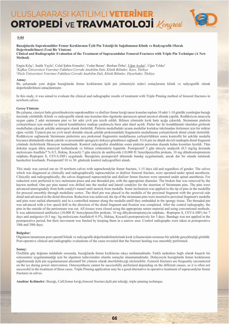
S-04
Buzağılarda Suprakondüler Femur Kırıklarının Üçlü Pin Tekniği ile Sağaltımının Klinik ve Radyografik Olarak Değerlendirilmesi (Yeni Bir Yöntem) Clinical and Radiographic Evaluation of the Treatment of Supracondular Femoral Fractures with Triple Pin Technique (A New Method)
Engin Kılıç1, Sadık Yayla2, Celal Şahin Ermutlu1, Vedat Baran1, Burhan Özba1, Uğur Aydın1, Uğur Yıldız1 1Kafkas Üniversitesi Veteriner Fakültesi Cerrahi Anabilim Dalı, Klinik Bilimler, Kars, Türkiye 2Dicle Üniversitesi Veteriner Fakültesi Cerrahi Anabilim Dalı, Klinik Bilimler, Diyarbakır, Türkiye
Amaç: Bu çalışmada yeni doğan buzağılarda femur kırıklarının üçlü pin yöntemiyle tedavi sonuçlarının klinik ve radyografik olarak değerlendirilmesi amaçlanmıştır.
In this study, it was aimed to evaluate the clinical and radiographic results of treatment with Triple Pinning method of femoral fractures in newborn calves.
Gereç-Yöntem: Bu çalışma, cinsiyet farkı gözetilmeksizin suprakondüler ve diafizer femur kırığı tanısı konulan toplam 10 adet 1-10 günlük yenidoğan buzağı üzerinde yürütüldü. Klinik ve radyografik olarak tanı konulan tüm olgularda operasyon spinal anestezi altında yapıldı. Redüksiyon amacıyla uygun çapta 2 adet steinmann pini ve bir adet yivli pin tercih edildi. Bilinen yöntemle kırık hattı açığa çıkarıldı. Steinmann pinlerin yerleştirilmesi için medial ve lateral kondülüslere matkap yardımıyla birer adet tünel açıldı. Pinler her iki kondülüsteki tünelden girilerek medulladan çıkacak şekilde anterograt olarak ilerletildi. Pinlerin medulladaki ucuna medullar kortekse takılmadan ilerlemesi için bir miktar eğim verildi. Üçüncü pin ise yivli tarafı distalde olacak şekilde proksimaldeki fragmentin medullasına yerleştirilerek distal yönde ilerletildi. Redüksiyon sağlanarak Steinmann pinlerinin ucu proksimal fragmentin medullasına yerleştirildikten sonra kontrollü bir şekilde medulla boyunca dönüşümlü olarak çakıldı ve prosimalde spongiyöz dokuya gömülmesi sağlandı. Yivli pin ise düşük devirli matkapla distal fragment yönünde ilerletilerek fiksasyon tamamlandı. Kontrol radyografisi alındıktan sonra pinlerin periostun dışında kalan kısımları kesildi. Tüm dokular uygun dikiş materyali kullanılarak ve bilinen yöntemlerle kapatıldı. Postoperatif 3 gün süreyle analjezik (0.5 mg/kg dozunda meloxicam-Anafleks® % 0.5, Hektaş, Kocaeli) 7 gün süreyle de antibiyotik (10,000 IU benzilpenicillin prokain, 10 mg dihidrostreptomisin sulphate,-Reptopen S, CEVA-DIF) uygulandı. Buzağılara postoperatif dönemde bandaj uygulanmadı, ancak dar bir alanda tutularak hareketleri kısıtlandı. Postoperatif 10 ve 30. günlerde kontrol radyografileri alındı.
This study was carried out on 10 newborn calves with suprachondylar femur fracture, 1-15 days old and regardless of gender. The calves which was diagnosed as clinically and radiographically supracondylar or diafizer femoral fracture, were operated under spinal anesthesia. Clinically and radiographically, the calves diagnosed supracondylar and diafizer femur fracture were operated under spinal anesthesia. For reduction were preferred to two steinmann pines and one threaded pin, with the appropriate diameter. The broken line was removed by the known method. One per pins tunnel was drilled into the medial and lateral condyles for the insertion of Steinmann pins. The pins were advanced anterogradely from both condyl's tunnel until unstick from medulla. Some inclination was applied to the tip of pins in the medullla for proceed smoothly through medullary cortex. The third pin was placed in the medulla of the proximal fragment with the grooved side distal and advanced in the distal direction. Reduction was achieved, the tip of the steinmann pins were inserted to proximal fragment's medulla and pins were nailed alternately and in a controlled manner along the medulla untill they embedded in the spongy tissue. The threaded pin was advanced with a low speed drill in the direction of the distal fragment and fixation was completed. After the control radiography, the pins in the outside of the periosteum was cut. All tissues were closed using the appropriate suture material and using conventional methods. It was administered antibiotics (10,000 IU benzylpenicillin prokain, 10 mg dihydrostreptomycin sulphate, -Reptopen S, CEVA-DIF) for 7 days and analgesics (0.5 mg / kg meloxicam-Anafleks® 0.5%, Hektaş, Kocaeli) postoperatively for 3 days. Bandage was not applied in the postoperative period, but their movement was limited by keeping them in a narrow area. Control radiographs were taken at postoperative 10th and 30th days.
Bulgular: Olguların tamamının post-operatif klinik ve radyografik değerlendirilmelerinde kırık iyileşmesinin sorunsuz bir şekilde gerçekleştiği görüldü. Post-operative clinical and radiographic evaluations of the cases revealed that the fracture healing was smoothly performed.
Sonuç: Özellikle güç doğuma müdahale sırasında, buzağılarda femur kırıklarına sıkça rastlanmaktadır. Farklı nedenlere bağlı olarak başarılı bir osteosentez uygulanamadığı için bu olguların tedavisinden olumlu sonuçlar alınamamaktadır. Dolayısıyla buzağılarda femur kırıklarının sağaltımında üçlü pin uygulamasının alternatif bir yöntem olarak önerilebileceği söylenebilir. Femoral fractures are frequently encountered on the ice during power intervention. Osteosynthesis cannot be successfully performed depending on the different causes, so it is often not successful in the treatment of these cases. Triple Pinning application may be a good alternative in operative treatment of supracondylar femur fractures in calves.
Anahtar Kelimeler: Buzağı, Calf;femur kırığı,femoral fracture;üçlü pin tekniği, triple pinning technique.
66
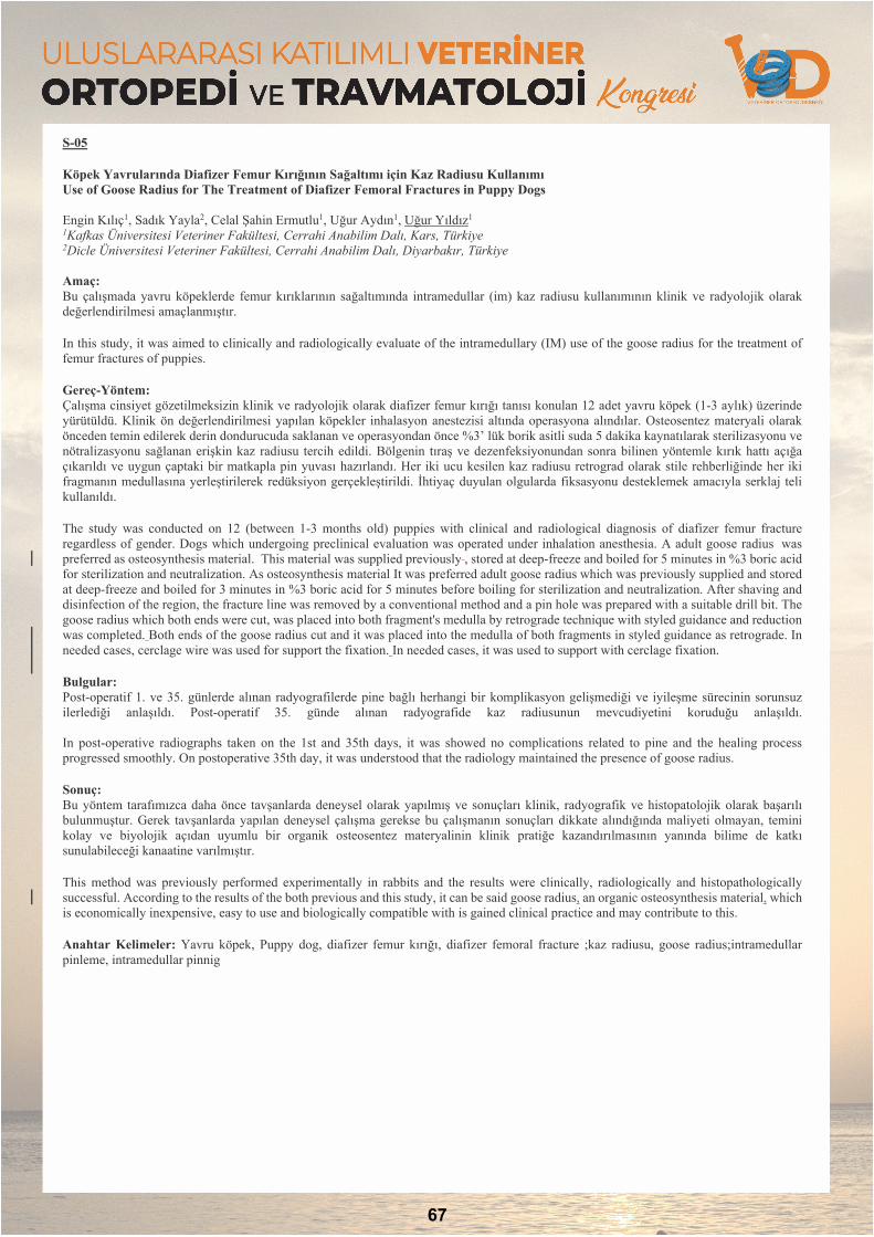
S-05
Köpek Yavrularında Diafizer Femur Kırığının Sağaltımı için Kaz Radiusu Kullanımı Use of Goose Radius for The Treatment of Diafizer Femoral Fractures in Puppy Dogs
Engin Kılıç1, Sadık Yayla2, Celal Şahin Ermutlu1, Uğur Aydın1, Uğur Yıldız1 1Kafkas Üniversitesi Veteriner Fakültesi, Cerrahi Anabilim Dalı, Kars, Türkiye 2Dicle Üniversitesi Veteriner Fakültesi, Cerrahi Anabilim Dalı, Diyarbakır, Türkiye
Amaç: Bu çalışmada yavru köpeklerde femur kırıklarının sağaltımında intramedullar (im) kaz radiusu kullanımının klinik ve radyolojik olarak değerlendirilmesi amaçlanmıştır.
In this study, it was aimed to clinically and radiologically evaluate of the intramedullary (IM) use of the goose radius for the treatment of femur fractures of puppies.
Gereç-Yöntem: Çalışma cinsiyet gözetilmeksizin klinik ve radyolojik olarak diafizer femur kırığı tanısı konulan 12 adet yavru köpek (1-3 aylık) üzerinde yürütüldü. Klinik ön değerlendirilmesi yapılan köpekler inhalasyon anestezisi altında operasyona alındılar. Osteosentez materyali olarak önceden temin edilerek derin dondurucuda saklanan ve operasyondan önce %3’ lük borik asitli suda 5 dakika kaynatılarak sterilizasyonu ve nötralizasyonu sağlanan erişkin kaz radiusu tercih edildi. Bölgenin tıraş ve dezenfeksiyonundan sonra bilinen yöntemle kırık hattı açığa çıkarıldı ve uygun çaptaki bir matkapla pin yuvası hazırlandı. Her iki ucu kesilen kaz radiusu retrograd olarak stile rehberliğinde her iki fragmanın medullasına yerleştirilerek redüksiyon gerçekleştirildi. İhtiyaç duyulan olgularda fiksasyonu desteklemek amacıyla serklaj teli kullanıldı.
The study was conducted on 12 (between 1-3 months old) puppies with clinical and radiological diagnosis of diafizer femur fracture regardless of gender. Dogs which undergoing preclinical evaluation was operated under inhalation anesthesia. A adult goose radius was preferred as osteosynthesis material. This material was supplied previously , stored at deep-freeze and boiled for 5 minutes in %3 boric acid for sterilization and neutralization. As osteosynthesis material It was preferred adult goose radius which was previously supplied and stored at deep-freeze and boiled for 3 minutes in %3 boric acid for 5 minutes before boiling for sterilization and neutralization. After shaving and disinfection of the region, the fracture line was removed by a conventional method and a pin hole was prepared with a suitable drill bit. The goose radius which both ends were cut, was placed into both fragment's medulla by retrograde technique with styled guidance and reduction was completed. Both ends of the goose radius cut and it was placed into the medulla of both fragments in styled guidance as retrograde. In needed cases, cerclage wire was used for support the fixation. In needed cases, it was used to support with cerclage fixation.
Bulgular: Post-operatif 1. ve 35. günlerde alınan radyografilerde pine bağlı herhangi bir komplikasyon gelişmediği ve iyileşme sürecinin sorunsuz ilerlediği anlaşıldı. Post-operatif 35. günde alınan radyografide kaz radiusunun mevcudiyetini koruduğu anlaşıldı.
In post-operative radiographs taken on the 1st and 35th days, it was showed no complications related to pine and the healing process progressed smoothly. On postoperative 35th day, it was understood that the radiology maintained the presence of goose radius.
Sonuç: Bu yöntem tarafımızca daha önce tavşanlarda deneysel olarak yapılmış ve sonuçları klinik, radyografik ve histopatolojik olarak başarılı bulunmuştur. Gerek tavşanlarda yapılan deneysel çalışma gerekse bu çalışmanın sonuçları dikkate alındığında maliyeti olmayan, temini kolay ve biyolojik açıdan uyumlu bir organik osteosentez materyalinin klinik pratiğe kazandırılmasının yanında bilime de katkı sunulabileceği kanaatine varılmıştır.
This method was previously performed experimentally in rabbits and the results were clinically, radiologically and histopathologically successful. According to the results of the both previous and this study, it can be said goose radius, an organic osteosynthesis material, which is economically inexpensive, easy to use and biologically compatible with is gained clinical practice and may contribute to this.
Anahtar Kelimeler: Yavru köpek, Puppy dog, diafizer femur kırığı, diafizer femoral fracture ;kaz radiusu, goose radius;intramedullar pinleme, intramedullar pinnig
67

S-06
Yüksekten Düşen Kedilerin Retrospektif Değerlendirilmesi (2017-2019): 330 Olgu Retrospective Evaluation of Cats with High-Rise (2017-2019): 330 Cases
Kamil Serdar Inal1, Taylan Önyay1, Mehmet Suat Yılmaz1, Birsen Deniz Özbakır1, Cenk Yardımcı1, Hatice Özlem Nisbet1, Ahmet Özak1 1Ondokuz Mayıs Üniversitesi, 19 Mayıs Üniversitesi, Veteriner Fakültesi Cerrahi Anabilim Dalı, Samsun, Türkiye
Amaç: Bu çalışmada; yüksekten düşen kedilerde cinsiyet, yükseklik ve düşülen zemin faktörünün morbidite / mortaliye olan etkisinin retrospektif olarak değerlendirilmesi amaçlandı.
This study aims to retrospectively evaluate the effects of gender, height and the floor type on the mortality and morbidity of cats fallen from high-rises.
Gereç-Yöntem: Çalışma materyalini yüksekten düşme şikayeti ile OMÜ Veteriner Fakültesi Hayvan Hastanesi’ne getirilen değişik ırk, yaş ve cinsiyette 330 kedi oluşturdu. Tüm olguların rutin klinik ve radyografik muayeneleri yapıldıktan sonra lezyonun şiddetine göre konservatif ya da cerrahi sağaltım uygulandı.
The study material was composed from 330 cats of varying breeds, ages and genders that were brought to the teaching hospital of Ondokuz Mayıs University, Faculty of Veterinary Medicine. Clinical and radiological examinations of each case were performed and conservative management or surgical intervention was carried out depending on the severity of the lesion
Bulgular: Olguların 238’inin dişi, 92’sinin erkek olduğu tespit edildi. Anamnez doğrultusunda olguların 64’ünün 4.kat, 62’sinin 5.kat, 42’sinin 3.kat, 32’sinin 2.kat, 15’inin 7.kat, 12’sinin 6.kat, 11’inin 8.kat, 7’sinin 10.kat, 1’inin 11.kat ve 1’inin 13.kattan düştüğü belirlendi. 78 kedinin ise düştüğü yükseklik bilinmiyordu. Olguların ortalama düşme yüksekliği 4,5. kat, ortalama yaşları ise 14,3 ay olarak tespit edildi. Olguların 227’sinin beton zemine, 25’inin toprak zemine, 1’inin araç tavanına, 1’inin ise önce ağaç sonra toprak zemine düştüğü öğrenilirken, 76 olgunun düştüğü zemin bilinmiyordu. Olguların büyük çoğunluğunda göğüs travması ilişkili lezyonlar tespit edilirken, ortopedik lezyonlar da oldukça önemli bir orana sahipti.
Ninety two of the cases were male and 238 were female. According to the case histories, 64 cases fell from 4th, 62 from 5th, 42 from 3rd, 32 from 2nd, 15 from 6th, 11 from 8th, 7 from 10th, 1 from 11thand 1 from 13thstoreys. Case histories of 78 cats were unclear concerning the height of the fall. The average of the storeys cats fell from was 4.5 and the mean age of cats included in the study was 14.3 months. Two hundred twenty seven of the cases fell to concrete, 25 to grass, 1 to the roof of a car and 1 first fell to a tree then to grass, whereas histories of 76 cases were unclear in regard to the floor type they fell on. Most of the cases had lesions concerning chest trauma and orthopedic lesions had a high incidence as well.
Sonuç: Kedilerin yüksekten düşme sendromunda paraşüt etkisinin çok önemli olduğu, 5.kat ve üzerine çıkıldığında ortopedik lezyonlarda artış olduğu belirlendi. Zeminin morbidite/mortalite üzerinde çok önemli etkisi olduğu tespit edildi.
In regards to cats falling from high rises, the parachute effect was found to be very important and orthopedic lesions had a higher incidence on 5thstory and above. The ground had an important impact on the morbidity/mortality.
Anahtar Kelimeler: kedi;travma;kırık;yüksekten düşme sendromu
68
POSTERBİLDİRİLER

P-01
Kangal Irkı Bir Köpekte Kronik Dirsek Luksasyonunun Artrodez ile Başarılı Sağaltımı ve Postoperatif Bandaj Komplikasyonu Successful Treatment of Chronic Elbow Luxation with Arthrodesis and Postoperative Bandage Complications in a Kangal Dog
Semih Altan1 , Berna Ersöz Kanay1, Emine Çatalkaya1, Sadık Yayla1 1Dicle Üniversitesi, Veteriner Fakültesi- Cerrahi Anabilim Dalı, Diyarbakır, Türkiye
Giriş ve Amaç: Artrodez, bir eklemin cerrahi füzyonu anlamına gelir. Başka bir deyişle, eklemi oluşturan kemikler kalıcı olarak birleştirilir, böylece ekstremitenin bu bölümünde hareket olmaz. Artrodez, genellikle sadece eklem fonksiyonunu korumada başka seçenek olmadığında yapılan bir ağrı kesici ve kurtarma prosedürüdür. Kronik dirsek çıkıklarının sağaltımı oldukça zordur. Bu tür durumlarda artrodez uygulamaları ampütasyona iyi bir alternatiftir ancak yüksek derecede teknik zorlukları vardır. Bu vaka sunumunda; 5 yaşlı kangal melezi bir köpekte karşılaşılan travmatik kronik dirsek çıkığının artrodez uygulanarak yapılan başarılı operatif sağaltımı ve postoperatif 2. ayda karşılaşılan komplikasyona ait sonuçların paylaşılması amaçlanmıştır. Arthrodesis refers to the surgical fusion of a joint. In other words, the bones forming the joint are permanently joined so that there is no movement in this part of the limb. Arthrodesis is a pain relief and recovery procedure, usually performed only when there is no other option to maintain joint function. Chronic elbow luxation is difficult to treat. In such cases, arthrodesis is a good alternative to amputation but has a high degree of technical difficulties. In this case report; it was aimed to share the results of the successful surgical treatment of traumatic chronic elbow luxation in a 5-year-old crossbreed dog by arthrodesis and the complications encountered in the postoperative second month in this dog.
Olgunun Tanımı: Olguyu 5 yaşlı kangal melezi erkek bir köpeği oluşturdu. Köpek, sadist bir uygulamaya maruz kalması sonucu yaklaşık 25 günden beri devam eden topallık şikâyetiyle cerrahi kliniğine getirildi. Yapılan klinik ve radyolojik muayenelerde sonucunda kronik lateral dirsek lukzasyonu teşhisi konuldu. Röntgen görüntülerinden eklem bölgesinde dejeneratif değişikliklerin varlığı ve osteofitik üremelerin yoğun olması nedeniyle plaka artrodezi uygulanarak sağaltılmasına karar verildi. Bu amaçla hazırlanan 3,5 mm 12 delikli DCP plağı, ulnar ostektomiyi ve eklem yüzlerinin kazınmasını takiben yapılan redüksiyon sonrası yaklaşık 110º’lik açıyla ve kullanılan 11 kortikal vida vasıtasıyla dirsek eklemine uygulandı. Plaka yerleştirildikten hemen sonra eklem aralıklarına kontralateral humerusun trochantör majus’undan taze olarak alınan otojen kansellöz kemik grefti uygulandı. Postoperatif PVC destekli bandaj uygulanarak antibiyotik ve analjezik reçete edildi. Hastanın takibi 15 gün aralıklarla yapılan radyolojik muayenelerle sağlandı. İlk 1 ay sonunda yapılan muayenelerde topallığın azaldığı dikkat çekti. Ancak hasta sahibinin; köpeğin mizaç olarak oldukça agresif olması nedeniyle uygulanan bandajı düzenli olarak kontrol edememesi ve yaklaşık 120 km’lik bir uzaklıktan köpeği getirmekte zorlanması nedeniyle 2. ayda bandaja bağlı basınç nekrozları ve dekübital ülserlerle karakterize komplikasyon gelişti. Yapılan tedavilere rağmen iyileşme sağlanamaması sonucu ekstremiteye ampütasyon uygulanmak zorunda kalındı. The case consisted of a 5-year-old male crossbreed kangal dog. The dog was brought to the surgery clinic with the complaint of lameness that had been going on for about 25 days after being exposed to a sadistic trauma. Chronic lateral elbow luxation was diagnosed with clinical findings and radiological examinations. Because of the presence of degenerative changes and intense osteophytic reproduction on the joint from the X-ray images and, it was decided to treat with plate arthrodesis. For this purpose, a 3.5 mm 12-hole DCP plate at an angle of 110º was applied to the elbow joint after ulnar ostectomy and scraping of the joint surfaces using 11 cortical screws. Autogenous cancellous bone graft was obtained from the trochanter majus of the contralateral humerus after the plate placement. Antibiotic and analgesic were prescribed by postoperative PVC-supported bandage. The dog was followed-up with radiological examinations performed at 15-day intervals. The examinations performed at the end of the first month showed that lameness decreased. However, complications characterized by pressure necrosis and decubital ulcers due to bandage were developed at postop 2 months due to the dog's being aggressively temperamentally unable to control, the bandage regularly and having difficulty in bringing the dog from a distance of approximately 120 km. Amputation was performed due to the lack of improvement despite the treatments.
Sonuç: Birçok hayvan türünde travmatik eklem çıkıkları ile karşılaşılmaktadır. Lateral dirsek eklemi çıkıkları da bunlardan birisidir. Özellikle ilk 24-48 saat içerisinde kapalı redüksiyon ve bandaj ile başarılı sonuçlar alınmasına rağmen olay üzerinde geçen süre uzadıkça tedavide başarı oranı oldukça düşmekte ve süreç ampütasyona kadar gidebilmektedir. Bu tür durumlarda yapılan artrodez işlemini takiben iyi bir postoperatif bakım tedavinin başarılı olması için hayati öneme sahiptir. Bu sağlanmadığı taktirde füzyon yetersizliği, implant/ fiksasyon yetersizliği (vida gevşemesi / kırılma veya plaka kırılması), kemik kırıkları, enfeksiyon, deri nekrozu ve bandaj / alçıya bağlı komplikasyonlarla karşılaşılabilmektedir. Sunulan bu vakada uygulanan operatif işlemler başarılı olsa da iki kemik arasında yeterli düzeyde füzyon oluşuncaya kadar (6-8 hafta) destekli bandajla immobilizasyonun sağlanması esnasında komplikasyon geliştiği görülmüştür. Sonuç olarak bu süreçte bandaja bağlı oluşabilecek bu komplikasyonların önlenmesine yönelik tedbirlerin alınması, yapılan tedavinin başarılı olmasını önemli düzeyde etkiyebileceği görüldüğü için bu vakanın sunulmasının önemli olduğu kanısına varılmıştır.
Traumatic joint luxations are encountered in many animal species. Lateral elbow luxation is one of them. Although successful results are obtained with closed reduction and bandage especially in the first 24-48 hours, the success rate in treatment decreases considerably and the process may go up to amputation. Following arthrodesis in such cases, good postoperative care is vital to the success of the treatment. Failure to do so may result in fusion failure, implant / fixation failure (screw loosening / fracture or plate fracture), bone fractures, infection, skin necrosis, and bandage / plaster-related complications. Although the operative procedures performed in this case were successful, it was seen that complications developed during the immobilization with supported bandage until sufficient fusion occurred between the two bones (6-8 weeks). As a result, it was concluded that the presentation of this case is important since it is seen that taking measures to prevent these complications may affect the success of the treatment.
Anahtar Kelimeler: Kronik dirsek çıkığı;arthrodez;köpek;komplikasyon
70
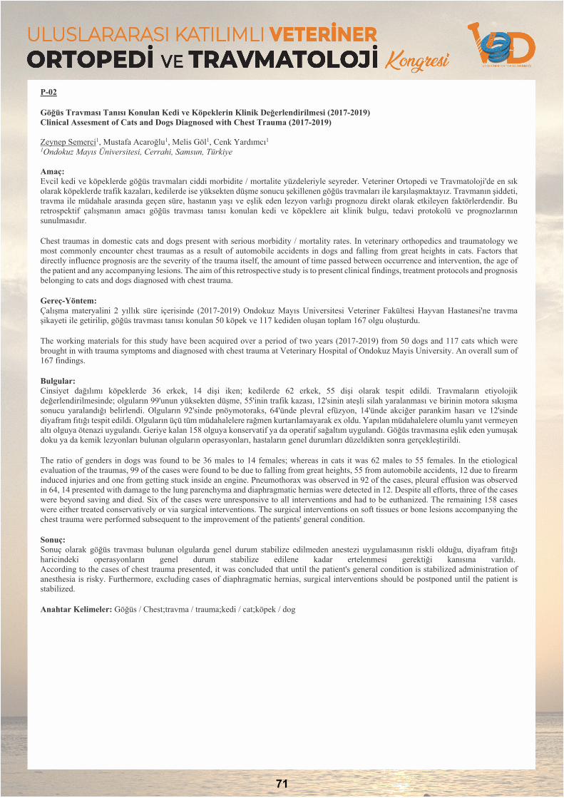
P-02
Göğüs Travması Tanısı Konulan Kedi ve Köpeklerin Klinik Değerlendirilmesi (2017-2019) Clinical Assesment of Cats and Dogs Diagnosed with Chest Trauma (2017-2019)
Zeynep Semerci1, Mustafa Acaroğlu1, Melis Göl1, Cenk Yardımcı1 1Ondokuz Mayıs Üniversitesi, Cerrahi, Samsun, Türkiye
Amaç: Evcil kedi ve köpeklerde göğüs travmaları ciddi morbidite / mortalite yüzdeleriyle seyreder. Veteriner Ortopedi ve Travmatoloji'de en sık olarak köpeklerde trafik kazaları, kedilerde ise yüksekten düşme sonucu şekillenen göğüs travmaları ile karşılaşmaktayız. Travmanın şiddeti, travma ile müdahale arasında geçen süre, hastanın yaşı ve eşlik eden lezyon varlığı prognozu direkt olarak etkileyen faktörlerdendir. Bu retrospektif çalışmanın amacı göğüs travması tanısı konulan kedi ve köpeklere ait klinik bulgu, tedavi protokolü ve prognozlarının sunulmasıdır.
Chest traumas in domestic cats and dogs present with serious morbidity / mortality rates. In veterinary orthopedics and traumatology we most commonly encounter chest traumas as a result of automobile accidents in dogs and falling from great heights in cats. Factors that directly influence prognosis are the severity of the trauma itself, the amount of time passed between occurrence and intervention, the age of the patient and any accompanying lesions. The aim of this retrospective study is to present clinical findings, treatment protocols and prognosis belonging to cats and dogs diagnosed with chest trauma.
Gereç-Yöntem: Çalışma materyalini 2 yıllık süre içerisinde (2017-2019) Ondokuz Mayıs Universitesi Veteriner Fakültesi Hayvan Hastanesi'ne travma şikayeti ile getirilip, göğüs travması tanısı konulan 50 köpek ve 117 kediden oluşan toplam 167 olgu oluşturdu.
The working materials for this study have been acquired over a period of two years (2017-2019) from 50 dogs and 117 cats which were brought in with trauma symptoms and diagnosed with chest trauma at Veterinary Hospital of Ondokuz Mayis University. An overall sum of 167 findings.
Bulgular: Cinsiyet dağılımı köpeklerde 36 erkek, 14 dişi iken; kedilerde 62 erkek, 55 dişi olarak tespit edildi. Travmaların etiyolojik değerlendirilmesinde; olguların 99'unun yüksekten düşme, 55'inin trafik kazası, 12'sinin ateşli silah yaralanması ve birinin motora sıkışma sonucu yaralandığı belirlendi. Olguların 92'sinde pnöymotoraks, 64'ünde plevral efüzyon, 14'ünde akciğer parankim hasarı ve 12'sinde diyafram fıtığı tespit edildi. Olguların üçü tüm müdahalelere rağmen kurtarılamayarak ex oldu. Yapılan müdahalelere olumlu yanıt vermeyen altı olguya ötenazi uygulandı. Geriye kalan 158 olguya konservatif ya da operatif sağaltım uygulandı. Göğüs travmasına eşlik eden yumuşak doku ya da kemik lezyonları bulunan olguların operasyonları, hastaların genel durumları düzeldikten sonra gerçekleştirildi.
The ratio of genders in dogs was found to be 36 males to 14 females; whereas in cats it was 62 males to 55 females. In the etiological evaluation of the traumas, 99 of the cases were found to be due to falling from great heights, 55 from automobile accidents, 12 due to firearm induced injuries and one from getting stuck inside an engine. Pneumothorax was observed in 92 of the cases, pleural effusion was observed in 64, 14 presented with damage to the lung parenchyma and diaphragmatic hernias were detected in 12. Despite all efforts, three of the cases were beyond saving and died. Six of the cases were unresponsive to all interventions and had to be euthanized. The remaining 158 cases were either treated conservatively or via surgical interventions. The surgical interventions on soft tissues or bone lesions accompanying the chest trauma were performed subsequent to the improvement of the patients' general condition.
Sonuç: Sonuç olarak göğüs travması bulunan olgularda genel durum stabilize edilmeden anestezi uygulamasının riskli olduğu, diyafram fıtığı haricindeki operasyonların genel durum stabilize edilene kadar ertelenmesi gerektiği kanısına varıldı. According to the cases of chest trauma presented, it was concluded that until the patient's general condition is stabilized administration of anesthesia is risky. Furthermore, excluding cases of diaphragmatic hernias, surgical interventions should be postponed until the patient is stabilized.
Anahtar Kelimeler: Göğüs / Chest;travma / trauma;kedi / cat;köpek / dog
71
P-03
Kedilerde Unilateral Temporomandibular Eklem Ankilozunun İntra-Artiküler Ostektomi ile Sağaltımı Intra-Articular Ostectomy Technique to Treat Unilateral Temporomandibular Joint Ankylosis in Cats
Kamil Serdar Inal1, Taylan Önyay1, Cenk Yardımcı1, Ahmet Özak1 1Ondokuz Mayıs Üniversitesi, 19 Mayıs Üniversitesi Veteriner Fakültesi Cerrahi Anabilim Dalı, Samsun, Türkiye
Amaç: Bu çalışmanın amacı; kedilerde unilateral temporomandibular eklem (TME) ankilozunun basit bir ostektomi tekniği ile sağaltımının ve fonoksiyonel sonuçlarının aktarılmasıdır.
The purpose of this study is to explain a simple ostectomy procedure used to treat unilateral temporomandibular joint (TMJ) ankylosis in cats in order to show its functional outcome.
Materyal ve Metot: Çalışma materyalini unilateral TME ankilozu olan üç kedi oluşturdu. Kedilere gerek güvenli bir entübasyon uygulanabilmesi gerekse de postoperatif dönemde üç günlük beslenme için faringostomi uygulandı. Three cats with one sided TMJ ankylosis were presented. Pharyngostomies were performed to each cat for safe intubation and placing feeding tubes for three-day long postoperative period.
Bulgular: Kedilerin tümü, postoperatif 4.günden itibaren yumuşak gıda ile beslenebiliyorlardı. İkinci haftadan itibaren tüm olguların ağrısız ve fonksiyonel olarak çiğneyebildiği gözlendi. Olguların uzun dönem sonuçları da gayet tatminkardı.
Each patient was able to consume soft food at the fourth day after surgery. In two weeks, they were able to chew kibbles and did not show any pain or discomfort in the post-operative period. Long term functional outcomes of the patients were also favorable.
Sonuç: Biz bu tekniğin unilateral TME ankilozu olan kedilerde başarılı ve etkin olarak kullanılabileceğine inanıyoruz. Tüm olgular sorunsuz olarak iyileşti ancak, olgu sayısının yetersiz oluşu olası komplikasyonların öngörülmesi açısından yetersizdir.
We believe that, this technique may be successfull and can efficiently be use in cases with unilateral TMJ ankylosis in cats. Each cat in the study recovered without any problems, though it is important to note that the number of cases are insufficient to estimate possible complications.
Anahtar Kelimeler: Ankilioz;kedi;ostektomi;temporomandibular eklem
72
P-04
Kedi ve Köpeklerde Çoklu Kırıkların Retrospektif Değerlendirilmesi (2016-2019) Retrospective Evaluation of Multiple Fractures in Cats and Dogs (2016-2019)
Kamil Serdar Inal1, Taylan Önyay1, Mehmet Suat Yılmaz1, Cenk Yardımcı1, Ahmet Özak1 1Ondokuz Mayıs Üniversitesi, 19 Mayıs Üniversitesi Veteriner Fakültesi Cerrahi Anabilim Dalı, Samsun, Türkiye
Amaç: Bu retrospektif çalışmada; çoklu kemik kırığı bulunan kedi ve köpeklerde, travmanın etiyolojisi, kırıkların lokalizasyonu, eşlik eden lezyonlar, sağaltım seçenekleri ile sonuçları değerlendirildi.
In this retrospective study, the etiology, fracture localization, accompanying lesions, treatment options and results of multiple fractures of cats and dogs were evaluated.
Gereç-Yöntem: Çalışma materyalini OMÜ Veteriner Fakültesi Hayvan Hastanesine 2016-2019 yılları arasında travma şikayeti ile getirilip, vücudunda en az iki farklı kırık tespit edilen değişik ırk, yaş ve cinsiyetteki 48 olgu oluşturdu. Klinik ve radyolojik muayeneleri yapılan hastalara, kırıkların ve varsa eşlik eden lezyonların durumuna bağlı olarak farklı sağaltım yöntemleri uygulandı.
The study material of this study were 48 dogs of different breeds, age and sex that were diagnosed with at least 2 fractures on their bodies. After clinical and radiological assesment of the patients, appropriate treatment options were administered depending on the fracture and any accompanying lesions.
Bulgular: Olguların 35’i kedi (20 dişi, 15 erkek), 13 köpek (11 erkek, 2 dişi) olup; kedilerde ortalama yaş 11 ay, köpeklerde 15,3 aydı. Etiyolojik değerlendirme yapıldığında lezyonlar kedilerin 27’sinde yüksekten düşme, 3’ünde trafik kazası, 1’inde köpek saldırısı sonucunda oluşurken, 5’inde sebep bilinmiyordu. Lezyonların; köpeklerin 10’unda trafik kazası, 2’sinde yüksekten düşme ve 1’inde ise ateşli silah yaralanması sonucunda şekillendiği tespit edildi. Kırıkların dağılımı kedilerde; 15 femur, 11 humerus, 12 tibia, 10 radius/ulna, 3 metakarpus, 1 tarsal, 1 mandibular ve bir vertebra kırığı iken; köpeklerde, 7 femur, 5 radius/ulna, 5 tibia, 2 humerus, 2 olekranon, 1 metatarsus ve 1 patella kırığı idi. Sağaltımda 9 olguda bandaj, 36 olguda ise operatif sağaltım teknikleri uygulandı. 1 hasta sahibi operasyonu reddederken, 2 hasta müdahale sırasında ex oldu.
35 cases were cats (20queens, 15 males) and 13 were dogs (11 males and 2 bitches); the average age of was 11 months for cats and 15.3 months for dogs. The etiologies for cats were 27 high-rise falls, 3 motor vehicle accidents, 1 dog bite and 5 of them were of unknown etiology. For dogs, the lesions were caused by motor vehicle accidents for 10, high-rise falls for 2 and a gunshot wound for 1. The fracture location dispersion was 15 femur, 11 humeri, 12 tibias, 10 radius/ulna, 3 metacarpus, 1 tarsal,1 mandibular and 1 vertebra for cats and 7 femur, 5 radius/ulna, 5 tibia, 2 humerus, 2 olecranon, 1 metatarsus and 1 patella for dogs. Splints were used in 9 cases and surgery was performed in 36. One patient owner refused treatment while 2 patients died during surgery.
Sonuç: Yüksek enerjili travmalar sonucunda şekillenen çoklu kırıklar, oldukça masraflı ve zor olgulardır. Yüksek operasyon maliyetlerinden dolayı, ekonomik durumu uygun olmayan hasta sahiplerini bandaj seçeneğini kullanmak zorunda kalmaktadır. Ancak bandaj uygulanan hastalarda iyileşme süreci oldukça zor geçmektedir.
Multiple fractures caused by high energy traumas are difficult and expensive to treat. Many patient owners that are not well to do have to choose treatment with splints due to economic reasons. In addition, the recovery process of patients with splints is quite difficult.
Anahtar Kelimeler: Kırık;kedi;köpek;travma
73
P-05
Kedilerde Yüksekten Düşme Vakalarının Değerlendirilmesi (Klinik Bir Çalışma, 44 Olgu) Evaluation of High Fall Cases in Cats (A Clinical Study, 44 Cases)
Emine Çatalkaya1, Semih Altan1, Berna Ersöz Kanay1, Sadık Yayla1, Nahit Saylak1 1Dicle Üniversitesi Veteriner Fakültesi, Cerrahi Anabilim Dalı, Diyarbakır, Türkiye
Amaç: Kedilerde cerrahi hastalıklar arasında yüksekten düşme sendromu olarak da tanımlanan kedilerin binaların balkon veya pencerelerinden düşmesi sonrasında görülen ortopedik problemler oldukça önemli bir yer tutmaktadır. Bölgemizde kedilerde sıklıkla karşılaşılan yüksekten düşme vakalarının klinik olarak değerlendirilmesi amaçlandı.
Orthopedic problems seen after falling from the balcony or windows of the buildings, which are defined as high rise syndrome among cats, are very important. The aim of this study was to evaluate the cases with high rise syndrome which frequently seen in cats in our region.
Gereç-Yöntem: Çalışma, Dicle Üniversitesi Veteriner Fakültesi Cerrahi Anabilim Dalı kliniklerine 01.01.2019- 10.09.2019 tarihleri arasında yüksekten düşme şikayeti ile getirilen farklı ırk, yaş ve cinsiyetteki 44 kedi üzerinde yürütüldü. Olguların teşhisi anamnezi takiben klinik ve radyolojik muayeneye göre yapıldı. Tedavi olarak destekli bandaj uygulaması, kafes istirahati, osteosentez, laminektomi ve açık redüksiyon yapıldı.
The study was carried out on 44 cats of different breeds, ages and sex who were admitted to Dicle University Faculty of Veterinary Medicine Department of Surgery between 01.01.2019 and 10.09.2019. The diagnosis was made according to the clinical and radiological examination following the anamnesis. Supported bandage application, cage rest, osteosynthesis, laminectomy and open reduction were performed.
Bulgular: Belirtilen tarihler arasında kliniğimize getirilen kedilerin % 21.78’ini (44 olgu, 22 erkek, 21 dişi) yüksekten düşme vakaları oluşturdu. Bu olguların yaşları 20 günlük ile 3 yaş arasında idi. 15 olguda (%34) klinik olarak herhangi bir problem saptanmadı. Diğer olgular içerisinde femur kırığı (n=13), humerus kırığı (n=4), tibia kırığı (n=7), antebrahium kırığı (n=1), 5. lumbal vertebra kırığı (n=1), 10. torakal vertebra kırığı (n=1), 5-6. lumbal vertebra luksasyonu (n=1), tibial paraliz (n=1), dirsek eklemi çıkığı (n=1), koksofemoral luksasyon (n=1), metakarpus kırığı (n=1) tespit edildi. Ayrıca bu olguların birinde tibia kırığı bilateraldi ve başka bir olguda ise femur ve tibia kırığı, diğer bir olguda ise metakarpal kırıkları ile kollum femoris kırığı bir arada idi. Bu çalışmada disloke olmayan kapalı kırıklar için destekli bandaj (6 olgu), disloke olan kırıklarda osteosentez (18 olgu), 5-6 lumbal vertebralar arasında luksasyon için laminektomi (1 olgu), koksofemoral luksasyon için De Vita pin uygulaması (1 olgu), vertebra kırıkları için kafes istirahati (2 olgu), dirsek eklemi çıkığı için açık redüksiyon (1 olgu), tibial paraliz için ise medikal tedavi uygulandığı belirlendi. Tedavi uygulanan kedilerin 42’sinde fonksiyonel bir iyileşmenin olduğu görülürken, 2’sinde genel durumun kötüye gitmesiyle öldüğü belirlendi.
21.78% (44 cases, 22 males, 21 females) of the cats brought to our clinic between the dates mentioned were caused by high fall cases. The ages of these cases were between 20 days and 3 years. No clinical problem was detected in 15 cases (34%). Femoral fracture (n = 13), humeral fracture (n = 4), tibial fracture (n = 7), antebrachial fracture (n = 1), 5th lumbar vertebral fracture (n = 1), 10th thoracal vertebral fracture (n = 1), 5-6th lumbar vertebral luxation (n=1), tibial paralysis (n = 1), elbow joint dislocation (n = 1), coxofemoral luxation (n = 1), metacarpal fracture (n = 1) detected. In addition, tibial fracture was bilateral in one of these cases and femoral and tibial fracture in another case, metacarpal fractures and femoral neck fracture in another case. In this study, supported bandage for non-dislocated closed fractures (6 cases), osteosynthesis in dislocated fractures (18 cases), laminectomy for luxation between 5-6th lumbal vertebrae (1 case), De Vita pin application for coxofemoral luxation (1 case), cage rest for vertebral fractures (2 case), open reduction for elbow joint dislocation (1 case), and medical treatment for tibial paralysis. While 42 of the treated cats had a functional improvement, 2 of them died due to deterioration of the general health status.
Sonuç: Bu çalışmadan elde edilen verilere göre yüksekten düşme vakalarında kedilerde lezyonun yerinin belirlenmesi önemli olmasının yanısıra disloke olmayan kapalı kırıklarda destekli bandaj, diğer kırık ve luksasyonlarda ise operatif sağaltım yöntemlerinin başarılı bir şekilde uygulandığı sonucuna varıldı.
According to the data obtained from this study, it is concluded that it is important to determine the location of the lesion in high fall cases in cats, that also supportive bandage in non-dislocated closed fractures, for other fractures and luxations operative treatment methods were successfully applied.
Anahtar Kelimeler: Yüksekten düşme falling from high;kedi cat;travma trauma
74

P-06
Bir Kedide Osteoblastik Osteosarkom Olgusu A Case Of Osteoblastıc Osteosarcoma In A Cat
Birkan Karslı1, Merve Bakıcı1, Tuğçe Sümer2, Zeynep Pekcan1 1 Kırıkkale Üniversitesi Veteriner Fakültesi Cerrahi Anabilim Dalı, Kırıkkale 2Kırıkkale Üniversitesi Veteriner Fakültesi Patoloji Anabilim Dalı, Kırıkkale
Osteosarkom (OS), insan ve hayvanlarda görülen en yaygın malign kemik tümörüdür ve kedilerde primer malign kanserlerinin %70-80’ini oluşturur.
Bu olgu sunumunu, Kırıkkale üniversitesi veteriner fakültesi araştırma ve uygulama hastanesine dispne şikayeti ile getirilen 11 yaşlı erkek tekir kedi oluşturmaktadır. Yapılan muayenelerde toraks bölgesinde palpe edilebilen ve alınan radyografik görüntülerde toraks içerisine infiltre olan bir kitle tespit edildi. Bakılan kan parametrelerinde tam kan sayımında tüm parametreler normal iken serum ALP değerinde yüksek oranda artış görülmüştür.
Eksizyonel biyopsi tekniği ile çıkarılan kitlenin histopatolojik incelemesi sonucunda osteoblastik osteosarkom tanısı konuldu. Kedi osteosarkomlarında düşük metastaz hızı ve sadece ampütasyon/geniş lokal rezeksiyonla bile ortalama yaşam süresinin 24-44 ay arasında olması nedeniyle adjuvan terapi önerilmedi. Postoperatif 9. ay kontrollerinde toraks kranialinde yeni bir kitlenin varlığı tespit edildi. Fakat hayvanın genel durumunun iyi olması, ileri yaşlı olması, şiddetli dispne ve benzeri solunum problemlerinin olmaması nedeniyle tekrar operasyona gerek duyulmadı. İlerleyen zamanlarda hastanın kötüleşmesi durumunda hayvan sahibinin de istediği doğrultusunda ötenazi yapılmasına karar verildi.
Osteosarcoma (OS) is the most common malignant bone tumor in humans and animals and constitutes 70-80% of primary malignant cancers in cats.
This case report consists of 11-year-old male tabby cat with dyspnea brought to Kırıkkale University Veterinary Faculty Research and Application Hospital. In the examination, a mass was palpable in the thorax region and it was determined that an infiltration was detected into the thorax on radiographic images. While the whole blood count was normal in all blood parameters, there was a high increase in serum ALP value.
Histopathological examination of the mass was excised by excisional biopsy revealed a diagnosis of osteoblastic osteosarcoma. Adjuvant therapy was not recommended in cat osteosarcomas because of the low metastasis rate and the mean survival of 24-44 months even with amputation / wide local resection. A new mass was detected in the cranial of the thorax at the 9th postoperative month. However re-operation was not required due to the general condition of the animal was good, was an elderly age and no severe dyspnea and similar respiratory problems. In the event that the patient worsens in the future, it was decided to perform euthanasia according to the animal owner's wishes.
75

P-07
Bir Kedide Simfizis Mandibula Ayrılmasında Rostral Mandibula Etrafından Serklaj Teli Uygulaması Application of Cerclage Wire Around Rostral Mandible in a Cat with Mandibular Symphysis Fracture
Müge Bircan Akkartal, Mustafa Akkartal Nova Veteriner Kliniği, Balıkesir, Türkiye Nova Veterinary Clinic, Balıkesir, Turkey
Amaç: Simfizis mandibulada ayrılma kedilerde oldukça yaygın görülmekte ve kedilerde ki kırıkların %14.5'ini oluşturmaktadır. Büyük bir çoğunluğu, basit ama etkili bir teknik olan serklaj teliyle sağaltılabilir. Seçilecek sağaltım yöntemi, kırık bölgesine ve çevre doku hasarına bağlı olarak değişebilir. Sağaltımda öncelikli amaç, kaynama ve buna bağlı olarak iyileşme sağlanırken, normal dental oklüzyona izin verebilecek bir stabilizasyon sağlamaktır. Bu poster sunumunda, bir kedide simfizis mandibula ayrılmasında rostral mandibula etrafından serklaj teli uygulanmış ve sağaltım sonuçları meslektaşlarımızla paylaşılmıştır.
Mandibular symphysis fracture is commonly seen in cats, making up around 14.5% of total fracture cases. Majority of them can be fixed with the basic, yet effective technique of cerclage wire application. Fixation method may differ based on the location of the fracture and the damage of the surrounding tissue. The primary objective of the therapy includes bone healing and the achievement of stabilization that allows normal dental occlusion. The overall aim of this poster presentation is to share with our collegues the results of cerclage wire application around rostral mandible in a cat with mandibular symphysis fracture.
Materyal ve Metot: Olguyu tahmini 4 yaş civarı, dişi, sokak kedisi oluşturdu.
The patient was a 4-year-old female stray cat.
Bulgular: Klinik muayenede simfizis mandibulada deformasyon, aşırı salivasyon ve deride yer yer nekrotik alanlar görüldü. Hastanın aşırı kaşektik ve dehidre olduğu farkedildi. Alınan anamnezde, 10 gün önce trafik kazası geçirdiği, başka bir klinikte tedavisinin ve operasyonun yapıldığı belirtildi. Operasyonda serklaj telinin kanin dişlerin etrafından sekiz şeklinde dolandırılarak sabitlendiği izlenirken mevcut durumda serklaj telinin gevşediği ve stabilizasyonun bozulduğu fark edildi. Kliniğimizde ilk 15 günlük süreçte hasta sıvı sağaltımı ile desteklenip dehidrasyonu giderildi, gevşeyen serklaj teli çıkartıldı, maddi kayıplı nekrotik deri hipokloröz asit solüsyonuyla(Crystalin®- NHP) günde 4 sefer yıkandı ve Prescription diyet mama ile sıkça beslenerek kilo alımı sağlandı. Genel durumu stabil hale gelen hasta için operasyon kararı verildi. Operasyonda, doku debride edildikten sonra simfizis mandibula ayrılmasını onarmak için farklı bir teknik; rostral mandibula etrafından serklaj teli uygulandı (Circumferential serklaj tekniği). Tel, her iki kanin dişin kaudalinde konumlanmış pembe kanülden geçirilerek mandibulanın ventralinde düğümlendi ve sabitlendi. İmmobilizasyon sağlandıktan sonra hastanın oklüzyonu muayene edilerek tam bir kapanış sağlandığı kaydedildi. Rostral mandibula bölgesinde bulunan deri kaybı unilateral tek pediküllü ilerletme flebi yöntemiyle kapatıldı. Hastanın deri dikişleri 10.günde alınırken, serklaj 45.günde uzaklaştırıldı. Postoperatif takibi yapılan olguda 1 yıllık süreçte herhangi bir komplikasyonla karşılaşılmadı.
Clinical exam revealed mandibular symphysis deformation, hypersalivation and dermal necrosis along with cachexia and dehydration. It was reported that the patient has been hit by a car 10 days prior to the exam and operated by another clinic. The cerclage wire was arranged in a figure of 8 shape around the canine teeth; however, it was loose resulting in the lack of stabilization. The patient was hospitalized for 15 days in our clinic. The treatment plan included frequent feeding regimen with a prescription diet, IV fluid therapy, removal of the loose cerclage wire and application of hypochlorous acid solution (Crystalin®- NHP) on the necrotic lesions of the skin four times daily. Adecision was made for surgery after the physical condition of the patient was stabilized. The operation was based on a relatively different technique of cerclage wire application around rostral mandible for the healing of mandibular symphysis fracture (circumferential cerclage technique). The wire was slipped through pink cannulas inserted behind both canine teeth and stabilized on the ventral aspect of mandible followed by immobilization and the examination of occlusion to ensure full fixation. The skin tissue loss over the rostral mandible region was compensated via unilateral single pedicle advancement flap technique. Skin suture was removed at day 10 and the cerclage was at day 45. The patient was followed up for a year postoperatively and no complications recorded.
Sonuç: Simfizis mandibula ayrılmaları hakkında literatürde birçok tedavi yöntemi tarif edilmiş olmasına rağmen, olgumuzda uygulanan yöntem bütün türlerde başarıyla kullanılabilir ve tercih edilebilir tedavi yöntemidir. Güncel yayınlarda kanin dişlere ”8” şeklinde serklaj uygulamasının vakamızda uygulanan yönteme destek amaçlı yapılabileceği (çift tel tekniği) veya alternatif olarak “8” şeklindeki serklaj yöntemi tercih edilirse de telin uygulandıktan sonra akrilik ile kaplanması gerektiğine değinilmiştir. Although there is a variety of techniques determined for mandibular symphysis fracture repair in the medical literature, the technique used in this case can be applied to all species successfully. In recent publications, it has been reported that 8 shaped cerclage application to canine teeth can be performed to support the method applied in our case (Double wire technique). Alternatively, the 8 shaped cerclage method is preferred, but after application the wire should be coated with acrylic.
Anahtar Kelimeler: Bir Kedide Simfizis Mandibula Ayrılmasında Rostral Mandibula Etrafından Serklaj Teli Uygulaması Mandibular symphysis, trauma, cat, cerclage wire
76
SponsorlarımızaTeşekkürlerimizle
ANA SPONSORLAR
SPONSORLAR
BASIN SPONSORU
Sponsorlar alfabetik olarak sıralanmıştır